Embed Size (px)
Citation preview
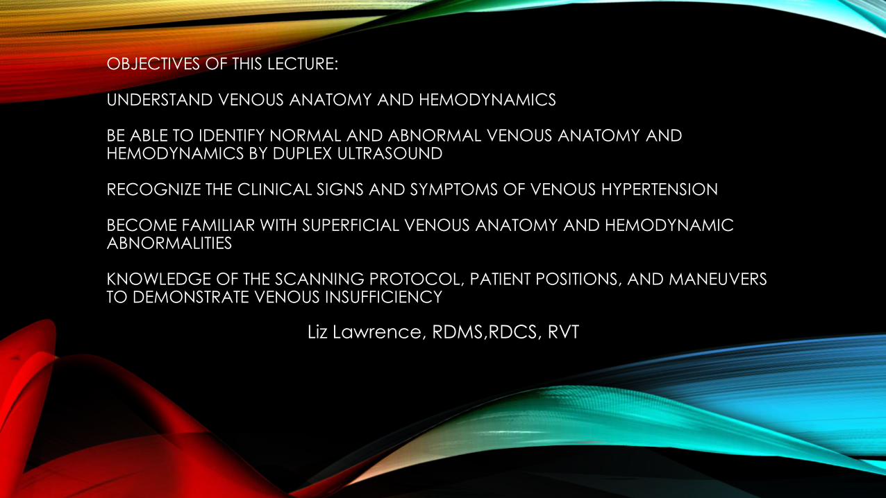
OBJECTIVES OF THIS LECTURE:
UNDERSTAND VENOUS ANATOMY AND HEMODYNAMICS
BE ABLE TO IDENTIFY NORMAL AND ABNORMAL VENOUS ANATOMY AND HEMODYNAMICS BY DUPLEX ULTRASOUND
RECOGNIZE THE CLINICAL SIGNS AND SYMPTOMS OF VENOUS HYPERTENSION
BECOME FAMILIAR WITH SUPERFICIAL VENOUS ANATOMY AND HEMODYNAMIC ABNORMALITIES
KNOWLEDGE OF THE SCANNING PROTOCOL, PATIENT POSITIONS, AND MANEUVERS TO DEMONSTRATE VENOUS INSUFFICIENCY
Liz Lawrence, RDMS,RDCS, RVT
VENOUS HEMODYNAMICSWHAT HAPPENS WHEN FLOW IS WRONG……
Liz Lawrence, RDMS,RDCS, RVT

KNOW YOUR ANATOMY
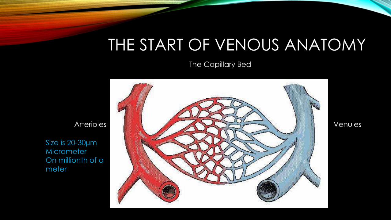
THE START OF VENOUS ANATOMYThe Capillary Bed
Arterioles Venules
Size is 20-30µmMicrometerOn millionth of a meter
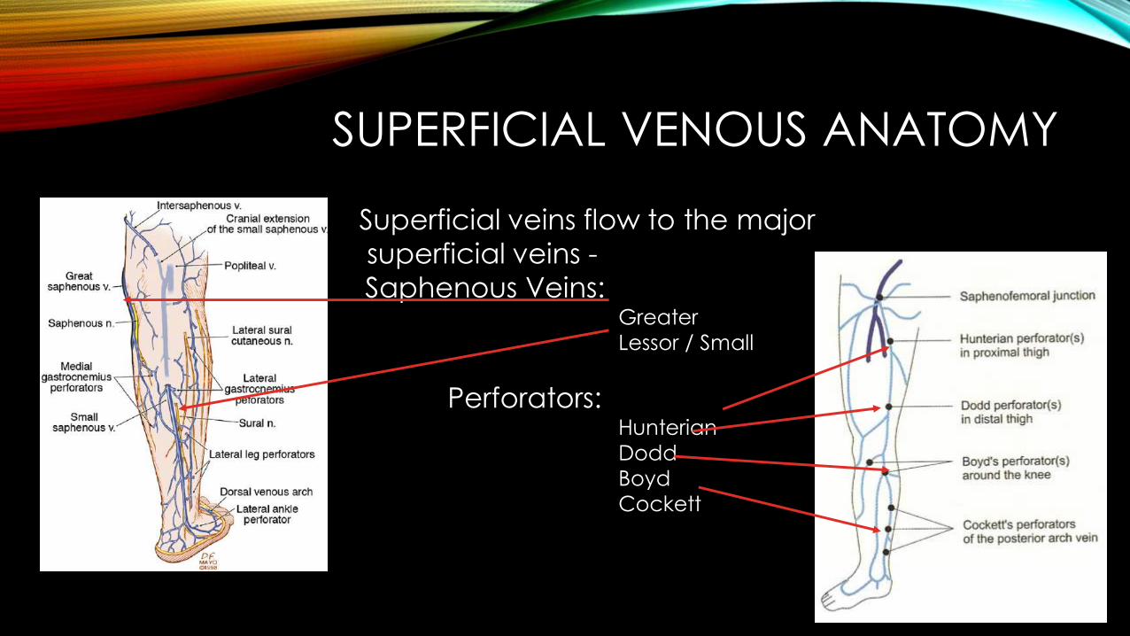
SUPERFICIAL VENOUS ANATOMY
Superficial veins flow to the major superficial veins -Saphenous Veins:
GreaterLessor / Small
Perforators: HunterianDoddBoydCockett
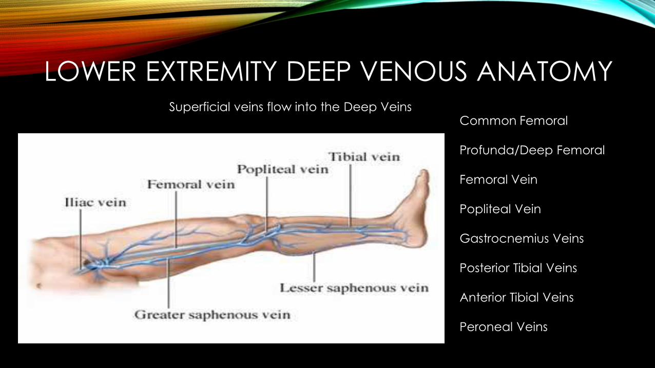
LOWER EXTREMITY DEEP VENOUS ANATOMYCommon Femoral
Profunda/Deep Femoral
Femoral Vein
Popliteal Vein
Gastrocnemius Veins
Posterior Tibial Veins
Anterior Tibial Veins
Peroneal Veins
Superficial veins flow into the Deep Veins
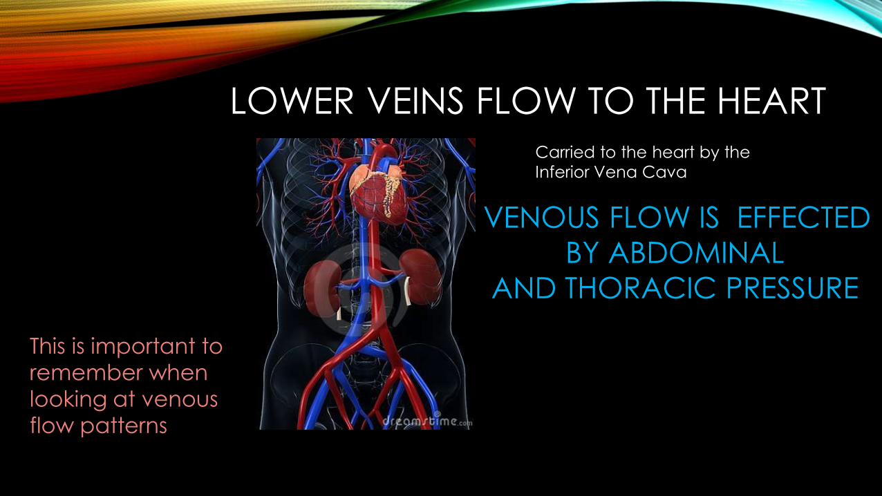
LOWER VEINS FLOW TO THE HEART
This is important to remember when looking at venousflow patterns
VENOUS FLOW IS EFFECTED BY ABDOMINAL
AND THORACIC PRESSURE
Carried to the heart by the Inferior Vena Cava
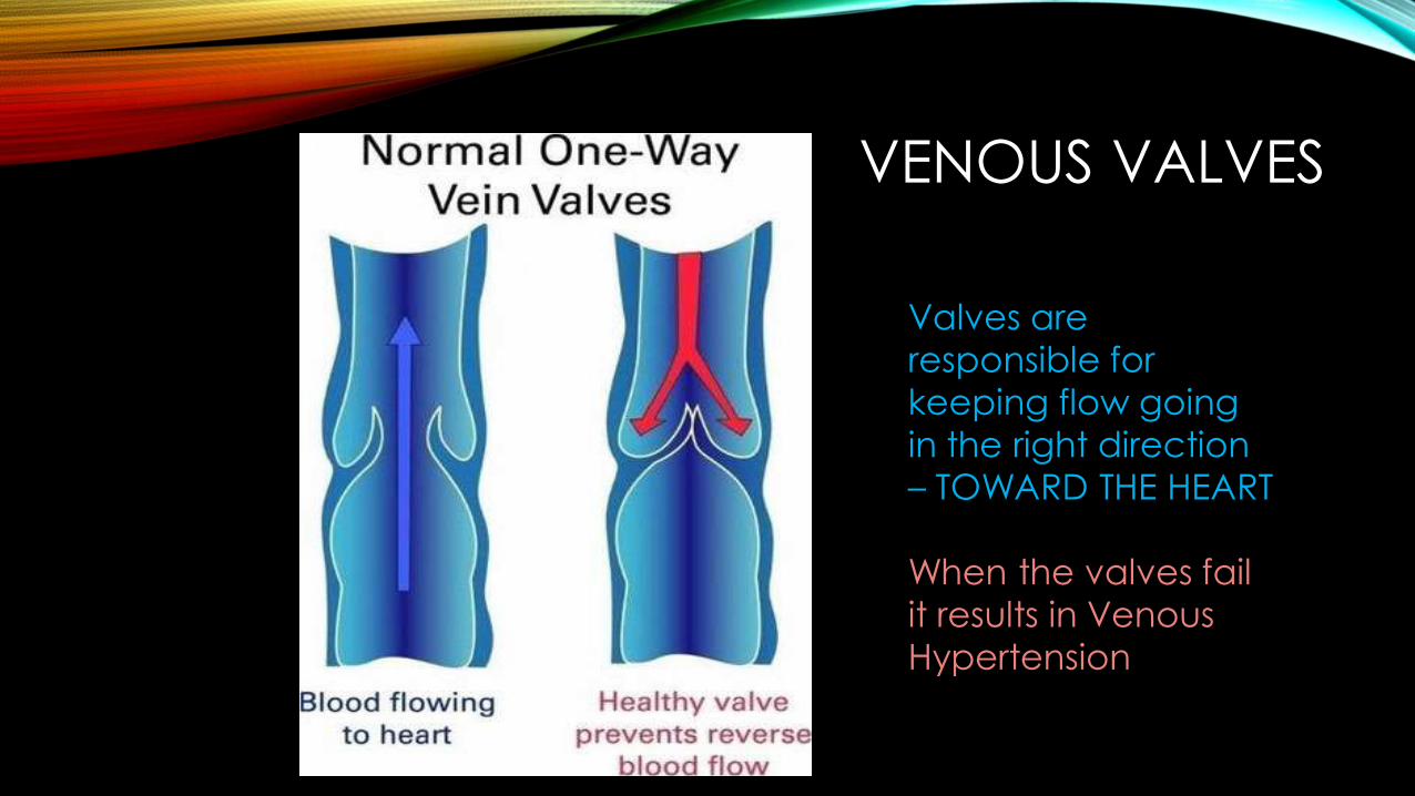
VENOUS VALVES
Valves are responsible for keeping flow going in the right direction – TOWARD THE HEART
When the valves fail it results in Venous Hypertension
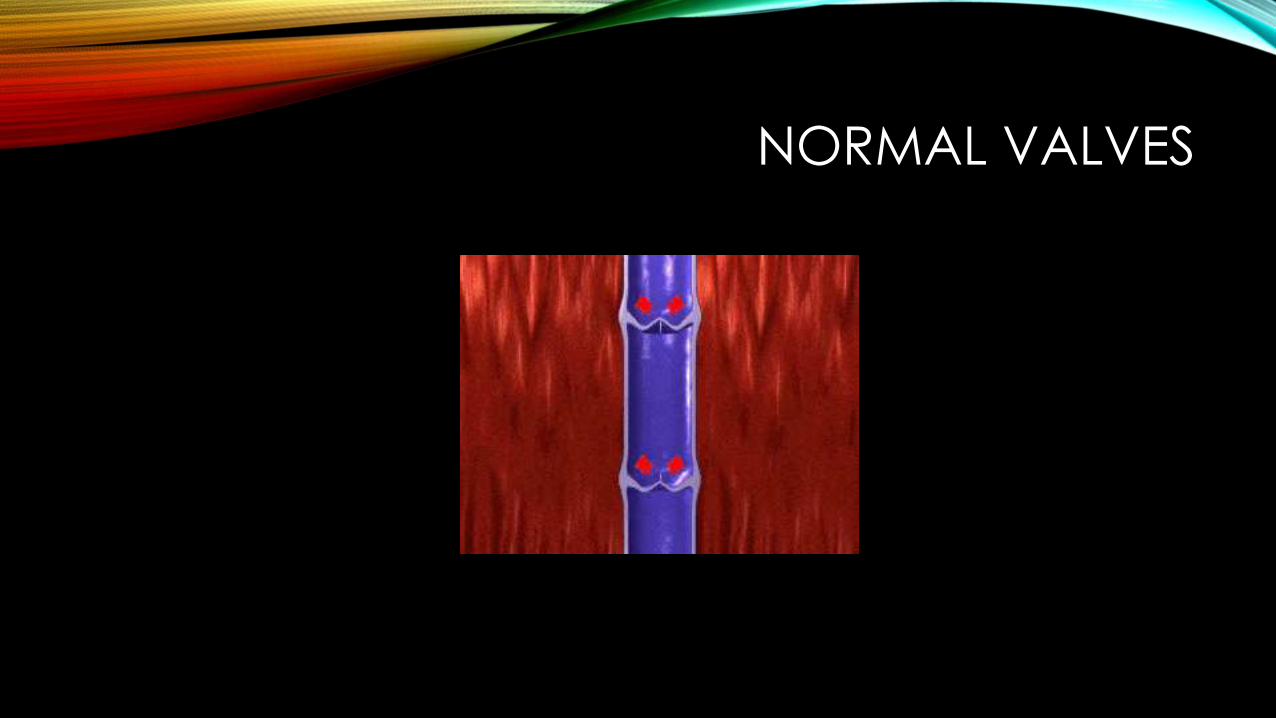
NORMAL VALVES
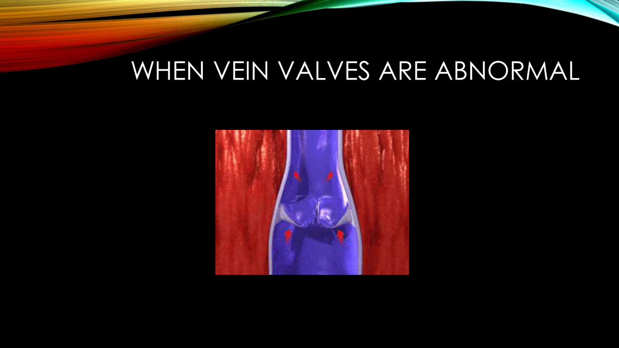
WHEN VEIN VALVES ARE ABNORMAL
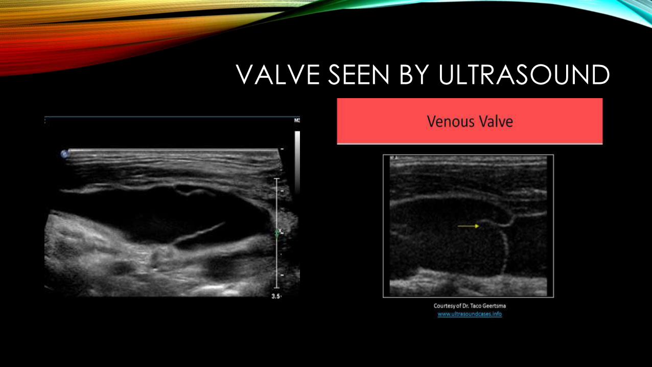
VALVE SEEN BY ULTRASOUND
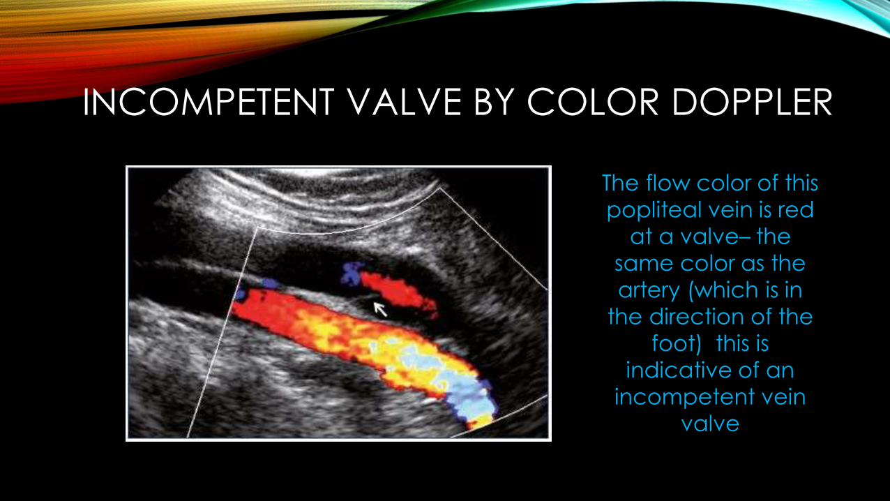
INCOMPETENT VALVE BY COLOR DOPPLER
The flow color of this popliteal vein is red
at a valve– the same color as the artery (which is in
the direction of the foot) this is
indicative of an incompetent vein
valve
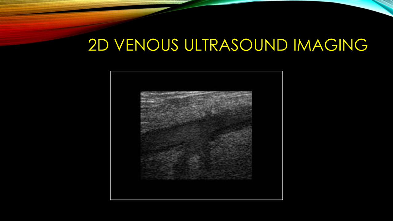
2D VENOUS ULTRASOUND IMAGING
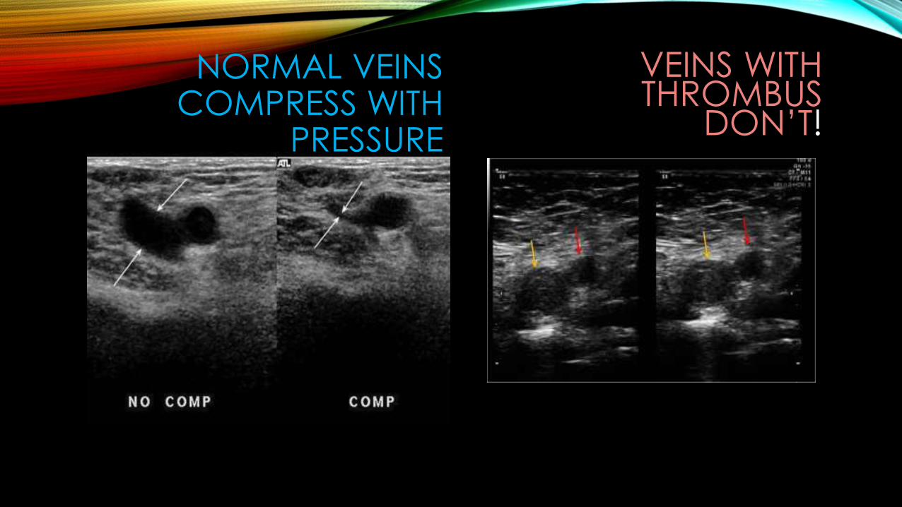
NORMAL VEINS COMPRESS WITH
PRESSURE
VEINS WITH THROMBUS
DON’T!
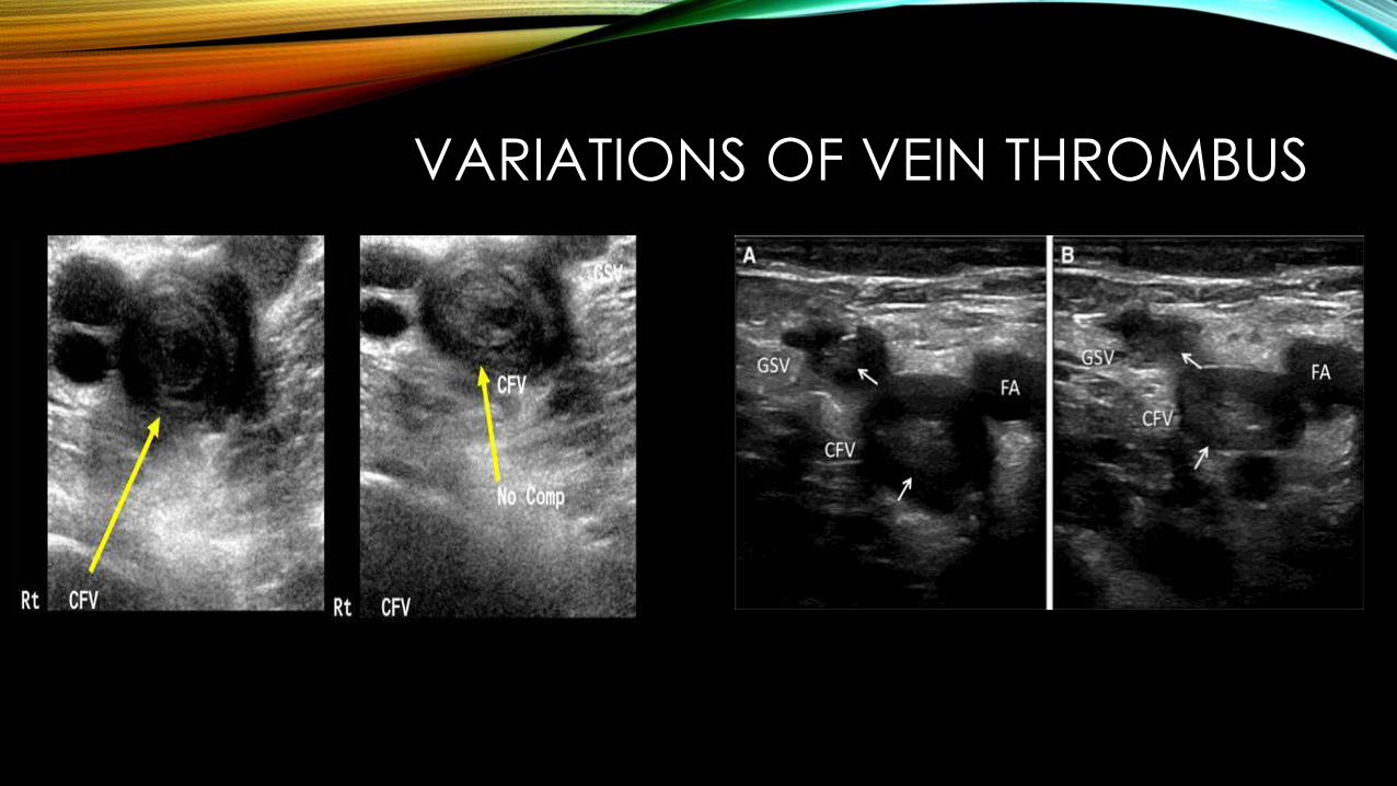
VARIATIONS OF VEIN THROMBUS
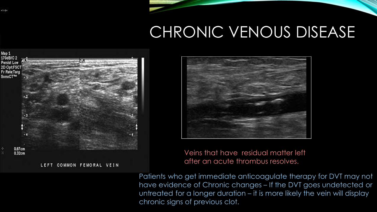
CHRONIC VENOUS DISEASE
Veins that have residual matter left after an acute thrombus resolves.
Patients who get immediate anticoagulate therapy for DVT may not have evidence of Chronic changes – If the DVT goes undetected or untreated for a longer duration – it is more likely the vein will display chronic signs of previous clot.
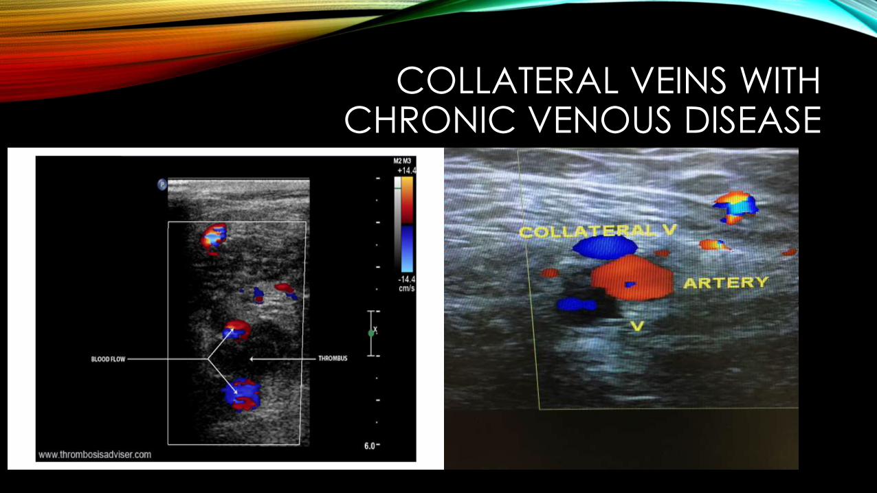
COLLATERAL VEINS WITH CHRONIC VENOUS DISEASE
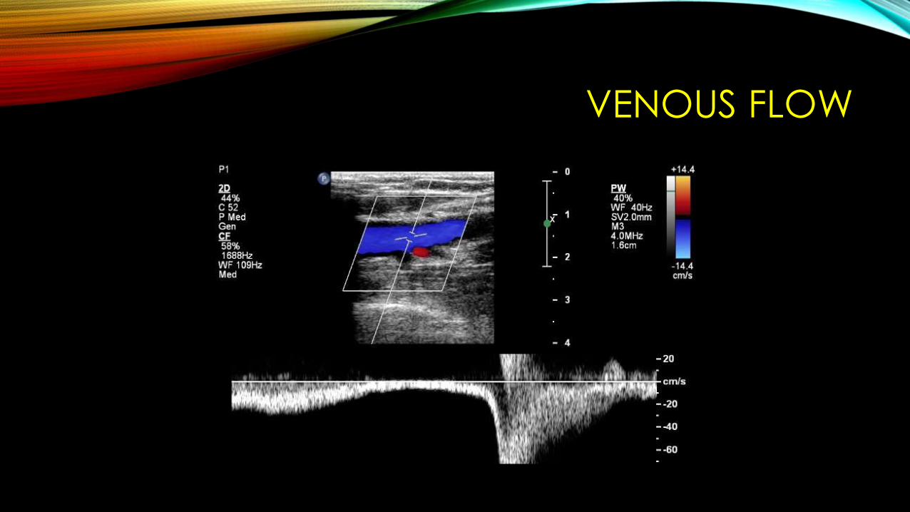
VENOUS FLOW
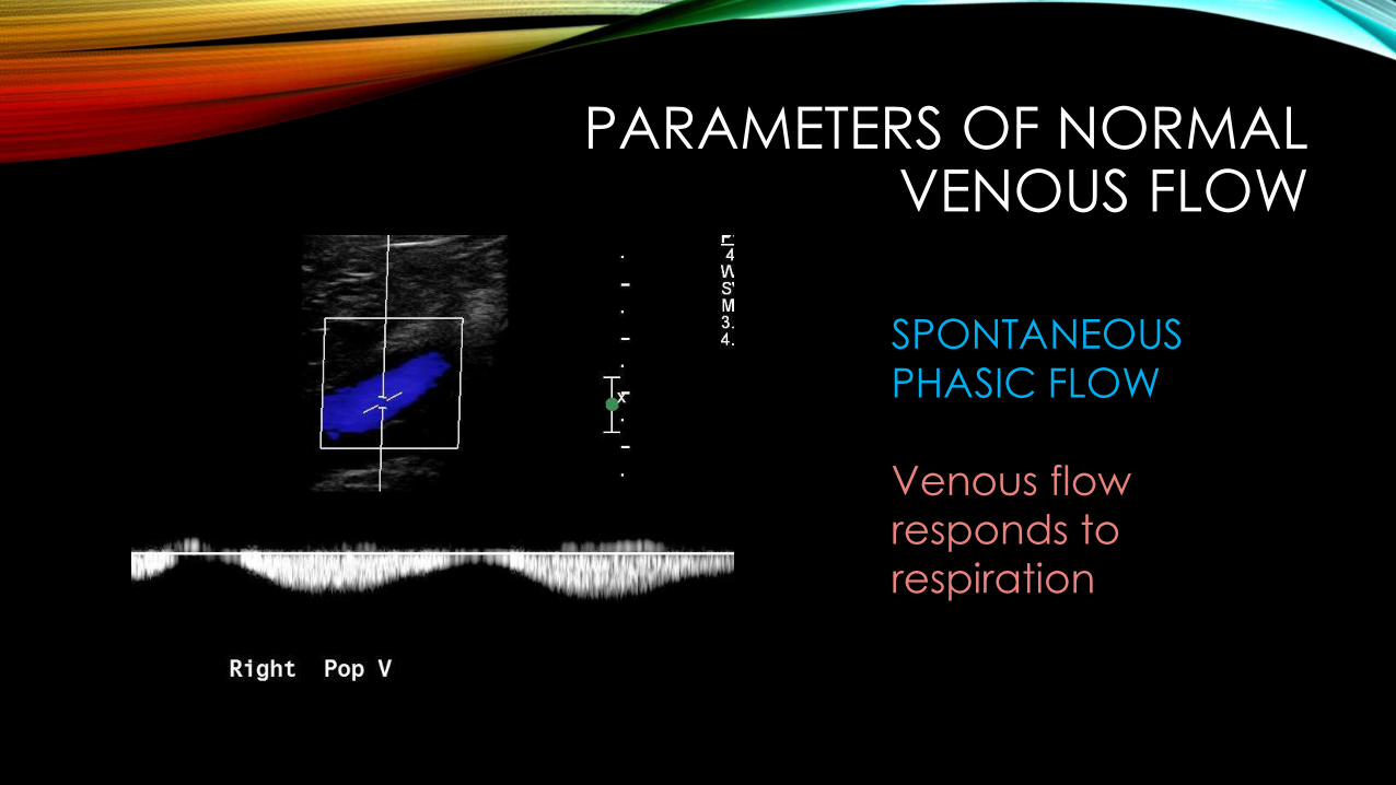
PARAMETERS OF NORMAL VENOUS FLOW
SPONTANEOUSPHASIC FLOW
Venous flow responds to respiration
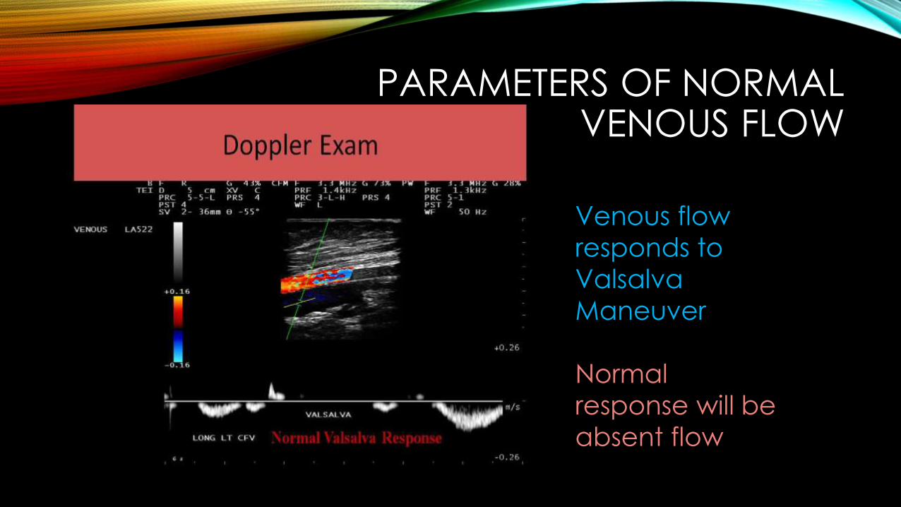
PARAMETERS OF NORMAL VENOUS FLOW
Venous flow responds to Valsalva Maneuver
Normal response will be absent flow
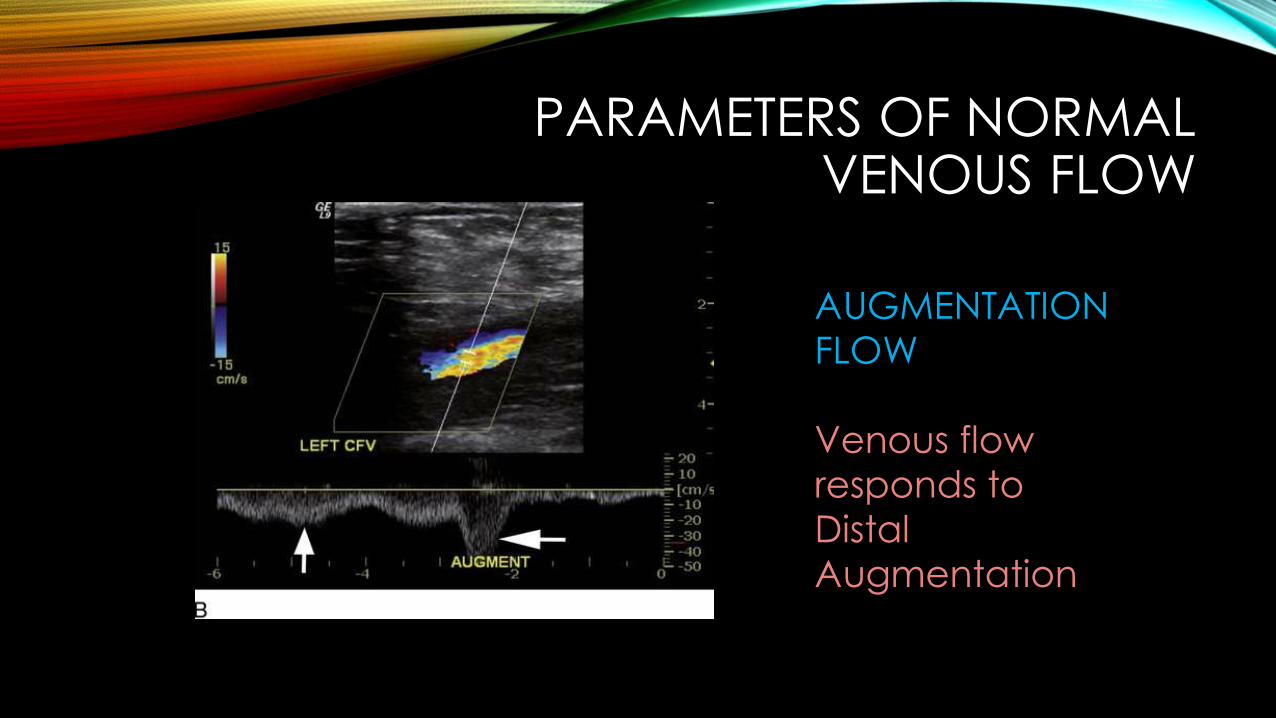
PARAMETERS OF NORMAL VENOUS FLOW
AUGMENTATIONFLOW
Venous flow responds to Distal Augmentation
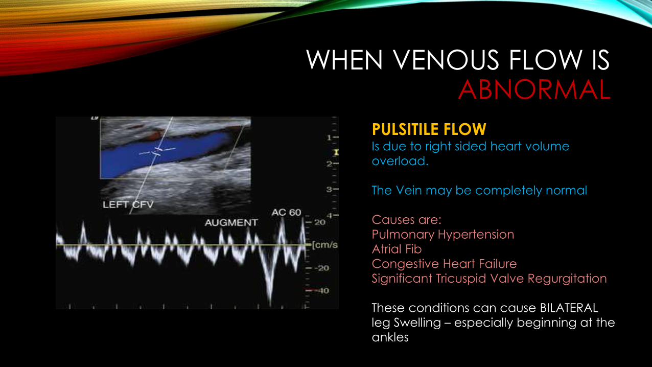
WHEN VENOUS FLOW IS ABNORMAL
PULSITILE FLOW Is due to right sided heart volume overload.
The Vein may be completely normal
Causes are: Pulmonary HypertensionAtrial FibCongestive Heart FailureSignificant Tricuspid Valve Regurgitation
These conditions can cause BILATERAL leg Swelling – especially beginning at the ankles
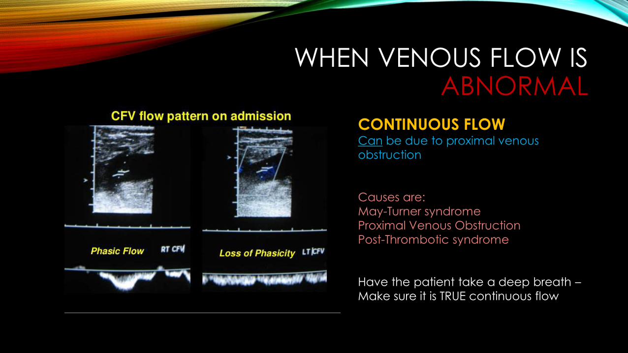
WHEN VENOUS FLOW IS ABNORMAL
CONTINUOUS FLOWCan be due to proximal venous obstruction
Causes are: May-Turner syndromeProximal Venous ObstructionPost-Thrombotic syndrome
Have the patient take a deep breath –Make sure it is TRUE continuous flow
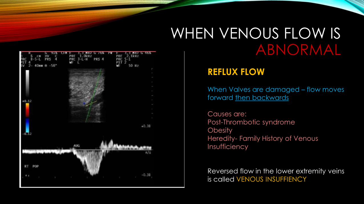
WHEN VENOUS FLOW IS ABNORMAL
REFLUX FLOW
When Valves are damaged – flow moves forward then backwards
Causes are: Post-Thrombotic syndromeObesityHeredity- Family History of Venous Insufficiency
Reversed flow in the lower extremity veins is called VENOUS INSUFFIENCY
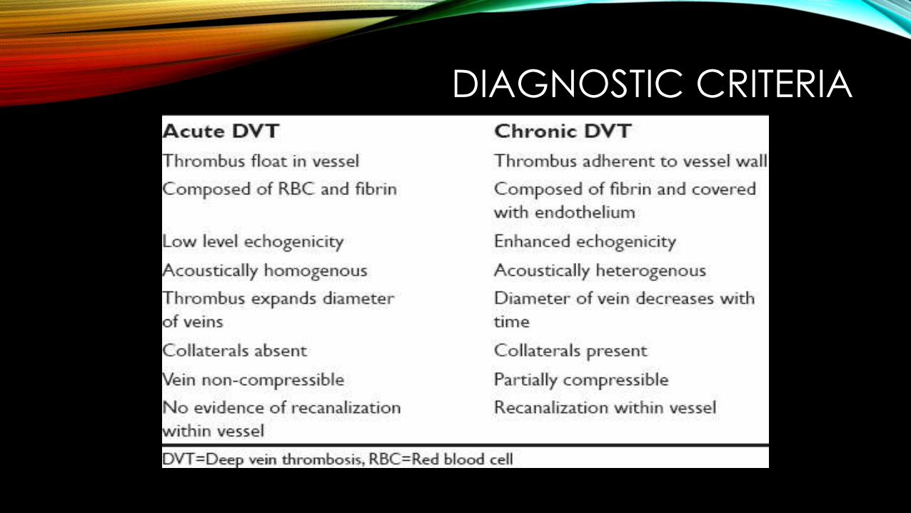
DIAGNOSTIC CRITERIA
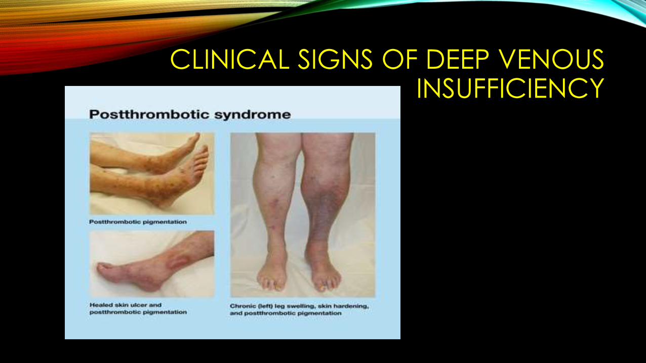
CLINICAL SIGNS OF DEEP VENOUS INSUFFICIENCY
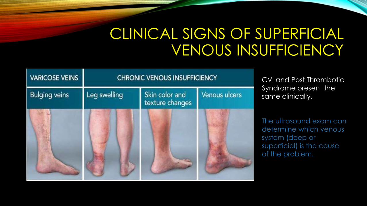
CLINICAL SIGNS OF SUPERFICIAL VENOUS INSUFFICIENCY
CVI and Post ThromboticSyndrome present the same clinically.
The ultrasound exam can determine which venous system (deep or superficial) is the cause of the problem.
28
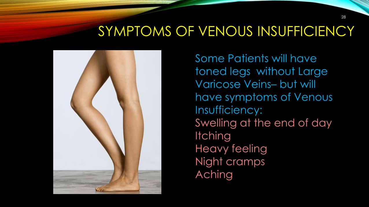
SYMPTOMS OF VENOUS INSUFFICIENCY
Some Patients will have toned legs without Large Varicose Veins– but will have symptoms of Venous Insufficiency:Swelling at the end of dayItchingHeavy feelingNight crampsAching
SUPERFICIAL VENOUS DUPLEX EXAMPATIENT POSITION
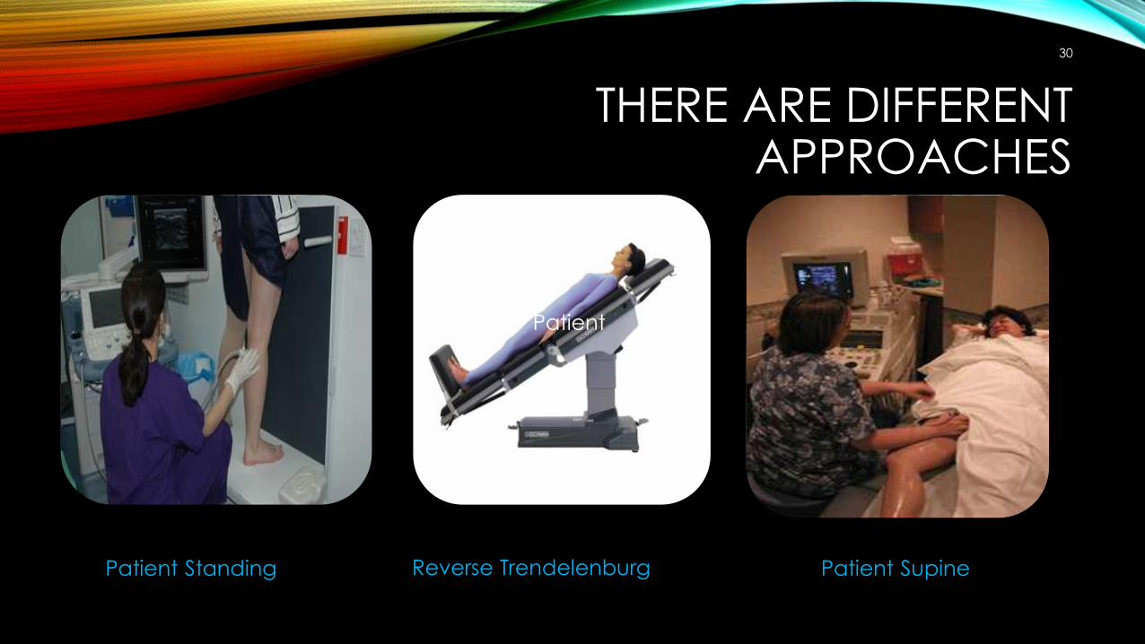
THERE ARE DIFFERENT APPROACHES
30
Patient Standing
Patient
Reverse Trendelenburg Patient Supine
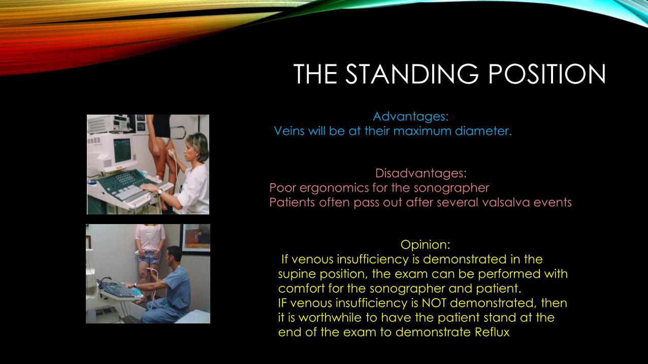
THE STANDING POSITIONAdvantages:
Veins will be at their maximum diameter.
Disadvantages: Poor ergonomics for the sonographerPatients often pass out after several valsalva events
Opinion:If venous insufficiency is demonstrated in the
supine position, the exam can be performed with comfort for the sonographer and patient.IF venous insufficiency is NOT demonstrated, then it is worthwhile to have the patient stand at the end of the exam to demonstrate Reflux
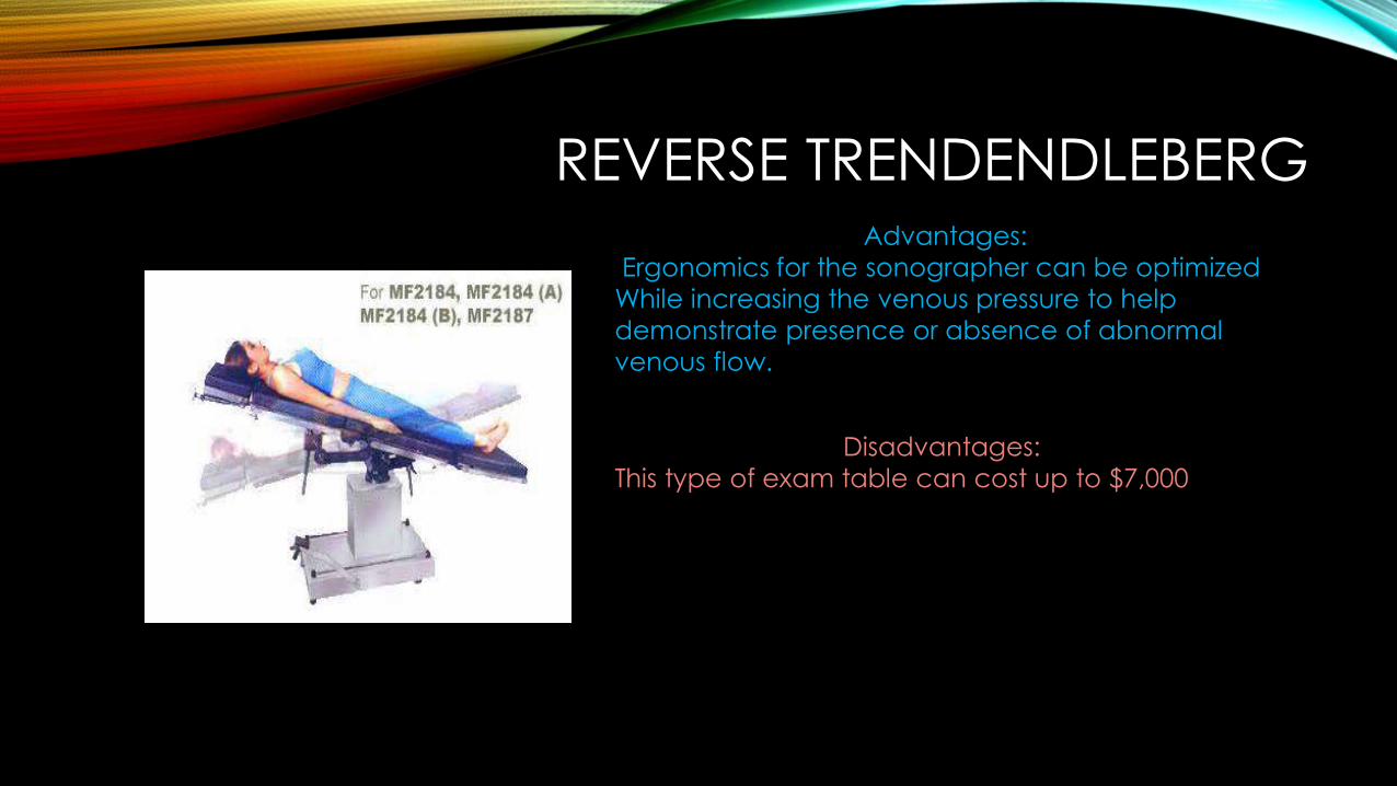
REVERSE TRENDENDLEBERG
Disadvantages: This type of exam table can cost up to $7,000
Advantages: Ergonomics for the sonographer can be optimized
While increasing the venous pressure to help demonstrate presence or absence of abnormal venous flow.
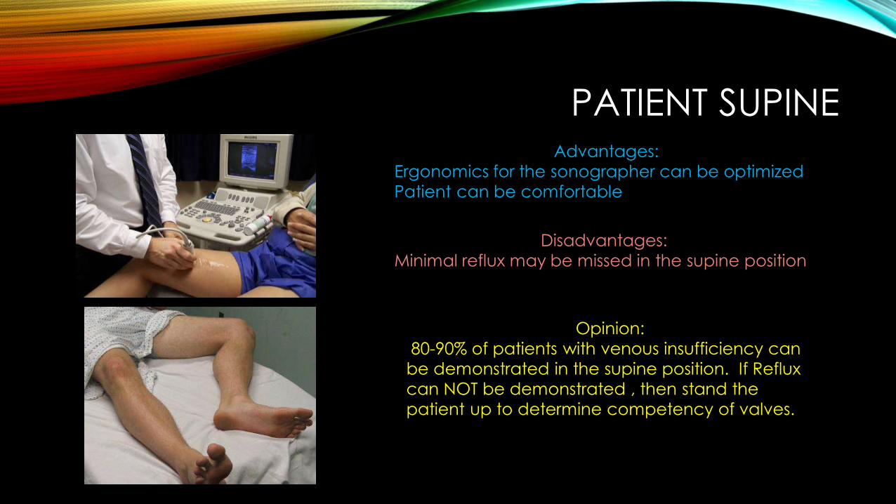
PATIENT SUPINEAdvantages:
Ergonomics for the sonographer can be optimizedPatient can be comfortable
Disadvantages: Minimal reflux may be missed in the supine position
Opinion:80-90% of patients with venous insufficiency can
be demonstrated in the supine position. If Reflux can NOT be demonstrated , then stand the patient up to determine competency of valves.
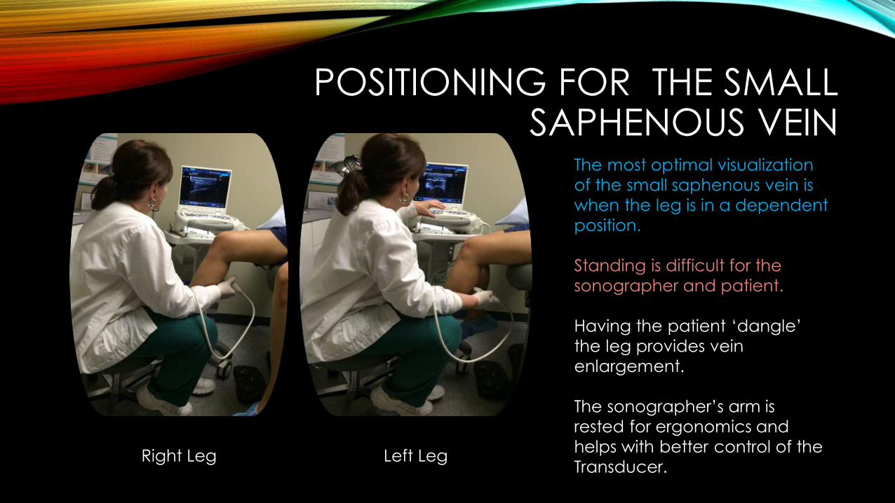
POSITIONING FOR THE SMALL SAPHENOUS VEIN
The most optimal visualization of the small saphenous vein is when the leg is in a dependent position.
Standing is difficult for the sonographer and patient.
Having the patient ‘dangle’ the leg provides vein enlargement.
The sonographer’s arm is rested for ergonomics and helps with better control of the Transducer.Right Leg Left Leg
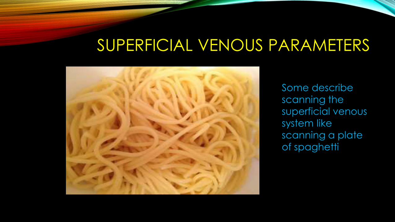
SUPERFICIAL VENOUS PARAMETERS
Some describe scanning the superficial venous system like scanning a plate of spaghetti
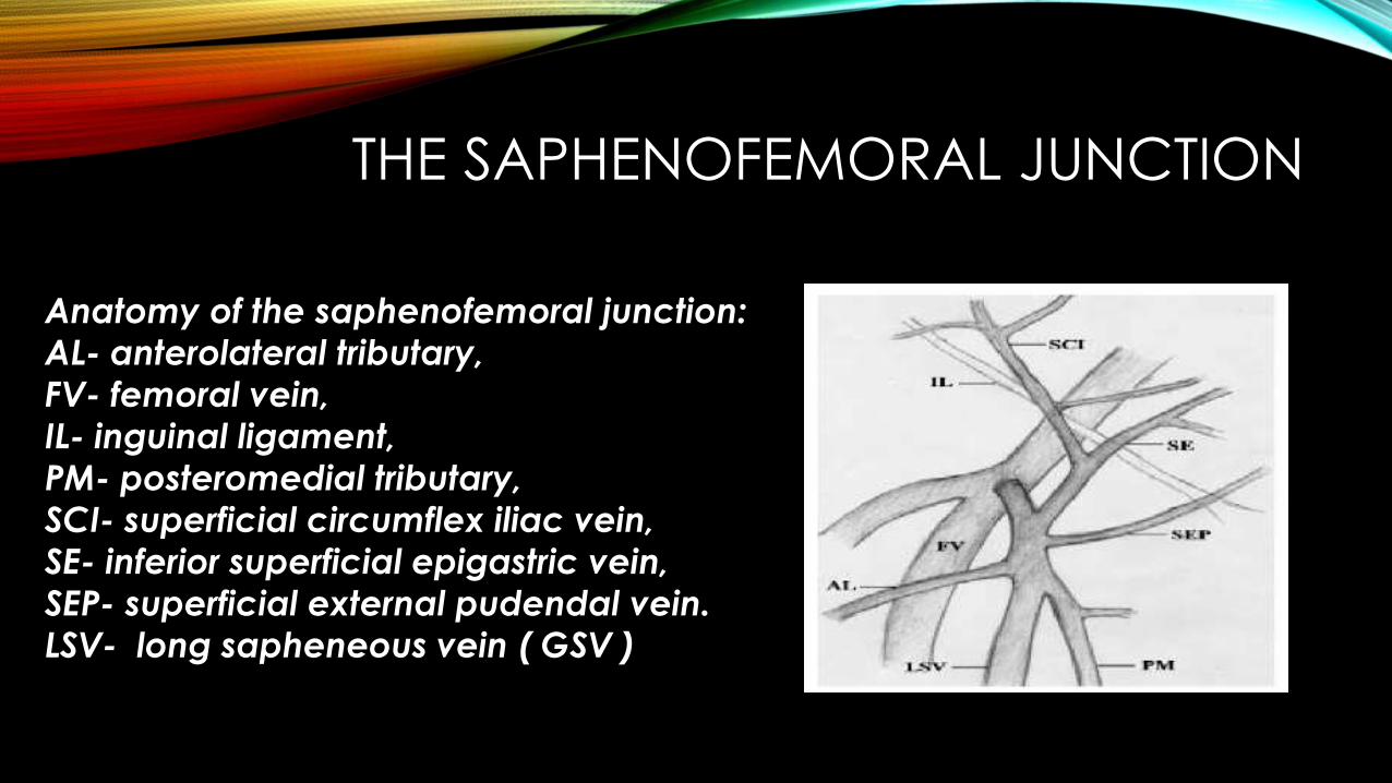
THE SAPHENOFEMORAL JUNCTION
Anatomy of the saphenofemoral junction: AL- anterolateral tributary,FV- femoral vein, IL- inguinal ligament, PM- posteromedial tributary, SCI- superficial circumflex iliac vein, SE- inferior superficial epigastric vein, SEP- superficial external pudendal vein.LSV- long sapheneous vein ( GSV )
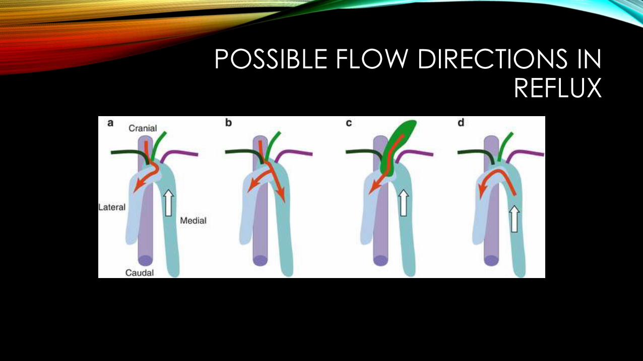
POSSIBLE FLOW DIRECTIONS IN REFLUX
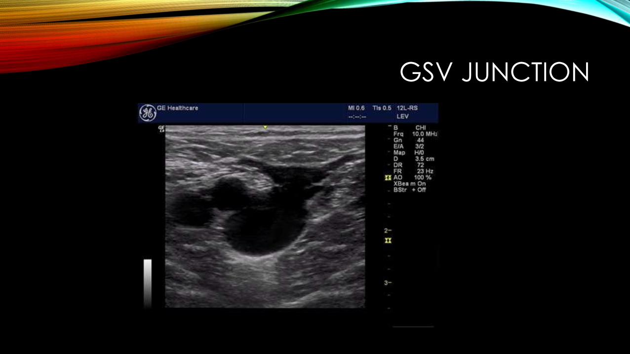
GSV JUNCTION
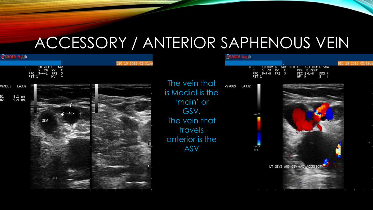
ACCESSORY / ANTERIOR SAPHENOUS VEIN
The vein that is Medial is the
‘main’ or GSV.
The vein that travels
anterior is the ASV
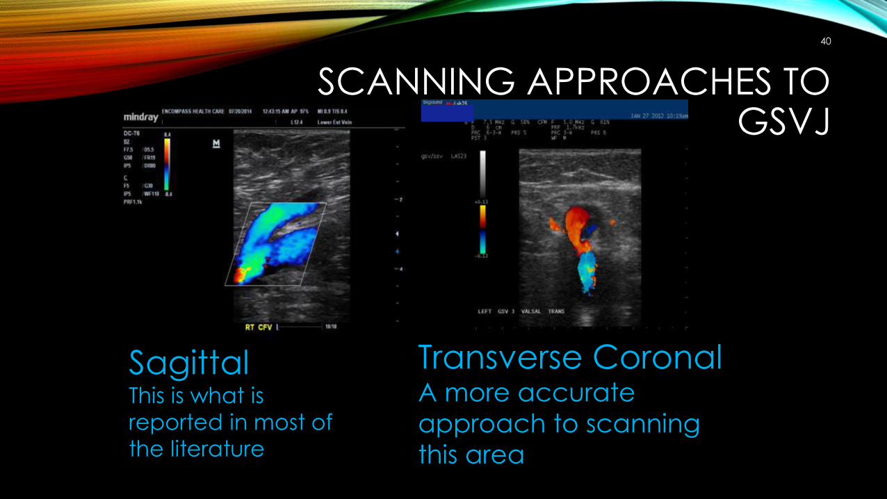
SCANNING APPROACHES TO GSVJ
40
SagittalThis is what is reported in most of the literature
Transverse CoronalA more accurate approach to scanning this area

WHY TRANSVERSE CORONAL WORKS
41
By positioning the transducer in a way to ‘look down the barrel’ of the vein, the ultrasound color and Doppler angle is better aligned with flow and if the reflux flow is eccentric, this position will detect and determine the angle of the reflux.
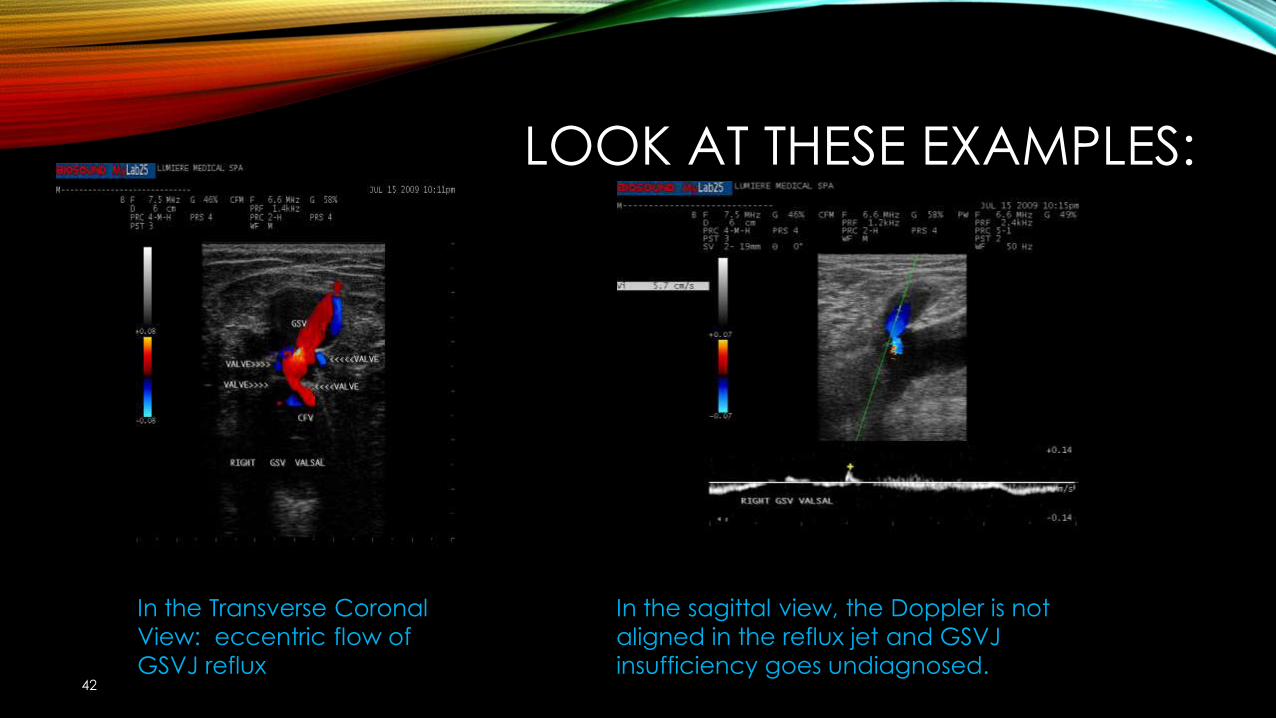
LOOK AT THESE EXAMPLES:
42
In the Transverse Coronal View: eccentric flow of GSVJ reflux
In the sagittal view, the Doppler is not aligned in the reflux jet and GSVJ insufficiency goes undiagnosed.
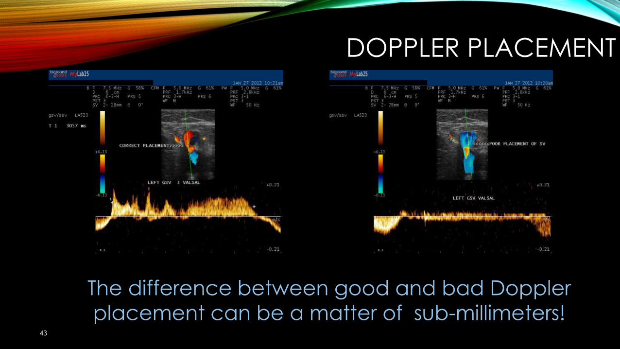
DOPPLER PLACEMENT
43
The difference between good and bad Doppler placement can be a matter of sub-millimeters!
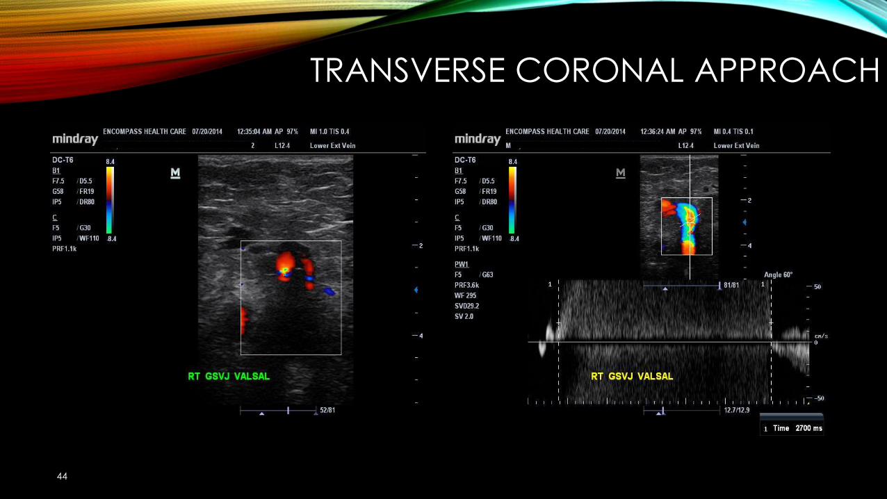
TRANSVERSE CORONAL APPROACH
44
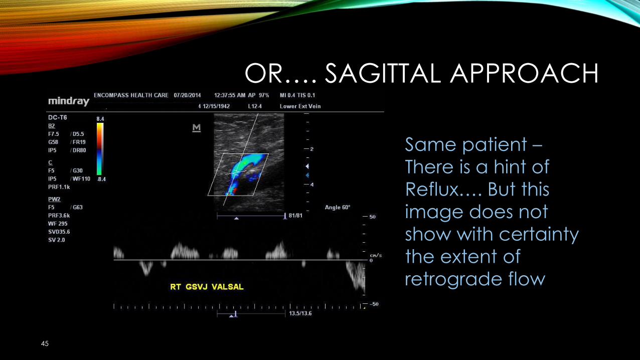
OR…. SAGITTAL APPROACH
45
Same patient –There is a hint of Reflux…. But this image does not show with certainty the extent of retrograde flow
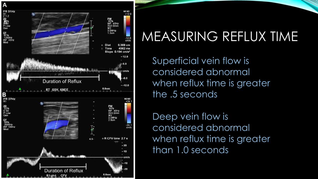
MEASURING REFLUX TIME
Superficial vein flow is considered abnormal when reflux time is greater the .5 seconds
Deep vein flow is considered abnormal when reflux time is greater than 1.0 seconds
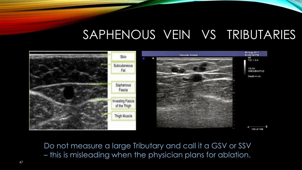
SAPHENOUS VEIN VS TRIBUTARIES
47
Do not measure a large Tributary and call it a GSV or SSV – this is misleading when the physician plans for ablation.
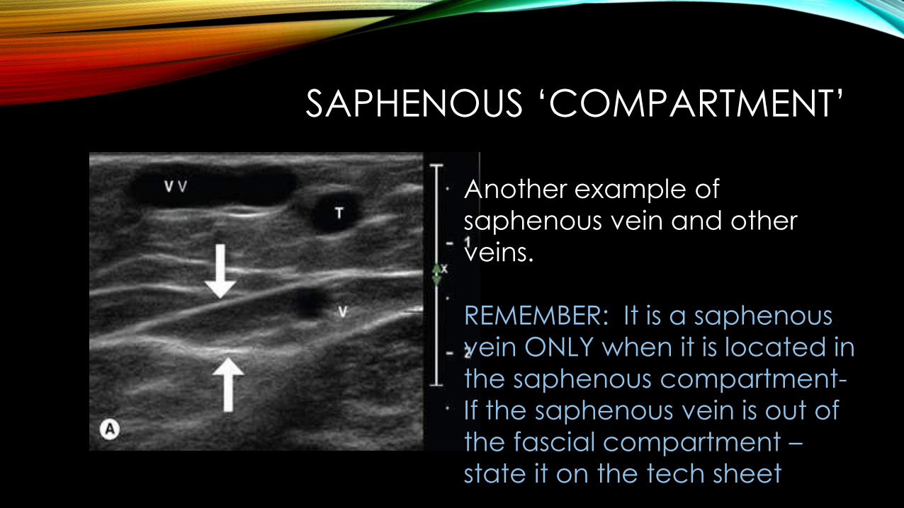
SAPHENOUS ‘COMPARTMENT’
Another example of saphenous vein and other veins.
REMEMBER: It is a saphenous vein ONLY when it is located in the saphenous compartment-If the saphenous vein is out of the fascial compartment –state it on the tech sheet
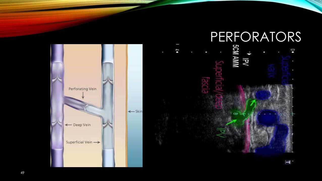
PERFORATORS
49
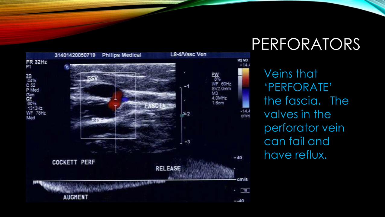
PERFORATORS
Veins that ‘PERFORATE’ the fascia. The valves in the perforator vein can fail and have reflux.
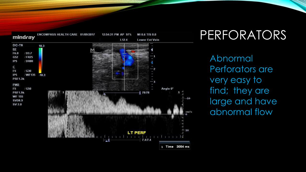
PERFORATORS
Abnormal Perforators are very easy to find; they are large and have abnormal flow
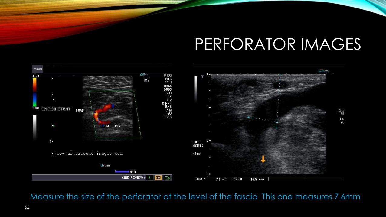
PERFORATOR IMAGES
52
Measure the size of the perforator at the level of the fascia This one measures 7.6mm
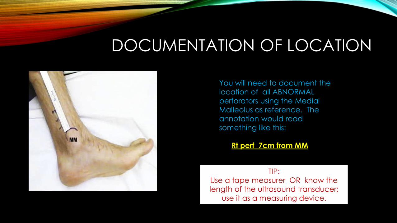
DOCUMENTATION OF LOCATION
You will need to document the location of all ABNORMAL perforators using the Medial Malleolus as reference. The annotation would read something like this:
Rt perf 7cm from MM
TIP:Use a tape measurer OR know the length of the ultrasound transducer;
use it as a measuring device.
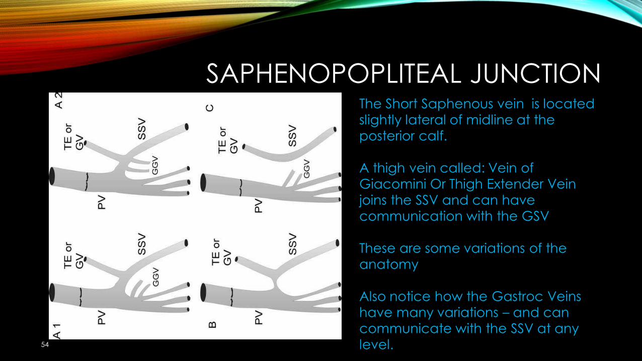
SAPHENOPOPLITEAL JUNCTION
54
The Short Saphenous vein is located slightly lateral of midline at the posterior calf.
A thigh vein called: Vein of Giacomini Or Thigh Extender Vein joins the SSV and can have communication with the GSV
These are some variations of the anatomy
Also notice how the Gastroc Veins have many variations – and can communicate with the SSV at any level.
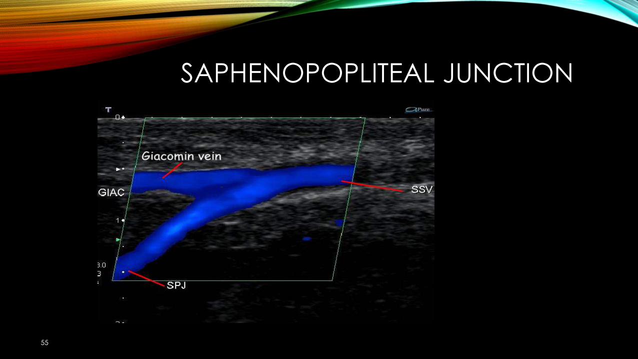
SAPHENOPOPLITEAL JUNCTION
55
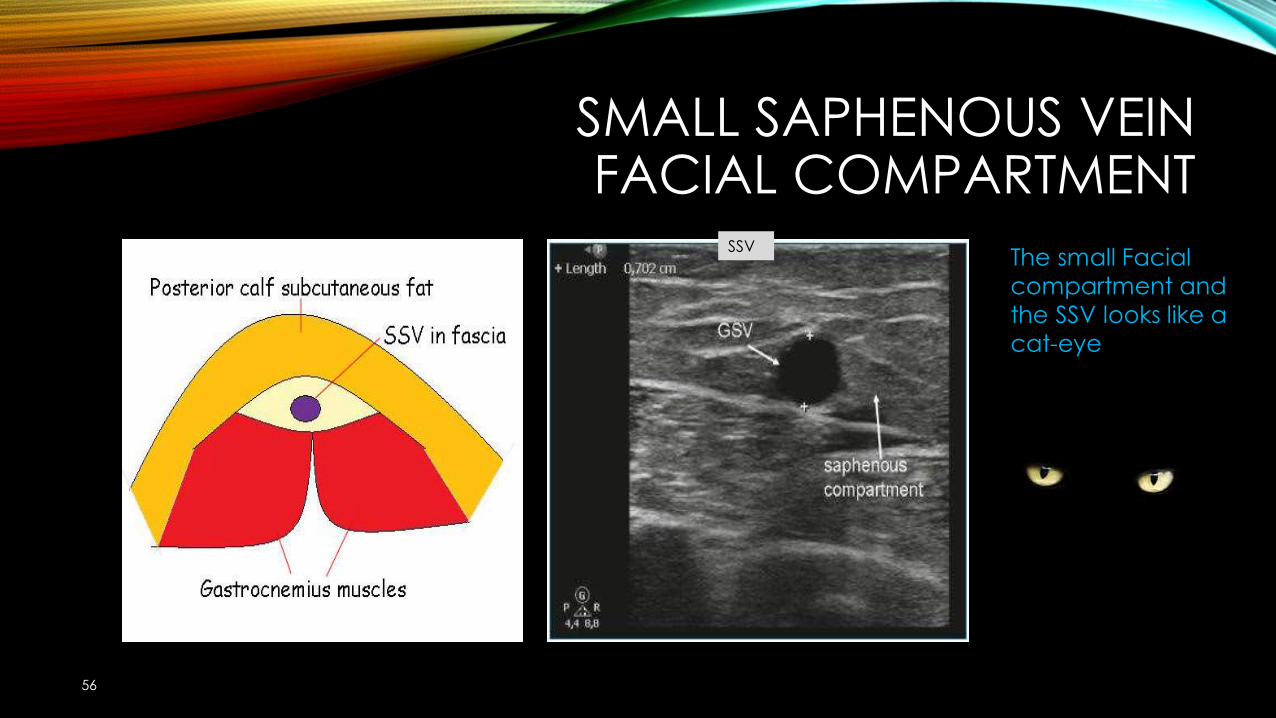
SMALL SAPHENOUS VEINFACIAL COMPARTMENT
56
SSV The small Facial compartment and the SSV looks like a cat-eye
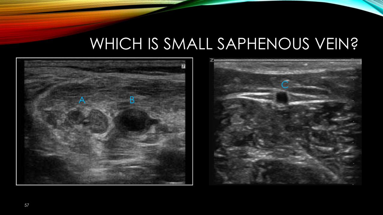
WHICH IS SMALL SAPHENOUS VEIN?
57
A BC
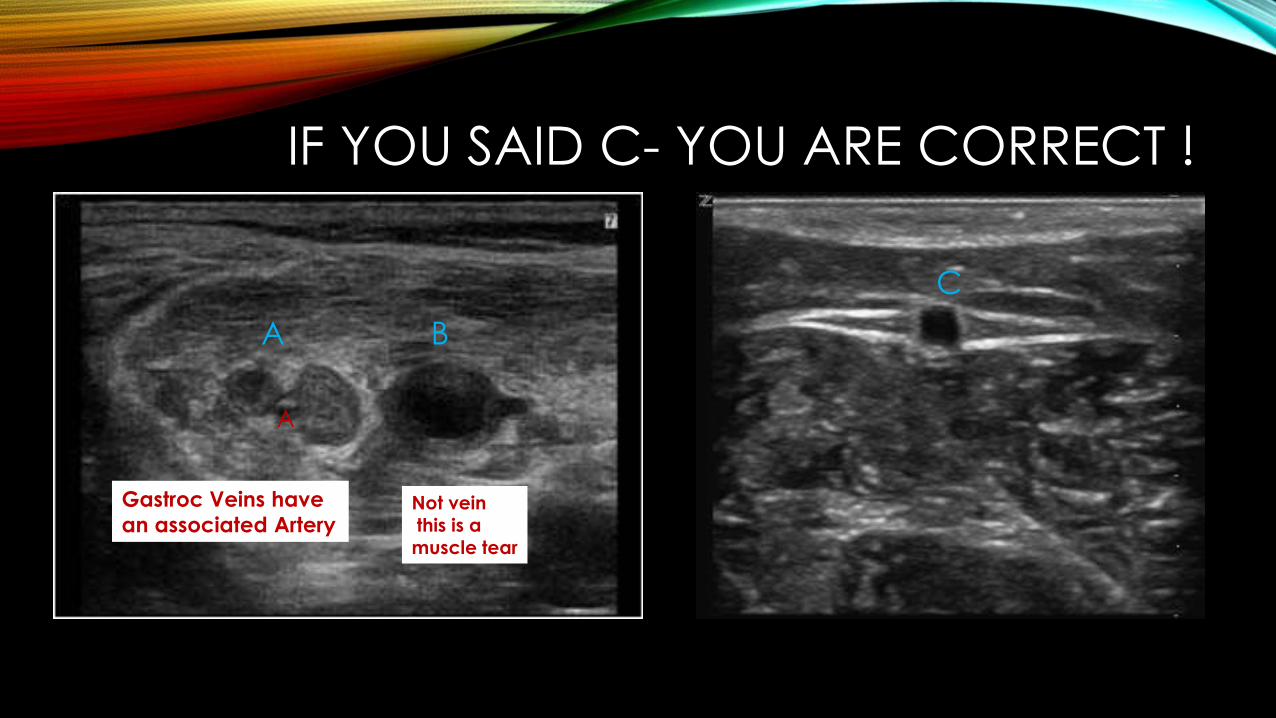
IF YOU SAID C- YOU ARE CORRECT !
A BC
Not veinthis is a muscle tear
Gastroc Veins have an associated Artery
A
MANEUVERS- TO DEMONSTRATE REFLUX
59
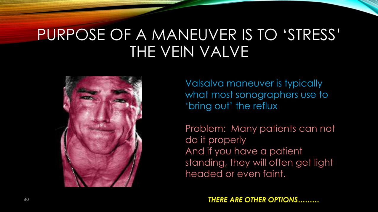
PURPOSE OF A MANEUVER IS TO ‘STRESS’ THE VEIN VALVE
60
Valsalva maneuver is typically what most sonographers use to ‘bring out’ the reflux
Problem: Many patients can not do it properlyAnd if you have a patient standing, they will often get light headed or even faint.
THERE ARE OTHER OPTIONS………
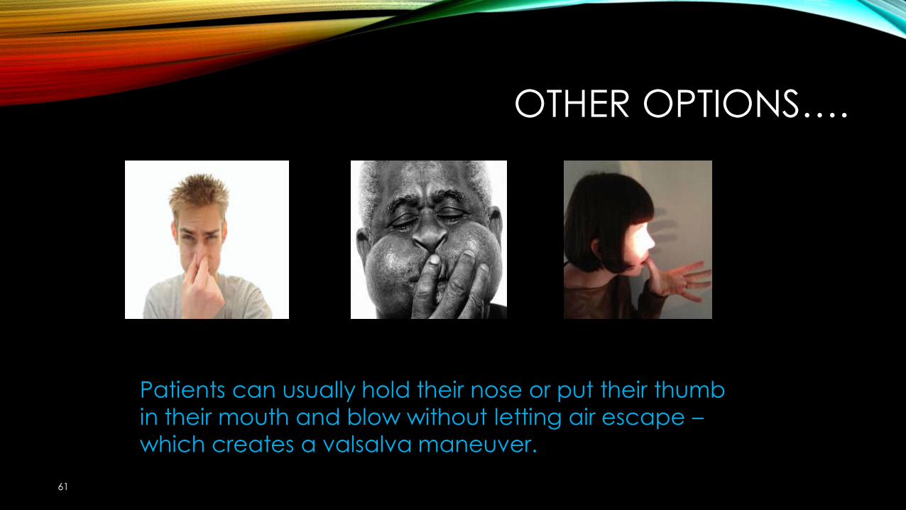
OTHER OPTIONS….
61
Patients can usually hold their nose or put their thumb in their mouth and blow without letting air escape –which creates a valsalva maneuver.
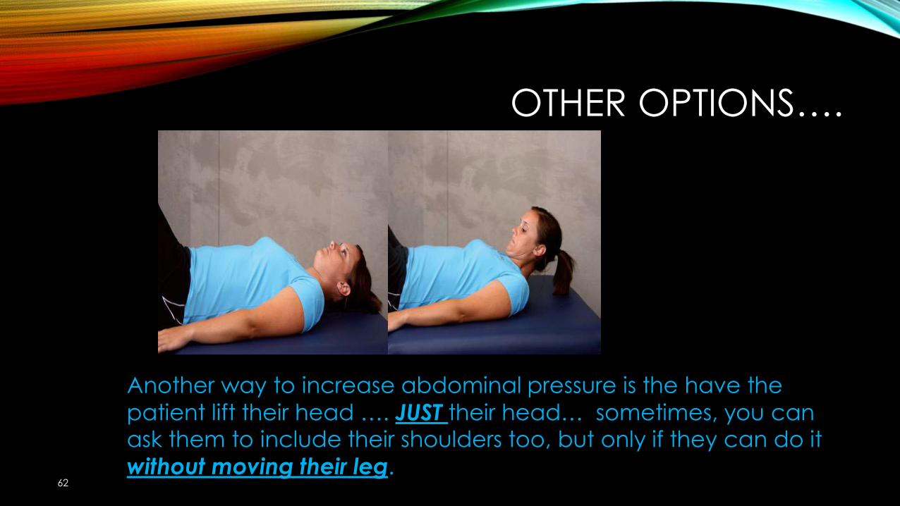
OTHER OPTIONS….
62
Another way to increase abdominal pressure is the have the patient lift their head …. JUST their head… sometimes, you can ask them to include their shoulders too, but only if they can do it without moving their leg.
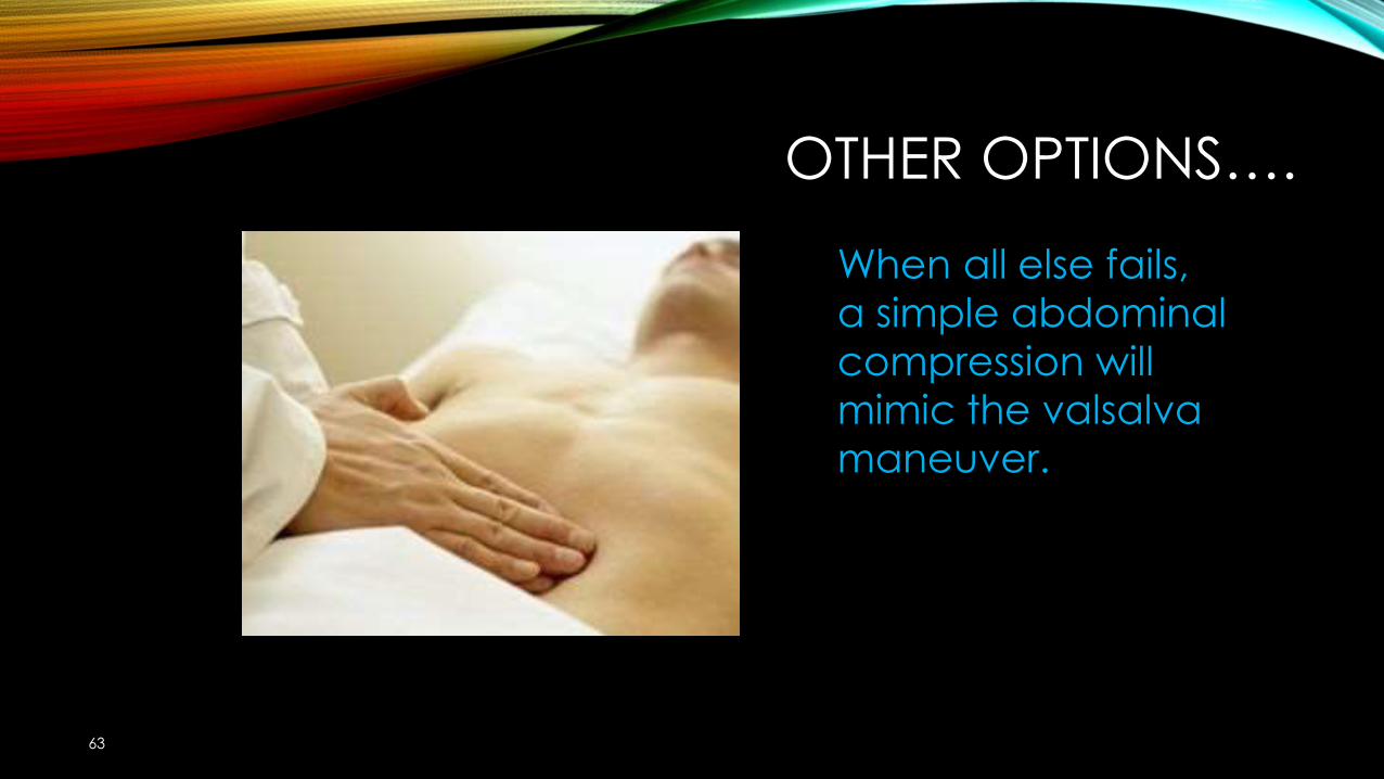
OTHER OPTIONS….
63
When all else fails, a simple abdominal compression will mimic the valsalva maneuver.
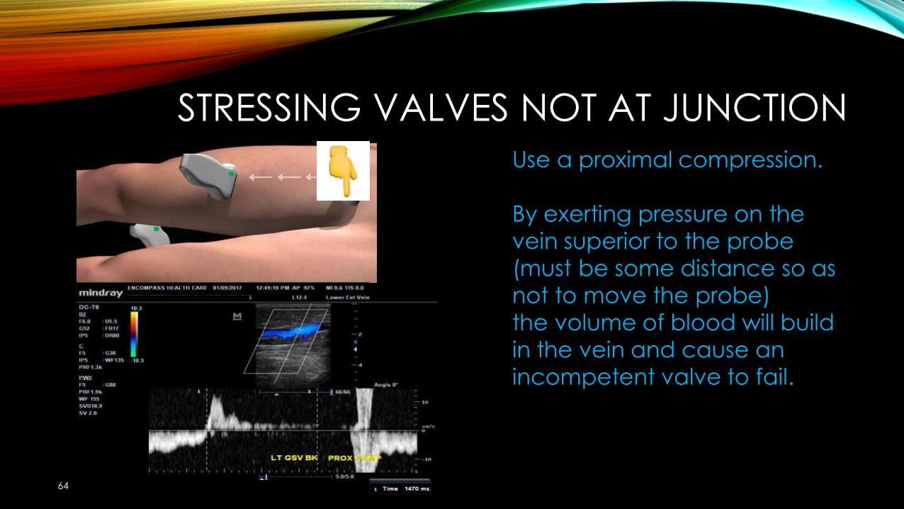
STRESSING VALVES NOT AT JUNCTION
64
Use a proximal compression.
By exerting pressure on the vein superior to the probe (must be some distance so as not to move the probe) the volume of blood will build in the vein and cause an incompetent valve to fail.
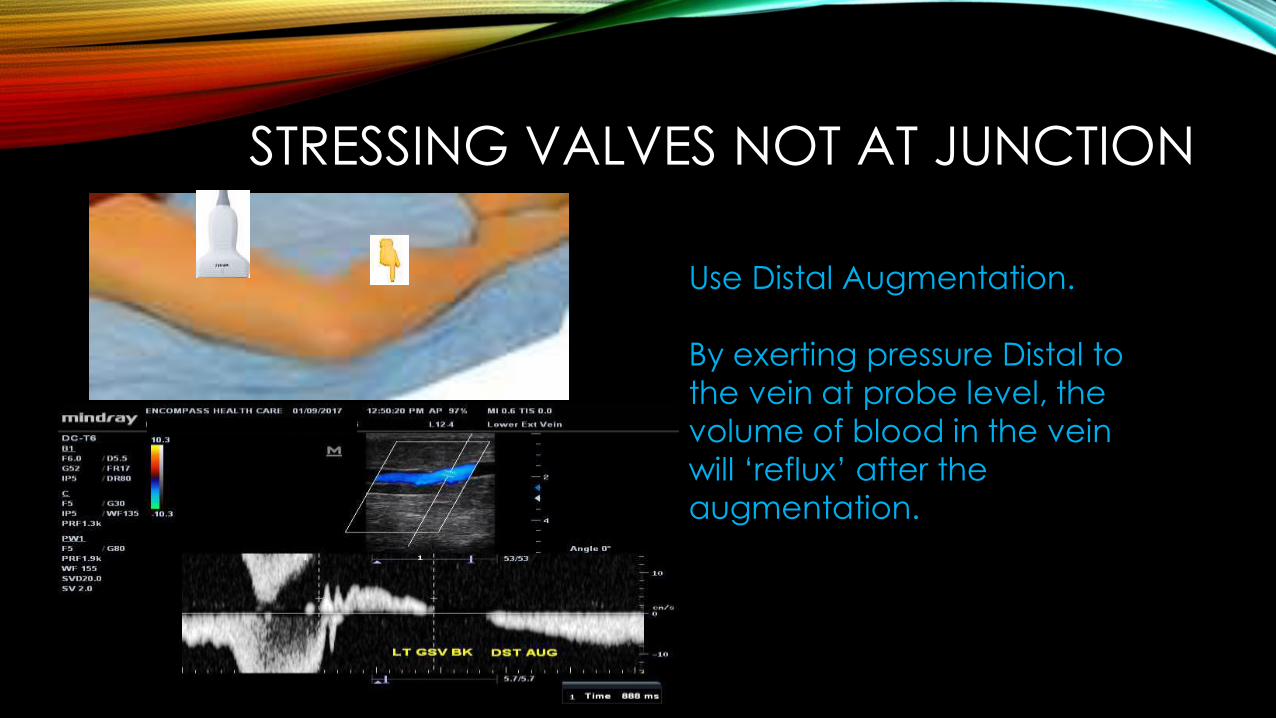
STRESSING VALVES NOT AT JUNCTION
Use Distal Augmentation.
By exerting pressure Distal to the vein at probe level, the volume of blood in the vein will ‘reflux’ after the augmentation.
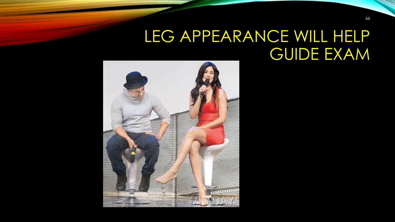
LEG APPEARANCE WILL HELP GUIDE EXAM
66
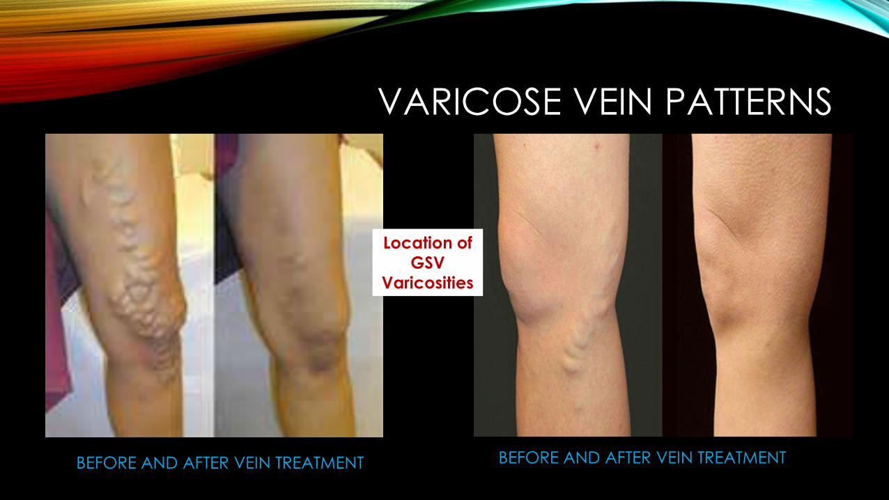
VARICOSE VEIN PATTERNS
BEFORE AND AFTER VEIN TREATMENT BEFORE AND AFTER VEIN TREATMENT
Location of GSV
Varicosities
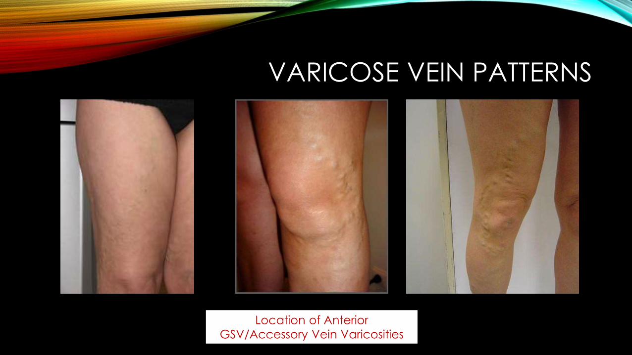
VARICOSE VEIN PATTERNS
Location of Anterior GSV/Accessory Vein Varicosities
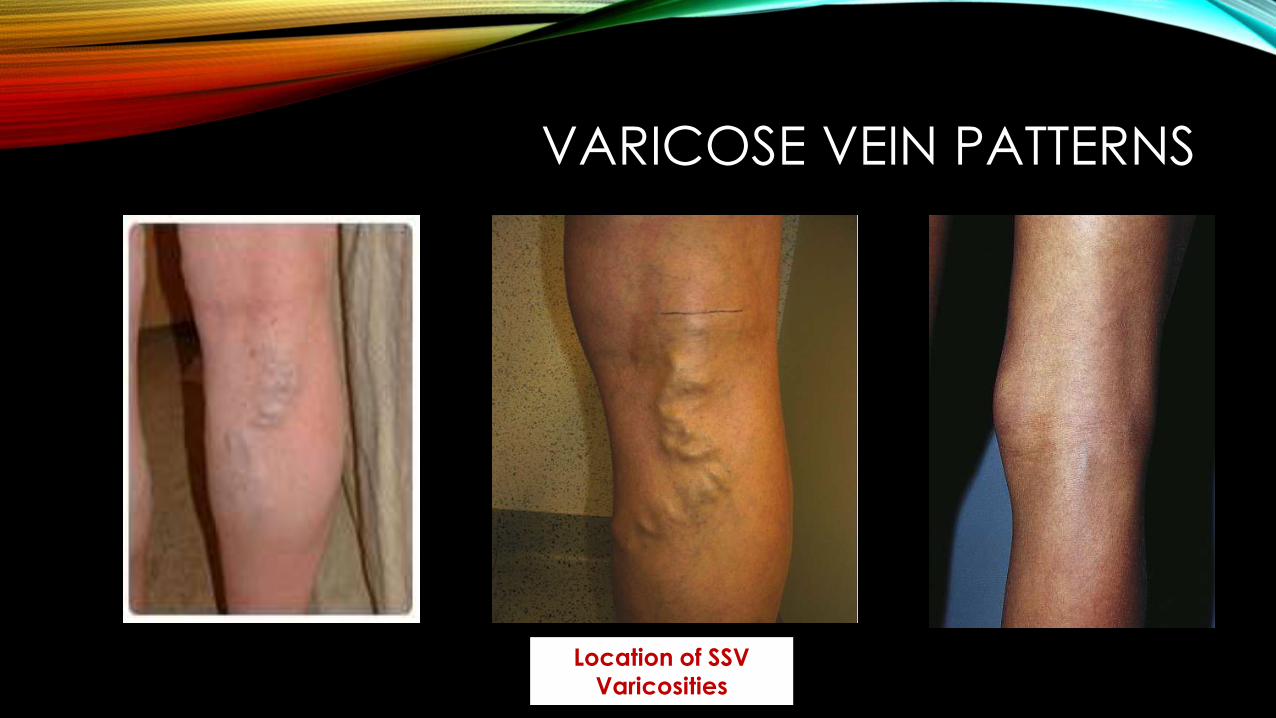
VARICOSE VEIN PATTERNS
Location of SSV Varicosities

VARICOSE VEIN PATTERNS
Location of Thigh Extendor / Vein of Giacomini Varicosities
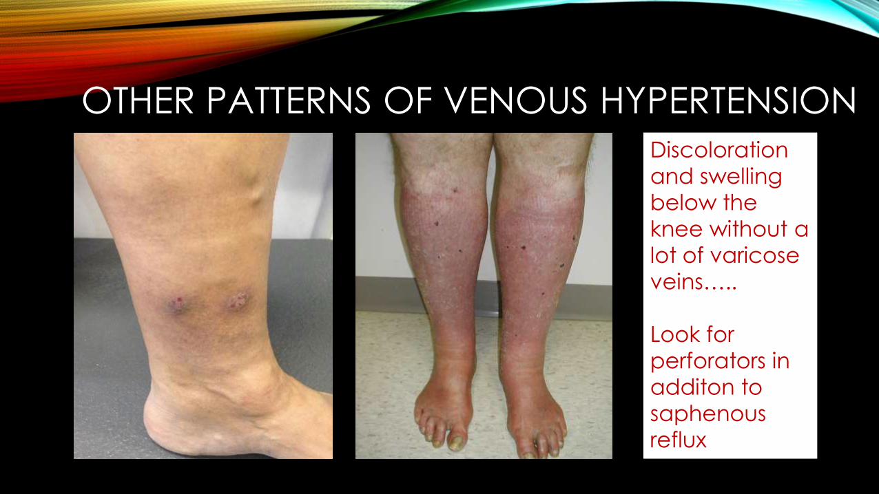
OTHER PATTERNS OF VENOUS HYPERTENSIONDiscoloration and swelling below the knee without a lot of varicose veins…..
Look for perforators in additon to saphenous reflux
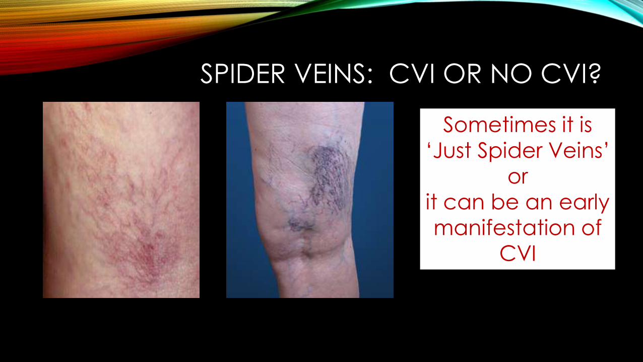
SPIDER VEINS: CVI OR NO CVI?
Sometimes it is ‘Just Spider Veins’
or it can be an early manifestation of
CVI
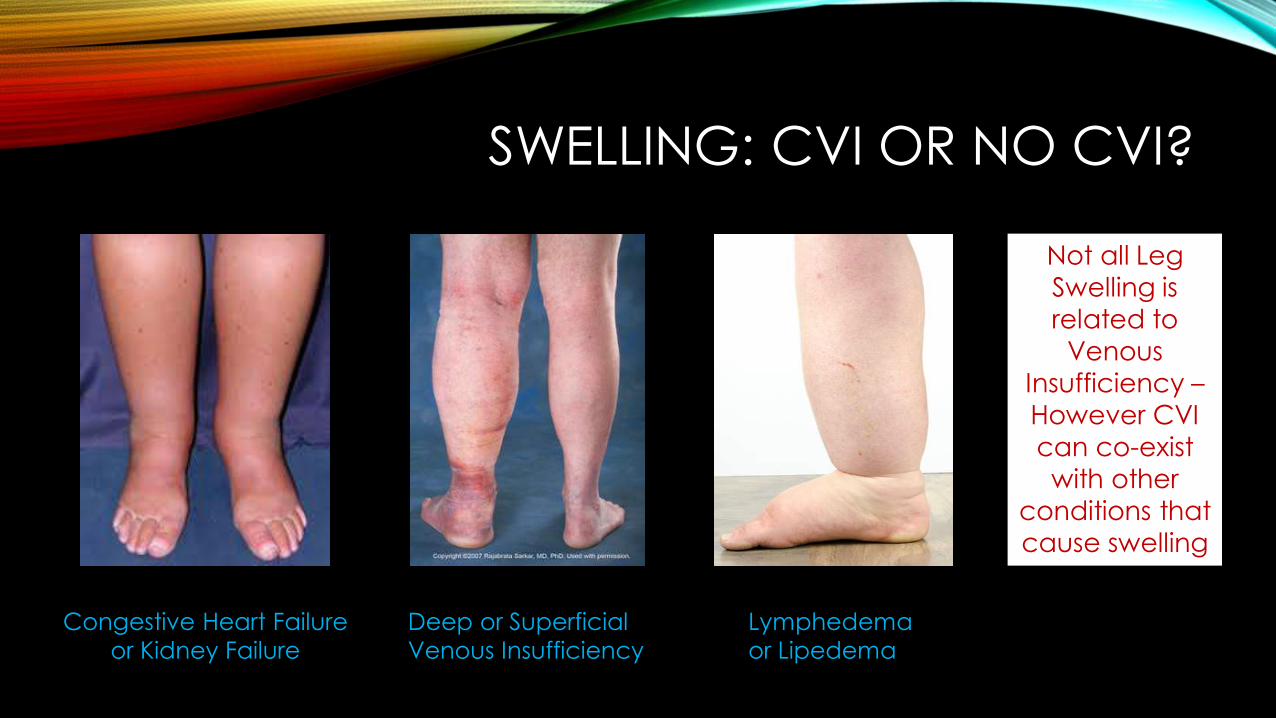
SWELLING: CVI OR NO CVI?
Not all Leg Swelling is related to
Venous Insufficiency –However CVI can co-exist with other
conditions that cause swelling
Congestive Heart Failure or Kidney Failure
Lymphedema or Lipedema
Deep or Superficial Venous Insufficiency
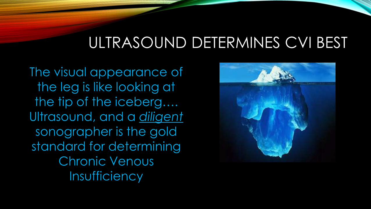
ULTRASOUND DETERMINES CVI BEST
The visual appearance of the leg is like looking at the tip of the iceberg….
Ultrasound, and a diligentsonographer is the gold
standard for determining Chronic Venous
Insufficiency