Embed Size (px)
Citation preview
CELL JOURNAL(Yakhteh), Vol 19, No 3, Oct-Dec (Autumn) 2017 352
Original Article
VDR and CYP24A1 Expression Analysis in IranianRelapsing-Remitting Multiple Sclerosis Patients
Hashem Sadeghi, M.Sc.1#, Mohammad Taheri, M.Sc.1#, Elham Sajjadi, M.Sc.2, Abolfazl
Movafagh, Ph.D.3, Shahram Arsang-Jang, Ph.D.4, Arezou Sayad, Ph.D.1*
1. Department of Medical Genetics, School of Medicine, Shahid Beheshti University of Medical Sciences, Tehran, Iran
2. Department of Hematology, School of Paramedical Sciences, Shahid Beheshti University of Medical Sciences, Tehran, Iran
3. Department of Medical Genetics, School of Medicine, Cancer Research Centre, Shahid Beheshti University of Medical Sciences, Tehran, Iran
4. Department of Epidemiology and Biostatistics, Faculty of Health, Qom University of Medical Sciences, Qom, Iran
*Corresponding Address: P.O.BOX: 1985717443, Department of Medical Genetics, School of Medicine, Shahid Beheshti University of Medical Sciences, Tehran, Iran
Email: [email protected]
#The first two authors equally contributed to this manuscript.
Received: 26/Apr/2016, Accepted: 27/Aug/2016AbstractObjective: Multiple sclerosis (MS) is a common disease of the central nervous system. This disease may be initiated by either vitamin deficiency or triggered by abnormality in CYP24A1 and vitamin D receptor.
Materials and Methods: In this case-control study, the expression of genes encoding vitamin D receptor (VDR) and CYP24A1 in relapsing-remitting MS (RR-MS) patients was compared with normal individuals in the Iranian population. RNA from whole blood of 50 RR-MS patients (HLA-DRB1*15-negative and responders to interferon-beta with a normal vitamin D level) and 50 normal controls was extracted. The levels of CYP24A1 and VDR expression were measured using real-time quantitative poly-merase chain reaction. Results: The RR-MS group had a significantly more than 2 times higher expression level of VDR than the normal group (P=0.04). On the other hand, there was a 0.89 times de-crease in the expression level of CYP24A1 in RR-MS patients which was not statistically significant. There was no linear correlation between the risk of expanded disability status scale of Kurtzke (EDSS) and the expression level of either CYP24A1 or VDR. In addi-tion, the expression level of CYP24A1 or VDR was not correlated with the duration of the disease. Conclusion: Up-regulation of VDR is likely to happen in RR-MS patients in the Iranian population. We did not observe a gene expression-phenotype correlation for CYP24A1 which may be due to limited statistical power as a result of the small sample size. Although the individuals taking part in this study had normal levels of vitamin D, the increase in VDR expression levels may perhaps be a response to a defect in vitamin D processing. Another possibility is that despite an increase in VDR expression level, factors such as micro-RNAs may result in their deactivation while an increase in VDR expression level can be seen as a compensatory response. Of course, further studies are required to identify the mechanism of action of vitamin D by analyzing genes involved in its signaling pathway, particularly VDR and CYP24A1.
Keywords: VDR, CYP24A1, Expression, Multiple Sclerosis, Real Time-Polymerase Chain Reaction Cell Journal(Yakhteh), Vol 19, No 3, Oct-Dec (Autumn) 2017, Pages: 352-360
Citation: Sadeghi H, Taheri M, Sajjadi E, Movafagh A, Arsang-Jang Sh, Sayad A. VDR and CYP24A1 expression analysis in Iranian relapsing-remitting multiple sclerosis patients. Cell J. 2017; 19(3): 352-360. doi: 10.22074/cellj.2017.4192.
CELL JOURNAL(Yakhteh), Vol 19, No 3, Oct-Dec (Autumn) 2017 353
IntroductionMultiple sclerosis (MS) is as a complex
autoimmune inflammatory disease of the central nervous system which leads to myelin injury. MS, as an autoimmune disease, may be initiated by genetic and environmental factors (1-5). Among environmental factors, research in the past decade has been focused on the association between vitamin D deficiency and the risk of MS (6). Vitamin D is derived from ultraviolet B (UVB) (7) and dietary habits (8, 9). MS is more prevalent in environments lacking sufficient UVB. This suggests that geographical location serves as a factor for vitamin D synthesis (10). The inactive form of vitamin D (25-hydroxyvitamin D) is hydroxylated to its active form (1,25-dihydroxyvitamin D) by 25(OH)D-1alfa-hydroxylase (CYP27B1) (11). The active form of vitamin D then binds to the intracellular vitamin D receptor (VDR) and induces the expression of 1,25(OH)2D-24-hydroxylase (24-OHase; CYP24A1) which initiates the degradation of the physiologically active form of vitamin D3 (12).
The role of vitamin D on MS has been highlighted in numerous epidemiologic studies and related fields (13). However, the mechanisms by which vitamin D may affect MS are not known. Given that genetic factors may affect MS, we have studied genetic variation in HLA and cytokine genes, and have analyzed expression of genes encoding TNF-related apoptosis inducing Ligand (TRAIL) and matrix metalloproteinase-9 (MMP9) in Iranian patients with MS previously (14-18). Genes encoding VDR and CYP24A1, as a key enzyme in vitamin D metabolism, are reported to be involved in MS in certain countries (19, 20). Also, a few gene expression studies on vitamin D metabolism in MS have been carried out thus far (21). Also, there have been a few studies with sufficient sample size on the nerve tissue, and studies analyzing these genes in the blood have been very limited in number (19-20). Nonetheless, no such study has been done to examine the expression of these genes in the Iranian population so far. We therefore aimed to examine the expression levels of VDR and CYP24A1 in RR-MS patients in Iran.
Materials and MethodsIn this case-control study, blood samples were
taken from 50 relapsing-remitting MS (RR-MS) patients (29 females and 21 males) and 50 healthy as a control group (31 females and 19 males).
Magnetic resonance imaging (MRI) was used to identify MS in patients based on the McDonald criteria (22, 23). All the patients taking part in this study were HLA-DRB1*15-negative, clinically stable and responsive to interferon-beta. Cinnovex™ was administered to all patients as part of their treatment. Vitamin D levels were shown to be normal in both groups.
Blood samplingFor each individual, 5 ml of peripheral blood
was obtained. The local Ethics Committee of Shahid Beheshti University of Medical Sciences (IR.SBMU.MSP.REC.1395.47) approved this study, including the blood collection procedure. Written consent was obtained from all individuals. The blood samples were collected at Iran’s MS Society Clinic.Quantitative real time-polymerase chain reaction
Total RNA was extracted using Geneall Hybrid-RTM blood RNA extraction kit (General Biotechnology, Korea), in line with the manufacturer’s instructions. The Average OD260/280 of the extracted RNA was 1.9 and had a concentration of 100 ng/microliter. Next, cDNA was synthesized using the Biosystems High-Capacity cDNA Reverse Transcription Kit (Applied Biosystems, USA). For designing the specific probes and primers, allele ID 7 (Premier Biosoft, Palo Alto, USA) was used. The sequences of all probes and primers are given in Table 1. Primers for VDR and CYP24A1 were designed to span the exon-exon junction. In addition, DNase was used to remove DNA contamination. Real-time quantitative polymerase chain reaction (PCR) was carried out in a Corbett Rotor Gene 6000 machine (Corbet Life Science) by using the BiosystemsTaqMan® Universal PCR Master Mix (Applied Biosystems, USA).
VDR and CYP24A1 Expression in MS
CELL JOURNAL(Yakhteh), Vol 19, No 3, Oct-Dec (Autumn) 2017 354
Sadeghi et al.
Table 1: The sequences of probes and primers
Gene name Primer and probe sequences (5ˊ-3ˊ) Primer and probe length
Product length
Accession number
Targeted splice variants
Average of Amplification efficiency
HPRT1 F:AGCCTAAGATGAGAGTTC 18 88 NM_000194.2 Single splice variant
1.98
R: CACAGAACTAGAACATTGATA 21
FAM-CATCTGGAGTCCTATTGACATCGC-TAMRA
24
VDR F: TGGCTTTCACTTCAATGCTATGA 23 126 NM_000376.2 Variant 1 1.98
R: CGTCGGTTGTCCTTGGTGAT 20 NM_001017535.1 Variant 2
FAM-ACTTCCGGCCTCCAGTTCGTATGGAC-TAMRA
26 NM_001017536.1 Variant 3
XM_011538720.2 variant X1
XM_006719587.3 variant X2
CYP24A1 F: TATCGCGACTACCGCAAAGA 20 145 NM_000782.4 Variant 1 1.97
R: CGGCCAAGACCTCATTGATT 20 NM_001128915.1 Variant 2
FAM-TCCGGACCCGCTGCCAGTCTT-TAMRA
21 XM_005260304.4 variant X1
XM_017027691.1 variant X2
XM_017027692.1 variant X3
XM_017027693.1 variant X4
Statistical methodsIndependent sample t test was used to compare
mean expression values. P values and 95% confidence interval (CI) were estimated for mean differences based on bootstrapping. Pearson correlation coefficient was used to examine whether the variables under study were correlated. The level of significance was set at 0.05. The analyses were implemented in SPSS 18 (Chicago, IL, USA).
Results
Clinical details of MS patients and healthy ivdividuals are given in Table 2.VDR and CYP24A1 expression levels and risk of relapsing-remitting multiple sclerosis
To compare the expression level of VDR and
CYP24A1 in RR-MS patients with normal individuals, the groups were defined as i. The total number of participants (regardless of age and sex) and ii. Age-based and sex-based subgroups (based on the age (<30, 30-40, >40 years old) and sex of participants respectively). The expression level of VDR in MS patients was significantly higher than normal individuals. This increase was limited to the total group. Sex- and age-based comparisons showed no statistically significant differences (Table 3).
CYP24A1 expression levelCompared with normal individuals, CYP24A1
expression level in MS patients showed a decrease in all categories (i.e. the total and the two subgroups), however, none reached statistical significance (Table 4).
CELL JOURNAL(Yakhteh), Vol 19, No 3, Oct-Dec (Autumn) 2017 355
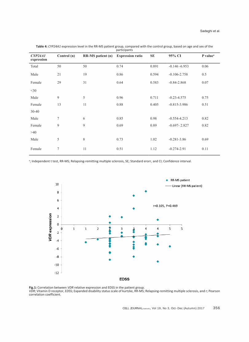
VDR and CYP24A1 expression levels are not correlated with expanded disability status scale or disease duration
The correlation between the expression levels of both genes and Kurtzke expanded disability status scale (EDSS) was measured among the RR-MS patients. There was no significant linear
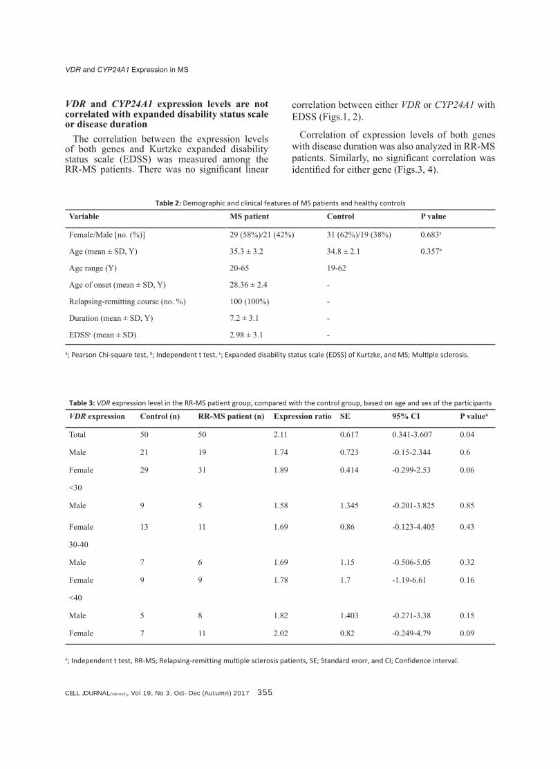
Table 2: Demographic and clinical features of MS patients and healthy controls
Variable MS patient Control P value
Female/Male [no. (%)] 29 (58%)/21 (42%) 31 (62%)/19 (38%) 0.683a
Age (mean ± SD, Y) 35.3 ± 3.2 34.8 ± 2.1 0.357b
Age range (Y) 20-65 19-62
Age of onset (mean ± SD, Y) 28.36 ± 2.4 -
Relapsing-remitting course (no. %) 100 (100%) -
Duration (mean ± SD, Y) 7.2 ± 3.1 -
EDSSc (mean ± SD) 2.98 ± 3.1 -
a; Pearson Chi-square test, b; Independent t test, c; Expanded disability status scale (EDSS) of Kurtzke, and MS; Multiple sclerosis.
Table 3: VDR expression level in the RR-MS patient group, compared with the control group, based on age and sex of the participants
VDR expression Control (n) RR-MS patient (n) Expression ratio SE 95% CI P valuea
Total 50 50 2.11 0.617 0.341-3.607 0.04
Male 21 19 1.74 0.723 -0.15-2.344 0.6
Female 29 31 1.89 0.414 -0.299-2.53 0.06
<30
Male 9 5 1.58 1.345 -0.201-3.825 0.85
Female 13 11 1.69 0.86 -0.123-4.405 0.43
30-40
Male 7 6 1.69 1.15 -0.506-5.05 0.32
Female 9 9 1.78 1.7 -1.19-6.61 0.16
<40
Male 5 8 1.82 1.403 -0.271-3.38 0.15
Female 7 11 2.02 0.82 -0.249-4.79 0.09
a; Independent t test, RR-MS; Relapsing-remitting multiple sclerosis patients, SE; Standard erorr, and CI; Confidence interval.
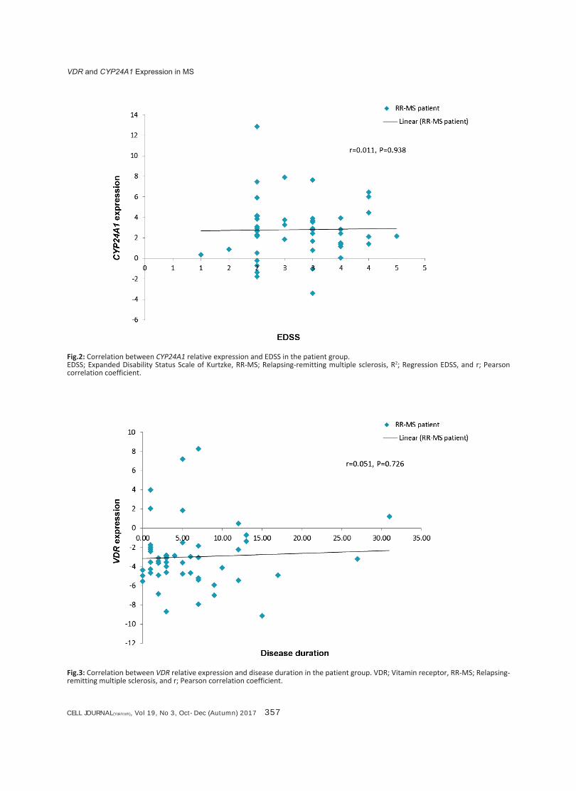
correlation between either VDR or CYP24A1 with EDSS (Figs.1, 2).
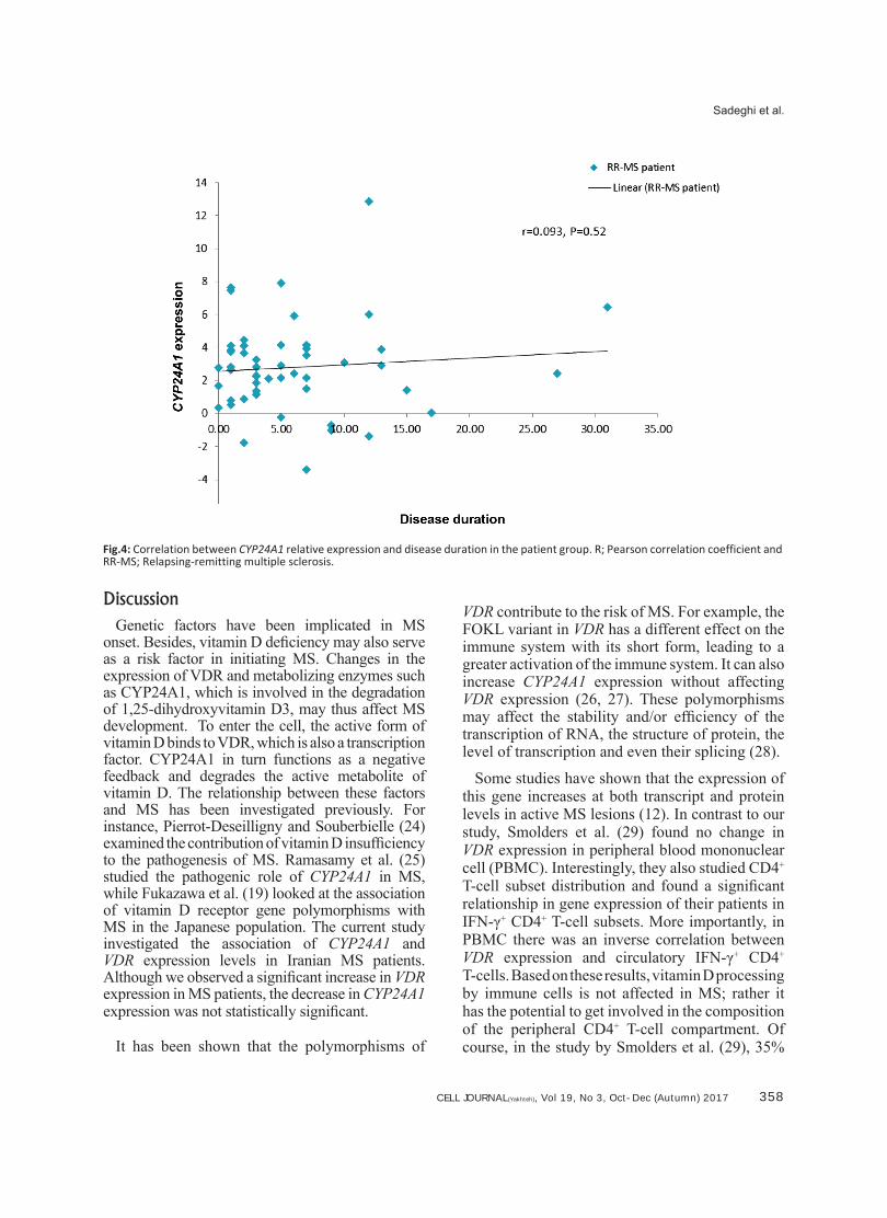
Correlation of expression levels of both genes with disease duration was also analyzed in RR-MS patients. Similarly, no significant correlation was identified for either gene (Figs.3, 4).
VDR and CYP24A1 Expression in MS
CELL JOURNAL(Yakhteh), Vol 19, No 3, Oct-Dec (Autumn) 2017 356
Sadeghi et al.
Table 4: CYP24A1 expression level in the RR-MS patient group, compared with the control group, based on age and sex of the participants
CYP24A1 expression
Control (n) RR-MS patient (n) Expression ratio SE 95% CI P valuea
Total 50 50 0.74 0.891 -0.146 -6.953 0.06
Male 21 19 0.86 0.594 -0.106-2.758 0.5
Female 29 31 0.64 0.583 -0.84-2.868 0.07
<30
Male 9 5 0.96 0.711 -0.23-4.575 0.75
Female 13 11 0.88 0.405 -0.815-3.986 0.51
30-40
Male 7 6 0.85 0.98 -0.554-4.213 0.82
Female 9 9 0.69 0.89 -0.697- 2.827 0.82
>40
Male 5 8 0.73 1.02 -0.281-3.86 0.69
Female 7 11 0.51 1.12 -0.274-2.91 0.11
a; Independent t test, RR-MS; Relapsing-remitting multiple sclerosis, SE; Standard erorr, and CI; Confidence interval.
Fig.1: Correlation between VDR relative expression and EDSS in the patient group. VDR; Vitamin D receptor, EDSS; Expanded disability status scale of kurtzke, RR-MS; Relapsing-remitting multiple sclerosis, and r; Pearson correlation coefficient.
CELL JOURNAL(Yakhteh), Vol 19, No 3, Oct-Dec (Autumn) 2017 357
Fig.2: Correlation between CYP24A1 relative expression and EDSS in the patient group. EDSS; Expanded Disability Status Scale of Kurtzke, RR-MS; Relapsing-remitting multiple sclerosis, R2; Regression EDSS, and r; Pearson correlation coefficient.
Fig.3: Correlation between VDR relative expression and disease duration in the patient group. VDR; Vitamin receptor, RR-MS; Relapsing-remitting multiple sclerosis, and r; Pearson correlation coefficient.
VDR and CYP24A1 Expression in MS
CELL JOURNAL(Yakhteh), Vol 19, No 3, Oct-Dec (Autumn) 2017 358
Sadeghi et al.
Fig.4: Correlation between CYP24A1 relative expression and disease duration in the patient group. R; Pearson correlation coefficient and RR-MS; Relapsing-remitting multiple sclerosis.
DiscussionGenetic factors have been implicated in MS
onset. Besides, vitamin D deficiency may also serve as a risk factor in initiating MS. Changes in the expression of VDR and metabolizing enzymes such as CYP24A1, which is involved in the degradation of 1,25-dihydroxyvitamin D3, may thus affect MS development. To enter the cell, the active form of vitamin D binds to VDR, which is also a transcription factor. CYP24A1 in turn functions as a negative feedback and degrades the active metabolite of vitamin D. The relationship between these factors and MS has been investigated previously. For instance, Pierrot-Deseilligny and Souberbielle (24) examined the contribution of vitamin D insufficiency to the pathogenesis of MS. Ramasamy et al. (25) studied the pathogenic role of CYP24A1 in MS, while Fukazawa et al. (19) looked at the association of vitamin D receptor gene polymorphisms with MS in the Japanese population. The current study investigated the association of CYP24A1 and VDR expression levels in Iranian MS patients. Although we observed a significant increase in VDR expression in MS patients, the decrease in CYP24A1 expression was not statistically significant.
It has been shown that the polymorphisms of
VDR contribute to the risk of MS. For example, the FOKL variant in VDR has a different effect on the immune system with its short form, leading to a greater activation of the immune system. It can also increase CYP24A1 expression without affecting VDR expression (26, 27). These polymorphisms may affect the stability and/or efficiency of the transcription of RNA, the structure of protein, the level of transcription and even their splicing (28).
Some studies have shown that the expression of this gene increases at both transcript and protein levels in active MS lesions (12). In contrast to our study, Smolders et al. (29) found no change in VDR expression in peripheral blood mononuclear cell (PBMC). Interestingly, they also studied CD4+ T-cell subset distribution and found a significant relationship in gene expression of their patients in IFN-γ+ CD4+ T-cell subsets. More importantly, in PBMC there was an inverse correlation between VDR expression and circulatory IFN-γ+ CD4+
T-cells. Based on these results, vitamin D processing by immune cells is not affected in MS; rather it has the potential to get involved in the composition of the peripheral CD4+ T-cell compartment. Of course, in the study by Smolders et al. (29), 35%
CELL JOURNAL(Yakhteh), Vol 19, No 3, Oct-Dec (Autumn) 2017 359
of patients received a high level of vitamin D (high vitamin D dose, 20,000 IU/day) and their 25 (OH) D serum level was thus significantly higher than the control group. A higher serum level than in normal controls probably affects expression level of genes related to vitamin D metabolism, thereby confounding the results. This could even affect the existing correlation of gene expression levels in different PBMC cell subsets (12, 29).
The mechanism involved in the activation of the enzymatic pathway of vitamin D metabolism in PBMC or T-cells is not yet known, nor is the way such enzymes may influence the pathobiology of MS (29). Recent studies have attempted to elucidate the molecular mechanism of the immunological effects of calcidiol metabolites in patients with MS, showing that an increase in calcidiol could cause a decrease in the activity of MS. Such effects, to some extent, could be initiated by interferon-beta (IFN-β) (30). Given that vitamin D is a strong regulator of the immune system, inflammatory activities that stimulate T-helper cells toward greater inflammation may be blocked by vitamin D (31, 32). The mechanism of action of vitamin D is however complex and is likely to be influenced by various factors.
In this study, there was no significant correlation between VDR and CYP24A1 expression levels and clinical findings, such as the level of physical disability in MS patients (according to the EDSS criterion) and disease duration. It is likely that the data on the amount and duration of the intake of vitamin D and serum levels of different metabolites of this vitamin may aid the interpretation of gene expression data. Also the interaction between genes and uncertainty about the transcript and protein levels increases the analytical complexity. For instance, although the people taking part in this study had normal levels of vitamin D, the increase in VDR expression may be a response to the defective vitamin D processing. This increase in VDR expression may also be due to factors such as microRNAs which may cause VDR and metabolizing enzymes down-regulation, thus the up-regulation may be seen as a compensatory response. Further studies with greater number of subjects are necessary to validate the results reported in the current work. Also, additional studies are recommended to investigate the expression level of other genes involved in
vitamin D synthesis, type polymorphisms and examine protein-level expression of VDR and CYP24A1. The likelihood of such links can may also be assessed in other subsets of T cells and T-regulatory cells.
ConclusionThe role of vitamin D in MS has been highlighted
in numerous epidemiologic studies and related fields. However, the mechanism by which vitamin D affects MS is yet to be identified. All the genes that affect vitamin D metabolism belong to the genetic causes of MS. Two of the genes that play a key role in vitamin D metabolism are VDR and CYP24A1. Although CYP24A1 was not dys-regulated, up-regulation of VDR was observed in RR-MS patients in Iranian population. Expression analyses of the whole vitamin D pathway in RR-MS patients may therefore shed further light on the underlying molecular basis of MS.
AcknowledgmentsThe present study was financially supported by
The Research Department of The School of Medi-cine, Shahid Beheshti University of Medical Sci-ences (Grant No. 8884). The authors declare that they have no conflicts of interest in relation to this research and its publication.
References1. Peltonen L. Old suspects found guilty the first genome
profile of multiple sclerosis. N Engl J Med. 2007; 357(9): 927-929.
2. Ebers GC, Sadovnick AD, Risch NJ. A genetic basis for familial aggregation in multiple sclerosis. Nature. 1995; 377(6545): 150-151.
3. Ebers GC, Sadovnick AK, Dyment DD, Yee IM, Willer CJ, Risch N. Parent-of-origin effect in multiple sclerosis: observations in half-siblings. Lancet. 2004; 363(9423): 1773-1774.
4. Orton SM, Herrera BM, Yee IM, Valdar W, Ramagopalan SV, Sadovnick AD, et al. Sex ratio of multiple sclerosis in Canada: a longitudinal study. Lancet Neurol. 2006; 5(11): 932-936.
5. Dyment DA, Yee IM, Ebers GC, Sadovnick AD. Multiple sclerosis in stepsiblings: recurrence risk and ascertain-ment. J Neurol Neurosurg Psychiatry. 2006; 77(2): 258-259.
6. Pierrot-Deseilligny C, Souberbielle JC. Is hypovitaminosis D one of the environmental risk factors for multiple sclero-sis? Brain. 2010; 133(7): 1869-1888.
7. Calvo MS, Whiting SJ, Barton CN. Vitamin D intake: a global perspective of current status. J Nutr. 2005; 135(2): 310-316.
8. Holick MF. High prevalence of vitamin D inadequacy and implications for health. Mayo Clin Proc. 2006; 81(3): 353-373.
VDR and CYP24A1 Expression in MS
CELL JOURNAL(Yakhteh), Vol 19, No 3, Oct-Dec (Autumn) 2017 360
Sadeghi et al.
9. Holick MF. Vitamin D deficiency. N Engl J Med. 2007; 357(3): 266-281.
10. Marrie RA. Environmental risk factors in multiple sclerosis aetiology. Lancet Neurol. 2004; 3(12): 709-718.
11. Malloy PJ, Pike JW, Feldman D. The vitamin D receptor and the syndrome of hereditary 1, 25-dihydroxyvitamin D-resistant Rickets 1. Endocr Rev. 1999; 20(2): 156-188.
12. Smolders J, Schuurman KG, van Strien ME, Melief J, Hendrickx D, Hol EM, et al. Expression of vitamin D re-ceptor and metabolizing enzymes in multiple sclerosis—affected brain tissue. J Neuropathol Exp Neurol. 2013; 72(2): 91-105.
13. Sundstrom P, Salzer J. Vitamin D and multiple sclerosis-from epidemiology to prevention. Acta Neurol Scand. 2015; 132(199): 56-61.
14. Sayad A, Allameh A, Sayad A, Noruzinia M, Sarzaeem A. The influence of-330 IL-2 gene polymorphism on relaps-ing remitting and secondary progressive multiple sclerosis in Iranian patients. Neurol Asia. 2013;18(1): 83-86.
15. Sayad A, Allameh A, Sayad A, Noruzinia M, Akbari MT, Sarzaeem A, et al. The association of-475 and-631 inter-leukin-2 gene polymorphism with multiple sclerosis in Ira-nian patients. Cell J. 2013; 15(2): 124-129.
16. Sayad A. The association of− 330 interleukin-2 gene poly-morphism and HLA-DR15 allele in Iranian patients with multiple sclerosis. Int J Immunogenet. 2014; 41(4): 330-334.
17. Taheri M, Nemati S, Movafagh A, Saberi M, Mirfakhraie R, Eftekharian MM, et al. TRAIL gene expression analysis in multiple sclerosis patients. Hum Antibodies. 2016; 24(1-2): 33-38.
18. Yazdandoost Hamedani Sh, Taheri M, Omrani MD, Sajjadi E, Mazdeh M, Tabatabaei Panah AS, et al. Up regulation of MMP9 gene expression in female patients with multiple sclerosis. Hum Antibodies. 2016; 1(1): 1-6.
19. Fukazawa T, Yabe I, Kikuchi S, Sasaki H, Hamada T, Mi-yasaka K, et al. Association of vitamin D receptor gene polymorphism with multiple sclerosis in Japanese. J Neu-rol Sci. 1999; 166(1): 47-52.
20. Tajouri L, Ovcaric M, Curtain R, Johnson MP, Griffiths LR, Csurhes P, et al. Variation in the vitamin D receptor gene is associated with multiple sclerosis in an Australian popu-lation. J Neurogenet. 2005; 19(1): 25-38.
21. Rezaie Z, Taheri M, Kohan L, Sayad A. Down-regulation of CYP27B1 gene expression in Iranian patients with relapsing-remitting multiple sclerosis. Hum Antibodies. 2016; 1-5 (A head of print).
22. McDonald WI, Compston A, Edan G, Goodkin D, Hartung
HP, Lublin FD, et al. Recommended diagnostic criteria for multiple sclerosis: guidelines from the International Panel on the diagnosis of multiple sclerosis. Ann Neurol. 2001; 50(1): 121-127.
23. Polman CH, Reingold SC, Edan G, Filippi M, Hartung HP, Kappos L, et al. Diagnostic criteria for multiple sclerosis: 2005 revisions to the “McDonald Criteria”. Ann Neurol. 2005; 58(6): 840-846.
24. Pierrot-Deseilligny C, Souberbielle JC. Contribution of vi-tamin D insufficiency to the pathogenesis of multiple scle-rosis. Ther Adv Neurol Disord. 2013; 6(2): 81-116.
25. Ramasamy A, Trabzuni D, Forabosco P, Smith C, Walker R, Dillman A, et al. Genetic evidence for a pathogenic role for the vitamin D3 metabolizing enzyme CYP24A1 in mul-tiple sclerosis. Mult Scler Relat Disord. 2014; 3(2): 211-219.
26. O Neill V, Asani FF, Jeffery TJ, Saccone DS, Bornman L.Vitamin D receptor gene expression and function in a south african population: ethnicity, vitamin D and Fokl. PLoS One. 2013; 8(6): 676-679.
27. Van Etten E, Verlinden L, Giulietti A, Ramos-Lopez E, Branisteanu DD, Ferreira GB, et al. The vitamin D recep-tor gene FokI polymorphism: functional impact on the im-mune system. Eur J Immunol. 2007; 37(2): 395-405.
28. Tizaoui K, Kaabachi W, Hamzaoui A, Hamzaoui K. Asso-ciation between vitamin D receptor polymorphisms and multiple sclerosis: systematic review and meta-analysis of case-control studies. Cell Mol Immunol. 2015; 12(2): 243-252.
29. Smolders J, Thewissen M, Theunissen R, Peelen E, Knip-penberg S, Menheere P, et al. Vitamin D-related gene ex-pression profiles in immune cells of patients with relapsing remitting multiple sclerosis. J Neuroimmunol. 2011; 235(1-2): 91-97.
30. Munger KL, Kochert K, Simon KC, Kappos L, Polman CH, Freedman MS, et al. Molecular mechanism underlying the impact of vitamin D on disease activity of MS. Ann Clin Transl Neurol. 2014; 1(8): 605-617.
31. Peelen E, Damoiseaux J, Muris AH, Knippenberg S, Smolders J, Hupperts R, et al. Increased inflammasome related gene expression profile in PBMC may facilitate T helper 17 cell induction in multiple sclerosis. Mol Immunol. 2015; 63(2): 521-529.
32. Berge T, Leikfoss IS, Brorson IS, Bos SD, Page CM, Gus-tavsen MW, et al. The multiple sclerosis susceptibility genes TAGAP and IL2RA are regulated by vitamin D in CD4+ T cells. Genes Immun. 2016; 17(2): 118-127.


![VDR G4[e] S-VDR G4[e] - INTERSCHALT · Innovation for shipping VDR G4[e] S-VDR G4[e] VOYAGE DATA RECORDER SYSTEMS Comply with all IMO performance standards Offer …](https://img.pdfslide.us/doc/110x75/5b5e09397f8b9a310a8b9cbf/vdr-g4e-s-vdr-g4e-innovation-for-shipping-vdr-g4e-s-vdr-g4e-voyage.jpg)