Embed Size (px)
Citation preview

Vascular Registry Review
VENOUS
Peripheral Venous Anatomy
Think direction of flow!!
Proximal is still closer to heart and distal away. BUT … veins flow distal to proximal. That means they start at the bottom and end at the top. Anatomy is based on direction of flow. Veins never bifurcate, they form confluences.
Empty blood from = Where blood is coming from *** example : hepatic veins empty blood from the liver
Drain/empty blood into = Where blood is going *** example : hepatic veins drain blood into the IVC
Veins always flow up into the next vessel. When 2 veins join each other and form a new vein, they both flow into the new vein. They do not empty into each other. The junction is ALWAYS the new vein
Lower Extremities
Deep veins accompany arteries.
Venae comitantes = corresponding veins. Paired deep
veins. In LE = all calf veins PTV, ATV, PER
PTV and PER drain into tibeoperoneal trunk. TP trunk
and ATVs join to form the POP v.
Popliteal becomes the FV at Hunters Canal.
FV and DFV form the CFV. CFV terminates when it
drains into the EIV at the inguinal canal.
EIV and IIV empty into CIV.
IVC is formed by the confluence of the RT and LT CIVs.
Not for Distribution 59

Vascular Registry Review
Superficial veins
Do not have an artery. Located between the
fascial layers within the subcutaneous fat
Greater saphenous vein = longest vein in body.
Medial leg. Drains into the CFV at the
saphenofemoral junction.
Small saphenous vein = posterior calf. Drains
into POP.
Perforatoring veins = Connect superficial to
deep. Used in connection with the calf muscle
pump. Most are found below the calf, superior
to medial malleolus (connecting post accessory
GSV to post tibs)
Communicating vein = Connect veins of same system.
Venous sinuses = Potential reservoirs or channels that allow blood to accumulate
in the calf and during calf contraction > ejected into peroneals and PTVs.
Important role in calf muscle pump.
Abdominal venous system
IVC courses superiorly. Renal veins drain into IVC
Hepatic veins drain the liver (Hepatofugal) and
empty into IVC. All drains into Rt atrium of heart.
Not for Distribution 60

Vascular Registry Review
Portal venous system
Not related to IVC or its veins
Drains blood from digestive tract and spleen into liver.
SMV and splenic form Portal confluence. Both drain
into MPV.
MPV drains into liver (supplies 80% of liver’s blood) =
Hepatopedal flow
Upper Extremities
Venae comitantes = Radial, Ulnar, Brachial
Superficial veins = Cephalic and Basilic
Radial and Ulnar v drain into Brachial. Brachial and
basilic (superficial vein that courses medial) join
together to form the axillary v. Both drain into
axillary.
Axillary and cephalic (superficial vein that courses
lateral) join to become the subclavian vein. Both
drain up into the subclavian.
Venous return to heart
Subclavian and IJV join to form the
brachiocephalic or innominate.
Rt and Lt innominate drain into the SVC
SVC carries blood to Rt atrium of heart
Not for Distribution 61

Vascular Registry Review
Venous structure
Same 3 layers as arteries = intima, media, adventitia
*** difference = medial layer is thinner in veins. Collapsible
Venous valves - bicuspid extensions of intimal layer. Purpose is to prevent
backflow and maintain unidirectional flow. Venous blood has greatest
hemodynamic challenge to overcome = gravity. Needs valves to assist
Due to hydrostatic pressure greatest closest to ankle….
Most # valves = veins in calf. Decrease in # closer to abdomen.
NO valves = veins in chest and abdomen. Ex- IVC, SVC, iliac veins, etc
Arms have very few valves, 1 per vein.
Perforators - must have at least 1 valve
Not for Distribution 62
Vascular Registry Review
Venous Hemodynamics
Venous compliance
Ability to handle large changes in volume without greatly affecting the pressure.
Veins can expand to accommodate more volume
Transmural pressure - pressure on the wall.
Difference in pressure from outside (interstitial) and
pressure from the inside (intraluminal).
***Depends on volume
Circular: Full and more volume. Has less resistance which allows for emptying
Elliptical: Less volume but higher resistance. Allows for venous filling
Respiration “Phasic flow”
Big role in venous return to heart. Creates pressure gradient for flow into
abdomen (for LE) or chest (for UE)
Inspiration: Inc abdominal pressure / Dec thoracic pressure.
*** flow needs pressure gradient (hi to low pressure)
If abd pressure is high = flow from legs STOP
If chest pressure is low = flow from arms FLOWS into chest
When veins are not flowing/emptying = they are filling.
Filling = Inc volume = Inc pressure
Expiration: Dec abdominal pressure / Inc thoracic pressure
*** LOW abd pressure and HIGH venous pressure in legs = FLOW
LE venous flows into abdomen
UE venous flow halts and fills
Not for Distribution 63

Vascular Registry Review
Valsalva: Take deep breath in, hold it, and bear down (tightening abdomen as if
having a bowel movement). Increases both abdomen and chest pressures
>> ALL VENOUS FLOW HALTS
After release of valsalva = venous flow should augment
If there is augmentation of flow DURING manuever = reflux flow
Hydrostatic pressure
Has same influence on venous as arterial. Makes it more difficult for venous flow
to go up
*** Supine = 0mmHg at ankle
*** Standing = 100mmHg at ankle
Calf muscle pump
Venous heart. To propel blood up towards the heart. Only works if valves are
competent. Venous sinuses fill during relaxation. Perforators allow blood to flow
from superficial to deep systems. During contraction, muscle squeezes sinuses
and deep veins to empty up. Valves in superficial and perforators normally
should close during contraction.
Muscle contraction = Empties Relaxation = Filling
Venous hypertension
Any abnormal increase in venous pressure due to high venous volume
Not for Distribution 64
Effective Calf Pump
>60% blood ejected from calfIncreased venous return to heartDecreases venous poolingDecreases venous volumeDecreases venous pressure
Ineffective Calf Pump
Incompetent valvesIncreased venous poolingIncreases venous volumeIncreases venous pressureVenous hypertension

Vascular Registry Review
Venous Disease and Clinical History
2 types of disease - Acute obstruction (thrombosis) and Chronic insufficiency
ACUTE DVT
Thrombosis - RBCs trapped in fibrin web. Most frequently originate at venous valves or sinuses (stasis). Greatest clinical danger: pulmonary embolism Most likely complication: venous insufficiency (damage to valves)
Symptoms: Pain, swelling, redness, warmth
Virchow’s triad : risk factors and contributing to formation of clot
1. Trauma Intrinsic- damage to inside. Catheters, PICC lines, IV Extrinsic- from outside. Accident, fall
2. Stasis Bed rest Obesity Pregnancy Paralytics Congestive heart failure Surgery related
3. Hypercoagulability Increased clotting of blood Pregnancy Cancer Oral contraceptives Inherited states (factor V Leiden, protein C, protein S)
Not for Distribution 65

Vascular Registry Review
Other sources of thrombus
• Paget-Schroetter Stress or effort thrombosis of subclavian or axillary due to repetitive trauma to vessel. (Ex-Baseball pitcher with unilateral UE pain and swelling) Venous component of TOS
• Lt CIV compression AKA May-Thurner syndrome Compression of LT CIV by RT CIA as artery crosses over it. Stasis >thrombosis
• Nutcracker syndrome Compression of Lt renal vein by SMA and aorta. LRV may thrombose
• SVC syndrome Thrombosis or compression of SVC by mass. Causes bilateral facial swelling, bilateral UE swelling, and dyspnea
Limb threatening acute venous disease
Severe acute extensive ileofemoral DVT. So obstructive it affects the arterial circulations. Combo of DVT and arterial symptoms
• Phlegmasia alba dolens PAIN, SWELLING, WHITE Triggers arterial spasms
• Phlegmasia cerulea dolens PAIN, SWELLING, BLUE Reduces arterial inflow
VENOUS INSUFFICIENCY
Valvular incompetence. Valves leak, no longer maintain unidirectional flow. Flow refluxes caudally (back down). Causes VENOUS HYPERTENSION
Causes
Congenital Avalvular veins Inherited incompetent valves Klippel-Trenauny (hypoplastic or absent deep veins)
Post-thrombotic / post-phlebitic syndrome Pt had previous DVT. Damaged valves lead to chronic insufficiency
Other acquired causes for increased venous pressure: obesity or pregnancy
Not for Distribution 66

Vascular Registry Review
Venous walls dilate and stretch, collaterals (varicose veins) develop. Dilated
tortuous superficial venous collaterals. Caused by chronic venous hypertension
Primary: hereditary
Secondary: due to obstructive process
High venous pressure to back up into capillaries. Under pressure allow leakage
of fluids into tissue = edema and brawny discoloration
Brawny: leakage of fibrin, RBCs into tissue. RBCs die and create hemosiderin =
brown. Most common location is superior to medial malleolus
Symptoms of venous insufficiency are the same whatever the cause
• Edema • Heaviness • Varicose veins • Brawny • Lipodermatosclerosis (severe and chronic= hardening of skin due to
constant irritation of tissue. Bottle neck appearance) • Venous ulcers
Edema
Fluid enters tissues. May be caused by venous hypertension. Increased capillary pressure
Pitting = manual pressure applied and leaves dent in skin. Fluid retention, renal dysfunction, inc venous pressure
Non-pitting = tissue so engorged with fluid, no indent. Caused by lymphedema
Not for Distribution 67
VenousMedial malleolus/calf
Shallow
Oozy
Less painful
Irregular
Other: brawny, swelling
Arterial Toes/bony regions
Deep
Dry
Very painful
Regular
Other: Dry skin, thick toenails

Vascular Registry Review
Venous PPG
Capabilities and limitations
Presence/absence of venous insufficiency Determine superficial vs deep insufficiency
Not quantify insufficiency / severity
Physical principles
Documents capillary blood volume
Same technology as arterial, except setting is DC (direct current) AC (alternating current) > arterial DC (direct current) > venous (detects slower flow changes)
Technique
Pt sitting with legs dangling
PPG placed above medial malleolus (not directly on varicose veins)
*** Activate calf muscle pump to empty blood from calf Exaggerated flexions and dorsiflexions See how long it takes to fill back up = Venous Refill Time (VRT)
Interpretation
Venous Refill Time VRT
Normal = Gradual venous filling >20sec Incompetent valves = Fills up quickly <20sec
1. If initial reading is normal = No venous insufficiency
2. If initial reading is abnormal = repeat with tourniquet. Purpose of the tourniquet is to cut off influence of the superficial system. Helps to know if insufficiency is coming form the deep or superficial.
Normalizes with the tourniquet = Superficial insufficiency
Never normalizes = Deep insufficiency
Not for Distribution 68

Vascular Registry Review
If initial reading is abnormal and tourniquet needs to be placed, 1st place the tourniquet above the knee = rule out GSV
If still abnormal, place below the knee = rule out SSV
Venous Air Plethysmography
Same purpose as Venous PPG.
Can quantify venous reflux and measure venous function
• Pt has large cuff on calf and completes various maneuvers
1: Leg elevated 2: Standing 3: 1 toe-up 4: 10 toe-ups 5: Resting
• Ejection Fraction >60% is normal
• If initially abnormal, can be repeated with tourniquet to distinguish superficial from deep insuff (same as PPG)
Not for Distribution 69
Venous PPG reading
>20 sec VRT Normal<20 sec VRT InsufficiencyTourniquet normal AK GSV insuffTourniquet normal BK SSV insuffNever normalizes Deep insuff

Vascular Registry Review
CW Doppler
Capabilities and limitations
Presence/absence of venous insufficiency Presence of DVT Cannot specifically choose vessel or sampling depth (CW) No imaging, only waveform contour reading
More likely to have false negatives or false positives
• False Negative = Test seems negative but there really is disease Partial non-occlusive DVT Venous collaterals Bifid system (duplicate deep veins)
• False Positive = Test has abnormal reading but really is normal Extrinsic compression (pregnancy, tight clothing, tumors, ascites) Severe PAD (reduced inflow=reduced outflow) COPD Doppler angle/tech error
Technique
Similar to CW in arterial 45-60 degree angle to skin surface
Pt position = reverse trendelenburg (limb lower than heart). Increased hydrostatic pressure encourages venous filling and allows for easier evaluation of the LE veins LE externally rotated with knee slightly bent
Done in segments (groin, thigh, behind knee, ankle)
Not for Distribution 70
Vascular Registry Review
Interpretation
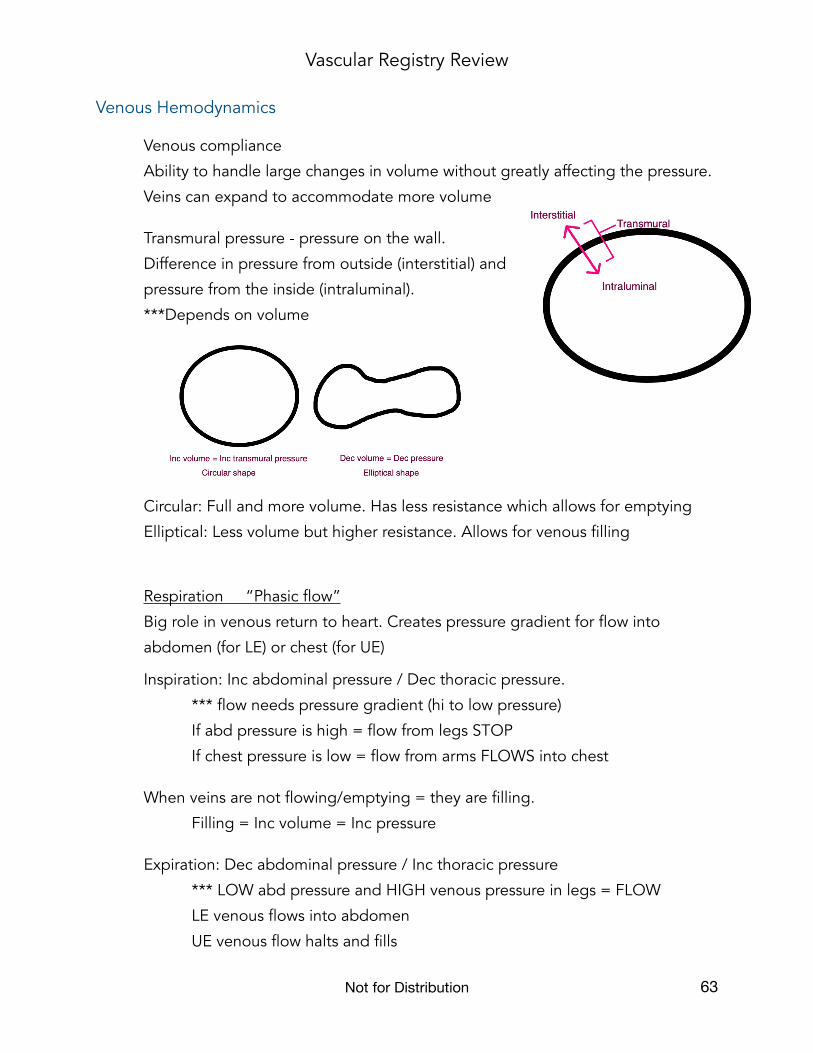
NORMAL peripheral venous waveform
• Spontaneity - naturally occurring. Most larger deep veins should be spontaneous. Non-spontaneous flow may be seen in tibials, superficial veins, and radial/ulnar Abnormally non-spontaneous = location of obstruction
• Phasicity - flows with respiration. Normally is seen in peripheral veins Non-phasic or continuous flow = proximal obstruction Pulsatile =Normal for veins close to heart (IVC, subclavian veins, IJV). In peripheral veins = CHF, tricuspid regurgitation, or fluid overload.
• Augmentation with distal compression or proximal release - Normal flow should augment or increase when compressed distally or after the release of the proximal compression No augmentation = obstruction between probe and compression/release
• Proximal compression or Valsalva - both increase the proximal pressure and are done to test competence of venous valves. Adds stress or pressure on valves. During maneuvers, venous flow should halt or stop. Normal result after release is to augment. Augments DURING = Valvular incompetence
Not for Distribution 71

Vascular Registry Review
Venous Duplex
Capabilities and limitations
Location of disease (DVT or reflux) Acute vs Chronic DVT Partial vs complete
Less limitations
Technique
Patient position: reverse trendelenburg (limb lower than heart)
B-mode : perform with and without compressions
Color: show full filling of vessel and directionality
PW doppler: waveform morphology
Insufficiency testing: Pt in reverse trendelenburg or standing. Add valsalva maneuver or proximal compressions to stress valves.
Interpretation
Normal
Normal veins fully compress = Coaptation. No thrombus
Fully fill with color
Phasic, spontaneous and augmentation
No reflux when standing or during valsalva/prox comp
Not for Distribution 72
Vascular Registry Review
Abnormal
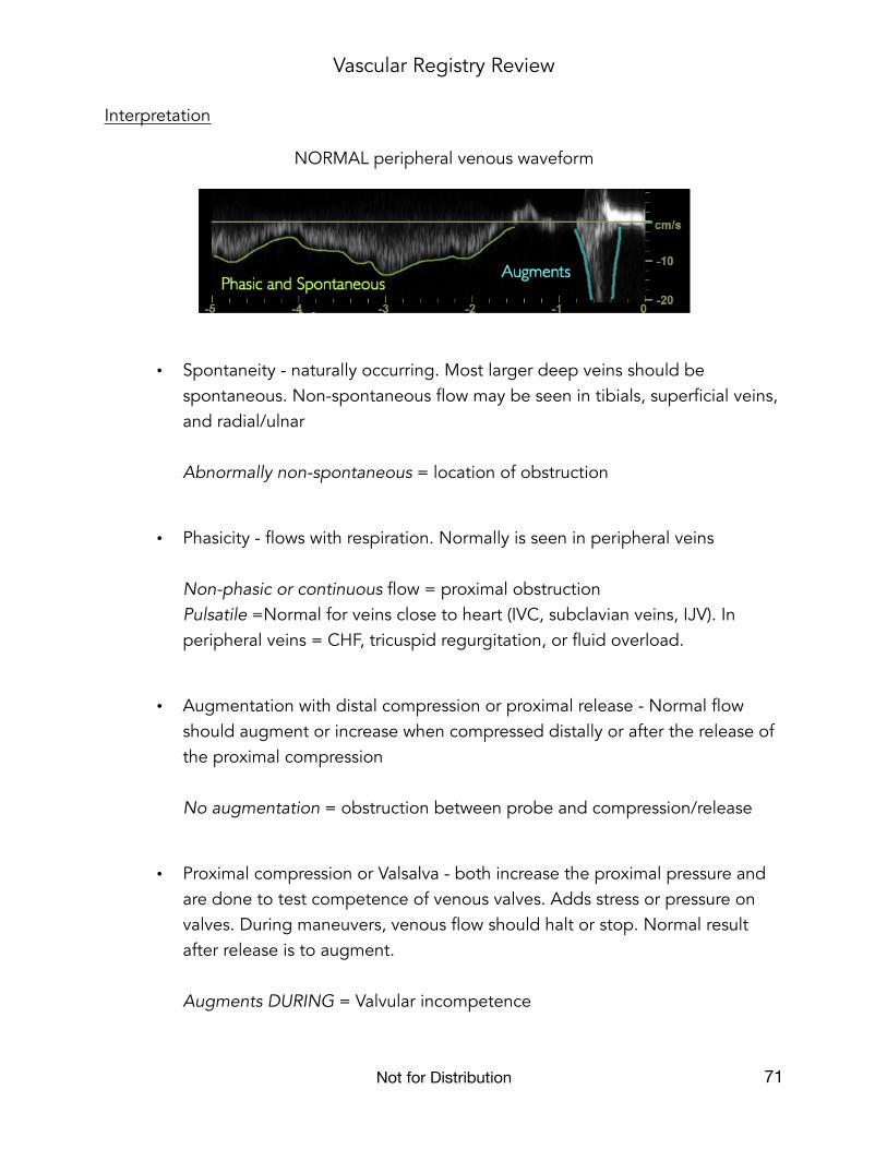
• Acute DVT Dark, dilated, incompressible Larger vein size, spongy texture, poorly attached the walls Acute - anechoic, hypoechoic. Subacute - possible hypo to echogenic. Still dilated and spongy looking ** If superficial vein is thrombosed = NOT DVT. “Superficial thrombophlebitis”
Color - no signal or trace amounts along walls Continuous doppler = proximal obstruction No augmentations = indicate proximal or distal DVT
• Chronic DVT Smaller vessel size, thicker walls. Hyperechoic striations, linear bands scattered within vessel. Flow recanalizes throughout vessel, patchy color flow. And partially compressible vessel. Collateral veins may also be seen Doppler - may be normal. May have evidence of reflux
• Venous insufficiency Flow augments DURING valsalva or proximal compression. Flow is retrograde through valves during the maneuvers. Reflux lasting >0.5sec = valvular incompetence. (Normal valve closure time is about 0.5sec) Varicose veins should be documented. Should be compressible. Will demonstrate augmented flow during valsalva or proximal compression Perforating veins: If easily visible and >3mm = insufficiency. Normal flow direction is superficial to deep (blue or negative). During valsalva, turns red when abnormal
Not for Distribution 73

Vascular Registry Review
• Additional findings
Lymph nodes - usually in groin. Hypoechoic with echogenic hilum. Abn = >2cm
Edema - fluid found infiltrating tissues
Joint effusion - fluid at joint, anterior to bone
Bakers cyst - cyst filled with synovial fluid in medial pop fossa
Abdominal venous duplex
• Portal vein. Hepatopedal flow (towards the liver) and minimally phasic, almost continuous. All veins related to this system should reflect similar flow patterns. Portal hypertension: Increased pressure in portal system most likely caused by cirrhosis of liver or other chronic liver disease. Increased resistance of liver cause flow reversal = hepatofugal PV flow and portosystemic collaterals. Abdominal varices caused by portal hypertensions may be found near spleen, stomach, and esophagus. Dilated coronary vein (drains stomach). Recanalized paraumbilical vein may also be present.
• IVC and hepatic veins. HV hepatofugal and triphasic pattern. All veins draining into this system also will be similar
Budd-Chiari: occlusion of hepatic veins and possible IVC
Enlargement of hepatic veins and IVC: Caused by rt sided heart failure
IVC tumor: Invasion from Renal cell carcinoma. Check kidneys
Not for Distribution 74

Vascular Registry Review
Alternate testing
D-dimer Serum blood test for thrombolytic activity in body. May indicate presence of DVT if positive. May have false positives
Contrast venogram 2 types: ascending and descending Same idea as angio. Dye is injected into vein and x-rays show the flow of dye material
Ascending: Goes up R/O DVT
Dye injected into distal superficial vein on dorsum of foot
Full filling = No obstruction
No filling/partial filling/railroad tracks = DVT
Descending: Goes down R/O Valvular incompetence
Dye injected into CFV
No filling = No reflux
Filling = Reflux
Pulmonary embolism testing In patients with +DVT indications and shortness of breath VQ scan - ventilation quotient. Measures air and blood flow in lungs Most sensitive = CT pulmonary angiography
Not for Distribution 75

Vascular Registry Review
Treatment
Controlling of risk factors
Reduce Virchow’s Triad:
- reduce injury
- decrease venous stasis.
Compression stockings, leg elevation, pneumatic compression devices
- therapy for hypercoagulable states
Anticoagulant therapy for prophylaxis Prophylaxis = preventative Decreases risk of DVT in high risk pt perioperatively Unfractionated heparin, low molecular weight
Anticoagulant therapy for Acute DVT and PE
Weight based, loading dose of unfractionated heparin, administered intravenously. Pt can ambulate 30min after initial dose. 4-5days then oral anticoagulant
Heparin is an anticoagulant (prevents propagation of clot). NOT break down clot (thrombolytic). The body always has thrombolytic abilities.
Thrombolytic treatment is for complicated cases or limb threatening DVT (phlegmasia dolens)
Thrombectomy/Embolectomy
Last resort with limb or life threatening diseases. If pt does not respond to thrombolytic therapy (phlegmasia)
Vena Caval filter
Placed infrarenal IVC in patients that are high risk of pulmonary embolism.
Not for Distribution 76

Vascular Registry Review
Varicose veins/ Insufficiency
Deep venous insufficiency cannot be corrected. Only symptoms minimized and reduced progression of disease. Superficial insuff can be treated by removing or closing the GSV or SSV to stop supplying the varicose veins.
Ablation procedures: Radio freq and laser 1. Catheter placed into distal superficial vein 2. US guided to place tip distal to SFJ. ***landmark superficial epigastric vein 3. US guided injection of tumescent anesthesia surrounding vein down the leg. Creates halo around vein- helps in protecting surrounding tissues from burning and to compress vein to ensure good contact with catheter tip 4. Ablation heats inside of vein, damaging it, and cause thrombus. Vein permanently sealed 5. Varicose veins can be surgically removed - phlebectomy. Or left to disappear on own
Sclerotherapy: Injections for the removal/closure of spider veins
Venous ulcers: Unna boot, hyperbaric oxygen chamber
Portal hypertension
TIPSS transjugular intrahepatic portosystemic shunt
Provides communication or shunt between portal vein and hepatic veins to decompress the portal vein and normalize flow direction.
Rt portal vein > Rt hepatic vein. If successful, flow will be hepatopedal at proximal anastomosis (portal) and hepatofugal at distal anastomosis (hepatic)
Not for Distribution 77

Vascular Registry Review
Quality Assurance
Gold standard testing: Comparing the “truth” (invasive exam) with the duplex
True positive- our test found disease and there really was disease
True negative- our test was normal and pt really is normal
False positive- our test said it was abnormal, but pt really is normal. False alarm
False negative- our test said no disease, but it really was abnormal. We missed it
• Sensitivity Ability to DETECT disease
• Specificity Ability to DETECT or IDENTIFY normal
• Positive predictive value How good at predicting disease
• Negative predictive value How good at predicting normal
• Accuracy - between all values above
Not for Distribution 78

Vascular Registry Review
Practice:
125 patients received LE venous duplex studies and venogram for venous insufficiency
64 patients had reflux by both the duplex study and the venogram
53 patients demonstrated competent by both methods
6 patients were thought to be normal on ultrasound but abnormal findings on venogram
2 patients had documented reflex on duplex but venogram demonstrated no abnormality
Sensitivity 64/70 91%
Specificity 53/55 96%
PPV 64/66 96%
NPV 53/59 89%
Accuracy 117/125 93%
**Accuracy will fall between sensitivity and specificity and also between PPV and NPV
Not for Distribution 79
Vascular Registry Review
Physics Review
** basic physics coverage as applied to Vascular
Transducer choice and principles of frequency
Operating or transducer frequency chosen based on scanning depth and depends on 2 main factors
• Scan type
• Body habitus
Increase frequency = better axial resolution / poorer penetration
To scan deeper > decrease frequency
Peripheral duplex exams
7-12 MHz linear array
More superficial = higher frequency to improve resolution
Larger body habitus or deeper vessels = lower end of frequency range
Abdominal duplex exams
2-6 MHz curved linear array
Need larger field of view/sector
In general deeper structures, overall lower frequencies
Thinner patient = higher 6 MHz
Obese patient = lower 2 or 3 MHz
Intraoperative
12-15 MHz array
Very high frequency because probe placed directly on vessel
Not for Distribution 80

Vascular Registry Review
Image Optimization
Incident angles
Imaging B-mode surfaces/vessel walls
• 90 degrees AKA perpendicular
Improving resolutions
• Axial: depends on transducer frequency Increase frequency to improve vis of vessel wall thickness or plaque
• Lateral: depends on beam width / narrow beam Focal position = at or below area of interest Increase # focal zones Increase line density Decrease sector angle
• Temporal: frame rate / work faster Decrease line density / sector angle Decrease # focal zones Decrease color box size
Artifacts
• Reverberation / Comet tail / Ring down Several bright false echoes echoes deep to real reflector Examples: micro calcifications, gas bubbles, syringe needle, catheter
• Posterior shadowing Severe attenuation. Dark band deep to highly reflecting object Examples: bony structures, calcified plaque
• Posterior enhancement Lack of attenuation from fluid-filled structures Examples: cysts, hematoma
• Mirror image Copy of echoes deep to real anatomy/specular reflector. May be seen in color Example: liver/diaphragm, larger artery
Not for Distribution 81

Vascular Registry Review
The DOPPLER EFFECT
Object in motion will produce echoes that come back at a slightly different frequency than what was sent out.
Moving TOWARDS the sound source = HIGHER received frequency Moving AWAY from sound source = LOWER received Frequency
The difference between the reflected and transmitted frequencies is called the Frequency Shift or Doppler Shift.
If the received freq is greater than the transmitted, it’s a positive shift.
If the received freq is less than the transmitted, it’s a negative shift
Doppler shift tells us presence, direction, and magnitude
3 things that affect the shift:
• Velocity Directly related to shift
Increase blood velocity = Increase doppler shift
• Frequency Directly related to shift
Increase transducer frequency = Increase doppler shift
• Angle Inversely related to shift
Increase angle (closer to 90) = Decrease doppler shift
At 90 degrees/perpendicular = NO shift
At 0 degrees/parallel = Greatest shift
Not for Distribution 82

Vascular Registry Review
COLOR
Doppler shift info colorized and superimposed or ‘pasted’ onto B-Mode image (duplex). Only displays direction and average velocities. Qualitative
Color scale display What color is assigned for positive and for negative. PRF (displayed doppler shift freq) and the range it’s able to detect (maximum velocity)
The scale should be set to the type of flow you are evaluating in order to be displayed accurately Slow flow = Low scale Fast flow = High scale
How angle affects brightness and color
Closer to parallel = higher doppler shift and therefore, brighter color. Perpendicular (as shown by arrow) = NO doppler shift and so black. Flow separations and any vessel that loops or curves will show both red and blue as part of the flow moves towards the transducer and then away.
Determining flow direction
First, notice the scale. The red is negative because it’s on the bottom. Negative means away or downhill. Box is steered to left, so flow is going away from us (downhill) to the LEFT. The negative color is always going towards the side the box is steered. The positive color is the opposite.
Not for Distribution 83
How to correctly use Color Doppler
Steer the box in the direction of the vessel angle.
The size of the box should just cover area of interest.
Adjust the scale to fit the type of flow you are evaluating.
Adjust color gain so color fills in vessel but does not ‘bleed’ out of the vessel walls.
Vascular Registry Review
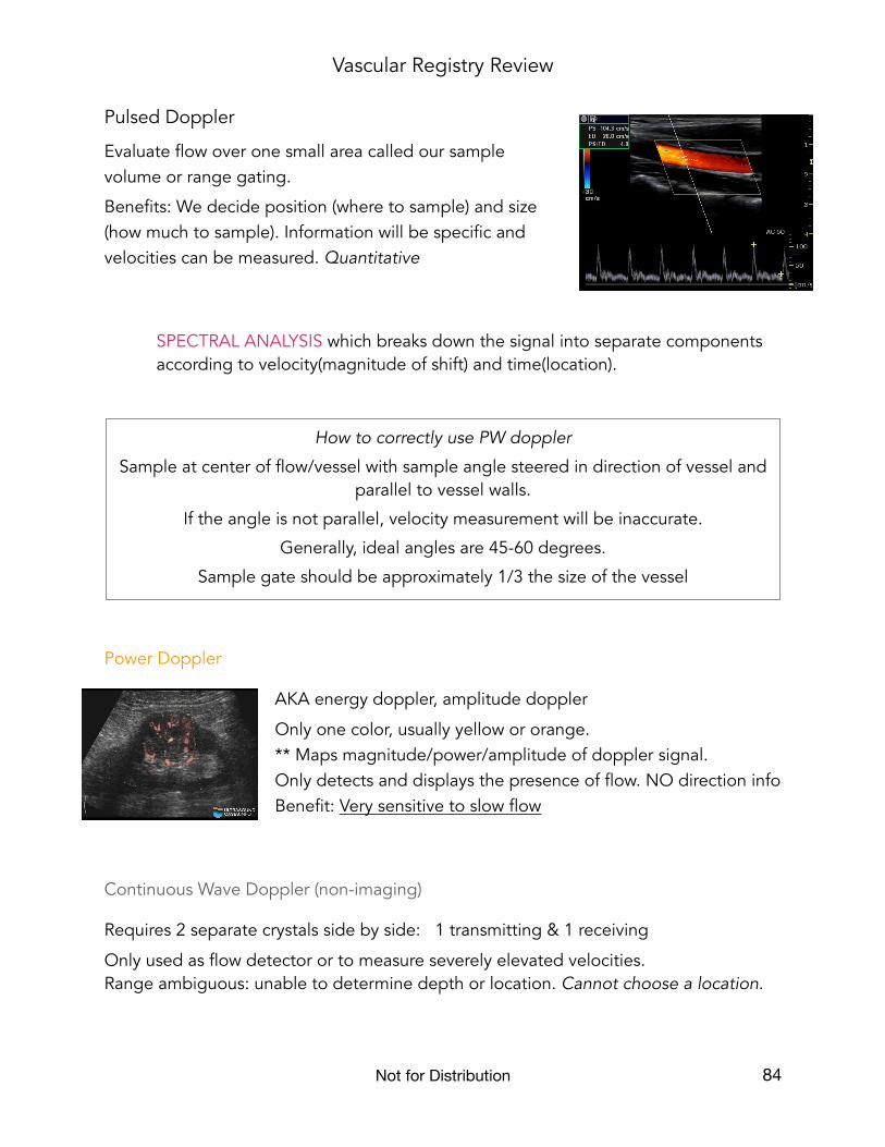
Pulsed Doppler
Evaluate flow over one small area called our sample volume or range gating.
Benefits: We decide position (where to sample) and size (how much to sample). Information will be specific and velocities can be measured. Quantitative
SPECTRAL ANALYSIS which breaks down the signal into separate components according to velocity(magnitude of shift) and time(location).
Power Doppler
AKA energy doppler, amplitude doppler
Only one color, usually yellow or orange. ** Maps magnitude/power/amplitude of doppler signal. Only detects and displays the presence of flow. NO direction info Benefit: Very sensitive to slow flow
Continuous Wave Doppler (non-imaging)
Requires 2 separate crystals side by side: 1 transmitting & 1 receiving
Only used as flow detector or to measure severely elevated velocities. Range ambiguous: unable to determine depth or location. Cannot choose a location.
Not for Distribution 84
How to correctly use PW doppler
Sample at center of flow/vessel with sample angle steered in direction of vessel and parallel to vessel walls.
If the angle is not parallel, velocity measurement will be inaccurate.
Generally, ideal angles are 45-60 degrees.
Sample gate should be approximately 1/3 the size of the vessel

Vascular Registry Review
Doppler Optimization
Scale Needs to match the type of flow you are evaluating
• Decrease the scale = Not sensitive enough
• Increase the scale = Aliasing
Wall filters and High Pass filters Filters LOW FREQUENCY/HIGH AMPLITUDE.
• Decrease WF = Not sensitive enough
Gain Fine tuning
• Increase gain to enhance the strength of the doppler signal
• Decrease if bleeding out of vessel
Problem: ALIASING
Shift exceeds the Nyquist limit (1/2 PRF)
• Increase scale or PRF
• Lower baseline
• Decrease transducer frequency
• Increase doppler angle
Problem: SLOW FLOW
Low velocity flow will produce a small doppler shift. The opposite to aliasing. Simply the problem is the shift is too small to see.
• LOW scale
• LOW wall filter
• Increase frequency
• Decrease angle (closer to parallel)
• Increase color gain
• Increase packet size
• Use power doppler
Not for Distribution 85