Embed Size (px)
Citation preview
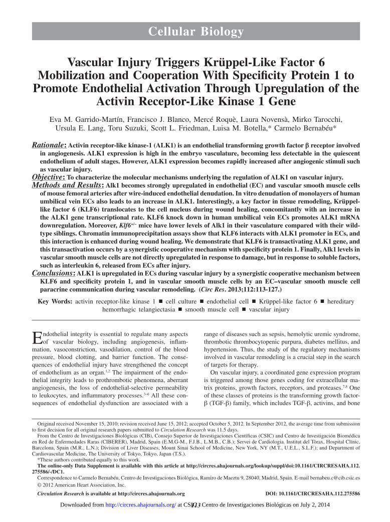
Ursula E. Lang, Toru Suzuki, Scott L. Friedman, Luisa M. Botella and Carmelo BernabéuEva M. Garrido-Martín, Francisco J. Blanco, Mercé Roquè, Laura Novensà, Mirko Tarocchi,
Activin Receptor-Like Kinase 1 GeneSpecificity Protein 1 to Promote Endothelial Activation Through Upregulation of the Vascular Injury Triggers Krüppel-Like Factor 6 Mobilization and Cooperation With
Print ISSN: 0009-7330. Online ISSN: 1524-4571 Copyright © 2012 American Heart Association, Inc. All rights reserved.is published by the American Heart Association, 7272 Greenville Avenue, Dallas, TX 75231Circulation Research
doi: 10.1161/CIRCRESAHA.112.2755862013;112:113-127; originally published online October 9, 2012;Circ Res.
http://circres.ahajournals.org/content/112/1/113World Wide Web at:
The online version of this article, along with updated information and services, is located on the
http://circres.ahajournals.org/content/suppl/2012/10/09/CIRCRESAHA.112.275586.DC1.htmlData Supplement (unedited) at:
http://circres.ahajournals.org//subscriptions/
is online at: Circulation Research Information about subscribing to Subscriptions:
http://www.lww.com/reprints Information about reprints can be found online at: Reprints:
document. Permissions and Rights Question and Answer about this process is available in the
located, click Request Permissions in the middle column of the Web page under Services. Further informationEditorial Office. Once the online version of the published article for which permission is being requested is
can be obtained via RightsLink, a service of the Copyright Clearance Center, not theCirculation Researchin Requests for permissions to reproduce figures, tables, or portions of articles originally publishedPermissions:
at CSIC - Centro de Investigaciones Biológicas on July 2, 2014http://circres.ahajournals.org/Downloaded from at CSIC - Centro de Investigaciones Biológicas on July 2, 2014http://circres.ahajournals.org/Downloaded from at CSIC - Centro de Investigaciones Biológicas on July 2, 2014http://circres.ahajournals.org/Downloaded from at CSIC - Centro de Investigaciones Biológicas on July 2, 2014http://circres.ahajournals.org/Downloaded from at CSIC - Centro de Investigaciones Biológicas on July 2, 2014http://circres.ahajournals.org/Downloaded from at CSIC - Centro de Investigaciones Biológicas on July 2, 2014http://circres.ahajournals.org/Downloaded from at CSIC - Centro de Investigaciones Biológicas on July 2, 2014http://circres.ahajournals.org/Downloaded from at CSIC - Centro de Investigaciones Biológicas on July 2, 2014http://circres.ahajournals.org/Downloaded from at CSIC - Centro de Investigaciones Biológicas on July 2, 2014http://circres.ahajournals.org/Downloaded from at CSIC - Centro de Investigaciones Biológicas on July 2, 2014http://circres.ahajournals.org/Downloaded from at CSIC - Centro de Investigaciones Biológicas on July 2, 2014http://circres.ahajournals.org/Downloaded from at CSIC - Centro de Investigaciones Biológicas on July 2, 2014http://circres.ahajournals.org/Downloaded from at CSIC - Centro de Investigaciones Biológicas on July 2, 2014http://circres.ahajournals.org/Downloaded from at CSIC - Centro de Investigaciones Biológicas on July 2, 2014http://circres.ahajournals.org/Downloaded from at CSIC - Centro de Investigaciones Biológicas on July 2, 2014http://circres.ahajournals.org/Downloaded from
113
Cellular Biology
Endothelial integrity is essential to regulate many aspects of vascular biology, including angiogenesis, inflam-
mation, vasoconstriction, vasodilation, control of the blood pressure, blood clotting, and barrier function. The conse-quences of endothelial injury have strengthened the concept of endothelium as an organ.1,2 The impairment of the endo-thelial integrity leads to prothrombotic phenomena, aberrant angiogenesis, the loss of endothelial-selective permeability to leukocytes, and inflammatory processes.3–6 All these con-sequences of endothelial dysfunction are associated with a
range of diseases such as sepsis, hemolytic uremic syndrome, thrombotic thrombocytopenic purpura, diabetes mellitus, and hypertension. Thus, the study of the regulatory mechanisms involved in vascular remodeling is a crucial step in the search of targets for therapy.
On vascular injury, a coordinated gene expression program is triggered among those genes coding for extracellular ma-trix proteins, growth factors, receptors, and proteases.7,8 One of these classes of proteins is the transforming growth factor-β (TGF-β) family, which includes TGF-β, activins, and bone
Original received November 15, 2010; revision received June 15, 2012; accepted October 5, 2012. In September 2012, the average time from submission to first decision for all original research papers submitted to Circulation Research was 11.5 days.
From the Centro de Investigaciones Biológicas (CIB), Consejo Superior de Investigaciones Científicas (CSIC) and Centro de Investigación Biomédica en Red de Enfermedades Raras (CIBERER), Madrid, Spain (E.M.G-M., F.J.B., L.M.B., C.B.); Servei de Cardiologia. Institut del Tòrax, Hospital Clínic, Barcelona, Spain (M.R., L.N.); Division of Liver Diseases, Mount Sinai School of Medicine, New York, NY (M.T., U.E.L., S.L.F.); and Department of Cardiovascular Medicine, The University of Tokyo, Tokyo, Japan (T.S.).
*These authors contributed equally to this work.The online-only Data Supplement is available with this article at http://circres.ahajournals.org/lookup/suppl/doi:10.1161/CIRCRESAHA.112.
275586/-/DC1.Correspondence to Carmelo Bernabéu, Centro de Investigaciones Biológica, Ramiro de Maeztu 9, 28040, Madrid, Spain. E-mail [email protected]© 2012 American Heart Association, Inc.
Circulation Research is available at http://circres.ahajournals.org DOI: 10.1161/CIRCRESAHA.112.275586
RES
Circulation Research
0009-7330
10.1161/CIRCRESAHA.112.275586
201483
Garrido-Martín et al KLF6 Transactivates ALK1 on Vascular Injury
Circulation ResearchMonth 2012AQ1
00
January
2013
© 2011 American Heart Association, Inc.
Rationale: Activin receptor-like kinase-1 (ALK1) is an endothelial transforming growth factor β receptor involved in angiogenesis. ALK1 expression is high in the embryo vasculature, becoming less detectable in the quiescent endothelium of adult stages. However, ALK1 expression becomes rapidly increased after angiogenic stimuli such as vascular injury.
Objective: To characterize the molecular mechanisms underlying the regulation of ALK1 on vascular injury.Methods and Results: Alk1 becomes strongly upregulated in endothelial (EC) and vascular smooth muscle cells
of mouse femoral arteries after wire-induced endothelial denudation. In vitro denudation of monolayers of human umbilical vein ECs also leads to an increase in ALK1. Interestingly, a key factor in tissue remodeling, Krüppel-like factor 6 (KLF6) translocates to the cell nucleus during wound healing, concomitantly with an increase in the ALK1 gene transcriptional rate. KLF6 knock down in human umbilical vein ECs promotes ALK1 mRNA downregulation. Moreover, Klf6+/− mice have lower levels of Alk1 in their vasculature compared with their wild-type siblings. Chromatin immunoprecipitation assays show that KLF6 interacts with ALK1 promoter in ECs, and this interaction is enhanced during wound healing. We demonstrate that KLF6 is transactivating ALK1 gene, and this transactivation occurs by a synergistic cooperative mechanism with specificity protein 1. Finally, Alk1 levels in vascular smooth muscle cells are not directly upregulated in response to damage, but in response to soluble factors, such as interleukin 6, released from ECs after injury.
Conclusions: ALK1 is upregulated in ECs during vascular injury by a synergistic cooperative mechanism between KLF6 and specificity protein 1, and in vascular smooth muscle cells by an EC–vascular smooth muscle cell paracrine communication during vascular remodeling. (Circ Res. 2013;112:113-127.)
Key Words: activin receptor-like kinase 1 ◼ cell culture ◼ endothelial cell ◼ Krüppel-like factor 6 ◼ hereditary hemorrhagic telangiectasia ◼ smooth muscle cell ◼ vascular injury
Vascular Injury Triggers Krüppel-Like Factor 6 Mobilization and Cooperation With Specificity Protein 1 to
Promote Endothelial Activation Through Upregulation of the Activin Receptor-Like Kinase 1 Gene
Eva M. Garrido-Martín, Francisco J. Blanco, Mercé Roquè, Laura Novensà, Mirko Tarocchi, Ursula E. Lang, Toru Suzuki, Scott L. Friedman, Luisa M. Botella,* Carmelo Bernabéu*
Gomathy & Uma
at CSIC - Centro de Investigaciones Biológicas on July 2, 2014http://circres.ahajournals.org/Downloaded from
114 Circulation Research January 4, 2013
morphogenetic proteins (BMPs).9 Several lines of evidence support a pivotal role for TGF-β in response to injury: (1) TGF-β expression is upregulated after injury8,10,11; (2) infusion of TGF-β polypeptide or transfection of TGF-β cDNA into injured arteries increases extracellular matrix production asso-ciated with the repair process12; (3) antibodies against TGF-β1 suppress intimal hyperplasia13; (4) radiation-mediated vascu-lar injury causes a rapid and persistent increase in expression of TGF-β receptors and mediators14; (5) BMP-9 is involved in postnatal retinal vascular remodeling15; and (6) TGF-β has a role in intimal thickening after vascular injury.16,17 Moreover, increased TGF-β1 activity underlies the wound–healing response in liver,18,19 kidney, lung,12 and vascular tissue.20,21
TGF-β family members exert their effects via type I (TβRI) and type II (TβRII) serine/threonine kinase receptors, helped by the co-receptors (TβRIII) and transduce the signal from the membrane to the nucleus through the intracellular Smad fac-tors.9 In endothelial cells (ECs), the TGF-β signaling acquires an important level of regulation attributable to the coexistence of 2 different type I receptors, the ubiquitous activin receptor-like kinase 5 (ALK5), and ALK1.22 ALK1 is highly expressed in the vascular structures of the embryo,23–26 and it is essential during vascular development as demonstrated by the lethality of the ALK1/Activin A receptor type II-like 1 (ACVRL1) gene disruption. The Alk1 knock-out embryos die at E10.5, because of the absence of mature blood vessels in the yolk sac, show-ing aberrant hyperdilated vascular structures and clumps of blood cells.27,28 Moreover, the heterozygous mutation of ALK1 results in a vascular dysplasia called hereditary hemorrhagic telangiectasia type 2 (HHT2), characterized by skin and mu-cosa telangiectases as well as liver and lung arteriovenous malformations (AVMs).29,30 Despite the essential role exerted by ALK1 in the vasculogenic process during embryonic de-velopment, its expression is diminished in the quiescent en-dothelium during adult life.24 The activation of the EC ALK1 expression is crucially upregulated in certain locations in re-sponse to several angiogenic stimuli.24,31
Krüppel-like factor 6 (KLF6) is a transcriptional regulator that mediates cellular differentiation and tissue development through
its roles in growth-related signal transduction pathway, cell pro-liferation, apoptosis, and angiogenesis.32–34 KLF6 is considered as a damage–response factor that promotes tissue remodeling because of its ability of transactivating several target genes by direct binding to their promoters.19,35 These genes comprise sev-eral members of the TGF-β signaling pathway such as TGF-β1, its receptors TβRI (ALK5) and TβRII,36 the co-receptor endoglin (ENG),37 urokinase-type plasminogen activator (uPA),38 and col-lagen 1A (Col1A).35 Furthermore, we have recently described a specific functional relationship between KLF6 and TGF-β pathway by the direct formation of a ternary Smad3-specificity protein 1 (Sp1)-KLF6 complex.39 These effects suggest that KLF6 is a common regulatory factor for all the TGF-β func-tions related to injury, so KLF6 seems to orchestrate the repair mechanisms to return the endothelium to its regular state and to avoid the complications derived of its dysfunction.40
In this article, we have explored the regulation of ALK1 expression under vascular injury. Our results demonstrate the transactivation of ALK1 gene by KLF6 and as a consequence the ALK1 upregulation in the migrating ECs. These data pro-vide new insights in the molecular mechanisms mediated by KLF6 for the coordination of the vascular remodeling process and provide additional evidences for a pivotal role of ALK1 in the activated state of the EC during the angiogenic response after vascular injury.
MethodsCell culture,37,41 expression vectors,35,42,43 transfection and reporter assays,41 stable infection of primary EC cultures,44 real-time poly-merase chain reaction,41 in vitro EC denudation,37 immunofluo-rescence microscopy,37 flow cytometry,37 immunohistochemistry, mechanical injury model in mouse femoral arteries,45,46 laser micro-dissection, and chromatin immunoprecipitation41 are described in an expanded manner in the Material and Methods section of the Online Data Supplement.
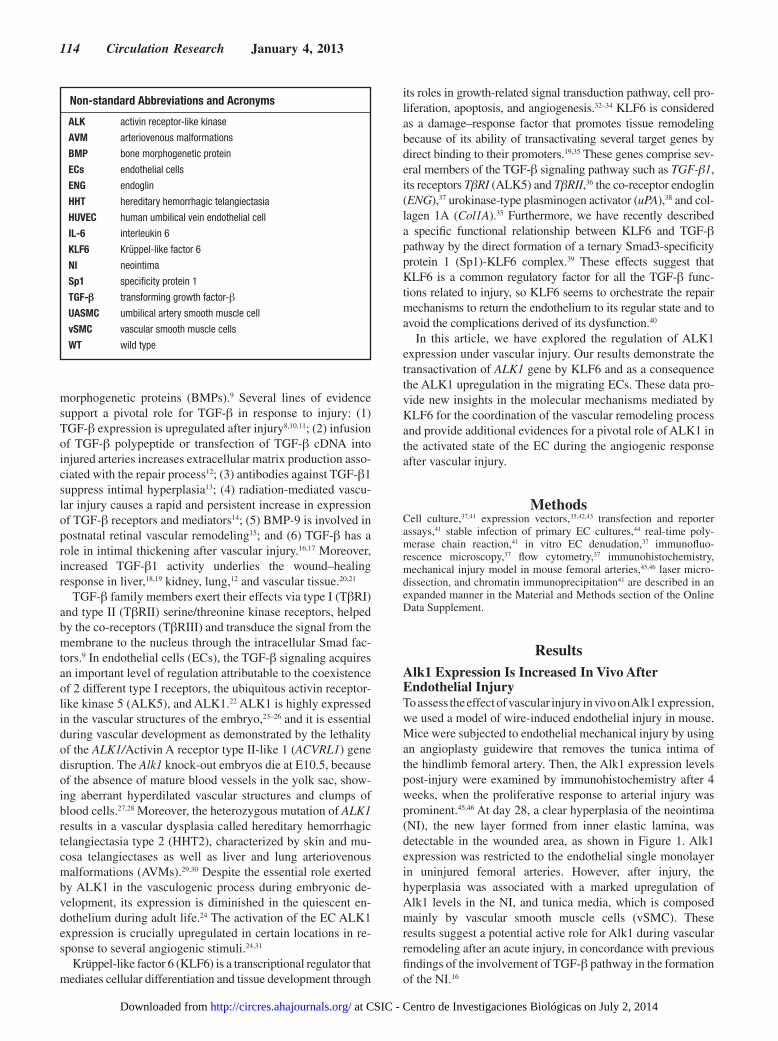
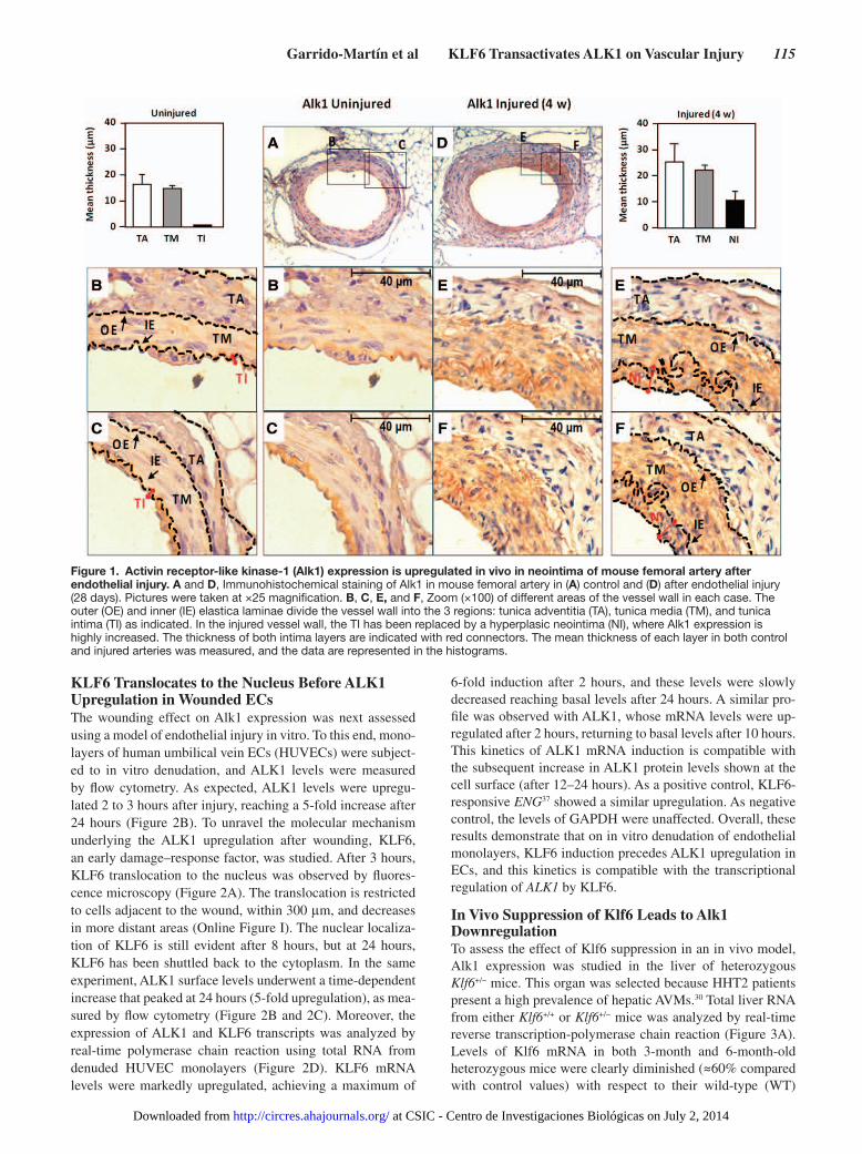
ResultsAlk1 Expression Is Increased In Vivo After Endothelial InjuryTo assess the effect of vascular injury in vivo on Alk1 expression, we used a model of wire-induced endothelial injury in mouse. Mice were subjected to endothelial mechanical injury by using an angioplasty guidewire that removes the tunica intima of the hindlimb femoral artery. Then, the Alk1 expression levels post-injury were examined by immunohistochemistry after 4 weeks, when the proliferative response to arterial injury was prominent.45,46 At day 28, a clear hyperplasia of the neointima (NI), the new layer formed from inner elastic lamina, was detectable in the wounded area, as shown in Figure 1. Alk1 expression was restricted to the endothelial single monolayer in uninjured femoral arteries. However, after injury, the hyperplasia was associated with a marked upregulation of Alk1 levels in the NI, and tunica media, which is composed mainly by vascular smooth muscle cells (vSMC). These results suggest a potential active role for Alk1 during vascular remodeling after an acute injury, in concordance with previous findings of the involvement of TGF-β pathway in the formation of the NI.16
Non-standard Abbreviations and Acronyms
ALK activin receptor-like kinase
AVM arteriovenous malformations
BMP bone morphogenetic protein
ECs endothelial cells
ENG endoglin
HHT hereditary hemorrhagic telangiectasia
HUVEC human umbilical vein endothelial cell
IL-6 interleukin 6
KLF6 Krüppel-like factor 6
NI neointima
Sp1 specificity protein 1
TGF-β transforming growth factor-β
UASMC umbilical artery smooth muscle cell
vSMC vascular smooth muscle cells
WT wild type
at CSIC - Centro de Investigaciones Biológicas on July 2, 2014http://circres.ahajournals.org/Downloaded from
Garrido-Martín et al KLF6 Transactivates ALK1 on Vascular Injury 115
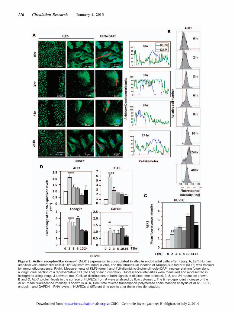
KLF6 Translocates to the Nucleus Before ALK1 Upregulation in Wounded ECsThe wounding effect on Alk1 expression was next assessed using a model of endothelial injury in vitro. To this end, mono-layers of human umbilical vein ECs (HUVECs) were subject-ed to in vitro denudation, and ALK1 levels were measured by flow cytometry. As expected, ALK1 levels were upregu-lated 2 to 3 hours after injury, reaching a 5-fold increase after 24 hours (Figure 2B). To unravel the molecular mechanism underlying the ALK1 upregulation after wounding, KLF6, an early damage–response factor, was studied. After 3 hours, KLF6 translocation to the nucleus was observed by fluores-cence microscopy (Figure 2A). The translocation is restricted to cells adjacent to the wound, within 300 μm, and decreases in more distant areas (Online Figure I). The nuclear localiza-tion of KLF6 is still evident after 8 hours, but at 24 hours, KLF6 has been shuttled back to the cytoplasm. In the same experiment, ALK1 surface levels underwent a time-dependent increase that peaked at 24 hours (5-fold upregulation), as mea-sured by flow cytometry (Figure 2B and 2C). Moreover, the expression of ALK1 and KLF6 transcripts was analyzed by real-time polymerase chain reaction using total RNA from denuded HUVEC monolayers (Figure 2D). KLF6 mRNA levels were markedly upregulated, achieving a maximum of
6-fold induction after 2 hours, and these levels were slowly decreased reaching basal levels after 24 hours. A similar pro-file was observed with ALK1, whose mRNA levels were up-regulated after 2 hours, returning to basal levels after 10 hours. This kinetics of ALK1 mRNA induction is compatible with the subsequent increase in ALK1 protein levels shown at the cell surface (after 12–24 hours). As a positive control, KLF6-responsive ENG37 showed a similar upregulation. As negative control, the levels of GAPDH were unaffected. Overall, these results demonstrate that on in vitro denudation of endothelial monolayers, KLF6 induction precedes ALK1 upregulation in ECs, and this kinetics is compatible with the transcriptional regulation of ALK1 by KLF6.
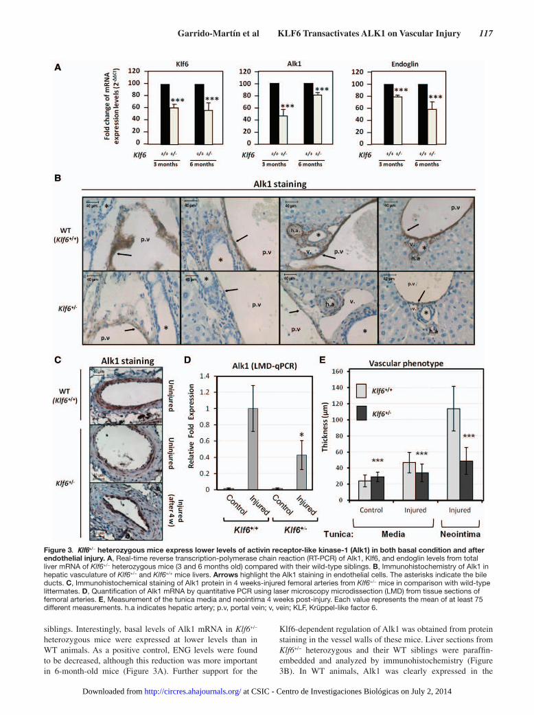
In Vivo Suppression of Klf6 Leads to Alk1 DownregulationTo assess the effect of Klf6 suppression in an in vivo model, Alk1 expression was studied in the liver of heterozygous Klf6+/− mice. This organ was selected because HHT2 patients present a high prevalence of hepatic AVMs.30 Total liver RNA from either Klf6+/+ or Klf6+/− mice was analyzed by real-time reverse transcription-polymerase chain reaction (Figure 3A). Levels of Klf6 mRNA in both 3-month and 6-month-old heterozygous mice were clearly diminished (≈60% compared with control values) with respect to their wild-type (WT)
Figure 1. Activin receptor-like kinase-1 (Alk1) expression is upregulated in vivo in neointima of mouse femoral artery after endothelial injury. A and D, Immunohistochemical staining of Alk1 in mouse femoral artery in (A) control and (D) after endothelial injury (28 days). Pictures were taken at ×25 magnification. B, C, E, and F, Zoom (×100) of different areas of the vessel wall in each case. The outer (OE) and inner (IE) elastica laminae divide the vessel wall into the 3 regions: tunica adventitia (TA), tunica media (TM), and tunica intima (TI) as indicated. In the injured vessel wall, the TI has been replaced by a hyperplasic neointima (NI), where Alk1 expression is highly increased. The thickness of both intima layers are indicated with red connectors. The mean thickness of each layer in both control and injured arteries was measured, and the data are represented in the histograms.
at CSIC - Centro de Investigaciones Biológicas on July 2, 2014http://circres.ahajournals.org/Downloaded from
116 Circulation Research January 4, 2013
Figure 2. Activin receptor-like kinase-1 (ALK1) expression is upregulated in vitro in endothelial cells after injury. A, Left, Human umbilical vein endothelial cells (HUVECs) were wounded in vitro, and the intracellular location of Krüppel-like factor 6 (KLF6) was tracked by immunofluorescence. Right, Measurements of KLF6 (green) and 4',6-diamidino-2-phenylindole (DAPI) nuclear staining (blue) along a longitudinal section of a representative cell (red line) of each condition. Fluorescence intensities were measured and represented in histograms using Image J software tool. Cellular distributions of both signals at distinct time points (0, 3, 8, and 24 hours) are shown. B and C, ALK1 protein levels in the surface of HUVECs from A were analyzed by flow cytometry. The time-dependent increase of the ALK1 mean fluorescence intensity is shown in C. D, Real-time reverse transcription-polymerase chain reaction analysis of ALK1, KLF6, endoglin, and GAPDH mRNA levels in HUVECs at different time points after the in vitro denudation.
at CSIC - Centro de Investigaciones Biológicas on July 2, 2014http://circres.ahajournals.org/Downloaded from
Garrido-Martín et al KLF6 Transactivates ALK1 on Vascular Injury 117
siblings. Interestingly, basal levels of Alk1 mRNA in Klf6+/− heterozygous mice were expressed at lower levels than in WT animals. As a positive control, ENG levels were found to be decreased, although this reduction was more important in 6-month-old mice (Figure 3A). Further support for the
Klf6-dependent regulation of Alk1 was obtained from protein staining in the vessel walls of these mice. Liver sections from Klf6+/− heterozygous and their WT siblings were paraffin-embedded and analyzed by immunohistochemistry (Figure 3B). In WT animals, Alk1 was clearly expressed in the
Figure 3. Klf6+/− heterozygous mice express lower levels of activin receptor-like kinase-1 (Alk1) in both basal condition and after endothelial injury. A, Real-time reverse transcription-polymerase chain reaction (RT-PCR) of Alk1, Klf6, and endoglin levels from total liver mRNA of Klf6+/− heterozygous mice (3 and 6 months old) compared with their wild-type siblings. B, Immunohistochemistry of Alk1 in hepatic vasculature of Klf6+/− and Klf6+/+ mice livers. Arrows highlight the Alk1 staining in endothelial cells. The asterisks indicate the bile ducts. C, Immunohistochemical staining of Alk1 protein in 4 weeks-injured femoral arteries from Klf6+/− mice in comparison with wild-type littermates. D, Quantification of Alk1 mRNA by quantitative PCR using laser microscopy microdissection (LMD) from tissue sections of femoral arteries. E, Measurement of the tunica media and neointima 4 weeks post-injury. Each value represents the mean of at least 75 different measurements. h.a indicates hepatic artery; p.v, portal vein; v, vein; KLF, Krüppel-like factor 6.
at CSIC - Centro de Investigaciones Biológicas on July 2, 2014http://circres.ahajournals.org/Downloaded from
118 Circulation Research January 4, 2013
endothelium of liver vessels. By contrast, Alk1 signal was much weaker in Klf6+/− heterozygous mice. Furthermore, Alk1 staining of quiescent endothelium from femoral arteries was found to be lower in Klf6+/− compared with Klf6+/+ mice (Figure 3C). Also, on wire-induced endothelial injury, Alk1 protein was induced in both Klf6+/+ and Klf6+/− mice (Figure 3C), but laser microdissection studies showed that the upregulated Alk1 mRNA levels in Klf6+/− were lower than in Klf6+/+ animals (Figure 3D). In addition, a distinct vascular phenotype was observed (Figure 3C and 3E). Thus, although the increase in the tunica media thickness was ≈2-fold in WT mice, only a slight augmentation (1.2-fold) was observed in Klf6+/− mice. Remarkably, on injury, the NI of Klf6+/+ mice was >2-fold thicker than that of Klf6+/− animals. Taken together, these results agree with the crucial role of Alk1 in vascular remodeling and strongly support the involvement of Klf6 in the regulation of Alk1 gene expression.
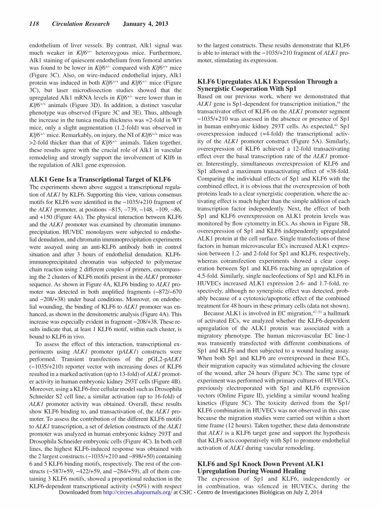
ALK1 Gene Is a Transcriptional Target of KLF6The experiments shown above suggest a transcriptional regula-tion of ALK1 by KLF6. Supporting this view, various consensus motifs for KLF6 were identified in the −1035/+210 fragment of the ALK1 promoter, at positions −815, −739, −148, −109, −86, and +150 (Figure 4A). The physical interaction between KLF6 and the ALK1 promoter was examined by chromatin immuno-precipitation. HUVEC monolayers were subjected to endothe-lial denudation, and chromatin immunoprecipitation experiments were assayed using an anti-KLF6 antibody both in control situation and after 3 hours of endothelial denudation. KLF6-immunoprecipitated chromatin was subjected to polymerase chain reaction using 2 different couples of primers, encompass-ing the 2 clusters of KLF6 motifs present in the ALK1 promoter sequence. As shown in Figure 4A, KLF6 binding to ALK1 pro-moter was detected in both amplified fragments (−872/−670 and −208/+38) under basal conditions. Moreover, on endothe-lial wounding, the binding of KLF6 to ALK1 promoter was en-hanced, as shown in the densitometric analysis (Figure 4A). This increase was especially evident in fragment −208/+38. These re-sults indicate that, at least 1 KLF6 motif, within each cluster, is bound to KLF6 in vivo.
To assess the effect of this interaction, transcriptional ex-periments using ALK1 promoter (pALK1) constructs were performed. Transient transfections of the pGL2-pALK1 (−1035/+210) reporter vector with increasing doses of KLF6 resulted in a marked activation (up to 13-fold) of ALK1 promot-er activity in human embryonic kidney 293T cells (Figure 4B). Moreover, using a KLF6-free cellular model such as Drosophila Schneider S2 cell line, a similar activation (up to 16-fold) of ALK1 promoter activity was obtained. Overall, these results show KLF6 binding to, and transactivation of, the ALK1 pro-moter. To assess the contribution of the different KLF6 motifs to ALK1 transcription, a set of deletion constructs of the ALK1 promoter was analyzed in human embryonic kidney 293T and Drosophila Schneider embryonic cells (Figure 4C). In both cell lines, the highest KLF6-induced response was obtained with the 2 largest constructs (−1035/+210 and −898/+50) containing 6 and 5 KLF6 binding motifs, respectively. The rest of the con-structs (−587/+59, −422/+59, and −284/+59), all of them con-taining 3 KLF6 motifs, showed a proportional reduction in the KLF6-dependent transcriptional activity (≈50%) with respect
to the largest constructs. These results demonstrate that KLF6 is able to interact with the −1035/+210 fragment of ALK1 pro-moter, stimulating its expression.
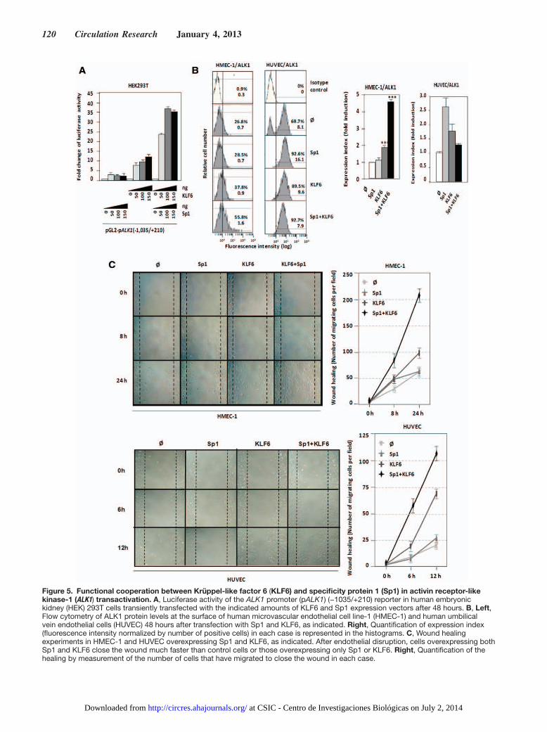
KLF6 Upregulates ALK1 Expression Through a Synergistic Cooperation With Sp1Based on our previous work, where we demonstrated that ALK1 gene is Sp1-dependent for transcription initiation,41 the transactivator effect of KLF6 on the ALK1 promoter segment −1035/+210 was assessed in the absence or presence of Sp1 in human embryonic kidney 293T cells. As expected,41 Sp1 overexpression induced (≈4-fold) the transcriptional activ-ity of the ALK1 promoter construct (Figure 5A). Similarly, overexpression of KLF6 achieved a 12-fold transactivating effect over the basal transcription rate of the ALK1 promot-er. Interestingly, simultaneous overexpression of KLF6 and Sp1 allowed a maximum transactivating effect of ≈38-fold. Comparing the individual effects of Sp1 and KLF6 with the combined effect, it is obvious that the overexpression of both proteins leads to a clear synergistic cooperation, where the ac-tivating effect is much higher than the simple addition of each transcription factor independently. Next, the effect of both Sp1 and KLF6 overexpression on ALK1 protein levels was monitored by flow cytometry in ECs. As shown in Figure 5B, overexpression of Sp1 and KLF6 independently upregulated ALK1 protein at the cell surface. Single transfections of these factors in human microvascular ECs increased ALK1 expres-sion between 1.2- and 2-fold for Sp1 and KLF6, respectively, whereas cotransfection experiments showed a clear coop-eration between Sp1 and KLF6 reaching an upregulation of 4.5-fold. Similarly, single nucleofections of Sp1 and KLF6 in HUVECs increased ALK1 expression 2.6- and 1.7-fold, re-spectively, although no synergistic effect was detected, prob-ably because of a cytotoxic/apoptotic effect of the combined treatment for 48 hours in these primary cells (data not shown).
Because ALK1 is involved in EC migration,47–51 a hallmark of activated ECs, we analyzed whether the KLF6-dependent upregulation of the ALK1 protein was associated with a migratory phenotype. The human microvascular EC line-1 was transiently transfected with different combinations of Sp1 and KLF6 and then subjected to a wound healing assay. When both Sp1 and KLF6 are overexpressed in these ECs, their migration capacity was stimulated achieving the closure of the wound, after 24 hours (Figure 5C). The same type of experiment was performed with primary cultures of HUVECs, previously electroporated with Sp1 and KLF6 expression vectors (Online Figure II), yielding a similar wound healing kinetics (Figure 5C). The toxicity derived from the Sp1/KLF6 combination in HUVECs was not observed in this case because the migration studies were carried out within a short time frame (12 hours). Taken together, these data demonstrate that ALK1 is a KLF6 target gene and support the hypothesis that KLF6 acts cooperatively with Sp1 to promote endothelial activation of ALK1 during vascular remodeling.
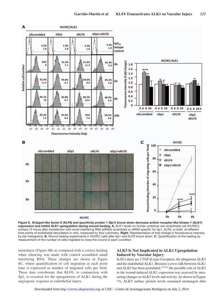
KLF6 and Sp1 Knock Down Prevent ALK1 Upregulation During Wound HealingThe expression of Sp1 and KLF6, independently or in combination, was silenced in HUVECs, during the
at CSIC - Centro de Investigaciones Biológicas on July 2, 2014http://circres.ahajournals.org/Downloaded from
Garrido-Martín et al KLF6 Transactivates ALK1 on Vascular Injury 119
wounding process by transfection of specific small interfering RNAs (Online Figure III). Figure 6A shows that silencing of Sp1, KLF6, or both resulted in the abolishment of the wound-mediated ALK1 induction as determined by
flow cytometry analysis. When cellular migration was measured, as a function depending on ALK1 expression, in the presence of small interfering RNA of Sp1, KLF6, or both, HUVECs were unable to close the disrupted
Figure 4. Krüppel-like factor 6 (KLF6) transactivates activin receptor-like kinase-1 (ALK1) promoter. A, KLF6 interacts with ALK1 promoter in human umbilical vein endothelial cells (HUVECs), and this interaction is increased after vascular injury. Left, Chromatin immunoprecipitation (ChIP) of KLF6 over the 2 main KLF6-sites rich regions of the ALK1 5´-proximal promoter in HUVECs. Right, KLF6 binding was measured by densitometry of the individual bands and values of the (KLF6-IgG)/Input ratios were represented in both the control situation and 3 hours after the endothelial denudation B, Dose–response effect of KLF6 on the transcriptional activity of ALK1 promoter in human embryonic kidney (HEK) 293T cells (left) and Drosophila Schneider S2 cells (right). C, Effect of KLF6 on the transcriptional activity of 5´ deleted constructs of ALK1 promoter. Left, Scheme of the different ALK1 promoter constructs shows the distribution of the KLF6 consensus binding sites found along the ALK1 promoter (black boxes). Right, Transient cotransfection of KLF6 expression vector with different 5´deleted constructs of ALK1 promoter in both HEK293T and Schneider S2 cells.
at CSIC - Centro de Investigaciones Biológicas on July 2, 2014http://circres.ahajournals.org/Downloaded from
120 Circulation Research January 4, 2013
Figure 5. Functional cooperation between Krüppel-like factor 6 (KLF6) and specificity protein 1 (Sp1) in activin receptor-like kinase-1 (ALK1) transactivation. A, Luciferase activity of the ALK1 promoter (pALK1) (−1035/+210) reporter in human embryonic kidney (HEK) 293T cells transiently transfected with the indicated amounts of KLF6 and Sp1 expression vectors after 48 hours. B, Left, Flow cytometry of ALK1 protein levels at the surface of human microvascular endothelial cell line-1 (HMEC-1) and human umbilical vein endothelial cells (HUVEC) 48 hours after transfection with Sp1 and KLF6, as indicated. Right, Quantification of expression index (fluorescence intensity normalized by number of positive cells) in each case is represented in the histograms. C, Wound healing experiments in HMEC-1 and HUVEC overexpressing Sp1 and KLF6, as indicated. After endothelial disruption, cells overexpressing both Sp1 and KLF6 close the wound much faster than control cells or those overexpressing only Sp1 or KLF6. Right, Quantification of the healing by measurement of the number of cells that have migrated to close the wound in each case.
at CSIC - Centro de Investigaciones Biológicas on July 2, 2014http://circres.ahajournals.org/Downloaded from
Garrido-Martín et al KLF6 Transactivates ALK1 on Vascular Injury 121
monolayer (Figure 6B) as compared with a correct healing when silencing was made with control scrambled small interfering RNA. These changes are shown in Figure 6C, where quantification of cell migration at each point time is expressed as number of migrated cells per field. These data corroborate that KLF6, in conjunction with Sp1, is essential for the upregulation of ALK1 during the angiogenic response to endothelial injury.
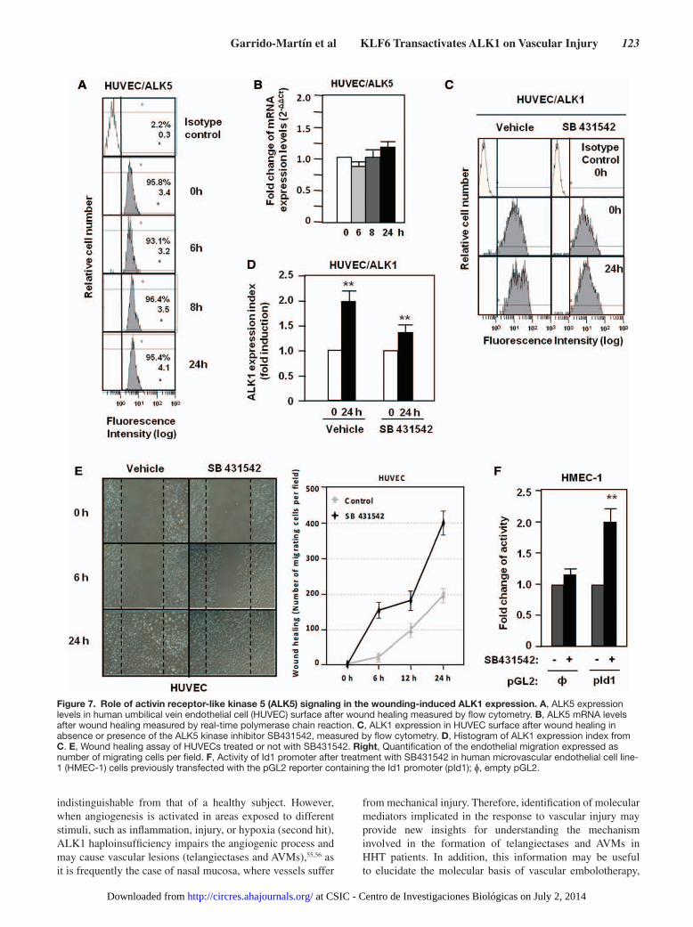
ALK5 Is Not Implicated in ALK1 Upregulation Induced by Vascular InjuryIn ECs there are 2 TGF-β type I receptors, the ubiquitous ALK5 and the endothelial ALK1. Because a cross-talk between ALK1 and ALK5 has been postulated,47,50,52 the possible role of ALK5 in the wound-induced ALK1 expression was assessed by mea-suring changes in ALK5 levels and activity. As shown in Figure 7A, ALK5 surface protein levels remained unchanged after
Figure 6. Krüppel-like factor 6 (KLF6) and specificity protein 1 (Sp1) knock down decrease activin receptor-like kinase-1 (ALK1) expression and inhibit ALK1 upregulation during wound healing. A, ALK1 levels on human umbilical vein endothelial cell (HUVEC) surface 72 hours after transfection with small interfering RNA (siRNA) scrambled or siRNA specific for Sp1, KLF6, or both, at different time points of endothelial denudation in vitro, measured by flow cytometry. Right, Representation of fold change in fluorescence intensity by bar histograms. B, Wound healing experiments in HUVEC cells after Sp1 and KLF6 knock down. C, Quantification of the healing by measurement of the number of cells migrated to close the wound in each condition.
at CSIC - Centro de Investigaciones Biológicas on July 2, 2014http://circres.ahajournals.org/Downloaded from
122 Circulation Research January 4, 2013
wound healing, and the same was true for the corresponding mRNA levels (Figure 7B). To analyze the influence of ALK5 signal, ECs were treated with SB431542, a specific ALK5 ki-nase inhibitor. In spite of the suppression of ALK5 signaling, ALK1 expression was upregulated on wound healing, although at a slightly lower extent than cells treated with vehicle (Figure 7C and 7D). On scratching endothelial monolayers, the migra-tion of cells is stimulated when the ALK5 pathway is inhibited (Figure 7E), whereas the ALK5 inhibitor stimulated the pro-moter activity of the Id1 target gene in ECs (Figure 7F). The efficiency of SB431542 treatment is demonstrated in HUVECs by the decrease in mRNA levels of PAI-1, a target gene of ALK5 (Online Figure IV). These results suggest that whereas wound-mediated upregulation of ALK1 is mostly independent of the ALK5 signaling pathway, inhibition of ALK5 activity favors signaling through ALK1, as shown by the stimulation of cell migration and activation of the Id1 target.
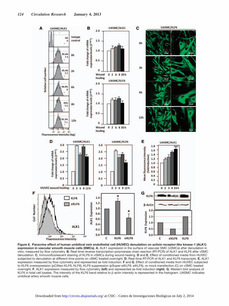
ALK1 Is Upregulated in Smooth Muscle Cells After Wounding Through a Paracrine Communication With Endothelial CellsAs seen in Figure 1, endothelial injury induces ALK1 upregu-lation in cells of the tunica media, suggesting the involvement of vSMC. As vSMCs are in close contact with ECs, we have explored the wounding effect in vSMCs. For this purpose, primary cultures of umbilical artery smooth muscle cells (UASMCs) were subjected to a scratching process and the surface levels of ALK1 were measured by flow cytometry. As observed in Figure 8A, UASMCs express high levels of ALK1 at their surface, but these levels remain unchanged along the wound healing process at variance with the increase of ALK1 in ECs after wounding. Accordingly, levels of ALK1 mRNA were unaffected (Figure 8B); the same was true for KLF6, as opposed to the increased levels of KLF6 observed in ECs after wound healing (Figure 2B–2D). Staining of KLF6 in UASMC at different times after wound healing did not reveal any nuclear translocation either (Figure 8C), at variance to ECs, where nuclear translocation is evident after 3 hours (Figure 2A). Interestingly, when UASMCs were cultured with condi-tioned media from wounded monolayers of ECs, a significant increase in ALK1 was evident at mRNA and protein levels, already after 2 hours (Figure 8D and 8E). Furthermore, KLF6 mRNA levels were increased in parallel with ALK1 mRNA in UASMCs grown in conditioned media from ECs subjected to wound healing. The increase in ALK1 protein was sustained up to 24 hours (Figure 8E). Next, we assessed the contribution of the wound healing-dependent induction of KLF6 in ECs to the upregulated expression of ALK1 in UASMCs. KLF6 was overexpressed (using pCiNeo-KLF6) or suppressed (us-ing pSuperKLF6) in HUVECs, and the corresponding condi-tioned media were used to culture vSMCs. As shown in Figure 8F, culture media from KLF6 overexpressing HUVECs in-duced a modest, but significant, increase (1.3-fold) in the expression levels of ALK1 of UASMCs. By contrast, cul-ture media from HUVECs with a knocked down expression of KLF6-induced a marked reduction of ALK1 levels (0.6-fold) compared with mock treated cells. Of note, changes in ALK1 levels paralleled those of KLF6 in vSMCs (Figure 8G). In summary, these results suggest that ALK1 is not directly
upregulated in vSMCs in response to injury. Instead, endo-thelial injury triggers indirectly a similar KLF6-dependent stimulation of ALK1, in vSMCs. This response would con-stitute a paracrine mechanism operating from endothelium (intima) to smooth muscle cells (tunica media). In an attempt to analyze candidates responsible for this paracrine response, several cytokines and growth factors involved in angiogenesis were analyzed by ELISA in conditioned media from ECs after injury. The results showed a sustained increase of interleukin 6 (IL-6) in wounded cultures of HUVECs (from 6 to 24 hours after wounding; Online Figure VI), suggesting the involve-ment of this cytokine in the upregulated expression of ALK1 in vSMCs after endothelial injury. To investigate the putative regulation of IL-6 by KLF6, immunohistochemistry staining of IL-6 in 4 weeks-injured femoral arteries from Klf6+/− mice in comparison with WT littermates was carried out (Online Figure VIIA). In uninjured vessels from WT and heterozygous animals, the presence of IL-6 was almost undetectable. By contrast, IL-6 staining was clearly detected on wire injury in different layers of the femoral artery. However, the increased signal of IL-6 in WT mice was higher than that of Klf6+/− lit-termates, suggesting that KLF6 regulates IL-6 gene expres-sion. Supporting this view, several putative consensus motifs for KLF6 were identified on in silico analysis of both human and mouse IL-6 proximal promoter sequences (Online Figure VIII). Furthermore, ectopic expression of KLF6 stimulated >3-fold the promoter activity of a luciferase reporter construct driven by the human IL-6 promoter sequence (Online Figure VIIB). Taken together, these experiments suggest that IL-6 is a target gene of KLF6. It can be speculated that KLF6 induc-tion on vascular injury modulates the expression of a set of target genes, including IL-6, which in turn upregulate ALK1. An in-depth analysis of these genes may shed light on the mo-lecular mechanism triggered to achieve vascular repair.
DiscussionALK1 functions are closely related to vascular biology.28,53,54 During embryogenesis, ALK1 is highly expressed in the vascular bed because of its critical requirement for a correct vasculogenic process, whereas in the adult life, the endothelium reaches quiescence and ALK1 expression levels drop.24 In adults, the signaling network triggered by ALK1 is framed in the angiogenic process, when activated EC proliferate and migrate to develop new vessels from the preexistent ones, in response to certain stimuli.23,50 After formation of the new sprouts, a resolution phase is needed, in which TGF-β–mediated signaling blocks the proliferation and stabilizes the new vessel by the deposition of extracellular matrix.47,49 Therefore, TGF-β signaling in the EC during angiogenesis is crucial and needs to be highly regulated to control the balance between activating and resolving signals in each stage of the process. Having this in mind, a vascular damage would require an immediate ALK1 upregulation for the correct coordination of the subsequent repair mechanisms, whereas ALK5-controlled pathway should remain unchanged immediately after injury.
In HHT, patients harboring mutations in ALK1 are able to develop normal vessels, and their vascular system is, overall,
at CSIC - Centro de Investigaciones Biológicas on July 2, 2014http://circres.ahajournals.org/Downloaded from
Garrido-Martín et al KLF6 Transactivates ALK1 on Vascular Injury 123
indistinguishable from that of a healthy subject. However, when angiogenesis is activated in areas exposed to different stimuli, such as inflammation, injury, or hypoxia (second hit), ALK1 haploinsufficiency impairs the angiogenic process and may cause vascular lesions (telangiectases and AVMs),55,56 as it is frequently the case of nasal mucosa, where vessels suffer
from mechanical injury. Therefore, identification of molecular mediators implicated in the response to vascular injury may provide new insights for understanding the mechanism involved in the formation of telangiectases and AVMs in HHT patients. In addition, this information may be useful to elucidate the molecular basis of vascular embolotherapy,
Figure 7. Role of activin receptor-like kinase 5 (ALK5) signaling in the wounding-induced ALK1 expression. A, ALK5 expression levels in human umbilical vein endothelial cell (HUVEC) surface after wound healing measured by flow cytometry. B, ALK5 mRNA levels after wound healing measured by real-time polymerase chain reaction. C, ALK1 expression in HUVEC surface after wound healing in absence or presence of the ALK5 kinase inhibitor SB431542, measured by flow cytometry. D, Histogram of ALK1 expression index from C. E, Wound healing assay of HUVECs treated or not with SB431542. Right, Quantification of the endothelial migration expressed as number of migrating cells per field. F, Activity of Id1 promoter after treatment with SB431542 in human microvascular endothelial cell line-1 (HMEC-1) cells previously transfected with the pGL2 reporter containing the Id1 promoter (pId1); ɸ, empty pGL2.
at CSIC - Centro de Investigaciones Biológicas on July 2, 2014http://circres.ahajournals.org/Downloaded from
124 Circulation Research January 4, 2013
Figure 8. Paracrine effect of human umbilical vein endothelial cell (HUVEC) denudation on activin receptor-like kinase-1 (ALK1) expression in vascular smooth muscle cells (SMCs). A, ALK1 expression in the surface of vascular SMC (vSMCs) after denudation in vitro, measured by flow cytometry. B, Real-time reverse transcription-polymerase chain reaction (RT-PCR) of ALK1 and KLF6 after vSMC denudation. C, Immunofluorescent staining of KLF6 in vSMCs during wound healing. D and E, Effect of conditioned media from HUVEC subjected to denudation at different time points on vSMC treated overnight. D, Real-time RT-PCR of ALK1 and KLF6 transcripts. E, ALK1 expression measured by flow cytometry and represented as fold induction. F and G, Effect of conditioned media from HUVEC subjected to KLF6 overexpression (pCiNeo-KLF6; KLF6), KLF6 suppression (pSuper-siKLF6; siKLF6), or mock transfection (C) on vSMC treated overnight. F, ALK1 expression measured by flow cytometry (left) and represented as fold induction (right). G, Western blot analysis of KLF6 in total cell lysates. The intensity of the KLF6 band relative to β-actin intensity is represented in the histogram. UASMC indicates umbilical artery smooth muscle cells.
at CSIC - Centro de Investigaciones Biológicas on July 2, 2014http://circres.ahajournals.org/Downloaded from
Garrido-Martín et al KLF6 Transactivates ALK1 on Vascular Injury 125
a common method used to treat pulmonary AVMs in HHT patients that involves vascular damage and remodeling induced with coils or balloons.56
Using both in vitro and in vivo models, we demonstrated that ALK1, but not ALK5, levels become strongly upregulated at the surface of ECs after vascular injury. We observed the ex-istence of a temporal relationship between ALK1 upregulation and KLF6 translocation to the nucleus, in an in vitro endotheli-al wound healing model; a relationship that is compatible with a transcriptional involvement of KLF6 in ALK1 gene expres-sion regulation. Supporting this observation, ALK1 protein and mRNA levels are much lower in liver vasculature and in femoral arteries of Klf6+/− mice than those of WT animals, and ectopic expression of KLF6 is able to markedly transactivate the ALK1 promoter. Our recent studies on cloning and char-acterization of ALK1 gene promoter41 prompted us to explore the potential regulation by KLF6 of the ALK1 gene. Based on the in silico analysis of the ALK1 promoter sequence, 6 puta-tive consensus binding sites for KLF6 were found along the −1035/+210 pALK1 fragment. The motifs are located at posi-tions −815, −739, −148, −109, −86, and +150. By chromatin immunoprecipitation we show that KLF6 indeed binds to the ALK1 promoter, and we demonstrate that, at least, 1 KLF6 motif is functional, within each one of the 2 KLF6 clusters located in −872/−670 and −208/+38, respectively. Of note, KLF6 binding to ALK1 promoter is evident under basal con-ditions, being this interaction increased after vascular injury. Remarkably, 3 of the KLF6 motifs are surrounding the major transcriptional start site (+1) driven by the transcription factor Sp1 through binding to the GC-rich regions in the TATA-less proximal promoter of ALK1.41 These characteristics empha-size the importance of Sp1 in the basal mechanism of ALK1 transcription and explain the strong synergistic cooperation observed between KLF6 and Sp1 potentiating ALK1 tran-scriptional activity. Similarly, we have previously demonstrat-ed the direct physical interaction and functional cooperation between Sp1 and KLF6 on the ENG promoter, in response to vascular injury.37 There are also experimental evidences of similar regulatory mechanisms on the expression of other TGF-β family-related genes involved in vascular repair.36,37 Among them are TGF-β1, TβRI/ALK5, TβR-II,36 as well as other important key regulators of the vascular physiology like Col1A35 and uPA.38 Of note, the transcriptional activation of uPA by KLF6 activates latent TGF-β1 in vascular ECs.38 All these genes are intimately involved in endothelial homeosta-sis. Thus, even though KLF6 is ubiquitously expressed, after endothelium injury its endothelial expression is markedly in-creased, playing key roles in vascular biology.34
Recently, we described the TGF-β regulation of KLF6 and its splice variants and the cooperative transactivation effect on common target genes through a Smad3/Sp1/KLF6 inter-action,39 highlighting the tight relationship between KLF6 and the TGF-β pathway. ALK1 and ENG are involved in a common signaling pathway within the TGF-β system,22,55,56 in agreement with the fact that ALK1 and ENG gene mutations result in similar syndromic diseases, HHT2 and HHT1, re-spectively.56 HHT is characterized by the presence of vascular lesions associated with fragile vessels and impaired vascular remodeling, likely a consequence of a deficient endothelial
signaling in response to TGF-β. Indeed, experiments using heterozygous mouse models, alk1+/− or eng+/−, have shown that vascular lesions develop on an angiogenic stimulus, such as vascular injury, because of an inappropriate EC wound–heal-ing response.23,57 In this sense, ENG cooperates with ALK1 in the proliferating responses of ECs and opposes to ALK5-promoted responses, including growth arrest, differentiation of ECs, recruitment of pericytes, and production of extracel-lular matrix proteins.50,52
The signal(s) that triggers the KLF6 nuclear translocation remains to be elucidated. It can be postulated that the loss of intercellular contacts sustained by VE-cadherins and integrins might be a primary stimulus for KLF6 translocation in ECs affected by the wound. This nuclear translocation would pro-mote an immediate stimulation of KLF6 target genes, includ-ing ENG, ALK1, and KLF6 itself, to start the repair process. In the context of vascular homeostasis after endothelial damage, crucial players are the vSMCs in close contact with the EC layer, contributing to vessel stabilization and recovery. Alk1 is highly expressed in the vSMC layers surrounding the tunica intima as seen in Figure 1, and it is especially conspicuous in the process of NI formation after femoral denudation. At vari-ance with ECs, neither ALK1 upregulation nor KLF6 translo-cation in cultured vSMCs was observed on direct wounding. Thus, a cross-talk between endothelial and smooth muscle cell layers appears necessary for a correct homeostasis of the ves-sel wall. Indeed, in vitro experiments have shown that ALK1 is induced in vSMCs through a paracrine signal from the wound-ed endothelium. In this regard, the increase of IL-6 along the wounding process of ECs suggests that this factor is a putative candidate to contribute to the paracrine effect on vSMCs sur-rounding the endothelium. Thus, the release of soluble factors, such as IL-6, from the injured endothelium would serve to expand the repair signal by upregulating ALK1 from the inner to the more distant layers of the vessel. In addition, among the soluble factors secreted in vivo, it would be worth analyzing the levels of BMP-9, a specific ligand for ALK1 and ENG.58–60 BMP-9 is synthesized by the liver, circulates in human adult blood, is involved in vascular remodeling, and induces vas-cular quiescence.15,61 However, the in vitro effects of BMP-9 on endothelial proliferation and migration are controversial, as some reports have described an inhibition, whereas others have described a stimulation of these functions.58,59,62 Because EC proliferation and migration are increased on vascular in-jury, it will be of interest to find out whether the upregulation of ALK1 and ENG in this setting is associated with changes in BMP-9 levels.
Overall, the parallelism between ALK1 and ENG in their pathophysiological functions as well as in their regulated gene expression in response to endothelial damage supports their involvement in the TGF-β–dependent events triggered by a vascular injury to recover the endothelial homeostasis. Because haploinsufficiency is the mechanism of pathogenicity in HHT1 and HHT2,56,63 those stimuli able to upregulate the gene expression of ALK1 and ENG constitute therapeutic targets to counteract the haploinsufficiency. In this regard, the characterization of KLF6 as a mediator of vascular injury in the induction of ALK1 and ENG expression deserves an independent investigation.
at CSIC - Centro de Investigaciones Biológicas on July 2, 2014http://circres.ahajournals.org/Downloaded from
126 Circulation Research January 4, 2013
AcknowledgmentsWe thank Drs Jorge Allina and Zahra Ghiassi-Nejad for their valu-able discussions and help, Dr Manuel Fresno for IL-6 reporter plas-mid, and Lucía Recio and Carmen Langa for excellent technical as-sistance. The CIBER de Enfermedades Raras is an initiative of the Instituto de Salud Carlos III (ISCIII) of Spain.
Sources of FundingThis work was supported by the Ministerio de Ciencia e Innovación of Spain (MICINN grants SAF2007-61827 and SAF2010-19222 to C.B. and SAF2008-01218 to L.M.B., and predoctoral fellowship BES-2005–7974 to E.M.G-M), Genoma España (MEICA), Centro de Investigación Biomédica en Red de Enfermedades Raras (CIBERER), Red HERACLES (RD6/009/0008 to M.R.), and National Institutes of Health & National Center for Research Resources (DK56621 and DK37340 to S.L.F.).
DisclosuresNone.
References 1. Goligorsky MS. Endothelial cell dysfunction and nitric oxide synthase.
Kidney Int. 2000;58:1360–1376. 2. Hack CE, Zeerleder S. The endothelium in sepsis: source of and a target
for inflammation. Crit Care Med. 2001;29:S21–S27. 3. Gotlieb AI, Koo EW. Endothelial injury. CMAJ. 1990;142:349. 4. Sutton TA, Fisher CJ, Molitoris BA. Microvascular endothelial injury and dys-
function during ischemic acute renal failure. Kidney Int. 2002;62:1539–1549. 5. Murakami M, Simons M. Regulation of vascular integrity. J Mol Med.
2009;87:571–582. 6. Rajashekhar G, Willuweit A, Patterson CE, Sun P, Hilbig A, Breier G,
Helisch A, Clauss M. Continuous endothelial cell activation increases angiogenesis: evidence for the direct role of endothelium linking angio-genesis and inflammation. J Vasc Res. 2006;43:193–204.
7. Reidy MA, Irvin C, Lindner V. Migration of arterial wall cells. Expression of plasminogen activators and inhibitors in injured rat arter-ies. Circ Res. 1996;78:405–414.
8. Wysocki SJ, Zheng MH, Fan Y, Lamawansa MD, House AK, Norman PE. Expression of transforming growth factor-beta1 (TGF-beta1) and urokinase-type plasminogen activator (u-PA) genes during arterial repair in the pig. Cardiovasc Res. 1996;31:28–36.
9. Santibañez JF, Quintanilla M, Bernabeu C. TGF-β/TGF-β receptor sys-tem and its role in physiological and pathological conditions. Clin Sci. 2011;121:233–251.
10. Frank S, Madlener M, Werner S. Transforming growth factors beta1, beta2, and beta3 and their receptors are differentially regu-lated during normal and impaired wound healing. J Biol Chem. 1996;271:10188–10193.
11. Shi Y, O’Brien JE Jr, Fard A, Zalewski A. Transforming growth factor-beta 1 expression and myofibroblast formation during arterial repair. Arterioscler Thromb Vasc Biol. 1996;16:1298–1305.
12. Border WA, Noble NA. Transforming growth factor beta in tissue fibro-sis. N Engl J Med. 1994;331:1286–1292.
13. Wolf YG, Rasmussen LM, Ruoslahti E. Antibodies against transforming growth factor-beta 1 suppress intimal hyperplasia in a rat model. J Clin Invest. 1994;93:1172–1178.
14. Kruse JJ, Floot BG, te Poele JA, Russell NS, Stewart FA. Radiation-induced activation of TGF-beta signaling pathways in relation to vascu-lar damage in mouse kidneys. Radiat Res. 2009;171:188–197.
15. Ricard N, Ciais D, Levet S, Subileau M, Mallet C, Zimmers TA, Lee SJ, Bidart M, Feige JJ, Bailly S. BMP9 and BMP10 are critical for postnatal retinal vascular remodeling. Blood. 2012;119:6162–6171.
16. Khan R, Agrotis A, Bobik A. Understanding the role of transform-ing growth factor-beta1 in intimal thickening after vascular injury. Cardiovasc Res. 2007;74:223–234.
17. Yokote K, Kobayashi K, Saito Y. The role of Smad3-dependent TGF-beta sig-nal in vascular response to injury. Trends Cardiovasc Med. 2006;16:240–245.
18. Meyer C, Meindl-Beinker NM, Dooley S. TGF-beta signaling in alcohol induced hepatic injury. Front Biosci. 2010;15:740–749.
19. Lalazar A, Wong L, Yamasaki G, Friedman SL. Early genes induced in hepatic stellate cells during wound healing. Gene. 1997;195:235–243.
20. Saltis J, Bobik A. Regulation by protein kinase C of transforming growth factor-beta 1 action on the proliferation of vascular smooth muscle cells from spontaneously hypertensive rats. Clin Exp Pharmacol Physiol. 1996;23:573–575.
21. Schwartz SM, Majesky MW, Murry CE. The intima: development and monoclonal responses to injury. Atherosclerosis. 1995;118: S125–S140.
22. Pardali E, Goumans MJ, ten Dijke P. Signaling by members of the TGF-beta family in vascular morphogenesis and disease. Trends Cell Biol. 2010;20:556–567.
23. Mahmoud M, Borthwick GM, Hislop AA, Arthur HM. Endoglin and ac-tivin receptor-like-kinase 1 are co-expressed in the distal vessels of the lung: implications for two familial vascular dysplasias, HHT and PAH. Lab Invest. 2009;89:15–25.
24. Seki T, Yun J, Oh SP. Arterial endothelium-specific activin receptor-like kinase 1 expression suggests its role in arterialization and vascular re-modeling. Circ Res. 2003;93:682–689.
25. Roelen BA, van Rooijen MA, Mummery CL. Expression of ALK-1, a type 1 serine/threonine kinase receptor, coincides with sites of vasculogenesis and angiogenesis in early mouse development. Dev Dyn. 1997;209:418–430.
26. Panchenko MP, Williams MC, Brody JS, Yu Q. Type I receptor serine-threonine kinase preferentially expressed in pulmonary blood vessels. Am J Physiol. 1996;270:L547–L558.
27. Oh SP, Seki T, Goss KA, Imamura T, Yi Y, Donahoe PK, Li L, Miyazono K, ten Dijke P, Kim S, Li E. Activin receptor-like kinase 1 modulates transforming growth factor-beta 1 signaling in the regulation of angio-genesis. Proc Natl Acad Sci USA. 2000;97:2626–2631.
28. Urness LD, Sorensen LK, Li DY. Arteriovenous malformations in mice lacking activin receptor-like kinase-1. Nat Genet. 2000;26:328–331.
29. Johnson DW, Berg JN, Baldwin MA, et al. Mutations in the activin re-ceptor-like kinase 1 gene in hereditary haemorrhagic telangiectasia type 2. Nat Genet. 1996;13:189–195.
30. Letteboer TG, Mager JJ, Snijder RJ, Koeleman BP, Lindhout D, Ploos van Amstel JK, Westermann CJ. Genotype-phenotype relationship in he-reditary haemorrhagic telangiectasia. J Med Genet. 2006;43:371–377.
31. Cunha SI, Pardali E, Thorikay M, Anderberg C, Hawinkels L, Goumans MJ, Seehra J, Heldin CH, ten Dijke P, Pietras K. Genetic and pharmaco-logical targeting of activin receptor-like kinase 1 impairs tumor growth and angiogenesis. J Exp Med. 2010;207:85–100.
32. Bieker JJ. Krüppel-like factors: three fingers in many pies. J Biol Chem. 2001;276:34355–34358.
33. Black AR, Black JD, Azizkhan-Clifford J. Sp1 and krüppel-like factor family of transcription factors in cell growth regulation and cancer. J Cell Physiol. 2001;188:143–160.
34. Atkins GB, Jain MK. Role of Krüppel-like transcription factors in endo-thelial biology. Circ Res. 2007;100:1686–1695.
35. Ratziu V, Lalazar A, Wong L, Dang Q, Collins C, Shaulian E, Jensen S, Friedman SL. Zf9, a Krüppel-like transcription factor up-regu-lated in vivo during early hepatic fibrosis. Proc Natl Acad Sci USA. 1998;95:9500–9505.
36. Kim Y, Ratziu V, Choi SG, Lalazar A, Theiss G, Dang Q, Kim SJ, Friedman SL. Transcriptional activation of transforming growth factor beta1 and its receptors by the Krüppel-like factor Zf9/core promoter-binding protein and Sp1. Potential mechanisms for autocrine fibrogen-esis in response to injury. J Biol Chem. 1998;273:33750–33758.
37. Botella LM, Sánchez-Elsner T, Sanz-Rodriguez F, Kojima S, Shimada J, Guerrero-Esteo M, Cooreman MP, Ratziu V, Langa C, Vary CP, Ramirez JR, Friedman S, Bernabéu C. Transcriptional activation of endoglin and transforming growth factor-beta signaling components by cooperative interaction between Sp1 and KLF6: their potential role in the response to vascular injury. Blood. 2002;100:4001–4010.
38. Kojima S, Hayashi S, Shimokado K, Suzuki Y, Shimada J, Crippa MP, Friedman SL. Transcriptional activation of urokinase by the Krüppel-like factor Zf9/COPEB activates latent TGF-beta1 in vascular endothe-lial cells. Blood. 2000;95:1309–1316.
39. Botella LM, Sanz-Rodriguez F, Komi Y, Fernandez-L A, Varela E, Garrido-Martin EM, Narla G, Friedman SL, Kojima S. TGF-beta reg-ulates the expression of transcription factor KLF6 and its splice vari-ants and promotes co-operative transactivation of common target genes through a Smad3-Sp1-KLF6 interaction. Biochem J. 2009;419:485–495.
40. Goumans MJ, Liu Z, ten Dijke P. TGF-beta signaling in vascular biology and dysfunction. Cell Res. 2009;19:116–127.
41. Garrido-Martin EM, Blanco FJ, Fernandez-L A, Langa C, Vary CP, Lee UE, Friedman SL, Botella LM, Bernabeu C. Characterization of the
at CSIC - Centro de Investigaciones Biológicas on July 2, 2014http://circres.ahajournals.org/Downloaded from
Garrido-Martín et al KLF6 Transactivates ALK1 on Vascular Injury 127
human Activin-A receptor type II-like kinase 1 (ACVRL1) promoter and its regulation by Sp1. BMC Mol Biol. 2010;11:51.
42. Gill G, Pascal E, Tseng ZH, Tjian R. A glutamine-rich hydrophobic patch in transcription factor Sp1 contacts the dTAFII110 component of the Drosophila TFIID complex and mediates transcriptional activation. Proc Natl Acad Sci USA. 1994;91:192–196.
43. Courey AJ, Tjian R. Analysis of Sp1 in vivo reveals multiple transcrip-tional domains, including a novel glutamine-rich activation motif. Cell. 1988;55:887–898.
44. Lv W, Chen L, Zhou DH, Wei B. Influence of specific blocking of the delta-like ligand 4/notch signal transduction pathway on the biologi-cal behavior of human umbilical vein endothelial cells. Cancer Biother Radiopharm. 2010;25:449–454.
45. Roque M, Fallon JT, Badimon JJ, Zhang WX, Taubman MB, Reis ED. Mouse model of femoral artery denudation injury associated with the rap-id accumulation of adhesion molecules on the luminal surface and recruit-ment of neutrophils. Arterioscler Thromb Vasc Biol. 2000;20:335–342.
46. Sanz-González SM, Barquín L, García-Cao I, Roque M, González JM, Fuster JJ, Castells MT, Flores JM, Serrano M, Andrés V. Increased p53 gene dosage reduces neointimal thickening induced by mechani-cal injury but has no effect on native atherosclerosis. Cardiovasc Res. 2007;75:803–812.
47. Goumans MJ, Valdimarsdottir G, Itoh S, Rosendahl A, Sideras P, ten Dijke P. Balancing the activation state of the endothelium via two dis-tinct TGF-beta type I receptors. EMBO J. 2002;21:1743–1753.
48. Li B, Yin W, Hong X, Shi Y, Wang HS, Lin SF, Tang SB. Remodeling retinal neovascularization by ALK1 gene transfection in vitro. Invest Ophthalmol Vis Sci. 2008;49:4553–4560.
49. Goumans MJ, Lebrin F, Valdimarsdottir G. Controlling the angiogenic switch: a balance between two distinct TGF-b receptor signaling path-ways. Trends Cardiovasc Med. 2003;13:301–307.
50. Lebrin F, Goumans MJ, Jonker L, Carvalho RL, Valdimarsdottir G, Thorikay M, Mummery C, Arthur HM, ten Dijke P. Endoglin promotes endothelial cell proliferation and TGF-beta/ALK1 signal transduction. EMBO J. 2004;23:4018–4028.
51. Fernandez LA, Garrido-Martin EM, Sanz-Rodriguez F, Pericacho M, Rodriguez-Barbero A, Eleno N, Lopez-Novoa JM, Duwell A, Vega MA, Bernabeu C, Botella LM. Gene expression fingerprinting for human hereditary hemorrhagic telangiectasia. Hum Mol Genet. 2007;16:1515–1533.
52. Blanco FJ, Santibanez JF, Guerrero-Esteo M, Langa C, Vary CP, Bernabeu C. Interaction and functional interplay between endoglin and
ALK-1, two components of the endothelial transforming growth factor-beta receptor complex. J Cell Physiol. 2005;204:574–584.
53. Lux A, Salway F, Dressman HK, Kröner-Lux G, Hafner M, Day PJ, Marchuk DA, Garland J. ALK1 signalling analysis identifies angiogen-esis related genes and reveals disparity between TGF-beta and constitu-tively active receptor induced gene expression. BMC Cardiovasc Disord. 2006;6:13.
54. Ota T, Fujii M, Sugizaki T, Ishii M, Miyazawa K, Aburatani H, Miyazono K. Targets of transcriptional regulation by two distinct type I receptors for transforming growth factor-beta in human umbilical vein endothelial cells. J Cell Physiol. 2002;193:299–318.
55. López-Novoa JM, Bernabeu C. The physiological role of endog-lin in the cardiovascular system. Am J Physiol Heart Circ Physiol. 2010;299:H959–H974.
56. Shovlin CL. Hereditary haemorrhagic telangiectasia: pathophysiology, diagnosis and treatment. Blood Rev. 2010;24:203–219.
57. Park SO, Wankhede M, Lee YJ, Choi EJ, Fliess N, Choe SW, Oh SH, Walter G, Raizada MK, Sorg BS, Oh SP. Real-time imaging of de novo arteriovenous malformation in a mouse model of hereditary hemorrhagic telangiectasia. J Clin Invest. 2009;119:3487–3496.
58. David L, Mallet C, Mazerbourg S, Feige JJ, Bailly S. Identification of BMP9 and BMP10 as functional activators of the orphan activin receptor-like kinase 1 (ALK1) in endothelial cells. Blood. 2007;109:1953–1961.
59. Scharpfenecker M, van Dinther M, Liu Z, van Bezooijen RL, Zhao Q, Pukac L, Löwik CW, ten Dijke P. BMP-9 signals via ALK1 and inhibits bFGF-induced endothelial cell proliferation and VEGF-stimulated an-giogenesis. J Cell Sci. 2007;120:964–972.
60. Alt A, Miguel-Romero L, Donderis J, Aristorena M, Blanco FJ, Round A, Rubio V, Bernabeu C, Marina A. Structural and functional insights into endoglin ligand recognition and binding. PLoS ONE. 2012;7:e29948.
61. David L, Mallet C, Keramidas M, Lamandé N, Gasc JM, Dupuis-Girod S, Plauchu H, Feige JJ, Bailly S. Bone morphogenetic protein-9 is a cir-culating vascular quiescence factor. Circ Res. 2008;102:914–922.
62. Suzuki Y, Ohga N, Morishita Y, Hida K, Miyazono K, Watabe T. BMP-9 induces proliferation of multiple types of endothelial cells in vitro and in vivo. J Cell Sci. 2010;123:1684–1692.
63. Ricard N, Bidart M, Mallet C, Lesca G, Giraud S, Prudent R, Feige JJ, Bailly S. Functional analysis of the BMP9 response of ALK1 mutants from HHT2 patients: a diagnostic tool for novel ACVRL1 mutations. Blood. 2010;116:1604–1612.
What Is Known?• Activin receptor-like kinase-1 (ALK1) is an endothelial transforming
growth factor-β receptor involved in vascular remodeling and angio-genesis and whose expression is rapidly increased with angiogenic stimuli or on vascular injury.
• The heterozygous deficiency of ALK1 gives rise to hereditary hemor-rhagic telangiectasia type 2, characterized by aberrant dilated ves-sels, and lack of capillary beds in certain areas.
What New Information Does This Article Contribute?
• On vascular injury, nuclear translocation of the transcription factor Krüppel-like factor 6 (KLF6) activates ALK1 gene transcription.
• The mechanism also involves a paracrine signal from endothelial cells that lead to the upregulation of ALK1 in smooth muscle cells.
Endothelial integrity is essential to regulate angiogenesis and vascular remodeling, but the repair mechanisms involved on
endothelial injury are poorly understood. ALK1 is an endothe-lial receptor whose expression is rapidly increased with angio-genic stimuli or on vascular injury. In the present study, we find that after endothelial injury, KLF6 translocates to the nucleus binding and activating the ALK1 gene promoter in synergy with specificity protein 1 in endothelial cells. In addition, KLF6 translocation results in the release of soluble factors, includ-ing interleukin 6, which act on smooth muscle cells, increasing their ALK1 levels as well. This work demonstrates a key role of KLF6 in ALK1 upregulation after vascular damage, both in vitro and in vivo. These findings enhance our understanding of the mechanism involved in vascular remodeling on angiogenic stimuli, stenosis or after endothelial denudation during embo-lotherapy of vascular lesions. Thus, KLF6 may be a therapeutic target to counteract ALK1 deficiency in hereditary hemorrhagic telangiectasia type 2.
Novelty and Significance
at CSIC - Centro de Investigaciones Biológicas on July 2, 2014http://circres.ahajournals.org/Downloaded from
Garrido-Martin et al.
SUPPLEMENTAL MATERIAL Expanded Methods Cell culture The Human Embryonic Kidney (HEK 293T) cell line was cultured in Dulbecco´s Modified Eagle´s Medium (DMEM; Gibco, Paisley, UK). The Drosophila embryonic Schneider S2 cell line was grown in Drosophila-Enriched Schneider´s (DES) insect medium (Sigma Aldrich, St Louis, MO, US). The primary culture of Human Umbilical Vein-derived Endothelial Cells (HUVEC) was obtained from Lonza (Walkersville, MD, US). HMEC-1 line is an immortalized Human dermal Microvascular Endothelial Cell line generated from human neonatal foreskin.1,2 These cells retain the morphologic, phenotypic, and functional characteristics of normal human microvascular endothelial cells and have been used for wound healing experiments.3 The HMEC-1 line was kindly provided by Dr. Edwin W. Ades (Emory University School of Medicine, Atlanta, Georgia, USA). Both HMEC-1 and HUVEC were cultured in gelatin precoated plates and grown in Endothelial Basic Medium (EBM2) supplemented with Endothelial Growth Medium (EGM2; Lonza). Human Umbilical Artery Smooth Muscle Cells (UASMCs) were obtained from Lonza and cultured in Clonetics SmGM-2 BulletKit (CC-3182, Lonza). All the media were supplemented with 2 mM L-glutamine, 10% of fetal bovine serum (FBS; Gibco) and 100 U/ml penicillin and streptomycin (Gibco). When required, cells were treated with the ALK5 inhibitor SB431542 (S4317, Sigma-Aldrich) at 5μmol/L or with endothelial cell derived conditioned media, as indicated. For endothelial denudation injury, 50- to 300-µm–wide wounds were systematically created with a sterile pipette tip throughout a confluent monolayer of HUVECs or HMEC-1 cells. Plates were washed, fresh medium was added, and cells were cultured at 37ºC. In some cases, HMEC-1 cells and HUVECs were transiently transfected with pCIneo-Sp1 and pCIneo-KLF6, prior to in vitro endothelial cell denudation. Mice and mechanical injury experiments Generation of Klf6+/- mice of the C57BL/6 strain has been previously reported4. For the mechanical injury experiments, Klf6+/+ and Klf6+/- mice were anesthetized with Forane and underwent bilateral endoluminal injury to the common femoral artery by passing 3 times a 0.25 mm-diameter angioplasty guidewire as described previously.5 At 28 days post-injury, mice were killed and perfused in situ with 5 ml of PBS followed by 10 ml of freshly prepared 4% paraformaldehyde/PBS using a peristaltic pump at approximately 1 mL/min. Hind limbs were isolated in block and the fixation continued for 24-48 hr. Specimens were decalcified for 24 hr at RT with mild shaking in Osteodec (Bio-Optica). After washes with PBS, transverse segments (approximately 2 mm-thick) were cut at the level of the injury, embedded in paraffin, and 5 μm cross-sections were obtained throughout the injured fragment. Alk1 was stained as described above, followed by hematoxylin-eosin staining. For the study, fifteen Klf6+/+ and seven Klf6+/- mice underwent bilateral femoral artery injury, whereas four Klf6+/+ and two Klf6+/- animals were uninjured controls. Plasmids and DNA transfection assays The cloning of the -1,035/+210 sequence and four 5´-deleted fragments (-898/+59, -587/+59, -422/+59 and -284/+59) of ALK1 promoter (pALK1) in the pGL2-luc reporter plasmid was previously described.6 For KLF6 expression in Drosophila Schneider S2 cells, pAc-KLF63 was used. For expression in mammalian cells (HEK293T and HMEC-1), the plasmids used were pCIneo-Sp1 and pCIneo-KLF6.7,8 The pGL3-IL6-Luc reporter construct driven by a 651-bp promoter fragment of human IL-6 has been described9 and was kindly provided by Dr. Manuel Fresno (Centro de Biología Molecular Severo Ochoa (CSIC-UAM), Cantoblanco, Madrid, Spain). DNA transfections were performed using the Superfect Reagent (Qiagen, Hilden, Germany) following the manufacturer´s guide. For luciferase assays, HEK293T cells were co-transfected with 500 ng/well of pALK1 in pGL2-luc reporter-construct (pGL2-pALK1) and with different amounts of pCIneo-KLF6 and pCIneo-Sp1 for each condition, as indicated in the text. The pCIneo empty vector was used to equalize the amount of DNA in each well. Schneider S2 cells were transfected with 1 µg/well of pGL2-pALK1 and with different amounts of pAc-KLF6 for each condition, as indicated. The pAc empty vector was used to
1
Garrido-Martin et al.
complete the amount of DNA in each well. pRL-TK plasmid (Promega, Madison, WI, US) was co-transfected in both cases as a control for transfection efficiency. Forty eight hours after transfection, cell lysates were analyzed using dual-luciferase reporter assay system (Promega) in a Dynex luminometer (Dynex Technologies, Chantilly, VA, US). Transfection efficiency was normalized to Renilla luciferase activity. For overexpression of Sp1 and KLF6, HMEC-1 cells were transfected with Superfect, whereas HUVECs were nucleofected with Amaxa HUVEC nucleofector kit (VPB-1002, Lonza), using pmax-GFP vector as a control of efficiency. Both cell types were transfected with 1 μg/plate of pCIneo-Sp1 and/or pCIneo-KLF6, completing with pCIneo-empty vector. To assess the effectivity of SB431542 inhibitor, the sequence -1585/+88 of the ID1 human promoter inserted in pGL2 was used.10 Knock down assays with siRNA HUVECs were transfected with siRNA specific for KLF6 (S3374, Ambion), Sp1 (S13319, Ambion) or a negative control scrambled siRNA (Ambion) with lipofectamine 2000. Each siRNA was added to a final concentration of 20 nmol/L using OptiMEM (Gibco). For nucleofection experiments, cells were nucleofected with Amaxa HUVEC nucleofector kit (VPB-1002, Lonza). KLF6 was overexpressed with pCiNeo-KLF6 or suppressed with pSuperKLF6, using pmax-GFP vector as a control of efficiency. Real time PCR For quantitative analysis of the amount of ALK1, Endoglin, GAPDH (Glyceraldehyde-3-phosphate dehydrogenase), KLF6, ALK5 or Pai1 mRNA transcripts, total RNA was isolated from HUVECs or UASMCs using the RNeasy kit (Qiagen) and was reverse-transcripted using Moloney Murine Leukemia Virus (M-MLV) Reverse Transcriptase (iScript cDNA Synthesis kit; BioRad, Hercules, CA, US). The resultant cDNA was used as a template for real time PCR performed using the LightCycler 480 PCR Master SYBR Green (Roche Applied Biosciences, Indianapolis, IN, US) with the following forward (Fwd) and reverse (Rev) primers: hALK1 Fwd 5´-ATCTGAGCAGGGCGACAGC-3´ and Rev 5´-ACTCCCTGTGGTGCAGTCA-3´; hENG Fwd 5´-GCCCCGAGAGGTGCTTCT-3´ and Rev 5´-TGCAGGAAGACACTGCTGTTTAC-3´; hGAPDH Fwd 5´-AGCCACATCGCTCAGACAC-3´ and Rev 5´-GCCCAATACGACCAAATCC-3´; hKLF6 Fwd 5´-CGGACGCACACAGGAGAAAA-3´ and Rev 5´-CGGTGTGCTTTCGGAAGTG-3´; hALK5 Fwd 5´-CATTAGATCGCCCTTTTATTTC-3´ and Rev 5´- CACAATAGTTCTCGCAATTGTT -3´; hPAI1 Fwd 5´-CACCCTCAGCATGTTCATTG-3´ and Rev 5´-GGTCATGTTGCCTTTCCAGT-3´. As an internal control, mRNA levels of 18S were measured using primers Fwd 5´-CTCAACACGGGAAACCTCAC-3´ and Rev 5´-CGCTCCACCAACTAAGAACG-3´. For quantitative RT-PCR measure of murine mRNA transcripts from Klf6+/- mice vs. wild type, the following primers were used: mAlk1 Fwd 5´-TGACCTCAAGAGTCGCAATG-3´ and Rev 5´-CTCGGGTGCCATGTATCTTT-3´; mEng Fwd 5´-CTTCCAAGGACAGCCAAGAG-3´ and Rev 5´-TTCTGGCAAGCACAAGAATG-3´; and mKlf6 Fwd 5´-GAGGCTGGCAGCGGAGCTTTG-3´ and Rev 5´-GTCGGGTTGGAAAAGACAGTCC-3´. As an internal control, mRNA levels of mGapdh were measured using primers mGapdh Fwd 5´-CAACGACCCCTTCATTGACC-3´ and Rev 5´- GATCTCGCTCCTGGAAGACG -3´. Amplicons were detected using a LightCycler 480 System II-384 (Roche Applied Biosciences). Triplicates of each experiment were performed. Immunofluorescence Microscopy In order to monitor the KLF6 translocation to the nucleus, HUVECs or UASMCs were grown to confluence onto 12 mm-diameter coverslips previously coated with 0.2% (v/v) gelatin (Sigma-Aldrich) in PBS. Then, monolayers were disrupted several times with micropipette tips, so that ~75% of the surface was denuded. At different time points, cells were fixed with 3.5% (v/v) formaldehyde in PBS. Afterwards, cells were permeabilized with 100 µg/mL L-α-lysophosphatidylcholine (Sigma-Aldrich) prior to the incubation with the primary antibody against human KLF6 (sc-7158; Santa Cruz Biotechnology, Santa Cruz, CA, US). After blocking, samples were incubated with Alexa 488 goat anti-rabbit IgG (Molecular Probes, Invitrogen; Eugene, OR, US). For nuclear counterstaining, the slides were incubated for 5 minutes with 4',6-diamidino-2-phenylindole (DAPI) at 10% in PBS and mounted with coverslips using Prolong mounting reagent (Molecular Probes, Invitrogen). Results
2
Garrido-Martin et al.
were observed with a spectral confocal microscope (Leica Microsystems; Bannockburn, IL, US). For the semiquantification of fluorescence intensity, the Image J™ software tool was used. Flow cytometry For the analysis of ALK1 expression levels at the endothelial cell surface, HUVECs collected at different time-points after the endothelial injury were fixed with 3.5% paraformaldehyde/PBS during 5 minutes and incubated with a monoclonal antibody against human ALK1 (MAB370, R&D Systems) or a polyclonal antibody against human ALK5 (SC-398, V-22 epitope, Santa Cruz Biotechnology). As a negative control, a primary irrelevant antibody from the same isotype of anti-ALK1 (IgG2A) or a pool of rabbit IgGs were used, in each case. After washing, cells were incubated with Alexa 488 goat anti-mouse or goat anti rabbit IgG (Molecular Probes, Invitrogen). The fluorescence intensity was estimated with a EPICS XL flow cytometer (Coulter, Hialeah, FL, US). A minimum of 10,000 cells were counted for each experimental point. When necessary, the percentage of positive cells and the mean fluorescence intensity are indicated in each histogram (Figs. 5B, 6A, 7A, 7C and 8A). As a control to assess that ALK1 detection in HUVEC and UASMC corresponds to surface expression and not to intracellular ALK1, cells were incubated with 100 µg/mL L-α-lysophosphatidylcholine (Sigma-Aldrich) to permeabilize the cell membrane, prior to the incubation with the primary antibody (Supplementary Fig. V). Immunohistochemistry Paraffin embedded sections of livers or femoral arteries were prewarmed at 60ºC and deparaffinized with xylene prior to hydration with a series of ethanol graded dilutions followed by distilled water. Then, slides were subjected to epitope retrieval with citrate buffer pH 6.0 for 45 min at 95ºC in a water bath. The endogenous peroxidase activity of the tissue as well as unspecific epitopes were blocked. All the reagents used belong to the Novolink Polymer Detection System kit for IHC (Novocastra, Millipore; Billerica, MA, US). Alk1 staining was detected with rat anti-mouse monoclonal anti-Alk1 antibody (CMA106; Cell Sciences. Canton, MA, US) and with biotin goat anti-rat IgG (H+L) (Molecular Probes), followed by incubation with streptavidin-HRP (Cat #21126; Pierce. Rockford, IL, US). IL-6 staining was detected with a rabbit polyclonal antibody anti-IL-6 (ab6672; Abcam, Cambridge, MA, US). For development of the peroxidase activity, 3,3'-diaminobenzidine (DAB) chromogen was used. Nuclei were counterstained with Mayer´s haematoxylin 0.02% followed by immersion in ammonia water. Slides were dehydrated in a graded ethanol series and immersed in xylene previous to mounting in HiMo (Bio-Optica, Milano, Italy). Laser microdissection (LMD) and qPCR analysis Ten μm paraffin-embedded sections from hind limbs containing the mouse femoral artery were mounted on PET-slides (Leica Microsystems) and stained with Mayer’s hematoxylin following a standard protocol. Tissue structures were visualized and then LMD was performed using a fully motorized LMD6000 system (Leica) under a 20× objective. Microdissected tissues were collected in triplicates in the lid of 0.5 ml microtubes, pelleted and resuspended in 250 μL TRIzol reagent (Invitrogen) for RNA isolation. Total RNA was quantified in a NanoVue Spectrophotometer and equal RNA amounts retrotranscribed to cDNA with the iScript cDNA Synthesis Kit (Bio-Rad). Then, qPCR was set up to detect murine Alk1 using the primers pair indicated above in an iQ5 PCR thermal cycler (Bio-Rad). The 18S gene was used as an internal housekeeping gene control. Data were analyzed using the CFX Manager software (Bio-Rad). Chromatin immunoprecipitation (ChIP) ChIP was performed with ChIP-IT Express kit (Active Motif, Rixensart, Belgium), following the manufacturer´s instructions. Briefly, HUVEC were grown to confluence and subsequently fixed with 1% formaldehyde in Opti-MEM medium (Gibco). Cells were scrapped in the presence of PMSF (phenylmethylsulphonyl fluoride) and lysed. Nuclei were separated using a dounce homogenizer and digested with enzymatic shearing cocktail for 15 min. One aliquot of this sheared chromatin was used as “input chromatin” and the rest was incubated with protein G magnetic beads and 10 μg of rabbit polyclonal antibody anti-human KLF6 (R-173; sc-7158, Santa Cruz Biotechnology), anti-histone H3 rabbit antiserum (07-690, Upstate Biotechnology, Inc. Charlottesville, VA, US) or control IgG (sc-
3
Garrido-Martin et al.
2027, Santa Cruz Biotechnology). Protein G magnetic beads bound to the immune complexes were pelleted, washed and eluted. Then, the crosslinking was reversed and the samples were incubated with proteinase K. Primers used for PCR were selected by mapping the two main KLF6-sites-rich regions in the promoter sequence. The first region encompasses from -864 to -662 (202 bp) and the second from -200 to +40 (240 bp). Sequences of both couples of primers are the following: First region Fwd 5´-GTCAGCAGAGTTCCAGGGAG-3´ and Rev 5´-TTAGCCCTGAGGATGGTTTG-3´; and second region Fwd 5´-CCCACGGCCTGAGTCCAAGG-3´ and Rev 5´-GGCCCAGCTCCTCCACTCC-3´. For negative and positive control PCRs, primers from ChIP-IT control human kit (Cat#53010, Active motif) were used (data not shown). The experiment was performed in control situation and after 3 hr of monolayer disruption. ELISA of angiogenic factors HUVECs monolayers were grown in EBM/EGM2 media until confluence and scratched with a pipet tip. At 0h, 2h, 6h, 8h, 12h and 24h after wounding, culture supernatants were collected and subjected to ELISA using a commercial kit (Human Angiogenesis ELISA Strip I for Profiling 8 Cytokines; #EA-1101, Signosis, Sunnyvale, CA, US), which allows profiling and measuring of angiogenic proteins, including TNF-α, IGF1, VEGF, IL6, FGFb, TGF-β, EGF and leptin. Western blot analysis Cells were lysed in Laemmli buffer and protein concentrations were determined (Bradford, BioRad). Protein samples were separated by 10% SDS-PAGE and electrotransferred to nitrocellulose membranes using an iBlot gel transfer system (Invitrogen). Immunodetection was carried out with the rabbit polyclonal antibody anti-KLF6 (R-173; sc-7158, Santa Cruz Biotechnology) or the mouse monoclonal antibody anti-β-actin (A-2103, Sigma), followed by incubation with the appropriate secondary antibody, anti-rabbit or anti-mouse IgG, both coupled to HRP (Dako, Glostrup, Denmark). Protein bands were revealed using the SuperSignal chemiluminescent substrate (Pierce, Rockford, IL, USA) according to the manufacturer’s instructions. In silico analysis of promoter sequences For the in silico analysis of GC-boxes containing putative KLF6 and Sp1 motifs in the IL-6 proximal promoter, we retrieved the upstream sequence of the human (ID: 3569) and mouse (ID: 16193) genes. These sequences were submitted to the Genomatix MatInspector software tool: http://www.genomatix.de/products/MatInspector. Statistics Data were subjected to statistical analysis and results are shown as mean ±SD. Differences in mean values were analyzed using Student´s t-test. In the figures, the statistically significant values are marked with asterisks (*p<0.05; **p<0.01; ***p<0.005; ns= not significant). References 1. Ades EW, Candal FJ, Swerlick RA, George VG, Summers S, Bosse DC, Lawley TJ. HMEC-1: establishment of an immortalized human microvascular endothelial cell line. J. Invest. Dermatol. 1992;99:683-690. 2. Xu Y, Swerlick RA, Sepp N, Bosse D, Ades EW, Lawley TJ. Characterization of expression and modulation of cell adhesion molecules on an immortalized human dermal microvascular endothelial cell line (HMEC-1). J. Invest. Dermatol. 1994;102:833-837. 3. Botella LM, Sanchez-Elsner T, Sanz-Rodriguez F, Kojima S, Shimada J, Guerrero-Esteo M, Cooreman MP, Ratziu V, Langa C, Vary CP, Ramirez JR, Friedman S, Bernabeu C. Transcriptional activation of endoglin and transforming growth factor-beta signaling components by cooperative interaction between Sp1 and KLF6: their potential role in the response to vascular injury. Blood. 2002;100:4001-4010. 4. Matsumoto N, Kubo A, Liu H, Akita K, Laub F, Ramirez F, Keller G, Friedman SL. Developmental regulation of yolk sac hematopoiesis by Kruppel-like factor 6. Blood. 2006;107:1357-1365.
4
Garrido-Martin et al.
5
5. Roque M, Fallon JT, Badimon JJ, Zhang WX, Taubman MB, Reis ED. Mouse model of femoral artery denudation injury associated with the rapid accumulation of adhesion molecules on the luminal surface and recruitment of neutrophils. Arterioscler. Thromb. Vasc. Biol. 2000;20:335-342. 6. Garrido-Martin EM, Blanco FJ, Fernandez LA, Langa C, Vary CP, Lee UE, Friedman SL, Botella LM, Bernabeu C. Characterization of the human Activin-A receptor type II-like kinase 1 (ACVRL1) promoter and its regulation by Sp1. BMC Mol. Biol. 2010;11:51. 7. Ratziu V, Lalazar A, Wong L, Dang Q, Collins C, Shaulian E, Jensen S, Friedman SL. Zf9, a Kruppel-like transcription factor up-regulated in vivo during early hepatic fibrosis. Proc. Natl. Acad. Sci. USA. 1998;95:9500-9505. 8. Friedman SL. Molecular regulation of hepatic fibrosis, an integrated cellular response to tissue injury. J. Biol. Chem. 2000;275:2247-2250. 9. Eickelberg O, Pansky A, Mussmann R, Bihl M, Tamm M, Hildebrand P, Perruchoud AP, Roth M. Transforming growth factor-beta1 induces interleukin-6 expression via activating protein-1 consisting of JunD homodimers in primary human lung fibroblasts. J. Biol. Chem. 1999;274:12933-12938. 10. Tournay O, Benezra R. Transcription of the dominant-negative helix-loop-helix protein Id1 is regulated by a protein complex containing the immediate-early response gene Egr-1. Mol. Cell. Biol. 1996;16:2418-2430.
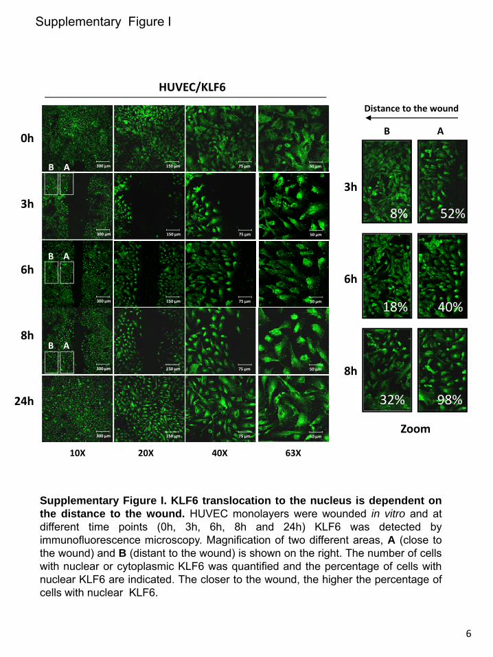
0h
3h
6h
8h
24h
Supplementary Figure I. KLF6 translocation to the nucleus is dependent onthe distance to the wound. HUVEC monolayers were wounded in vitro and atdifferent time points (0h, 3h, 6h, 8h and 24h) KLF6 was detected byimmunofluorescence microscopy. Magnification of two different areas, A (close tothe wound) and B (distant to the wound) is shown on the right. The number of cellswith nuclear or cytoplasmic KLF6 was quantified and the percentage of cells withnuclear KLF6 are indicated. The closer to the wound, the higher the percentage ofcells with nuclear KLF6.
300 μm
300 μm
300 μm
300 μm
300 μm
10X
Supplementary Figure I
B A
B A
HUVEC/KLF6
75 μm 50 μm150 μm
75 μm 50 μm150 μm
75 μm 50 μm150 μm
75 μm 50 μm150 μm
75 μm 50 μm150 μm
20X 40X 63X
B A
B A
3h
6h
8h
Distance to the wound
8% 52%
18% 40%
32% 98%
Zoom
6
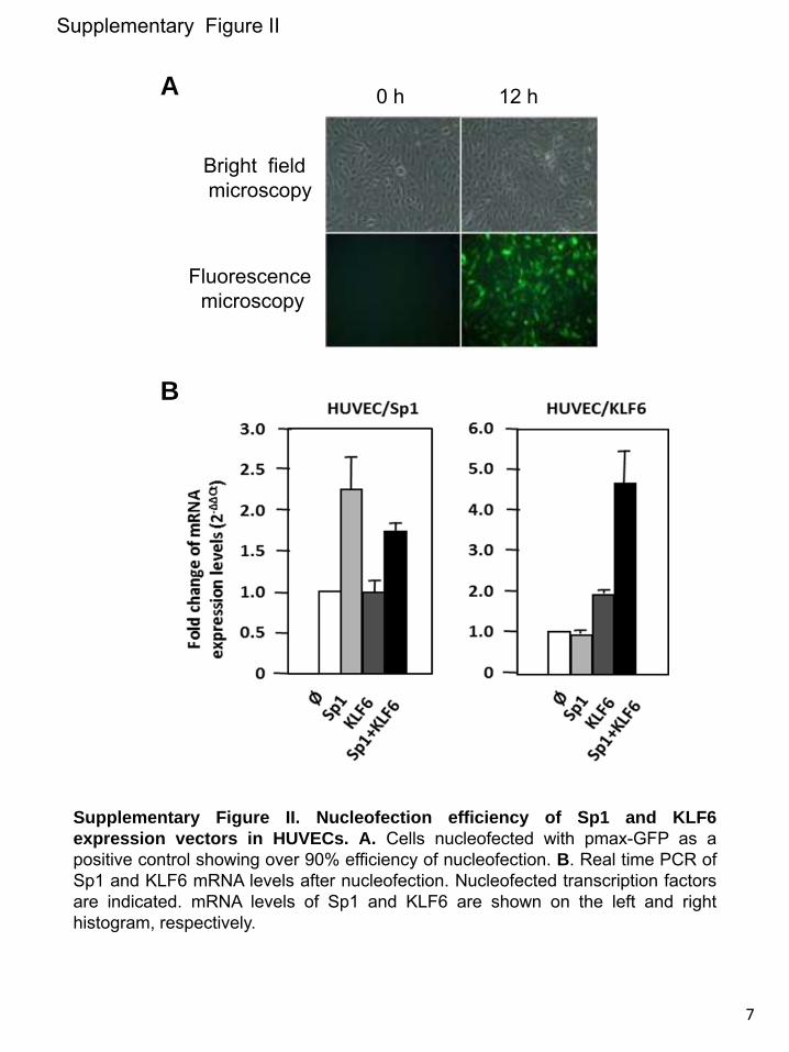
Supplementary Figure II. Nucleofection efficiency of Sp1 and KLF6expression vectors in HUVECs. A. Cells nucleofected with pmax-GFP as apositive control showing over 90% efficiency of nucleofection. B. Real time PCR ofSp1 and KLF6 mRNA levels after nucleofection. Nucleofected transcription factorsare indicated. mRNA levels of Sp1 and KLF6 are shown on the left and righthistogram, respectively.
Supplementary Figure II
0 h 12 h
Bright fieldmicroscopy
Fluorescencemicroscopy
A
B
7
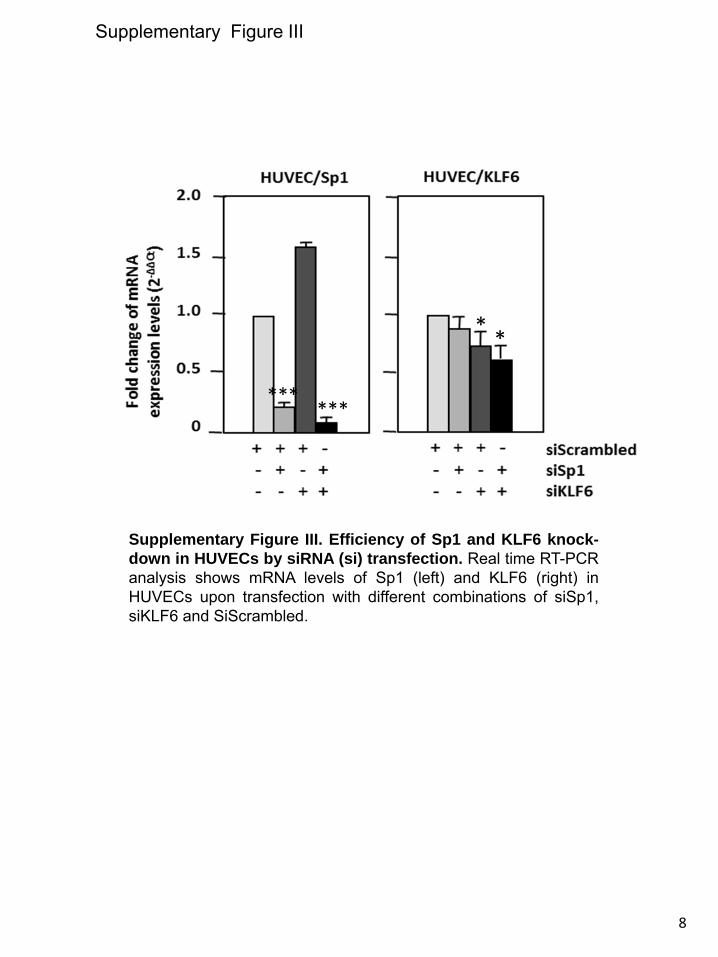
Supplementary Figure III. Efficiency of Sp1 and KLF6 knock-down in HUVECs by siRNA (si) transfection. Real time RT-PCRanalysis shows mRNA levels of Sp1 (left) and KLF6 (right) inHUVECs upon transfection with different combinations of siSp1,siKLF6 and SiScrambled.
Supplementary Figure III
* *
******
8
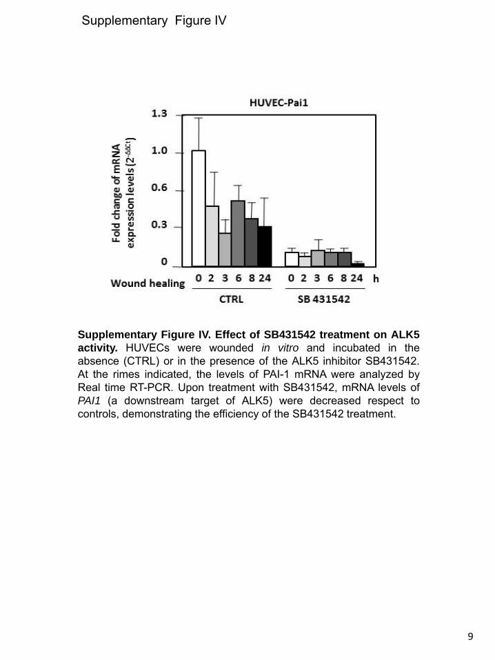
Supplementary Figure IV
Supplementary Figure IV. Effect of SB431542 treatment on ALK5activity. HUVECs were wounded in vitro and incubated in theabsence (CTRL) or in the presence of the ALK5 inhibitor SB431542.At the rimes indicated, the levels of PAI-1 mRNA were analyzed byReal time RT-PCR. Upon treatment with SB431542, mRNA levels ofPAI1 (a downstream target of ALK5) were decreased respect tocontrols, demonstrating the efficiency of the SB431542 treatment.
9
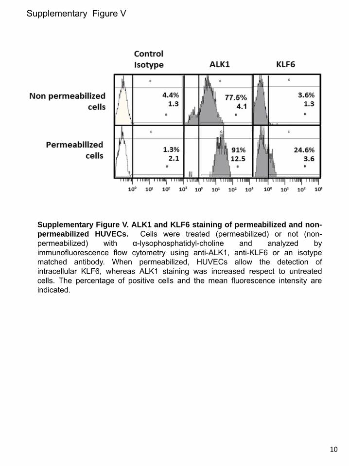
Supplementary Figure V
Supplementary Figure V. ALK1 and KLF6 staining of permeabilized and non-permeabilized HUVECs. Cells were treated (permeabilized) or not (non-permeabilized) with α-lysophosphatidyl-choline and analyzed byimmunofluorescence flow cytometry using anti-ALK1, anti-KLF6 or an isotypematched antibody. When permeabilized, HUVECs allow the detection ofintracellular KLF6, whereas ALK1 staining was increased respect to untreatedcells. The percentage of positive cells and the mean fluorescence intensity areindicated.
10
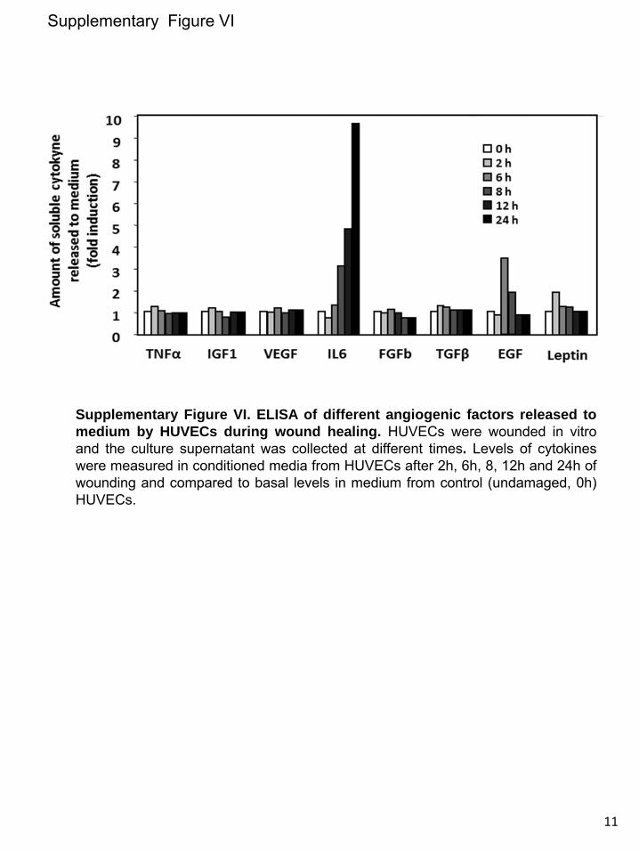
Supplementary Figure VI. ELISA of different angiogenic factors released tomedium by HUVECs during wound healing. HUVECs were wounded in vitroand the culture supernatant was collected at different times. Levels of cytokineswere measured in conditioned media from HUVECs after 2h, 6h, 8, 12h and 24h ofwounding and compared to basal levels in medium from control (undamaged, 0h)HUVECs.
Supplementary Figure VI
11
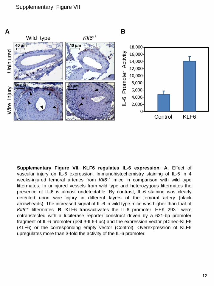
Supplementary Figure VII. KLF6 regulates IL-6 expression. A. Effect ofvascular injury on IL-6 expression. Immunohistochemistry staining of IL-6 in 4weeks-injured femoral arteries from Klf6+/- mice in comparison with wild typelittermates. In uninjured vessels from wild type and heterozygous littermates thepresence of IL-6 is almost undetectable. By contrast, IL-6 staining was clearlydetected upon wire injury in different layers of the femoral artery (blackarrowheads). The increased signal of IL-6 in wild type mice was higher than that ofKlf6+/- littermates. B. KLF6 transactivates the IL-6 promoter. HEK 293T werecotransfected with a luciferase reporter construct driven by a 621-bp promoterfragment of IL-6 promoter (pGL3-IL6-Luc) and the expression vector pCIneo-KLF6(KLF6) or the corresponding empty vector (Control). Overexpression of KLF6upregulates more than 3-fold the activity of the IL-6 promoter.
Supplementary Figure VII
12
18,000‐16,000‐14,000‐12,000–10,000–8,000–6,000–4,000–2,000–
0‐
IL-6
Pro
mot
erA
ctiv
ityControl KLF6
Wild type Klf6+/-
Uni
njur
edW
irein
jury
40 µm
40 µm40 µm
40 µm
A B
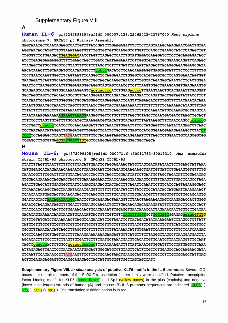
13
Supplementary Figure VIII A Human IL-6; gi|224589819|ref|NC_000007.13|:22765423-22767000 Homo sapiens chromosome 7, GRCh37.p5 Primary Assembly GAGTAAATGCCCAACAGAGGTCACTGTTTTATCGATCTTGAAGAGATCTCTTCTTAGCAAAGCAAAGAAACCGATTGTGAAGGTAACACCATGTTTGGTAAATAAGTGTTTTGGTGTTGTGCAAGGGTCTGGTTTCAGCCTGAAGCCATCTCAGAGCTGTCTGGGTCTCTGGAGACTGGAGGGACAACCTAGTCTAGAGCCCATTTGCATGAGACCAAGGATCCTCCTGCAAGAGACACCATCCTGAGGGAAGAGGGCTTCTGAACCAGCTTGACCCAATAAGAAATTCTTGGGTGCCGACGCGGAAGCAGATTCAGAGCCTAGAGCCGTGCCTGCGTCCGTAGTTTCCTTCTAGCTTCTTTTGATTTCAAATCAAGACTTACAGGGAGAGGGAGCGATAAACACAAACTCTGCAAGATGCCACAAGGTCCTcctttGACATCCCCAACAAAGAGGTGAGTAGTATTCTCCCCCTTTCTGCCCTGAACCAAGTGGGCTTCAGTAATTTCAGGGCTCCAGGAGACCTGGGGCCCATGCAGGTGCCCCAGTGAAACAGTGGTGAAGAGACTCAGTGGCAATGGGGAGAGCACTGGCAGCACAAGGCAAACCTCTGGCACAGAGAGCAAAGTCCTCACTGGGAGGATTCCCAAGGGGTCACTTGGGAGAGGGCAGGGCAGCAGCCAACCTCCTCTAAGTGGGCTGAAGCAGGTGAAGAAAGTGGCAGAAGCCACGCGGTGGCAAAAAGGAGTCacacactccACCTGGAGacgccTTGAAGTAACTGCACGAAATTTGAGGATGGCCAGGCAGTTCTACAACAGCCGCTCACAGGGAGAGCCAGAACACAGAAGAACTCAGATGACTGGTAGTATTACCTTCTTCATAATCCCAGGCTTGGGGGGCTGCGATGGAGTCAGAGGAAACTCAGTTCAGAACATCTTTGGTTTTTACAAATACAAATTAACTGGAACGCTAAATTCTAGCCTGTTAATCTGGTCACTGAAAAAAAATTTTTTTTTTTTCAAAAAACATAGCTTTAGCTTATTTTTTTTCTCTTTGTAAAACTTCGTGCATGACTTCAGCTTTACTCTTTGTCAAGACATGCCAAAGTGCTGAGTCACTAATAAAAGAAAAAAAGAAAGTAAAGGAAGAGTGGTTCTGCTTCTTAGCGCTAGCCTCAATGACGACCTAAGCTGCACTTTTCCCCCTAGTTGTGTCTTGCCATGCTAAAGGACGTCACATTGCACAATCTTAATAAGGTTTCCAATCAGCCccacccGCTCTGGCCccacccTCACCCTCCAACAAAGATTTATCAAATGTGGGATTTTCCCATGAGTCTCAATATTAGAGTCTCAACCCCCAATAAATATAGGACTGGAGATGTCTGAGGCTCATTCTGCCCTCGAGCCCACCGGGAACGAAAGAGAAGCTCTATCTCCCCTCCAGGAGCCCAGCTATGAACTCCTTCTCCACAAGTAAGTGCAGGAAATCCTTAGCCCTGGAACTGCCAGCGGCGGTCGAGCCCTGTGTGAGGGAGGGGTGTGTGGCCCAGGGAGGGCTGGCGGGCGGCCAGCA
B Mouse IL-6; gi|372099105|ref|NC_000071.6|:30011733-30013310 Mus musculus
strain C57BL/6J chromosome 5, GRCm38 C57BL/6J TTATTTTAGTGTAATTTTTTTCTTCACATTGAATTCTAGGAGAAACTATGCTAGTGATATATAATTCTTGAACTATTAAACATGGGAGCATAAGAAAACAAGAATCTTAAGGCAATCTGCAGAGTGAAGAAGCTGATTGTGATCCTGAGAGTGTGTTTTGTAAATGGTTTTGGATTTTATGTACAGAGCCTACTTTCAGCCTGGAATCATTCTGAATGCTAGCTAGATATCTGGAGACAGGTGGACAGAAAACCAGGAACTAGTCTGAAAAAGAAACTAACCAAAGGGAAGAAGTCTGTTTAAGTTTGACCCAGCCTAGAAGACTTGAGCATTGGAGGGGTTATTCAGAGTGAGACGTACCACCTTCAGATTCAAATCCTGTCATCCAGTAGAAGGGAGCTTCAAACACAAGCTAGCTAAGATACAATGAGGTCCTTCTTCGATATCTTTATCTTCCATATACCATGAATCAAAGAAACTTCAACAACATGAGGACTGCAACAGACCTTCAAGCCTCCTTGCATGACCTGGAAATGTTTTGGGGTGTCCTGGCAGCAGTGGGATCAGCACTAACAGATAAGGGCAACTCTCACAGAGACTAAAGGTCTTAACTAAGAAGATAGCCAAGAGACCACTGGGGAGAATGCAGAGAATAGGCTTGGACTTGGAAGCCAAGATTGCTTGACAACAGACAGAAGATATTTCTGTACTTCACCCACTTTACCCACCTGGCAACTCCTGGAAACAACTGCACAAAATTTGGAGGTGAACAAACCATTAGAAACAACTGGTCCTGACAAGACACAGGAAAAACAAGCAATATGCAACATTACTGTCTGTTGTCCAGGTTGGGTGCTGGGGGTGGGAGAGggagtGTGTGTCTTTGTATGATCTGAAAAAACTCAGGTCAGAACATCTGTAGATCCTTACAGACATACAAAAGAATCCTAGCCTCTTATTCATGTGTGTGTGTGTGTGTGTGTGTGTGTGTGTGTGTGTGTGTGTGTGTATGTGTGTGTCGTCTGTCATGCGCGCGTGCCTGCGTTTAAATAACATCAGCTTTAGCTTCTCTTTCTCCTTATAAAACATTGTGAATTTCAGTTTTCTTTCCCATCAAGACATGCTCAAGTGCTGAGTCACTTTTAAAGAAAAAAAAGAAGAGTGCTCATGCTTCTTAGGGCTAGCCTCAAGGATGACTTAAGCACACTTTCCCCTTCCTAGTTGTGATTCTTTCGATGCTAAACGACGTCACATTGTGCAATCTTAATAAGGTTTCCAATCAGCCccacccACTCTGGCCccacccccacccTCCAACAAAGATTTTTATCAAATGTGGGATTTTCCCATGAGTCTCAAAATTAGAGAGTTGACTCCTAATAAATATGAGACTGGGGATGTCTGTAGCTCATTCTGCTCTGGAGCCCACCAAGAACGATAGTCAATTCCAGAAACCGCTATGAAGTTCCTCTCTGCAAGTAAGTGAAGGCAGTTCCTTGCCCTCTGGCGGAGCTATTGAGACTGTGAGAGAGGAGTGTGAGGCAGAGAGCCAGCATTGTGGGTTGGCCAGCAGCCATC Supplementary Figure VIII. In silico analysis of putative KLF6 motifs in the IL-6 promoter. Several GC-boxes that recruit members of the Sp/KLF transcription factors family were identified. Putative transcription factor binding motifs for KLF6 (green boxes) and Sp1 (yellow boxes) in the plus (capitals) and negative (lower case letters) strands of human (A) and mouse (B) IL-6 promoter sequences are indicated. KLF6(+); klf6(-); SP1(+); sp1(-). The translation initiation codon is in red.