Embed Size (px)
Citation preview
Krüppel-like transcription factor 9 (KLF9) belongs to the Krüppel-like factors (KLFs) families, which consists of 17 evolutionarily conserved zinc finger containing proteins with diverse regulatory functions. KLF9 involved in the development of mouse, oxidative stress and regulating gene transcription in eukaryocyte [1-3]. KLF9 influences the adhesion, apoptosis, and proliferation [4-6]. And the expression is down regulated in many type of tumor such breast cancer, hepatocellular carcinoma and restore the expression would inhibit the migration and induce the apoptosis of cancer cell respectively. The results above indicated the anti-oncogenic function of KLF9. Previous, we found that KLF9 was associated with the prognosis of pancreatic cancer, but the underlying mechanism has not delineated in pancreatic cancer [7-9].
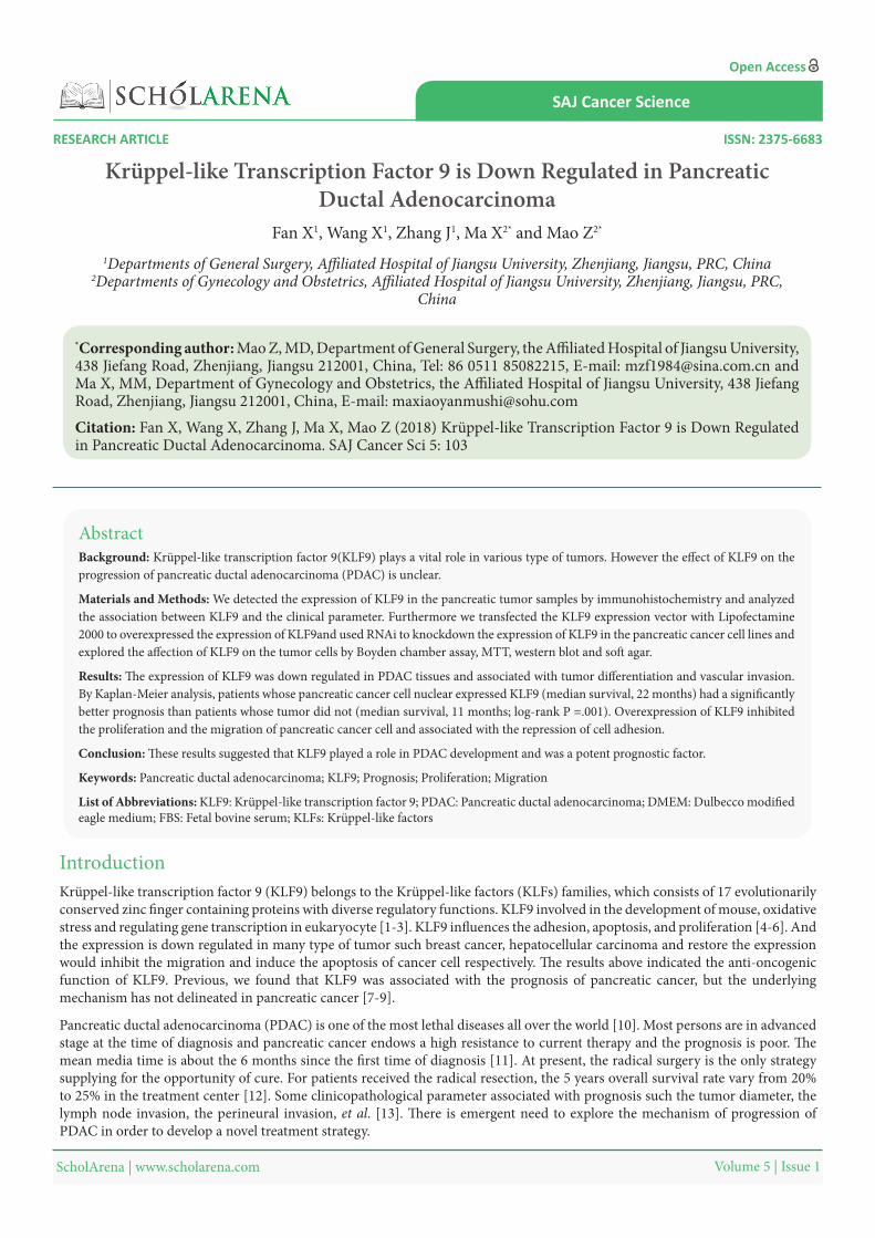
Krüppel-like Transcription Factor 9 is Down Regulated in Pancreatic Ductal Adenocarcinoma
Fan X1, Wang X1, Zhang J1, Ma X2* and Mao Z2*
1Departments of General Surgery, Affiliated Hospital of Jiangsu University, Zhenjiang, Jiangsu, PRC, China2Departments of Gynecology and Obstetrics, Affiliated Hospital of Jiangsu University, Zhenjiang, Jiangsu, PRC,
China
*Corresponding author: Mao Z, MD, Department of General Surgery, the Affiliated Hospital of Jiangsu University, 438 Jiefang Road, Zhenjiang, Jiangsu 212001, China, Tel: 86 0511 85082215, E-mail: [email protected] and Ma X, MM, Department of Gynecology and Obstetrics, the Affiliated Hospital of Jiangsu University, 438 Jiefang Road, Zhenjiang, Jiangsu 212001, China, E-mail: [email protected]
Citation: Fan X, Wang X, Zhang J, Ma X, Mao Z (2018) Krüppel-like Transcription Factor 9 is Down Regulated in Pancreatic Ductal Adenocarcinoma. SAJ Cancer Sci 5: 103
RESEARCH ARTICLE
Volume 5 | Issue 1 ScholArena | www.scholarena.com
SAJ Cancer Science
ISSN: 2375-6683
Open Access
Abstract
Keywords: Pancreatic ductal adenocarcinoma; KLF9; Prognosis; Proliferation; Migration
Background: Krüppel-like transcription factor 9(KLF9) plays a vital role in various type of tumors. However the effect of KLF9 on the progression of pancreatic ductal adenocarcinoma (PDAC) is unclear.
Materials and Methods: We detected the expression of KLF9 in the pancreatic tumor samples by immunohistochemistry and analyzed the association between KLF9 and the clinical parameter. Furthermore we transfected the KLF9 expression vector with Lipofectamine 2000 to overexpressed the expression of KLF9and used RNAi to knockdown the expression of KLF9 in the pancreatic cancer cell lines and explored the affection of KLF9 on the tumor cells by Boyden chamber assay, MTT, western blot and soft agar.
Results: The expression of KLF9 was down regulated in PDAC tissues and associated with tumor differentiation and vascular invasion. By Kaplan-Meier analysis, patients whose pancreatic cancer cell nuclear expressed KLF9 (median survival, 22 months) had a significantly better prognosis than patients whose tumor did not (median survival, 11 months; log-rank P =.001). Overexpression of KLF9 inhibited the proliferation and the migration of pancreatic cancer cell and associated with the repression of cell adhesion.
Conclusion: These results suggested that KLF9 played a role in PDAC development and was a potent prognostic factor.
Introduction
List of Abbreviations: KLF9: Krüppel-like transcription factor 9; PDAC: Pancreatic ductal adenocarcinoma; DMEM: Dulbecco modified eagle medium; FBS: Fetal bovine serum; KLFs: Krüppel-like factors
Pancreatic ductal adenocarcinoma (PDAC) is one of the most lethal diseases all over the world [10]. Most persons are in advanced stage at the time of diagnosis and pancreatic cancer endows a high resistance to current therapy and the prognosis is poor. The mean media time is about the 6 months since the first time of diagnosis [11]. At present, the radical surgery is the only strategy supplying for the opportunity of cure. For patients received the radical resection, the 5 years overall survival rate vary from 20% to 25% in the treatment center [12]. Some clinicopathological parameter associated with prognosis such the tumor diameter, the lymph node invasion, the perineural invasion, et al. [13]. There is emergent need to explore the mechanism of progression of PDAC in order to develop a novel treatment strategy.
SAJ Cancer Sci 2
Volume 5 | Issue 1 ScholArena | www.scholarena.com
During the development of cancer, numerous genetic and epigenetic mutations were considered as contribution. As the more understanding of the molecular and genetic change involved in the pancreatic cancer, we hope to identify the novel marker that could predict the prognosis and response to treatment better. Genetic alterations important for pancreatic cancer development have been considered as biomarker of prognosis such as the K-ras, SMAD4 [14]. But the former is activated in most of the pancreatic cancer which limited the usefulness. Also epigenetic changes have a relationship with the development of the pancreatic cancer, including the promoter methylation and silence of KLF9 gene [14].
Previous, we found that KLF was down regulated in the PDAC and considered as an independent factor associated with the prognosis [9]. The molecular mechanism is uncovered. The purpose of this study is to explore preliminarily the underlying mechanism of KLF9 in PDAC. In presents study, we found that the expression of KLF9 was down regulated in PDAC tissues and associated with tumor differentiation and vascular invasion and poor overall survival, which were coincided with previous results. Furthermore, restored expression of KLF9 in pancreatic cancer cells inhibited the proliferation and migration.
The cells used in this experiment were obtained from the American Type Culture Collection™ (Manassas, VA, USA) and cultured in Dulbecco’s modified Eagle’s medium (Invitrogen™, Carlsbad, CA,USA), supplemented with 10% fetal bovine serum (FBS;PAA Laboratories™, Pasching, Austria), 10U/ml penicillin, and 10U/ml streptomycin, at 37 °C in a humidified atmosphere containing 5% CO2.
Materials and Methods
One hundred and fifty six primary pancreatic cancer samples and their corresponding normal tissues, which were at least 3 cm away from the tumor, were obtained from pancreatic cancer patients treated at Affiliated Hospital of Jiangsu University (ZhenJiang, china) from 2005 to 2015. All the patients write the informed consent, and were naive to any treatment including neoadjuvant therapy, radiotherapy. All specimens were frozen in liquid nitrogen immediately after surgical excision and or directly stored at -80 °C until use. Our study was approved by the Institutional Review Board of Affiliated Hospital of Jiangsu University.
Formalin-fixed paraffin-embedded tissue sections (4μm thick) were stained using the immunoperoxidase method with avidin-biotin complex as described previously [15]. The monoclonal anti-KLF9 antibody was from Santa Cruz Biotechnology (Santa Cruz™, CA, USA). Sections were incubated with the anti-KLF9 monoclonal antibody at 1:500 dilutions overnight at 4 °C. The absence of nonspecific staining was confirmed by the control staining omitting the primary antibody. Labeling for marker was carried out using the Envision Plus Detection Kit (DAKO™, Carpinteria, CA) followed the manufacturer’s protocol. Nuclei were counterstained with hematoxylin. Immunohistochemical results were judged by three pathologists who were unaware of the clinical data. The staining results referred to KLF9 high or low expression were judged by German semiquantitative scoring system which considers both the stain intensity and the area extent as previous described [16].
Western blotting was done as described previously [17]. Briefly, Rabbit anti-human KLF9 polyclonal antibody and N-cadherin, E-cadherin were purchased from Santa Cruz Biotechnology (Santa Cruz, CA). Anti-GAPDH antibody was from Cell Signaling (Boston, MA, USA). Secondary antibodies, rabbit anti-mouse IgG (Sigma) and goat anti-rabbit IgG (Cell Signaling Technology) were used at a dilution of 1:1500. Primary antibodies were diluted in 0.1 mol/L Tris-HCl, 1.5 mol/L NaCl, 0.05% Tween-20 (v/v) (TBST) containing 1% BSA and NaN3 in appropriate dilution. The immunoreactive protein bands were visualized using an ECL kit (Pierce, Rockford, IL, USA).
In MTT assay, MIAPaCa2, MIAPaCa2/V, and MIAPaCa2/KLF9 cells were plated into 96-well plates at 2×10³ cells per well, cultured in 1% FBS/DMEM for various durations, and cell numbers were measured by MTT assay according to the protocol provided by MTT manufacturer (Roche Applied Science). The cells were dissolve by DMSO and detected at OD540 nm, each clone was plated in triplicate for each experiment, and each experiment was repeated at least three times.
Cell Culture and Tissue samples
Immunohistochemistry
Western blot analysis
MTT assay
The MIAPaCa2, MIAPaCa2/V, and MIAPaCa2/KLF9 cells were plated onto six-well plate at 1×10⁴ cell. After 7 days, colonies were stained with Comassie blue and the ones greater than 50 cells were auto-counted by the photographed machine. All experiments were repeated three times.
Adhesion-dependent colony formation test
The migration assay was carried out sing a 12-well Boyden Chamber (Neuro Probe) with an 8μm pore size. Approximately 1×10⁵
cells were seeded into upper wells of the Boyden Chamber and incubated for 6 h at 37 °C in medium containing 1% FBS. Medium with 10% FBS was used as a chemoattractant in the bottom wells. Cells that did not migrate through the pores of the Boyden Chamber were manually removed with a rubber swab. Cells that migrated to the lower side of the membrane were stained with hematoxylin and eosin and random photographed five filed using an inverted microscope and counted the cell numbers. Each clone was plated in triplicate for each experiment, and each experiment was repeated at least three times.
In vitro migration assays
SAJ Cancer Sci 3
Volume 5 | Issue 1 ScholArena | www.scholarena.com
Statistical analyses were performed with the SPSS software (Chicago, IL, USA, version 16.0 for Window). Continuous variables were expressed as mean ± SD or median ± range; categorical variables were expressed as percentages. Chi-square test and Fisher exact test were used to examine the correlation between the KLF9 expression and clinicopathological characteristics. Kaplan-Meier estimates and log-rank tests were used for overall survival (OS) analysis. For all analyses, P < 0.05 was considered statistically significant. All the figures were created by Photoshop.
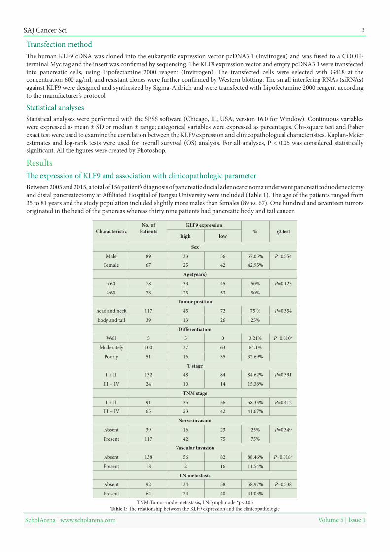
Between 2005 and 2015, a total of 156 patient’s diagnosis of pancreatic ductal adenocarcinoma underwent pancreaticoduodenectomy and distal pancreatectomy at Affiliated Hospital of Jiangsu University were included (Table 1). The age of the patients ranged from 35 to 81 years and the study population included slightly more males than females (89 vs. 67). One hundred and seventeen tumors originated in the head of the pancreas whereas thirty nine patients had pancreatic body and tail cancer.
The human KLF9 cDNA was cloned into the eukaryotic expression vector pcDNA3.1 (Invitrogen) and was fused to a COOH-terminal Myc tag and the insert was confirmed by sequencing. The KLF9 expression vector and empty pcDNA3.1 were transfected into pancreatic cells, using Lipofectamine 2000 reagent (Invitrogen). The transfected cells were selected with G418 at the concentration 600 μg/ml, and resistant clones were further confirmed by Western blotting. The small interfering RNAs (siRNAs) against KLF9 were designed and synthesized by Sigma-Aldrich and were transfected with Lipofectamine 2000 reagent according to the manufacturer’s protocol.
Transfection method
Statistical analyses
ResultsThe expression of KLF9 and association with clinicopathologic parameter
χ2 test%KLF9 expressionNo. of
PatientsCharacteristiclowhigh
Sex
P=0.55457.05%563389Male
42.95%422567Female
Age(years)
P=0.12350%453378<60
50%532578≥60
Tumor position
P=0.35475 %7245117head and neck
25%261339body and tail
Differentiation
P=0.010*3.21%055Well
64.1%6337100Moderately
32.69%351651Poorly
T stage
P=0.39184.62%8448132I + II
15.38%141024III + IV
TNM stage
P=0.41258.33%563591I + II
41.67%422365III + IV
Nerve invasion
P=0.34925%231639Absent
75%7542117Present
Vascular invasion
P=0.018*88.46%8256138Absent
11.54%16218Present
LN metastasis
P=0.53858.97%583492Absent
41.03%402464Present
TNM:Tumor-node-metastasis, LN:lymph node.*p<0.05Table 1: The relationship between the KLF9 expression and the clinicopathologic
SAJ Cancer Sci 4
Volume 5 | Issue 1 ScholArena | www.scholarena.com
Table 1 summarizes the relationship between the KLF9 expression and clinicopathological characteristics. There was no significant difference between KLF9 -high and -low expression regarding gender, age, tumor site, TNM stage, perineural invasion, lymph node metastasis. However, significant statistical difference existed with respect to differentiation (P = 0.010) and vascular invasion (P = 0.018).
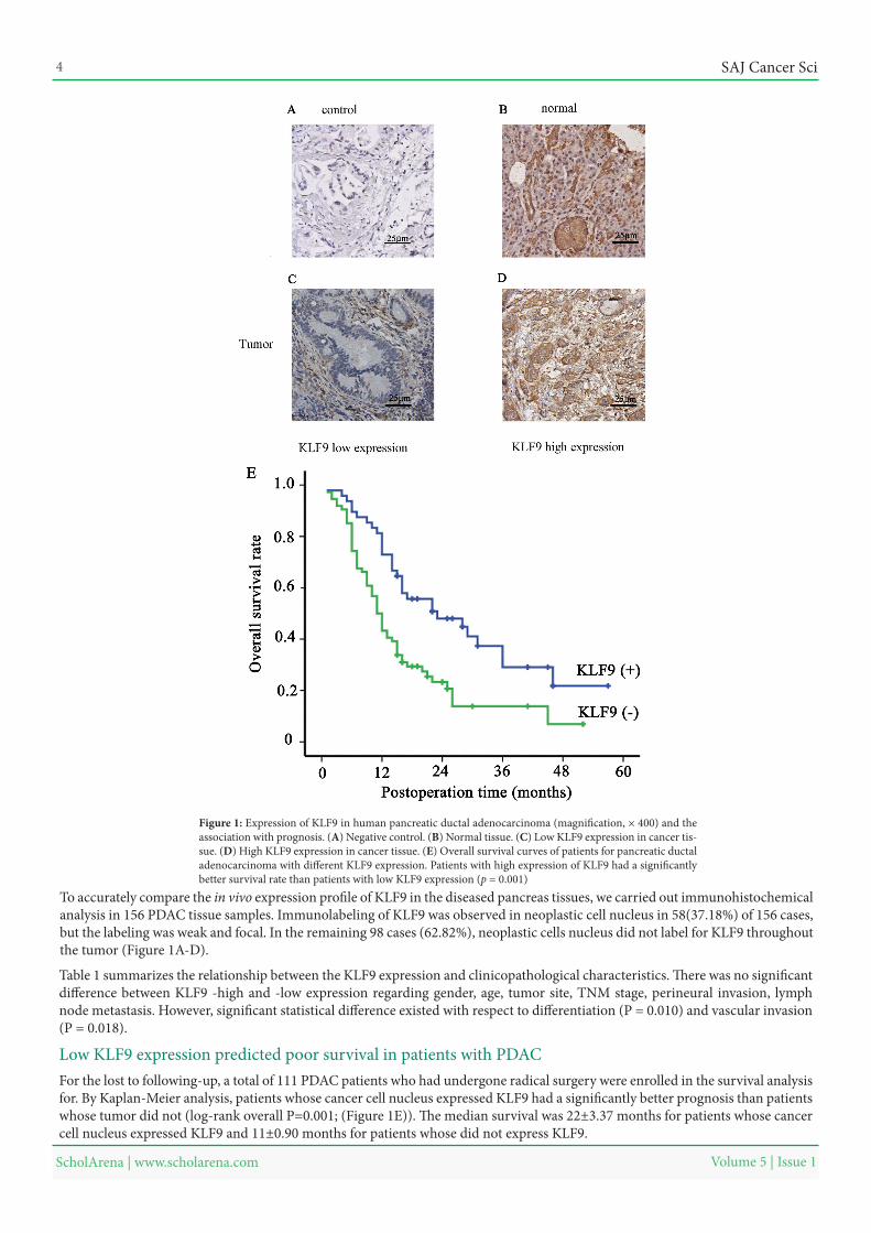
Low KLF9 expression predicted poor survival in patients with PDACFor the lost to following-up, a total of 111 PDAC patients who had undergone radical surgery were enrolled in the survival analysis for. By Kaplan-Meier analysis, patients whose cancer cell nucleus expressed KLF9 had a significantly better prognosis than patients whose tumor did not (log-rank overall P=0.001; (Figure 1E)). The median survival was 22±3.37 months for patients whose cancer cell nucleus expressed KLF9 and 11±0.90 months for patients whose did not express KLF9.
Figure 1: Expression of KLF9 in human pancreatic ductal adenocarcinoma (magnification, × 400) and the association with prognosis. (A) Negative control. (B) Normal tissue. (C) Low KLF9 expression in cancer tis-sue. (D) High KLF9 expression in cancer tissue. (E) Overall survival curves of patients for pancreatic ductal adenocarcinoma with different KLF9 expression. Patients with high expression of KLF9 had a significantly better survival rate than patients with low KLF9 expression (p = 0.001)
To accurately compare the in vivo expression profile of KLF9 in the diseased pancreas tissues, we carried out immunohistochemical analysis in 156 PDAC tissue samples. Immunolabeling of KLF9 was observed in neoplastic cell nucleus in 58(37.18%) of 156 cases, but the labeling was weak and focal. In the remaining 98 cases (62.82%), neoplastic cells nucleus did not label for KLF9 throughout the tumor (Figure 1A-D).
SAJ Cancer Sci 5
Volume 5 | Issue 1 ScholArena | www.scholarena.com
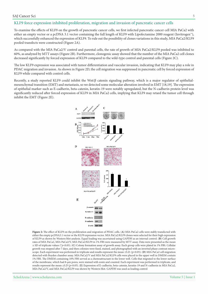
To examine the effects of KLF9 on the growth of pancreatic cancer cells, we first infected pancreatic cancer cell MIA PaCa2 with either an empty vector or a pcDNA 3.1 vector containing the full length of KLF9 with Lipofectamine 2000 reagent (Invitrogen™), which successfully enhanced the expression of KLF9. To rule out the possibility of clones variations in this study, MIA PaCa2/KLF9 pooled transfects were constructed (Figure 2A).
As compared with the MIA PaCa2/V control and parental cells, the rate of growth of MIA PaCa2/KLF9 pooled was inhibited to 60%, as analyzed by MTT assays (Figure 2B). Furthermore, clonogenic assay showed that the number of the MIA PaCa2 cell clones decreased significantly by forced expression of KLF9 compared to the wild-type control and parental cells (Figure 2C).
The low KLF9 expression was associated with tumor differentiation and vascular invasion, indicating that KLF9 may play a role in PDAC migration and invasion. As shown in Figure 2D, the cell migration was suppressed in pancreatic cell by forced expression of KLF9 while compared with control cells.
Recently, a study reported KLF9 could inhibit the Wnt/β catenin signaling pathway, which is a major regulator of epithelial-mesenchymal transition (EMT) and metastasis, so we detected some molecular alteration involved in EMT [18,19]. The expression of epithelial marker such as E-cadherin, beta-catenin, keratin-19 were notably upregulated, but the N-cadherin protein level was significantly reduced after forced expression of KLF9 in MIA PaCa2 cells, implying that KLF9 may retard the tumor cell through inhibit the EMT (Figure 2E).
Figure 2: The effect of KLF9 on the proliferation and migration of PDAC cells. (A) MIA PaCa2 cells were stably transfected with either the empty pcDNA3.1 vector or the KLF9 expression vector. MIA PaCa2/KLF9 clones were selected for their high expression of KLF9 as shown by Western blot analysis. Equal loading was ascertained using GAPDH as an internal control. (B) Cell growth rates of MIA PaCa2, MIA PaCa2/V, MIA PaCa2/KLF9 in 1% FBS were measured by MTT assay. Data were presented as the mean ± SD of triplicate values (*p<0.05). (C) Colony formation assay of growth assay. Each group cells were plated in 1% FBS. Cellular growth was stopped after 7 days, and then colonies were fixed, stained, and photographed with an inverted phase contrast micro-scope. Each experiment was performed in triplicate and results represent the mean ±S.D. (p<0.05). (D) MIA PaCa2 cell migration detected with Boyden chamber assay. MIA PaCa2/V and MIA PaCa2/KLF9 cells were placed in the upper well in DMEM contain 1% FBS. The DMEM containing 10% FBS served as a chemoattractant in the lower well. Cells that migrated to the lower surface of the membrane, which had 8-μm pores, were stained with eosin and counted. Each experiment was performed in triplicate, and results represent the mean ±S.D (p<0.05). (E) Expression of E-cadherin, beta-catenin, keratin-19 and N-cadherin in MIA PaCa2, MIA PaCa2/V, and MIA PaCa2/KLF9 was shown by Western blot. GAPDH was used as loading control
KLF9 force expression inhibited proliferation, migration and invasion of pancreatic cancer cells
SAJ Cancer Sci 6
Volume 5 | Issue 1 ScholArena | www.scholarena.com
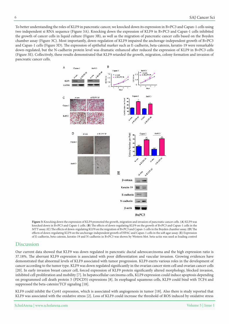
To better understanding the roles of KLF9 in pancreatic cancer, we knocked down its expression in B×PC3 and Capan-1 cells using two independent si RNA sequence (Figure 3A). Knocking down the expression of KLF9 in B×PC3 and Capan-1 cells inhibited the growth of cancer cells in liquid culture (Figure 3B), as well as the migration of pancreatic cancer cells based on the Boyden chamber assay (Figure 3C). Most importantly, down-regulation of KLF9 impaired the anchorage-independent growth of B×PC3 and Capan-1 cells (Figure 3D). The expression of epithelial marker such as E-cadherin, beta-catenin, keratin-19 were remarkable down-regulated, but the N-cadherin protein level was dramatic enhanced after reduced the expression of KLF9 in B×PC3 cells (Figure 3E). Collectively, these results demonstrated that KLF9 retarded the growth, migration, colony formation and invasion of pancreatic cancer cells.
Figure 3: Knocking down the expression of KLF9 promoted the growth, migration and invasion of pancreatic cancer cells. (A) KLF9 was knocked down in B×PC3 and Capan-1 cells. (B) The effects of down-regulating KLF9 on the growth of B×PC3 and Capan-1 cells in the MTT assay. (C) The effects of down-regulating KLF9 on the migration of B×PC3 and Capan-1 cells in the Boyden chamber assay. (D) The effects of down-regulating KLF9 on the anchorage-independent growth of HPAC and Capan-1 cells in the soft agar assay. (E) Expression of E-cadherin, beta-catenin, keratin-19 and N-cadherin in B×PC3 was shown by Western blot. beta-actin was used as loading control
Discussion Our current data showed that KLF9 was down regulated in pancreatic ductal adenocarcinoma and the high expression ratio is 37.18%. The aberrant KLF9 expression is associated with poor differentiation and vascular invasion. Growing evidences have demonstrated that abnormal levels of KLF9 associated with tumor progression. KLF9 exerts various roles in the development of cancer according to the tumor type. KLF9 was down regulated significantly in the ovarian cancer stem cell and ovarian cancer cells [20]. In early invasion breast cancer cell, forced expression of KLF9 protein significantly altered morphology, blocked invasion, inhibited cell proliferation and mobility [7]. In hepatocellular carcinoma cells, KLF9 expression could induce apoptosis depending on programmed cell death protein 5 (PDCD5) expressions [8]. In esophageal squamous cells, KLF9 could bind with TCF4 and suppressed the beta-catenin/TCF signaling [18].
KLF9 could inhibit the Cyr61 expression, which is associated with angiogenesis in tumor [18]. Also there is study reported that KLF9 was associated with the oxidative stress [2]. Loss of KLF9 could increase the threshold of ROS induced by oxidative stress
SAJ Cancer Sci 7
Volume 5 | Issue 1 ScholArena | www.scholarena.com
necessary for induction of cell death and promote the tumor cells survival. Under hypoxia circumstance, loss of KLF9 expression may be in favor of angiogenesis formation induced by tumor cells.
The most important conclusion in these findings is that after analyzing the potential relationship between the KLF9 expression and survival time, we observed that patients with low KLF9 expression had a poorer overall survival, which agreed with the results of many other studies that aberrant KLF9 expression indicated a poor outcome in the cancers [7,21]. The results were coinciding with our previous study [9]. It should be mentioned here that the samples involved in this study were not big enough and there maybe underlying difference between the pancreatic proximal and distal tumor. Future study enrolled larger sample size and more diversified tumor stages are needed in order to independently verify our current findings.
The KLF9 expression was significantly correlated with the tumor differentiation and vascular invasion, implying that KLF9 may play a role in pancreatic ductal adenocarcinoma cell proliferation, migration. To verify this speculation, we forced expression KLF9 in PDAC cells, and found that overexpression of KLF9 significantly suppressed pancreatic cancer cell proliferation, migration. Meanwhile, EMT markers such as N-cadherin expression was down regulated and E-cadherin expression was upregulated, indicating an inhibition of EMT. Former studies improved that KLF9 could retard ESCC tumor growth and migration through competitive binding with TCF disturbing the β-catenin/TCF complex inhibiting the Wnt signaling pathway [18]. The Wnt signaling pathway has versatile function on cell physiologic process and plays an important role in the EMT. The underlying mechanism of KLF9 effect on the EMT markers expression in pancreatic cancer need further explore.
Our study confirmed the relationship between KLF9 expression and the prognosis of pancreatic ductal adenocarcinoma and decipher elementary the potential mechanism of KLF9 on the development of PDAC. The multiple effects of KLF9 signaling upon different cell types present within the tumor microenvironment indicates that targeting of KLF9 signaling might have important implications to defer tumor progression.
ConclusionIn the present study suggested that KLF9 expression was related to tumor differentiation and vascular invasion. Meanwhile, aberrant expression of KLF9 was correlated with unfavorable clinical prognostic factors and decreased survival for human pancreatic ductal adenocarcinoma. Considering the contributions of KLF9 to tumor progression, KLF9 may be a potential target for pancreatic cancer therapy.
AcknowledgementThis work was sponsored by National Natural Science foundation of China, Grant numbers: 81502372; Natural Science Foundation of Jiangsu Province (Youth Fund), Grant numbers: BK20130475; Jiangsu Provincial Medical Youth Talent (QNRC2016838, QNRC2016839); Zhenjiang Science and Technology Pillar Program, Grant numbers: SH2014035; The Foundation for Young Scientists of affiliated Hospital of Jiangsu University (Grant numbers: JDFYRC2013009, JDFYRC2016002).
References1. Martin KM, Metcalfe JC, Kemp PR (2001) Expression of Klf9 and Klf13 in mouse development. Mech Dev 103: 149-51.2. Zucker SN, Fink EE, Bagati A, Mannava S, Bianchi-Smiraglia A, Bogner PN, et al. (2014) Nrf2 amplifies oxidative stress via induction of Klf9. Mol Cell 53: 916-28.3. Kang L, Lai MD (2007) [BTEB/KLF9 and its transcriptional regulation]. Yi Chuan 29: 515-22.4. Huang S, Wang C, Yi Y, Sun X, Luo M, et al. (2015) Kruppel-like factor 9 inhibits glioma cell proliferation and tumorigenicity via downregulation of miR-21. Cancer Lett 356: 547-55.5. Brown AR, Simmen RC, Raj VR, Van TT, MacLeod SL, et al. (2015) Kruppel-like factor 9 (KLF9) prevents colorectal cancer through inhibition of interferon-re-lated signaling. Carcinogenesis 36: 946-55.6. Simmen FA, Su Y, Xiao R, Zeng Z, Simmen RC (2008) The Kruppel-like factor 9 (KLF9) network in HEC-1-A endometrial carcinoma cells suggests the carcino-genic potential of dys-regulated KLF9 expression. Reprod Biol Endocrinol 6: 41.7. Limame R, de Beeck KO, Van Laere S, Croes L, De Wilde A, et al. (2014) Expression profiling of migrated and invaded breast cancer cells predicts early metastatic relapse and reveals Kruppel-like factor 9 as a potential suppressor of invasive growth in breast cancer. Oncoscience 1: 69-81.8. Fu DZ, Cheng Y, He H, Liu HY, Liu YF (2014) The fate of Kruppel-like factor 9-positive hepatic carcinoma cells may be determined by the programmed cell death protein 5. Int J Oncol 44: 153-60.9. Mao Z, Fan X, Zhang J, Wang X, Ma X, et al. (2017) KLF9 Is a Prognostic Indicator in Human Pancreatic Ductal Adenocarcinoma. Anticancer Res 37: 3795-9.10. Siegel RL, Miller KD, Jemal A (2016) Cancer statistics, 2016. CA Cancer J Clin 66: 7-30.11. Barton MK (2016) Germline mutations in pancreatic cancer become better defined. CA Cancer J Clin 66: 93-4.12. Wolfgang CL, Herman JM, Laheru DA, Klein AP, Erdek MA, et al. (2013) Recent progress in pancreatic cancer. CA Cancer J Clin 63: 318-48.13. Winter JM, Tang LH, Klimstra DS, Brennan MF, O’Reilly EM, et al. (2011) Evaluation of putative prognostic tissue biomarkers in pancreatic ductal adenocar-cinoma. J Clin Oncol 29: 4020.14. Tatarian T, Winter JM (2016) Genetics of Pancreatic Cancer and Its Implications on Therapy. Surg Clin North Am 96: 1207-21.15. Sun ZJ, Wang Y, Cai Z, Chen PP, Tong XJ, et al. (2008) Involvement of Cyr61 in growth, migration, and metastasis of prostate cancer cells. Br J Cancer 99: 1656-67.
SAJ Cancer Sci 8
Volume 5 | Issue 1 ScholArena | www.scholarena.com
16. Deng YZ, Yao F, Li JJ, Mao ZF, Hu PT, et al. (2012) RACK1 suppresses gastric tumorigenesis by stabilizing the beta-catenin destruction complex. Gastroenter-ology 142: 812-23.e815.17. Deng YZ, Chen PP, Wang Y, Yin D, Koeffler HP, et al. (2007) Connective tissue growth factor is overexpressed in esophageal squamous cell carcinoma and promotes tumorigenicity through beta-catenin-T-cell factor/Lef signaling. J Biol Chem 282: 36571-81.18. Qiao F, Yao F, Chen L, Lu C, Ni Y,et al. (2016) Kruppel-like factor 9 was down-regulated in esophageal squamous cell carcinoma and negatively regulated be-ta-catenin/TCF signaling. Mol Carcinog 55: 280-91.19. Liu X, Li Z, Song Y, Wang R, Han L, et al. (2016) AURKA induces EMT by regulating histone modification through Wnt/beta-catenin and PI3K/Akt signaling pathway in gastric cancer. Oncotarget 7: 33152-64.20. Huang Y, Ju B, Tian J, Liu F, Yu H, et al. (2014) Ovarian cancer stem cell-specific gene expression profiling and targeted drug prescreening. Oncol Rep 31: 1235-48.21. Rackow BW, Taylor HS (2010) Submucosal uterine leiomyomas have a global effect on molecular determinants of endometrial receptivity. Fertil Steril 93: 2027-34.