Embed Size (px)
Citation preview


2
Valvular Heart Disease
M Saugi Abduh

Valvular Heart Disease
Heart contains
– Two atrioventricular valves Mitral
Tricuspid
– Two semilunar valves Aortic
Pulmonic
Valvular Disease

4
Normal StructureMitral Valve
Cross sectional Area 4-6cm²
Anterior and Posterior Leaflets
Chordae Tendineae Papillary Muscles

5
Mitral StenosisEtiology & Pathology
Rheumatic Fever- 99%
Other
– Congenital
– Carcinoid
– Lupus
– Amyloid
– Infective Endocarditis
– Mucopolysaccharide Disease
6
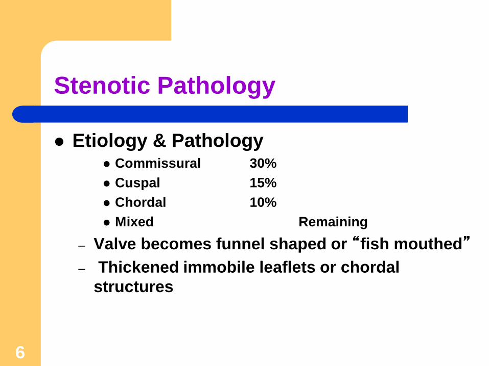
Stenotic Pathology
Etiology & Pathology Commissural 30%
Cuspal 15%
Chordal 10%
Mixed Remaining
– Valve becomes funnel shaped or “fish mouthed”
– Thickened immobile leaflets or chordal
structures
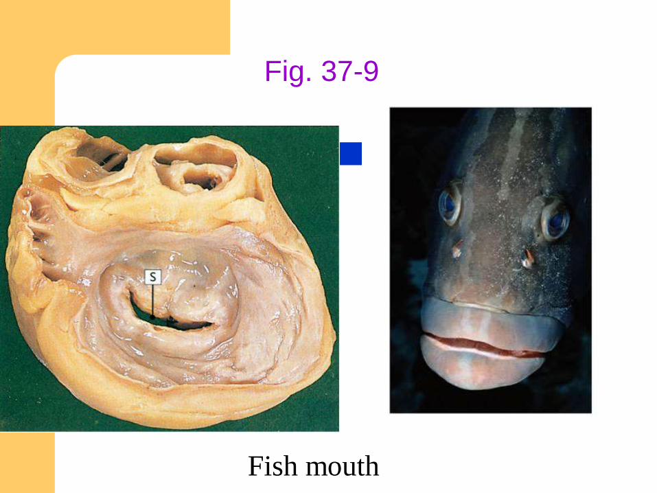
Fig. 37-9
Fish mouth

8
Pathophysiology
Mild MS- orifice <2 cm²
Critical MS- <1 cm²
– A-V pressure gradient >20mmHg
– Increased LA Pressure
– Increase Pulmonary Venous + Capillary Pressures
– Increase Pulmonary Artery Systolic Pressure
– Decrease RV Function (when PAS>30-60mmHg)


10
History
Exertional Dyspnea
Cough/Wheezing
Orthopnea/PND/CHF
Hemoptysis-Rupture of Pulmonary Vein-
Bronchial Vein Shunts
11
History
Chest Pain-Increase RV Pressures or
Unknown Etiology
Systemic Emboli (LA clots)
– Increased LA size, Decreased C.O., Atrial Fib

12
Physical Exam
Auscultation Diastolic Rumble
Assoc Murmur of MR
Loud S1-thickened leaflets
Increased P2-pulmonary hypertension
Decreased B/P if C.O. decreased
Prominent a wave if sinus rhythm present

13
Physical Exam
Mitral Facies-pink, purple facial patches
due to decrease CO and systemic
vasoconstriction
Hepatomegally
Edema
Ascites
Hydrothorax With Right Heart Failure

14
Diagnosis
ECG
– Left Atrial Abnormality
P wave becomes bifid and greater than 0.12 sec in
duration in V1 and Lead II
– RVH- right axis deviation
– R wave > S wave in V1

15
Diagnosis
Chest X-ray
– Dilated LA, RA, RV
– Elevated Left Main stem Bronchus
– Interstitial Edema
Echo- Cornerstone of Diagnosis
– Thickened Calcified Leaflets
– Doming of Leaflets on Opening

16
Natural History
Asymptomatic for 15-20yrs following
Rheumatic Fever
Additional 5-10 yrs for progression from mild to
severe stenosis
Stenosis progression approximately .09 cm²/yr

17
Natural History
Presurgical Survival Rates
– NYHA Class II 80%-10yrs
– Class III 38%-10yrs, 62% 5yrs
– Class IV 15%-5yrs
19
Percutaneous Balloon Angioplasty
Moderate-Severe MS
Mild MS- if Pulmonary Artery Pressures or
Wedge Pressure Elevate with Exercise

20
Valve Replacement
Indications– Combined MS/MR
– <1.5 cm²-NYHA III or IV
– <1 cm²
– Class II if Pulmonary Artery Pressure >70mmHg
Mortality– 3-8%
Valve Type-Prosthetic or Bioprosthetic

21
Mitral Regurgitation
Etiology
– Rheumatic Heart Disease
– Infective Endocarditis
– Collagen Vascular Disease
– Cardiomyopathy
– Ischemic Heart Disease
– Mitral Valve Prolapse-most common cause for valve
surgery in US

22
Pathophysiology
LV Compensation
– Increased End Diastolic Volume
– Increased Wall Tension
– Increased Preload
– Increased LV Emptying
– Normal Ejection Fraction should be Super Normal
>65% to maintain forward cardiac output and B/P
23
Pathophysiology
LV Decompensation
– Increase End Systolic Volume
– Increased End Diastolic Volume
– Leads to Annulus Dilatation (MR begets MR)
– Decreased Ejection Fraction and Stroke Volume

24
Pathophysiology
Ejection Fraction in Mitral Regurgitation
– >65% normal in compensated MR
– 50-65% mild impairment
– 40-50% moderate-severe impairment
– <35% advanced impairment
As ejection fraction decreases operative risk
increases.
25
History
Shortness of Breath
Exertional Dyspnea
Congestive Heart Failure
Right Heart Failure
Significant symptoms in chronic MR usually do
not develop until LV decompensation occurs.

26
History
Medical Treatment Survival
– 80% 5yr
– 60% 10yr
– 30-45% 5yr if MR severe
27
Diagnosis
Physical Exam
– Holosystolic Murmur
– Increase Carotid Impulse
ECG
– LA abnormality
– LVH
– RVH
Chest X-ray
– Increase LA, LV, RV, Interstitial Edema

28
Diagnosis
Echo
– Transesophageal superior to transthoracic
– Evaluation of Chamber Sizes, Regurgitant Jet,
Leaflets

29
Management of Acute MR
Medical
– After load Reduction (Nitropresside & Intra
aortic balloon pump)
Decrease impedance to LV ejection
Decrease regurgitant volume into left atrium
– Inotropic Support (Dobutamine)-if LV function
reduced

30
Management of Acute MR
Surgical Intervention
– Progressive LV Failure or Hemodynamic
Deterioration
– CHF
– Hypertension
– Valve Disruption
31
Management of Chronic MR
Medical
– Digoxin
– Diuretics*
– After load Reduction
– Anticoagulation in A-fib
– Endocarditis Prophylaxis

32
Management of Chronic MR
Surgical
– Indications
Asymptomatic Class I
– EF < 60% or LV Systolic Diameter >45mm
Severe MR Class II, III, or IV
– generally considered for surgery unless EF <30%
– Valve Repair vs. Replacement

33
Aortic ValveNormal Structure
Valve sits at the base of Aortic Root
Three Leaflets (cusps)-non coronary, right
coronary, left coronary
Normal cross-sectional area 3-4cm²
34
Aortic Stenosis Etiology and Pathology
Valvular
Supravalvular
Subvalvular
Hyperthrophic Cardiomyopathy

35
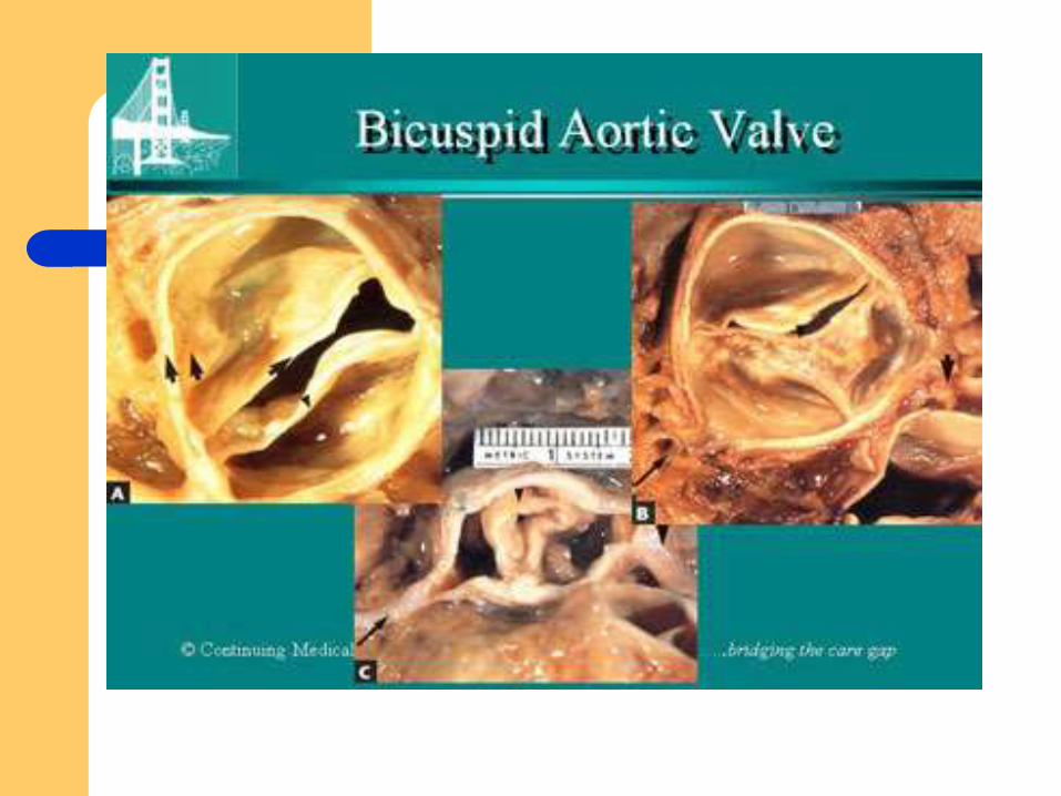
Congenital Aortic Stenosis
Unicuspid– Presents less than one year of age
Bicuspid– Adult Presentation
– Chronic turbulent flow
– Leads to fibrosis, rigidity, calcification
Tricuspid– Leaflets of unequal size
36
Acquired Aortic Stenosis
Rheumatic– Rare
– Usually mitral valve also involved
Degenerative or Senile– Most common cause of adult AS
– Most common cause of valve replacement
– Inflammatory or Infectious component??
– >age 65 30% Aortic Sclerosis

37

38
Hemodynamics
Critical (Surgical) AS
– Peak systolic pressure gradient > 50mmHg in the
presence of normal cardiac output
– Valve area <0.7-0.8cm²
Moderate AS
– 1-1.5cm²
Mild AS
– 1.5-2cm²
Aortic Sclerosis
39
History
Long latent period of increasing obstruction
Symptoms usually begin in 5th or 6th decade
Angina in 2/3 of patients– Hypertrophied myocardium
– Increased ventricular systolic pressure
– All of which increase myocardial oxygen consumption
– Oxygen supply-demand imbalance leads to subendocardial ischemia

42
Diagnosis
Physical Examination
– Systolic Murmur
Diamond-Shaped, harsh, left sternal boarder to right
intercostal spaces, neck and apex
Late peak, obliteration of S2, Dx of Critical AS
– Pulses Parvus
Delayed and Prolonged Carotid Impulse

43
Diagnosis
ECG
– Classic LVH
Chest X-ray
– Concentric LVH
– Calcification of Aortic Valve
Echo
– calculation of LV-Aortic pressure gradient and
valve area

44
Diagnosis
Cardiac Catherization

45
Medical Management
Endocarditis Prophylaxis
Limit Physical Activity
Watch Beta Blockers and Diuretics
*Treatment of Critical AS in viable
candidates is surgery

46
Surgery (Valve Replacement)
Indications
– Symptomatic Patients -valve area 0.7-0.8cm² or
less
– Asymptomatic Patients-progressive LV
dysfunction (EF <35%) or hypotensive response
to mild exercise
Delaying surgery in asymptomatic patients with good
exercise tolerance is controversial

47
Aortic RegurgitationEtiology and Pathology
Valvular– Rheumatic-Fibrotic Retraction of Leaflets
Ankylosing Spondylitis, Behcets, Psoriatic Arthritis, Giant Cell Arteritis
– Degenerative AS-75% w/AR
– Infective Endocarditis-Leaflet Destruction
– Trauma-ascending aortic tear
– Bicuspid aortic valve-prolapse or incomplete closure
– Myxomatous Degeneration-like MVP
– Appetite suppressant drugs-serotonin related valve deposits

48
Etiology and Pathology
Aortic Root Disease-More common than primary
valvular. Root Dilatation leads to non-coaptation of leaflets.
– Degenerative-Hypertensive Aortic Dilatation
– Cystic Medial Necrosis-Classic Marfans
Syndrome
– Aortic Dissection
– Syphilitic Aortitis
– Rheumatic Disease-same as valvular

49
50
Physical Examination
Diastolic Murmur
– Left sternal boarder
– Decrescendo, high pitched
– Best heard Sitting Up, End Expiration
– Longer murmur equals worse AR

51
Diagnosis
ECG– LVH
Chest X-ray– Cardiomegaly predominantly inferior and leftward
Echo– Can aid in detecting etiology, quantifying degree of
regurgitation, and assessing LV size and function
Cardiac Catheterization

52
Medical Treatment
Symptomatic Moderate-Severe AR
– Limit exertional activity
– Aggressively treat B/P
– Diuretics
– Salt Restriction
– Digoxin
– Vasodilators (Nifedipine?)

53
Surgical Treatment
Indications
– Defer surgery for chronic severe AR if good
exercise tolerance, EF greater than 50%, end
systolic diameter < 50 mmHg, and end diastolic
diameter < 70 mmHg
– Be aware that progressive decline in LV function or
size increases surgical morbidity and mortality

54
Surgical Treatment
Mortality
– 3-8% perioperative
– 5-10% late mortality with significant preop LV
dysfunction

Tricuspid and Pulmonic Valve Disorders
Uncommon
Both conditions cause an increase in blood volume in R atrium and R ventricle
Result in Right sided heart failure

Diagnostic Tests
Echo- assess valve motion and chamber size
CXR
EKG
Cardiac cath- get pressures

Medications
Like Heart Failure– ACE inhibitors
– Digoxin
– Diuretics
– Vasodilators
– Beta blockers
– Anticoagulants
– *Prophylactic antibiotics

Medical/ Surgical Treatment Percutaneous balloon valvuloplasty
Surgical therapy for valve repair or replacement:
– Valve repair is typically the surgical procedure of choice
Open commissurotomy- open stenotic valves
Annuloplasty- can be used for both
– Valve replacement may be required for certain patients Heart valve surgery
Mechanical-need anticoagulant
Biologic-only last about 15 years
Ross Procedure
MedlinePlus: Interactive Health Tutorials


60
Physical Examination
de Mussett’s Sign (head bobbing)
Corrigan’s Pulse “water hammer”– Abrupt Distention with Quick Collapse
Bisferiens-pulse
– 2 peaks
Traube’s Sign
– Pistol shot sounds over femoral pulse
Duroziez’s Sign
– Murmur over femoral pulse with compression

61
Physical Examination
Quinckes Sign
– Capillary pulsations
Muller’s Sign
– Systolic pulsations of uvula
Hill’s Sign
– Popliteal pulse exceed brachial pulse by >
60mmHg
62
Physical Examination
Korotkoff Sounds
– Can persist to 0mmHg
– Wide Pulse pressure


![^/v }v ] o v}( } µ }v]vîìîì - Unissula](https://img.pdfslide.us/doc/110x75/618a266d3d48751e16498aee/v-v-o-v-vv-unissula.jpg)