Embed Size (px)
Citation preview

Valve Disease PearlsIssues in heart valve disease for the primary care provider
John J. Reusch, Jr., MD, FACCColorado ACP MeetingFebruary 3, 2017
Cardiology Department

Conflict of Interest• No disclosures

3 | © 2011 Kaiser Foundation Health Plan, Inc. For internal use only.January 11, 2017
Learning Objectives
Understand the role of the echocardiogram in the
management of heart valve disease.
Understand the difference between primary and secondary
heart valve disease.
Understand the difference between primary and secondary
pulmonary hypertension and the role of echo in pulmonary
hypertension.

Murmur
Symptoms
Imaging (echo)
Past Medical History
4 | © 2011 Kaiser Foundation Health Plan, Inc. For internal use only.January 11, 2017
How Patients present with valve disease

Heart Murmur – are there patients who don’t need an echo?
Short answer – No. Echo is a great tool.
Impressive Murmur? – Intensity, duration, location, consistency
Active, asymptomatic patient?
An active, asymptomatic patient with an unimpressive murmur does not require an echo (but they might want one!)

Symptoms of valve disease are nonspecific
DOE
Reduced exercise capacity
CHF symptoms: SOB, orthopnea, PND
Edema is less specific and often non-cardiac in origin

Echo is only one piece of the data
Meticulous History and physical
Presence/absence of symptoms
Symptoms are usually slowly progressive; patients may be unaware
ECG
CXR
Routine labs
Echo

8 | © 2011 Kaiser Foundation Health Plan, Inc. For internal use only.January 11, 2017
Echo in valve disease
(Etiology and severity of valve lesion)
But most important:
– LV size and function
– RV size and function
– RV/PA systolic pressure

Primary vs SecondaryHeart valve Disease
The vast majority of valve disease is secondary to another condition (MR secondary to LVSD, TR secondary to RVSD/pulmonary disease)
Treat the underlying condition

Primary Valve Disease
Primary valve disease (AS, AI, MVP with MR, rheumatic MS) has no effective medical treatment
It’s a mechanical problem that requires careful f/u, and when appropriate, a mechanical solution

Medical management of valve disease
CHF – usual Rx
HTN – usual Rx
Pulmonary disease
Obesity/OSA
(Oral Health)
(Influenza and pneumococcal vaccinations)
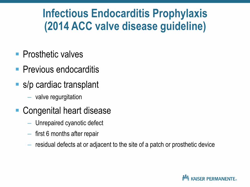
Infectious Endocarditis Prophylaxis (2014 ACC valve disease guideline)
Prosthetic valves
Previous endocarditis
s/p cardiac transplant
– valve regurgitation
Congenital heart disease
– Unrepaired cyanotic defect
– first 6 months after repair
– residual defects at or adjacent to the site of a patch or prosthetic device

Infectious Endocarditis Prophylaxis (2014 ACC valve disease guideline)
Not recommended for non-dental procedures (EGD, colonoscopy, cystoscopy, other surgery, TEE)
No longer recommended for any other valve lesion (MVP, Bicuspid AV, MR, TR, etc.)

Echo in valve disease (2014 ACC valve disease guideline)
Class I: TTE is recommended in the initial evaluation of patients with known or suspected VHD to confirm the diagnosis, establish etiology, determine severity, assess hemodynamic consequences, determine prognosis, and evaluate for timing of intervention.
Class I: TTE is recommended in patients with known VHD with any change in symptoms or physical examination findings.
Class I: Periodic monitoring with TTE is recommended in asymptomatic patients with known VHD at intervals depending on valve lesion, severity, ventricular size, and ventricular function.

Echo estimate of pulmonary artery systolic pressure
Requires presence of TR – at least a trace
Measure TR velocity
Simplified Bernoulli equation: 4V2
3 m/s
RA pressure – typically assume 5 mmHg
RV pressure + RA pressure = estimated PA systolic pressure

When can I not worry about Pulmonary Hypertension?
TR velocity <3.5 m/s
Estimated PA systolic pressure <50
Especially when there is a likely explanation
– (LV dysfunction, pulmonary disease, obesity, OSA)
Treat the underlying problem

When do I need to care about Pulmonary Hypertension?
TR velocity >3.5 m/s
Estimated PA systolic pressure >50
When there isn’t a likely explanation
Especially when there is RV enlargement and dysfunction

Primary vs. SecondaryPulmonary Hypertension
99% is secondary
Address the underlying problem:
– Lung disease
– Hypoxia
– Obesity
– OSA
– LV dysfunction (systolic, diastolic)

Right-Sided Valve Disease
Tricuspid and Pulmonic regurgitation are VERY COMMOM
PR is almost never an issue (will not discuss)
TR is typically a bystander – almost NEVER a PRIMARYclinical problem.
So TR does not require echo follow-up in and of itself.
The important issues are :– RV (and LV) size and function
– Pulmonary HTN
– Pulmonary disease/hypoxia
– Obesity/OSA

Specific Valve Lesions
Aortic Stenosis


Clinical follow-up of AS
Most common primary valve disease
2 types: Bicuspid AV (age <65), Senile calcific (age >75)
Hemodynamic progression leading to symptoms occurs in all asymptomatic patients with AS.
Survival during asymptomatic phase is similar to age-matched controls. Low risk of sudden death (<1% per year)
Substantial overlap in hemodynamic severity between asymptomatic and symptomatic patients
No single parameter indicates the need for AVR.
Combination of symptoms, valve anatomy, and hemodynamics

Clinical follow-up of AS
Symptoms: DOE, decreased exercise tolerance.
The classical symptoms of syncope, angina, and HF are late manifestations of disease, most often seen in patients in whom early symptom onset was not recognized and intervention was inappropriately delayed.
The only effective treatment is surgical or trans catheter AVR (there is no medical therapy)
Once even mild AS symptoms are present, outcomes are extremely poor unless outflow obstruction is relieved.
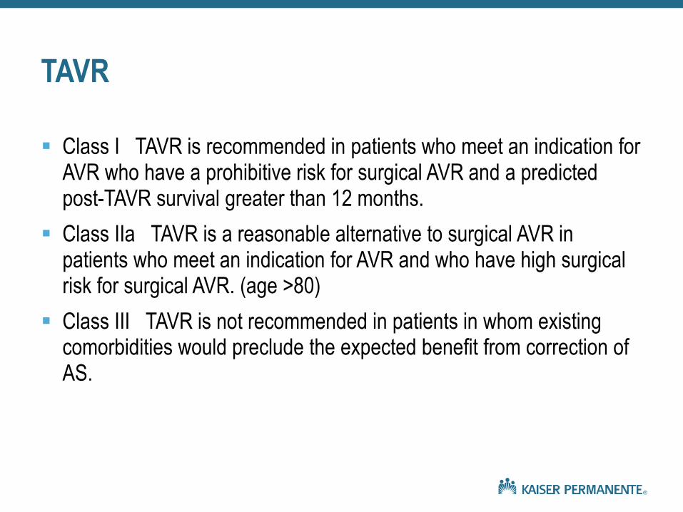
TAVR
Class I TAVR is recommended in patients who meet an indication for AVR who have a prohibitive risk for surgical AVR and a predicted post-TAVR survival greater than 12 months.
Class IIa TAVR is a reasonable alternative to surgical AVR in patients who meet an indication for AVR and who have high surgical risk for surgical AVR. (age >80)
Class III TAVR is not recommended in patients in whom existing comorbidities would preclude the expected benefit from correction of AS.


Aortic Regurgitation
Acute AR: IE, aortic dissection.
Chronic AR: bicuspid aortic valve, dilation of the ascending aorta or the sinuses of Valsalva, rheumatic heart disease
AR is mainly chronic and slowly progressive
Vasodilators (dihydropyridine CCB’s, ACE inhibitors/ARBs) may be helpful, but unproven and not recommended routinely.
In symptomatic patients who are candidates for surgery, medical therapy is not a substitute for AVR.

Bicuspid Aortic Valve
Most patients with a bicuspid aortic valve will develop AS or AR over their lifetime.
Ascending aortic aneurysm – frequently associated with BAV
The echo report should include aortic measurements at the aortic annulus, sinuses, sinotubular junction, and mid-ascending aorta.
Coarctation of the Aorta – Doppler interrogation of the proximal descending aorta

Bicuspid Aortic Valve
In 20% to 30% of patients with bicuspid valves, other family members also have bicuspid valve disease and/or an associated aortopathy.
A specific genetic cause has not been identified, and the patterns of inheritance are variable.
Important to take a family history and inform patients that other family members may be affected.
Many valve experts recommend screening all first-degree relatives of patients with bicuspid aortic valve. Insufficient data to know whether this is appropriate.

BAV – Aortic Aneurysm
Serial evaluation of the ascending aorta by echo, CMR, or CT angiography is recommended in patients with BAV and an aortic diameter greater than 4.0 cm
Examination interval determined by the degree and rate of progression of aortic dilation and by family history.
Aortic diameter greater than 4.5 cm – evaluation should be performed annually.
No proven drug therapies to reduce the rate of progression of aortic dilation. BB often used/recommended.

BAV – Aortic Aneurysm
Class I Operative repair of the ascending aorta is indicated in patients with BAV when ascending aorta is greater than 5.5 cm. (Level of Evidence: B)
Previous guidelines have recommended surgery when the degree of aortic dilation is >5.0 cm – evidence supporting these previous recommendations was limited and anecdotal
Surgery is recommended with aortic dilation of 5.1 cm to 5.5 cm only if there is a family history of aortic dissection or rapid progression of dilation.
BSA can be considered in decision-making


Mitral stenosis
Class I: TTE is indicated in patients with signs or symptoms of MS to establish the diagnosis, quantify hemodynamic severity (mean pressure gradient, mitral valve area, and pulmonary artery pressure), assess concomitant valvular lesions, and demonstrate valve morphology (to determine suitability for mitral commissurotomy). (Level of Evidence: B)
Class I: TEE should be performed in patients who are being considered for percutaneous mitral balloon commissurotomy to assess the presence or absence of left atrial thrombus and to further evaluate the severity of MR. (Level of Evidence: B)

Balloon Commissurotomy


Secondary Mitral Regurgitation
Far and away the most common cause of MR
This issue is not the valve, the issue is CHF, LV systolic dysfunction.
Treat the CHF!
Interventions on the valve to address MR/CHF have largely been unsuccessful
Echo can be done as needed to manage CHF (decisions about ICD and Bi-V pacing, assessing prognosis, aggressiveness of medical Rx)
There is little reason to do f/u echo for the MR.

Primary Mitral Regurgitation
Trace, mild MR does not need f/u echo.
> Moderate MR does require f/u echo
Moderate MR with normal LV size/function and no symptoms – if stable over a few years can be followed more loosely
> Moderate MR needs more close f/u – approximately annually.
Bottom line – LV size and function: if severe MR, enlarging LV, and falling LVEF, cardiology consultation for MVR
Symptoms can be quite clear or quite tricky