Embed Size (px)
Citation preview
HAL Id: tel-02127553https://tel.archives-ouvertes.fr/tel-02127553
Submitted on 13 May 2019
HAL is a multi-disciplinary open accessarchive for the deposit and dissemination of sci-entific research documents, whether they are pub-lished or not. The documents may come fromteaching and research institutions in France orabroad, or from public or private research centers.
L’archive ouverte pluridisciplinaire HAL, estdestinée au dépôt et à la diffusion de documentsscientifiques de niveau recherche, publiés ou non,émanant des établissements d’enseignement et derecherche français ou étrangers, des laboratoirespublics ou privés.
Valorisation des activités biologiques de la diatoméemarine Haslea ostrearia
Charlotte Falaise
To cite this version:Charlotte Falaise. Valorisation des activités biologiques de la diatomée marine Haslea ostrearia. Mi-crobiologie et Parasitologie. Université du Maine, 2019. Français. �NNT : 2019LEMA1011�. �tel-02127553�
THESE DE DOCTORAT
LE MANS UNIVERSITE COMUE UNIVERSITE BRETAGNE LOIRE
ECOLE DOCTORALE N° 598 Sciences de la Mer et du littoral Spécialité : « Biologie marine - Microbiologie »
Valorisation des Activités Biologiques de la Diatomée Marine Haslea ostrearia Thèse présentée et soutenue à Le Mans, le 5 avril 2019 Unité de recherche : Mer Molécules Santé (MMS) - EA2160, Le Mans Université UFR Sciences et Techniques, Avenue Olivier Messiaen 72085 Le Mans Cedex 9 Thèse N° : 2019LEMA1011
Par
Charlotte FALAISE
Rapporteurs avant soutenance :
Lucie BEAULIEU, Professeure Université Laval de Québec
Laurent PICOT, Maitre de conférences, HDR La Rochelle Université
Composition du Jury : Patrick CORMIER, Professeur Sorbonne Université
Juliette FAUCHOT, Maîtresse de conférences Caen Normandie Université
Présidente du jury : Claire HELLIO, Professeure Bretagne Occidentale Université
Marie-Agnès TRAVERS, Chargée de recherche, HDR Ifremer SG2M-LGPMM, La Tremblade
Directeur de thèse Jean-Luc MOUGET, Professeur Le Mans Université
Co-directeur de thèse Vincent LEIGNEL, Maître de conférences, HDR Le Mans Université


I
“Always look on the bright side of life [whistles]”
Brian

II
III
REMERCIEMENTS
Cette thèse a été réalisée au laboratoire Mer, Molécules, Santé de Le Mans Université et a
bénéficié d’une allocation du Ministère de l’Enseignement Supérieur et de la Recherche
(2015-2018). Ce projet a également bénéficié de financements du programme de recherche
et d'innovation Horizon 2020 de l'Union européenne//au titre de la convention de subvention
n° 734708 / GHANA / H2020-MSCA-RISE-2016.
Je voudrais d’abord remercier les membres du jury qui ont accepté d’examiner mon travail,
merci aux rapporteurs Lucie Beaulieu et Laurent Picot, ainsi qu’aux examinateurs Patrick
Cormier, Juliette Fauchot, Claire Hellio, Vincent Leignel et Marie-Agnès Travers.
Merci à mes encadrants de m’avoir donné l’opportunité de réaliser ce travail de doctorat et
un merci tout particulier à mon directeur de thèse Jean-Luc Mouget. Merci pour tes conseils,
tes encouragements pendant mes moments de doutes et ta disponibilité tout au long de mon
doctorat. Merci d’avoir toujours pris le temps de répondre à mes questions et également
d’avoir été aussi réactif pour me renvoyer toutes tes corrections (et particulièrement en cette
fin de thèse !). Ces trois années ont été une belle aventure, que j’ai pu enrichir de séjours
dans différents laboratoires en France et à l’étranger grâce à ton réseau et au programme
européen. Donc un grand merci pour ton encadrement, j’ai été très contente que tu diriges
ma thèse, de découvrir l’univers de la marennine et de voir la vie en bleu !
Merci aux membres de mon CSI, Tristan Renault et Sébastien Lefebvre, pour leurs regards
extérieurs et leurs conseils sur mon travail de doctorat.
Merci à tous mes collègues du Laboratoire MMS du Mans, j’ai eu beaucoup de plaisir à
travailler à vos côtés, à discuter autour d’un café et à partager les pauses repas. Merci
Brigitte, Justine et Marie pour nos séances de bavardages et de rigolades, nos pique-niques
au soleil et parties de Mölkky, mais également pour votre aide et vos conseils tout au long
de ma thèse. Merci Nathalie et Aurore d’être toujours disponibles et à l’écoute et en
particulier pour votre soutien au début de mon doctorat. Merci à ma co-bureau Myriam pour
toutes nos belles conversations, pour les brassages de meubles et pour les chocolats
IV
sur-emballés ; tu as vraiment apporté la touche d’énergie positive dont j’avais besoin en
cette fin de thèse ! Merci Françoise et Gérard pour les pauses café animées et les
discussions variées. Merci à vous tous, Benoit, Annick, et merci aussi Marie pour ton aide
dans la préparation des TP, Valérie pour ton éternelle bonne humeur et pour avoir géré tout
un tas de dossiers au cours de ces années. Bref, un grand merci à tous d’avoir rendu la vie
au travail si agréable !
Merci également à tous ceux qui ne sont aujourd’hui plus au labo mais avec qui ça a été un
réel plaisir de travailler, merci Sophie, Alexandre et Kajani. Merci Romain pour tout ce que tu
m’as transmis et pour avoir veillé même à distance que ma thèse se passait bien. Bien sûr
une pensée spéciale pour les « anciens » thésards, merci pour le soutien moral et l’entraide,
merci Stéphane ma co-bureau de début de thèse, merci Hélène, Fiddy, Stan, Bing et Wided.
La relève est maintenant assurée : merci Matteo, Orlane, Mariem et Julie !
Merci à Intan et Hamza, j’ai fait mes premiers pas d’encadrante de stage avec vous et
j’espère vous avoir transmis au moins autant que ce que vous m’avez apporté.
Merci à toute la team GHaNA et marennine, à Pamela, Emmanuelle, Boris et bien sûr merci
à toi Lila, pour nos nombreux fous rires même dans nos moments de galère à essayer de
trouver un sens à nos résultats, merci pour ta délicieuse cuisine (qui me manque !) et pour ta
joie de vivre .
Merci Patrick pour ton accueil à Roscoff, ça a été un réel plaisir de travailler avec toi. J’ai
beaucoup apprécié mes séjours à la station biologique, tant pour la sympathie et l’accueil
chaleureux de toute l’équipe TCCD que pour le cadre de travail et les magnifiques couchers
de soleil qu’offre la ville !
Merci aux équipes du LGPMM de la station de la Tremblade. Merci Cyrille pour ton accueil,
tes conseils et pour tous nos échanges intéressants, merci à Marie-Agnès, Joël et à tous les
chercheurs et doctorants pour leur aide et avoir rendu ces séjours à La Tremblade si
agréables.
V
Merci aux équipes de l’ISMER, pour leur accueil et leur aide lors de mes trois séjours
rimouskois ! Merci beaucoup Réjean de m’avoir permis de travailler dans ton labo, merci
pour ta disponibilité, ton optimisme et de m’avoir offert la possibilité de participer à différents
congrès. Merci Nathalie, Jordan, Sarah-Béatrice et François pour votre aide à la station
aquicole. Merci Karine et Kim de m’avoir donné accès au labo de bactériologie et pour tous
vos conseils. Merci Bertrand et Renée pour le temps que vous m’avez accordé et pour votre
aide en biologie moléculaire. Merci Efflam et Jean-Nicolas pour votre soutien au cours de
mes expérimentations. Enfin merci à tous ceux qui ont contribué à rendre ces séjours au
Canada aussi enrichissants sur les plans professionnels que personnels, merci notamment à
Constance pour nos balades dans la neige ou au bord du Saint Laurent, à Inès pour nos
sessions de travail tardives et nos discussions autour d’une poutine, et bien sûr un grand
merci à mes supers colocataires Marion et Florian pour tous ces moments partagés !
Je tiens également à remercier tous mes proches qui m’ont soutenue et encouragée au
cours de mon doctorat.
Un merci tout particulier à ma famille, « pour l’amour, le partage… la générosité !!! ». Merci
Kathleen et Leslie d’être toujours là pour moi, je ne pourrais pas imaginer avoir des sœurs
plus géniales, vous êtes des piliers dans ma vie et je sais qu’on pourra toujours compter les
unes sur les autres. Merci maman pour tout ce que tu fais pour moi, d’être toujours aussi
positive et confiante, de t’assurer que je vais bien (que je bois du thym-miel-citron en hiver !)
et pour toute la jovialité et la force que tu me transmets. A big thank you to my little Nany,
thank you for always being there for me and for all your love and your support. Merci papa
d’avoir été aussi présent pendant mon doctorat, de m’avoir soutenu, de t’être intéressé à
mes travaux et de m’avoir même insufflé des pistes de réflexion ! Merci à Mathieu, à
Aurélien et bien sûr à Robinson qui grandit tellement vite et qui égaye nos vies avec toute
son énergie et sa bonne humeur. Un grand merci à vous tous, la famille proche et plus
lointaine, c’est un vrai bonheur de se sentir si bien entourée !
Merci bien sûr à tous mes ami(e)s aux côtés de qui je peux recharger les batteries, me
confier et rigoler. Merci à Elodie, Manon, Melou, Coco, Damien, Thomas et Marion, même si
VI
on ne se voit pas forcément souvent je sais que je peux compter sur vous et ça fait toujours
un bien immense de vous retrouver ! Merci aussi à toute la team de Poitiers, pour nos joyeux
moments de détentes et de festivités !
Un énorme MERCI à mes colocs Marie-Laure, Elodie, Léonie, Carole (et Loulou !). Quel
plaisir de rentrer le soir à la maison et de papoter autour d’une « infu » et des cookies (ou
d’une bière et des Munster Munch selon l’heure). Merci pour tous ces chouettes moments
partagés dans « notre maison du bonheur », nos discussions parfois philosophiques et
profondes et parfois beaucoup moins, nos larmes de rire ou de tracas, nos activités
artistiques, nos playlists musicales qui tournent en boucle, nos séances de sport, nos
soirées soupe-télé ou apéros ensoleillés. Merci d’avoir été là pour moi, à l’écoute,
attentionnées et d’avoir rendu ces trois années si riches et si belles !
And last but not least, merci à toi David, pour tout. Tu as toujours su me motiver, trouver les
mots pour m’encourager, me tirer vers le haut et me faire relativiser quand je commençais à
dramatiser sur ma vie de thésarde (parce-que non, ce n’est pas si grave si les bactéries
n’ont pas poussé ou que les moules ne veulent pas pondre). Merci de t’être autant impliqué
et intéressé à mon travail ; je ne suis pas sûre de pouvoir parler des moteurs CFM et LEAP
aussi bien que tu parles de microalgues ou de réponse hormétique ! Merci de ta patience et
de ton soutien. J’ai une chance folle de t’avoir auprès de moi, et maintenant l’aventure du
doctorat terminée j’ai hâte d’en entamer une toute nouvelle avec toi.
VII
SOMMAIRE
REMERCIEMENTS……………………………………………………………………….……II
SOMMAIRE………………………………………………………………………………….VII
LISTE DES FIGURES…………………………………………………………………………XI
LISTE DES TABLEAUX…………………………………………………………………….XVII
LISTE DES FIGURES ET TABLEAUX SUPPLEMENTAIRES………………………………..XVII
LISTE DES PRODUCTIONS SCIENTIFIQUES……………………………………………...XVIII
CHAPITRE I. INTRODUCTION GÉNÉRALE ........................................................................ 1
1 Les diatomées ................................................................................................ 3 1.1 Ecologie des diatomées ............................................................................. 3 1.2 Caractères généraux des diatomées ......................................................... 4 1.3 Classification des diatomées ..................................................................... 6 1.4 Cycle de vie et reproduction des diatomées .............................................. 7 1.5 Utilisation et valorisation des diatomées .................................................. 10
2 Les diatomées du genre Haslea .................................................................. 11 2.1 Caractéristiques générales du genre Haslea ........................................... 11 2.2 La diatomée bleue Haslea ostrearia ........................................................ 12
2.2.1 Caractéristiques morphologiques ................................................................. 12 2.2.2 Écologie ........................................................................................................ 13 2.2.3 Physiologie .................................................................................................... 13 2.2.4 Reproduction ................................................................................................. 14 2.2.5 Culture........................................................................................................... 14 2.2.6 Distribution géographique ............................................................................. 16
2.3 Haslea ostrearia, unique en son genre ? La diversité insoupçonnée des Haslea bleues. ................................................................................................... 17
3 Les pigments bleus d’Haslea ...................................................................... 21 3.1 La marennine et le verdissement des huîtres .......................................... 21
3.1.1 Les huîtres « fines de claires vertes » .......................................................... 21 3.1.2 La fixation de la marennine sur les branchies .............................................. 22 3.1.3 La consommation des huîtres vertes, une spécialité française .................... 23
3.2 La marennine et les pigments de type marennine ................................... 24 3.2.1 Production et excrétion de la marennine par Haslea .................................... 24 3.2.2 Caractérisations préliminaires du pigment .................................................... 25 3.2.3 Techniques de purification du pigment ......................................................... 26 3.2.4 Un nouveau procédé pour obtenir des solutions enrichies en pigment bleu 27
4 Activités biologiques et valorisations potentielles des Haslea bleues et de leurs pigments ............................................................................................... 30
4.1 Les biotechnologies bleues ..................................................................... 30 4.1.1 Le secteur de l’agroalimentaire ..................................................................... 30
VIII
4.1.2 Les secteurs de la cosmétique et de la pharmacologie ................................ 32 4.2 L’aquaculture ........................................................................................... 33
4.2.1 Les microalgues comme probiotiques .......................................................... 33 4.2.2 Les Haslea bleues pour limiter les infections microbiennes ? ...................... 34
4.3 Perspectives et limites actuelles ............................................................. 35 5 Contexte et objectifs du sujet de recherche ............................................. 36
CHAPITRE II. MICROALGAL COMPOUNDS AGAINST PATHOGENS: EMPHASIZE ON
DISEASES IN AQUACULTURE ....................................................................................... 39
1 Introduction .................................................................................................. 43 2 Antibacterial activity from microalgae ....................................................... 46
2.1 Antibacterial activity from microalgae against human pathogenic bacteria ..................................................................................................................46
2.1.1 Toward improving extraction techniques ...................................................... 49 2.1.2 Diversity of antibacterial compounds extracted from microalgae ................. 50
2.2 Use of microalgae against pathogenic bacteria in aquaculture ............... 52 2.2.1 Benefits of the “green-water” technique against bacterial diseases in
aquaculture .................................................................................................................. 54 2.2.2 Microalgae to improve the live-food quality .................................................. 56 2.2.3 In vitro efficiency of microalgal compounds against marine bacteria ........... 56 2.2.4 Interactions between microalgae and marine bacteria ................................. 59
3 3. Antifungal activity from microalgae ....................................................... 61 3.1 Antifungal activity from microalgae against human pathogens ............... 61 3.2 Potential use of microalgae against fungal diseases in aquaculture ....... 64
4 Antiviral activity from microalgae .............................................................. 66 4.1 Antiviral activity from microalgae against human pathogenic viruses ..... 66 4.2 Potential use of microalgae against viruses in aquaculture .................... 70
5 Conclusion ................................................................................................... 73
CHAPITRE III. EFFECTS OF MARENNINE SOLUTIONS ON THE GROWTH OF MARINE VIBRIO
................................................................................................................................. 75
CHAPITRE III.Partie 1. Complex relationships between the blue pigment marennine and marie bacteria of the genus Vibrio .................................................................... 77
1 Introduction .................................................................................................. 80 2 Materials and Methods ................................................................................ 82
2.1 Vibrio strains ............................................................................................ 82 2.2 Vibrio exposure to Blue Water (BW) solutions ........................................ 82
2.2.1 Preparation of bacterial inocula .................................................................... 83 2.2.2 Blue Water (BW) production ......................................................................... 83 2.2.3 Antibacterial essay ........................................................................................ 84
2.3 Growth curve analyzes and statistics ...................................................... 85
IX
3 Results .......................................................................................................... 86 3.1 Different patterns of Vibrio growth curves evidenced by the screening experiment ......................................................................................................... 86 3.2 Experiment with BW concentration range ................................................ 89
4 Discussion .................................................................................................... 92 5 Conclusion .................................................................................................... 95
CHAPITRE III. Partie 2. Co-culture of Haslea ostrearia with an oyster virulent Vibrio strain ...................................................................................................................... 101
1 Introduction ................................................................................................ 104 2 Results and discussion ............................................................................. 106 3 Conclusion .................................................................................................. 110 4 Materials and Methods............................................................................... 111
CHAPITRE IV. NEW INSIGHTS INTO THE EFFECTS OF MARENNINE SOLUTIONS ON MARINE
ORGANISMS ............................................................................................................. 113
CHAPITRE IV. Partie 1. Harmful or harmless: biological effects of marennine on marine organisms................................................................................................... 115
1 Introduction ................................................................................................ 118 2 Materials and Methods............................................................................... 121
2.1 Preparation of the purified extracellular marennine (EMn), Blue Water (BW) and Concentrated Supernatant (CS) ...................................................... 122 2.2 Exposure of the mussel Mytilus edulis larvae to BW ............................. 123 2.3 Exposure of the barnacle Chthamalus bisinuatus larvae to BW ............ 124 2.4 Exposure of the winter flounder Pseudopleuronectes americanus larvae to BW 125 2.5 Exposure of the sea urchins Sphaerechinus granularis and Paracentrotus lividus to EMn and CS ..................................................................................... 126 2.6 Exposure of Vibrio species to BW and CS ............................................ 128 2.7 Exposure of fish gill cell lines to EMn .................................................... 129 2.8 Exposure of the oyster Crassostrea gigas haemocytes to EMn ............ 130 2.9 Statistics ................................................................................................ 131
3 Results ........................................................................................................ 132 3.1 Differences in the solutions containing the extracellular marennine ...... 132 3.2 Effects of BW on Mytilus edulis larvae ................................................... 132 3.3 Effect of BW on the barnacle Chthamalus bisinuatus larvae ................. 135 3.4 Effect of BW on the winter flounder Pseudopleuronectes americanus larvae ................................................................................................................135 3.5 Effects of CS and purified EMn on Sphaerechinus granularis and Paracentrotus lividus ....................................................................................... 136 3.6 Antibacterial effects of BW and CS on Vibrio species ........................... 139 3.7 Effects of EMn on fish gill cell line RTgill-W1 ......................................... 140
X
3.8 Effects of EMn on Crassostrea gigas haemocytes ................................ 141 4 Discussion .................................................................................................. 142
4.1 Limitations of the study .......................................................................... 143 4.2 The unpredictable “Jekyll and Hyde”, good and bad nature of marennine ................................................................................................................145
5 Conclusion ................................................................................................. 148
CHAPITRE IV. Partie 2. Preliminary results on marennine effects on the mussel Mytilus edulis larvae .............................................................................................. 149
1 Introduction ................................................................................................ 152 2 Results and discussion ............................................................................. 154
2.1 Mytilus edulis larvae challenged with Vibrio .......................................... 154 2.2 Mytilus edulis exposure to different BW concentrations ........................ 155
2.2.1 Effect of BW on Mytilus edulis larval mortality ............................................ 155 2.2.2 Molecular response of Mytilus edulis larvae under BW exposure .............. 156
3 Conclusion ................................................................................................. 160 4 Materials and methods .............................................................................. 161
4.1 Experimental design .............................................................................. 161 4.1.1 Mytilus edulis rearing procedure and exposure to BW ............................... 161 4.1.2 Mytilus edulis challenged with Vibrio species ............................................. 161
4.2 Sample collection .................................................................................. 162 4.3 Studied genes and RT-qPCR ................................................................ 163 4.4 Statistical analysis ................................................................................. 164
CHAPITRE V. DISCUSSION GÉNÉRALE ET PERSPECTIVES ......................................... 165
1 Contexte des travaux de recherche ......................................................... 167 2 Bilan des effets biologiques de la marennine ......................................... 169 3 Valorisation de la marennine en aquaculture : limites actuelles et perspectives de recherche .............................................................................. 173
RÉFÉRENCES BIBLIOGRAPHIQUES ........................................................................... 179
ANNEXES ................................................................................................................ 213
Annexe 1. Falaise et al. (2016), Marine Drugs [texte complet] Annexe 2. Falaise et al. (2019), Aquatic Toxicology [texte complet] Annexe 3. Falaise et al. (2019), Marine Drugs [texte complet] Annexe 4. Prasetiya et al. (sous presse), Plant Ecology & Evolution [texte complet] Annexe 5. Chapitres de Livre : Falaise, Dewi et al. (2018) ; Gastineau et al. (2018) Annexe 6. Essais préliminaires
XI
L ISTE DES F IGURES
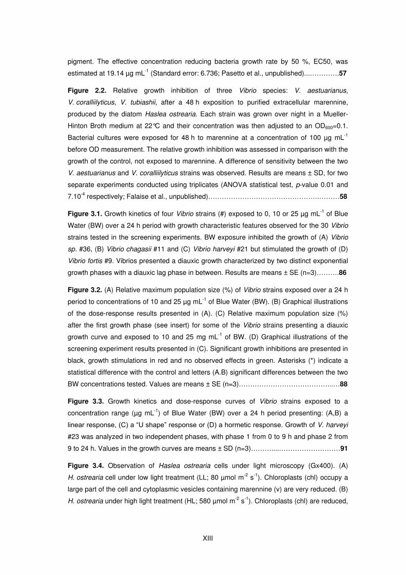
Figure 1.1. Diversité de la structure des frustules siliceux des diatomées nettoyés à l’acide
et observés en microscopie électronique à balayage (MEB). (A,F) Diatomées centriques
radiales, (B,C) diatomées pennées et (D,E) diatomées centriques polaires. Les frustules des
diatomées sont constitués de deux valves : l’épivalve et l’hypovalve liées par un ensemble
de bandes cingulaires (le cingulum). Barres d’échelles : 10 µm. Cette figure est adaptée de
photographies provenant de Kociolek et al. (2015a, 2015b) pour (A,B,C) et de Losic et al.
(2009) pour (D,E,F)…………………………………………………………………………………...5
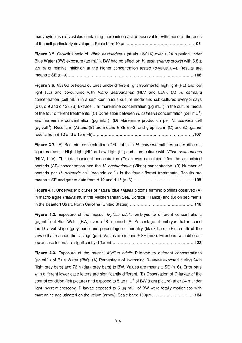
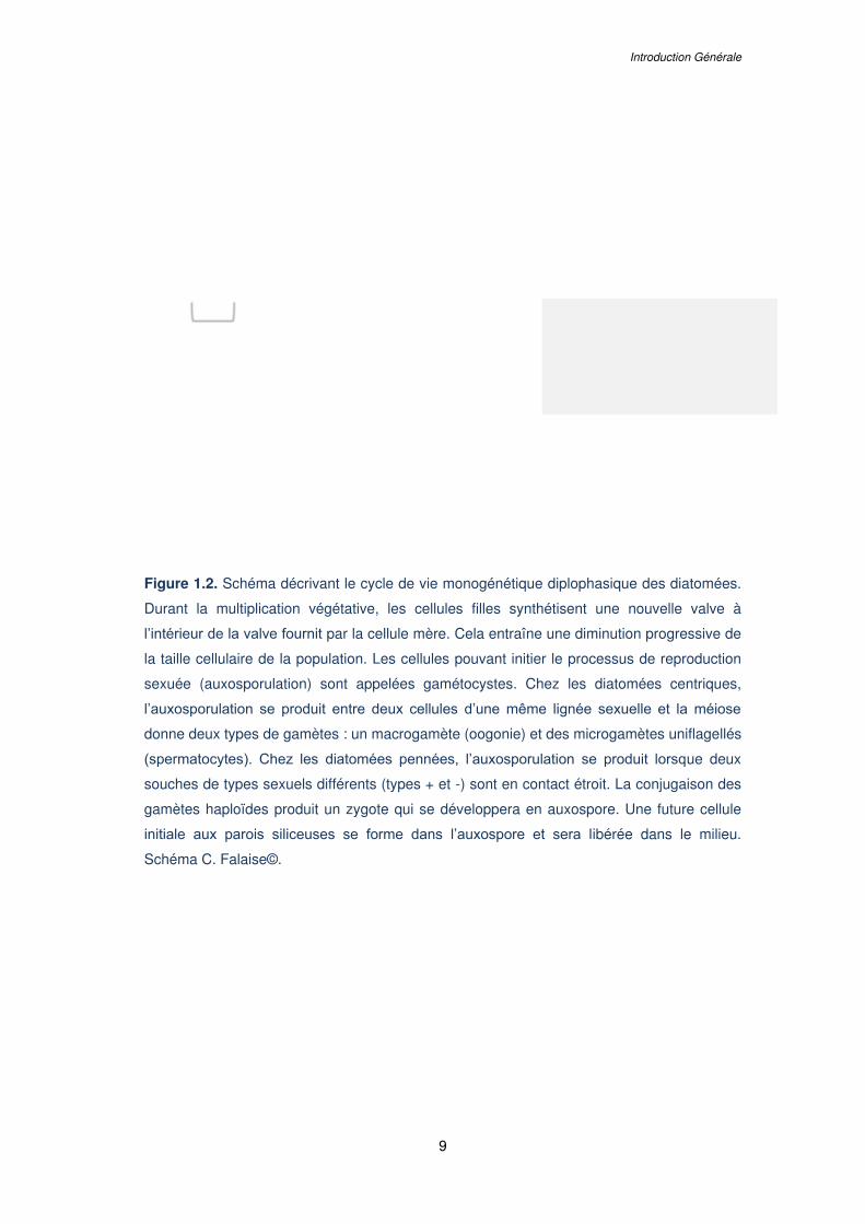
Figure 1.2. Schéma décrivant le cycle de vie monogénétique diplophasique des diatomées.
Durant la multiplication végétative, les cellules filles synthétisent une nouvelle valve à
l’intérieur de la valve fournit par la cellule mère. Cela entraîne une diminution progressive de
la taille cellulaire de la population. Les cellules pouvant initier le processus de reproduction
sexuée (auxosporulation) sont appelées gamétocystes. Chez les diatomées centriques,
l’auxosporulation se produit entre deux cellules d’une même lignée sexuelle et la méiose
donne deux types de gamètes : un macrogamète (oogonie) et des microgamètes uniflagellés
(spermatocytes). Chez les diatomées pennées, l’auxosporulation se produit lorsque deux
souches de types sexuels différents (types + et -) sont en contact étroit. La conjugaison des
gamètes haploïdes produit un zygote qui se développera en auxospore. Une future cellule
initiale aux parois siliceuses se forme dans l’auxospore et sera libérée dans le milieu……....9
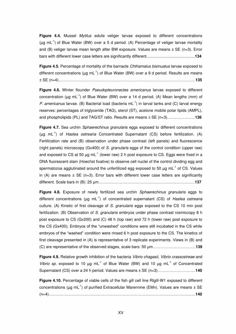
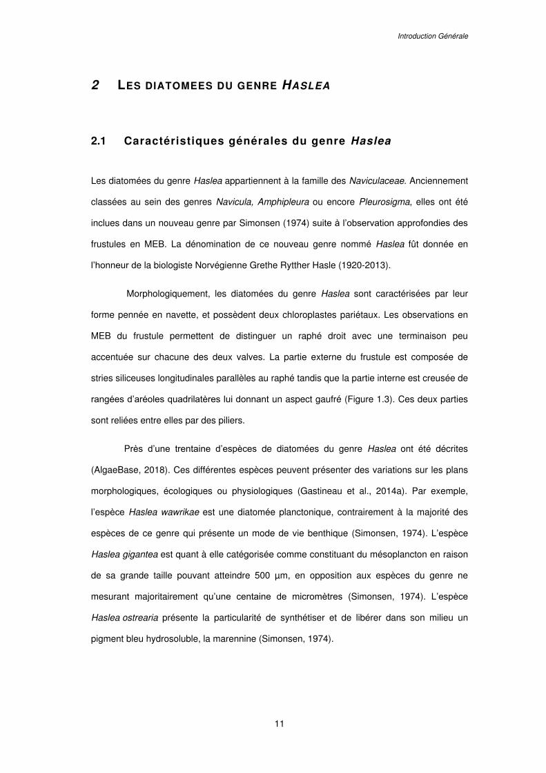
Figure 1.3. Photographies de la diatomée pennée Haslea ostrearia. (A) Observations en
microscopie permettant de distinguer deux chloroplastes pariétaux et la présence de
vésicules contenant un pigment bleu (la marennine). Barre d’échelle 5 µm. (B-E)
Photographies du frustule siliceux en microscopie électroniques à balayage (MEB) avec
observations des apex de la cellule (B,C) et du centre de la cellule (D,E) en vue externe
(B,D) et interne (C,E). Barres d’échelle 1 µm. Photographie optique communiquée par
A. Alverson et photographies MEB d’après Gastineau et al. (2014a)…………………...…….12
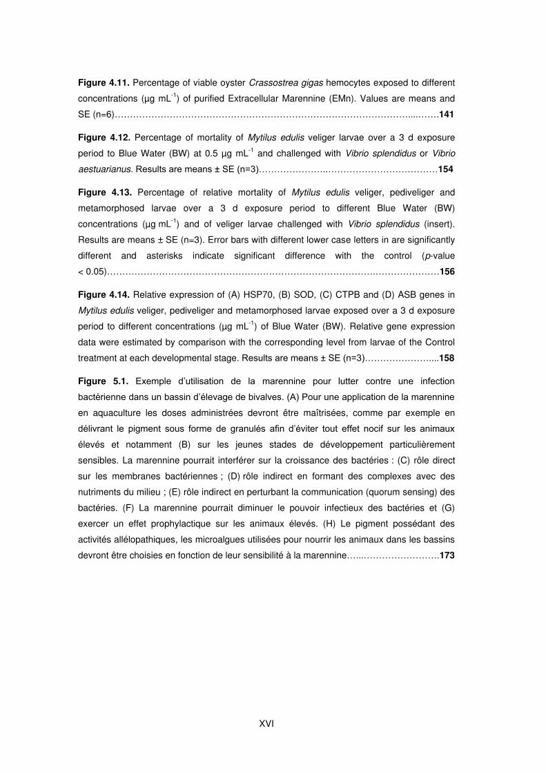
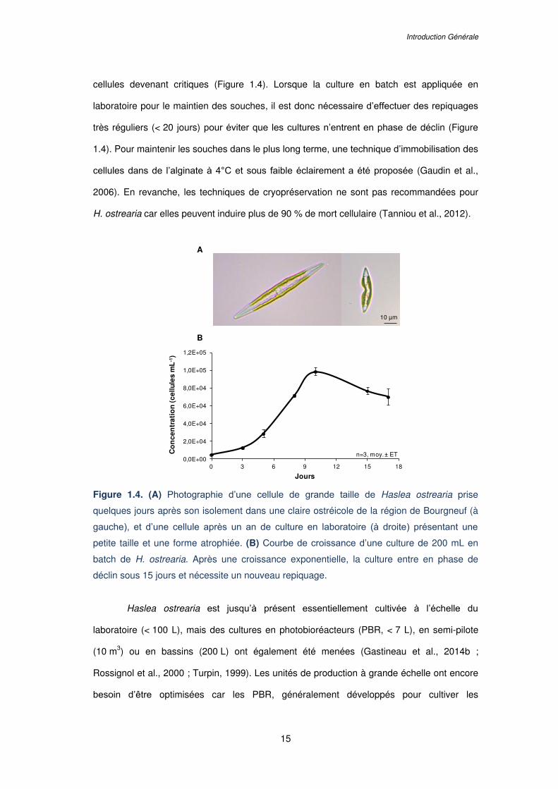
Figure 1.4. (A) Photographie d’une cellule de grande taille de Haslea ostrearia prise
quelques jours après son isolement dans une claire ostréicole de la région de Bourgneuf (à
gauche), et d’une cellule après un an de culture en laboratoire (à droite) présentant une
petite taille et une forme atrophiée. (B) Courbe de croissance d’une culture de 200 mL en
batch de H. ostrearia. Après une croissance exponentielle, la culture entre en phase de
déclin sous 15 jours et nécessite un nouveau repiquage……………………………………….15
Figure 1.5. Répartition mondiale de Haslea ostrearia d’après la littérature. La présence de
H. ostrearia a été déduite de l’observation de diatomées avec des apex bleus ou de la
présence d’huîtres aux branchies vertes ; d’après Gastineau et al. (2014a)…………………16
XII
Figure 1.6. (A) Comparaisons de données morphométriques des frustules de plusieurs
espèces de Haslea bleues. (B) Arbre phylogénétique de différentes espèces de Haslea basé
sur le gène chloroplastique rbcL (les valeurs des bootstraps du maximum de vraisemblance
(en noir) et du maximum de parcimonie (en bleu) ont été obtenues avec 1000 réplicats).
Photographies au microscope électronique à balayage (MEB) de la fin du raphé central de
plusieurs espèces d'Haslea référencées dans l'arbre phylogénétique. Une fine barre
parallèle au raphé central (flèche blanche) peut être observée chez certaines espèces de
diatomées bleues telles que H. nusantara et H. silbo. Cette caractéristique morphologique
est commune aux diatomées non bleues telles que H. pseudostrearia. Cette fine barre n'est
pas observée parmi les espèces de diatomées bleues génétiquement plus lointaines telles
que H. ostrearia, H. karadagensis et H. provincialis. Barres d'échelle = 1 µm. Figures
adaptées de Gastineau et al. (2014a) et Falaise et al. (2016a)………………………………..18
Figure 1.7. Efflorescences de Haslea bleues observées (A) en mer Mediterranée (Corse,
France), (B,C) dans l’océan Atlantique (Caroline du Nord, États-Unis) et (D) en mer
Adriatique (Dalmatie, Croatie). Photographies communiquées par D. Sirjacobs (A),
N. Lindquist (B,C) et Y. Hardivillier (D)……………………………………………...…………….20
Figure 1.8. (A) Affinage des huîtres dans des bassins d’eau de mer peu profonds appelés
« claires » dans la région de Marennes-Oléron. (B) Huître « fine de claire » et huître « fine
de claire verte » (label Rouge français) dont les branchies ont été verdies par la présence de
Haslea ostrearia et de son pigment (marennine) dans les claires lors de l’affinage…………22
Figure 1.9. (A) Technique de purification de la marennine intracellulaire (IMn) et
extracellulaire (EMn) de Haslea ostrearia proposée par Pouvreau et al. (2006b). La Blue
Water (BW), un surnageant de culture concentré en marennine, est obtenue par
ultrafiltration de la EMn. (B) Spectres UV-Visible caractéristiques des fractions de IMn et
EMn purifiées, adapté de Pouvreau et al. (2006b)………………………………………………27
Figure 1.10. Nouveau procédé de concentration de la marennine et des pigments de « type
marennine » des espèces de Haslea bleues (n° de brevet 1872316). Procédé développé en
collaboration entre le laboratoire Mer, Molécule, Santé (MMS) et l’Institut des Molécules et
Matériaux du Mans (I3M) de le Mans Université………………………………………………...29
Figure 2.1. Growth inhibition of Vibrio splendidus by purified marennine, the blue pigment
produced by Haslea ostrearia. (A) V. splendidus was grown over night at 25°C, cells were
then washed with sterile water 2 times and adjusted to an OD600=0.5. Cells were added to
wells with marennine in the following concentrations: 0, 0.1, 1, 10, 100 and 1000 µg mL-1.
Kinetics were run at OD600 for 48 h, with measurements taken every 30 min (n=3). (B)
Relative values were graphed in order to account for the absorbance differences due to the
XIII
pigment. The effective concentration reducing bacteria growth rate by 50 %, EC50, was
estimated at 19.14 µg mL-1 (Standard error: 6.736; Pasetto et al., unpublished)...………….57
Figure 2.2. Relative growth inhibition of three Vibrio species: V. aestuarianus,
V. coralliilyticus, V. tubiashii, after a 48 h exposition to purified extracellular marennine,
produced by the diatom Haslea ostrearia. Each strain was grown over night in a Mueller-
Hinton Broth medium at 22°C and their concentration was then adjusted to an OD600=0.1.
Bacterial cultures were exposed for 48 h to marennine at a concentration of 100 µg mL-1
before OD measurement. The relative growth inhibition was assessed in comparison with the
growth of the control, not exposed to marennine. A difference of sensitivity between the two
V. aestuarianus and V. coralliilyticus strains was observed. Results are means ± SD, for two
separate experiments conducted using triplicates (ANOVA statistical test, p-value 0.01 and
7.10-4 respectively; Falaise et al., unpublished)………………………………………….………58
Figure 3.1. Growth kinetics of four Vibrio strains (#) exposed to 0, 10 or 25 µg mL-1 of Blue
Water (BW) over a 24 h period with growth characteristic features observed for the 30 Vibrio
strains tested in the screening experiments. BW exposure inhibited the growth of (A) Vibrio
sp. #36, (B) Vibrio chagasii #11 and (C) Vibrio harveyi #21 but stimulated the growth of (D)
Vibrio fortis #9. Vibrios presented a diauxic growth characterized by two distinct exponential
growth phases with a diauxic lag phase in between. Results are means ± SE (n=3)……….86
Figure 3.2. (A) Relative maximum population size (%) of Vibrio strains exposed over a 24 h
period to concentrations of 10 and 25 µg mL-1 of Blue Water (BW). (B) Graphical illustrations
of the dose-response results presented in (A). (C) Relative maximum population size (%)
after the first growth phase (see insert) for some of the Vibrio strains presenting a diauxic
growth curve and exposed to 10 and 25 mg mL-1 of BW. (D) Graphical illustrations of the
screening experiment results presented in (C). Significant growth inhibitions are presented in
black, growth stimulations in red and no observed effects in green. Asterisks (*) indicate a
statistical difference with the control and letters (A.B) significant differences between the two
BW concentrations tested. Values are means ± SE (n=3)…………………………………...…88
Figure 3.3. Growth kinetics and dose-response curves of Vibrio strains exposed to a
concentration range (µg mL-1) of Blue Water (BW) over a 24 h period presenting: (A,B) a
linear response, (C) a “U shape” response or (D) a hormetic response. Growth of V. harveyi
#23 was analyzed in two independent phases, with phase 1 from 0 to 9 h and phase 2 from
9 to 24 h. Values in the growth curves are means ± SD (n=3)………....………………………91
Figure 3.4. Observation of Haslea ostrearia cells under light microscopy (Gx400). (A)
H. ostrearia cell under low light treatment (LL; 80 µmol m-2 s-1). Chloroplasts (chl) occupy a
large part of the cell and cytoplasmic vesicles containing marennine (v) are very reduced. (B)
H. ostrearia under high light treatment (HL; 580 µmol m-2 s-1). Chloroplasts (chl) are reduced,
XIV
many cytoplasmic vesicles containing marennine (v) are observable, with those at the ends
of the cell particularly developed. Scale bars 10 μm…………………………………………..105
Figure 3.5. Growth kinetic of Vibrio aestuarianus (strain 12/016) over a 24 h period under
Blue Water (BW) exposure (µg mL-1). BW had no effect on V. aestuarianus growth with 6.8 ±
2.9 % of relative inhibition at the higher concentration tested (p-value 0.4). Results are
means ± SE (n=3)………………………………………………………………………………….106
Figure 3.6. Haslea ostrearia cultures under different light treatments: high light (HL) and low
light (LL) and co-cultured with Vibrio aestuarianus (HLV and LLV). (A) H. ostrearia
concentration (cell mL-1) in a semi-continuous culture mode and sub-cultured every 3 days
(d 6, d 9 and d 12). (B) Extracellular marennine concentration (µg mL-1) in the culture media
of the four different treatments. (C) Correlation between H. ostrearia concentration (cell mL-1)
and marennine concentration (µg mL-1). (D) Marennine production per H. ostrearia cell
(µg cell-1). Results in (A) and (B) are means ± SE (n=3) and graphics in (C) and (D) gather
results from d 12 and d 15 (n=6)…………………………………………………………………107
Figure 3.7. (A) Bacterial concentration (CFU mL-1) in H. ostrearia cultures under different
light treatments: High Light (HL) or Low Light (LL) and in co-culture with Vibrio aestuarianus
(HLV, LLV). The total bacterial concentration (Total) was calculated after the associated
bacteria (AB) concentration and the V. aestuarianus (Vibrio) concentration. (B) Number of
bacteria per H. ostrearia cell (bacteria cell-1) in the four different treatments. Results are
means ± SE and gather data from d 12 and d 15 (n=6)……………………………………….108
Figure 4.1. Underwater pictures of natural blue Haslea blooms forming biofilms observed (A)
in macro-algae Padina sp. in the Mediterranean Sea, Corsica (France) and (B) on sediments
in the Beaufort Strait, North Carolina (United States)………………………………………….118
Figure 4.2. Exposure of the mussel Mytilus edulis embryos to different concentrations
(µg mL-1) of Blue Water (BW) over a 48 h period. (A) Percentage of embryos that reached
the D-larval stage (grey bars) and percentage of mortality (black bars). (B) Length of the
larvae that reached the D stage (µm). Values are means ± SE (n=3). Error bars with different
lower case letters are significantly different……………………………………………..………133
Figure 4.3. Exposure of the mussel Mytilus edulis D-larvae to different concentrations
(µg mL-1) of Blue Water (BW). (A) Percentage of swimming D-larvae exposed during 24 h
(light grey bars) and 72 h (dark grey bars) to BW. Values are means ± SE (n=6). Error bars
with different lower case letters are significantly different. (B) Observation of D-larvae of the
control condition (left picture) and exposed to 5 µg mL-1 of BW (right picture) after 24 h under
light invert microscopy. D-larvae exposed to 5 µg mL-1 of BW were totally motionless with
marennine agglutinated on the velum (arrow). Scale bars: 100µm…………………………..134
XV
Figure 4.4. Mussel Mytilus edulis veliger larvae exposed to different concentrations
(µg mL-1) of Blue Water (BW) over a 5 d period. (A) Percentage of veliger larvae mortality
and (B) veliger larvae mean length after BW exposure. Values are means ± SE (n=3). Error
bars with different lower case letters are significantly different............................................134
Figure 4.5. Percentage of mortality of the barnacle Chthamalus bisinuatus larvae exposed to
different concentrations (µg mL-1) of Blue Water (BW) over a 9 d period. Results are means
± SE (n=4)…………………………………………………………………………………………..135
Figure 4.6. Winter flounder Pseudopleuronectes americanus larvae exposed to different
concentration (µg mL-1) of Blue Water (BW) over a 14 d period. (A) Mean lengths (mm) of
P. americanus larvae. (B) Bacterial load (bacteria mL-1) in larval tanks and (C) larval energy
reserves: percentages of triglyceride (TAG), sterol (ST), acetone mobile polar lipids (AMPL),
and phospholipids (PL) and TAG/ST ratio. Results are means ± SE (n=3)…………………136
Figure 4.7. Sea urchin Sphaerechinus granularis eggs exposed to different concentrations
(µg mL-1) of Haslea ostrearia Concentrated Supernatant (CS) before fertilization. (A)
Fertilization rate and (B) observation under phase contrast (left panels) and fluorescence
(right panels) microscopy (Gx400) of S. granularis eggs of the control condition (upper raw)
and exposed to CS at 50 µg mL-1 (lower raw) 2 h post exposure to CS. Eggs were fixed in a
DNA fluorescent stain (Hoechst fixative) to observe cell nuclei of the control dividing egg and
spermatozoa agglutinated around the unfertilized egg exposed to 50 µg mL-1 of CS. Values
in (A) are means ± SE (n=3). Error bars with different lower case letters are significantly
different. Scale bars in (B): 25 µm……………………………………………………………….137
Figure 4.8. Exposure of newly fertilized sea urchin Sphaerechinus granularis eggs to
different concentrations (µg mL-1) of concentrated supernatant (CS) of Haslea ostrearia
culture. (A) Kinetic of first cleavage of S. granularis eggs exposed to the CS 10 min post
fertilization. (B) Observation of S. granularis embryos under phase contrast rosmiccopy 8 h
post exposure to CS (Gx200) and (C) 48 h (top raw) and 72 h (lower raw) post exposure to
the CS (Gx400). Embryos of the “unwashed” conditions were still incubated in the CS while
embryos of the “washed” condition were rinsed 6 h post exposure to the CS. The kinetics of
first cleavage presented in (A) is representative of 3 replicate experiments. Views in (B) and
(C) are representative of the observed stages, scale bars: 50 µm…………………………...139
Figure 4.9. Relative growth inhibition of the bacteria Vibrio chagasii, Vibrio crassostreae and
Vibrio sp. exposed to 10 µg mL-1 of Blue Water (BW) and 10 µg mL-1 of Concentrated
Supernatant (CS) over a 24 h period. Values are means ± SE (n=3)………………………..140
Figure 4.10. Percentage of viable cells of the fish gill cell line Rtgill-W1 exposed to different
concentrations (µg mL-1) of purified Extracellular Marennine (EMn). Values are means ± SE
(n=4)…………………………………………………………………………………………………140
XVI
Figure 4.11. Percentage of viable oyster Crassostrea gigas hemocytes exposed to different
concentrations (µg mL-1) of purified Extracellular Marennine (EMn). Values are means and
SE (n=6)……………………………………………………………………………………....…….141
Figure 4.12. Percentage of mortality of Mytilus edulis veliger larvae over a 3 d exposure
period to Blue Water (BW) at 0.5 µg mL-1 and challenged with Vibrio splendidus or Vibrio
aestuarianus. Results are means ± SE (n=3)…………………..………………………………154
Figure 4.13. Percentage of relative mortality of Mytilus edulis veliger, pediveliger and
metamorphosed larvae over a 3 d exposure period to different Blue Water (BW)
concentrations (µg mL-1) and of veliger larvae challenged with Vibrio splendidus (insert).
Results are means ± SE (n=3). Error bars with different lower case letters in are significantly
different and asterisks indicate significant difference with the control (p-value
< 0.05)…………………………………………………………………………….…………………156
Figure 4.14. Relative expression of (A) HSP70, (B) SOD, (C) CTPB and (D) ASB genes in
Mytilus edulis veliger, pediveliger and metamorphosed larvae exposed over a 3 d exposure
period to different concentrations (µg mL-1) of Blue Water (BW). Relative gene expression
data were estimated by comparison with the corresponding level from larvae of the Control
treatment at each developmental stage. Results are means ± SE (n=3)…………………....158
Figure 5.1. Exemple d’utilisation de la marennine pour lutter contre une infection
bactérienne dans un bassin d’élevage de bivalves. (A) Pour une application de la marennine
en aquaculture les doses administrées devront être maîtrisées, comme par exemple en
délivrant le pigment sous forme de granulés afin d’éviter tout effet nocif sur les animaux
élevés et notamment (B) sur les jeunes stades de développement particulièrement
sensibles. La marennine pourrait interférer sur la croissance des bactéries : (C) rôle direct
sur les membranes bactériennes ; (D) rôle indirect en formant des complexes avec des
nutriments du milieu ; (E) rôle indirect en perturbant la communication (quorum sensing) des
bactéries. (F) La marennine pourrait diminuer le pouvoir infectieux des bactéries et (G)
exercer un effet prophylactique sur les animaux élevés. (H) Le pigment possédant des
activités allélopathiques, les microalgues utilisées pour nourrir les animaux dans les bassins
devront être choisies en fonction de leur sensibilité à la marennine…...…………………….173
XVII
L ISTE DES TABLEAUX
Table 2.1. Antibacterial activity from microalgae against human pathogenic bacteria..........47
Table 2.2. Antibacterial activity from microalgae against diseases in aquaculture……….....53
Table 2.3. Antifungal activity from microalgae.......................................................................62
Table 2.4. Antiviral activity from microalgae..........................................................................68
Table 4.1. Summary of the marennine based solutions and marine organisms used in the
present study........................................................................................................................121
Table 4.2. Summary of the marennine based solutions effects on the marine organisms
tested....................................................................................................................................142
Table 4.3. Details of genes and primers used in the study..................................................164
L ISTE DES F IGURES ET TABLEAUX SUPPLEMENTAIRES
Supplementary Figure 3.1. Growth curves of 30 Vibrio strains (#) from 10 different species
exposed over a 24 h period to 0, 10 or 25 µg mL-1 of Blue Water (BW), the concentrated
supernatant of Haslea ostrearia containing the extracellular marennine. Results are means ±
SE (n=3)………………………………………………………………………………………………96
Supplementary Figure 3.2. Growth kinetics and dose-response curves of Vibrio strains
exposed to a concentration range (µg mL-1) of Blue Water (BW) over a 24 h period. Values
are means ± SD (n=3). ……………………………………………………………………………..98
Supplementary Table 3.1. Preferential distribution, virulence to oysters and Blue Water
(BW) effect over a 24 h period and over the first phase of growth of the 30 Vibrio strains
tested......................................................................................................................................99
XVIII
L ISTE DES PRODUCTIONS SCIENTIF IQUES
Articles Scientifiques
Prasetiya, F.S., Gastineau, R., Poulin, M., Lemieux, C., Turmel, M., Syakti, A.D., Hardivillier,
Y., Widowati, I., Risjani, Y., Iskandar, I., Subroto, T., Falaise, C., Mouget, J.-L., Leignel, V.,
sous press. Haslea nusantara, a new blue diatom from the Java Sea, Indonesia:
Morphology, biometry and molecular characterization. Plant Ecology and Evolution.
Falaise C., James A., Travers M-A., Zanella M., Badawi M., Mouget J-L., 2019. Complex
relationships between the blue pigment marennine and marine bacteria of the genus Vibrio,
Marine Drugs 17(3), 160-172.
Falaise, C., Cormier, P., Tremblay, R., Audet, C., Deschênes, J.-S., Turcotte, F., François,
C., Seger, A., Hallegraeff, G., Lindquist, N., Sirjacobs, D., Gobert, S., Lejeune, P., Demoulin,
V., Mouget, J.-L., 2019. Harmful or harmless: Biological effects of marennine on marine
organisms. Aquatic Toxicology 209, 13–25.
Falaise, C., François, C., Travers, M.-A., Morga, B., Haure, J., Tremblay, R., Turcotte, F.,
Pasetto, P., Gastineau, R., Hardivillier, Y., Leignel, V., Mouget, J.-L., 2016. Antimicrobial
compounds from eukaryotic microalgae against human pathogens and diseases in
aquaculture. Marine Drugs 14, 159-186.
Chapitres de Livre
Gastineau, R., Prasetiya, F.S., Falaise, C., Cognie, B., Decottignies, P., Morancais, M.,
Méléder, V., Davidovich, N.A., Turcotte, F., Tremblay, R., Pasetto, P., Dittmer, J., Bardeau,
J.-F., Pouvreau, J.-B., Mouget, J.-L., 2018. Chapter 16. Marennine-Like Pigments: Blue
Diatom of Green Oyster Cult?, in: Blue Biotechnology, Production and Use of Marine
Molecules. Stéphane La Barre and Stephen S. Bates (Eds.). Wiley-VCH, Weinheim,
Germany.
Falaise, C., Dewi, I.C., Hellio, C., Bourgougnon, N., Mouget, J.-L., 2018. Chapter 12.
Anticancer, antiviral, antibacterial and antifungal properties in microalgae, in: Microalgae in
Health and Disease Prevention. Levine I. and Fleurence J. (Eds.). Elsevier Science,
Amsterdam, Netherlands.
XIX
Dépôt de Brevet Français
Falaise C., Zebiri L., Jacquette B., Pasetto P., Mouget J-L., Pigment bleu hydrosoluble de
type marennine – Haslea. Numéro : 1872316, dépôt : décembre 2018.
Communications Orales
Falaise C., James A., Tremblay R., Cormier P., Mouget J-L., Harmful or harmless: effects of
marennine on marine invertebrates and bacteria from the genus Vibrio. 67th Annual Meeting
of the British Phycological Society (BPS), 7th-10th January 2019, SAMS-Oban, Scotland.
Mouget J-L., Falaise C., Perkins R., Pasetto P., Witkowski A., Gastineau R., The genus
Haslea: dress up something old as something new. 67th Annual Meeting of the British
Phycological Society (BPS), 7th-10th January 2019, SAMS-Oban, Scotland.
Falaise C., Tremblay R., Cormier P., François C., James A., Seger A., Hayashi H., Hallegraeff
G., Mouget J.L., Can the blue diatom Haslea ostrearia with strong biological activity be
considered as a harmful alga? International Conference on Harmful Algae (ICHA), 21st-26th
October 2018, Nantes, France.
Zebiri L., Falaise C., Yusuf M., Dittmer J., Pasetto P., Mouget J-L., Dr Marennine and Mr
Blue pigment: a diatom polymeric compound fit for food or for feed? Biopolymers,
29th November-1st December 2017, Nantes, France.
Prasetiya F., Mouget JL., Decottignies P., Morançais M., Comeau L., Turcotte F., Gastineau
R., Iskandar, Subroto T., Falaise C., Tremblay R., & Cognie B., Greening by the blue
diatom Haslea ostrearia: consequences on bivalve feeding behaviour, physiological and
biochemichal state. Molluscan Forum, 22th November 2017, London, England.
Falaise C., Cormier P., Mouget J.L., Leignel V., Hardivillier Y., Lemarchand K., Tremblay R.,
Effects of marennine, the blue pigment synthesized by the diatom Haslea ostrearia, on early
developmental stages of the mussel Mytilus edulis and the sea urchin Sphaerechinus
granularis. Physiomar, 18th-22th September 2017, Cambridge, England.
Dewi I.C., Falaise C., François C., Travers M.-A., Morga B., Haure J., Widowati I.,
Yanuhar U., Hardivillier Y., Leignel V., Mouget J.-L., Antimicrobial compounds from
microalgae: potential application of the diatom Haslea ostrearia in aquaculture. International
Society of Applied Phycology (ISAP), 18th-23th June 2017, Nantes, France.
XX
Communications Écrites (Posters)
Falaise C., Leignel V., Hardivillier Y., Mouget J-L., Marennine : Docteur Jekyll ou Mister
Hyde ? Expose ta thèse, 17 Novembre 2017, Le Mans, France.
Falaise C., Zebiri L., François C., Travers M.A., Dittmer J., Pasetto P., Mouget J.L.,
Marennine, the still mysterious pigment with antibacterial activities from Haslea ostrearia.
ANTIMIC, 21st-23th June 2017, Quebec city, Canada.
Falaise C., Gastineau R., Poulin M., Leignel V., Hardivillier Y., Widowati, I., Syakti, A.D.,
Lemieux, C., Turmel, M., Mouget, J.L, Haslea nusantara sp. nov., a novel species of blue
diatom from Indonesia. 24th International Diatom Symposium, 21st-26th August 2016,
Quebec city, Canada.

1
CHAPITRE I.
INTRODUCTION GÉNÉRALE

2
Introduction Générale
3
1 LES DIATOMEES
Les diatomées sont des algues microscopiques caractérisées par la présence d’une paroi
cellulaire en silice, nommée le frustule. Elles sont communément appelées « microalgues
brunes » en raison de la couleur brunâtre donnée par la fucoxanthine, un pigment qui
masque la couleur verte des pigments chlorophylliens des cellules (Round et al., 1990). Les
diatomées forment un groupe de microalgues très diversifié dont le nombre est estimé
potentiellement à 200 000 espèces et dont moins de 10 % d’entre elles seraient décrites
(Guiry et al. 2012 ; Mann et Vanormelingen, 2013 ; Medlin, 2018).
1.1 Ecologie des diatomées
Les diatomées sont majoritairement unicellulaires mais peuvent parfois former des colonies
(Hoagland et al., 1982). Elles peuvent être soit planctoniques et se développer dans la
colonne d’eau, soit benthiques et croître sur des substrats rocheux (e.g. épilithiques),
sédimentaires (e.g. épipéliques ou épisammiques) ou sur d’autres végétaux (e.g. épiphytes).
Les diatomées sont ubiquistes et sont retrouvées dans presque tout type de milieu : des
eaux douces aux marais salants, des mers tropicales aux glaces polaires (Smol et Stoermer,
2010). On retrouve également des diatomées en association avec d’autres organismes
vivants, par exemple dans le plumage d’oiseaux marins (Croll et Holmes, 1982), sur la peau
de cétacés (Bennett, 1920) ou sur la carapace de tortues (Wetzel et al., 2010). Elles peuvent
également être endosymbiotes de foraminifères (Lee, 2011), parasites d’éponges
(Bavestrello et al., 2000) ou bien hôtes de cyanobactéries (Stewart et al., 1983).
Principaux constituants du phytoplancton et à la base des réseaux trophiques
marins, les diatomées sont responsables d’environ 45 % de la production primaire des
océans en ne représentant pourtant que 1 % de la biomasse terrestre photosynthétique
(Benoiston et al., 2017 ; Field et al., 1998). Le microphytoplancton (principalement constitué
de diatomées) pourrait même contribuer jusqu’à 70 % de la production primaire dans les
Chapitre I.
4
systèmes de remontées d’eau (upwelling) côtiers et 50 % dans les régions tempérées et
subpolaires durant les saisons printanières et estivales (Uitz et al., 2010). Les diatomées
représentent des maillons essentiels des cycles de la silice (Tréguer et De La Rocha, 2013)
et du carbone océaniques, en contribuant notamment à la production de carbone organique
dans les zones euphotiques profondes par transport à travers les réseaux trophiques
(Tréguer et al., 2018).
1.2 Caractères généraux des diatomées
“Few objects are more beautiful than the minute siliceous cases of diatoms: were they only
created to be admired under microscope?” (Darwin, 1859).
Le mot diatomée provient du grec diatomos qui signifie « deux parts», en référence à
l’exosquelette de la microalgue constitué de deux valves siliceuses distinctes : l’épivalve
et l’hypovalve. L’hypovalve (partie inférieure) a une taille sensiblement inférieure et
s’insère à l’épivalve (partie supérieure). Les deux valves sont liées l’une à l’autre par des
ceintures connectives qui forment le cingulum (Figure 1.1). La taille des diatomées varie
généralement de 2 à 500 µm mais peut pour de rares espèces (e.g. Ethmodiscus rex,
Thalassiothrix antarctica) atteindre une taille macroscopique de 2 à 5 mm (Sarthou et al.,
2005 ; Smetacek, 2000).
Selon la forme de leur frustule, les diatomées peuvent être catégorisées en trois
groupes morphologiques : les diatomées pennées de forme allongée et à symétrie
bilatérale, les centriques radiales qui présentent des valves circulaires à symétrie radiale,
et les centriques polaires dont les valves n’ont généralement pas une forme circulaire et
présentent une symétrie multipolaire (Figure 1.1).
Introduction Générale
5
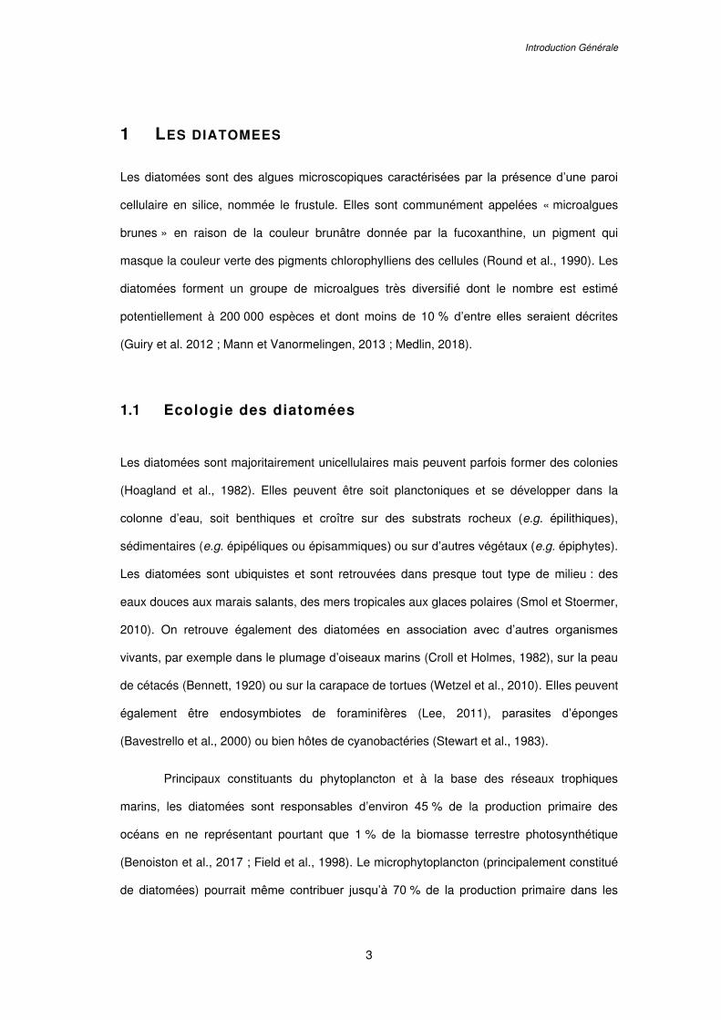
Figure 1.1. Diversité de la structure des frustules siliceux des diatomées nettoyés à l’acide
et observés en microscopie électronique à balayage (MEB). (A,F) Diatomées centriques
radiales, (B,C) diatomées pennées et (D,E) diatomées centriques polaires. Les frustules des
diatomées sont constitués de deux valves : l’épivalve et l’hypovalve liées par un ensemble
de bandes cingulaires (le cingulum). Barres d’échelles : 10 µm. Cette figure est adaptée de
photographies provenant de Kociolek et al. (2015a, 2015b) pour (A,B,C) et de Losic et al.
(2009) pour (D,E,F).
Les frustules des diatomées présentent une grande variété de formes. Ils sont
percés de pores et ornementés d’aréoles ou encore de stries qui constituent des motifs
propres à chaque espèce. Ces caractères morphologiques, visibles en microscopie
électronique à balayage (MEB), sont en effet utilisés pour identifier les diatomées au
niveau spécifique. Les pores ou les conduits présents sur le frustule permettent à la
cellule de communiquer et d’échanger avec le milieu extérieur en captant notamment des
frustule
A B
C
E
D
F
Chapitre I.
6
éléments dissous dans l’eau et en sécrétant des composés cellulaires dans le milieu
extérieur. C’est le cas du raphé (i.e. fente dans la paroi de la valve mettant en contact la
cellule et le milieu extérieur) chez les diatomées pennées ou du fultoportula et du
rimoportula (i.e. structures tubulaires par lesquelles s’effectuent la sécrétion de
substances vers le milieu extérieur) chez les diatomées centriques (Round et al., 1990 ;
Ruck et Theriot, 2011). Bien que la fonction pour la cellule de cet exosquelette siliceux soit
encore mal connue, il est suggéré que le frustule protégerait les diatomées de potentiels
prédateurs (Hamm et al., 2003) ou constituerait une barrière physique limitant les
infections virales ou fongiques (Hanic et al., 2009). Il est également démontré que la silice
biogène (SiO2) qui constitue les frustules protège les cellules des dommages induits par
les rayonnements ultraviolets (Ingalls et al., 2010). Le frustule pourrait également
permettre le positionnement des diatomées dans la colonne d’eau en contenant la
pression de turgescence engendrée par le protoplaste (Raven et Waite, 2004).
1.3 Classification des diatomées
Les diatomées auraient émergé d’un ancêtre commun il y a plus de 250 millions d’années
(Medlin, 2011). Il a été suggéré que l’origine des diatomées résulterait d’endosymbioses
multiples entre des cellules hétérotrophes et des ancêtres autotrophiques de microalgues
vertes puis de microalgues rouges (Armbrust, 2009 ; Benoiston et al., 2017). Cependant,
l’origine endosymbiotique avec un ancêtre des microalgues vertes est aujourd’hui encore
soumise à débat (Deschamps et Moreira, 2012 ; Kociolek et Hamsher, 2017).
La classification des diatomées est elle aussi en constante évolution, au gré de
l’identification de nouvelles espèces et des progrès technologiques et scientifiques. Avant
les années 90, la classification des diatomées était principalement basée sur des critères
morphologiques par l’observation des frustules en MEB selon une morpho-taxonomie
établie par Round et al. (1990). Les diatomées étaient alors catégorisées en tant que
division (Bacillariophyta) regroupant trois classes : les pennées raphides, avec la
Introduction Générale
7
présence d’un raphé sur au moins l’une des valves (Bacillariophyceae), les pennées
araphides, sans raphé (Fragilariophyceae) et les diatomées centrées
(Coscinodiscophyceae). Cette classification basée sur la morphologie a été sujette à
modification (Hoek et al., 1995) jusqu’à être remise en cause au début des années 2000
avec l’émergence des techniques de phylogénies moléculaires (Kooistra et al., 2003). Une
nouvelle classification des diatomées, basée sur des analyses moléculaires, a donc
récemment été établie par Medlin (2016) en classant les diatomées dans la lignée des
Héterokontophytes, division des Bacillarophyta :
Sous-division des Coscinodiscophytina
o Classe des Coscinodiscophyceae (centrées radiales)
Sous-division des Bacillariophytina
o Classe des Mediophyceae (centrées polaires)
o Classe des Bacillariophyceae (pennées)
Sous-classe des Urneidophycidae (pennées araphides)
Sous-classe des Fragilariophycidae (pennées araphides)
Sous-classe des Bacillariophycidae (pennées raphides)
1.4 Cycle de vie et reproduction des diatomées
Les diatomées sont des cellules diploïdes qui se multiplient par divisions végétatives.
Lors de la mitose, les valves du frustule de la cellule mère deviendront les épivalves des
deux cellules filles qui synthétiseront chacune l’hypovalve manquante, de plus petite taille
(Figure 1.2). De ce fait, les phases de multiplications asexuées entrainent une diminution
progressive de la taille cellulaire moyenne de la population ; jusqu’à atteindre une taille
minimale critique au-delà de laquelle les plus petites cellules ne sont plus viables. Afin de
restaurer des cellules de grandes tailles, les diatomées effectuent périodiquement une
phase de reproduction sexuée, appelée auxosporulation. La cellule doit atteindre une
gamme de taille favorable, spécifique à chaque espèce, pour que la reproduction sexuée
Chapitre I.
8
se produise (Kaczmarska et al., 2013). Ces cellules, dont la taille optimale est atteinte
pour initier la méiose et la production de gamètes sont les gamétocystes.
Les diatomées centrées et pennées font appel à deux processus distincts lors de
la reproduction sexuée :
Les diatomées centrées sont homothalliques, c'est-à-dire qu’il n’existe pas de
lignées de types sexuels différents. Au moment de la reproduction sexuée, les diatomées
centrées se différencient en cellule mâle ou en cellule femelle et produisent, suite à la
méiose, deux types de gamètes : les spermatocytes (e.g. petits gamètes flagellés mâles)
et les oogonies (e.g. gamètes femelles non mobiles de grande taille). La conjugaison des
gamètes entraîne la formation d’un zygote diploïde (Figure 1.2).
Les diatomées pennées sont majoritairement hétérothalliques, c’est à dire que
deux types de gamètes sont produits par deux cellules de types sexuels différents. Au
moment de l’auxosporulation, les gamétocystes de types sexuels différents (type + et
type -) s’apparient deux à deux, ce qui amorcera le processus de la méiose. Dans
certains cas (anisogamie), les gamétocystes « + » produisent des gamètes actifs
(mobiles) et les gamétocystes « - » des gamètes passifs (immobiles). Après ouverture
des valves des gamétocytes, les gamètes « + » se déplacent pour fusionner avec les
gamètes « - », restés associés au frustule du gamétocyste « - ». La conjugaison des
gamètes entraîne la formation de deux zygotes diploïdes (Figure 1.2). Dans d'autres cas,
chaque gamétocyste produit un gamète mobile et un autre immobile (isogamie).
Chez les diatomées centrées comme pennées le zygote diploïde s'allonge et se
développe en auxospore dans laquelle une cellule initiale de grande taille se forme. La
cellule initiale est ensuite libérée dans le milieu et formera à son tour une nouvelle lignée
par multiplications végétatives (Figure 1.2).
Introduction Générale
9
Figure 1.2. Schéma décrivant le cycle de vie monogénétique diplophasique des diatomées.
Durant la multiplication végétative, les cellules filles synthétisent une nouvelle valve à
l’intérieur de la valve fournit par la cellule mère. Cela entraîne une diminution progressive de
la taille cellulaire de la population. Les cellules pouvant initier le processus de reproduction
sexuée (auxosporulation) sont appelées gamétocystes. Chez les diatomées centriques,
l’auxosporulation se produit entre deux cellules d’une même lignée sexuelle et la méiose
donne deux types de gamètes : un macrogamète (oogonie) et des microgamètes uniflagellés
(spermatocytes). Chez les diatomées pennées, l’auxosporulation se produit lorsque deux
souches de types sexuels différents (types + et -) sont en contact étroit. La conjugaison des
gamètes haploïdes produit un zygote qui se développera en auxospore. Une future cellule
initiale aux parois siliceuses se forme dans l’auxospore et sera libérée dans le milieu.
Schéma C. Falaise©.
Méiose
Gamètes (n)
Conjugaison
Zygote (2n)
Auxospore
Cellule initiale
Ph
ase hap
loïd
e
Diatomées centrées
Diatomées pennées
Type + Type -
oogonie
spermatocytes gamètes + gamètes -
Gamètocyste (2n)
Chapitre I.
10
1.5 Utilisation et valorisation des diatomées
Divers secteurs s’intéressent à l’utilisation et à la valorisation des diatomées. Les structures
à potentiel de valorisation sont soit la cellule vivante entière, soit des métabolites produits
par la diatomée ou encore le frustule siliceux.
La principale utilisation des diatomées est dans le secteur de l’aquaculture,
notamment pour les écloseries de bivalves. Elles peuvent être délivrées dans les bassins
d’élevage comme aliment « fourrage » ou intégrées dans la composition de compléments
alimentaires (Bellou et al., 2016 ; Muller-Feuga, 2000). Les diatomées sont en effet réputées
pour leur contenu lipidique riche en acides gras polyinsaturés, essentiels à la croissance des
larves (Brown et al., 1997 ; Guedes et Malcata, 2012). Les diatomées benthiques sont
également utilisées comme indicateur de la qualité biologique de l’eau. L’indice biologique
des diatomées (IBD) a été normalisé par l’AFNOR pour estimer la qualité des cours d’eau de
France métropolitaine (Norme AFNOR (NF T90-354), 2007, 2016). En paléontologie, la
diatomite (e.g. roche formée par les frustules des diatomées fossiles) peut être étudiée pour
la reconstitution de paléo-environnements (Mackay et al., 2003 ; Smol et Stoermer, 2010).
Cette roche est également connue pour être utilisée, en association avec de la
nitroglycérine, dans la fabrication de la dynamite. L’utilisation des frustules siliceux intéresse
également le secteur des nanotechnologies pour des applications dans des composés
électroniques, optiques ou encore dans la fabrication de batteries (La Barre et Bates, 2018).
Certains composés produits par les diatomées peuvent également trouver une application
en pharmacologie. C’est notamment le cas de l’acide domoïque produit par certaines
diatomées du genre Pseudo-nitzschia et dont des applications sont envisagées en
neuropharmacologie. En effet, ce composé, ingéré suite à la consommation de fruits de mer
contaminés, provoque des pertes de mémoire parfois permanentes et pourrait représenter
un modèle pour l’étude des maladies de d’Alzheimer ou de Parkinson (Lefebvre et
Robertson, 2010 ; Trainer et al., 2012).
Introduction Générale
11
2 LES DIATOMEES DU GENRE HASLEA
2.1 Caractéristiques générales du genre Haslea
Les diatomées du genre Haslea appartiennent à la famille des Naviculaceae. Anciennement
classées au sein des genres Navicula, Amphipleura ou encore Pleurosigma, elles ont été
inclues dans un nouveau genre par Simonsen (1974) suite à l’observation approfondies des
frustules en MEB. La dénomination de ce nouveau genre nommé Haslea fût donnée en
l’honneur de la biologiste Norvégienne Grethe Rytther Hasle (1920-2013).
Morphologiquement, les diatomées du genre Haslea sont caractérisées par leur
forme pennée en navette, et possèdent deux chloroplastes pariétaux. Les observations en
MEB du frustule permettent de distinguer un raphé droit avec une terminaison peu
accentuée sur chacune des deux valves. La partie externe du frustule est composée de
stries siliceuses longitudinales parallèles au raphé tandis que la partie interne est creusée de
rangées d’aréoles quadrilatères lui donnant un aspect gaufré (Figure 1.3). Ces deux parties
sont reliées entre elles par des piliers.
Près d’une trentaine d’espèces de diatomées du genre Haslea ont été décrites
(AlgaeBase, 2018). Ces différentes espèces peuvent présenter des variations sur les plans
morphologiques, écologiques ou physiologiques (Gastineau et al., 2014a). Par exemple,
l’espèce Haslea wawrikae est une diatomée planctonique, contrairement à la majorité des
espèces de ce genre qui présente un mode de vie benthique (Simonsen, 1974). L’espèce
Haslea gigantea est quant à elle catégorisée comme constituant du mésoplancton en raison
de sa grande taille pouvant atteindre 500 µm, en opposition aux espèces du genre ne
mesurant majoritairement qu’une centaine de micromètres (Simonsen, 1974). L’espèce
Haslea ostrearia présente la particularité de synthétiser et de libérer dans son milieu un
pigment bleu hydrosoluble, la marennine (Simonsen, 1974).
Chapitre I.
12
2.2 La diatomée bleue Haslea ostrearia
2.2.1 Caractéristiques morphologiques
Les dimensions de Haslea ostrearia en milieu naturel varient de 60 à 120 µm de longueur et
6 à 12 µm de largeur (Robert, 1973, 1978). En laboratoire, la longueur maximale d’une
cellule initiale après reproduction sexuée est de 138 à 140 µm, et la taille minimale en deçà
de laquelle une cellule ne peut plus se diviser est de 17 µm (Davidovich et al., 2009 ;
Neuville et Daste, 1978). En microscopie optique deux chloroplastes pariétaux sont
observables ainsi que des vésicules bleues, essentiellement concentrées aux apex de la
cellule, contenant la marennine (Figure 1.3).
Figure 1.3. Photographies de la diatomée pennée Haslea ostrearia. (A) Observations en
microscopie permettant de distinguer deux chloroplastes pariétaux et la présence de
vésicules contenant un pigment bleu (la marennine). Barre d’échelle 5 µm.
(B-E) Photographies du frustule siliceux en microscopie électroniques à balayage (MEB)
avec observations des apex de la cellule (B,C) et du centre de la cellule (D,E) en vue
externe (B,D) et interne (C,E). Barres d’échelle 1 µm. Photographie optique communiquée
par A. Alverson et photographies MEB d’après Gastineau et al. (2014a).
A B
C
D
E
Introduction Générale
13
2.2.2 Écologie
Haslea ostrearia est une diatomée benthique qui peut former des biofilms sur des surfaces
sédimenteuses ou sur des macroalgues, ou d'autres supports immergés. Elle peut être
occasionnellement rencontrée en milieu pélagique lors de la remise en suspension des
sédiments ou par brassage des courants (Robert, 1983). Haslea ostrearia est une diatomée
euryhaline qui peut s’acclimater à des salinités allant de 1,5 à 4,0 % (Wraige et al., 1998).
Elle supporte également des gammes d’intensités lumineuses variant de 20 à
750 µmol m-1 s-1 (Mouget et al., 1999) et possède des mécanismes de défense contre le
stress UV (Rech et al., 2005).
2.2.3 Physiologie
Haslea ostrearia est photosynthétique et photoautotrophe, bien qu’elle puisse présenter une
photohétérotrophie ou photomixotrophie pour certaines substances azotées et carbonées
(Neuville et Daste, 1978 ; Robert et al., 1982). Comme toutes les diatomées, H. ostrearia
possède deux voies d’assimilation du carbone minéral : la voie de la ribulose-bisphosphate-
carboxylase (RuBPC) associée au cycle de Calvin-Benson, et la voie de la
phosphoénolpyruvatecarboxykinase (PEPCK ; Tremblin et Robert, 2001). Une voie
alternative de β-carboxylation associée à une activité anhydrase carbonique dépendante de
l’intensité lumineuse a également été démontrée (Rech et al., 2008 ; Tremblin et Robert,
2001). La composition biochimique d’H. ostrearia reste stable au cours du vieillissement des
cultures, avec une teneur en glucides et en protéines moyennant 50 mg / 10-9 cellules et
200 mg / 10-9 cellules respectivement (Robert, 1983). En revanche, la teneur en lipides varie
entre 60 et 200 mg / 10-9 cellules en fonction de la phase de croissance de la culture et les
proportions en acides gras, lipides neutres et glycolipides sont semblables à celles d’autres
diatomées (Robert, 1983). Haslea ostrearia synthétise des acides gras polyinsaturés ω-3
(Mimouni et al., 2003) et possède une voie de synthèse d’hydrocarbures insaturés
(i.e. alcanes) polyramifiés tels que des isoprénoïdes, dont certains à 25 carbones ont été
nommés haslènes en référence au genre Haslea (Allard et al., 2001 ; Wraige et al., 1998).
Les pigments retrouvés chez H. ostrearia sont les chlorophylles a et c, la diatoxanthine, la
Chapitre I.
14
diadinoxanthine, la fucoxanthine et le pigment bleu marennine. Dans une culture en mode
discontinu (batch), l’organisation des thylakoïdes et le comportement photosynthétique de
H. ostrearia varient au cours de l’accumulation de marennine dans la cellule (Nassiri et al.,
1998).
2.2.4 Reproduction
La reproduction sexuée chez H. ostrearia présente la particularité d’être hétérothallique, se
produisant entre cellules compatibles de types sexuels différents (Davidovich et al., 2009),
mais aussi homothallique, pouvant se produire entre deux cellules de même type sexuel
(Neuville et Daste, 1978). Ces deux types de reproductions sexuées sont préférentiellement
déclenchés sous faible éclairement (< 50 photons m-2 s-1) et lors de courtes photopériodes
(entre 6 et 10 h d’éclairement journalier), correspondant à des conditions hivernales
(Davidovich et al., 2009 ; Neuville et Daste, 1978). La qualité de la lumière joue également
un rôle dans l’induction de la reproduction sexuée (Mouget et al., 2009). La taille maximale
des gamétocystes est estimée à 68 µm, soit environ la moitié de la taille de la cellule initiale
(Davidovich et al., 2009).
2.2.5 Culture
Différents milieux de culture sont utilisés en laboratoire pour cultiver H. ostrearia. En général,
des solutions d’enrichissement telles que celles proposées par Provasoli et al. (1957) ou par
Robert (1983) sont ajoutées à de l’eau de mer naturelle filtrée ou à de l’eau de mer artificielle
(Harrison et al., 1980). Un milieu de culture a également été développé spécifiquement pour
H. ostrearia, nommé diatom artificial medium (Gagneux-Moreaux et al., 2007).
Sur le long terme, Haslea ostrearia, comme beaucoup de diatomées pennées, est
difficile à maintenir en culture en laboratoire en raison de la réduction de la taille des cellules
au cours de la multiplication végétative. Les mitoses successives conduisent au
développement de petites cellules tordues, de forme tératogène, dont l’état physiologique
est diminué (Falasco et al., 2009). De ce fait, une souche de H. ostrearia est difficilement
maintenue en laboratoire au delà de 2-3 ans en culture en batch : la taille et la forme des
Introduction Générale
15
cellules devenant critiques (Figure 1.4). Lorsque la culture en batch est appliquée en
laboratoire pour le maintien des souches, il est donc nécessaire d’effectuer des repiquages
très réguliers (< 20 jours) pour éviter que les cultures n’entrent en phase de déclin (Figure
1.4). Pour maintenir les souches dans le plus long terme, une technique d’immobilisation des
cellules dans de l’alginate à 4°C et sous faible éclairement a été proposée (Gaudin et al.,
2006). En revanche, les techniques de cryopréservation ne sont pas recommandées pour
H. ostrearia car elles peuvent induire plus de 90 % de mort cellulaire (Tanniou et al., 2012).
Figure 1.4. (A) Photographie d’une cellule de grande taille de Haslea ostrearia prise
quelques jours après son isolement dans une claire ostréicole de la région de Bourgneuf (à
gauche), et d’une cellule après un an de culture en laboratoire (à droite) présentant une
petite taille et une forme atrophiée. (B) Courbe de croissance d’une culture de 200 mL en
batch de H. ostrearia. Après une croissance exponentielle, la culture entre en phase de
déclin sous 15 jours et nécessite un nouveau repiquage.
Haslea ostrearia est jusqu’à présent essentiellement cultivée à l’échelle du
laboratoire (< 100 L), mais des cultures en photobioréacteurs (PBR, < 7 L), en semi-pilote
(10 m3) ou en bassins (200 L) ont également été menées (Gastineau et al., 2014b ;
Rossignol et al., 2000 ; Turpin, 1999). Les unités de production à grande échelle ont encore
besoin d’être optimisées car les PBR, généralement développés pour cultiver les
0,0E+00
2,0E+04
4,0E+04
6,0E+04
8,0E+04
1,0E+05
1,2E+05
0 3 6 9 12 15 18
Co
nce
ntr
atio
n (c
ellu
les
mL
-1)
Jours
10 µm
A
B
n=3, moy. ± ET
Chapitre I.
16
microalgues en suspension, ne conviennent pas pour H. ostrearia qui sédimente et forme
des biofilms.
2.2.6 Distribution géographique
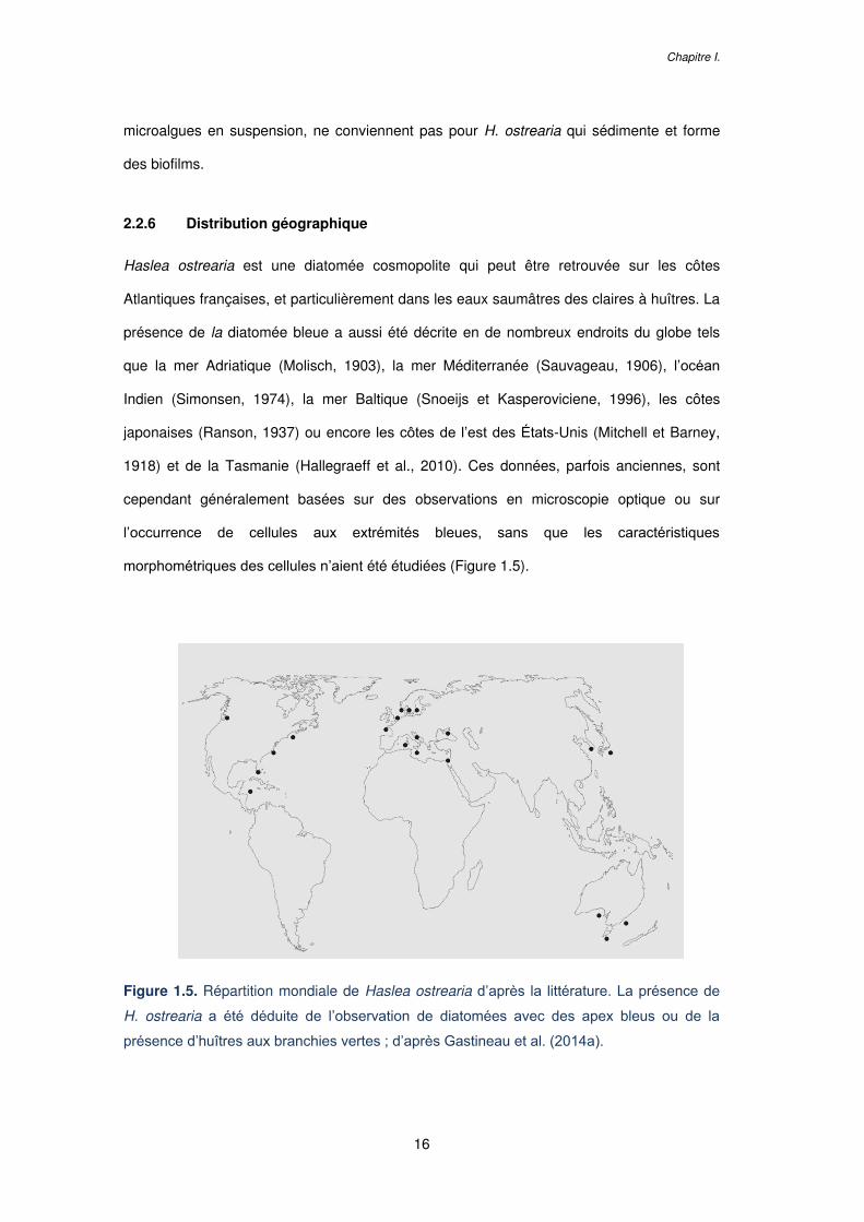
Haslea ostrearia est une diatomée cosmopolite qui peut être retrouvée sur les côtes
Atlantiques françaises, et particulièrement dans les eaux saumâtres des claires à huîtres. La
présence de la diatomée bleue a aussi été décrite en de nombreux endroits du globe tels
que la mer Adriatique (Molisch, 1903), la mer Méditerranée (Sauvageau, 1906), l’océan
Indien (Simonsen, 1974), la mer Baltique (Snoeijs et Kasperoviciene, 1996), les côtes
japonaises (Ranson, 1937) ou encore les côtes de l’est des États-Unis (Mitchell et Barney,
1918) et de la Tasmanie (Hallegraeff et al., 2010). Ces données, parfois anciennes, sont
cependant généralement basées sur des observations en microscopie optique ou sur
l’occurrence de cellules aux extrémités bleues, sans que les caractéristiques
morphométriques des cellules n’aient été étudiées (Figure 1.5).
Figure 1.5. Répartition mondiale de Haslea ostrearia d’après la littérature. La présence de
H. ostrearia a été déduite de l’observation de diatomées avec des apex bleus ou de la
présence d’huîtres aux branchies vertes ; d’après Gastineau et al. (2014a).
Introduction Générale
17
2.3 Haslea ostrearia , unique en son genre ? La diversité
insoupçonnée des Haslea bleues.
Haslea ostrearia a longtemps été considérée comme la seule diatomée du genre Haslea à
synthétiser un pigment bleu. Cependant, de récentes campagnes d’échantillonnage de
phytoplancton dans différents pays et continents, alliées à des analyses morphométriques
des frustules en MEB ainsi qu’à l’utilisation de marqueurs moléculaires ont permis de révéler
l’existence d’espèces différentes produisant également un pigment bleu hydrosoluble
(Gastineau et al., 2014a). La première nouvelle espèce identifiée, Haslea karadagensis, a
été échantillonnée en 2008 dans la mer Noire et décrite par Gastineau et al. (2012a). De
nouvelles espèces ont ensuite été identifiées aux îles Canaries et en mer Méditerranée,
nommées respectivement Haslea silbo (Gastineau, 2011) et Haslea provincialis (Gastineau
et al., 2016). Haslea silbo, initialement échantillonnée sur l’île de la Gomera, a par la suite
été identifiée en Caroline du Nord et dans la baie de Naples. Une autre espèce, encore
partiellement identifiée, a également été retrouvée aux îles Canaries, sur l’île de Gran
Canaria, et est pour l’instant nommée Haslea acoran (Gastineau, 2011). Ces différentes
espèces sont sexuellement incompatibles et présentent des variations d’ornementations sur
les frustules (Figure 1.6). Aussi, l’utilisation du marqueur moléculaire cox1 (gène codant pour
la première sous-unité de l’enzyme cytochrome oxydase) a démontré un pourcentage
d’identité de paires de bases (pb) de seulement 90 % entre ces nouvelles espèces et
H. ostrearia, tandis que deux sous-populations de H. ostrearia isolées sur les côtes
françaises (baie de Bourgneuf) et australiennes (baie de Coffin) présentent quant à elles
entre 98 et 100 % d’identité de pb (Gastineau et al., 2014a).
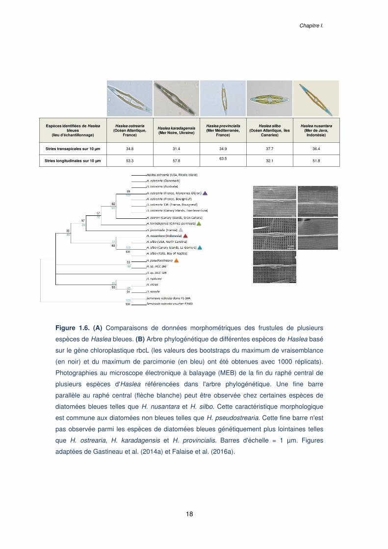
Chapitre I.
18
Figure 1.6. (A) Comparaisons de données morphométriques des frustules de plusieurs
espèces de Haslea bleues. (B) Arbre phylogénétique de différentes espèces de Haslea basé
sur le gène chloroplastique rbcL (les valeurs des bootstraps du maximum de vraisemblance
(en noir) et du maximum de parcimonie (en bleu) ont été obtenues avec 1000 réplicats).
Photographies au microscope électronique à balayage (MEB) de la fin du raphé central de
plusieurs espèces d'Haslea référencées dans l'arbre phylogénétique. Une fine barre
parallèle au raphé central (flèche blanche) peut être observée chez certaines espèces de
diatomées bleues telles que H. nusantara et H. silbo. Cette caractéristique morphologique
est commune aux diatomées non bleues telles que H. pseudostrearia. Cette fine barre n'est
pas observée parmi les espèces de diatomées bleues génétiquement plus lointaines telles
que H. ostrearia, H. karadagensis et H. provincialis. Barres d'échelle = 1 µm. Figures
adaptées de Gastineau et al. (2014a) et Falaise et al. (2016a).
Espèces identifiées de Hasleableues
(lieu d’échantillonnage)
Haslea ostrearia(Océan Atlantique,
France)
Haslea karadagensis(Mer Noire, Ukraine)
Haslea provincialis(Mer Méditerranée,
France)
Haslea silbo(Océan Atlantique, îles
Canaries)
Haslea nusantara(Mer de Java,
Indonésie)
Stries transapicales sur 10 µm 34.8 31.4 34.9 37.7 36.4
Stries longitudinales sur 10 µm 53.3 57.863.5
32.1 51.8
Haslea bleues
Haslea non bleues
A
B
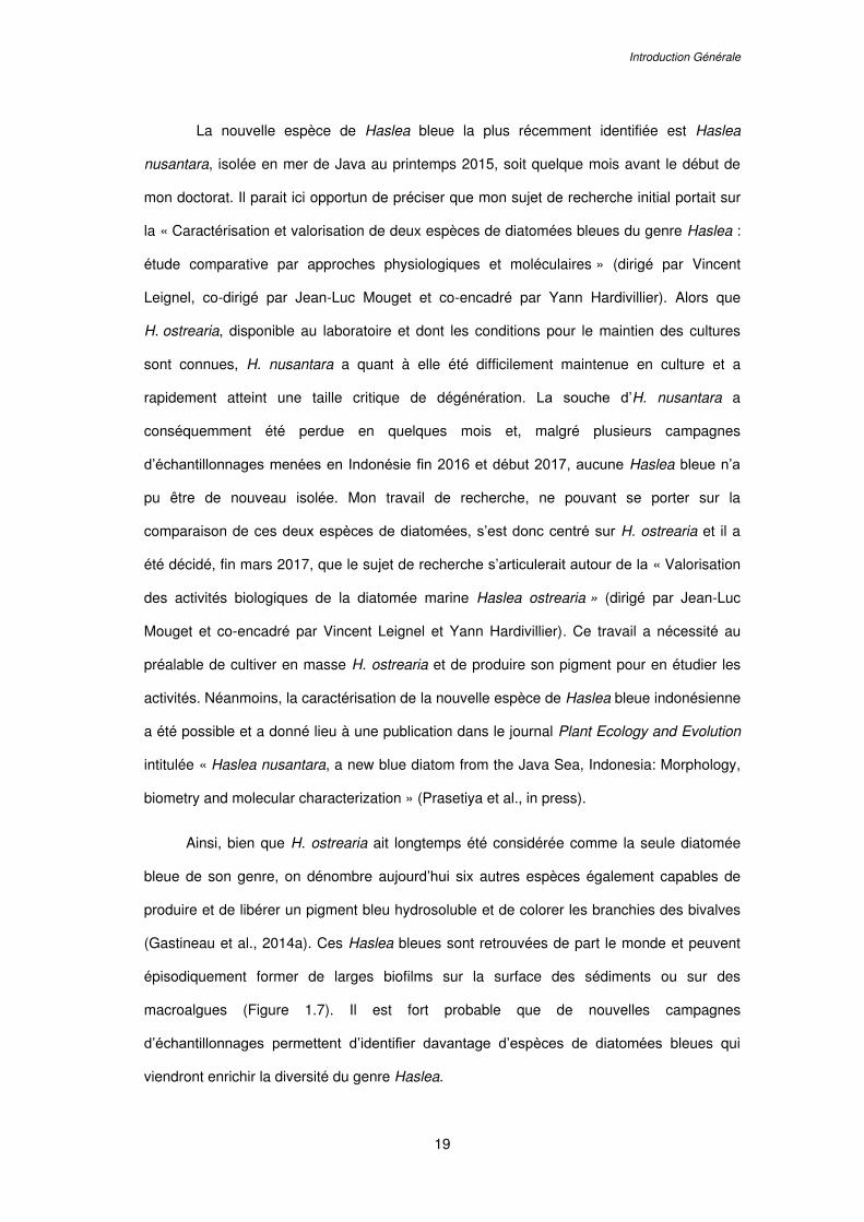
Introduction Générale
19
La nouvelle espèce de Haslea bleue la plus récemment identifiée est Haslea
nusantara, isolée en mer de Java au printemps 2015, soit quelque mois avant le début de
mon doctorat. Il parait ici opportun de préciser que mon sujet de recherche initial portait sur
la « Caractérisation et valorisation de deux espèces de diatomées bleues du genre Haslea :
étude comparative par approches physiologiques et moléculaires » (dirigé par Vincent
Leignel, co-dirigé par Jean-Luc Mouget et co-encadré par Yann Hardivillier). Alors que
H. ostrearia, disponible au laboratoire et dont les conditions pour le maintien des cultures
sont connues, H. nusantara a quant à elle été difficilement maintenue en culture et a
rapidement atteint une taille critique de dégénération. La souche d’H. nusantara a
conséquemment été perdue en quelques mois et, malgré plusieurs campagnes
d’échantillonnages menées en Indonésie fin 2016 et début 2017, aucune Haslea bleue n’a
pu être de nouveau isolée. Mon travail de recherche, ne pouvant se porter sur la
comparaison de ces deux espèces de diatomées, s’est donc centré sur H. ostrearia et il a
été décidé, fin mars 2017, que le sujet de recherche s’articulerait autour de la « Valorisation
des activités biologiques de la diatomée marine Haslea ostrearia » (dirigé par Jean-Luc
Mouget et co-encadré par Vincent Leignel et Yann Hardivillier). Ce travail a nécessité au
préalable de cultiver en masse H. ostrearia et de produire son pigment pour en étudier les
activités. Néanmoins, la caractérisation de la nouvelle espèce de Haslea bleue indonésienne
a été possible et a donné lieu à une publication dans le journal Plant Ecology and Evolution
intitulée « Haslea nusantara, a new blue diatom from the Java Sea, Indonesia: Morphology,
biometry and molecular characterization » (Prasetiya et al., in press).
Ainsi, bien que H. ostrearia ait longtemps été considérée comme la seule diatomée
bleue de son genre, on dénombre aujourd’hui six autres espèces également capables de
produire et de libérer un pigment bleu hydrosoluble et de colorer les branchies des bivalves
(Gastineau et al., 2014a). Ces Haslea bleues sont retrouvées de part le monde et peuvent
épisodiquement former de larges biofilms sur la surface des sédiments ou sur des
macroalgues (Figure 1.7). Il est fort probable que de nouvelles campagnes
d’échantillonnages permettent d’identifier davantage d’espèces de diatomées bleues qui
viendront enrichir la diversité du genre Haslea.
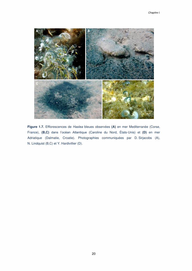
Chapitre I.
20
Figure 1.7. Efflorescences de Haslea bleues observées (A) en mer Mediterranée (Corse,
France), (B,C) dans l’océan Atlantique (Caroline du Nord, États-Unis) et (D) en mer
Adriatique (Dalmatie, Croatie). Photographies communiquées par D. Sirjacobs (A),
N. Lindquist (B,C) et Y. Hardivillier (D).
A B
C D
Introduction Générale
21
3 LES PIGMENTS BLEUS D’HASLEA
3.1 La marennine et le verdissement des huîtres
3.1.1 Les huîtres « fines de claires vertes »
La marennine produite par H. ostrearia est connue pour induire le verdissement des
branchies des huîtres dans les claires ostréicoles, des bassins d’eau de mer dans lesquels
les huîtres sont mises en affinage (Figure 1.8). Dans ces bassins peu profonds et riches en
nutriments, H. ostrearia peut proliférer et libérer de grandes quantités de marennine qui
rendra l’eau des claires verte. Les huîtres obtiendront ainsi par filtration des branchies
verdies en quelques jours seulement (Robert, 1983).
Ce phénomène de verdissement naturel présente un intérêt économique dans
l’activité ostréicole des régions de Marennes-Oléron et de la baie de Bourgneuf dans l’ouest
de la France, où l’affinage des huîtres en claires est pratiqué. Les huîtres verdies de
Marennes-Oléron portent en effet un label rouge particulier « huîtres fines de claires vertes »
et se vendent en moyenne 20 à 30 % plus cher que des huîtres affinées non verdies
(Figure 1.8). Ce prix de vente plus élevé peut s’expliquer par la rareté du produit : les blooms
de H. ostrearia dans les claires étant un phénomène erratique, mais également par une
différence de saveur entre les huîtres vertes et non vertes. Cependant, l’amélioration des
propriétés organoleptiques avancée par les amateurs d’huîtres ne proviendrait pas
nécessairement du pigment lui-même, mais pourrait plutôt résulter de la modification de la
communauté de phytoplancton des claires lors des blooms de H. ostrearia, modifiant ainsi
l’alimentation des huîtres et potentiellement leur goût. En effet, la prolifération de H. ostrearia
dans les claires génère un effet d’ombrage qui empêche la croissance d’autres microalgues
(Pouvreau et al., 2007). De plus, il a été montré que la marennine possède des propriétés
allélopathiques qui ralentissent, voire préviennent la croissance d’autres microalgues
(Pouvreau et al., 2007 ; Prasetiya et al., 2016).
Chapitre I.
22
Figure 1.8. (A) Affinage des huîtres dans des bassins d’eau de mer peu profonds appelés
« claires » dans la région de Marennes-Oléron. (B) Huître « fine de claire » et huître « fine
de claire verte » (label Rouge français) dont les branchies ont été verdies par la présence de
Haslea ostrearia et de son pigment (marennine) dans les claires lors de l’affinage.
3.1.2 La fixation de la marennine sur les branchies
Les mécanismes par lesquels la marennine se fixe aux branchies des huîtres sont encore
aujourd’hui mal connus. Il a été montré que les huîtres gardent la signature de la marennine
plusieurs mois sur leurs branchies même élevées en absence de H. ostrearia (Gastineau et
al., 2014b). L’étude de coupes histologiques d’huîtres verdies montre que la marennine
pénètre les plis des branchies et se fixe principalement dans les mucocytes ; des cellules
produisant et excrétant du mucus (Gastineau et al., 2018 ; Semba Prasetiya, 2015).
Il est à noter que le verdissement des branchies induit par la marennine n’est pas
exclusif aux huîtres et que d’autres organismes tels que la moule, le pétoncle, la coque ou la
palourde présentent également un verdissement des branchies suite à une exposition au
pigment (Gastineau et al., 2014b). Aussi, ce phénomène ne se produit pas uniquement dans
les claires ostréicoles mais peut survenir en milieu naturel, au niveau de baies où la mer
n’est pas trop agitée, ou dans des milieux totalement ouverts (voir Figure 1.7).
Marennes-Oléron
A B
Introduction Générale
23
3.1.3 La consommation des huîtres vertes, une spécialité française
Les huîtres vertes sont à l’heure actuelle presque exclusivement consommées en France
mais leur production et leur consommation commencent à se développer en Australie et aux
États-Unis. Dans l’hexagone, les huîtres vertes sont considérées depuis plusieurs siècles
comme un met de grande qualité et étaient d’ailleurs retrouvées à la table des rois de
France (Grelon, 1978), alors qu’ailleurs la méconnaissance du phénomène à l’origine de ce
verdissement et la couleur inhabituelle laissaient penser aux ostréiculteurs et aux
consommateurs que ces huîtres étaient polluées et impropres à la consommation (Boyce et
Herdman, 1896). D’ailleurs, les rapports du ministère du Commerce américain de 1916
indiquaient que les branchies vertes étaient un « problème », alertant qu’« aucune méthode
de contrôle des branchies vertes n'a été déterminée » (US Report of the Secretary of
Commerce and Reports of Bureaus, 1916). En effet, avant de pouvoir déterminer que le
verdissement des branchies était causé par une microalgue, différentes hypothèses avaient
été avancées pour tenter d’expliquer ce phénomène, décrit depuis le XVIIème siècle (Sprat,
1667). Ainsi, il a été suggéré que les branchies vertes étaient un signe de maladie des
huîtres comme l’obésité ou le disfonctionnement de la glande digestive (Pasquier, 1818 ;
Valenciennes, 1841). Il avait également été suggéré que le verdissement pouvait être dû à
des causes environnementales telles qu’un effet du soleil, la présence d’eaux saumâtres ou
de macroalgues vertes (Bosc et Lair, 1817 ; Sprat, 1667), ou résulterait d’interactions des
huîtres avec des métaux tels que le cuivre (Boon, 1977).
Ce n’est qu’au début du XIXème siècle que Gaillon identifie la présence de
microorganismes « verts » (appréciation incorrecte de la couleur due aux moyens
d'observation limités de l'auteur) comme étant à l’origine du verdissement. Ces
microorganismes sont d’abord considérés comme étant d’origine animale et sont nommés
Vibrio ostrearius (Gaillon, 1820). L’appellation « Navicule bleue » ou Navicula ostrearia fût
ensuite proposée par le botaniste Bory de Saint Vincent, qui suggéra une appartenance de
ces microorganismes au règne végétal (Bory de Saint-Vincent, 1826). La microalgue bleue a
ainsi été classée dans le genre Navicula pendant près de 150 ans puis, grâce aux progrès
Chapitre I.
24
de la microscopie, elle fût transférée dans le genre Haslea, crée spécialement pour cette
espèce aux caractéristiques morphologiques bien particulières (Simonsen, 1974).
3.2 La marennine et les pigments de type marennine
La marennine est le pigment bleu produit par l’espèce H. ostrearia. Le nom du pigment,
initialement marennin, fût proposé par (Lankester, 1886) en référence à la région de
Marennes-Oléron. La marennine est un pigment hydrosoluble, insoluble dans les solvants
organiques (Pouvreau et al., 2006a), et dont la couleur varie selon le pH, allant de violet à
bleu dans des conditions acides et passant de vert clair à vert-gris dans des conditions
basiques. Bien que H. ostrearia et son pigment aient fait l’objet de nombreuses études au
cours des dernières décennies, la voie de biosynthèse du pigment n'est pas encore
déterminée et sa structure chimique pas totalement élucidée.
Les pigments bleus produits par les autres espèces de Haslea bleues récemment
identifiées sont nommés pigments « de type marennine », en l’absence d’identification de
caractéristiques chimiques plus spécifiques. A l’heure actuelle, H. ostrearia et la marennine
demeurent davantage étudiées que les autres Haslea bleues et leurs pigments « de type
marennine ».
3.2.1 Production et excrétion de la marennine par Haslea
Bien que le processus soit encore mal identifié, il semblerait que la marennine soit produite
via une voie de synthèse cytoplasmique (Nassiri et al., 1998). Des vésicules contenant la
marennine se forment dans le cytoplasme avec une phase de concentration transitoire aux
deux extrémités cellulaires. La marennine est ensuite libérée de la cellule par exocytose
dans de petites vésicules qui éclatent au bout de quelques minutes permettant la libération
de la marennine dans le milieu. Les conditions qui favorisent la production et la libération de
marennine sont elles aussi incertaines. Des études ont suggéré une stimulation de
production par des substances organiques comme les glucosamines (Ranson, 1937) ou
Introduction Générale
25
encore par un stress salin (Bachrach et Simonet, 1936 ; Neuville et Daste, 1978) ou des
déficiences en fer ou vitamines (Neuville et Daste, 1978 ; Robert, 1983). La lumière est
également avancée comme étant un facteur influençant la production du pigment. Des
conditions de culture sous fort éclairement avec de longues photopériodes, ainsi que la
qualité de la lumière avec les longueurs d’ondes dans le bleu semblent favoriser la
production de marennine (Mouget et al., 2005 ; Tremblin et al., 2000). Sur des algues
maintenues en phase de croissance exponentielle (pas de limitation en nutriments ni en
lumière), plus le niveau d'éclairement est élevé, plus la production de marennine est
importante (Prasetiya et al., 2016).
Le coût métabolique de la production de marennine implique nécessairement un intérêt
pour la cellule et un rôle écologique, mais les fonctions biologiques du pigment restent
incertaines. La marennine n’est pas directement impliquée dans la photosynthèse ou la
captation lumineuse (Schubert et al., 1995). Des auteurs ont néanmoins suggéré une
implication indirecte de la marennine dans la photosynthèse en modifiant quantitativement et
qualitativement la lumière traversant la colonne d’eau (Mouget et al., 1999 ; Tremblin et al.,
2000). Une fonction écologique peut également être avancée en raison des effets
allélopathiques (Pouvreau et al., 2007 ; Prasetiya et al., 2016) et antimicrobiens (Gastineau
et al., 2014b) exercés par la marennine et prémunirait Haslea d’un moyen de compétition
envers d’autres microalgues ou bactéries, voire de potentiels prédateurs.
3.2.2 Caractérisations préliminaires du pigment
Aujourd’hui encore, la nature chimique de la marennine questionne. Différentes hypothèses
se sont succédées pour tenter de caractériser ce pigment. Des auteurs ont ainsi relié la
marennine à des caroténoïdes (Ranson, 1937), des produits de dégradation de la
chlorophylle (Bachrach et Simonet, 1936 ; Geneves et al., 1976 ; Robert, 1983), ou encore
des pigments de nature protéique tels que les phycobiliprotéines des cyanobactéries (Bocat,
1907). L’hypothèse la plus généralement admise est celle proposée par Pouvreau et al.
(2006a) qui ont décrit la marennine comme un polymère complexe portant des groupements
polyphénoliques. Une étude récente a également permis de caractériser le squelette
Chapitre I.
26
carboné de la molécule et d’identifier la présence de différents sucres (Gastineau et al.,
2014b). La marennine existerait sous deux formes : intracellulaire (IMn) et extracellulaire
(EMn) dont les poids moléculaires ont respectivement été estimés à 10,7 kDa et 9,9 kDa
(Pouvreau et al., 2006a).
3.2.3 Techniques de purification du pigment
Une méthode de purification de la IMn et de la EMn a été développée par Pouvreau et al.
(2006b). Elle comprend deux phases d’ultrafiltration, à 30 kDa et 3 kDa, ainsi qu’une phase
de chromatographie par échange d’anions, une étape de dialyse puis de lyophilisation
(Figure 1.9). Cette technique aboutit à l’obtention d’une poudre de marennine, partiellement
purifiée, permettant notamment de réaliser des essais biochimiques afin de caractériser
davantage le pigment. Des spectres « types » de la IMn et de la EMn en spectrophotométrie
UV-Visible et Raman ont ainsi pu être établis (Pouvreau et al., 2006a). Le spectre UV-Visible
de la marennine à pH 8 présente trois pics identifiables autour de 247, 322 et 675 nm
(Figure 1.9). Les spectres UV-Visible et Raman des pigments de « type marennine » de
H. silbo, H. provincialis et H. nusantara sont semblables à ceux de la marennine. Seul le
pigment de type marennine produit par H. karadagensis, qui apparait plus foncé que la
marennine en microscopie optique, présente des propriétés spectrales légèrement
différentes (Gastineau et al., 2012a).
Néanmoins, le procédé de purification développé par Pouvreau et al. (2006b) n’est
pas adapté pour le traitement de gros volumes de culture car il nécessite du matériel
coûteux (e.g. colonnes de chromatographies à échanges d’ions) et ne présente qu’un faible
rendement. Une telle technique de récupération de la marennine rend donc difficilement
envisageable son utilisation pour des essais biologiques à large échelle ou pour la
concentration du pigment en vue d’applications biotechnologiques et industrielles. La Blue
Water (BW), une solution de marennine extracellulaire concentrée par ultrafiltration
(Figure 1.9), peut être utilisée pour des essais biologiques. Elle peut en effet être produite
plus rapidement et en plus grande quantité que la marennine purifiée, mais cette technique
nécessite également d’effectuer des ultrafiltrations sur d’importants volumes de culture.
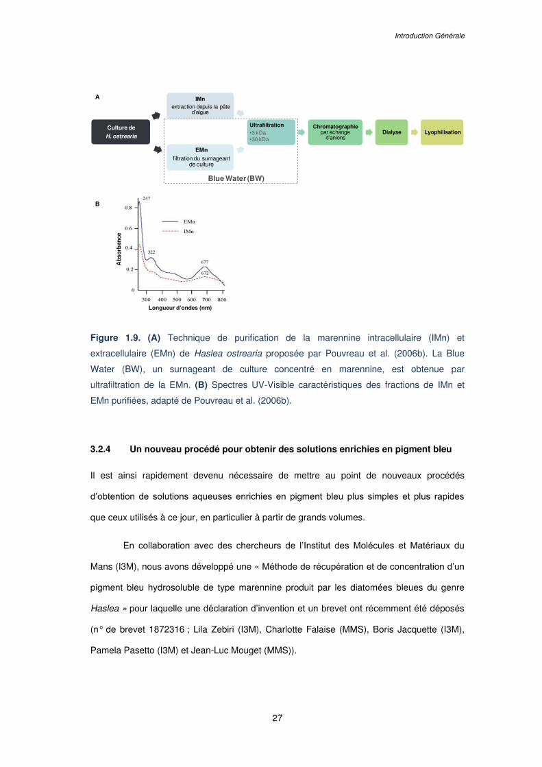
Introduction Générale
27
Figure 1.9. (A) Technique de purification de la marennine intracellulaire (IMn) et
extracellulaire (EMn) de Haslea ostrearia proposée par Pouvreau et al. (2006b). La Blue
Water (BW), un surnageant de culture concentré en marennine, est obtenue par
ultrafiltration de la EMn. (B) Spectres UV-Visible caractéristiques des fractions de IMn et
EMn purifiées, adapté de Pouvreau et al. (2006b).
3.2.4 Un nouveau procédé pour obtenir des solutions enrichies en pigment bleu
Il est ainsi rapidement devenu nécessaire de mettre au point de nouveaux procédés
d’obtention de solutions aqueuses enrichies en pigment bleu plus simples et plus rapides
que ceux utilisés à ce jour, en particulier à partir de grands volumes.
En collaboration avec des chercheurs de l’Institut des Molécules et Matériaux du
Mans (I3M), nous avons développé une « Méthode de récupération et de concentration d’un
pigment bleu hydrosoluble de type marennine produit par les diatomées bleues du genre
Haslea » pour laquelle une déclaration d’invention et un brevet ont récemment été déposés
(n° de brevet 1872316 ; Lila Zebiri (I3M), Charlotte Falaise (MMS), Boris Jacquette (I3M),
Pamela Pasetto (I3M) et Jean-Luc Mouget (MMS)).
A
B
Longueur d’ondes (nm)
Ab
sorb
ance
IMnextraction depuis la pâte
d’algue
Ultrafiltration•3 kDa•30 kDa
Chromatographiepar échange
d’anionsDialyse Lyophilisation
Blue Water (BW)
Culture de
H. ostrearia
EMn
filtration du surnageantde culture
Chapitre I.
28
Cette méthode présente l’avantage d’être beaucoup plus rapide que celle
développée par Pouvreau et al. (2006b), moins onéreuse et permet d’obtenir une solution de
marennine concentrée avec un spectre UV-Visible semblable à la EMn. Cette technique peut
s’appliquer à la marennine, mais également aux pigments de « type marennine » puisqu’elle
a été testée avec des cultures de H. provincialis, H. silbo et H. nusantara. L’invention
concerne un procédé d’obtention d’une solution aqueuse enrichie en pigment bleu
comprenant diverses étapes : 1) la filtration d’un surnageant de culture de Haslea contenant
la forme extracellulaire du pigment, 2) la précipitation du pigment bleu à l’hydroxyde de
sodium, 3) la séparation de la phase précipitée de la phase aqueuse, 4) la solubilisation du
pigment bleu compris dans la phase précipitée, 5) une dialyse et enfin 6) l’ultrafiltration de la
solution concentrée (Figure 1.10).
Grâce à cette technique, l’utilisation d’une solution concentrée en pigment bleu
pourrait être envisagée à l’échelle industrielle. Cependant, la structure chimique du pigment
n’étant pas définie, l’application de la marennine ou des pigments de type marennine dans
les secteurs de l’agroalimentaire ou de la pharmacologie ne peut pour l’instant pas être
considérée. En revanche, ces solutions aqueuses enrichies en pigment bleu pourront être
valorisées dans les secteurs de l’aquaculture ou de la cosmétologie, pour lesquels
l’identification de la molécule ne présente pas un impératif pour l’autorisation de mise sur le
marché.
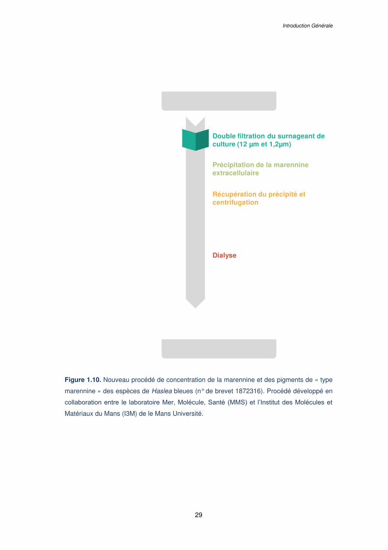
Introduction Générale
29
Figure 1.10. Nouveau procédé de concentration de la marennine et des pigments de « type
marennine » des espèces de Haslea bleues (n° de brevet 1872316). Procédé développé en
collaboration entre le laboratoire Mer, Molécule, Santé (MMS) et l’Institut des Molécules et
Matériaux du Mans (I3M) de le Mans Université.
Double filtration du surnageant de culture (12 µm et 1,2µm)
Précipitation de la marennine extracellulaire
Récupération du précipité et centrifugation
Dialyse
Double ultrafiltration
(“cut off” 30 kDa – 1 kDa)
Cultures de Haslea ostrearia
Solution de marennine concentrée
Solubilisation du pigment
Chapitre I.
30
4 ACTIVITES BIOLOGIQUES ET VALORISATIONS
POTENTIELLES DES HASLEA BLEUES ET DE LEURS
PIGMENTS
4.1 Les biotechnologies bleues
4.1.1 Le secteur de l’agroalimentaire
En plus d’applications en aquaculture, les pigments de type marennine peuvent avoir des
débouchés sous-exploités mais néanmoins pertinents dans le domaine émergent des
biotechnologies bleues. Pour le moment, le principal marché à considérer est « le pigment
bleu naturel ». De nos jours, les colorants bleus les plus utilisés pour les aliments et les
boissons sont le Brillant Blue (FD&C Blue n°1, E133), l’Indigo Carmine (FD&C Blue n°2,
E132), approuvés dans l'Union européenne et aux États-Unis, ainsi que le bleu patenté V
(E131) autorisé dans certains pays européens (Directive 94/36/CE ; Food and Drug
Administration (FDA), 2016). Cependant, ces colorants bleus sont d’origine synthétique
(Sharma et al., 2011). Étant donné que les colorants artificiels questionnent quant à leurs
effets sur la santé et ont tendance à être évités par les consommateurs, la recherche
d'homologues naturels ou de nouvelles molécules est actuellement un grand défi pour
l'industrie alimentaire (Buchweitz, 2016 ; Frick, 2003).
Les composés de couleur bleue peuvent être retrouvés dans les plantes, les
animaux, les champignons ou plus fréquemment chez les microbes, et appartiennent à
différentes classes chimiques telles que les flavonoïdes ou les quinones (Newsome et al.,
2014). Des pigments bleus tels que les phycocyanines ou allophycocyanines sont
notamment retrouvés chez les cyanobactéries, d’ailleurs anciennement nommées les
« algues bleues-vertes ».
Bien que présentes dans la nature, les teintes bleues sont très difficiles à reproduire
dans les aliments. En effet les pigments sont, par définition, insolubles dans le milieu qui les
contient (e.g. en suspension dans un liquide, en dispersion dans un solide), à la différence
Introduction Générale
31
des colorants qui sont des substances colorées solubles dans le milieu qu’elles colorent. Les
termes de pigments et colorants font souvent l’objet de mésusages et sont génériquement
utilisés pour décrire une « substance colorée » ou « donnant une couleur ». Les pigments
d’origine organique ou minérale doivent donc être extraits et solubilisés par des méthodes
telles que l’utilisation de solvant, la distillation ou la chromatographie pour pouvoir être
utilisés en tant que colorants.
Le développement d'un nouveau colorant bleu doit répondre à plusieurs critères en
termes d'intensité de couleur, de solubilité, de stabilité à la chaleur, à l’acidité, la lumière et
l'oxygène, ainsi qu'en termes d'exigence de sécurité alimentaire. À ce jour, seuls quelques
colorants bleus naturels sont utilisés dans l'industrie alimentaire. Le bleu spiruline, nom
commercial de la phycocyanine, est un pigment bleu principalement produit par les
cyanobactéries mais également par certaines algues eucaryotes, telles que les cryptophytes
et les rhodophytes (Eriksen, 2008). Les extraits de spiruline sont utilisés depuis longtemps
en Asie et ont récemment été approuvés par la FDA des États-Unis pour colorer les
bonbons et les gommes à mâcher (FDA, 2016). Cependant, la phycocyanine ne peut pas
remplacer l'utilisation du colorant synthétique Brillant Blue dans la plupart des aliments et
boissons en raison de sa faible stabilité (Newsome et al., 2014). Le bleu Jagua, dérivé du
fruit de Genipa Americana L., est également considéré comme une source prometteuse de
pigment bleu naturel pour une application agroalimentaire en raison de sa bonne stabilité
(Brauch et al., 2016). Son utilisation dans les aliments est déjà approuvée au Japon, mais
des évaluations de sécurité supplémentaires sont nécessaires pour les marchés des États-
Unis et de l'Union européenne (Wrolstad et Culver, 2012).
La marennine et les pigments de « type marennine » pourraient ainsi représenter
des composés bleus intéressants pour l'industrie alimentaire, notamment en raison de leur
solubilité dans l'eau et de l'intensité de leur couleur. Cependant, l’évaluation de la stabilité à
long terme, de la toxicité potentielle ou des propriétés mutagènes de la marennine
représente une étape importante pour son utilisation en tant qu’additif alimentaire.
Néanmoins, la consommation humaine d'huîtres vertes pendant des siècles sans
Chapitre I.
32
enregistrement de maladies ni de réactions anaphylactiques laisse supposer la non-toxicité
de ce pigment bleu dans un contexte de consommation alimentaire régulière.
4.1.2 Les secteurs de la cosmétique et de la pharmacologie
Outre de potentielles applications dans l'industrie alimentaire, une meilleure connaissance
des propriétés de la marennine et des pigments de type marennine pourrait permettre leur
utilisation dans les secteurs de la cosmétologie ou de la pharmacologie. Jusqu'à présent,
très peu d'études ont été menées dans le domaine de la cosmétologie et celles-ci n'ont pas
permis de mettre en évidence des activités cosmétologiques intéressantes. Les effets anti-
inflammatoires de la marennine ont été évalués avec l'application d'une crème contenant
10 % (m/m) de marennine sur un œdème de l'oreille d’une souris. L’application de la crème
a permis une inhibition de l’œdème de 62,5 %, un effet modéré comparativement à celui du
dermocorticoïde contrôle utilisé pour l’étude (100 % d’inhibition, à 0,1 % m/m), supposant un
faible potentiel de valorisation de la marennine dans des crèmes apaisantes (Gastineau et
al., 2014b). Le potentiel photoprotecteur de la marennine a également été étudié in vitro
pour évaluer les facteurs de protection contre le soleil et les UVA, mais la molécule n’a pas
présenté de photoprotection topique intéressante (Gastineau et al., 2014b). Cependant, il a
été prouvé que la marennine possède de fortes activités antioxydantes, supérieures même à
certains antioxydants couramment utilisés dans les aliments, tels que l'apegénine flovonoïde
(Pouvreau et al., 2008). L’utilisation de la marennine ou des pigments de « type marennine »
en tant que substance antioxydante ou agent colorant naturel dans des produits
cosmétiques représente donc une application potentielle intéressante ; la production de
nouvelles crèmes à base de marennine a d’ailleurs débuté dans une start up canadienne.
En ce qui concerne les applications pharmaceutiques potentielles, la marennine a
démontré plusieurs effets biologiques, notamment antiviraux, antiprolifératifs et antioxydants.
Les extraits hydrosolubles et les formes EMn et IMn purifiées ont montré une activité contre
le virus de l'herpès simplex de type 1 (HSV-1 ; Bergé et al., 1999 ; Gastineau et al., 2012b).
Les deux formes extracellulaire et intracellulaire purifiées du pigment de type marennine
extrait de H. karadagensis ont également démontré des activités anti-herpétiques en
Introduction Générale
33
inhibant l'infection et la destruction des cellules (Gastineau et al., 2012c). De plus, les
propriétés antiprolifératives d’extraits bruts de marennine ou de la forme purifiée ont
également été démontrées sur diverses lignées de cellules cancéreuses humaines telles
que SKOV-3 (cancer de l'ovaire), SW116 (cancer du côlon) ou M113
(mélanome ; Carbonnelle et al., 1998 ; Gastineau et al., 2012b). Cependant, la marennine
n'a montré aucun effet sur un large éventail de bactéries pathogènes terrestres importantes
pour la sécurité alimentaire, telles que Pseudomonas aeruginosa, Staphylococcus aureus ou
Salmonella typhimurium (Gastineau et al., 2012b).
4.2 L’aquaculture
4.2.1 Les microalgues comme probiotiques
La définition des probiotiques change avec l’apport de nouvelles connaissances dans les
domaines d'applications concernés, tel que l'aquaculture (Newaj-Fyzul et al., 2014 ;
Pandiyan et al., 2013). L’une des définitions la plus généralement acceptée est donnée par
l’Organisation des Nations Unies pour l’alimentation et l’agriculture (ONUAA, ou plus
couramment FAO pour Food and Agriculture Organization of the United Nations) et par
l’Organisation mondiale de la santé (OMS ou WHO pour World Health Organization), qui
considèrent les probiotiques comme des « microorganismes vivants qui, lorsqu'ils sont
administrés en quantité suffisante ou consommés en quantité suffisante en tant qu’aliments,
confèrent un avantage à la santé de l’hôte » (FAO/WHO, 2001).
Les microalgues ont longtemps été utilisées en aquaculture pour nourrir et favoriser
la croissance de divers animaux aquatiques ou pour les processus d’affinage des stades
adultes (Muller-Feuga, 2000). Elles jouent un rôle majeur dans le secteur de l’aquaculture,
qui est le secteur de production alimentaire présentant le plus fort taux de croissance au
niveau mondial, avec une croissance annuelle estimée à 6 % entre 2000 et 2016 (FAO,
2018a). L’addition régulière de phytoplancton dans des bassins d’élevage est appelée
technique « d’eau verte » (Neori, 2010 ; Papandroulakis et al., 2001). Cette utilisation de
Chapitre I.
34
microalgues comme probiotiques présente de nombreux avantages pour les animaux
d’élevage, tels que l’amélioration de la santé générale et une meilleure résistance aux
maladies (Cahu et al., 1998 ; Van der Meeren et al., 2007), grâce à une meilleure nutrition
(Marques et al., 2006) et à une amélioration de la qualité de l’eau du milieu de culture
(Palmer et al., 2007). Plusieurs espèces de microalgues sont également connues pour leurs
activités biologiques contre divers microbes marins, tels que des bactéries ou des
champignons responsables de maladies en aquaculture (ces aspects seront développés
dans le Chapitre II. de ce manuscrit).
4.2.2 Les Haslea bleues pour limiter les infections microbiennes ?
Les Haslea bleues ont également été étudiées pour leur utilisation potentielle en tant que
probiotiques en aquaculture et pour une utilisation dans des systèmes en « eaux vertes ».
La marennine purifiée extraite de H. ostrearia et le pigment purifié de H. karadagensis ont
démontré une activité antibactérienne contre trois bactéries pathogènes d’intérêt en
aquaculture : Polaribacter irgensii, Pseudoalteromonas elyakowii et Vibrio aestuarianus
(Gastineau et al., 2012b, 2012c). La marennine purifiée présente également des activités
antibactériennes contre d'autres espèces de Vibrio, telles que V. anguillarum et
V. tasmaniensis (Pouvreau, 2006 ; Gastineau et al., 2014). De plus, il a été démontré que la
marennine en solution dans l'eau de mer peut augmenter la survie et l'état physiologique des
larves de moules bleues Mytilus edulis et de pétoncles Placopecten magellanicus exposées
à une souche virulente de V. splendidus (Turcotte et al., 2016). Haslea ostrearia et son
pigment bleu pourraient donc représenter un probiotique intéressant pour le contrôle des
agents pathogènes dans les écloseries. En outre, la marennine purifiée extraite de
H. karadagensis a démontré une activité antifongique in vitro contre Corollospora maritima,
Lulworthia sp. et Dendryphiella salina ; des champignons souvent impliqués dans des
phénomènes de biofouling (i.e. encrassement biologique) et qui peuvent s'accumuler sur les
coquilles ou les cages d'huîtres (Gastineau et al., 2012c). Ainsi, compte tenu des activités
biologiques de la marennine et des pigments de type marennine contre plusieurs agents
pathogènes connus pour induire des maladies en aquaculture, l’utilisation des espèces
Introduction Générale
35
bleues du genre Haslea pourrait présenter de nombreux avantages. Cependant, d'autres
études sont nécessaires pour 1) évaluer la concentration en marennine requise pour exercer
une activité antimicrobienne sans induire de toxicité pour les animaux d’élevage, et 2) définir
la faisabilité de la production de ces diatomées benthiques à une échelle industrielle. De
plus, des informations sur le mode d'action de la marennine, et notamment contre les
bactéries marines, sont également nécessaires. Jusqu'à présent, certains indices ont été
apportés par Tardy-Laporte et al. (2013) qui ont démontré qu'un pigment de type marennine
purifié, extrait de H. provincialis, induisait une perturbation de la membrane cellulaire
d'Escherichia coli, mais le mécanisme d'action exact n'a pas encore été élucidé.
4.3 Perspectives et limites actuelles
La marennine et les pigments de « type marennine » sont des substances naturelles
prometteuses pouvant être utilisées comme composés prophylactiques en aquaculture ou
comme pigment bleu naturel dans les industries agroalimentaire, pharmaceutique ou
cosmétique. Les principaux défis liés à la valorisation de ces pigments bleus sont de définir
leurs structures chimiques, de déterminer leur absence de toxicité et d’améliorer les modes
de culture des espèces de Haslea et les procédés d’extraction des composés d’intérêt, en
particulier si la production de marennine à une échelle industrielle est envisagée.
Chapitre I.
36
5 CONTEXTE ET OBJECTIFS DU SUJET DE RECHERCHE
Les travaux de ce doctorat s’inscrivent au sein d’un projet de recherche européen intitulé
GHaNA (The Genus Haslea, New marine resources for blue biotechnology and Aquaculture,
H2020-MSCA-RISE-2016) et porté par Jean-Luc Mouget. Le projet GHaNA s’articule autour
de différents axes de recherche tels que l’étude de la physiologie des Haslea bleues, le
criblage de produits à haute valeur ajoutée (e.g. silice, acides gras), le développement
d’unités de production (PBR), la bioprospection pour la découverte et l’identification de
nouvelles espèces ou encore la valorisation de Haslea et de leurs pigments bleus dans le
secteur de l’aquaculture.
Mon projet de thèse se rattache ainsi à deux de ces axes de recherche : la
bioprospection, avec des campagnes d’échantillonnages menées notamment sur les îles
Canaries et l’isolement de souches d’Haslea bleues et, essentiellement, l’étude des activités
biologiques de la marennine en vue d’une potentielle valorisation en aquaculture.
Comme précédemment évoqué dans l’introduction générale de ce manuscrit, la
marennine a démontré in vitro un effet sur la prolifération de certaines bactéries marines et
également un effet protecteur in vivo, dit prophylactique, à faible dose sur des larves de
bivalves. De tels résultats ont donc encouragé la poursuite des recherches en vue de
l’utilisation de solutions concentrées en marennine pour une application dans les bassins
d’élevage, notamment ceux de bivalves. Cependant, avant d’envisager une telle application
de la marennine, il est nécessaire d’identifier davantage les effets de ce pigment sur la
croissance de bactéries d’intérêt en aquaculture, de s’assurer de son innocuité sur d’autres
organismes marins ou de définir des doses seuils d’exposition pour prévenir de tout effet
adverse sur la survie ou le développement des animaux exposés au pigment.
Ainsi, ce manuscrit se développe en quatre parties principales présentant 1) un état
de l’art des activités antimicrobiennes des microalgues, et particulièrement envers des
microorganismes responsables d’infections dans le secteur de l’aquaculture (Chapitre II) ; 2)
l'étude des effets de la marennine sur la croissance de bactéries marines du genre Vibrio
pouvant être impliquées dans des épisodes de mortalité notamment chez les bivalves
Introduction Générale
37
(Chapitre III) ; 3) l'étude des effets de la marennine sur la survie et le développement
d’organismes marins d’intérêt en aquaculture (Chapitre IV) ; et enfin 4) une discussion
générale soulevant des perspectives de recherche suite aux résultats obtenus dans ce
travail de thèse (Chapitre V).
S Y N O P S I S
Chapitre II. Microalgal compounds against pathogens: emphasize on diseases in
aquaculture.
Ce chapitre dresse un état de l’art de la découverte et de l’identification de composés
extraits de microalgues avec des activités antimicrobiennes pour des valorisations dans les
secteurs de la santé, mais également dans le secteur de l’aquaculture.
Dans cette revue de littérature, seuls les composés de microalgues stricto sensu sont
présentés, les activités antimicrobiennes des composés extraits de cyanobactéries ont quant
à elles été développées au sein d’un chapitre de livre intitulé « Anticancer, antiviral,
antibacterial and antifungal properties in microalgae » et publié dans l’ouvrage Microalgae in
Health and Disease Prevention (Falaise et al., 2018).
Chapitre III. Effects of marennine solutions on the growth of marine Vibrio
Les bactéries du genre Vibrio sont les bactéries les plus fréquemment impliquées dans les
épisodes de mortalités associés à des infections bactériennes chez les bivalves (Travers et
al., 2015). Cependant, peu d’études ont été menées concernant les effets de la marennine
sur la croissance de bactéries du genre Vibrio. De plus, les évaluations des activités anti-
Vibrio précédemment réalisées se sont basées sur des méthodes in vitro de diffusion sur
boite de Petri (Gastineau et al., 2012b ; Pouvreau, 2006), qui sont des techniques
principalement qualitatives, qui ne permettent pas de rendre compte quantitativement des
effets d’une substance sur la croissance bactérienne (Jorgensen et al., 2007).
Ce chapitre se divise en deux parties et vise à estimer les effets de solutions de
marennine sur la croissance de bactéries du genre Vibrio. La partie 1) « Complex
Chapitre I.
38
relationships between the blue pigment marennine and marine bacteria of the genus Vibrio »
présente une approche in vitro des effets de la marennine sous forme de Blue Water (BW) à
différentes concentrations sur la croissance de trente souches de bactéries du genre Vibrio.
La partie 2) « Co-culture of Haslea ostrearia with an oyster virulent Vibrio strain » présente
une approche in vivo pour estimer les effets de la marennine libérée par H. ostrearia sur la
croissance d’une souche de Vibrio dans un système de co-culture. Les parties 1) et 2) sont
respectivement présentées sous forme d’article scientifique et de Short communication.
Chapitre IV. New insights into the effects of marennine solutions on marine organisms
À la lumière des diverses activités biologiques exercées par la marennine telles que des
actions antimicrobiennes ou antioxydantes, l’utilisation de ce pigment est présentée comme
bénéfique dans un secteur tel que l’aquaculture (Gastineau et al., 2014b). Cependant, bien
que quelques études récentes aient souligné l’occurrence d’effets adverses sur la survie ou
le comportement de larves ou juvénile de bivalves suite à une exposition à la marennine
(Prasetiya et al., 2017 ; Turcotte et al., 2016), les effets du pigment sur les organismes
marins ont peu été explorés.
Ce chapitre se divise en deux parties et vise à identifier les effets de la marennine sur
divers animaux marins, choisis selon l’intérêt qu’ils représentent comme organisme modèle
pour des essais écotoxicologiques, ou pour leur intérêt en aquaculture. La partie 1)
« Harmful or harmless: biological effects of marennine on marine organisms » présente les
effets de différentes solutions de marennine (sous forme de marennine extracellulaire
purifiée (EMn), de BW et de solution de marennine concentrée selon le nouveau procédé
présenté dans la section 3.2.4) sur la survie et le développement d’organismes marins
appartenant à divers phyla. La partie 2) « Preliminary results on marennine effects on the
mussel Mytilus edulis larvae » propose une approche biomoléculaire pour évaluer les effets
de la marennine sous forme de BW chez des larves de moules à différents stades de
développement. Les parties 1) et 2) sont respectivement présentées sous forme d’article
scientifique et de Short communication.
39
CHAPITRE II.
MICROALGAL COMPOUNDS
AGAINST PATHOGENS: EMPHASIZE
ON DISEASES IN AQUACULTURE

40
Microalgal compounds against pathogens: emphasize on diseases in aquaculture
41
Chapitre II. Microalgal compounds against pathogens: emphasize on
diseases in aquaculture.
Ce chapitre a bénéficié de l’expertise de Cyrille François2, Joël Haure2, Marie-Agnès
Travers2, Réjean Tremblay3, François Turcotte3, Pamela Pasetto4, Romain Gastineau1, Yann
Hardivillier 1, Vincent Leignel 1 et Jean-Luc Mouget1.
Cette revue de littérature, qui a été publiée dans le Journal Marine Drugs en septembre
2016 sous l’intitulé « Antimicrobial compounds from ekaryotic microalgae against human
pathogens and diseases in aquaculture », contient également des résultats originaux. Ces
résultats ont été obtenus suite à des expérimentations menées au Laboratoire de Génétique
et de Pathologie des Mollusques Marins (IFREMER, La Tremblade) sous la supervision de
Cyrille François et Marie-Agnès Travers en février et avril 2016.
Les résultats originaux contenus dans cet article ont été présentés sur des Posters lors du
congrès ANTIMIC à Québec (Canada), les 21-23 juin 2017 et du congrès Biopolymers à
Nantes (France), les 29 novembre-1er décembre 2017, ainsi que lors d’une présentation
orale au congrès de l’International Society of Applied Phycology (ISAP) à Nantes (France),
les 18-23 juin 2017.
Affiliations des différents contributeurs de ce travail :
1Laboratoire Mer Molécules Santé (EA 2160/FR CNRS 3473 IUML), Le Mans Université,
France ; 2Laboratoire de Génétique et de Pathologie des Mollusques Marins
(RBE/SG2M/LGPMM/Ifremer), Station La Tremblade, France ; 3Institut des Sciences de la
Mer de Rimouski, Université du Québec à Rimouski, Canada ; 4Institut des Molécules et
Matériaux du Mans (UMR CNRS 6283), Le Mans Université, France.
Chapitre II.
42
Abstract
The search for novel compounds of marine origin has increased since the last decades for
their application in various areas such as pharmaceutical, human or animal nutrition,
cosmetics or bioenergy. In this context of blue technology development, microalgae are of
particular interest due to their immense biodiversity and their simple growth needs. In this
review, we discuss about the promising use of microalgae and microalgal compounds as
sources of natural antibiotics against human pathogens but also their potential to limit
microbial infections in aquaculture. An alternative to conventional chemical antibiotics is
needed as the microbial resistance to these drugs is increasing in human and animals.
Furthermore, using natural antibiotics for livestock could meet the consumer demand to
avoid chemicals in food, would support a sustainable aquaculture and present the advantage
of being environmentally friendly. Using natural and renewable microalgal compounds is still
at its early days, but considering the improvement in culture and extraction process, the
valuation of microalgae will surely extend in the future.
Keywords: biological activity; antimicrobial; antibacterial; antifungal; antiviral; Haslea;
microalgae; sustainable aquaculture
Microalgal compounds against pathogens: emphasize on diseases in aquaculture
43
1 INTRODUCTION
Microalgae are present in almost all ecosystems around the world. They evolved in extreme
competitive environments, abundant in phytoplankton grazers and microbial pathogens such
as bacteria, viruses and fungi. In order to survive, they had to develop tolerance or defense
strategies. The variety of these mechanisms resulted in a high diversity of compounds
synthesized from diverse metabolic pathways. It appears that many of these metabolites
present very specific chemical structures that are not encountered among terrestrial
organisms, and sometimes with a structural complexity very difficult , often impossible to
produce by hemi-synthesis or complete synthesis (Borowitzka, 1995).
Since the last decades, it has been a great trend for research and industrial application
of marine compounds and biotechnology (Bhatnagar and Kim, 2010; Dyshlovoy and
Honecker, 2015; Jha and Zi-rong, 2004; Kang et al., 2015; Mayer et al., 2013; Suleria et al.,
2015). Among the large spectrum of marine organisms, microalgae represent a promising
resource for blue technologies, due to their rapid growth and usually simple nutriment
requirements need. Furthermore, many microalgal species are able to grow on saline water
or wastewater, this represents an invaluable advantage, considering freshwater resources
are becoming scarce. Microalgae are usually very versatile and able to acclimate to various
and changing environments. They offer the opportunity to discover novel molecules or
produce known molecules at lower cost. Furthermore, the nature and amount of their
secondary metabolites can be manipulated through changing culture conditions, due to a
tremendous phenotypic plasticity. Many valuable compounds can be extracted from
microalgae, including among others pigments, fatty acids, lipids, proteins, polysaccharides,
vitamins or minerals (Encarnacao et al., 2015; Shimizu, 2003; Singh et al., 2005; Talero et
al., 2015). If an important research effort in microalgal biotechnology is made to promote the
production of biofuels (Ahmad et al., 2011; Ghasemi et al., 2012; Halim et al., 2012), the
important variety of compounds produced by microalgae enables a broad spectrum of
applications such as pharmaceutical, cosmetics, human and animal nutrition, environmental
restoration and protection or bioenergy (Barra et al., 2014; Cadoret et al., 2012;
Priyadarshani and Biswajit, 2012; Raposo et al., 2013a; Spolaore et al., 2006; Yaakob et al.,
Chapitre II.
44
2014). Several compounds have shown potent biological activities, such as antioxidant,
anticoagulant, anti-inflammatory, antimicrobial or antitumoral (Encarnacao et al., 2015;
Raposo et al., 2013b, 2015a; Singh et al., 2005). The possible use of these compounds as a
source of prebiotics, nutraceuticals, chemopreventive agents or antimicrobial drugs was
investigated and has demonstrated promising results (Amaro et al., 2011; Raposo et al.,
2015b, 2016; Smith et al., 2010; Talero et al., 2015). Microalgae are also valuable for their
production of a diverse range of pigments such as chlorophyll, phicobiliproteins or
carotenoids that can be used as dyes for food industry or cosmetic.
The earliest and most common use of microalgae is aquaculture. They have been used
as food source and feed additive to promote the growth of various aquatic animals,
especially for the larvae of bivalves and prawns and for the live food used to feed the larvae
of finfish and crustaceans (Borowitzka, 1997). Microalgae are not only essential as a food
source but they also permit to improve the quality of aquaculture stock. For example, the
carotenoid astaxanthin, especially abundant in the green microalga Haematococcus
pluvialis, can be supplied to give or increase color to the flesh of salmon and trout (Lorenz
and Cysewski, 2000) and the blue pigment marennine produced by the diatom Haslea
ostrearia gives a blue-green color on the gills and labial palps of oysters, increasing their
market value (Gastineau et al., 2014b).
The so-called “green-water” and “pseudo green-water” techniques do not refer to
H. ostrearia cultures, but to natural phytoplankton populations in outdoor ponds and to the
regular addition of selected microalgae to provide favorable and continuous nutrition to
larvae or to the live food, respectively (Muller-Feuga, 2000; Neori, 2010; Papandroulakis et
al., 2001). Both have proven multiple benefits over the “clear water” system, for which a
series of external filters allows to maintain the water quality, in terms of survival and growth
of several animal species (Naas et al., 1992; Reitan et al., 1997; Supamattaya et al., 2005;
Van der Meeren et al., 2007). Indeed, “green-water” cultures help to provide food with high
nutritional value with chemical and digestive stimulants, and to improve and stabilize the
quality of the culture medium (Cahu et al., 1998; Palmer et al., 2007). Such culture
techniques allow increasing the good animal health and their resistance to diseases, thanks
Microalgal compounds against pathogens: emphasize on diseases in aquaculture
45
not only to a better nutritional immunology (Marques et al., 2006), but also to the production
of antimicrobial compounds by some microalgae. The “green-water” culture, and especially
the “pseudo green-water” culture present many advantages as they allow direct supply of
nutrients, are easy to manage, environmentally friendly and could lower the use of antibiotics
in rearing systems (Tendencia and dela Peña, 2003).
Considering the remarkable biodiversity of microalgae and the improvement in culture,
screening and extraction techniques, it is likely these microorganisms will represent an
important source of new products in the future as part as blue technology. So far, bioactive
compounds from cyanobacteria have been more studied than those from eukaryotic
microalgae, probably due to their simpler culture methods, and have been the subject of
several recent papers (Burja et al., 2001; Costa et al., 2012; Gupta et al., 2013; Vijayakumar
and Menakha, 2015).
The present work is thus an update of previous works (Amaro et al., 2011; Bhatnagar
and Kim, 2010; Borowitzka, 1995), and a complement to recent review on freshwater
microalgae (Pradhan et al., 2014). It aims to present the available information about the
biological activities of eukaryotic microalgae, by focusing on their (i) antibacterial, (ii)
antifungal and (iii) antiviral properties, with a focus on the activity against human pathogens
and their potential application in aquaculture against various microbial diseases.
Chapitre II.
46
2 ANTIBACTERIAL ACTIVITY FROM MICROALGAE
2.1 Antibacterial activity from microalgae against human
pathogenic bacteria
The increasing resistance of pathogenic bacteria against a significant number of antibiotics,
with consequences in human health and human mortality, had been a great concern for the
past decades and had forced the efforts to find new antibacterial substances. Some bacteria
may indeed cause several serious diseases in humans and some others can also provoke
foodborne illness inducing moderate to severe nausea, vomiting and diarrhea. Since the
pioneer work of Pratt in 1944, which demonstrated the activity of the green alga Chlorella
against several Gram-positive and Gram-negative bacteria (Pratt et al., 1944), the interest
for antibacterial compounds from microalgae has been growing. Numerous studies followed
to detect compounds with antibacterial activity in microalgae, either to develop new drugs
against bacterial infections, or to develop additives for food preservations.
Large screening programs have thus been conducted to assess the potential
antibacterial activity of various microalgal extracts against pathogenic and foodborne
bacteria. Numerous microalgal species from distinct taxonomical groups originating from
various areas (Mudimu et al., 2014; Ördög et al., 2004; Pane et al., 2015), mainly from
marine environment (Chang et al., 1993; Duff et al., 1966; Kellam and Walker, 1989; Ohta et
al., 1993; Srinivasakumar and Rajashekhar, 2009; Viso et al., 1987), but also from
freshwater environment (Cannell et al., 1988; Katircioglu et al., 2005), or even from the soil
(Safonova and Reisser, 2005) were shown to have potent antibacterial activity against both
Gram-positive and Gram-negative bacteria (Table 2.1). As screening studies can sometimes
include hundreds of different microalgae (Cannell et al., 1988; Kellam and Walker, 1989;
Ördög et al., 2004), Table 2.1 only presents the microalgae with the highest antibacterial
activity or the wider spectrum of activity from these screenings.

Microalgal compounds against pathogens: emphasize on diseases in aquaculture
47
It appeared from these studies that the production of antibiotics is largely dependent on
the microalgal species (Bhagavathy et al., 2011). The presence of antibiotic agents can vary
widely between different species from a same class, even if some studies presume that the
antibacterial activity is predominantly found in the members from the classes
Bacillariophyceae and Chrysophyceae (Duff et al., 1966; Kellam and Walker, 1989). The
antibacterial activity can also differs between ecotypes of a same species. Indeed, the green
microalga Dunaliella sp. isolated from high pollution waters was proved to be more active
against bacteria than its ecotypes isolated from low pollution waters (Lustigman, 1988).
Table 2.1. Antibacterial activity from microalgae against human pathogenic bacteria.
Microalgae species Antibacterial
compound/fraction Gram + bacteria growth inhibition
Gram – bacteria growth inhibition References
Green microalgae
Chlamydomonas reinhardtii
Aqueous or methanolic and exanolic extracts
Bacillus subtilis, Staphylococcus
aureus, Staphylococcus
epidermidis
Escherichia coli, Pseudomonas
aeruginosa, Salmonella thyphi
(Ghasemi et al., 2007)
Chlorella minutissima Ethanolic extracts S. aureus E. coli, P. aeruginosa (Ördög et al., 2004)
Chlorella pyrenoidosa
Various organic solvent extracts B. subtilis, S. aureus E. coli, P. aeruginosa (Abedin and
Taha, 2008)
Chlorella vulgaris Chlorellin B. subtilis, S.aureus,
Streptococcus pyogenes
E. coli, P. aeruginosa (Pratt et al., 1944)
Chlorella vulgaris Aqueous or
methanolic and hexanolic extracts
B. subtilis, S. aureus, S. epidermidis
E. coli, P. aeruginosa, S. thyphi
(Ghasemi et al., 2007)
Chlorococcum HS-101
alpha-linolenic acid B. subtilis,
Bacillus cereus, S. aureus, MRSA
E. aerogenes (Chang et al., 1993; Ohta et
al., 1993, 1995)
Chlorococcum humicola
Various organic solvent extracts and
purified pigments (carotenoid, chlorophyll)
B. subtilis, S. aureus
E. coli, P. aeruginosa, Salmonella
typhimurium, Klebsiella
pneumoniae, Vibrio cholerae
(Bhagavathy et al., 2011)
Desmococcus olivaceus
Ethanolic extracts S. aureus E. coli, P. aeruginosa (Ördög et al., 2004)
Dunaliella primolecta Polyunsatured fatty
acids: alpha-linolenic acid
B. cereus, B subtilis, S. aureus, MRSA
Enterobacter aerogenes
(Chang et al., 1993; Ohta et
al., 1995)
Dunaliella salina
Indolic derivative, polyunsaturated fatty
acids, beta-ionone and neophytadiene
S.aureus E. coli, P.aeruginosa
(Herrero et al., 2006; Mendiola
et al., 2008; Pane et al.,
2015)
Dunaliella sp. Lysed cells S. epidermidis, Micrococcus luteus Proteus vulgaris (Lustigman, 1988)
Haematococcus pluvialis
Short-chain fatty acids S.aureus E. coli
(Rodríguez-Meizoso et al.,
2010; Santoyo et al., 2009)
Klebsormidium sp. Pellet B. subtilis Ne (Mudimu et al., 2014)
Pseudokirchneriella subcapitata Methanolic extracts S.aureus P.aeruginosa (Pane et al., 2015)
Scenedesmus obliquus
Long chain fatty acids S. aureus
E. coli, P. aeruginosa, Salmonella sp.
(Guedes et al., 2011)
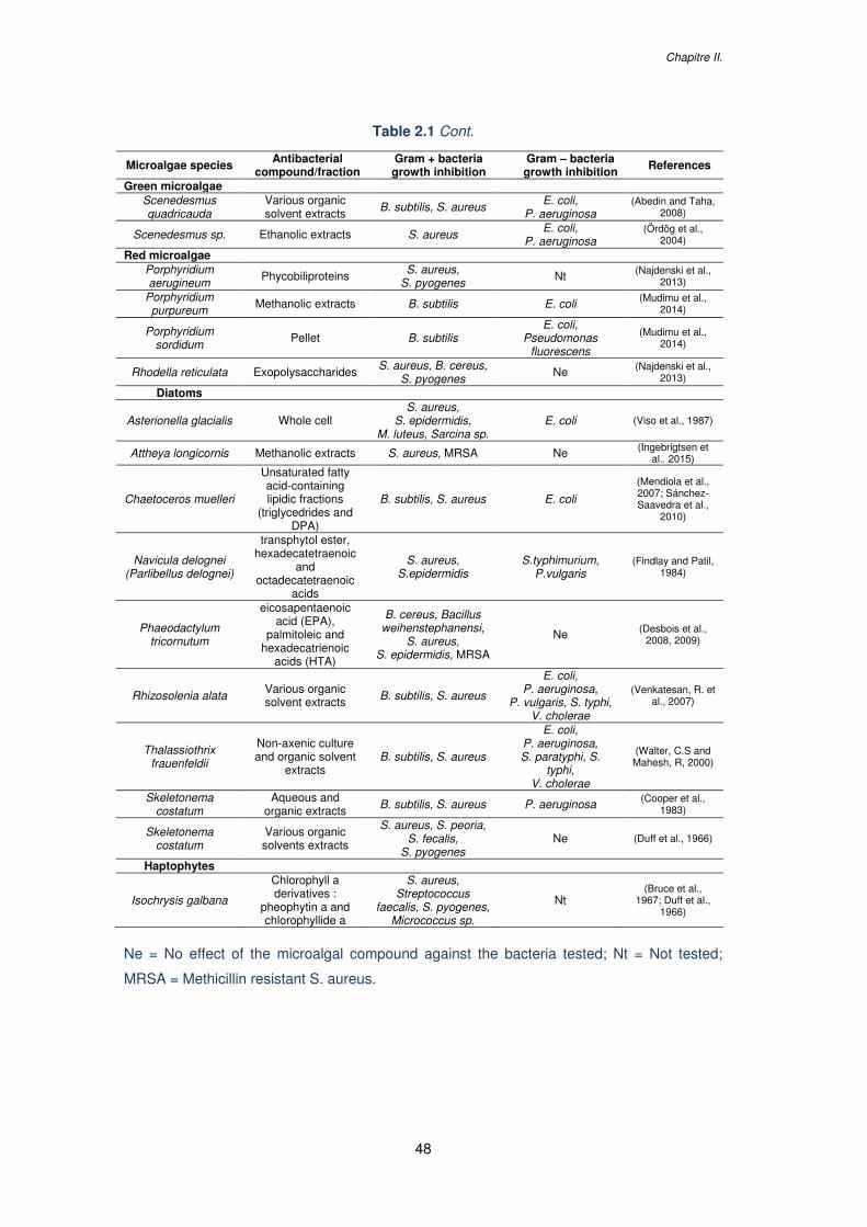
Chapitre II.
48
Table 2.1 Cont.
Microalgae species Antibacterial compound/fraction
Gram + bacteria growth inhibition
Gram – bacteria growth inhibition
References
Green microalgae Scenedesmus quadricauda
Various organic solvent extracts B. subtilis, S. aureus E. coli,
P. aeruginosa (Abedin and Taha,
2008)
Scenedesmus sp. Ethanolic extracts S. aureus E. coli, P. aeruginosa
(Ördög et al., 2004)
Red microalgae Porphyridium aerugineum Phycobiliproteins S. aureus,
S. pyogenes Nt (Najdenski et al., 2013)
Porphyridium purpureum Methanolic extracts B. subtilis E. coli (Mudimu et al.,
2014)
Porphyridium sordidum
Pellet B. subtilis E. coli,
Pseudomonas fluorescens
(Mudimu et al., 2014)
Rhodella reticulata Exopolysaccharides S. aureus, B. cereus, S. pyogenes
Ne (Najdenski et al., 2013)
Diatoms
Asterionella glacialis Whole cell S. aureus,
S. epidermidis, M. luteus, Sarcina sp.
E. coli (Viso et al., 1987)
Attheya longicornis Methanolic extracts S. aureus, MRSA Ne (Ingebrigtsen et al., 2015)
Chaetoceros muelleri
Unsaturated fatty acid-containing lipidic fractions
(triglycedrides and DPA)
B. subtilis, S. aureus E. coli
(Mendiola et al., 2007; Sánchez-Saavedra et al.,
2010)
Navicula delognei (Parlibellus delognei)
transphytol ester, hexadecatetraenoic
and octadecatetraenoic
acids
S. aureus, S.epidermidis
S.typhimurium, P.vulgaris
(Findlay and Patil, 1984)
Phaeodactylum tricornutum
eicosapentaenoic acid (EPA),
palmitoleic and hexadecatrienoic
acids (HTA)
B. cereus, Bacillus weihenstephanensi,
S. aureus, S. epidermidis, MRSA
Ne (Desbois et al., 2008, 2009)
Rhizosolenia alata Various organic solvent extracts B. subtilis, S. aureus
E. coli, P. aeruginosa,
P. vulgaris, S. typhi, V. cholerae
(Venkatesan, R. et al., 2007)
Thalassiothrix frauenfeldii
Non-axenic culture and organic solvent
extracts B. subtilis, S. aureus
E. coli, P. aeruginosa, S. paratyphi, S.
typhi, V. cholerae
(Walter, C.S and Mahesh, R, 2000)
Skeletonema costatum
Aqueous and organic extracts B. subtilis, S. aureus P. aeruginosa (Cooper et al.,
1983)
Skeletonema costatum
Various organic solvents extracts
S. aureus, S. peoria, S. fecalis,
S. pyogenes Ne (Duff et al., 1966)
Haptophytes
Isochrysis galbana
Chlorophyll a derivatives :
pheophytin a and chlorophyllide a
S. aureus, Streptococcus
faecalis, S. pyogenes, Micrococcus sp.
Nt (Bruce et al.,
1967; Duff et al., 1966)
Ne = No effect of the microalgal compound against the bacteria tested; Nt = Not tested;
MRSA = Methicillin resistant S. aureus.
Microalgal compounds against pathogens: emphasize on diseases in aquaculture
49
2.1.1 Toward improving extraction techniques
Beside the microalgal species, the presence of the antibacterial compound in the microalgal
extracts is also highly dependent on the solvent used during the extraction. As the biological
activity is rarely found in aqueous extracts (Cannell et al., 1988; Venkatesan, R. et al., 2007;
Walter, C.S and Mahesh, R, 2000), it seems that compounds with an activity against
foodborne and human pathogenic bacteria in microalgae are mostly hydrophobic and can be
more readily extracted with organic solvents. Some authors found that antibacterial activity
was generally found in methanol extracts (Cannell et al., 1988; Mudimu et al., 2014), while
some other studies described a better extraction using acetone (Venkatesan, R. et al., 2007;
Walter, C.S and Mahesh, R, 2000), benzene and ethyl acetate extracts (Bhagavathy et al.,
2011) or petroleum ether and hexane (Herrero et al., 2006).
Other techniques have been tested to extract bioactive compounds from microalgae,
such as the supercritical CO2 extracts, pressurized liquid extraction (PLE) or subcritical water
extraction (SWE). These techniques are considered as “greener” than the traditional
techniques as they do not need large quantities of organic solvents, allow a faster extraction
and are more selective toward the compounds of interest (Herrero et al., 2013). Supercritical
CO2 method allowed obtaining lipid fractions from Chaetoceros muelleri with antibacterial
activity against Staphyloccocus aureus and Escherichia coli (Mendiola et al., 2007), while a
classic extraction with solvents such as hexane, dichloromethane and methanol did not
demonstrate any activity against E. coli (del Pilar Sanchez-Saavedra, M. et al., 2010). PLE
and SWE permitted to extract antimicrobial agents from H. pluvialis in the red phase with
good efficiency (Herrero et al., 2006; Rodríguez-Meizoso et al., 2010; Santoyo et al., 2009).
SWE presents the advantage not to require the use of toxic solvent, and low temperatures
such as 50°C can allow a good extraction. SWE could therefore represent an interesting
green technique to obtain extracts for natural ingredients, and particularly for the food
industry.
Chapitre II.
50
2.1.2 Diversity of antibacterial compounds extracted from microalgae
In some studies, the antibacterial compounds present in the organic extracts were
characterized. These bioactive compounds can be pigments, such as phycobiliproteins
(Najdenski et al., 2013) or chlorophyll derivatives (Bruce et al., 1967; Jørgensen, 1962), but
they are most of time free fatty acids. Short chain fatty acids from H. pluvialis (Rodríguez-
Meizoso et al., 2010; Santoyo et al., 2009) and long chain fatty acids from Scenedesmus
obliquus (Guedes et al., 2011) present antibacterial activity against E. coli and S. aureus.
The polyunsaturated fatty acids from Chlorococcum strain HS-101 and Dunaliella primolecta
demonstrated antibacterial activity against the methicillin-resistant S. aureus (MRSA), a
bacteria causing infections that kill thousands of people per year and which is highly
resistant to conventional antibiotics (Ohta et al., 1995). Desbois et al. also found fatty acids
from the diatom Phaeodactylum tricornutum with a very potent antibacterial activity against
the MRSA and have characterized three different unsaturated fatty acids involved in the
antibacterial activity: the polyunsaturated fatty acid eicosapentaenoic acid (EPA), the
monounsaturated fatty acid palmitoleic acid (PA) and the relatively unusual polyunsaturated
fatty acid hexadecatrienoic acid (HTA ; Desbois et al., 2008, 2009). It has also been
observed that the fusiform morphotype of this diatom produced greater levels of EPA, PA
and HTA compared to its oval morphotype (Desbois et al., 2010).
In natural environments, fatty acids are released when the microalgal cell loses its
integrity and they seem to be involved in an ‘activated’ defence mechanism to protect an
algal population against grazing predators (Jüttner, 2001) and when pathogenic bacteria are
in the vicinity of the algae (Smith et al., 2010). Moreover, it has previously been shown that
fatty acids possess bactericidal properties against a diverse range of bacteria (Kabara et al.,
1972). The exact mechanism with which the bioactive compounds exert their antibacterial
activity is not yet fully elucidated, but it seems that bacterial cellular membranes would be
the main site of action (Galbraith and Miller, 1973). There are some evidences of deleterious
effects of fatty acids in the bacterial membrane, causing a cell leakage, a reduction of
nutrient intake and a reduction of the cellular respiration (Smith et al., 2010). The
Microalgal compounds against pathogens: emphasize on diseases in aquaculture
51
antibacterial action of fatty acids may also be mediated by the inhibition of bacterial fatty acid
synthesis (Zheng et al., 2005).
These mechanisms could explain why Gram-positive bacteria are more susceptible to
microalgae bioactive compounds than Gram-negative bacteria. In fact, the bacterial growth
inhibition is generally lower when microalgae are tested against Gram-negative bacteria, and
in some cases the tested extracts do not present any bactericidal effect. As examples, the
phycobiliproteins and exopolysaccharides from the red microalgae Porphyridium aerugineum
and Rhodella reticulata respectively, were active against the Gram-positive bacteria
S. aureus and Streptoccocus pyogenes but presented no effect against the Gram-negative
bacteria E. coli and Pseudomonas aeruginosa (Najdenski et al., 2013). The diatom
P. tricornutum did not demonstrated antibacterial effect against these two Gram-negative
bacteria either, whereas a good antibacterial activity against the gram-positive MRSA was
observed (Desbois et al., 2009). Thus, the difference in sensitivities between Gram–positive
and Gram–negative bacteria may be due to their complex membrane permeability, making
the penetration and the bactericidal action of the compound more difficult.
The potent activity of microalgal compounds, especially free fatty acids, against various
bacteria straightens further development in the search for drugs and food preservatives from
microalgae. Bacterial resistance to free fatty acids has not been encountered yet (Petschow
et al., 1996; Sun et al., 2003), so their exploitation in medicine deserves to be further
investigated (Desbois et al., 2009). Furthermore, as consumers tend to avoid synthetic
preservatives, microalgae could be good candidates as natural sources against food-borne
pathogens.
Chapitre II.
52
2.2 Use of microalgae against pathogenic bacteria in
aquaculture
Bacteria are the main cause of infections in intensive aquaculture of finfish and shellfish
worldwide (Austin, 2012), occurring as well in hatcheries and nurseries as in rear and grow-
out ponds. These infections can lead to serious mass mortalities and imply considerable
economic losses. A wide range of bacteria are known to infect farmed and wild species with
minor to severe consequences on health and survival. Among the important bacterial
diseases in finfish we can cite those caused by Aeromonas sp. and Pseudomonas sp.,
inducing hemorrhages in many fresh water species, or Vibrio sp. inducing vibrioses in marine
fish species (Austin and Austin, 2007). Vibrio are able to infect a wide variety of hosts
(Vandenberghe et al., 2003) and are also the most common and harmful shrimp pathogenic
bacteria, with luminous species such as Vibrio harveyi involved in mass mortalities in shrimp
hatcheries (Aguirre-Guzmán et al., 2004).
Antibiotics have been largely used in intensive fish farming but, as for humans and
terrestrial animals, bacterial resistance in aquaculture is increasing and most antibiotics are
no longer effective (Fernández-Alarcón et al., 2010; Miranda and Zemelman, 2002). The
presence of drug residues in tissues of aquatic animals and the risk of transferring resistant
bacteria to humans have led to a great concern about the use of synthetic antibiotics for
public health. Strict regulations have thus been established, and a few antibiotics are now
licensed in aquaculture. Various alternative and natural compounds are available today to
control aquatic pathogenic bacteria, mainly derived from plants. They present the
advantages of decreasing the side effects observed with synthetic antibiotics and being less
expansive (Citarasu, 2012). In this context, several microalgae species have been
investigated for their antibacterial activity in vitro, and in co-culture with pathogenic bacteria,
and some studies have also been conducted in vivo with the “green water” technique and
using microalgae as food supplement (Table 2.2).
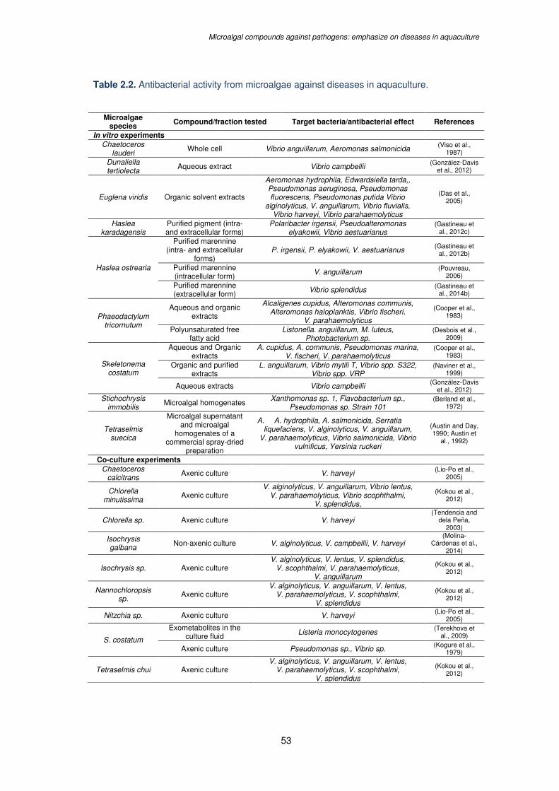
Microalgal compounds against pathogens: emphasize on diseases in aquaculture
53
Table 2.2. Antibacterial activity from microalgae against diseases in aquaculture.
Microalgae species
Compound/fraction tested Target bacteria/antibacterial effect References
In vitro experiments Chaetoceros
lauderi Whole cell Vibrio anguillarum, Aeromonas salmonicida (Viso et al.,
1987)
Dunaliella tertiolecta
Aqueous extract Vibrio campbellii (González-Davis et al., 2012)
Euglena viridis Organic solvent extracts
Aeromonas hydrophila, Edwardsiella tarda,, Pseudomonas aeruginosa, Pseudomonas fluorescens, Pseudomonas putida Vibrio
alginolyticus, V. anguillarum, Vibrio fluvialis, Vibrio harveyi, Vibrio parahaemolyticus
(Das et al., 2005)
Haslea karadagensis
Purified pigment (intra- and extracellular forms)
Polaribacter irgensii, Pseudoalteromonas elyakowii, Vibrio aestuarianus
(Gastineau et al., 2012c)
Haslea ostrearia
Purified marennine (intra- and extracellular
forms) P. irgensii, P. elyakowii, V. aestuarianus (Gastineau et
al., 2012b)
Purified marennine (intracellular form) V. anguillarum (Pouvreau,
2006)
Purified marennine (extracellular form) Vibrio splendidus (Gastineau et
al., 2014b)
Phaeodactylum tricornutum
Aqueous and organic extracts
Alcaligenes cupidus, Alteromonas communis, Alteromonas haloplanktis, Vibrio fischeri,
V. parahaemolyticus
(Cooper et al., 1983)
Polyunsaturated free fatty acid
Listonella. anguillarum, M. luteus, Photobacterium sp.
(Desbois et al., 2009)
Skeletonema costatum
Aqueous and Organic extracts
A. cupidus, A. communis, Pseudomonas marina, V. fischeri, V. parahaemolyticus
(Cooper et al., 1983)
Organic and purified extracts
L. anguillarum, Vibrio mytili T, Vibrio spp. S322, Vibrio spp. VRP
(Naviner et al., 1999)
Aqueous extracts Vibrio campbellii (González-Davis et al., 2012)
Stichochrysis immobilis
Microalgal homogenates Xanthomonas sp. 1, Flavobacterium sp., Pseudomonas sp. Strain 101
(Berland et al., 1972)
Tetraselmis suecica
Microalgal supernatant and microalgal
homogenates of a commercial spray-dried
preparation
A. A. hydrophila, A. salmonicida, Serratia liquefaciens, V. alginolyticus, V. anguillarum,
V. parahaemolyticus, Vibrio salmonicida, Vibrio vulnificus, Yersinia ruckeri
(Austin and Day, 1990; Austin et
al., 1992)
Co-culture experiments Chaetoceros
calcitrans Axenic culture V. harveyi (Lio-Po et al., 2005)
Chlorella minutissima
Axenic culture V. alginolyticus, V. anguillarum, Vibrio lentus,
V. parahaemolyticus, Vibrio scophthalmi, V. splendidus,
(Kokou et al., 2012)
Chlorella sp. Axenic culture V. harveyi (Tendencia and
dela Peña, 2003)
Isochrysis galbana Non-axenic culture V. alginolyticus, V. campbellii, V. harveyi
(Molina-Cárdenas et al.,
2014)
Isochrysis sp. Axenic culture V. alginolyticus, V. lentus, V. splendidus,
V. scophthalmi, V. parahaemolyticus, V. anguillarum
(Kokou et al., 2012)
Nannochloropsis sp.
Axenic culture V. alginolyticus, V. anguillarum, V. lentus,
V. parahaemolyticus, V. scophthalmi, V. splendidus
(Kokou et al., 2012)
Nitzchia sp. Axenic culture V. harveyi (Lio-Po et al., 2005)
S. costatum
Exometabolites in the culture fluid Listeria monocytogenes (Terekhova et
al., 2009)
Axenic culture Pseudomonas sp., Vibrio sp. (Kogure et al., 1979)
Tetraselmis chui Axenic culture V. alginolyticus, V. anguillarum, V. lentus,
V. parahaemolyticus, V. scophthalmi, V. splendidus
(Kokou et al., 2012)

Chapitre II.
54
Table 2.2. Cont.
Microalgae species
Compound/fraction tested Target bacteria/antibacterial effect References
In vivo experiments
C. minutissima
30 min incubation of enriched Artemia metanauplii
Decrease of the bacterial load in Artemia and diminution of presumptive
Vibrio
(Makridis et al., 2006)
D. tertiolecta Daily diet of Artemia franciscana Protection against V. campbellii and V. proteolyticus
(Marques et al., 2006)
H. ostrearia
Incubation of Mytilus edulis and Placopecten magellanicus larvae
with supernatant containing extracellular pigments
Higher survival and physiological conditions of larvae challenged with
V. splendidus
(Turcotte et al., 2016)
Tetraselmis sp.
4 h incubation of Artemia franciscana
Diminution of associated bacteria, better bacterial diversity and the flora
less dominated by V. alginolyctus
(Olsen et al.,2000)
T. suecica
Food supplement for the Atlantic salmon Solmo salar
Reduction of A. hydrophila, A. salmonicida, S. liquefaciens, V. anguillarum, V. salmonicida,
Y. ruckeri infections, reduction of bacterial populations in water tanks and increase of the microbial communities in
the digestive tract
(Austin et al., 1992)
Food supplement for the broodstock and partial live larvae
feed for the white prawn Fenneropenaeus inducus
Reduction of Vibrio numbers in water tank, better egg hatching rate and
survival rate of the larvae
(Regunathan C. and Wesley S.G.,
2004)
2.2.1 Benefits of the “green-water” technique against bacterial diseases in
aquaculture
The “green water” technique has demonstrated beneficial effects on health, survival rate and
resistance of different organisms (Cahu et al., 1998; Marques et al., 2006; Naas et al., 1992;
Papandroulakis et al., 2001; Reitan et al., 1997; Supamattaya et al., 2005; Van der Meeren
et al., 2007). Addition of the microalga Isochrysis galbana allowed a better viability and a
faster grow of the turbot Scophthalmus maximus larvae as well as a lower proliferation of
opportunistic bacteria (Salvesen et al., 1999). The green microalga Tetraselmis suecica first
showed good antibacterial activity in vitro against several Vibrio species (Austin and Day,
1990) and was then proved to reduce in vivo the number of various bacterial species in the
water tank of the Atlantic salmon Salmo salar (Austin et al., 1992). Adding T. suecica also
reduced the number of Vibrio species in broodstock gut, eggs and larvae of the white prawn
Fenneropenaeus indicus, resulting in improved egg hatching and larval survival (Regunathan
C. and Wesley S.G., 2004). A better survival and physiological conditions of the blue mussel
Mytilus edulis larvae and the scallop Placopecten magellanicus larvae were observed when
Microalgal compounds against pathogens: emphasize on diseases in aquaculture
55
incubated with supernatant containing marennine, the extracellular blue pigment of the
diatom H. ostrearia, at concentrations as low as 0.1 mg/L (Turcotte et al., 2016). The use of
the diatoms Skeletonema costatum and Chaetoceros calcitrans to feed Penaeus monodon
larvae allowed a growth inhibition of the luminous bacteria Vibrio harveyi (Lavilla-Pitogo et
al., 1998). However, this seemed mainly due to the microbial flora associated with the
diatoms rather than their metabolic products. Indeed, the effectiveness of the “green water”
culture in preventing bacterial infections and outbreaks can also be attributed to the
presence of antibacterial factors in the bacterial, fungal and phytoplankton microbiota
associated with this culture technique (Lio-Po et al., 2005).
Yet, several co-culture experiments with axenic microalgae demonstrated the ability of
some species to produce and release compounds with potent activity against pathogenic
bacteria. Axenic cultures of Chlorella minutissima, Tetraselmis chui, Isochrysis sp. and
Nannochloropsis sp. limited the growth of various Vibrio species (Kokou et al., 2012), and
growth of the luminous bacteria V. harveyi highly decreased when co-cultured with pure
Chlorella sp. (Tendencia and dela Peña, 2003), C. calcitrans or Nitzschia sp. (Lio-Po et al.,
2005).
Finally, genetically modified microalgae were also used in vivo. In a recent study, better
growth, resistance to bacteria and survival rate were observed in the shrimp postlarvae
P. monodon fed with “fusant” Chlorella and Dunaliella (Kusumaningrum and Zainuri, 2015).
The so-called “fusant” microalgae resulted from the generation of a unique cell through
somatic hybridization by fusion of the two protoplasts. This fusion technology is especially
used to transfer agronomically useful traits to plants (Davey et al., 2005) and would allow an
improvement of valuable metabolites production from these two microalgae. Another
genetically modified microalga tested in “green water” systems is the transgenic line of
Nannochloropsis oculata, able to produce the antimicrobial bovine lactoferricin (LFB)
peptide. These LFP-containing transgenic microalgae were developed and tested as food
supplement for the medaka fish Oryzias latipes infected with Vibrio parahaemolyticus, which
displayed a significantly better survival rate after 24h (Li and Tsai, 2009).
Chapitre II.
56
2.2.2 Microalgae to improve the live-food quality
Microalgae also show advantages for the live food quality (Marques et al., 2006; Øie et al.,
1994), by reducing the number of associated pathogenic bacteria such as Vibrio and
allowing a lower risk of transmission to fish larvae. Daily addition of Dunaliella tertiolecta to
feed the brine shrimp Artemia franciscana, considered as an essential part of the live food
chain for the culture of fish and shellfish larvae, conferred a full protection against
Vibrio campbellii and Vibrio proteolyticus (Marques et al., 2006). A 4 h incubation of
A. franciscana with the microalgae Tetraselmis sp. resulted in a diminution by 75 % of
associated bacteria, with a better bacterial diversity and the flora less dominated by Vibrio
alginolyticus (Olsen et al., 2000). Similar observations were made by Makridis et al. with
incubation of Artemia with T. chui and C. minutissima (Makridis et al., 2006). These authors
proposed that the reduction of Vibrio cells in Artemia cultures could either be due to
compounds released by microalgae or to Gram-positive associated bacteria.
2.2.3 In vitro efficiency of microalgal compounds against marine bacteria
Several in vitro studies demonstrated the ability of various microalgae to produce
antibacterial compounds effective against relevant marine pathogenic bacteria. The whole
cells of Chaetoceros lauderi (Viso et al., 1987), supernatant and homogenates of T. suecica
(Austin and Day, 1990; Austin et al., 1992) or homogenates from Stichochrysis immobilis
(Berland et al., 1972) induced a growth inhibition of various marine bacteria. However,
Berland et al. noted that limited attention should be paid to antibacterial activity obtained with
broken cells, as in natural conditions substances synthesized have to be released into the
water to target another organism (Berland et al., 1972). Microalgae producing antibacterial
compounds that are not released in the medium cannot indeed be considered gainful in
“green water” techniques, but these compounds can be highly useful in the design of novel
drugs.
Only a few compounds with activity against marine bacteria have been characterized,
such as the polyunsaturated free fatty acid in P. tricornutum, identified as ecosapentaenoic
acid (Desbois et al., 2009). An antibacterial activity was also demonstrated in vitro with the
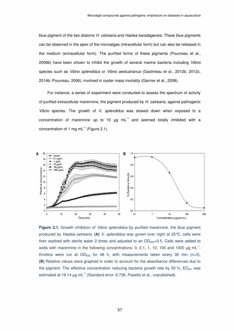
Microalgal compounds against pathogens: emphasize on diseases in aquaculture
57
blue pigment of the two diatoms H. ostrearia and Haslea karadagensis. These blue pigments
can be observed in the apex of the microalgae (intracellular form) but can also be released in
the medium (extracellular form). The purified forms of these pigments (Pouvreau et al.,
2006b) have been shown to inhibit the growth of several marine bacteria including Vibrio
species such as Vibrio splendidus or Vibrio aestuarianus (Gastineau et al., 2012b, 2012c,
2014b; Pouvreau, 2006), involved in oyster mass mortality (Garnier et al., 2008).
For instance, a series of experiment were conducted to assess the spectrum of activity
of purified extracellular marennine, the pigment produced by H. ostrearia, against pathogenic
Vibrio species. The growth of V. splendidus was slowed down when exposed to a
concentration of marennine up to 10 µg mL-1 and seemed totally inhibited with a
concentration of 1 mg mL-1 (Figure 2.1).
Figure 2.1. Growth inhibition of Vibrio splendidus by purified marennine, the blue pigment
produced by Haslea ostrearia. (A) V. splendidus was grown over night at 25°C, cells were
then washed with sterile water 2 times and adjusted to an OD600=0.5. Cells were added to
wells with marennine in the following concentrations: 0, 0.1, 1, 10, 100 and 1000 µg mL-1.
Kinetics were run at OD600 for 48 h, with measurements taken every 30 min (n=3).
(B) Relative values were graphed in order to account for the absorbance differences due to
the pigment. The effective concentration reducing bacteria growth rate by 50 %, EC50, was
estimated at 19.14 µg mL-1 (Standard error: 6.736; Pasetto et al., unpublished).
A B
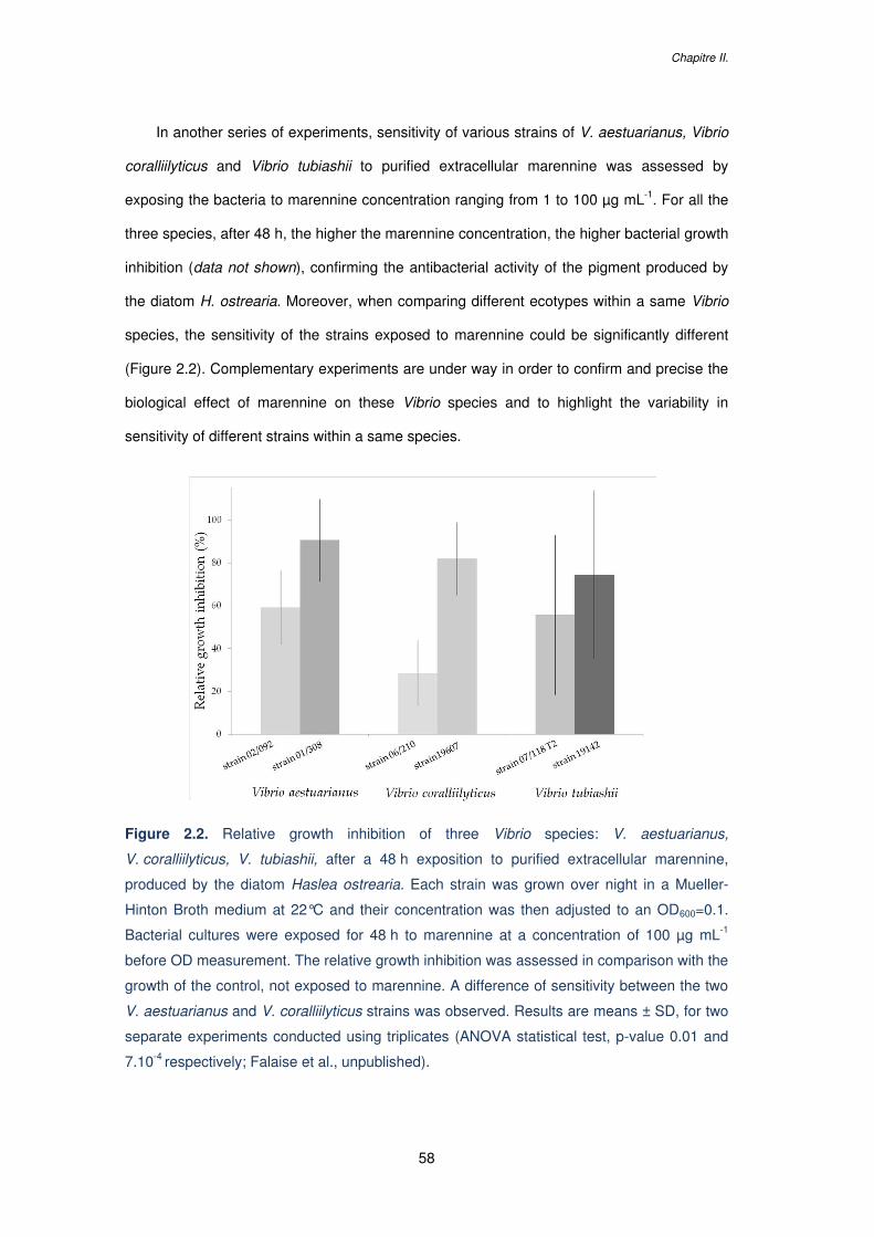
Chapitre II.
58
In another series of experiments, sensitivity of various strains of V. aestuarianus, Vibrio
coralliilyticus and Vibrio tubiashii to purified extracellular marennine was assessed by
exposing the bacteria to marennine concentration ranging from 1 to 100 µg mL-1. For all the
three species, after 48 h, the higher the marennine concentration, the higher bacterial growth
inhibition (data not shown), confirming the antibacterial activity of the pigment produced by
the diatom H. ostrearia. Moreover, when comparing different ecotypes within a same Vibrio
species, the sensitivity of the strains exposed to marennine could be significantly different
(Figure 2.2). Complementary experiments are under way in order to confirm and precise the
biological effect of marennine on these Vibrio species and to highlight the variability in
sensitivity of different strains within a same species.
Figure 2.2. Relative growth inhibition of three Vibrio species: V. aestuarianus,
V. coralliilyticus, V. tubiashii, after a 48 h exposition to purified extracellular marennine,
produced by the diatom Haslea ostrearia. Each strain was grown over night in a Mueller-
Hinton Broth medium at 22°C and their concentration was then adjusted to an OD600=0.1.
Bacterial cultures were exposed for 48 h to marennine at a concentration of 100 µg mL-1
before OD measurement. The relative growth inhibition was assessed in comparison with the
growth of the control, not exposed to marennine. A difference of sensitivity between the two
V. aestuarianus and V. coralliilyticus strains was observed. Results are means ± SD, for two
separate experiments conducted using triplicates (ANOVA statistical test, p-value 0.01 and
7.10-4 respectively; Falaise et al., unpublished).
Microalgal compounds against pathogens: emphasize on diseases in aquaculture
59
2.2.4 Interactions between microalgae and marine bacteria
The antibacterial mechanisms of microalgae are still unclear and the bioactive compounds
released by the cells could either be bactericidal or prevent the bacterial multiplication. The
very rapid growth inhibition of various Vibrio species in co-culture with Chlorella sp.,
Isochrysis sp. or Nannochloropsis sp. with no recovery after few days (Kokou et al., 2012;
Tendencia and dela Peña, 2003) allows considering a bactericidal action of the extracellular
substances produce by some microalgae (Molina-Cárdenas et al., 2014). Austin et al. also
observed in vitro a prompt inhibition of several Vibrio species by T. suecica and noticed a
very rapid decrease in bacterial mobility with an elongation and vacuolisation of the cells in
less than 20 min. Though, a reduction of the inhibitory activity was observed after only 5 h. It
was suggested that the bioactive substance could have been denatured or adsorbed by
some bacterial cells, allowing others to grow (Austin and Day, 1990; Austin et al., 1992).
In some cases, the antibacterial activity of microalgae can be induced by the presence
of bacteria in the vicinity of the microalgae, or can be constitutive and always present in the
algal culture medium (Safonova and Reisser, 2005). The constitutive production of
antibacterial exometabolites by some microalgae was highlighted with the growth diminution
of Listeria monocytogenes in co-culture with the cell-free culture media of S. costatum
(Terekhova et al., 2009).
Microalgae can influence marine bacteria in different ways. They can either inhibit or
stimulate bacterial growth, or have no apparent effect, depending on the target bacteria
(Berland et al., 1972; Kogure et al., 1979). As an example, the diatom S. costatum inhibits
the growth of Pseudomonas and Vibrio in co-culture, but enhances the growth of
Flavobacterium (Kogure et al., 1979). The production of antibacterial compounds by
microalgae such as lipids or fatty acids varies according to the taxonomic group, the growth
conditions, the available nutrients and their concentration in the medium, the light intensity,
the temperature or the pH. The development stage of the algal culture is also highly
significant as it is assumed that various secondary metabolites are produced and released in
the medium at different growth phases (Borowitzka, 1995). Terekhova et al. showed that
only the exometabolites produced by S. costatum during the middle steady-state growth

Chapitre II.
60
phase presented an antibacterial activity against L. monocytogenes while compounds
released during the exponential growth phase had no effect on these bacteria (Terekhova et
al., 2009). Cooper et al. have also demonstrated the direct relation between cell growth
phase and antibacterial activity, and showed that P. tricornutum presented a better activity
against a wide spectrum of marine bacteria during the exponential growth phase compared
to the stationary phase, while the reverse relationship was found for S. costatum (Cooper et
al., 1983).
Microalgal compounds against pathogens: emphasize on diseases in aquaculture
61
3 3. ANTIFUNGAL ACTIVITY FROM MICROALGAE
3.1 Antifungal activity from microalgae against human
pathogens
Searching for antifungal compounds from microalgae started much later than screening for
antibacterial activity. As a matter of fact, fungi have been considered as harmful human
pathogens since the 70’s, when mortality induced by fungal infections and the frequency of
nosocomial mycoses increased in hospitalized patients. Increase of fungal infections was
mainly due to therapies that depress patients’ immune system such as the use of intensive
and aggressive chemotherapy regimens, the expansion of organ transplant programs and
the spread of the AIDS epidemic. (Alangaden, 2011; Anaissie and Bodey, 1989). The
incidence of invasive aspergillosis (induced by Aspergillus species), and the number of
cases of candidemia (an infection caused by Candida species), have been rising inexorably
from that time (Manuel and Kibbler, 1998; Wey SB et al., 1988). The growing use of
antifungal agents in recent years has led to the development of drug resistance
(Accoceberry and Noël, 2006; Ghannoum and Rice, 1999). Thus, there is a need for novel
drugs and several study were recently conducted to find fungicide activity from marine
natural products against human pathogenic fungi (Cheung et al., 2014; El Amraoui et al.,
2014; Hong et al., 2015; Shishido et al., 2015; Wang et al., 2015b; Xu et al., 2015), including
antifungal agents from microalgae (Table 2.3).
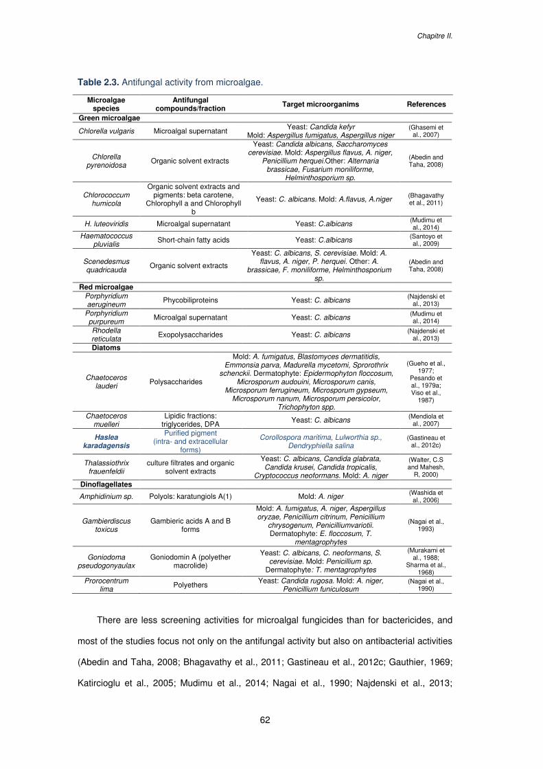
Chapitre II.
62
Table 2.3. Antifungal activity from microalgae.
Microalgae species
Antifungal compounds/fraction Target microorganims References
Green microalgae
Chlorella vulgaris Microalgal supernatant Yeast: Candida kefyr Mold: Aspergillus fumigatus, Aspergillus niger
(Ghasemi et al., 2007)
Chlorella pyrenoidosa
Organic solvent extracts
Yeast: Candida albicans, Saccharomyces cerevisiae. Mold: Aspergillus flavus, A. niger,
Penicillium herquei.Other: Alternaria brassicae, Fusarium moniliforme,
Helminthosporium sp.
(Abedin and Taha, 2008)
Chlorococcum humicola
Organic solvent extracts and pigments: beta carotene,
Chlorophyll a and Chlorophyll b
Yeast: C. albicans. Mold: A.flavus, A.niger (Bhagavathy et al., 2011)
H. luteoviridis Microalgal supernatant Yeast: C.albicans (Mudimu et al., 2014)
Haematococcus pluvialis
Short-chain fatty acids Yeast: C.albicans (Santoyo et al., 2009)
Scenedesmus quadricauda Organic solvent extracts
Yeast: C. albicans, S. cerevisiae. Mold: A. flavus, A. niger, P. herquei. Other: A.
brassicae, F. moniliforme, Helminthosporium sp.
(Abedin and Taha, 2008)
Red microalgae Porphyridium aerugineum Phycobiliproteins Yeast: C. albicans (Najdenski et
al., 2013)
Porphyridium purpureum Microalgal supernatant Yeast: C. albicans (Mudimu et
al., 2014)
Rhodella reticulata Exopolysaccharides Yeast: C. albicans (Najdenski et
al., 2013)
Diatoms
Chaetoceros lauderi Polysaccharides
Mold: A. fumigatus, Blastomyces dermatitidis, Emmonsia parva, Madurella mycetomi, Sprorothrix
schenckii. Dermatophyte: Epidermophyton floccosum, Microsporum audouini, Microsporum canis,
Microsporum ferrugineum, Microsporum gypseum, Microsporum nanum, Microsporum persicolor,
Trichophyton spp.
(Gueho et al., 1977;
Pesando et al., 1979a; Viso et al.,
1987)
Chaetoceros muelleri
Lipidic fractions: triglycerides, DPA Yeast: C. albicans (Mendiola et
al., 2007)
Haslea karadagensis
Purified pigment (intra- and extracellular
forms)
Corollospora maritima, Lulworthia sp., Dendryphiella salina
(Gastineau et al., 2012c)
Thalassiothrix frauenfeldii
culture filtrates and organic solvent extracts
Yeast: C. albicans, Candida glabrata, Candida krusei, Candida tropicalis,
Cryptococcus neoformans. Mold: A. niger
(Walter, C.S and Mahesh,
R, 2000)
Dinoflagellates
Amphidinium sp. Polyols: karatungiols A(1) Mold: A. niger (Washida et al., 2006)
Gambierdiscus toxicus
Gambieric acids A and B forms
Mold: A. fumigatus, A. niger, Aspergillus oryzae, Penicillium citrinum, Penicillium
chrysogenum, Penicilliumvariotii. Dermatophyte: E. floccosum, T.
mentagrophytes
(Nagai et al., 1993)
Goniodoma pseudogonyaulax
Goniodomin A (polyether macrolide)
Yeast: C. albicans, C. neoformans, S. cerevisiae. Mold: Penicillium sp.
Dermatophyte: T. mentagrophytes
(Murakami et al., 1988;
Sharma et al., 1968)
Prorocentrum lima Polyethers Yeast: Candida rugosa. Mold: A. niger,
Penicillium funiculosum (Nagai et al.,
1990)
There are less screening activities for microalgal fungicides than for bactericides, and
most of the studies focus not only on the antifungal activity but also on antibacterial activities
(Abedin and Taha, 2008; Bhagavathy et al., 2011; Gastineau et al., 2012c; Gauthier, 1969;
Katircioglu et al., 2005; Mudimu et al., 2014; Nagai et al., 1990; Najdenski et al., 2013;

Microalgal compounds against pathogens: emphasize on diseases in aquaculture
63
Ördög et al., 2004; Viso et al., 1987; Walter, C.S and Mahesh, R, 2000). As for antibacterial
activity, antifungal activity varies widely depending on microalgal species, type of solvent
used to extract the compound and the microorganism tested. It does not seem to be a
taxonomic trend for the antifungal activity, and the capability to produce antifungal
compounds would have evolved independently of phylogenetic relationship in microalgae
(Mudimu et al., 2014). However, Pesando et al. noticed a significant activity of the genus
Chaetoceros (Pesando et al., 1979b) and Kellam et al. indicated also that marine microalgae
showed more potential in the search for new antifungal agents than do freshwater species
(Kellam et al., 1988).
As illustrated on Table 2.3, several antifungal compounds from various microalgae have
been characterized. Polysaccharides with high molecular weight were identified in the diatom
C. lauderi. They presented a large spectrum of activity against dermatophytes, moulds and
phyto-fungi, but no activity was detected against the yeasts tested (Gueho et al., 1977;
Pesando et al., 1979a; Viso et al., 1987). Gambieric acids from the dinoflagellate
Gambierdiscus toxicus had also an antifungal activity against several dermatophytes and
moulds but had not activity against yeasts like Candida albicans or Saccharomyces
cerevisiae (Nagai et al., 1993). The diatom Thalassiothrix frauenfeldii was meanwhile active
against yeasts and moulds but not against dermathophytes (Walter, C.S and Mahesh, R,
2000). Other compounds such as pigments like beta-carotene, chlorophyll-a and chlorophyll-
b from Chlorococcum humicola (Bhagavathy et al., 2011), or phycobiliproteins from
Porphyridium aerugineum have also demonstrated antifungal activities (Najdenski et al.,
2013). The polyene-polyhydroxy metabolites amphidinols were extracted from the
dinoflagellate Amphidinium klebsii (Houdai et al., 2004; Satake et al., 1991). Polyenes are
metabolites known for having a potent antifungal activity as they target the biosynthetic
pathway of ergosterols, found in fungi membranes (Ghannoum and Rice, 1999). These few
results illustrate that the search for novel antifungal compounds from microalgae has not
been greatly developed so far, although an increasing number of fungi display drug
resistance phenomenon.
Chapitre II.
64
3.2 Potential use of microalgae against fungal diseases in
aquaculture
An increase in fungal infections has been observed in the last few decades with the
modernization and intensification of aquaculture at an industrial scale, resulting in huge loss
of aquaculture industries. Fungal infections in aquaculture may cause severe diseases and
mortality events leading to economical impacts. They are often considered secondary to
other factors or pathogens such as consequences of water quality problems, fish trauma by
rough handling or temperature shock, bacterial diseases or parasites (Yanong, 2003).
Several fungi are known to induce diseases by developing in the skin and the gills of the
infected fish or in eggs, or by producing toxins (Noga, 2011). Indeed, mycotoxins can
provoke many disorders and can accumulate in fish tissues, representing a risk for public
health (Anater et al., 2016).
Chemicals used to treat infected animals are limited and, due to the increasing
resistance of fungi against conventional drugs, environmental legislations and consumer's
safety, the alternative ‘herbal formulations’ and alternative save and cheap methods have
become of renewed interest (Trakranrungsie, N., 2011). Studies have been conducted in
order to find new antimycotics of natural origin that may have no harmful effect either on fish,
fish eggs, human health or on ecosystems, such as plant extracts (Hussein et al., 2002; Mori
et al., 2002; Udomkusonsri et al., 2007) or essential oils (Hussein et al., 2000; Mousavi et al.,
2009).
So far, very few studies have been conducted to assess the antifungal activity from
microalgae against pathogenic fungi in aquaculture systems (Table 2.3). Gastineau et al.
have demonstrated in vitro the antifungal activity of the pigment produced by the diatom
H. karadagensis against three marine fungal species Corollospora maritima, Dendryphiella
salina, Lulworthia sp., which can be involved in the phenomenon of biofouling (Gastineau et
al., 2012c). Organic extracts of the green microalgae Chlorella pyrenoidosa and
Scenedesmus quadricauda have demonstrated antifungal activity against Fusarium
monofiliform (Abedin and Taha, 2008) reported to cause black gill disease in shrimps
Microalgal compounds against pathogens: emphasize on diseases in aquaculture
65
(Rhoobunjongde et al., 1991). This result is of great interest as Fusarium sp. were recently
shown to produce toxins that accumulate in fish (Tolosa et al., 2014). Aspergillus fumigatus
is also a fungus susceptible to produce toxins and it thus represents a potential contaminant
for seafood, and particularly for marine bivalves (Grovel et al., 2003). It can be inhibited by
compounds produced by C. lauderi (Viso et al., 1987), G. toxicus (Nagai et al., 1993), and
Chlorella vulgaris (Ghasemi et al., 2007).
More generally, fungi from the taxa Fusarium and Aspergillus are found in many
countries in water, sediments and marine invertebrates, and their presence in shellfish
farming areas evidences the necessity to pay attention to shellfish contamination by such
fungi (Matallah-Boutiba et al., 2012). Furthermore, reports of fungi causing deleterious
effects are frequently related (Karthikeyan and Gopalakrishnan, 2014; Karthikeyan et al.,
2015), which encourage a larger screening of microalgae for the production of novel
antifungal compounds.
Chapitre II.
66
4 ANTIVIRAL ACTIVITY FROM MICROALGAE
4.1 Antiviral activity from microalgae against human
pathogenic viruses
Viral pathogens are the leading cause of human diseases and mortality worldwide.
Treatments to attack the pathogenic virus or its replication directly are difficult to design, as
they can have adverse effects on the infected host cells (Kitazato K et al., 2007). Thus,
treatments against diseases caused by virus are limited, and resistances to these available
treatments demonstrate the need for new medicines (Yasuhara-Bell and Lu, 2010). Many
drugs exhibiting selective inhibition of mammalian originate from synthetic organic chemicals
or from natural products, for instance secondary metabolites in plants (Kitazato K et al.,
2007). Along with the development of “blue” technology and extraction improvement, there is
a growing interest in marine-derived antiviral compounds. Thus, thousands of compounds
from various marine organisms such as algae, bacteria, fungi, marine invertebrates or
sponges have been screened (Cheung et al., 2014; Yasuhara-Bell and Lu, 2010; Moussa et
al., 2015; Vo et al., 2011) and some of them have demonstrated antiviral activities and are
commercially available.
Potential antiviral activity from algal compounds has been first demonstrated in the 50’s
by Gerber et al. who observed that the polysaccharides extracted from Gelidium
cartilagenium afforded protection for embryonated eggs against influenza B and mumps
viruses (Gerber et al., 1958), and the first brood-based studies of seaweeds for their antiviral
substances started in the 70’s (Ehresmann et al., 1977). Screenings for antiviral compounds
from macroalgae are still predominant (Bouhlal Rhimou, 2010; Luescher-Mattli, 2003;
Schaeffer and Krylov, 2000; Soares et al., 2012; Wijesekara et al., 2011), but the interest for
antiviral compounds from microalgae and cyanobacteria rapidly increased as they also
present relevant antiviral activities and are easier to culture. Cyanobacteria are promising
sources of antiviral compounds, and their simple growth needs make them good candidates
for the production of antiviral agents at an industrial scale (Arif et al., 2012; Vijayakumar and
Microalgal compounds against pathogens: emphasize on diseases in aquaculture
67
Menakha, 2015). The sulphated polysaccharide isolated from the cyanobacteria Spirulina
platensis, named spirulan, has demonstrated potent antiviral activity against the herpes
simplex virus type 1 (HSV-1) and also against the human immunodeficiency virus type 1
(HIV-1 ; Hayashi et al., 1996). A spirulan-like molecule isolated from Arthrospira platensis
has also been indicated to possess antiviral activities against these two viruses, with
absence of cytotoxic effects (Rechter et al., 2006).
Several studies have been conducted to test microalgae compounds against human
pathogenic viruses (Table 2.4). Antiviral compounds extracted from microalgae are mainly
polysaccharides. Their mechanisms of action against viruses are not fully understood but it
seems that they can inhibit different stages of the viral infection, such as the adhesion, the
penetration or the replication. Polysaccharides have gained interest in the biomedical and
pharmaceutical industries as they are easily available in nature and most of them are
nontoxic, safe, biodegradable and biocompatible (Ahmadi et al., 2015). However, numerous
polysaccharides with antiviral activities were not developed for clinical use. The reasons are
probably the very high molecular weights of some polysaccharides, preventing them to pass
through the different barriers of the body and the incapacity of enzymes to digest these large
and complex molecules, leading to their accumulation in the body and to potential cytotoxic
effects (Huleihel et al., 2001).
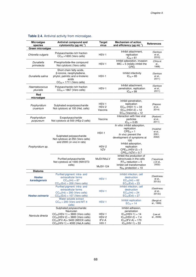
Chapitre II.
68
Table 2.4. Antiviral activity from microalgae.
Microalgae species
Antiviral compound and cytotoxicity (µg mL-1)
Target virus
Mechanism of action and efficiency (µg mL-1)
References
Green microalgae
Chlorella vulgaris Polysaccharide-rich fraction
CC50 >1600 (Vero cells) HSV-1 Inhibit attachment,
replication IC50 = 61
(Santoyo et al., 2010)
Dunaliella primolecta
Pheophorbide-like compound Not cytotoxic (Vero cells) HSV-1
Inhibit adsorption, invasion MIC = 5 (totally inhibit the
CPE)
(Ohta et al.,
1998)
Dunaliella salina
Short chain fatty acids, β-ionone, neophytadiene,
phytol, palmitic and α-linolenic acids
CC50 = 1711 (Vero cells)
HSV-1 Inhibit infectivity
IC50 = 85
(Santoyo et al., 2011)
Haematococcus pluvialis
Polysaccharide-rich fraction CC50 = 1867 (Vero cells) HSV-1
Inhibit attachment, penetration, replication
IC50 = 99
(Santoyo et al., 2011)
Red microalgae
Porphyridium cruentum
Sulphated exopolysaccharide Not cytotoxic at 100 (HeL cells)
HSV-1 HSV-2
Vaccina
Inhibit penetration, replication
EC50 (HSV-1) = 34 EC50 (HSV-2) = 12
EC50 (Vaccina) = 12
(Raposo et al., 2014)
Porphyridium purpureum
Exopolysaccharide Not cytotoxic at 500 (HEp-2 cells) Vaccina
Interaction with free viral particles
IC50 = 0.65
(Radonić et al., 2011)
Porphyridium sp.
Sulphated polysaccharide Not cytotoxic at 250 (Vero cells)
and 2000 (in vivo in rats)
HSV-1
In vitro: inhibit adsorption, replication CPE50 = 1
In vivo: prevent the development of symptoms at
100
(Huleihel et al., 2001, 2002)
HSV-2 VZV
Inhibit adsorption, replication
CPE50 (HSV-2) = 5 CPE50 (VZV) = 0.7
Purified polysaccharide Not cytotoxic at 1000 (NIH/3T3
cells)
MuSV/MuLV Inhibit the production of retroviruses in the cells
RT50 reduction = 5 (Talyshinsk
y et al., 2002)
MuSV-124 Inhibit cell transformation ffu50 protection = 10
Diatoms
Haslea karadagensis
Purified pigment: intra- and extracellular forms
CC50(Int) = 87 CC50(Ext) > 200 (Vero cells)
HSV-1
Inhibit infection, cell destruction
EC50(Int) = 62 EC50(Ext) = 23
(Gastineau et al.,
2012c)
Haslea ostrearia
Purified pigment: intra- and extracellular forms
CC50(Int) > 200 (Vero cells) CC50(Ext) = 107 (Vero cells)
HSV-1
Inhibit infection, cell destruction
EC50(Int) = 24 EC50(Ext) = 27
(Gastineau et al.,
2012c)
Water soluble extract CC50 > 200 (Vero and MT-4
cells) HSV-1 Inhibit replication
EC50 = 14 (Bergé et al., 1999)
Navicula directa
Sulphated polysaccharide: Naviculan
CC50(HSV-1) = 3800 (Vero cells) CC50(HSV-2) = 3800 (Vero cells) CC50(IFV-A)= 5400 (MDCK cells) CC50(HIV-1) = 4000 (HeLA cells)
HSV-1 HSV-2 IFV-A HIV-1
Inhibit adhesion, penetration
IC50(HSV-1) = 14 IC50(HSV-2) = 7.4 IC50(IFV-A) = 170 IC50(HIV-1) = 53
(Lee et al., 2006)
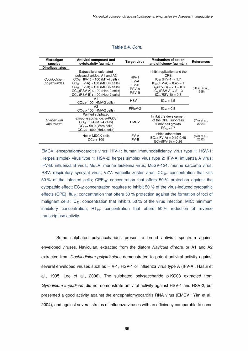
Microalgal compounds against pathogens: emphasize on diseases in aquaculture
69
Table 2.4. Cont.
Microalgae species
Antiviral compound and cytotoxicity (µg mL-1)
Target virus Mechanism of action and efficiency (µg mL-1)
References
Dinoflagellates
Cochlodinium polykrikoides
Extracellular sulphated polysaccharides: A1 and A2
CC50(HIV-1) > 100 (MT-4 cells) CC50(IFV-A) > 100 (MDCK cells) CC50(IFV-B) > 100 (MDCK cells) CC50(RSV-A) > 100 (Hep-2 cells) CC50(RSV-B) > 100 (Hep-2 cells)
HIV-1 IFV-A IFV-B RSV-A RSV-B
Inhibit replication and the CPE
IC50 (HIV-1) = 1.7 IC50(IFV-A) = 0.45 – 1 IC50(IFV-B) = 7.1 – 8.3
IC50(RSV-A) = 2 – 3 IC50(RSV-B) = 0.8
(Hasui et al., 1995)
A1
CC50 > 100 (HMV-2 cells) HSV-1 IC50 = 4.5
A2
CC50 > 100 (HMV-2 cells) PFluV-2 IC50 = 0,8
Gyrodinium impudicum
Purified sulphated exopolysaccharide: p-KG03
CC50 = 3,4 (MT-4 cells) CC50 = 59,9 (Vero cells)
CC50 > 1000 (HeLa cells)
EMCV
Inhibit the development of the CPE, suppress
tumor cell growth EC50 = 27
(Yim et al., 2004)
Not in MDCK cells CC50 > 100
IFV-A IFV-B
Inhibit adsorption EC50(IFV-A) = 0.19-0.48
EC50(IFV-B) = 0.26
(Kim et al., 2012)
EMCV: encephalomyocarditis virus; HIV-1: human immunodeficiency virus type 1; HSV-1:
Herpes simplex virus type 1; HSV-2: herpes simplex virus type 2; IFV-A: influenza A virus;
IFV-B: influenza B virus; MuLV: murine leukemia virus; MuSV-124: murine sarcoma virus;
RSV: respiratory syncytal virus; VZV: varicella zoster virus. CC50: concentration that kills
50 % of the infected cells; CPE50: concentration that offers 50 % protection against the
cytopathic effect; EC50: concentration requires to inhibit 50 % of the virus-induced cytopathic
effects (CPE); ffu50: concentration that offers 50 % protection against the formation of foci of
malignant cells; IC50: concentration that inhibits 50 % of the virus infection; MIC: minimum
inhibitory concentration; RT50: concentration that offers 50 % reduction of reverse
transcriptase activity.
Some sulphated polysaccharides present a broad antiviral spectrum against
enveloped viruses. Naviculan, extracted from the diatom Navicula directa, or A1 and A2
extracted from Cochlodinium polykrikoides demonstrated to potent antiviral activity against
several enveloped viruses such as HIV-1, HSV-1 or influenza virus type A (IFV-A ; Hasui et
al., 1995; Lee et al., 2006). The sulphated polysaccharide p-KG03 extracted from
Gyrodinium impudicum did not demonstrate antiviral activity against HSV-1 and HSV-2, but
presented a good activity against the encephalomyocarditis RNA virus (EMCV ; Yim et al.,
2004), and against several strains of influenza viruses with an efficiency comparable to some
Chapitre II.
70
existing drugs (Kim et al., 2012). Antiviral activities of microalgae against HSV type 1 and 2
are the most studied (Bergé et al., 1999; Gastineau et al., 2012b, 2012c; Hasui et al., 1995;
Huleihel et al., 2001, 2002; Lee et al., 2006; Ohta et al., 1998; Santoyo et al., 2010, 2011).
More than one third of the world population is affected by HSV-1 or HSV-2, infections that
cause infectious diseases such as oral and genital herpes (Moussa et al., 2015). The
efficiency and the low toxicity of some of the microalgal compounds tested attest their
advantageous use as antiviral agents. They could help to control viral diseases occurring in
humans, but also in animal species with economic value.
4.2 Potential use of microalgae against viruses in
aquaculture
Aquaculture production undergoes numerous viral diseases, which can affect organism
health and survival rate, and can sometimes lead to mass mortality. Viral diseases are
spreading and are consequently important limiting factors for the expansion of aquaculture
(Ahne, 1994). Many different viruses, from various virus families, are known to infect farmed
species, such as finfish, crustaceans or molluscs (Ahne, 1994; Bower et al., 1994; Crane
and Hyatt, 2011; Elston, 1997; Farley, 1978). As a few relevant examples, we can cite the
infectious pancreatic necrosis virus (IPNV), isolated from a very wide host range among
finfish (Crane and Hyatt, 2011), the yellow head virus (YHV) and the white spot syndrome
virus (WSSV) causing important losses in shrimp culture, or the oyster herpesvirus-1
(OsHV-1), leading to high mortality in marine bivalves and which can be transmitted between
different bivalve species (Arzul et al., 2001). It appears that diseases induced by RNA
viruses are the highest cause of ecological and socio-economical impacts in European
farmed finfish (Gomez-Casado et al., 2011). Viruses in aquacultured species are either
established for decades or are newly emerging because of the intensification of farming
practices that facilitates rapid transmission of diseases.
Viral diseases in aquaculture are challenging to manage.They are difficult to treat
directly and a few, if any, efficacious treatments are available other than destroying all
Microalgal compounds against pathogens: emphasize on diseases in aquaculture
71
organisms in infected farms and avoiding their movements to and from infected areas. In
some particular cases, vaccination is used in farmed finfish, mainly to treat trout and salmon
(Gomez-Casado et al., 2011; Sommerset et al., 2005). Most of these vaccines are made with
inactivate virus, which are less subject to strict regulatory constraints compared to activated
vaccines, but might not be highly effective. Furthermore, vaccines have more often to be
delivered by intra-peritoneal injections, which require fish to be anesthetised and can be
laboured. The vaccination issue did not arise in invertebrates, as it was widely assumed that,
unlike vertebrates, they do not have the capacity to develop long-term acquired immunity.
Nevertheless, there are evidences for specific immune memory in some invertebrates (Loker
et al., 2004) such as crustaceans (Rowley and Pope, 2012), and several studies have
demonstrated the antiviral protection of shrimp by “vaccination” (Johnson et al., 2008;
Venegas et al., 2000). However, vaccination has not shown any effect on bivalve molluscs
and drug treatments can hardly be considered for these species, usually grown in an open
environment (Arzul and Renault, 2002). The lack of bivalve cell lines has limited the isolation
of bivalve viruses, and even if some of them can be isolated in fish cell lines, the
unavailability of specific tools is a major scientific lock to make significant progress in bivalve
virology (Elston, 1997).
Plant and herbal extracts with activity against viral diseases in aquaculture production
have recently been reviewed by Sivasankar et al. (Sivasankar et al., 2015). Some extracts
have already been successfully tested in vivo, such as the plant extract of Cyanodon
dactylon against the WSS virus of the shrimp P. monodon (Balasubramanian et al., 2008).
Plant extracts, acting as immunostimulants, have the advantage of being easily delivered by
oral administration and may be eco-friendly as they are biodegradable.
In contrast, very few studies have been conducted to assess the antiviral activity of
microalgae against virus in aquaculture. Some polysaccharide extracts of various microalgae
have been tested against the viral hemorrhagic septicaemia virus (VHSV; Fabregas et al.,
1999), a virus of economic importance afflicting over 50 species of fresh water and marine
fish including salmonid fish (Crane and Hyatt, 2011). Endocellular extracts of Porphyridium
cruentum, D. tertiolecta, Ellipsoidon sp., Isochrysis galbana var Tiso and Chlorella
Chapitre II.
72
autotrophica inhibited the viral infection of VHSV in vitro in epithelioma papulosum cyprinid
(EPC) cells. Concentrations lower than 2 µg of extracts per mL of P. Cruentum and
D. tertiolecta were sufficient to detect an antiviral activity. Exocellular extracts of these algal
species were also able to inhibit the viral infection, except for I. galbana var Tiso. This study
has also demonstrated that there is no correlation between the content of sulphated
polysaccharides of each microalga and its capacity to inhibit the viral replication. Thus, the
observed antiviral effects would be due to different polysaccharide molecular species with
differences in molecular size.
A higher resistance to the WSSV was observed in the tiger shrimp P. menodon reared
in a “green water” system using commercially available extracts of Dunaliella salina
(Supamattaya et al., 2005). Culture bath treatments with the microalga C. minutissima
significantly reduced the mortality of Epinephelus marginatus showing signs of Viral
Encephalopathy and Retinopathy (VER ; Katharios et al., 2005). These results indicate that
the control of the disease is probably due to the antiviral effect of C. minutissima cultures,
which thus needs to be further investigated using in vitro testing of water-soluble algal
extracts.
Viral diseases are increasingly spreading, causing great economic loss for aquaculture
industries. No effective treatment has yet been developed, as vaccination possibility is
limited and chemical drugs are gradually avoided because of their toxicity and potential
residue accumulation. Plant and herbal extracts have shown potent antiviral activity against
several pathogenic viruses in aquaculture. They could be promising candidates for antiviral
agents as part of an environmentally friendly and sustainable aquaculture, although their
production and delivery processes could be limiting factors. In this context, the use of
microalgae as a source of antiviral agents should be further studied in the future, as water-
soluble compounds present in algal supernatants could be a valuable alternative.
Microalgal compounds against pathogens: emphasize on diseases in aquaculture
73
5 CONCLUSION
The diversity of microalgae is immense, with species, genera or even classes being
discovered every year. On the estimated millions of existing species, about 30 000 have
been described, but only a dozen are cultivated in a large scale for biotechnological
applications. The main obstacle for their commercial exploitation remains the production
cost, but it should be bypassed by the optimization of mass culturing conditions. Microalgae
are promising source of high-valued products, and their application as antimicrobials are only
in its onset. The development of novel drugs with no microbial resistance and the use of
environmentally friendly antibiotics in a context of sustainable aquaculture are needed. The
efficiency of various microalgal compounds against human or aquatic pathogens is very
encouraging and there is no doubt that their exploitation and application will expand.

74
75
CHAPITRE III.
EFFECTS OF MARENNINE
SOLUTIONS ON THE GROWTH OF
MARINE VIBRIO

76
77
CHAPITRE III.
Partie 1 :
Complex relationships between the blue pigment
marennine and marine bacteria of the genus Vibrio
Chapitre III. Partie 1
78
Chapitre III. Effects of marennine solutions on the growth of marine
Vibrio. Partie 1 : Complex relationships between the blue pigment
marennine and marine bacteria of the genus Vibrio.
Ce travail a bénéficié de l’expertise d’Adèle James2-3, Marie-Agnès Travers4, Marie Zanella1,
Myriam Badawi1 et Jean-Luc Mouget1.
Ces expérimentations, conduites au laboratoire MMS Le Mans, ont pu être réalisées grâce à
la mise à disposition par Frédérique Le Roux2-3 et Marie-Agnès Travers4 de diverses
souches de Vibrio.
Les résultats de ce chapitre ont été présentés lors d’une communication orale au congrès de
la British Phycological Society (PBS) à Oban (Écosse) les 7-10 janvier 2019.
Cet article a été publié dans le journal Marine Drugs en Mars 2019.
Affiliations des différents contributeurs de ce travail :
1Laboratoire Mer Molécule Santé (EA 2160/FR CNRS 3473 IUML), Le Mans Université,
France ; 2Unité Physiologie Fonctionnelle des Organismes Marins (UMR 6539
CNRS/UBO/IRD/Ifremer), Plouzané, France ; 3Laboratoire de Biologie Intégrative des
Modèles Marins, Sorbonne Universités (UPMC Paris 06/CNRS/UMR 8227), Station
Biologique de Roscoff, France ; 4Laboratoire de Génétique et de Pathologie des Mollusques
Marins Ifremer (RBE/SG2M/LGPMM/Ifremer), Station La Tremblade, France.
Complex relationships between marennine and bacteria of the genus Vibrio
79
Abstract
Marennine, the water-soluble blue pigment produced by the marine diatom Haslea ostrearia
is known to display antibacterial activities. Previous studies demonstrated a prophylactic
effect of marennine on bivalve larvae challenged with a pathogenic Vibrio splendidus,
suggesting that the blue Haslea is a good candidate for applications in aquaculture as a
source of natural antimicrobial agent. Indeed, the genus Vibrio is ubiquitous in aquaculture
ecosystems and regular events of pathogenic invasion causes among the biggest losses
worldwide. To better characterize the effects of marennine on Vibrios, a panel of 30 Vibrio
strains belonging to 10 different species was tested, including bivalve pathogenic species
(e.g. Vibrio crassostreae, Vibrio harveyi). Vibrio strains were first exposed to 10 and
25 µg mL-1 of Blue Water (BW), a concentrated culture supernatant of H. ostrearia containing
marennine. This screening evidenced a great diversity in responses, from growth stimulation
to a total inhibition, at both the interspecific or intraspecific level. In a second series of
experiments, 10 Vibrio strains were exposed to BW at concentrations ranging from 5 to
80 µg mL-1. Highest concentrations of BW did not systematically result in highest growth
inhibition as hormetic responses – opposite effects regarding the concentration – were
occasionally evidenced. Relationships between marennine and Vibrio strains appear more
complex than expected and justify further study, in particular on the mechanisms of action,
before considering applications as a natural prophylactic or antibiotic agent in aquaculture.
Key words: antibacterial activity; diauxie; Haslea; hormesis; marennine; Vibrio
Chapitre III. Partie 1
80
1 INTRODUCTION
The marine diatom Haslea ostrearia is characterized by the production of a specific blue-
green pigment, named marennine. This water-soluble pigment accumulates at the apices of
the cells before its release in the surrounding environment (Nassiri et al., 1998). H. ostrearia
is a ubiquitous diatom and is of special interest in the Atlantic French coast (e.g. Marennes
Bay, Bourgneuf Bay) where blooms in oyster ponds induce the greening of oyster gills that
increases the market value of bivalves. Blue diatoms other than H. ostrearia have been
identified in the last decade such as Haslea karadagensis (Black Sea; Gastineau et al.,
2012a), Haslea provincialis (Mediterranean Sea; Gastineau et al., 2016) and more recently
Haslea nusantara (Java Sea; Prasetiya et al., in press). All these species produce blue
pigments whose spectral characteristics slightly differ from marennine and are named
marennine-like pigments in the absence of more specific determination (e.g. Gastineau et
al., 2012a). Despite an increasing knowledge on blue Haslea biodiversity and distribution,
questions still remain about these blue pigments and their functions for the algae. Indeed,
marennine or marennine-like pigments are highly complex molecules and their chemical
structure remains undetermined. Some glycosidics units attached to one or various aromatic
rings have been evidenced, but the exact nature of the chromophore is still unknown yet
(Pouvreau et al., 2006a).
Regarding the function of the pigment for the microalga, the significant release of blue
pigments by Haslea species in seawater (in the range of 1–15 µg mL-1 in oyster ponds;
Turpin et al., 2001) combined with an increasing number of evidences that marennine
interacts with different marine organisms could advocate for a protective or a competitive
role. Indeed, allelopathic effects were demonstrated toward various microalgal species
(Pouvreau et al., 2007; Prasetiya et al., 2016) as well as antimicrobial effects against several
marine bacteria and fungi (reviewed in Falaise et al., 2016b). More particularly, in vitro
experiments demonstrated antibacterial effects of marennine against various marine
bacteria, including strains from the Vibrio genus such as Vibrio anguillarium (Pouvreau,
2006), Vibrio aestuarianus (Gastineau et al., 2012b) or Vibrio splendidus (Gastineau et al.,
2014b). The Vibrio genus is genetically and metabolically highly diverse and several species
Complex relationships between marennine and bacteria of the genus Vibrio
81
have been described as pathogenic for shellfish (Travers et al., 2015). Major pathogens
found in hatcheries or in fields belong to Splendidus, Coralliilyticus, Harveyi clade or to
V. aestuarianus and V. tapetis species. However, it is important to consider ecological
populations as all strains of a same species do not share colonization and toxicity
characteristics and thus are not pathogenic. Furthermore, different strains of a same Vibrio
species presented distinct sensitivities toward marennine (Falaise et al., 2016b). Hence,
bacterial response to marennine exposure can be species-, and strain-dependent. This
biological activity seems to be intrinsic to blue Haslea species as antibacterial activities were
also demonstrated with the marennine-like pigment produced by H. karadagensis against
V. aestuarianus and other species of interest in aquaculture (Gastineau et al., 2012c).
At the sight of such results, the use of blue Haslea and marennine has been
considered for aquaculture applications. Marennine biological activities have thus been
investigated in vivo, using blue mussel and giant scallop larvae exposed to a concentrated
supernatant of H. ostrearia culture enriched in extracellular marennine (Turcotte et al., 2016).
Low concentrations of this Blue Water (BW) solution significantly increased larval survival
when challenged with a pathogenic V. splendidus strain (Turcotte et al., 2016). This result is
very promising in aquaculture for shellfish and fish larval health, but a better characterization
of the interactions between marennine and pathogenic bacteria is needed. Thus the present
work aims to increase further our knowledge about antimicrobial activity of H. ostrearia blue
pigment, by assessing the effects of marennine on different species and strains of the genus
Vibrio that are threatening aquaculture sustainability.
Chapitre III. Partie 1
82
2 MATERIALS AND METHODS
2.1 Vibrio strains
Thirty Vibrio strains belonging to 10 species were tested for their sensitivity toward Blue
Water (BW), a concentrated supernatant of H. ostrearia culture containing the extracellular
marennine. Vibrio chagasii (strain #11, #12, #13), Vibrio crassostreae (#51, #52, #53), Vibrio
fortis (#7, #8, #9), Vibrio harveyi (#21, #22, #23), Vibrio orientalis (#1, #2, #3), Vibrio
splendidus (#90, #91, #93), V. tasmaniensis (#112, #113, #114), Vibrio sp. (isolated from
oyster tissues; #36, #37, #38), V sp. (isolated from sea water; #90, #91, #33) strains were
provided by the Genomics of Vibrio team (Laboratoire de Biologie Intégrative des Modèles
Marins (LBI2M), station biologique de Roscoff, France) and were previously described in
(Bruto et al., 2017). Vibrio aestuarianus strains (#07/115, #12/016, #03/008) were provided
by the Laboratoire de Génétique et de Pathologie des Mollusques Marins (LGPMM), Institut
Français de Recherche pour l’Exploitation de la Mer (IFREMER; La Tremblade, France).
2.2 Vibrio exposure to Blue Water (BW) solutions
The susceptibility of the Vibrio strains to BW was assessed with the method described in the
Clinical and Laboratory Standards Institute (CLSI) antimicrobial microdilution guidelines
(Clinical and Laboratory Standards Institute (CLSI), 2012). Bacterial inocula at a defined
concentration were exposed to different BW concentrations in 96 well microplates with flat
bottom and cover (BrandTechTMBRANDplatesTMpureGradeTM S 96-well Microplates, Fisher
Scientific). Bacterial growth was monitored by Optical Density (OD) measurements with a
microplate spectrophotometer (xMark, Bio-Rad). Bacterial growth was then recorded using
Microplate Manager 6 Software (MPM6) with OD measurement (at 600 nm, to avoid the
absorbance peak of marennine around 677 nm; Pouvreau et al., 2006a) of each well every
30 min for 24 h at ambient temperature.
Complex relationships between marennine and bacteria of the genus Vibrio
83
2.2.1 Preparation of bacterial inocula
Vibrio strains were kept at -80 °C in 25 % glycerol. Broth cultures were prepared with an
autoclaved cation-adjusted Mueller-Hinton Broth media (CaMHB; Thermo Fisher Scientific)
by addition of 1 % NaCl (pH 7.5 ± 0.2; salinity 32) and agar media prepared with an
autoclaved cation-adjusted Nutrient Agar (CaNA; Biokar) by addition of 2.3 % NaCl (final
pH 7.5 ± 0.2; salinity 32). Prior to the antibacterial assays, each Vibrio strain was inoculated
in CaMHB from the -80 °C sample, incubated overnight at 25 °C under moderate agitation
(130 rpm) and isolated on CaNA Petri dishes. After 1 day incubation at 25 °C, plates
containing the isolated colonies were kept at 4 °C for no more than a week. Three different
colonies per Petri dish were inoculated in CaMHB (biological replicates, n=3) and grown
overnight at ambient temperature. The next day, the OD (630 nm) of bacteria in the broth
culture was measured (V-10 Plus, Onda Spectrophotometer) and the absorbance was
adjusted at 0.1 by dilution in CaMHB. To obtain the bacterial inoculum, the solution was
further diluted by 1/100 in CaMHB as recommended by the CLSI guidelines (Clinical and
Laboratory Standards Institute (CLSI), 2012). The bacterial inocula were exposed to BW in
the microplates within 15 min after the dilution.
2.2.2 Blue Water (BW) production
The growth of the 30 Vibrio strains exposed to Blue Water (BW) solutions was studied over a
24 h period. BW was prepared from a concentrated supernatant of H. ostrearia culture
containing the extracellular marennine and was produced at the Station aquicole de Pointe-
au-Père, Institut des Sciences de la Mer à Rimouski - Université du Québec à Rimouski
(ISMER - UQAR; Québec, Canada) during spring 2017. H. ostrearia was cultured in 100 L
circular and flat bottom photobioreactors with filtered sea water (temperature: 20 °C;
salinity 28) at high irradiance (180 μmol photons m−2 s−1), in a 14/10 h light/dark cycle for 3
weeks, until marennine concentration reached around 6–7 µg mL-1 as described in (Turcotte
et al., 2016). The supernatant was then collected and concentrated ca. 20 times by
ultrafiltration (double cut off 3–30 kDa) as described in (Pouvreau et al., 2006b) for a final
estimated concentration of ca. 120 µg mL-1 (pH 7.7 ± 0.2; salinity 0) and stored in the dark at
Chapitre III. Partie 1
84
4 °C. BW concentration was assessed with spectrophotometric measurements (UV/Vis
Lambda 25 Perkin Elmer spectrophotometer, UV Winlab software) on a syringe filtered BW
solution (0.2 µm; Sarstedt) and 1 cm path length quartz cuvettes using the Beer-Lambert
equation (ε677=12.13 L g-1cm-1) as proposed by (Pouvreau et al., 2006b). Prior to the
antibacterial experiments, the BW stock solution was syringe-filtered on 0.4 µm, the salinity
and the pH were respectively adjusted to be similar with the CaMHB at 32 and 7.5 ± 0.2 by
addition of NaCl and HCl 0.1 M. BW dilutions were prepared with sterile ultra pure water
+ NaCl (pH 7.5 ± 0.2; salinity 32). The BW solutions at different concentrations were then
syringe-filtered through 0.2 µm and kept at 4 °C.
2.2.3 Antibacterial essay
In a first series of experiments, the 30 Vibrio strains corresponding to 10 different species
were screened and exposed to three BW concentrations: 0 (control); 10 and 25 µg mL-1. In a
second series of experiments, 10 Vibrio strains presenting different patterns of sensitivity to
marennine were exposed to a dilution range of BW: 0; 5; 10; 25; 50; 70 and 85 µg mL-1. For
the screening experiment, the final volume in each well of the microplates was 100 µL with a
final ratio of 1:1 (v/v) bacterial inoculum: BW. In the dilution range experiment, to reach
concentrations as high as 70 and 85 µg mL-1 with a BW stock solution of 117 µg mL-1, the
final volume in each well was adjusted to 200 µL with a final ratio of 1:4 (v/v) bacterial
inoculum: BW. Microplates were first filled with the BW solutions and bacterial inocula were
then added in each well using a single channel electronic micropipette (Eppendorf Research
Pro 50-1000 µL). After being completed, microplates were sealed with parafilm and placed in
the microplate spectrophotometer for the 24 h run. Experiments were conducted in triplicates
with technical triplicates for each condition. A negative control was also run per microplate
with only BW and CaMHB.
Complex relationships between marennine and bacteria of the genus Vibrio
85
2.3 Growth curve analyzes and statistics
Bacterial growth kinetics were analyzed using R 3.5.1- software. For the screening
experiment, the OD (600 nm) data obtained over the 24 h run were fitted with a bi-phasic
logistic growth equation defined as below:
. Interpretable metric parameters were then obtained, such as the maximum possible
population size in a particular environment for the first phase of growth (“k1” parameter) and
for the second phase of growth (“k2” parameter), or the growth rate (“r1” and “r2”
parameters). The “k2” parameter was chosen in the screening experiment to study the
effects of BW on the Vibrio strains. For the concentration range experiments, the “k”
parameter was also studied but growth curves were fitted with a simple logistic growth
equation as it was not possible to fit growth curves of a same strain under the different BW
exposures with the same bi-phasic logistic growth equation. Growth curves were analyzed
with the R package Growth Curver (Sprouffske and Wagner, 2016).
R software was also used for statistical analyzes. Shapiro-Wilkinson test was used
to verify data normality and Fisher test for the homogeneity of variance. There was no need
to perform data transformation. Differences between treatments were assessed with One-
Way ANOVAs and post hoc Tukey’s pairwise multiple comparison tests were used to
determine differences between pairs. Unless specified data are expressed as mean
± standard error (SE).
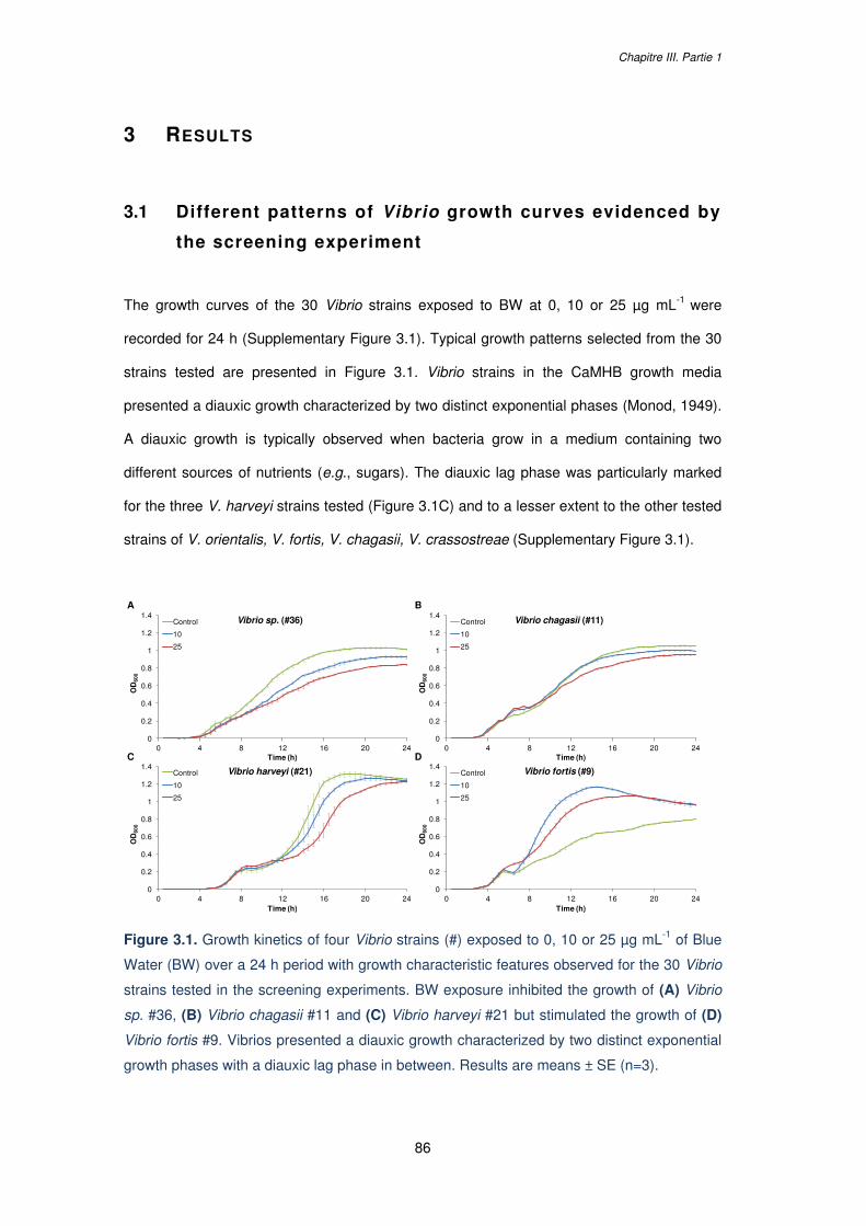
Chapitre III. Partie 1
86
3 RESULTS
3.1 Different patterns of Vibrio growth curves evidenced by
the screening experiment
The growth curves of the 30 Vibrio strains exposed to BW at 0, 10 or 25 µg mL-1 were
recorded for 24 h (Supplementary Figure 3.1). Typical growth patterns selected from the 30
strains tested are presented in Figure 3.1. Vibrio strains in the CaMHB growth media
presented a diauxic growth characterized by two distinct exponential phases (Monod, 1949).
A diauxic growth is typically observed when bacteria grow in a medium containing two
different sources of nutrients (e.g., sugars). The diauxic lag phase was particularly marked
for the three V. harveyi strains tested (Figure 3.1C) and to a lesser extent to the other tested
strains of V. orientalis, V. fortis, V. chagasii, V. crassostreae (Supplementary Figure 3.1).
Figure 3.1. Growth kinetics of four Vibrio strains (#) exposed to 0, 10 or 25 µg mL-1 of Blue
Water (BW) over a 24 h period with growth characteristic features observed for the 30 Vibrio
strains tested in the screening experiments. BW exposure inhibited the growth of (A) Vibrio
sp. #36, (B) Vibrio chagasii #11 and (C) Vibrio harveyi #21 but stimulated the growth of (D)
Vibrio fortis #9. Vibrios presented a diauxic growth characterized by two distinct exponential
growth phases with a diauxic lag phase in between. Results are means ± SE (n=3).
0
0.2
0.4
0.6
0.8
1
1.2
1.4
0 4 8 12 16 20 24
OD
600
Time (h)
Vibrio fortis (#9)Control
10
25
0
0.2
0.4
0.6
0.8
1
1.2
1.4
0 4 8 12 16 20 24
OD
600
Time (h)
Vibrio harveyi (#21)Control
10
25
0
0.2
0.4
0.6
0.8
1
1.2
1.4
0 4 8 12 16 20 24
OD
600
Time (h)
Vibrio chagasii (#11)Control
10
25
0
0.2
0.4
0.6
0.8
1
1.2
1.4
0 4 8 12 16 20 24
OD
600
Time (h)
Vibrio sp. (#36)Control
10
25
A
C
B
D
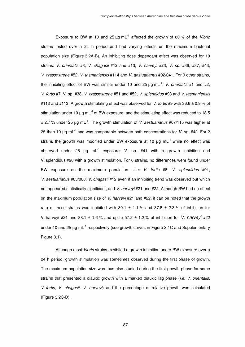
Complex relationships between marennine and bacteria of the genus Vibrio
87
Exposure to BW at 10 and 25 µg mL-1 affected the growth of 80 % of the Vibrio
strains tested over a 24 h period and had varying effects on the maximum bacterial
population size (Figure 3.2A-B). An inhibiting dose dependant effect was observed for 10
strains: V. orientalis #3, V. chagasii #12 and #13, V. harveyi #23, V. sp. #36, #37, #43,
V. crassostreae #52, V. tasmaniensis #114 and V. aestuarianus #02/041. For 9 other strains,
the inhibiting effect of BW was similar under 10 and 25 µg mL-1: V. orientalis #1 and #2,
V. fortis #7, V. sp. #38, V. crassostreae #51 and #52, V. splendidus #93 and V. tasmaniensis
#112 and #113. A growth stimulating effect was observed for V. fortis #9 with 36.6 ± 0.9 % of
stimulation under 10 µg mL-1 of BW exposure, and the stimulating effect was reduced to 18.5
± 2.7 % under 25 µg mL-1. The growth stimulation of V. aestuarianus #07/115 was higher at
25 than 10 µg mL-1 and was comparable between both concentrations for V. sp. #42. For 2
strains the growth was modified under BW exposure at 10 µg mL-1 while no effect was
observed under 25 µg mL-1 exposure: V. sp. #41 with a growth inhibition and
V. splendidus #90 with a growth stimulation. For 6 strains, no differences were found under
BW exposure on the maximum population size: V. fortis #8, V. splendidus #91,
V. aestuarianus #03/008, V. chagasii #12 even if an inhibiting trend was observed but which
not appeared statistically significant, and V. harveyi #21 and #22. Although BW had no effect
on the maximum population size of V. harveyi #21 and #22, it can be noted that the growth
rate of these strains was inhibited with 30.1 ± 1.1 % and 37.8 ± 2.3 % of inhibition for
V. harveyi #21 and 38.1 ± 1.6 % and up to 57.2 ± 1.2 % of inhibition for V. harveyi #22
under 10 and 25 µg mL-1 respectively (see growth curves in Figure 3.1C and Supplementary
Figure 3.1).
Although most Vibrio strains exhibited a growth inhibition under BW exposure over a
24 h period, growth stimulation was sometimes observed during the first phase of growth.
The maximum population size was thus also studied during the first growth phase for some
strains that presented a diauxic growth with a marked diauxic lag phase (i.e. V. orientalis,
V. fortis, V. chagasii, V. harveyi) and the percentage of relative growth was calculated
(Figure 3.2C-D).
Chapitre III. Partie 1
88
Figure 3.2. (A) Relative maximum population size (%) of Vibrio strains exposed over a 24 h
period to concentrations of 10 and 25 µg mL-1 of Blue Water (BW). (B) Graphical illustrations
of the dose-response results presented in (A). (C) Relative maximum population size (%)
after the first growth phase (see insert) for some of the Vibrio strains presenting a diauxic
growth curve and exposed to 10 and 25 mg mL-1 of BW. (D) Graphical illustrations of the
screening experiment results presented in (C). Significant growth inhibitions are presented in
black, growth stimulations in red and no observed effects in green. Asterisks (*) indicate a
statistical difference with the control and letters (A.B) significant differences between the two
BW concentrations tested. Values are means ± SE (n=3).
Vibrio species Percent of relative growthStrain number (#) BW 10 mg mL-1 BW 25 mg mL-1
Vibrio orientalis#1 - 19.9 ± 2.6 *** - 26.7 ± 2.2 ***#2 - 25.3 ± 2.3 ***aa - 37.5 ± 2.1 ***bb#3 - 20.6 ± 1.3 *** - 23.1 ± 0.6 ***
Vibrio fortis#7 - 11.4 ± 2.3 * - 5.5 ± 5.7 *#8 - 6.7 ± 3.8 - 9.1 ± 4.7#9 + 36.6 ± 0.9 ***aaa + 18.5 ± 2.7 ***bbb
Vibrio chagasii#11 - 13.3 ± 1.0 ***a - 18.6 ± 1.5 ***b#12 + 0.2 ± 12.0 - 23.0 ± 4.9#13 - 10.4 ± 4.9 - 19.0 ± 1.7 *
Vibrio harveyi#21 1.6 ± 2.1 - 7.2 ± 0.6#22 + 2.3 ± 0.7 + 2.2 ± 0.8#23 - 12.8 ± 0.8 ***aaa - 26.3 ± 1.5 ***bbb
Vibrio sp. (gills)#36 - 13.2 ± 5.4 a - 33.5 ± 1.3 **b#37 - 17.9 ± 1.3 ***aa - 23.6 ± 1.1 ***bb
#38 - 33.4 ± 4.2 *** - 34.6 ± 0.6 ***Vibrio sp.
#41 - 16.2 ± 5.6 *a + 5.4 ± 6.2 b#42 + 15.0 ± 3.4 * + 12.5 ± 5.0 *#43 - 6.8 ± 2.4 aa - 21.0 ± 2.7 **bb
Vibrio crassostreae#51 - 22.1 ± 0.7 ** - 30.4 ± 1.5 **#52 - 10.5 ± 1.6 *aa - 24.5 ± 2.3 ***bb#53 - 11.2 ± 1.3 * - 20.2 ± 2.0 **
Vibrio splendidus#90 + 16.6 ± 2.9 *a + 2.8 ± 5.2 b#91 - 7.5 ± 3.7 - 12.8 ± 6.5#93 - 33.5 ± 2.1 ** - 37.5 ± 0.5 **
Vibrio tasmaniensis#112 - 72.4 ± 36.8 *** - 100.0 0.0 ***#113 - 10.0 ± 2.7 * - 13.5 ± 1.3 **#114 - 19.6 ± 3.7 **a - 29.4 ± 0.9 ***b
Vibrio aestuarianus#02041 + 0.5 ± 3.7 a - 15.0 ± 3.5 *b#03008 - 1.5 ± 7.1 + 21.3 ± 3.5#07115 + 7.3 ± 2.6 *a + 15.1 ± 2.6 **b
50
25
0
25
50
0 10 20
Relative growth (%)
BW (µg mL-1)
V. orientalis
#1
#2
#3
50
25
0
25
50
0 10 20
Relative growth (%)
BW (µg mL-1)
V. fortis
#7
#8
#9
50
25
0
25
50
0 10 20
Relative growth (%)
BW (µg mL-1)
V. chagasii
#11
#12
#13
50
25
0
25
50
0 10 20
Relative growth (%)
BW (µg mL-1)
V. harveyi
#21
#22
#23
50
25
0
25
50
0 10 20
Relative growth (%)
BW (µg mL-1)
V. crassostreae
#51
#52
#53
50
25
0
25
50
0 10 20
Relative growth (%)
BW (µg mL-1)
V. sp. (gills)
#36
#37
#38
50
25
0
25
50
0 10 20
Relative growth (%)
BW (µg mL-1)
Vibrio sp.
#41
#42
#43
50
25
0
25
50
0 10 20
Relative growth (%)
BW (µg mL-1)
V. splendidus
#90
#91
#93
125
100
75
50
25
0
0 10 20
Relative growth (%)
BW (µg mL-1)
V. tasmaniensis
#112
#113
#114
50
25
0
25
50
0 10 20
Relative growth (%)
BW (µg mL-1)
V. aestuarianus
#02041
#03008
#07115
A B
Vibrio species Percent of relative growthStrain number (#) BW 10 mg mL-1 BW 25 mg mL-1
Vibrio orientalis+ 43.8 ± 2.0 ***aaa + 78.6 ± 6.6 ***bbb
#2 + 8.9 ± 0.6 **aaa + 25.4 ± 2.0 ***bbb
#3 + 41.4 ± 0.2 ***aaa + 68.0 ± 2.9 ***bbb
Vibrio fortis#7 - 7.6 ± 7.8 - 24.3 ± 12.2 *#8 - 13.2 ± 12.7 - 19.2 ± 19.8#9 + 52.9 ± 6.3 *aa + 122.7 ± 16.8 ***bb
Vibrio chagasii#11 + 33.1 ± 5.7 *** + 35.7 ± 4.3 ***#12 - 6.0 ± 4.3 + 7.3 ± 2.5#13 - 3.3 ± 11.6 + 4.8 ± 1.2
Vibrio harveyi#21 - 15.6 ± 3.4 - 4.0 ± 5.8#22 - 15.7 ± 3.8 * - 26.8 ± 3.2 **#23 + 21.8 ± 0.9 ** + 22.8 ± 3.2 **
Time (h)
OD
600
0
20
40
60
80
100
0 10 20
Relative growth (%)
BW (µg mL-1)
V. orientalis
#1
#2
#3
604020020406080100120140160
0 10 20Relative growth (%)
BW (µg mL-1)
V. fortis
#7
#8
#9
20
0
20
40
60
0 10 20Relative growth (%)
BW (µg mL-1)
V. chagasii
#11
#12
#13
40
30
20
10
0
10
20
30
0 10 20
Relative growth (%)
BW (µg mL-1)
V. harveyi
#21
#22
#23
C D
Complex relationships between marennine and bacteria of the genus Vibrio
89
The first growth phase of V. orientalis #1, #2 and #3 and V. fortis #9 was dose-
dependently stimulated under BW exposure. Bacterial growth during the first phase was also
stimulated by BW for V. chagasii #11 and V. harveyi #23 but no differences were found
between exposures to 10 and 25 µg mL-1. A growth inhibition was observed during the first
growth phase for V. harveyi #22 and V. fortis #7 while no effect of BW was recorded for
V. fortis #8, V. chagasii #12 and #13 and V. harveyi #21 during the first growth phase
(Figure 3.2C-D).
3.2 Experiment with BW concentration range
Different dose-response patterns were observed depending on the strain tested, including
linear response and “U shape” responses (Figure 3.3). V. fortis #8, V. orientalis #3,
V. aestuarianus #02/041, V. harveyi #23 presented a linear dose-response curve with
increasing growth inhibition along with an increasing concentration exposure (Figure 3.3A-B
and Supplementary Figure 3.2). For V. fortis #8 a significant inhibitory effect was observed
from 5 µg mL-1 with 5.1 ± 0.5 % of growth inhibition (p-value < 0.001) up to 64.6 ± 0.7 % at
85 µg mL-1 with an IC50 (concentration at which the response is inhibited by 50 %) around
50 µg mL-1 (Figure 3.3A). An increasing inhibitory effect was also observed for V. orientalis #3
from 5 µg mL-1 (p-value < 0.001) up to 50 µg mL-1, but there was no difference in growth
inhibition between 50 µg mL-1 and 85 µg mL-1 with a maximum inhibition level of about 37 %
(Supplementary Figure 3.2). A No Observed Effect Level (NOEL) was noted for
V. aestuarianus #02/041 between 0 µg mL-1 and 25 µg mL-1 (p-value 0.351) and the first
inhibitory effect was evidenced at 50 µg mL-1 with 12 ± 0.2 % of inhibition (p-value 0.015)
while the maximum effect was observed at 85 µg mL-1 with 23.4 ± 1.7 % of inhibition
(Supplementary Figure 3.2). For V. harveyi #23, the diauxic lag phase was particularly
marked and the growth response changed with BW concentration and time. Growth curves
were thus analyzed as two independent phases of growth with phase 1 from 0 to 9 h and
phase 2 from 9h to 24 h (Figure 3.3B). For the first growth phase a significant growth
stimulation was observed starting from 5 µg mL-1 (p-value < 0.001). The second growth
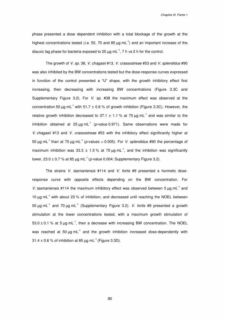
Chapitre III. Partie 1
90
phase presented a dose dependent inhibition with a total blockage of the growth at the
highest concentrations tested (i.e. 50, 70 and 85 µg mL-1) and an important increase of the
diauxic lag phase for bacteria exposed to 25 µg mL-1, 7 h vs 2 h for the control.
The growth of V. sp. 38, V. chagasii #13, V. crassostreae #53 and V. splendidus #90
was also inhibited by the BW concentrations tested but the dose-response curves expressed
in function of the control presented a “U” shape, with the growth inhibitory effect first
increasing, then decreasing with increasing BW concentrations (Figure 3.3C and
Supplementary Figure 3.2). For V. sp. #38 the maximum effect was observed at the
concentration 50 µg mL-1 with 51.7 ± 0.6 % of growth inhibition (Figure 3.3C). However, the
relative growth inhibition decreased to 37.1 ± 1.1 % at 70 µg mL-1 and was similar to the
inhibition obtained at 25 µg mL-1 (p-value 0.971). Same observations were made for
V. chagasii #13 and V. crassostreae #53 with the inhibitory effect significantly higher at
50 µg mL-1 than at 70 µg mL-1 (p-values < 0.005). For V. splendidus #90 the percentage of
maximum inhibition was 33.3 ± 1.5 % at 70 µg mL-1, and the inhibition was significantly
lower, 23.0 ± 0.7 % at 85 µg mL-1 (p-value 0.004; Supplementary Figure 3.2).
The strains V. tasmaniensis #114 and V. fortis #9 presented a hormetic dose-
response curve with opposite effects depending on the BW concentration. For
V. tasmaniensis #114 the maximum inhibitory effect was observed between 5 µg mL-1 and
10 µg mL-1 with about 23 % of inhibition, and decreased until reaching the NOEL between
50 µg mL-1 and 70 µg mL-1 (Supplementary Figure 3.2). V. fortis #9 presented a growth
stimulation at the lower concentrations tested, with a maximum growth stimulation of
53.0 ± 0.1 % at 5 µg mL-1, then a decrease with increasing BW concentration. The NOEL
was reached at 50 µg mL-1 and the growth inhibition increased dose-dependently with
31.4 ± 0.6 % of inhibition at 85 µg mL-1 (Figure 3.3D).
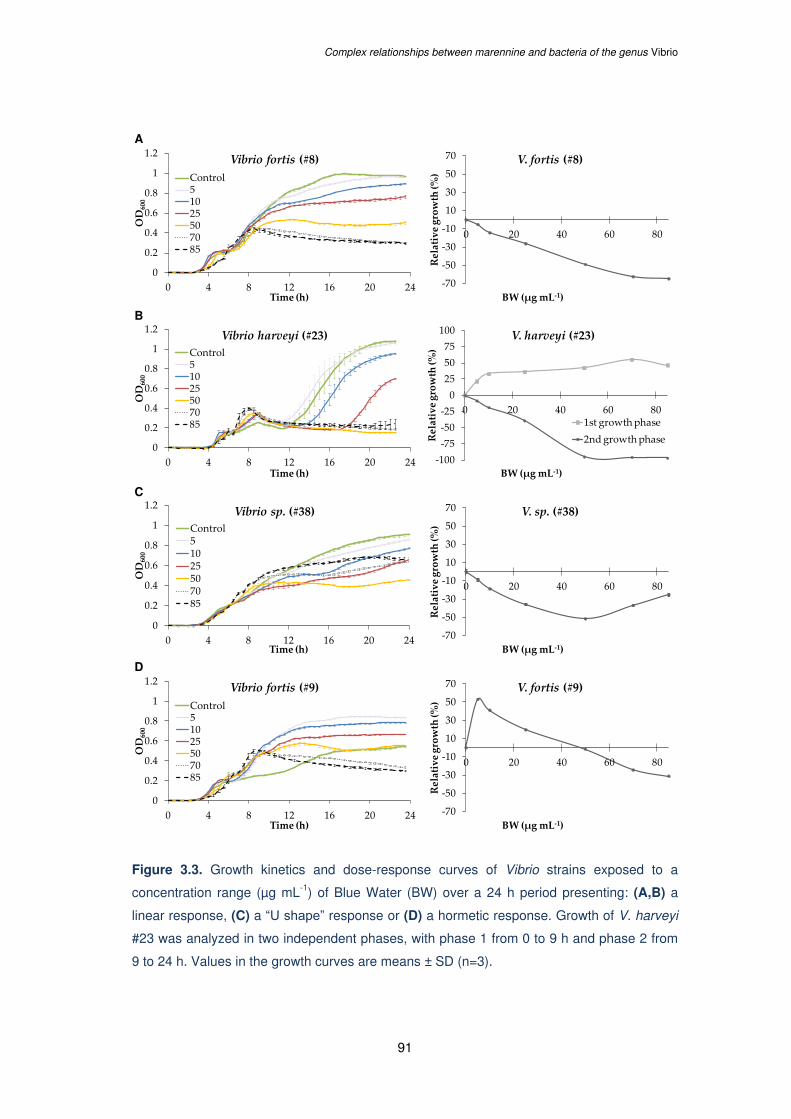
Complex relationships between marennine and bacteria of the genus Vibrio
91
Figure 3.3. Growth kinetics and dose-response curves of Vibrio strains exposed to a
concentration range (µg mL-1) of Blue Water (BW) over a 24 h period presenting: (A,B) a
linear response, (C) a “U shape” response or (D) a hormetic response. Growth of V. harveyi
#23 was analyzed in two independent phases, with phase 1 from 0 to 9 h and phase 2 from
9 to 24 h. Values in the growth curves are means ± SD (n=3).
0
0.2
0.4
0.6
0.8
1
1.2
0 4 8 12 16 20 24
OD600
Time (h)
Vibrio sp. (#38)Control51025507085
70
50
30
10
10
30
50
70
0 20 40 60 80
Relative growth (%)
BW (µg mL-1)
V. sp. (#38)
0
0.2
0.4
0.6
0.8
1
1.2
0 4 8 12 16 20 24
OD600
Time (h)
Vibrio fortis (#9)Control51025507085
70
50
30
10
10
30
50
70
0 20 40 60 80
Relative growth (%)
BW (µg mL-1)
V. fortis (#9)
0
0.2
0.4
0.6
0.8
1
1.2
0 4 8 12 16 20 24
OD600
Time (h)
Vibrio fortis (#8)Control51025507085
70
50
30
10
10
30
50
70
0 20 40 60 80
Relative growth (%)
BW (µg mL-1)
V. fortis (#8)
0
0.2
0.4
0.6
0.8
1
1.2
0 4 8 12 16 20 24
OD600
Time (h)
Vibrio harveyi (#23)Control51025507085
1007550250
25
50
75
100
0 20 40 60 80
Relative growth (%)
BW (µg mL-1)
V. harveyi (#23)
1st growth phase2nd growth phase
A
B
C
D
Chapitre III. Partie 1
92
4 D ISCUSSION
The effects of marennine on several Vibrio strains with different ecological and virulence
properties have been investigated by monitoring bacterial growth over a 24 h period after
addition of BW in the culture medium. The general trend for all the experiments is that BW
affected most of the Vibrio strains tested (80 %), but the results are contrasting. Indeed, BW
either inhibited or stimulated the growth of the Vibrio strains.
As BW solution (i.e. the concentrated supernatant of H. ostrearia containing
marennine) was used in the present work and not a purified form of the pigment, it could be
questioned if the observed effects were induced by marennine or by other molecules
possibly present in the non-axenic culture medium (e.g. exopolysaccharides (EPS) from
Haslea or epibiont bacteria). However, previous studies conducted with purified extracellular
marennine (EMn) on marine bacteria, including Vibrio species, confirmed that the pigment
itself did exert an antibacterial activity (Falaise et al., 2016b; Gastineau et al., 2012b, 2014b;
Pouvreau, 2006). Also, if blue Haslea species should be cultured at large scale to ensure the
production of marennine solutions for applications as antimicrobials in aquaculture, BW
represents the much easier and cheaper production process in comparison with the purified
pigment.
The observed effect of BW on vibrios was species- but also strain-dependent, as for
a same species, the sensitivity of distinct strains could significantly differ. This is in
accordance with previous observations on V. aestuarianus, V. coralliilyticus, V. tubiashii
exposed to purified marennine (Falaise et al., 2016b). Moreover, it seemed difficult to
establish a correlation between the effects of BW and the strain ecological characteristics
such as sampling season, distribution (sea water fractions and oyster tissues) or the
virulence of the strain toward bivalves (Supplementary Table 3.1).
However, the present work considerably extends our understanding of the variability
of marennine effect on bacteria of the Vibrio genus. The diversity observed in growth
responses was especially noteworthy in two species, V. fortis on the one hand, with a growth
inhibition for strains #7 and #8 and important growth stimulation for strain #9, and
Complex relationships between marennine and bacteria of the genus Vibrio
93
V. tasmaniensis on the other hand, with a total inhibition of strain #112 but no effect
observed on the strain #113.
Also, a diauxic growth was evidenced for all the Vibrio strains tested, a phenomenon
commonly observed when bacteria are grown in a medium containing two types of carbon
sources (e.g., sugars) and characterized by two distinct exponential phases with a diauxic
lag phase in between (Monod, 1949). Vibrio diauxic growth is not extensively evidenced in
the literature and was mainly illustrated using V. cholerae (Bag, 1974; Bhattacharya et al.,
2018; Chatterjee et al., 2017) but also V. alginolyticus (Vine et al., 2004). It is worth noting
that complete growth kinetics are rarely recorded for antibacterial assays as most
experiments conducted to screen bioactive compounds against marine Vibrio species used
the disc diffusion method (Banerjee et al., 2013; Cavallo et al., 2013; Das et al., 2005), as
the ones conducted with marennine and marennine-like pigments on V. aestuarianus
(Gastineau et al., 2012b, 2012c). The effect of purified marennine was previously tested with
the microdilution method on another V. tasmaniensis strain (Falaise et al., 2016b; Gastineau
et al., 2014b) and no obvious diauxic growth was observed then, which could be explained
by the use of a different growth media (marine broth media vs CaMHB in the present study),
a different time scale or a different treatment of data.
Bacterial diauxic growth has long been considered as a bacterial phase of enzymatic
acclimation to metabolize a different type of sugar, however, there are now growing
evidences that this phenomenon could rather correspond to the presence of two bacterial
subpopulations in the isogenic culture with different phenotypic adaptation and growth
strategy (Solopova et al., 2014). Based on this new hypothesis, diauxic growth curves in the
present study could evidence the existence of different subpopulations of Vibrio strains; the
Type 1 that can divide quickly by using the most suitable carbon source to sustain its growth,
and the Type 2 that grows more slowly but that would be able to metabolize a different
carbon source. According to Solopova et al. (2014), the Type 1 subpopulation, that cannot
switch to an alternative metabolic pathway, will stop dividing possibly because of the low
energy state of cells (Solopova et al., 2014). This phenotype heterogeneity has been
recently evidenced in V. cholerae (Chatterjee et al., 2017) and would result from the bet
Chapitre III. Partie 1
94
hedging strategy, an evolutionary strategy that allows colonies to cope with fluctuating
environments (Ackermann, 2015; Grimbergen et al., 2015; Martins and Locke, 2015). In the
present work, although bacterial growth was globally inhibited over the 24 h of the essay, a
growth stimulation of the subpopulation Type 1 was sometimes observed under BW
exposure (e.g. V. orientalis, V. chagasii, V. harveyi strains). It can be hypothesized that the
subpopulation Type 1 would be able to metabolize part of the sugars constituting the carbon
skeleton of the marennine molecule or, if any, other EPS present in BW solution to sustain
its growth, or that the stimulation would result from overcompensation mechanisms by the
bacteria under BW exposure, a mechanism that can be observed when cells undergo a
disruption in homeostasis (Calabrese, 2013). It may also be argued that, when exposed to
BW, it is the growth of subpopulation Type 2 that was mainly affected by the BW as
illustrated by the total absence of growth of V. harveyi #23 at the highest concentrations
tested (from 50 to 80 µg mL-1).
A global observation from the results shows that the dose-response curves of most
Vibrio strains did not follow a linear or threshold model with growth inhibition increasing with
concentrations. Indeed, some curves presented a “U-shape” response regarding the
antibacterial effect that decreased with increasing BW exposure (e.g. V. chagasii #13 or
V. sp #38). On top of this, opposite effects were observed at low and high concentrations, a
phenomenon known as hormesis (Calabrese and Baldwin, 2002). Indeed, the lowest BW
concentration tested (5 µg mL-1) highly stimulated the growth of V. fortis #9 up to 50 % while
the growth was significantly inhibited at higher concentrations (from 50 to 80 µg mL-1).
Hormetic dose-response relationships have raised a growing awareness in toxicological and
ecotoxicological studies and have been extensively documented over the past two decades
in different models as plants, algae or fungi (Belz and Duke, 2014; Calabrese and Baldwin,
2001; Cedergreen et al., 2006). Mechanisms of hormesis are not yet clearly understood and
it seems that only a subset of compounds with specific cellular mechanisms would mediate
hormetic responses (Kendig et al., 2010; Lushchak, 2014). Moreover, hormetic effects are
challenging to observe in laboratory conditions, being dependent on various factors such as
endpoint measurements or growth conditions (Calabrese and Baldwin, 1997). In the present
Complex relationships between marennine and bacteria of the genus Vibrio
95
study, hormetic response of Vibrios to BW was observed in the CaMHB medium, a culture
medium that was previously reported to promote the observation of hormesis in Escherichia
coli exposed to antibiotics, while the hormetic effect was not observed in Luria-Bertani (LB)
culture media (Wang et al., 2017).
The mechanisms of action responsible for the antibacterial activity of marennine
remain little explored. So far, the Haslea provincialis purified blue pigment was demonstrated
to interact with the outer membrane of the Gram – bacteria E. coli, rendering it more rigid
(Tardy-Laporte et al., 2013). Recent studies conducted on V. cholerae also evidenced a
disruption of the bacterial membrane integrity and a deformation of the cell architecture by
antibacterial agents such as polyphenols (Bhattacharya et al., 2018) or nanoparticles
(Sarwar et al., 2016). For now, it is still unclear how marennine and Vibrio interact in vivo and
if the pigment has a direct effect on Vibrio growth or if it could decrease its pathogenicity by
fixing on cell membranes, which both could explain better survival of bivalve larvae exposed
to marennine and challenged with V. splendidus (Turcotte et al., 2016).
5 CONCLUSION
In conclusion, the present work evidences much more complex interactions between
marennine and Vibrios than a standard linear correlation between the dose and the effect.
Moreover, the antibacterial activity of marennine is dependent on the species, the strain, and
also, possibly, on the population of bacteria, suggesting that the blue pigment would act on
specific targets. Marennine antibacterial mechanisms and low dose stimulation phenomenon
will have to be better understood before considering any application of Haslea and
marennine as antimicrobials in aquaculture.
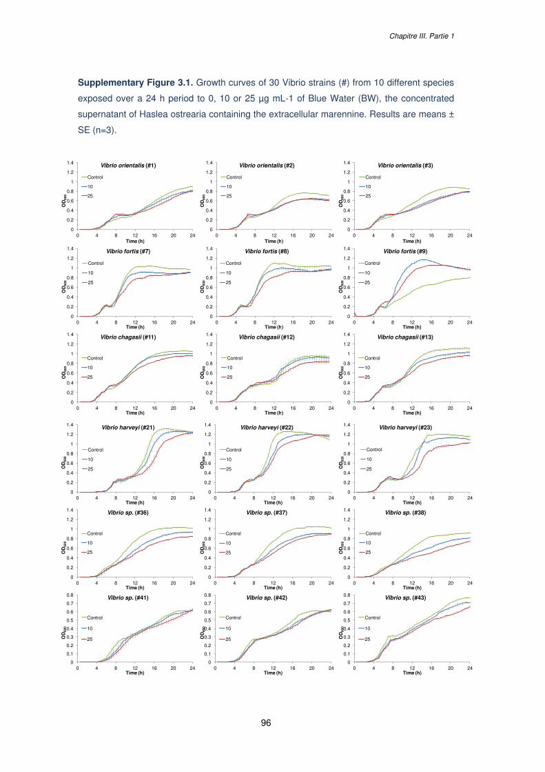
Chapitre III. Partie 1
96
Supplementary Figure 3.1. Growth curves of 30 Vibrio strains (#) from 10 different species
exposed over a 24 h period to 0, 10 or 25 µg mL-1 of Blue Water (BW), the concentrated
supernatant of Haslea ostrearia containing the extracellular marennine. Results are means ±
SE (n=3).
0
0.2
0.4
0.6
0.8
1
1.2
1.4
0 4 8 12 16 20 24
OD
600
Time (h)
Vibrio orientalis (#3)
Control
10
25
0
0.2
0.4
0.6
0.8
1
1.2
1.4
0 4 8 12 16 20 24
OD
600
Time (h)
Vibrio orientalis (#1)
Control
10
25
0
0.2
0.4
0.6
0.8
1
1.2
1.4
0 4 8 12 16 20 24
OD
600
Time (h)
Vibrio orientalis (#2)
Control
10
25
0
0.2
0.4
0.6
0.8
1
1.2
1.4
0 4 8 12 16 20 24
OD
600
Time (h)
Vibrio fortis (#7)
Control
10
25
0
0.2
0.4
0.6
0.8
1
1.2
1.4
0 4 8 12 16 20 24
OD
600
Time (h)
Vibrio fortis (#8)
Control
10
25
0
0.2
0.4
0.6
0.8
1
1.2
1.4
0 4 8 12 16 20 24
OD
600
Time (h)
Vibrio fortis (#9)
Control
10
25
0
0.2
0.4
0.6
0.8
1
1.2
1.4
0 4 8 12 16 20 24
OD
600
Time (h)
Vibrio chagasii (#11)
Control
10
25
0
0.2
0.4
0.6
0.8
1
1.2
1.4
0 4 8 12 16 20 24
OD
600
Time (h)
Vibrio chagasii (#12)
Control
10
25
0
0.2
0.4
0.6
0.8
1
1.2
1.4
0 4 8 12 16 20 24
OD
600
Time (h)
Vibrio chagasii (#13)
Control
10
25
0
0.2
0.4
0.6
0.8
1
1.2
1.4
0 4 8 12 16 20 24
OD
600
Time (h)
Vibrio harveyi (#21)
Control
10
25
0
0.2
0.4
0.6
0.8
1
1.2
1.4
0 4 8 12 16 20 24
OD
600
Time (h)
Vibrio harveyi (#22)
Control
10
25
0
0.2
0.4
0.6
0.8
1
1.2
1.4
0 4 8 12 16 20 24
OD
600
Time (h)
Vibrio harveyi (#23)
Control
10
25
0
0.2
0.4
0.6
0.8
1
1.2
1.4
0 4 8 12 16 20 24
OD
600
Time (h)
Vibrio sp. (#36)
Control
10
25
0
0.2
0.4
0.6
0.8
1
1.2
1.4
0 4 8 12 16 20 24
OD
600
Time (h)
Vibrio sp. (#37)
Control
10
25
0
0.2
0.4
0.6
0.8
1
1.2
1.4
0 4 8 12 16 20 24
OD
600
Time (h)
Vibrio sp. (#38)
Control
10
25
0
0.1
0.2
0.3
0.4
0.5
0.6
0.7
0.8
0 4 8 12 16 20 24
OD
600
Time (h)
Vibrio sp. (#41)
Control
10
25
0
0.1
0.2
0.3
0.4
0.5
0.6
0.7
0.8
0 4 8 12 16 20 24
OD
600
Time (h)
Vibrio sp. (#42)
Control
10
25
0
0.1
0.2
0.3
0.4
0.5
0.6
0.7
0.8
0 4 8 12 16 20 24
OD
600
Time (h)
Vibrio sp. (#43)
Control
10
25
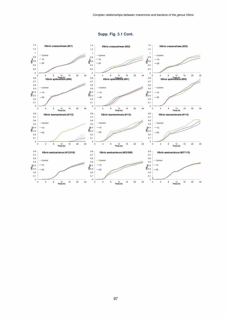
Complex relationships between marennine and bacteria of the genus Vibrio
97
Supp. Fig. 3.1 Cont.
0
0.2
0.4
0.6
0.8
1
1.2
1.4
0 4 8 12 16 20 24
OD
600
Time (h)
Vibrio crassostreae (#51)
Control
10
25
0
0.2
0.4
0.6
0.8
1
1.2
1.4
0 4 8 12 16 20 24
OD
600
Time (h)
Vibrio crassostreae (#52)
Control
10
25
0
0.2
0.4
0.6
0.8
1
1.2
1.4
0 4 8 12 16 20 24
OD
600
Time (h)
Vibrio crassostreae (#53)
Control
10
25
0
0.1
0.2
0.3
0.4
0.5
0.6
0.7
0.8
0 4 8 12 16 20 24
OD
600
Time (h)
Vibrio splendidus (#90)
Control
10
25
0
0.1
0.2
0.3
0.4
0.5
0.6
0.7
0.8
0 4 8 12 16 20 24
OD
600
Time (h)
Vibrio splendidus (#91)
Control
10
25
0
0.1
0.2
0.3
0.4
0.5
0.6
0.7
0.8
0 4 8 12 16 20 24
OD
600
Time (h)
Vibrio splendidus (#93)
Control
10
25
0
0.1
0.2
0.3
0.4
0.5
0.6
0.7
0.8
0 4 8 12 16 20 24
OD
600
Time (h)
Vibrio tasmaniensis (#112)
Control
10
25
0
0.1
0.2
0.3
0.4
0.5
0.6
0.7
0.8
0 4 8 12 16 20 24
OD
600
Time (h)
Vibrio tasmaniensis (#113)
Control
10
25
0
0.1
0.2
0.3
0.4
0.5
0.6
0.7
0.8
0 4 8 12 16 20 24O
D60
0Time (h)
Vibrio tasmaniensis (#114)
Control
10
25
0
0.1
0.2
0.3
0.4
0.5
0.6
0.7
0.8
0 4 8 12 16 20 24
OD
600
Time (h)
Vibrio aestuarianus (#12/016)
Control
10
25
0
0.1
0.2
0.3
0.4
0.5
0.6
0.7
0.8
0 4 8 12 16 20 24
OD
600
Time (h)
Vibrio aestuarianus (#03/008)
Control
10
25
0
0.1
0.2
0.3
0.4
0.5
0.6
0.7
0.8
0 4 8 12 16 20 24
OD
600
Time (h)
Vibrio aestuarianus (#07/115)
Control
10
25
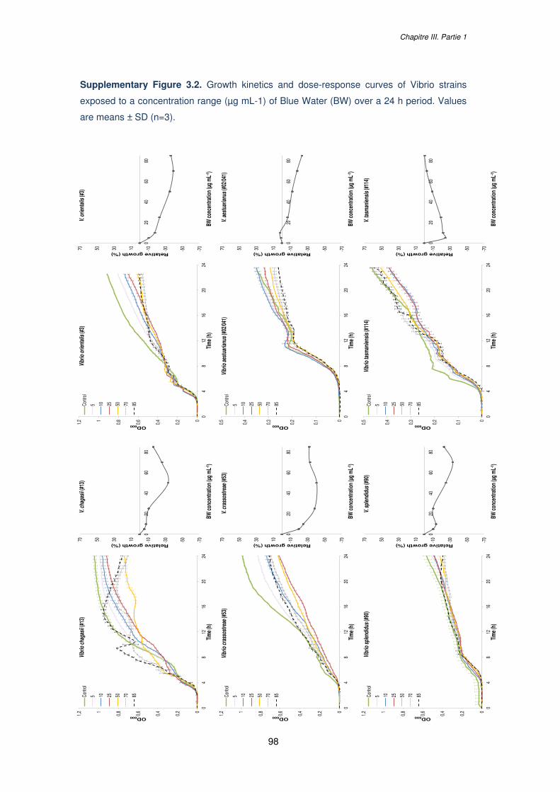
Chapitre III. Partie 1
98
Supplementary Figure 3.2. Growth kinetics and dose-response curves of Vibrio strains
exposed to a concentration range (µg mL-1) of Blue Water (BW) over a 24 h period. Values
are means ± SD (n=3).
00,20,40,60,811,2
04
812
1620
24
OD600
Tim
e (h)
Vibr
io c
haga
sii (
#13)
Contr
ol
5 10 25 50 70 85
00,10,20,30,40,5
04
812
1620
24
OD600
Tim
e (h)
Vibr
io ta
sman
iens
is (#
114)
Contr
ol
5 10 25 50 70 85
00,10,20,30,40,5
04
812
1620
24
OD600
Tim
e (h)
Vibr
io a
estu
aria
nus
(#02
/041)
Contr
ol
5 10 25 50 70 85
00,20,40,60,811,2
04
812
1620
24
OD600
Tim
e (h)
Vibr
io o
rient
alis
(#3)
Contr
ol
5 10 25 50 70 85
00,20,40,60,811,2
04
812
1620
24
OD600
Tim
e (h)
Vibr
io c
rass
ostr
eae
(#53
)Co
ntrol
5 10 25 50 70 85
00,20,40,60,811,2
04
812
1620
24
OD600
Tim
e (h)
Vibr
io s
plen
didu
s (#
90)
Contr
ol
5 10 25 50 70 85
-70
-50
-30
-1010305070
020
4060
80
Relative growth (%)
BW co
ncen
tratio
n (µ
g m
L-1)
V. c
haga
sii (
#13)
-70
-50
-30
-1010305070
020
4060
80
Relative growth (%)
BW co
ncen
tratio
n (µ
g m
L-1)
V. ta
sman
iens
is (#
114)
-70
-50
-30
-1010305070
020
4060
80
Relative growth (%)
BW co
ncen
tratio
n (µ
g m
L-1)
V. a
estu
aria
nus
(#02
/041)
-70
-50
-30
-1010305070
020
4060
80
Relative growth (%)
BW co
ncen
tratio
n (µ
g m
L-1)
V. o
rient
alis
(#3)
-70
-50
-30
-1010305070
020
4060
80
Relative growth (%)
BW co
ncen
tratio
n (µ
g m
L-1)
V. c
rass
ostr
eae
(#53
)
-70
-50
-30
-1010305070
020
4060
80
Relative growth (%)
BW co
ncen
tratio
n (µ
g m
L-1)
V. s
plen
didu
s (#
90)
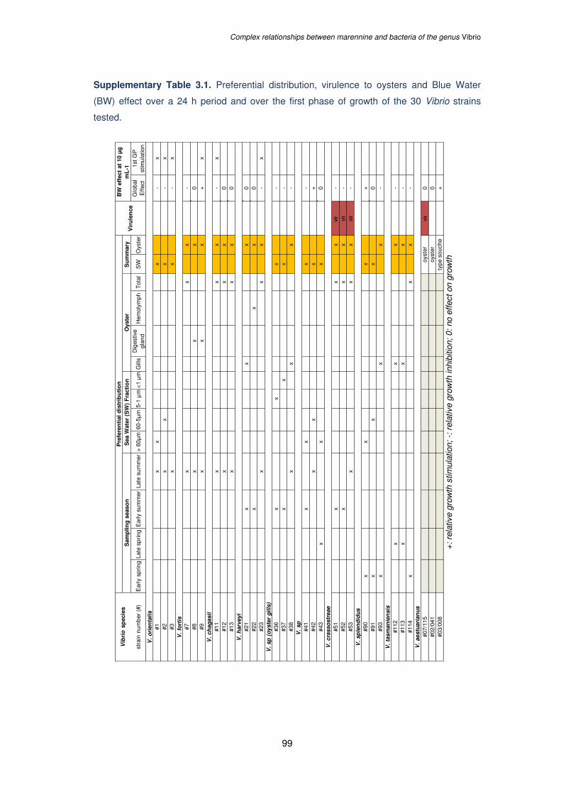
Complex relationships between marennine and bacteria of the genus Vibrio
99
Supplementary Table 3.1. Preferential distribution, virulence to oysters and Blue Water
(BW) effect over a 24 h period and over the first phase of growth of the 30 Vibrio strains
tested.
stra
in n
um
ber
(#)
Ear
ly s
prin
gLa
te s
prin
gE
arly
su
mm
erLa
te s
um
mer
> 6
0µm
60-5
µm
5-1
µm
<1
µm
Gill
sD
iges
tive
gla
nd
Hem
olym
ph
Tot
alS
WO
yste
rG
lob
al
Eff
ect
1st
GP
st
imu
latio
nV
. o
rien
tali
s
#1x
xx
-x
#2x
xx
-x
#3x
x-
xV
. fo
rtis
#7x
xx
-#8
xx
x0
#9x
xx
+x
V.
ch
ag
asii
#11
xx
x-
x#1
2x
xx
0#1
3x
xx
0V
. h
arv
eyi
#21
xx
x0
#22
xx
x0
#23
xx
x-
xV
. sp
(o
yste
r g
ills
)
#36
xx
x-
#37
xx
x-
#38
xx
x-
V.
sp
#41
xx
x-
#42
xx
x+
#43
xx
x0
V.
cra
sso
str
eae
#51
xx
xvi
r -
#52
xx
xvi
r-
#53
xx
xvi
r-
V.
sp
len
did
us
#90
xx
x+
#91
xx
x0
#93
xx
x-
V.
tasm
an
ien
sis
#112
xx
x-
#113
xx
x-
#114
xx
x-
V.
aestu
ari
an
us
#07/
115
vir
0#0
2/04
10
#03/
008
+
+: r
elat
ive
grow
th s
timul
atio
n; -
: rel
ativ
e gr
owth
inhi
bitio
n; 0
: no
effe
ct o
n gr
owth
Vir
ule
nce
BW
eff
ect
at 1
0 µ
g
mL
-1
typ
e so
uch
e
Vib
rio
sp
ecie
s
oyst
eroy
ster
Oys
ter
Su
mm
ary
Pre
fere
nti
al d
istr
ibu
tio
nS
amp
lin
g s
easo
nS
ea W
ater
(S
W)
Fra
ctio
n

100
101
CHAPITRE III.
Partie 2 :
Co-culture of Haslea ostrearia with an oyster
virulent Vibrio strain
Chapitre III. Partie 2
102
Chapitre III. Effects of marennine solutions on the growth of marine
Vibrio. Partie 2 : Co-culture of Haslea ostrearia with an oyster virulent
Vibrio strain.
Les travaux présentés dans la partie 2 de ce chapitre ont été conduits dans le cadre du
stage de Master 2 de l’étudiante Intan Chandra Dewi, co-encadrée avec Jean-Luc Mouget
entre janvier et juin 2017. Un protocole de co-culture Haslea-Vibrio a été imaginé et mis en
place pour ces travaux dont les résultats ont été présentés par Intan en juillet 2017 lors
d’une soutenance orale intitulée « Antibacterial activity from the supernatant containing
extracellular marennine produced by the marine diatom Haslea ostrearia ». Les résultats
produits au cours de ce stage ont également été présentés au congrès de l’International
Society of Applied Phycology (ISAP) à Nantes (France), les 18-23 juin 2017.
Co-culture of Haslea ostrearia with an oyster virulent Vibrio strain
103
Abstract
The effect of marennine, the water-soluble blue pigment produced by the diatom Haslea
ostrearia, was investigated on the growth of an oyster-virulent strain of Vibrio aestuarianus in
co-culture. To trigger H. ostrearia marennine production, cultures were exposed to high
irradiance level and the growth of V. aestuarianus was compared between cultures under a
high light treatment (HL) and cultures under a low light treatment (LL). In HL conditions, the
growth of H. ostrearia and marennine production per cell was significantly higher in
comparison with the LL condition but the growth of V. aestuarianus did not appear different
between the two conditions. Addition of Vibrio in the cultures had no effect on the growth of
the microalgae or on marennine production but lowered the associated bacteria (AB)
concentration (i.e. bacteria naturally present in the non-axenic H. ostrearia cultures),
highlighting complex interactions between AB, Vibrio and Haslea cells. From this experiment,
the effect of H. ostrearia and marennine on V. aestuarianus growth in co-culture could not
have been determined. Complementary experiments should be conducted with axenic
cultures of H. ostrearia to overcome the interactions between AB and Vibrio, but no culture
conditions are yet defined to sustain the growth of the microalgae without its associated
microbiota.
Keywords: Haslea ostrearia; Vibrio aestuarianus; marennine; co-culture
Chapitre III. Partie 2
104
1 INTRODUCTION
Bacterial species of the genus Vibrio are involved in diseases and mass mortality events in
shellfish cultures (Travers et al., 2015) with species such as V. aestuarianus particularly
virulent for oysters (Garnier et al., 2007). As the use of antibiotics is limited due to increasing
bacterial resistance and chemical residues in sea food tissues, alternative natural
compounds are developed to decrease risk for public health, especially compounds derived
from plants (Citarasu, 2012). Another strategy promotes the use of microalgae for the
biocontrol of pathogenic bacteria. For instance, antibacterial effects have been evidenced
with microalgae from different phyla (Bacillariophytes, Chlorophytes, Haphtophytes, etc.),
either in vitro using algal extracts or in vivo using algae co-cultured with bacteria, including
Vibrio species (reviewed in Falaise et al., 2016b). The diatom Haslea ostrearia, which
produces the water-soluble blue pigment marennine, has already been shown to display
antibacterial activities in vitro, especially against various Vibrio species such as
V. aestuarianus (Gastineau et al., 2012b), V. anguillarum (Pouvreau, 2006) and V.
splendidus (Gastineau et al., 2014; Falaise et al 2016b). This diatom has long been known to
be of great interest in oyster farming as marennine is also responsible for the “greening” of
oyster gills that increases their market value by 20 %. The possible use of H. ostrearia and
its pigment marennine as natural antibiotics has received more attention. Indeed a recent
study evidenced the effects of marennine on a large panel of Vibrio strains, with cases of
growth stimulation or growth inhibition under marennine exposure and demonstrated that the
interactions between the blue pigment and Vibrios could be very complex (see Chapter III
Part. 1). However, no in vivo studies have been conducted to assess the effects of
H. ostrearia on the Vibrio growth while the understanding of such interaction is much needed
before considering the use of Haslea and marennine in aquaculture.
In the present work, an oyster virulent strain of V. aestuarianus was co-cultured with
H. ostrearia. Two different light treatments were applied to the cultures: a high light (HL)
treatment and a low light (LL) treatment in order to enhance microalgal growth and
marennine production under HL condition (Figure 3.4; Mouget et al., 1999). The effect of
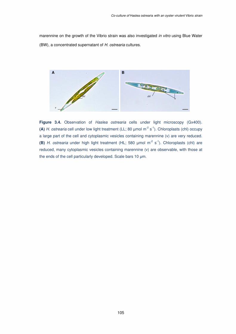
Co-culture of Haslea ostrearia with an oyster virulent Vibrio strain
105
marennine on the growth of the Vibrio strain was also investigated in vitro using Blue Water
(BW), a concentrated supernatant of H. ostrearia cultures.
Figure 3.4. Observation of Haslea ostrearia cells under light microscopy (Gx400).
(A) H. ostrearia cell under low light treatment (LL; 80 µmol m-2 s-1). Chloroplasts (chl) occupy
a large part of the cell and cytoplasmic vesicles containing marennine (v) are very reduced.
(B) H. ostrearia under high light treatment (HL; 580 µmol m-2 s-1). Chloroplasts (chl) are
reduced, many cytoplasmic vesicles containing marennine (v) are observable, with those at
the ends of the cell particularly developed. Scale bars 10 μm.
A B
chl chl
v
v
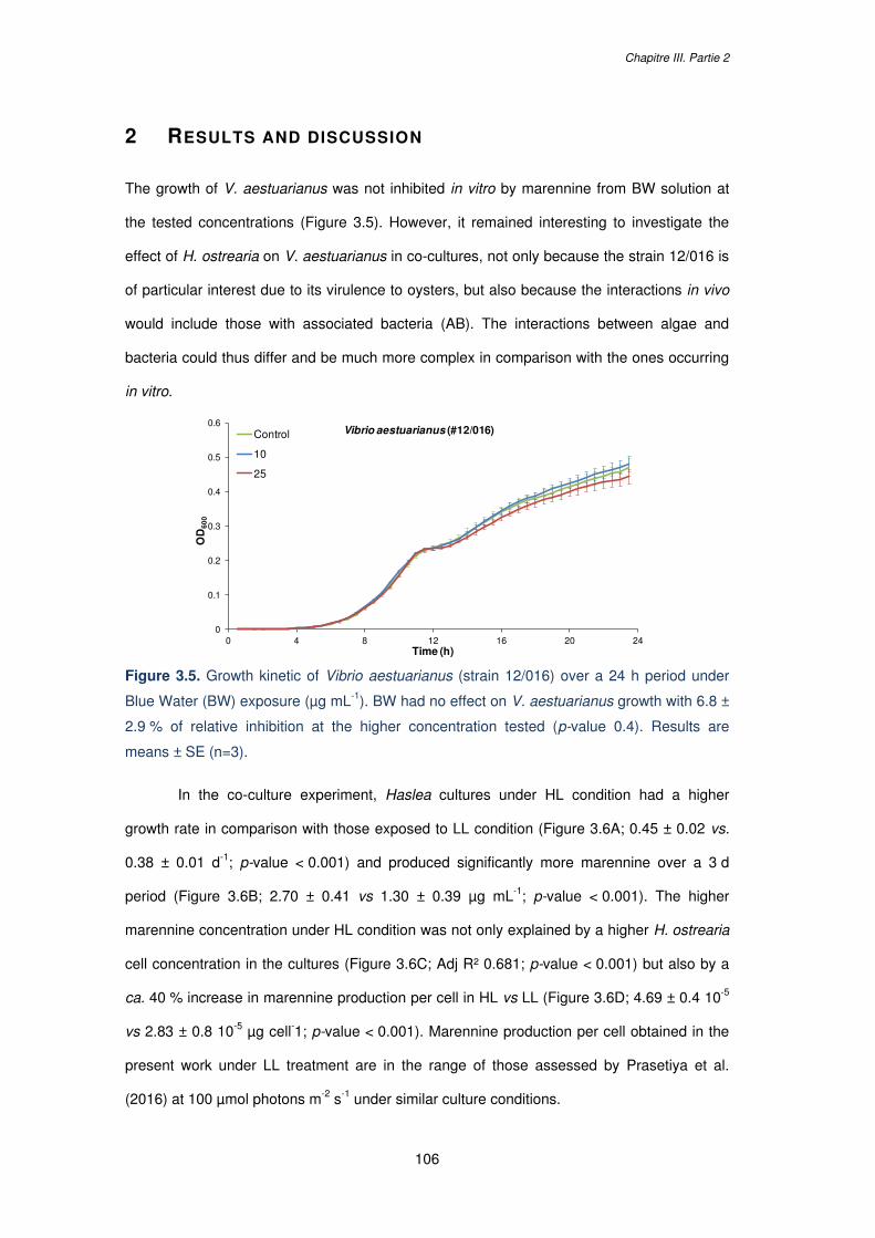
Chapitre III. Partie 2
106
2 RESULTS AND DISCUSSION
The growth of V. aestuarianus was not inhibited in vitro by marennine from BW solution at
the tested concentrations (Figure 3.5). However, it remained interesting to investigate the
effect of H. ostrearia on V. aestuarianus in co-cultures, not only because the strain 12/016 is
of particular interest due to its virulence to oysters, but also because the interactions in vivo
would include those with associated bacteria (AB). The interactions between algae and
bacteria could thus differ and be much more complex in comparison with the ones occurring
in vitro.
Figure 3.5. Growth kinetic of Vibrio aestuarianus (strain 12/016) over a 24 h period under
Blue Water (BW) exposure (µg mL-1). BW had no effect on V. aestuarianus growth with 6.8 ±
2.9 % of relative inhibition at the higher concentration tested (p-value 0.4). Results are
means ± SE (n=3).
In the co-culture experiment, Haslea cultures under HL condition had a higher
growth rate in comparison with those exposed to LL condition (Figure 3.6A; 0.45 ± 0.02 vs.
0.38 ± 0.01 d-1; p-value < 0.001) and produced significantly more marennine over a 3 d
period (Figure 3.6B; 2.70 ± 0.41 vs 1.30 ± 0.39 µg mL-1; p-value < 0.001). The higher
marennine concentration under HL condition was not only explained by a higher H. ostrearia
cell concentration in the cultures (Figure 3.6C; Adj R² 0.681; p-value < 0.001) but also by a
ca. 40 % increase in marennine production per cell in HL vs LL (Figure 3.6D; 4.69 ± 0.4 10-5
vs 2.83 ± 0.8 10-5 µg cell-1; p-value < 0.001). Marennine production per cell obtained in the
present work under LL treatment are in the range of those assessed by Prasetiya et al.
(2016) at 100 µmol photons m-2 s-1 under similar culture conditions.
0
0.1
0.2
0.3
0.4
0.5
0.6
0 4 8 12 16 20 24
OD
60
0
Time (h)
Vibrio aestuarianus (#12/016)Control
10
25
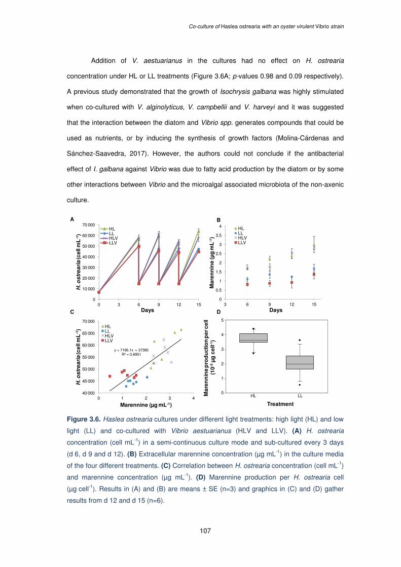
Co-culture of Haslea ostrearia with an oyster virulent Vibrio strain
107
Addition of V. aestuarianus in the cultures had no effect on H. ostrearia
concentration under HL or LL treatments (Figure 3.6A; p-values 0.98 and 0.09 respectively).
A previous study demonstrated that the growth of Isochrysis galbana was highly stimulated
when co-cultured with V. alginolyticus, V. campbellii and V. harveyi and it was suggested
that the interaction between the diatom and Vibrio spp. generates compounds that could be
used as nutrients, or by inducing the synthesis of growth factors (Molina-Cárdenas and
Sánchez-Saavedra, 2017). However, the authors could not conclude if the antibacterial
effect of I. galbana against Vibrio was due to fatty acid production by the diatom or by some
other interactions between Vibrio and the microalgal associated microbiota of the non-axenic
culture.
Figure 3.6. Haslea ostrearia cultures under different light treatments: high light (HL) and low
light (LL) and co-cultured with Vibrio aestuarianus (HLV and LLV). (A) H. ostrearia
concentration (cell mL-1) in a semi-continuous culture mode and sub-cultured every 3 days
(d 6, d 9 and d 12). (B) Extracellular marennine concentration (µg mL-1) in the culture media
of the four different treatments. (C) Correlation between H. ostrearia concentration (cell mL-1)
and marennine concentration (µg mL-1). (D) Marennine production per H. ostrearia cell
(µg cell-1). Results in (A) and (B) are means ± SE (n=3) and graphics in (C) and (D) gather
results from d 12 and d 15 (n=6).
0
0.5
1
1.5
2
2.5
3
3.5
4
3 6 9 12 15
Ma
ren
nin
e (µ
g m
L-1
)
Days
HLLLHLVLLV
A B
C D
Treatment
Ma
ren
nin
e p
rod
uct
ion
pe
r ce
ll(1
0-5µ
g c
ell-1
)
HL LL
5
4
3
2
1
0
y = 7196.1x + 37385R² = 0.6951
40 000
45 000
50 000
55 000
60 000
65 000
70 000
0 1 2 3 4
H. o
str
ea
ria
(ce
ll m
L-1
)
Marennine (µg mL-1)
HLLLHLVLLV
0
10 000
20 000
30 000
40 000
50 000
60 000
70 000
0 3 6 9 12 15
H. o
str
ea
ria
(ce
ll m
L-1
)
Days
HLLLHLVLLV
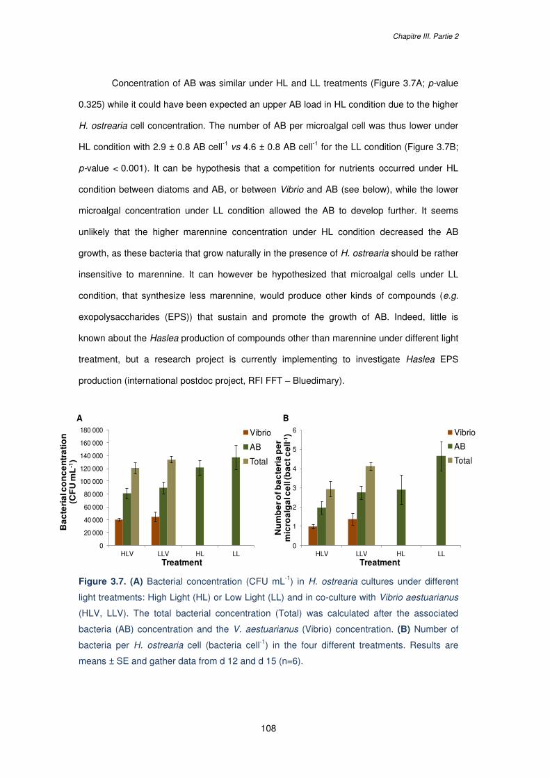
Chapitre III. Partie 2
108
Concentration of AB was similar under HL and LL treatments (Figure 3.7A; p-value
0.325) while it could have been expected an upper AB load in HL condition due to the higher
H. ostrearia cell concentration. The number of AB per microalgal cell was thus lower under
HL condition with 2.9 ± 0.8 AB cell-1 vs 4.6 ± 0.8 AB cell-1 for the LL condition (Figure 3.7B;
p-value < 0.001). It can be hypothesis that a competition for nutrients occurred under HL
condition between diatoms and AB, or between Vibrio and AB (see below), while the lower
microalgal concentration under LL condition allowed the AB to develop further. It seems
unlikely that the higher marennine concentration under HL condition decreased the AB
growth, as these bacteria that grow naturally in the presence of H. ostrearia should be rather
insensitive to marennine. It can however be hypothesized that microalgal cells under LL
condition, that synthesize less marennine, would produce other kinds of compounds (e.g.
exopolysaccharides (EPS)) that sustain and promote the growth of AB. Indeed, little is
known about the Haslea production of compounds other than marennine under different light
treatment, but a research project is currently implementing to investigate Haslea EPS
production (international postdoc project, RFI FFT – Bluedimary).
Figure 3.7. (A) Bacterial concentration (CFU mL-1) in H. ostrearia cultures under different
light treatments: High Light (HL) or Low Light (LL) and in co-culture with Vibrio aestuarianus
(HLV, LLV). The total bacterial concentration (Total) was calculated after the associated
bacteria (AB) concentration and the V. aestuarianus (Vibrio) concentration. (B) Number of
bacteria per H. ostrearia cell (bacteria cell-1) in the four different treatments. Results are
means ± SE and gather data from d 12 and d 15 (n=6).
A B
0
20 000
40 000
60 000
80 000
100 000
120 000
140 000
160 000
180 000
HLV LLV HL LL
Ba
cte
ria
l co
nc
en
tra
tio
n
(CF
U m
L-1
)
Treatment
Vibrio
AB
Total
0
1
2
3
4
5
6
HLV LLV HL LL
Nu
mb
er o
f ba
cte
ria
pe
r m
icro
alg
al c
ell
(ba
ctc
ell-1
)
Treatment
Vibrio
AB
Total
Co-culture of Haslea ostrearia with an oyster virulent Vibrio strain
109
It was also noteworthy that the AB load was significantly lowered in the presence of
V. aestuarianus under HL and LL conditions (Figure 3.7A; p-values 0.002 and < 0.001
respectively), that could suggest a competition between the epibiont bacteria and the
opportunistic Vibrio. The number of Vibrio per H. ostrearia cell was significantly lower under
HL condition in comparison with the LL condition (Figure 3.7B; p-value 0.013). However, it
cannot be deduced that this effect resulted from a higher marennine concentration under HL
condition but it would rather result from complex interactions such as competition between
AB, Vibrio and Haslea cells. The influence of the light treatment on the V. aestuarianus strain
tested was assessed without record of difference of light intensity on the bacterial growth
(data not shown). This is in accordance with a work conducted by Kokou et al. (2012) in
which the growth of six different Vibrio species was not influenced by light treatments. Same
authors demonstrated that the growth of Vibrio spp. was inhibited when co-cultured with
axenic microalgal cultures under light or dark treatment suggesting that the observed
antibacterial effect was not caused by oxygen radicals produced during photosynthesis by
the microalgae (Kokou et al., 2012).
The total bacterial concentration and total number of bacteria per cell was similar
under HL conditions in the presence or in the absence of Vibrio (Figure 3.7A-B; p-values
0.964 and 0.999 respectively). Same observations on the total bacterial concentration and
total number of bacteria per cell could be made under LL conditions in the presence or in the
absence of Vibrio (Figure 3.7A-B; p-values 0.742 and 0.413 respectively). This would
correspond to the maximal bacterial carrying capacity of the culture under the different light
treatment.
Chapitre III. Partie 2
110
3 CONCLUSION
In conclusion, the present work does not allow demonstrating an inhibiting effect of
marennine on V. aestuarianus growth in co-culture with H. ostrearia, mainly because of the
complex interactions between AB, Haslea cells and Vibrio. Addition of Vibrio to the cultures
did not result in a higher marennine concentration, while it could have been expected that
the release of this antimicrobial pigment would be triggered by the microalga under bacterial
exposure. However, the Vibrio inoculum was low and the total bacterial concentration was
similar in the absence or presence of Vibrio. To assess whether marennine released is
triggered by the presence of bacteria the experiment should be conducted with axenic
Haslea cultures and various inoculum concentrations tested. For instance, running a co-
culture experiment with axenic cultures remains challenging at the experimental level as
previous attempts of axenisation led either to culture death or to a significant reduction of cell
growth and marennine production. As H. ostrearia, such as other diatoms, cannot sustain
growth without the nutrients provided by the associated bacteria (Amin et al., 2012; Fuentes
et al., 2016; Lépinay et al., 2016), maintaining axenic cultures would only be considered with
the development of an optimal culture media with additional nutrient to compensate for the
absence of AB (e.g. vitamins). Although this study does not answer to the initial issue, it
provides new data on H. ostrearia cultures such as the quantification of marennine
production per cell under different light treatments, and also on the interaction between
H. ostrearia and its AB, with lower AB concentration at higher Haslea densities. This
information will be particularly relevant for future culture axenisation assays in which high
light conditions should be favored to promote Haslea growth and slow down the AB growth.
Co-culture of Haslea ostrearia with an oyster virulent Vibrio strain
111
4 MATERIALS AND METHODS
In the present work, a co-culture experiment was conducted between H. ostrearia (strain
NCC 495; isolated in Bourgneuf bay, France) and an oyster-virulent V. aestuarianus strain
(strain 12/016; provided by RBE - SG2M - LGPMM, IFREMER station La Tremblade,
France) to assess the marennine effects on V. aestuarianus growth. H. ostrearia was
cultured under two different light treatments: a high light treatment (HL; 580 µmol.m-2.s-1) and
a low light treatment (LL; 80 µmol.m-2.s-1) in order to obtain high marennine concentrations in
H. ostrearia cultures under the HL condition and low marennine concentration under the LL
condition (Figure 3.4).
The effect of different light treatments on V. aestuarianus growth was previously
assessed and no differences were recorded (data not shown). H. ostrearia was cultured in
250 mL Erlenmeyer flasks containing 100 mL of sterile sea water (SSW) prepared from a
commercial sea salt mix (Instant Ocean, Aquarium Systems®; pH 7.7 ± 0.2; salinity 32 ppm)
with enrichment solutions as described in Mouget et al. (2009). Three flasks were placed
under each light treatment in a control temperature room (16 °C) with illumination provided
by cool-white fluorescent tubes in a 14/10 h light/dark cycle. V. aestuarianus strain was
maintained at -80 °C in glycerol 50 % (v/v), cultured on Nutrient Broth (NB) + NaCl (Biokar)
and colonies isolated on Nutrient Agar 2 % (NA) + NaCl (Biokar; final pH and salinity for NB
and NA: 7.7 ± 0.2 and 32 ppm). The co-culture experiment was run over a 15 d period on a
semi-continuous mode with subcultures conducted every 3 d (on d 6, d 9 and d 12) with
H. ostrearia concentration adjusted to 15.000 cell mL-1. The semi continuous mode was used
to maintain cells in exponential growth phase, to ensure working on cells always in the same
physiological stage, without any nutrient deficiency due to competition or light limitation due
to self-shading. V. aestuarianus was added to 3 flasks per light treatment on d 0 at a final
concentration in the culture of 5.103 CFU mL-1. V. aestuarianus concentration was
determined with Optical Density measurements (OD; 630 nm) after establishing a calibration
curve (OD vs CFU mL-1). Before each subculture, H. ostrearia concentration was assessed
using an Nageotte hemocytometer and marennine concentration was determined on cell-free
Chapitre III. Partie 2
112
culture supernatant (syringe filtered through 0.2 μm, Sarstedt) using a spectrophotometer
(UV/Vis Lambda 25 Perkin Elmer spectrophotometer, UV Winlab software) and the Beer-
Lambert’s equation (ε677=12.13 L g-1cm-1) as proposed by Pouvreau et al. (2006c). Bacterial
concentration was estimated by counting the CFU on Petri dish after plating the samples
with an automatic plater (easySpiral Pro ®, Intersciences) and incubation at 27 °C over a
24 h period. Selective agar growth media were used to discriminate between V. aestuarianus
and H. ostrearia associated bacteria (AB) colonies (NA + NaCl media for V. aestuarianus
and NA + SSW for AB). For the result treatments, only data from d 12 and d 15 were taken
into account, which correspond to a stabilized system with significantly more marennine
under HL in comparison with the LL condition.
Also, prior to the co-culture experiment, the effect of marennine was assessed in
vitro on the V. aestuarianus growth following the same protocol described in Chapter III Part.
2, using a Blue Water (BW) solution (i.e. a concentrated supernatant of H. ostrearia
containing the extracellular marennine).
Statistical analyses were conducted on SigmaPlot 12.3 software. Differences
between treatments were assessed with One-Way ANOVAs. Normality was tested by the
Shapiro-Wilk test and the assumption of homoscedasticity of variance with Fisher’s test
(F-test). Post hoc Tukey’s pairwise multiple comparison tests were used to determine
differences between means. Unless specified, alpha value used was 0.05.
113
CHAPITRE IV.
NEW INSIGHTS INTO THE
EFFECTS OF MARENNINE
SOLUTIONS ON MARINE
ORGANISMS

114
115
CHAPITRE IV.
Partie 1 :
Harmful or harmless: biological effects of
marennine on marine organisms
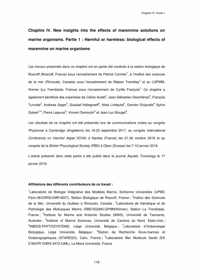
Chapitre IV. Partie 1
116
Chapitre IV. New insights into the effects of marennine solutions on
marine organisms. Partie 1 : Harmful or harmless: biological effects of
marennine on marine organisms
Les travaux présentés dans ce chapitre ont en partie été conduits à la station biologique de
Roscoff (Roscoff, France) sous l’encadrement de Patrick Cormier1, à l’Institut des sciences
de la mer (Rimouski, Canada) sous l’encadrement de Réjean Tremblay2 et au LGPMM,
Ifremer (La Tremblade, France) sous l’encadrement de Cyrille François3. Ce chapitre a
également bénéficié des expertises de Céline Audet2, Jean-Sébastien Deschênes2, François
Turcotte2, Andreas Seger4, Gustaaf Hallegraeff4, Niels Lindquist5, Damien Sirjacobs6, Sylvie
Gobert7-8, Pierre Lejeune8, Vincent Demoulin6 et Jean-Luc Mouget9.
Les résultats de ce chapitre ont été présentés lors de communications orales au congrès
Physiomar à Cambridge (Angleterre) les 18-22 septembre 2017, au congrès International
Conference on Harmful Algae (ICHA) à Nantes (France) les 21-26 octobre 2018 et au
congrès de la British Phycological Society (PBS) à Oban (Écosse) les 7-10 janvier 2019.
L’article présenté dans cette partie a été publié dans le journal Aquatic Toxicology le 17
janvier 2019.
Affiliations des différents contributeurs de ce travail :
1Laboratoire de Biologie Intégrative des Modèles Marins, Sorbonne Universités (UPMC
Paris 06/CNRS/UMR 8227), Station Biologique de Roscoff, France ; 2Institut des Sciences
de la Mer, Université du Québec à Rimouski, Canada ; 3Laboratoire de Génétique et de
Pathologie des Mollusques Marins (RBE/SG2M/LGPMM/Ifremer), Station La Tremblade,
France ; 4Institute for Marine and Antarctic Studies (IMAS), Université de Tasmanie,
Australie ; 5Institute of Marine Sciences, Université de Caroline du Nord, Etats-Unis ; 6INBIOS-PHYTOSYSTEMS, Liège Université, Belgique ; 7Laboratoire d’Océanologie
Biologique, Liège Université, Belgique ; 8Station de Recherche Sous-marines et
Océanographiques (STARESO), Calvi, France ; 9Laboratoire Mer Molécule Santé (EA
2160/FR CNRS 3473 IUML), Le Mans Université, France
Harmful or harmless: biological effects of marennine on marine organisms
117
Abstract
Marennine is a water-soluble blue-green pigment produced by the marine diatom Haslea
ostrearia. The diatom and its pigment are well known from oyster farming areas as the
source of the greening of oyster gills, a natural process increasing their market value in
Western France. Blooms of blue Haslea are also present outside oyster ponds and hence
marine organisms can be exposed, periodically and locally, to significant amounts of
marennine in natural environments. Due to its demonstrated antibacterial activities against
marine pathogenic bacteria (e.g. Vibrio) and possible prophylactic effects toward bivalve
larvae, marennine is of special interest for the aquaculture industry, especially bivalve
hatcheries. The present study aimed to provide new insights into the effects of marennine on
a large spectrum of marine organisms belonging to different phyla, including species of
aquaculture interest and organisms frequently employed in standardised ecotoxicological
assays. Different active solutions containing marennine were tested: partially purified
Extracellular Marennine (EMn), and concentrated solutions of marennine present in
H. ostrearia culture supernatant; the Blue Water (BW) and a new process called
Concentrated Supernatant (CS). Biological effects were meanwhile demonstrated in
invertebrate species for the three marennine-based solutions at the highest concentrations
tested (e.g., decrease of fertilization success, delay of embryonic developmental stages or
larval mortality). Exposure to low concentrations did not impact larval survival or
development and even tended to enhance larval physiological state. Furthermore, no effects
of marennine were observed on the fish gill cell line tested. Marennine could be viewed as a
Jekyll and Hyde molecule, which possibly affects the earliest stages of development of some
organisms but with no direct impacts on adults. Our results emphasize the need to determine
dosages that optimize beneficial effects and critical concentrations not to be exceeded
before considering the use of marennine in bivalve or fish hatcheries.
Key words: diatom; Haslea ostrearia; marennine; marine organisms; natural bioactive
compound
Chapitre IV. Partie 1
118
1 INTRODUCTION
Haslea ostrearia is a cosmopolitan marine pennate diatom that synthesizes and releases a
water-soluble blue-green pigment called marennine. This species can bloom erratically in
oyster ponds in Western France (Baie de Marennes-Oléron, Baie de Bourgneuf), a
phenomenon that has long been known to be responsible for the greening of oyster gills.
This phenomenon is of economic interest for the French oyster farming industry as the
market value of green oyster is significantly increased (by 20 to 30 %) due to changes in the
organoleptic properties of the oysters and the scarcity of the product, blooms in ponds being
an erratic phenomenon. In the last decade, new species of blue Haslea have been
discovered, some of them producing marennine-like pigments chemically distinct from the
originally described marennine molecule (Gastineau et al., 2012a, 2016). Blooms of blue
Haslea are also observed in natural environments, such as in the Mediterranean Sea and
East coast of the USA (Figure 4.1). Moreover, oysters with green gills have long been
observed worldwide, e.g., in Great Britain (Sprat, 1667), Denmark (Petersen, 1916), the USA
(Mitchell and Barney, 1917) and Australia (Hallegraeff and Mouget, personal
communications). Furthermore, the greening of gills has been reported in other organisms,
such as polychaetes, crabs, littorina, mussels (Ranson, 1927), sea-anemones (Gaillon,
1820), scallops and cockles (Gastineau et al., 2018), illustrating that many marine organisms
can be exposed to blue Haslea populations and marennine-like pigments not only in artificial
(oyster) ponds, but also in natural environments.
Figure 4.1. Underwater pictures of natural blue Haslea blooms forming biofilms observed (A)
in macro-algae Padina sp. in the Mediterranean Sea, Corsica (France) and (B) on sediments
in the Beaufort Strait, North Carolina (United States).
A B
Harmful or harmless: biological effects of marennine on marine organisms
119
Although our knowledge of the blue Haslea biodiversity has recently increased, little
is known about the chemical properties of marennine and marennine-like pigments or their
functions for the microalgae. Marennine is thought to be produced via a cytoplasmic
synthesis pathway (Nassiri et al., 1998) and transiently accumulates at the cell apices
(intracellular form of the pigment, IMn). Marennine is excreted from the cells, possibly by
exocytosis via small vesicles, which collapse and release an extracellular form of marennine
(EMn). The two forms of the pigment differ in their UV-visible spectral characteristics and
molecular mass (Pouvreau et al., 2006a). Marennine is a complex molecule composed of
glycosidic units (Gastineau et al., 2014b) attached to one or various aromatic rings
(Pouvreau et al., 2006a). A protocol to obtain a purified form of EMn or IMn has been
developed (Pouvreau et al., 2006b) and several authors have proposed different methods to
estimate marennine concentration in solution, despite incomplete knowledge of its chemical
structure (Pouvreau et al., 2006c; Robert et al., 2002).
Various studies have demonstrated that marennine (as purified molecule or raw
extract) has multiple biological activities, such as antioxidant (Pouvreau et al., 2008),
antiproliferative (Carbonnelle et al., 1998; Gastineau et al., 2012b), antiviral (Bergé et al.,
1999; Gastineau et al., 2012b) and antibacterial (Falaise et al., 2016b; Gastineau et al.,
2012b, 2014b). It has also been shown that marennine possesses allelopathic properties,
limiting the growth of various microalgae (Pouvreau et al., 2007; Prasetiya et al., 2016). The
biological activities of marennine are species- and even strain-dependent in the case of
bacteria (Falaise et al., 2016b), suggesting that marennine could act on specific molecular
targets. Tests conducted with Gram-negative bacteria have demonstrated activity of the
marennine-like pigment produced by Haslea provincialis (Gastineau et al., 2016) and
marennine produced by H. ostrearia, on the lipopolysaccharidic cell membrane of
Escherichia coli (Tardy-Laporte et al., 2013) and of Vibrio splendidus (Bouhlel et al., 2018),
rendering it more rigid.
In line with these results, particularly those demonstrating the capacity of marennine
to limit the proliferation of certain pathogenic marine bacteria (Falaise et al., 2016b;
Gastineau et al., 2012b, 2014b), further research has confirmed the protective effect of
Chapitre IV. Partie 1
120
marennine on the giant scallop (Placopecten magellanicus) and blue mussel (Mytilus edulis)
larvae when challenged with V. splendidus (Turcotte et al., 2016). The same study also
revealed that a 20 d exposure of mussel larvae to low concentrations of marennine
(0.1 µg mL-1) provided a significantly higher survival rate than the control, although exposure
to higher concentrations of marennine (1 µg mL-1) resulted in 100 % larval mortality. Even
brief exposure to marennine has been demonstrated to result in behavioural, physiological
and biochemical changes that were still present eight weeks after exposure (Prasetiya et al.,
2017).
These results underpin the interest to further study the effects of marennine on early
developmental stages of marine organisms, to identify which are possibly sensitive to
marennine, as well as defining exposure concentrations and durations that would be
beneficial to species of aquaculture interest. The present work presents a broad panel of
experiments conducted to provide an overview of the biological effects of marennine
solutions on various marine organisms belonging to different phyla. The organisms were
selected based on aquaculture interest or their established role in ecotoxicological model
assays. Different solutions containing marennine, prepared from H. ostrearia culture
supernatants, were used in the exposures: the purified EMn (Pouvreau et al., 2006b), the
Blue Water (BW; (Turcotte et al., 2016) and a newly patented Concentrated Supernatant
(CS; see Materials). Although EMn represents the most purified form of marennine currently
available, BW and CS are of particular interest if the use of marennine-based solutions is
considered at larger scales than laboratory experiments due to their easier and cheaper
method of production. Marennine exposures were conducted on embryos and/or larvae of
the mollusc M. edulis, the crustacean Chthamalus bisinuatus, the chordate
Pseudopleuronectes americanus and the echinoderms Sphaerechinus granularis and
Paracentrotus lividus. The effects of the blue pigment solutions were also investigated in
vitro on the fish gill cell line RTgill-W1, on the oyster Crassostrea gigas haemocytes, and on
prokaryotic models with bacteria of the genus Vibrio, providing a broad view of the diversity
of marennine effects on marine organisms.
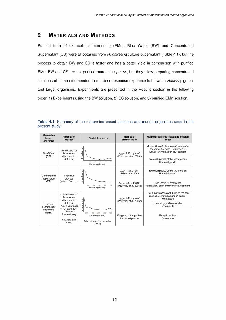
Harmful or harmless: biological effects of marennine on marine organisms
121
2 MATERIALS AND METHODS
Purified form of extracellular marennine (EMn), Blue Water (BW) and Concentrated
Supernatant (CS) were all obtained from H. ostrearia culture supernatant (Table 4.1), but the
process to obtain BW and CS is faster and has a better yield in comparison with purified
EMn. BW and CS are not purified marennine per se, but they allow preparing concentrated
solutions of marennine needed to run dose-response experiments between Haslea pigment
and target organisms. Experiments are presented in the Results section in the following
order: 1) Experiments using the BW solution, 2) CS solution, and 3) purified EMn solution.
Table 4.1. Summary of the marennine based solutions and marine organisms used in the present study.
Marennine based
solutions
Production process
UV-visible spectra Method of
quantificationMarine organisms tested and studied
effect
Blue Water(BW)
Ultrafiltration ofH. ostrearia
culture medium (3-30kDa)
ε677=12.13 L g-1cm-1
(Pouvreau et al. 2006c)
Mussel M. edulis, barnacle C. bisinuatusand winter flounder P. americanus:Larval survival and/or development
Bacterial species of the Vibrio genus: Bacterial growth
ConcentratedSupernatant
(CS)
Innovativeprocess
(patent n°1872316)
ε669=17.2 L g-1cm-1
(Robert et al. 2002)Bacterial species of the Vibrio genus:
Bacterial growth
ε677=12.13 L g-1cm-1
(Pouvreau et al. 2006c) Sea urchin S. granularis:
Fertilization, early embryonic development
Purified Extracellular Marennine
(EMn)
- Ultrafiltration ofH. ostrearia
culture medium (3-30kDa)
- Anion-Exchange chromatography
- Dialysis & freeze-drying
(Pouvreau et al. 2006c)
Adapted f rom Pouvreau et al. (2006)
ε677=12.13 L g-1cm-1
(Pouvreau et al. 2006c)
Preliminary assays with EMn on the sea urchins S. granularis and P. lividus:
Fertilization
Oyster C. gigas haemocytes:Cytotoxicity
Weighing of the purified EMn dried powder
Fish gill cell line:Cytotoxicity
Wavelength (nm)
Ab
sorb
ance
Wavelength (nm)
Ab
sorb
ance
Wavelength (nm)
Ab
sorb
ance
Chapitre IV. Partie 1
122
2.1 Preparation of the purified extracellular marennine
(EMn), Blue Water (BW) and Concentrated Supernatant
(CS)
The Blue Water (BW) and purified extracellular marennine (EMn) were produced at the
Station aquicole de Pointe-au-Père (Québec, Canada) and provided by the Institut des
sciences de la mer de Rimouski-Université du Québec à Rimouski (ISMER-UQAR; Québec,
Canada). The production process of BW and purified EMn were previously described
(Turcotte et al., 2016; Pouvreau et al., 2006b). Briefly, H. ostrearia strains (NCC 136),
isolated from Bourgneuf Bay (France) and provided by NCC (Nantes Culture Collection),
were cultured in 100 L photobioreactors until the extracellular marennine concentration
reached a maximum of 6 to 8 µg mL-1. Marennine concentration was determined on cell-free
culture supernatant (filtered through Sarstedt 0.2 μm syringe filters) using a
spectrophotometer (Cary 100 Bio UV-Visible, Agilent Technologies) and the Beer-Lambert’s
equation (ε677=12.13 L g-1cm-1) as proposed by Pouvreau et al. (2006c). The BW was then
obtained by concentration of the culture supernatant containing EMn by ultrafiltration (double
cut off 3-30 kDa; (Turcotte et al., 2016). To obtain the purified EMn, the BW was further
treated by an anion-exchange chromatography process and the fraction collected was
dialyzed and freeze-dried (Pouvreau et al., 2006b). BW and purified EMn were stored in the
dark at 4 °C and -20 °C respectively.
Concentrated Supernatant (CS) was produced in the Mer Molécule Santé (MMS)
and Institut des Molécules et Matériaux du Mans (IMMM) laboratories (Le Mans, France). A
strain of H. ostrearia (NCC 495) was batch cultured in 500 mL Erlenmeyers flask containing
250 mL of autoclaved sea water prepared from a commercial sea salt mix (Instant Ocean,
Aquarium Systems®; pH 7.6 ± 0.2; salinity 32) with an enrichment solution as described in
Mouget et al. (2009). Microalgal cultures were maintained in a 16 °C temperature-controlled
room at an irradiance of 200 μmol photon m−2 s−1, with illumination provided by cool-white
fluorescent tubes in a 14/10 h light/dark cycle. At the beginning of the stationary growth
phase the culture supernatant containing EMn was collected by decantation of the microalgal
Harmful or harmless: biological effects of marennine on marine organisms
123
cells and subsequent vacuum filtrations through 15 µm (150 mm Filter paper, Fisher
Scientific®) and 1.2 µm (37 mm glass microfiber filters, Whatman®). Filtered culture
supernatant was collected in 1 L glass bottles and the EMn was then concentrated using an
innovative technique recently patented (patent n° 1 872 316). The resulting solution was then
dialyzed using a 2 kDa dialysis membrane (Spectra/Por®6, Spectrum®). Dialysis tubes were
placed in ultra-pure water tanks for 3 d under agitation with the water changed every 24 h.
The dialyzed solution was then ultra-filtered (double cut-off 1 kD-30 kDa; Kros Flo®
Research IIi TFF System and Kros Flo® Automatic Backpressure valve, Spectrum®) and
further concentrated by evaporation with a Rotavapor (Vacuum controller CVC2,
Vacuubrand®; Rotary Elevator, Heating Bath Hei-Vap, Base Hei-Vap ML Adv/Pre,
Heidolph®). The pH of the resulting CS was neutralized if required to 7.5 ± 0.2 by addition of
NaOH 0.1 M. The CS was stored one week in the dark at 4 °C until use. The concentration
of marennine in the CS was determined using a spectrophotometer and the Beer-Lambert
equation (ε669=17.2 L g-1cm-1) as proposed by (Robert et al., 2002). UV-Vis spectra of the
different marennine based solutions were conducted using quartz cuvettes with 1 cm path
length (UV/Vis Lambda 25 Perkin Elmer spectrophotometer, UV Winlab software).
2.2 Exposure of the mussel Mytilus edulis larvae to BW
Adult mussels were obtained in summer 2017 from a farm in the Magdalen Islands
(47° 25’N, 61° 50’W, Quebec, Canada), characterized by pure M. edulis populations (Myrand
et al., 2009) and conditioned for spawning in the Station aquicole de Pointe-au-Père
(ISMER-UQAR; Québec, Canada) facilities for one month. Mussels were maintained in
180 L tanks in 1 μm filtered seawater at 20 °C flowing at 1 L min-1 and were continuously fed
with a mixture of Pavlova lutherii, Isochrysis galbana, and Nannochloropsis oculata
(ratio 1:1:1) supplied with a peristaltic pump at constant flow to maintain food load at 0.5 mg
L-1 as described in Hennebicq et al. (Hennebicq et al., 2013). Before spawning, a dozen
individuals were removed from the tank and washed by gently rubbing the shell with a brush
and diluted bleach and finally rinsed with filtered, UV sterilized sea water. Spawning was
Chapitre IV. Partie 1
124
induced by successive thermal shocks from 10 to 25 °C. Fertilized eggs were transferred to
60 L tanks at densities of 10 eggs µL-1 and embryos were maintained at 18 °C for 48 h.
D-larvae were collected by filtration of the water through a 20 µm mesh screen and
transferred to 60 L rearing tanks at densities of 10 D-larvae mL-1. Every 2-3 d, tanks were
washed and sterilized and the larvae fed with a mixture of P. lutherii, I. galbana and
Chaetoceros gracilis at a final concentration of 90 cells µL-1 (30 cell µL-1 for each component
of the diet; Turcotte et al., 2016).
Mytilus edulis embryos were collected prior to the transfer to the embryogenesis
tank, and D-larvae prior to the transfer to the rearing tank (48h post fertilization). Veliger
larvae were collected in the rearing tanks (14 d post fertilization) by filtration of the water
through a mesh screen. Embryos and larvae were exposed to BW in 12-well flat bottom
sterile microplates with each well containing 3 mL of diluted BW and 10-15 embryos or 10-15
larvae per well. Five concentrations of marennine from BW were tested: 0, 0.1, 0.5, 1 and
5 µg mL-1. Marennine concentration from BW was estimated as described in section 2.1 and
Table 4.1. BW was syringe-filtered through 0.2 µm and diluted in sterile sea water.
Microplates were kept in a controlled temperature room at 18 °C without addition of food
during the time of the experiment. The mortality of embryos and veliger larvae was assessed
using a light microscope (Olympus BX41; W. Carsen Co., Ltd., Don Mills, ON, Canada)
coupled to a digital camera (Evolution VF Color, MediaCybernetics, Silver Spring, MD, USA).
The percentages of swimming and motionless D-larvae were assessed by observation of the
microplates using an inverted microscope (Axiovert 100, Zeiss). Larval size was measured
with Image Pro-Express (Media Cybernetics) after addition of formaldehyde to the wells. A
minimum of 10 larvae were counted per well with at least 3 wells per concentration tested.
2.3 Exposure of the barnacle Chthamalus bisinuatus larvae
to BW
Experiments were conducted in June 2015 in the Center for Marine Biology of Sao Paulo
University (CEBIMar/USP; Sao Paulo, Brazil). Spawners were harvested by collecting rock
Harmful or harmless: biological effects of marennine on marine organisms
125
fragments in the intertidal zone of Calhetas Beach (23°49’ 28”S, 45°25’11”W). The fragments
were transferred to the laboratory and placed in seawater pumped from the adjacent bay, as
described in Kasten and Flores (Kasten and Flores, 2013). A light source was placed above
the tanks containing the breeders to recreate the natural photoperiod. Broodstock was
submerged for 30 min every 12.4 h to simulate the natural effect of the tide and stimulate
spawning. After a first submersion, the water used to submerge the broodstock was
siphoned and filtered to harvest the larvae. The larvae were placed in filtered seawater and
isolated using a dissecting microscope to obtain a number of larvae sufficient to run the
experiment. Larvae were placed individually in the wells of a 96-well microplate in 2 mL of
seawater containing 0, 0.05 or 0.1 µg mL-1 of marennine from BW without food to test only
the potential toxicity of BW. The BW was prepared and the concentration estimated as
described in section 2.1 (Table 4.1). Four larvae were tested at each concentration. The
microplate was placed on a stirring plate and no food was provided during the experiment.
Larvae were observed once a day and survival recorded over 9 d. The experiment was
repeated once with spat from a different production.
2.4 Exposure of the winter flounder Pseudopleuronectes
americanus larvae to BW
Larvae were reared as described by(Khemis et al., 2003) in 57 L cylindro-conical
polyethylene tanks and fed rotifers from d 4 to d 26 (Fraboulet et al., 2010). Rotifers were fed
a cocktail of microalgal concentrates (1:1:1 N. oculata: I. galbana: P. lutherii, Instant algae,
REED Mariculture) and a SELCO food supplement (1 g M-1 rotifers, INVE Aquaculture
Nutrition, Gransville, UT, USA). The larvae were kept in green water (addition of N. oculata
culture directly to basins). From d 2 to d 14, two concentrations of marennine from BW were
tested on the larvae, 0.05 and 0.1 µg mL-1, with three tanks per treatment (n = 3). BW
preparation, as described in section 2.1 (Table 4.1), was added to the tanks in the morning,
when water flow was cut to feed the larvae. Water flow was restarted at the end of the day
(for a total of about 8 h of exposure) and the BW was gradually evacuated from the tanks
Chapitre IV. Partie 1
126
with the flow of water outlet. A dose of BW was given every two days, for a total of seven
treatments per tank for the duration of the experiment. Procedures were the same for the
control treatments, without the addition of BW in the control tanks. The effect of BW
treatment on bacterial load and larval size was evaluated on d 2, d 6, d 10, and d 14, and the
size and physiological condition (assessment of energy reserves) of the larvae were
determined at the end of the experiment, i.e. at d 14 (12 d of treatment). Standard length
was measured on formaldehyde-preserved larval pictures as described in (Hjörleifsson and
Klein-MacPhee, 1992) using a dissecting microscope (Olympus SZ61) coupled to a digital
camera (Evolution VF; Media Cybernetics) and Image Pro-Plus measurement software 5.0
(Media Cybernetics). Lipids were extracted in a 2:1 mixture of dichloromethane: methanol
according to (Folch et al., 1957), the lipid classes (triglyceride [TAG], sterol [ST], acetone
mobile polar lipids [AMPL], and phospholipids [PL]) concentrations were quantified by TLC-
FID as described by (Parrish, 1987), and chromatograms analyzed using PeakSimple v3.21
software (SRI Inc.). To estimate the potential effect of BW on bacterial load in rearing water,
bacterial analyzes were carried out using a flow cytometer on water samples taken from the
tanks and frozen with glutaraldehyde according to (Seychelles et al., 2011).
2.5 Exposure of the sea urchins Sphaerechinus granularis
and Paracentrotus lividus to EMn and CS
A first series of assays were conducted in May 2016 with purified EMn on S. granularis and
P. lividus, model animals frequently used for in vivo ecotoxicological bioassays (Buttino et
al., 2016; Pinsino et al., 2010). Sea urchins were collected in the Brest area (France) and
obtained from the Centre de Ressources Biologiques Marines (CRBM) at the Roscoff
Biological Station (Roscoff, France). Other experiments on sea urchins were run in March
2017 using CS rather than purified EMn due to its limited production, and the species
S. granularis because it is available throughout the year.
Marennine-based solutions were prepared by weighing purified EMn freeze-dried
powder or after the estimation of CS concentration as described in section 2.1 (Table 4.1).
Harmful or harmless: biological effects of marennine on marine organisms
127
Solutions were diluted in 0.22 μm Millipore-filtered seawater (FSW) and the final solutions
were syringe filtered through 0.2 µm. Sea urchin spawning was induced by intracoelomic
injection of 0.1 M acetylcholine. Eggs were collected in FSW, rinsed twice by centrifugation
(2.000 g, 2 min) and re-suspended in FSW for a final 2 % (v/v) egg solution containing 0.1 %
(v/v) glycine. Sperm was kept dry at 4 °C until use. Experiments took place in a temperature-
controlled room at 16 °C.
Effects of purified EMn and CS were first assessed on fertilization. Eggs were
transferred to a 24-well culture plate and incubated during 10 min in marennine-based
solutions prior to addition of sperm. Solutions at different concentrations were added in wells
containing 1 mL of the egg suspension. Only FSW was added for the control condition.
pH strips were used to ensure that the pH did not vary between wells of the different
concentrations tested. For fertilization, 50 µL of dry sperm was diluted in 1 mL of FSW
shortly before use and 4 μL of diluted sperm added per mL of egg suspension. Observation
of the culture plate under phase contrast inverted microscopy allowed the determination of
the fertilization rate by counting at least 60 eggs per well (n = 3 wells per concentration
tested). For experiments conducted with CS, unfertilized eggs exposed to the highest CS
concentrations were rinsed 3 times in FSW using a benchtop centrifuge for 5 to 10 seconds
and exposed to sperm for fertilization as described above. Two hours post fertilization, eggs
exposed to CS were fixed on a DNA fluorescent stain (Hoechst fixative: Bisbenzymide
0.1 µg mL-1; methanol 75 %, glycerol 25 %) and observed under a fluorescence microscope
(ApoTome, Zeiss).
The effects of CS were also assessed post fertilization (kinetics of first cleavage and
early embryonic development). For fertilization, diluted sperm was added in a 50 mL tube
containing 25 mL of eggs suspended in FSW (2 % (v/v) egg suspension). When egg batches
exhibited greater than 90 % fertilization, sperm was removed by centrifugation in Heraeus
Labofuge centrifuge (2.000 g, swinging bucket rotor 2 min). Fertilized eggs were transferred
to a 24-well plate and CS solutions at 1; 10; 50 and 100 µg mL-1 added 10 min post
fertilization. The percentage of dividing eggs was determined under phase contrast inverted
light microscope; at least 60 eggs per well were counted. Three different sea urchin couples
Chapitre IV. Partie 1
128
were used to replicate the experiment. For the “washed” condition, embryos from 3 wells per
concentration tested were rinsed 6 h post fertilization as described earlier. Culture plates
were maintained under constant agitation at 16 °C and embryos were observed 8, 48 and
72 h post fertilization with a Leica DMi8 inverted microscope and pictures acquisition done
with LASX software. For each observation time, a 75 µL sample of each well was transferred
to a glass slide and pictures of the developmental stages predominantly observed were
taken. For post hatching developmental stages, a 1 µL drop of Janus green was added to
the samples. Bright Field microscopy technique (BF) was applied for embryos 8 h post
fertilization and a Differential Interference Contrast technique (DIC) for embryos 48 h and
72 h post fertilization.
2.6 Exposure of Vibrio species to BW and CS
Experiments were conducted during spring 2018 in MMS laboratory (Le Mans, France). BW
and CS were prepared as described in section 2.1 (Table 4.1), and the concentration of
marennine was determined with spectrophotometric measurements using the Beer-Lambert
law with the specific extinction coefficient (ε677=12.13 L g-1cm-1) proposed by (Pouvreau et
al., 2006a). CS and BW were diluted with sterile ultra-pure water. Salinity and pH were
adjusted by addition of NaCl and 0.1 M of NaOH or HCl (pH 7.5 ± 0.2; salinity 32) and the
solutions were then syringe-filtered through 0.22 µm (Sarstedt). The three Vibrio strains
tested, Vibrio chagasii (strain 8T3_5), Vibrio crassostreae (strain 8T2_1) and Vibrio sp.
(strain 7G1_11) were previously identified (Bruto et al., 2017) and kindly provided by the
Laboratory of Integrative Biology of Marine Models (CRBM/Roscoff, France). Bacterial
strains were kept at -80 °C in 25 % glycerol, inoculated in Mueller Hinton Broth (MHB) +
1 % NaCl (pH 7.5 ± 0.2; salinity 32) and incubated overnight at 25 °C. Isolations were done
on Petri dishes containing agar prepared with Nutrient Agar (Biokar) + 2.3 % NaCl (pH
7.5 ± 0.2; salinity 32). The plates were incubated at 25 °C for one day. The antibacterial
activity of BW and CS was assessed according to a method described in the Clinical and
Laboratory Standards Institute (CLSI) antimicrobial microdilution guidelines (Clinical and
Harmful or harmless: biological effects of marennine on marine organisms
129
Laboratory Standards Institute (CLSI), 2012). The day prior to the experiment, three different
colonies per Petri dish were inoculated in broth media and grown overnight at ambient
temperature. Wells of a 96-well sterile microplate with cover and flat bottom were filled with
50 µL of either CS or BW at a final concentration of 10 µg mL-1 (or sterile saline water for
the controls) and 50 µL of a bacterial inoculate. A negative control was also run with sterile
saline water and broth media without bacteria to ensure that no contamination occurred
during the experiment. To prepare the bacterial inoculate the optical density (OD) of the
broth culture was measured at 630 nm (V-10 Plus Onda Spectrophotometer), the OD was
adjusted to 0.1 by dilution in MHB + 1 % NaCl and the solution was then further diluted by
1/100 as recommended by the CLSI guidelines (Clinical and Laboratory Standards Institute
(CLSI), 2012). Microplates were inserted in a microplate spectrophotometer (xMark Bio-Rad)
for a 20 h run at ambient temperature. Growth was managed with microplate Manager 6
Software by taking the OD in each well every 30 min at 600 nm.
2.7 Exposure of fish gill cell lines to EMn
Experiments took place in August 2017 at the Institute for Marine and Antarctic Studies
(IMAS; Hobart, Australia). The gill epithelium cell line RTgill-W1 was obtained from the
American Type Culture Collection (ATTC; and originally isolated from the Rainbow trout
Oncorhyncis mykiss). The cell line was maintained and exposures to purified EMn conducted
as described in Dorantes-Aranda et al. (2011) in conventional 96-well plates. Gill cells were
seeded into a flat-bottom 96-well plate (655180, Greiner) at 2.5 x 10⁵ cells mL-1 in L-15
medium (L-1518, Sigma) and allowed to attach for 48 h in the dark. Confluence of cell
cultures was verified 12 h before experimental exposure and the L-15 medium replaced by
L-15/ex (Schirmer et al., 1997). The concentration of the original purified EMn solution was
estimated (540 µg mL-1 in 50 % methanol) as described in section 2.1 (Table 4.1), and the
solution was diluted in L-15/ex by factors of 1 x 10⁻¹, 5 x 10⁻², 1 x 10⁻², 5 x 10⁻³, 1x 10⁻³,
5 x 10⁻⁴ and 1 x 10⁻⁴, yielding final exposure concentrations of 0-54 µg mL-1. Methanol was
added to all dilutions to achieve a constant final MeOH concentration of 5 % across all
Chapitre IV. Partie 1
130
treatments, including the non-toxic control (L-15/ex). Gill cells were exposed to these
solutions for 2 h at 20 ± 1 ˚C in the dark (quadruplicate wells per concentration). After the
exposure, wells were rinsed twice with saline phosphate buffer (100 µL per well) and
incubated for a further 2 h in the dark with 100 µL of 5 % resazurin viability stain in L-15/ex
medium. Metabolic reduction of resazurin to resorufin by the gill cells was measured in a
microplate reader (Fluostar Omega, BMG Labtech) at excitation and emission wavelengths
of 540 and 590 nm, respectively. Results are expressed as percentage viability of the
nontoxic control (5 % MeOH in L-15/ex).
2.8 Exposure of the oyster Crassostrea gigas haemocytes
to EMn
Pacific oysters C. gigas were harvested in 2015-2016 in the hatchery of Laboratoire de
Génétique et de Pathologie des Mollusques Marins (LGPMM; La Tremblade, France) in
raceways supplied with a constant flow of seawater enriched with phytoplankton
(Skeletonema costatum, Tetraselmis suecica, I. galbana and C. gracilis). They were
maintained in safe conditions, free of known infectious pathogens. The shell of 20 adult
oysters was broken with metal clamps and haemolymph withdrawn from the adductor
muscle sinus (1 to 1.5 mL of haemolymph per oyster) using 1 mL sterile plastic syringes
equipped with a needle (0.90 mm x 25 mm). The haemolymph of the different individuals
was pooled, filtered through 60 µm nylon mesh and held on ice to prevent haemocyte
aggregation (Auffret and Oubella, 1997). Haemocytes were observed under light microscopy
using a Malassez-cell. Haemocytes were then exposed to different concentrations of purified
EMn: 0, 1, 50 and 100 µg mL-1. The purified EMn was obtained as described in section 2.1
(Table 4.1) and the solutions were prepared by weighing of the EMn dried powder on an
analytical balance (Sartorius Entris®) and by dilution on sterile sea water. Purified EMn
solutions were added to the haemolymph (1:1 ratio) for a final haemocyte concentration of
2.104 cells mL-1. Haemocytes were exposed to EMn during 1, 3 or 6 h and mortality was
quantified using 200 µL of cell suspension. Cells were incubated in the dark for 30 min on ice
Harmful or harmless: biological effects of marennine on marine organisms
131
with 50 µL of Propidium Iodide (PI, 1.0 g L-1, Interchim), a fluorescent DNA/RNA-specific dye
that only permeates through the membranes of dead cells and stains the nucleic acids.
Haemocyte samples were analyzed with flow cytometry using an EPICS XL 4 (Beckman
Coulter) and red fluorescence following the protocol of (Morga et al., 2009). Based on size
discrimination, only haemocytes were taken into account with 5.000 events counted per
sample. Results were depicted as cell cytograms and reported as log scale fluorescence
levels. Data were analyzed with Flowing Software 2.
2.9 Statistics
Statistical analyses were run using SigmaPlot 12.3 software for Windows. Differences
between treatments were assessed with One-Way or Two-Ways ANOVAs. Normality was
tested by the Shapiro-Wilk test and the assumption of homoscedasticity of variance with
Fisher’s test (F-test) and/or verified visually by the spread of residuals, as suggested by
(Quinn and Keough, 2002). Post hoc Tukey’s pairwise multiple comparison tests were used
to determine differences between means. Unless specified, alpha value used was 0.05.
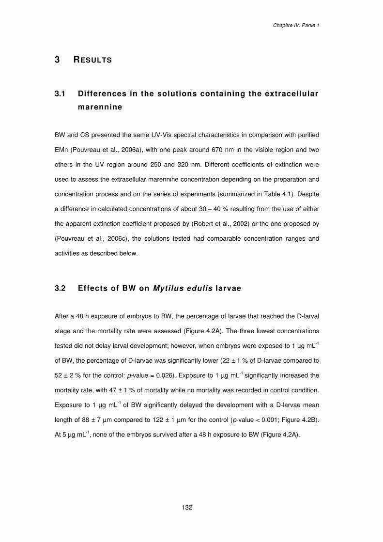
Chapitre IV. Partie 1
132
3 RESULTS
3.1 Differences in the solutions containing the extracellular
marennine
BW and CS presented the same UV-Vis spectral characteristics in comparison with purified
EMn (Pouvreau et al., 2006a), with one peak around 670 nm in the visible region and two
others in the UV region around 250 and 320 nm. Different coefficients of extinction were
used to assess the extracellular marennine concentration depending on the preparation and
concentration process and on the series of experiments (summarized in Table 4.1). Despite
a difference in calculated concentrations of about 30 – 40 % resulting from the use of either
the apparent extinction coefficient proposed by (Robert et al., 2002) or the one proposed by
(Pouvreau et al., 2006c), the solutions tested had comparable concentration ranges and
activities as described below.
3.2 Effects of BW on Mytilus edulis larvae
After a 48 h exposure of embryos to BW, the percentage of larvae that reached the D-larval
stage and the mortality rate were assessed (Figure 4.2A). The three lowest concentrations
tested did not delay larval development; however, when embryos were exposed to 1 µg mL-1
of BW, the percentage of D-larvae was significantly lower (22 ± 1 % of D-larvae compared to
52 ± 2 % for the control; p-value = 0.026). Exposure to 1 µg mL-1 significantly increased the
mortality rate, with 47 ± 1 % of mortality while no mortality was recorded in control condition.
Exposure to 1 µg mL-1 of BW significantly delayed the development with a D-larvae mean
length of 88 ± 7 µm compared to 122 ± 1 µm for the control (p-value < 0.001; Figure 4.2B).
At 5 µg mL-1, none of the embryos survived after a 48 h exposure to BW (Figure 4.2A).
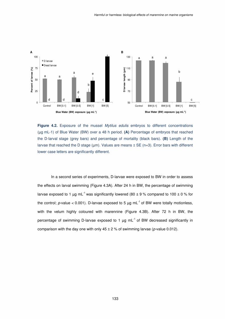
Harmful or harmless: biological effects of marennine on marine organisms
133
Figure 4.2. Exposure of the mussel Mytilus edulis embryos to different concentrations
(µg mL-1) of Blue Water (BW) over a 48 h period. (A) Percentage of embryos that reached
the D-larval stage (grey bars) and percentage of mortality (black bars). (B) Length of the
larvae that reached the D stage (µm). Values are means ± SE (n=3). Error bars with different
lower case letters are significantly different.
In a second series of experiments, D-larvae were exposed to BW in order to assess
the effects on larval swimming (Figure 4.3A). After 24 h in BW, the percentage of swimming
larvae exposed to 1 µg mL-1 was significantly lowered (80 ± 9 % compared to 100 ± 0 % for
the control; p-value < 0.001). D-larvae exposed to 5 µg mL-1 of BW were totally motionless,
with the velum highly coloured with marennine (Figure 4.3B). After 72 h in BW, the
percentage of swimming D-larvae exposed to 1 µg mL-1 of BW decreased significantly in
comparison with the day one with only 45 ± 2 % of swimming larvae (p-value 0.012).
a aa
b
cd d
d
e
f
0
25
50
75
100
Control BW [0.1] BW [0.5] BW [1] BW [5]
Perc
en
t o
f la
rvae (
%)
Blue Water (BW) exposure (µg mL-1)
D larvae
Dead larvae
a a a
b
50
70
90
110
130
Control BW [0.1] BW [0.5] BW [1] BW [5]
D larv
ae l
en
gth
(µ
m)
Blue Water (BW) exposure (µg mL-1)
A B
c
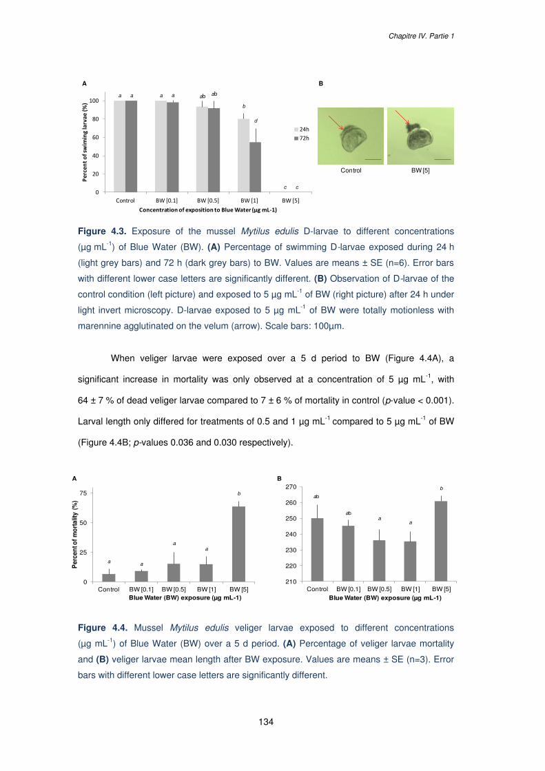
Chapitre IV. Partie 1
134
Figure 4.3. Exposure of the mussel Mytilus edulis D-larvae to different concentrations
(µg mL-1) of Blue Water (BW). (A) Percentage of swimming D-larvae exposed during 24 h
(light grey bars) and 72 h (dark grey bars) to BW. Values are means ± SE (n=6). Error bars
with different lower case letters are significantly different. (B) Observation of D-larvae of the
control condition (left picture) and exposed to 5 µg mL-1 of BW (right picture) after 24 h under
light invert microscopy. D-larvae exposed to 5 µg mL-1 of BW were totally motionless with
marennine agglutinated on the velum (arrow). Scale bars: 100µm.
When veliger larvae were exposed over a 5 d period to BW (Figure 4.4A), a
significant increase in mortality was only observed at a concentration of 5 µg mL-1, with
64 ± 7 % of dead veliger larvae compared to 7 ± 6 % of mortality in control (p-value < 0.001).
Larval length only differed for treatments of 0.5 and 1 µg mL-1 compared to 5 µg mL-1 of BW
(Figure 4.4B; p-values 0.036 and 0.030 respectively).
Figure 4.4. Mussel Mytilus edulis veliger larvae exposed to different concentrations
(µg mL-1) of Blue Water (BW) over a 5 d period. (A) Percentage of veliger larvae mortality
and (B) veliger larvae mean length after BW exposure. Values are means ± SE (n=3). Error
bars with different lower case letters are significantly different.
a a ab
b
c
a a ab
d
c0
20
40
60
80
100
Control BW [0.1] BW [0.5] BW [1] BW [5]
Percent o
f swim
ing larvae (%
)
Concentration of exposition to Blue Water (µg mL-1)
24h
72h
Control BW [5]
A B
a a
aa
b
0
25
50
75
Control BW [0.1] BW [0.5] BW [1] BW [5]
Per
cen
t of
mo
rtal
ity (
%)
Blue Water (BW) exposure (µg mL-1)
ab
aba
a
b
210
220
230
240
250
260
270
Control BW [0.1] BW [0.5] BW [1] BW [5]Blue Water (BW) exposure (µg mL-1)
A B
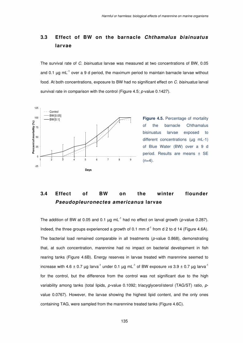
Harmful or harmless: biological effects of marennine on marine organisms
135
3.3 Effect of BW on the barnacle Chthamalus bisinuatus
larvae
The survival rate of C. bisinuatus larvae was measured at two concentrations of BW, 0.05
and 0.1 µg mL-1 over a 9 d period, the maximum period to maintain barnacle larvae without
food. At both concentrations, exposure to BW had no significant effect on C. bisinuatus larval
survival rate in comparison with the control (Figure 4.5; p-value 0.1427).
Figure 4.5. Percentage of mortality
of the barnacle Chthamalus
bisinuatus larvae exposed to
different concentrations (µg mL-1)
of Blue Water (BW) over a 9 d
period. Results are means ± SE
(n=4).
3.4 Effect of BW on the winter flounder
Pseudopleuronectes americanus larvae
The addition of BW at 0.05 and 0.1 µg mL-1 had no effect on larval growth (p-value 0.287).
Indeed, the three groups experienced a growth of 0.1 mm d-1 from d 2 to d 14 (Figure 4.6A).
The bacterial load remained comparable in all treatments (p-value 0.868), demonstrating
that, at such concentration, marennine had no impact on bacterial development in fish
rearing tanks (Figure 4.6B). Energy reserves in larvae treated with marennine seemed to
increase with 4.6 ± 0.7 µg larva-1 under 0.1 µg mL-1 of BW exposure vs 3.9 ± 0.7 µg larva-1
for the control, but the difference from the control was not significant due to the high
variability among tanks (total lipids, p-value 0.1092; triacyglycerol/sterol (TAG/ST) ratio, p-
value 0.0767). However, the larvae showing the highest lipid content, and the only ones
containing TAG, were sampled from the marennine treated tanks (Figure 4.6C).
-25
0
25
50
75
100
125
1 2 3 4 5 6 7 8 9
Pe
rce
nt o
f mo
rta
lity
(%
)
Days
ControlBW [0.05]BW [0.1]
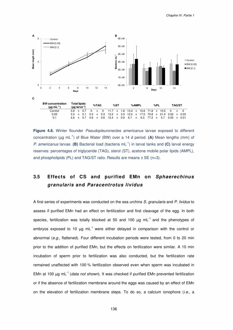
Chapitre IV. Partie 1
136
Figure 4.6. Winter flounder Pseudopleuronectes americanus larvae exposed to different
concentration (µg mL-1) of Blue Water (BW) over a 14 d period. (A) Mean lengths (mm) of
P. americanus larvae. (B) Bacterial load (bacteria mL-1) in larval tanks and (C) larval energy
reserves: percentages of triglyceride (TAG), sterol (ST), acetone mobile polar lipids (AMPL),
and phospholipids (PL) and TAG/ST ratio. Results are means ± SE (n=3).
3.5 Effects of CS and purified EMn on Sphaerechinus
granularis and Paracentrotus lividus
A first series of experiments was conducted on the sea urchins S. granularis and P. lividus to
assess if purified EMn had an effect on fertilization and first cleavage of the egg. In both
species, fertilization was totally blocked at 50 and 100 µg mL-1 and the phenotypes of
embryos exposed to 10 µg mL-1 were either delayed in comparison with the control or
abnormal (e.g., flattened). Four different incubation periods were tested, from 0 to 20 min
prior to the addition of purified EMn, but the effects on fertilization were similar. A 10 min
incubation of sperm prior to fertilization was also conducted, but the fertilization rate
remained unaffected with 100 % fertilization observed even when sperm was incubated in
EMn at 100 µg mL-1 (data not shown). It was checked if purified EMn prevented fertilization
or if the absence of fertilization membrane around the eggs was caused by an effect of EMn
on the elevation of fertilization membrane steps. To do so, a calcium ionophore (i.e., a
A B
C
0E+00
1E+06
2E+06
3E+06
4E+06
5E+06
6E+06
2 6 10 14
Bac
teri
a m
L-1
Days
Control
BW [0.05]
BW [0.1]
3
4
5
0 2 4 6 8 10 12 14
Mea
n l
eng
th (
mm
)
Days
Control
BW [0.05]
BW [0.1]
BW concentration(µg mL-1 )
Total lipids(µg larva-1)
%TAG %ST %AMPL %PL TAG/ST
Control 3.9 ± 0.7 0 ± 0 11.7 ± 1.8 13.4 ± 10.6 71.8 ± 10.5 0 ± 00.05 3.3 ± 0.1 0.3 ± 0.3 13.2 ± 3.3 12.0 ± 17.0 70.8 ± 21.4 0.02 ± 0.030.1 4.6 ± 0.7 0.6 ± 0.6 12.4 ± 0.9 6.7 ± 6.3 77.2 ± 5.7 0.05 ± 0.01

Harmful or harmless: biological effects of marennine on marine organisms
137
chemical that can activate echinoderm eggs by a release of intracellular calcium ions and
inducing the membrane elevation ; Steinhardt and Epel, 1974) was added in EMn incubated
eggs. For all concentrations of EMn tested, from 1 to 100 µg mL-1, the calcium ionophore
induced the egg activation demonstrating that purified EMn did not interfere with the
elevation of the fertilization membrane steps (data not shown).
Incubation of S. granularis eggs with CS prior to the addition of sperm also exhibited
a dose-dependent effect on fertilization (Figure 4.7A). Exposure to 10 µg mL-1 significantly
lowered the fertilization success with 85 ± 8 % of fertilization compared to 99 ± 2 % for the
control condition (p-value 0.032). When eggs were incubated in 50 µg mL-1, no fertilization
occurred although the sperm was highly active around the eggs (Figure 4.7B). When eggs
previously exposed to 50 µg mL-1 of CS were rinsed and removed in FSW without CS, the
fertilization rate was similar to the control condition (p-value 0.58) demonstrating that the
effect of CS on S. granularis eggs and fertilization was reversible (Figure 4.7A).
Figure 4.7. Sea urchin Sphaerechinus granularis eggs exposed to different concentrations
(µg mL-1) of Haslea ostrearia Concentrated Supernatant (CS) before fertilization.
(A) Fertilization rate and (B) observation under phase contrast (left panels) and fluorescence
(right panels) microscopy (Gx400) of S. granularis eggs of the control condition (upper raw)
and exposed to CS at 50 µg mL-1 (lower raw) 2 h post exposure to CS. Eggs were fixed in a
DNA fluorescent stain (Hoechst fixative) to observe cell nuclei of the control dividing egg and
spermatozoa agglutinated around the unfertilized egg exposed to 50 µg mL-1 of CS. Values
in (A) are means ± SE (n=3). Error bars with different lower case letters are significantly
different. Scale bars in (B): 25 µm.
a abb
c
ab
0
25
50
75
100
Control CS [1] CS [10] CS [50] CS [50] washed
Per
cen
t o
f fe
rtil
izat
ion
(%
)
Concentrated Supernatant (CS) exposure (µg mL-1)
BA
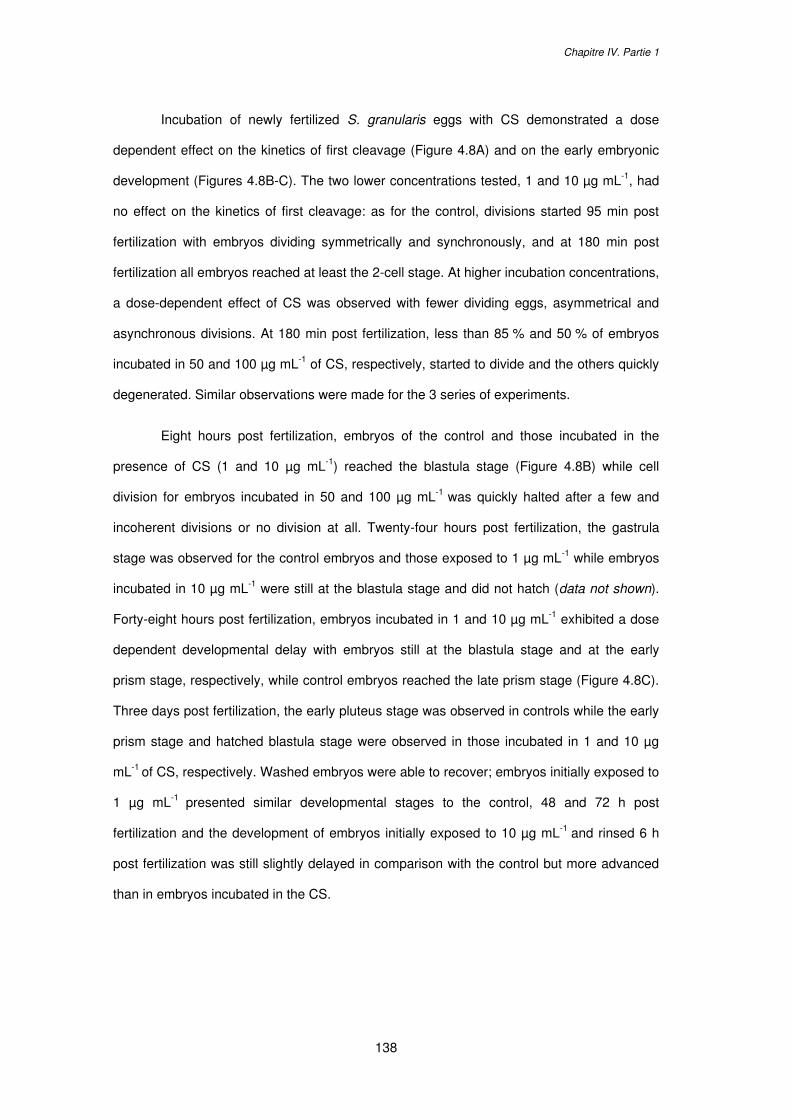
Chapitre IV. Partie 1
138
Incubation of newly fertilized S. granularis eggs with CS demonstrated a dose
dependent effect on the kinetics of first cleavage (Figure 4.8A) and on the early embryonic
development (Figures 4.8B-C). The two lower concentrations tested, 1 and 10 µg mL-1, had
no effect on the kinetics of first cleavage: as for the control, divisions started 95 min post
fertilization with embryos dividing symmetrically and synchronously, and at 180 min post
fertilization all embryos reached at least the 2-cell stage. At higher incubation concentrations,
a dose-dependent effect of CS was observed with fewer dividing eggs, asymmetrical and
asynchronous divisions. At 180 min post fertilization, less than 85 % and 50 % of embryos
incubated in 50 and 100 µg mL-1 of CS, respectively, started to divide and the others quickly
degenerated. Similar observations were made for the 3 series of experiments.
Eight hours post fertilization, embryos of the control and those incubated in the
presence of CS (1 and 10 µg mL-1) reached the blastula stage (Figure 4.8B) while cell
division for embryos incubated in 50 and 100 µg mL-1 was quickly halted after a few and
incoherent divisions or no division at all. Twenty-four hours post fertilization, the gastrula
stage was observed for the control embryos and those exposed to 1 µg mL-1 while embryos
incubated in 10 µg mL-1 were still at the blastula stage and did not hatch (data not shown).
Forty-eight hours post fertilization, embryos incubated in 1 and 10 µg mL-1 exhibited a dose
dependent developmental delay with embryos still at the blastula stage and at the early
prism stage, respectively, while control embryos reached the late prism stage (Figure 4.8C).
Three days post fertilization, the early pluteus stage was observed in controls while the early
prism stage and hatched blastula stage were observed in those incubated in 1 and 10 µg
mL-1 of CS, respectively. Washed embryos were able to recover; embryos initially exposed to
1 µg mL-1 presented similar developmental stages to the control, 48 and 72 h post
fertilization and the development of embryos initially exposed to 10 µg mL-1 and rinsed 6 h
post fertilization was still slightly delayed in comparison with the control but more advanced
than in embryos incubated in the CS.
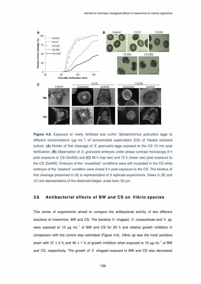
Harmful or harmless: biological effects of marennine on marine organisms
139
Figure 4.8. Exposure of newly fertilized sea urchin Sphaerechinus granularis eggs to
different concentrations (µg mL-1) of concentrated supernatant (CS) of Haslea ostrearia
culture. (A) Kinetic of first cleavage of S. granularis eggs exposed to the CS 10 min post
fertilization. (B) Observation of S. granularis embryos under phase contrast microscopy 8 h
post exposure to CS (Gx200) and (C) 48 h (top raw) and 72 h (lower raw) post exposure to
the CS (Gx400). Embryos of the “unwashed” conditions were still incubated in the CS while
embryos of the “washed” condition were rinsed 6 h post exposure to the CS. The kinetics of
first cleavage presented in (A) is representative of 3 replicate experiments. Views in (B) and
(C) are representative of the observed stages, scale bars: 50 µm.
3.6 Antibacterial effects of BW and CS on Vibrio species
This series of experiments aimed to compare the antibacterial activity of two different
solutions of marennine, BW and CS. The bacteria V. chagasii, V. crassostreae and V. sp.
were exposed to 10 µg mL-1 of BW and CS for 20 h and relative growth inhibition in
comparison with the control was estimated (Figure 4.9). Vibrio sp was the most sensitive
strain with 37 ± 2 % and 40 ± 1 % of growth inhibition when exposed to 10 µg mL-1 of BW
and CS, respectively. The growth of V. chagasii exposed to BW and CS also decreased
A B
C
Control CS [1] CS [10]
CS [50] CS [100]
unwashed unwashedwashed washed
48h
72h
CS [1] CS [10]Control
0
25
50
75
100
30 80 130 180
Per
cen
t o
f fi
rst
clea
vag
e (%
)
Time after fertilization (min)
Control
CS [1]
CS [10]
CS [50]
CS [100]
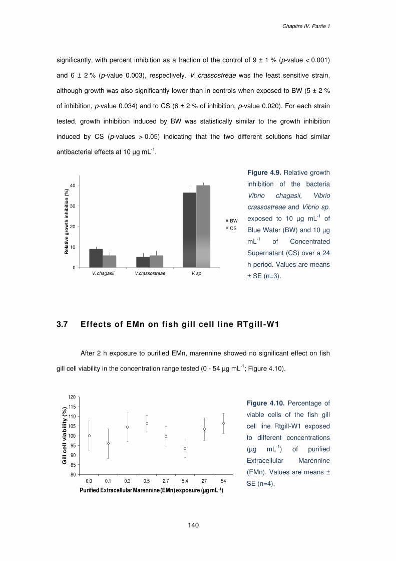
Chapitre IV. Partie 1
140
80
85
90
95
100
105
110
115
120
0.0 0.1 0.3 0.5 2.7 5.4 27 54
Gill c
ell v
iab
ilit
y (
%)
Purified Extracellular Marennine (EMn) exposure (µg mL -1)
significantly, with percent inhibition as a fraction of the control of 9 ± 1 % (p-value < 0.001)
and 6 ± 2 % (p-value 0.003), respectively. V. crassostreae was the least sensitive strain,
although growth was also significantly lower than in controls when exposed to BW (5 ± 2 %
of inhibition, p-value 0.034) and to CS (6 ± 2 % of inhibition, p-value 0.020). For each strain
tested, growth inhibition induced by BW was statistically similar to the growth inhibition
induced by CS (p-values > 0.05) indicating that the two different solutions had similar
antibacterial effects at 10 µg mL-1.
Figure 4.9. Relative growth
inhibition of the bacteria
Vibrio chagasii, Vibrio
crassostreae and Vibrio sp.
exposed to 10 µg mL-1 of
Blue Water (BW) and 10 µg
mL-1 of Concentrated
Supernatant (CS) over a 24
h period. Values are means
± SE (n=3).
3.7 Effects of EMn on fish gill cell line RTgill -W1
After 2 h exposure to purified EMn, marennine showed no significant effect on fish
gill cell viability in the concentration range tested (0 - 54 µg mL-1; Figure 4.10).
Figure 4.10. Percentage of
viable cells of the fish gill
cell line Rtgill-W1 exposed
to different concentrations
(µg mL-1) of purified
Extracellular Marennine
(EMn). Values are means ±
SE (n=4).
0
10
20
30
40
V. chagasii V.crassostreae V. sp
Rel
ativ
e g
row
th in
hib
itio
n (
%)
BW
CS
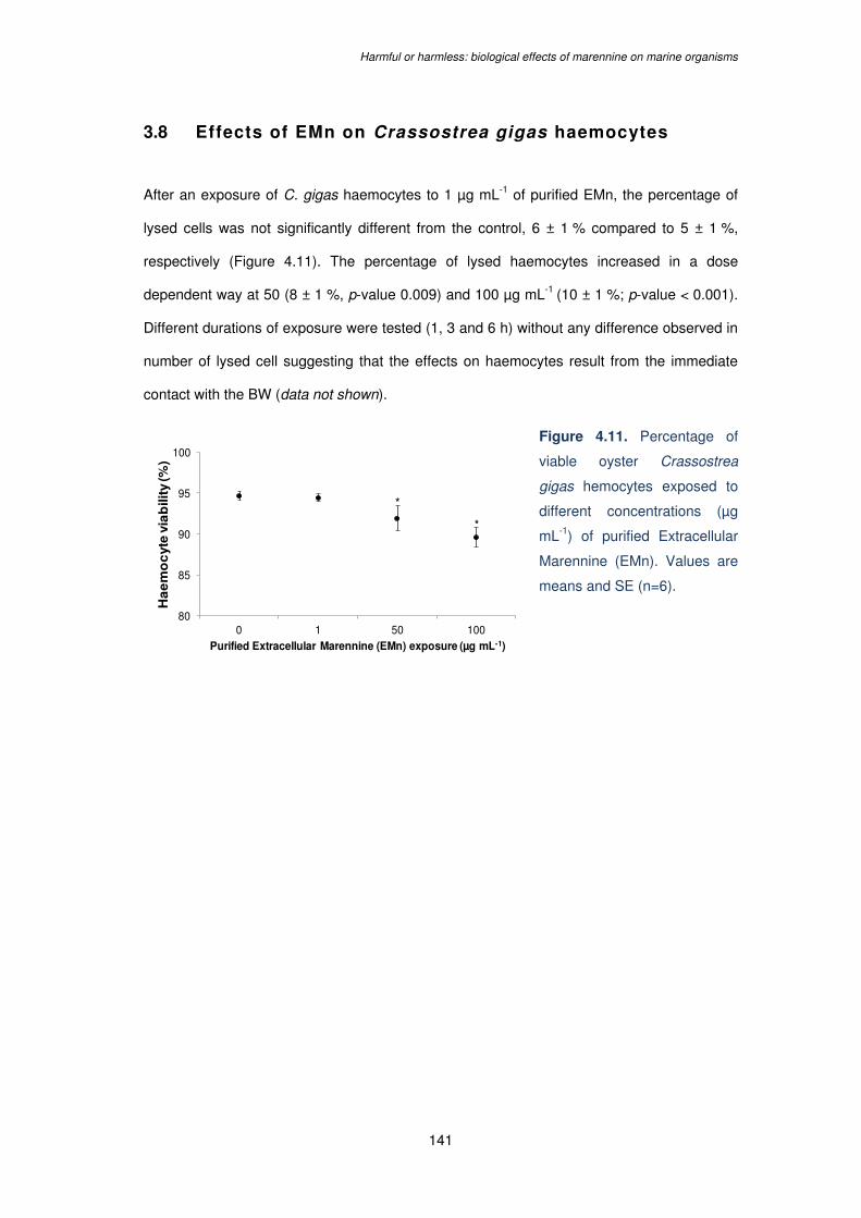
Harmful or harmless: biological effects of marennine on marine organisms
141
*
*
80
85
90
95
100
0 1 50 100
Ha
em
oc
yte
via
bili
ty (%
)
Purified Extracellular Marennine (EMn) exposure (µg mL-1)
3.8 Effects of EMn on Crassostrea gigas haemocytes
After an exposure of C. gigas haemocytes to 1 µg mL-1 of purified EMn, the percentage of
lysed cells was not significantly different from the control, 6 ± 1 % compared to 5 ± 1 %,
respectively (Figure 4.11). The percentage of lysed haemocytes increased in a dose
dependent way at 50 (8 ± 1 %, p-value 0.009) and 100 µg mL-1 (10 ± 1 %; p-value < 0.001).
Different durations of exposure were tested (1, 3 and 6 h) without any difference observed in
number of lysed cell suggesting that the effects on haemocytes result from the immediate
contact with the BW (data not shown).
Figure 4.11. Percentage of
viable oyster Crassostrea
gigas hemocytes exposed to
different concentrations (µg
mL-1) of purified Extracellular
Marennine (EMn). Values are
means and SE (n=6).
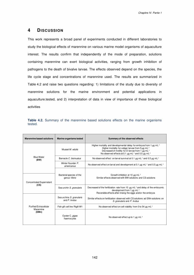
Chapitre IV. Partie 1
142
4 D ISCUSSION
This work represents a broad panel of experiments conducted in different laboratories to
study the biological effects of marennine on various marine model organisms of aquaculture
interest. The results confirm that independently of the mode of preparation, solutions
containing marennine can exert biological activities, ranging from growth inhibition of
pathogens to the death of bivalve larvae. The effects observed depend on the species, the
life cycle stage and concentrations of marennine used. The results are summarized in
Table 4.2 and raise two questions regarding: 1) limitations of the study due to diversity of
marennine solutions for the marine environment and potential applications in
aquaculture.tested, and 2) interpretation of data in view of importance of these biological
activities
Table 4.2. Summary of the marennine based solutions effects on the marine organisms tested.
Marennine based solutions Marine organisms tested Summary of the observed effects
Blue Water(BW)
Mussel M. edulis
Higher mortality and developmental delay for embryos from 1 µg mL-1
Higher mortality for veliger larvae from 5 µg mL-1
Decreased of motility for D-larvae from 1 µg mL-1
No observed effects at 0.1 µg mL-1 and 0.5 µg mL-1
Barnacle C. bisinuatus No observed effect on larval survival at 0.1 µg mL-1 and 0.5 µg mL-1
Winter flounder P. americanus
No observed effect on larval and development at 0.1 µg mL-1 and 0.5 µg mL-1
Bacterial species of the genus Vibrio
Growth inhibition at 10 µg mL-1
Similar effects observed with BW solutions and CS solutions
Concentrated Supernatant(CS)
Sea urchin S. granularis Decreased of the fertilization rate from 10 µg mL-1 and delay of the embryonic development from 1 µg mL-1
Reversible effects after rinsing the eggs and/or the embryos
Similar effects on fertilization observed with CS solutions ad EMn solutions on S. granularis and P. lividus
Purified Extracellular Marennine
(EMn)
Sea urchins S. granularis and P. lividus
Fish gill cell line Rtgill-W1 No observed effect on cell viability from 0 to 54 µg mL-1
Oyster C. gigashaemocytes
No observed effect up to 1 µg mL-1

Harmful or harmless: biological effects of marennine on marine organisms
143
4.1 Limitations of the study
This work brings together the results of different experiments, conducted over a three-year
period, in different laboratories and using three different marennine-based solutions (purified
EMn, BW and CS), as well as different methods to quantify marennine concentrations.
Moreover, cultures of H. ostrearia were grown on different media, which also may have
impacted marennine production and quality. The first series of experiments using purified
EMn and CS exposed against S. granularis and P. lividus, showed similar effects on sea
urchin egg fertilization. Furthermore, growth inhibition induced by BW and CS was
comparable within the same Vibrio species at 10 µg mL-1. We conclude that no important
modification in the biological activities therefore resulted from differences in preparation of
marennine solutions.
The concentration ranges tested could vary by about 30 – 40 %, depending on the
extinction coefficient used. Various marennine quantification methods have been published
so far, using an extinction coefficient determined from raw extract (Robert et al., 2002) or
purified marennine (Pouvreau et al., 2006c), or weighing of purified marennine dried powder
(Prasetiya et al., 2017; Turcotte et al., 2016 and Gastineau et al., 2012, respectively). As the
absolute quality and quantity of the pigment in different solutions could not be clearly
assessed without a complete characterization of this pigment, our results provided a range of
concentrations showing biological activities against various marine organisms. While the
higher concentrations (50 and 100 µg mL-1) provide clues about doses with deleterious
effects, the lower concentrations (from 0.01 to 10 µg mL-1) are more ecologically relevant. In
natural environments, marennine measurements indicate a range of 1 - 10 µg mL-1 (Turpin et
al., 2001), and natural blooms of blue Haslea have been recorded worldwide (Figure 4.1).
Haslea blooms are regularly observed in Calvi Bay (Corsica, France) as part of seaweed
surveys. These blooms, which mostly occur in spring, result in the development of a blue-
green biofilm at the surface of the thallus of seaweeds (i.e., Padina, Halopteris,
Acetabularia), on short turfs, and to a lower extent on sediments and rocks (STAtion de
REcherche Sous-marines et Océanographique - STARESO and Liège University;
Chapitre IV. Partie 1
144
V. Demoulin, D. Sirjacobs, S. Gobert and P. Lejeune, personal communications). Blue
Haslea can display very large patches reaching several square meters and be observed at
depths ranging from 2 to 8 meters. Such extensive blooms are also annually recorded from
November through March in central and southern coastal estuaries of North Carolina (USA),
in particular along the central coast from lower Core Sound westward across the lower North
River estuary and Back Sound to the western portions of Bogue Sound. These blooms of a
blue Haslea possibly different from H. ostrearia do not seem to be associated with the
development of biofilms on Padina sp. thalli, however they cause a wide-spread greening of
oysters, which could underpin emerging green oyster industries in the USA (N. Lindquist,
personal communications).
Thus it can be inferred that, at the microenvironment scale, high concentrations of
marennine could be encountered, and that many marine organisms can periodically and
locally face acute marennine exposure. All laboratory experiments to study biological activity
of marennine lasted hours or days, and the cumulated effect of long-lasting exposure to low
concentrations of marennine has not yet been tested. Greening of oyster gills can be viewed
as a cumulative, longer term effect. According to green oyster producers, the greening in
oyster ponds can occur in a matter of days, depending on Haslea growth and density. In the
laboratory, EMn rather than IMn was preferentially responsible for greening, which proved to
be time dependent and long-lasting (Gastineau et al., 2014b, 2018). However, green oysters
have also been observed in natural open environments, in absence of any record of Haslea
blooms, for instance in oyster leases in Australia (New South Wales and Tasmania). This
means that marennine could have biological activities in the long-term, from subacute and
chronic exposure, and further work is required to better assess the impact of this pigment in
the natural environment.
Harmful or harmless: biological effects of marennine on marine organisms
145
4.2 The unpredictable “Jekyll and Hyde” , good and bad
nature of marennine
On the one hand, our results showed that exposure to the tested marennine solutions could
lead to adverse effects against many marine animals, depending on the exposure dose.
Concentrations from 1 µg mL-1 significantly lowered the survival rate of M. edulis embryos
and locomotion of the D-larvae. Significantly higher mortality rates were observed for veliger
larvae exposed to 5 µg mL-1 of BW while no mortality was recorded at 1 µg mL-1, which
contrasts with results of a previous study where exposure of veliger larvae to 1 µg mL-1 of
BW led to 100 % mortality, but the duration of that experiment was 4 times longer, 20 d vs
5 d (Turcotte et al., 2016). Significant dose-response effects of marennine were also
observed with the sea urchin model. While high concentrations (50 and 100 µg mL-1)
resulted in a total blockage of fertilization and embryonic development, lower concentrations
(1 and 10 µg mL-1) of CS induced a developmental delay of S. granularis embryos. Previous
works also have demonstrated the effects of a water-soluble extract of the diatom
Thalassiosira rotula on cell division of the sea urchin P. lividus, with a blockage of cell
division at the higher doses tested and a delay and abnormal development of embryos at
lower doses (Buttino et al., 1999). Intact diatom cells of six different species could inhibit the
egg first cleavage of the sea urchins Strongylocentrotus droebachiensis and Echinus acutus
(Gudimova et al., 2016). Most interestingly, our results indicated that even at high
concentrations, the effects of CS on fertilization and embryonic development were reversible
after rinsing the eggs or embryos, suggesting non- toxicity of the CS, or inability of
marennine to act at the cell surface, or to penetrate the cell and exert long-term effects. This
confirms previous observations of the effects of water-soluble diatom extracts against
various marine invertebrates, including the sea urchin Psammechinus miliaris (Caldwell et
al., 2002).
The present results could suggest an inability of marennine to bind tightly at the cell
surface if considering the reversible effects observed in sea urchins, and to penetrate the cell
and exert irreversible damages given the high molecular weight of the molecule (> 10 kDa;
Chapitre IV. Partie 1
146
Pouvreau et al. 2006b). Marennine could however link to cell surface or membrane receptors
and trigger stress responses or even apoptotic signals by inducing changes in cell
metabolism. Furthermore, the variability of BW and CS effects observed at both the
interspecific and intraspecific levels on the three Vibrio species tested, but also those
previously shown with V. aestuarianus, V. coralliilyticus and V. tubiashii strains (Falaise et
al., 2016b) could reflect differences in membrane receptor affinities and in mechanisms of
action. Such variability should be considered before application of marennine in aquaculture
as an antibacterial agent. However, more data is necessary to confirm these hypotheses.
On the other hand, despite observed deleterious effect at the higher concentrations
tested, very low concentrations (0.05 and 0.1 µg mL-1) of BW did not exhibit a negative effect
on the barnacle C. bisinuatus survival nor on the winter flounder P. americanus. Both
flounder growth rate and bacterial load in the water remained unaffected throughout the
marennine exposure (0.05 and 0.1 µg mL-1), and physiological condition (energetic reserves)
improved in response to marennine treatment. These results compare with no observable
effect of marennine (0-54 µg L-1) on the viability (metabolic activity) of the rainbow trout gill
cell line RTgill-W1. Although the gill cell line exposure was conducted for only 2 h, results
provide a strong indication that marennine does not impair fish gill function at the
concentration ranges tested here. These results are further supported by (Turcotte et al.,
2016) who demonstrated that low exposure to BW (0.1 µg mL-1) enhanced survival and
physiological condition of M. edulis and P. magellanicus larvae. In addition, our results
showed that low concentrations of EMn (1 µg mL-1) did not present a cytotoxic effect against
haemocytes of adult oysters. Even though cytotoxic effects were evidenced at the highest
concentrations (50 and 100 µg mL-1), the point of inflection could not be determined with the
concentration range tested. However, it should be emphasized that even after centuries of
production of green oysters, no negative impacts on adult oysters, nor green oyster
consumers have been reported.
Despite the fact that diatoms are traditionally considered a high quality food source
enhancing growth and survival of many marine organisms, they can produce secondary
metabolites that function as grazing deterrents (Ianora and Miralto, 2010). Most of these
Harmful or harmless: biological effects of marennine on marine organisms
147
secondary metabolites are polyunsaturated fatty acids (PUFAs) and polyunsaturated
aldehydes (PUAs) with a special focus on oxylipins (Caldwell, 2009). Effects of diatom
aldehydes on reproductive and fertilization success or embryonic development of various
organisms such as crustaceans, echinoderms or arthropods have been widely studied
(Caldwell, 2009; Ianora and Miralto, 2010). Aldehyde production is a diatom chemical
defense strategy to limit growth of their grazers (Ban et al., 1997; Pohnert, 2005), but other
studies reported the absence of a correlation between diatom aldehyde and reproductive
biology and early development of marine invertebrates, suggesting the involvement of other
unidentified diatom metabolites (Poulet et al., 2006; Wichard et al., 2008). Unlike aldehydes,
that are likely to exert their activity following diatom cell destruction and ingestion, the water-
soluble marennine may target a larger spectrum of marine organisms and not only diatom
grazers.
Chapitre IV. Partie 1
148
5 CONCLUSION
In conclusion, the present work demonstrates that H. ostrearia supernatant containing
marennine represents a biologically active water-soluble solution with potential effects on
various marine organisms. More studies are required to better estimate the long term
impacts of blue Haslea blooms in natural environments, facilitated by laboratory studies,
investigating chronic exposure to low concentrations of marennine. If the use of H. ostrearia
supernatant is considered in hatcheries as a preventive or curative anti-infectious agent,
concentrated solutions such as BW or CS could be of interest in order to control the
delivered marennine doses. It is likely that the adverse effects of marennine only target the
early and more succeptable fragile developmental stages such as embryos and larvae,
depending on the exposure concentration, whereas adults remain unaffected.
149
CHAPITRE IV.
Partie 2 :
Preliminary results on marennine effects on the
mussel Mytilus edulis larvae
Chapitre IV. Partie 2
150
Chapitre IV. New insights into the effects of marennine solutions on
marine organisms. Partie 2 : Preliminary results on marennine effects
on the mussel Mytilus edulis.
Les travaux présentés dans la Partie 2 de ce chapitre ont été conduits à l’Institut des
Sciences de la Mer à Rimouski (ISMER, Canada) et à la station aquicole de l’ISMER à
Pointe-au-Père durant les mois de mai-août 2017, de novembre 2017 et de mai 2018. Ces
travaux ont bénéficié des expertises de Réjean Tremblay, Nathalie Gauthier et François
Turcotte pour les pontes de moules, élevages larvaires et production de Blue Water ; de
Karine Lemarchand et Kim Doiron pour les challenges bactériens ; et de Bertrand Genard et
Renée Gagné pour les expérimentations de biologie moléculaire.
Les résultats de ce chapitre ont été présentés lors de communications orales au congrès
Physiomar à Cambridge (Angleterre) les 18-22 septembre 2017, au congrès International
Conference on Harmful Algae (ICHA) à Nantes (France) les 21-26 octobre 2018 et au
congrès de la British Phycological Society (PBS) à Oban (Écosse) les 7-10 janvier 2019.
Preliminary results on marennine effects on Mytilus edulis larvae
151
Abstract
Marennine is the water-soluble blue pigment produced by the marine diatom Haslea
ostrearia. This pigment displays biological effects on micro-organisms, such as growth
inhibition of marine bacteria of the Vibrio genus or allopathic activities against several
microalgae. Moreover a previous study showed that mussel larvae exposed to 0.5 µg mL-1 of
Blue Water (BW), a concentrated marennine solution, had a better physiological condition
and survival when challenged with a pathogenic Vibrio splendidus strain. However, higher
exposure doses led to larval mortality. The present work aimed to assess the effects of BW
exposure on the expression of some genes in the mussel Mytilus edulis larvae. Three
different successive larval developmental stages were tested: veliger, pediveliger and
metamorphosed larvae, and four BW concentrations: 0, 0.5, 1 and 5 µg mL-1. A challenge
with V. splendidus on veliger larvae was also conducted to reproduce the prophylactic
effects previously observed, but the opportunistic bacteria did not induce a significant larval
mortality. A 3 d exposure to BW induced a significant larval mortality at the higher
concentration tested, with the early developmental stages (veliger, pediveliger) more
sensitive than the later ones (metamorphosed larvae). Transcriptomic analyses confirmed
that BW induced stress and enhanced a defense responses in veliger larvae with a higher
expression of the four target genes tested: Cathepsin B, Superoxide Dismutase, ATP
Synthase Beta and Heat Shock Protein 70. This work represents, to our knowledge, the first
analysis of gene expression in M. edulis larvae and the first molecular evidences of
marennine effects. Also, the present work proposes two reliable reference genes (Elongation
Factor 1 and Tubulin) with good stability in the three developmental stages tested that could
be used in future studies.
Key words: Haslea ostrearia; diatom; marennine; bioactive compound; Mytilus edulis; gene
expression
Chapitre IV. Partie 2
152
1 INTRODUCTION
Diatoms can produce secondary metabolites with toxigenic effects on various marine
organisms such as grazers, other algae or microbes (Ianora and Miralto, 2010). Most of the
diatom-derived compound studied so far are short chain polyunsaturated aldehydes (PUAs)
and other oxygenated fatty acid degradation products collectively named oxylipins (Caldwell,
2009). Their deleterious effects on fertilization, embryonic development or larval fitness were
demonstrated in various marine organisms and mostly on copepods, which are the major
diatom feeders in planktonic ecosystems (Adolph et al., 2004; Caldwell et al., 2002; Romano
et al., 2010). More recently, biomolecular studies evidenced the effects of diatom PUAs and
oxylipins on the expression of genes involved in apoptosis, detoxification process or stress
from copepod and sea urchin models (Lauritano et al., 2016; Varrella et al., 2016a).
However, as several studies reported the absence of a correlation between diatom PUAs or
oxylipins production and the observed adverse effects, the involvement of other cytotoxic
diatom-derived metabolites were suggested (Poulet et al., 2006; Wichard et al., 2008).
In the present work, the effects of marennine were investigated on the expression of
some genes in the mussel Mytilus edulis larvae. Marennine is the biologically active blue
pigment produced by the marine diatom Haslea ostrearia. Although H. ostrearia has never
been considered as a harmful diatom, there are growing evidences that this diatom and its
water-soluble pigment can impair growth or survival of marine organisms and, unlike PUAs
or oxylipins, not only diatom-grazers (see Chapter IV. Part 1). Allelopathic activities were
also demonstrated, marennine being able to inhibit the growth of several microalgal species
(Pouvreau et al., 2007; Prasetiya et al., 2016), as well as neutral, positive or negative
interactions with a large panel of bacteria of the genus Vibrio (see Chapter III. Part 1).
Moreover, marennine can affect the behavior (e.g. valve opening frequency) or the
physiology (e.g. clearance rate, oxygen consumption) of mussel and oyster juveniles
(Prasetiya et al., 2017), and can also impair embryonic development of sea urchins (see
Chapter IV. Part 1). But, despite potential deleterious effects of marennine, a study
conducted by Turcotte et al. (2016) demonstrated a prophylactic activity of the pigment on
the blue mussel M. edulis and on the giant scallop Placopecten magellanicus larvae.
Preliminary results on marennine effects on Mytilus edulis larvae
153
Exposure to low concentration (0.5 µg mL-1) of Blue water (BW), a concentrated marennine
solution, significantly improved the survival of larvae challenged with a virulent strain of
Vibrio splendidus over a 3 d period. It was found that the larvae exposed to BW had a better
physiological condition state, characterized by a higher accumulation of triacylglycerol
(TAG), an energetic lipid reserve (Gallager et al., 1986). As the bacterial concentration was
not different between the two treatments BW + Vibrio and Vibrio, it was suggested that BW
reduced bacterial pathogenicity and did not inhibit the growth of Vibrio strain. As bacterial
exposure triggers an oxidative stress in bivalve larvae (Genard et al., 2012), it was
suggested that the antioxidant properties of the pigment (Pouvreau et al., 2008) could afford
a protective action to mussel larvae. However, the same study showed that exposure of
larvae to 0.1 µg mL-1 of BW over a 20 d period tended to improve larval survival but that
exposure to 1 µg mL-1 led to 100 % larval mortality, highlighting the tiny limit between
beneficial and deleterious effects of the pigment on bivalve larvae (Turcotte et al., 2016).
The present work aimed to assess the molecular effects of BW on the mussel M. edulis
larvae at different developmental stages: veliger (17 days post fertilization, dpf), pediveliger
(23 dpf) and metamorphosed (30 dpf). Larvae were exposed over a 3 d period to different
BW doses: 0, 0.5, 1 and 5 µg mL-1. Gene expression of Cathepsin B (CTPB), Superoxide
Dismutase (SOD), ATP Synthase Beta (ASB) and Heat Shock Protein 70 (HSP70) was
measured by real time quantitative PCR (RT-qPCR) to evidence the effects of BW exposure
on larval defense and stress response. CTPB, SOD, ASB and HSP70 genes were
respectively chosen for their role in pathogen recognition, oxidative stress response, energy
metabolism and cytoprotection. Also, bacterial challenges with veliger larvae and Vibrio were
conducted in order to reproduce the prophylactic effects of BW observed in Turcotte et al.
(2016) and assess the resulting effects on gene expression.
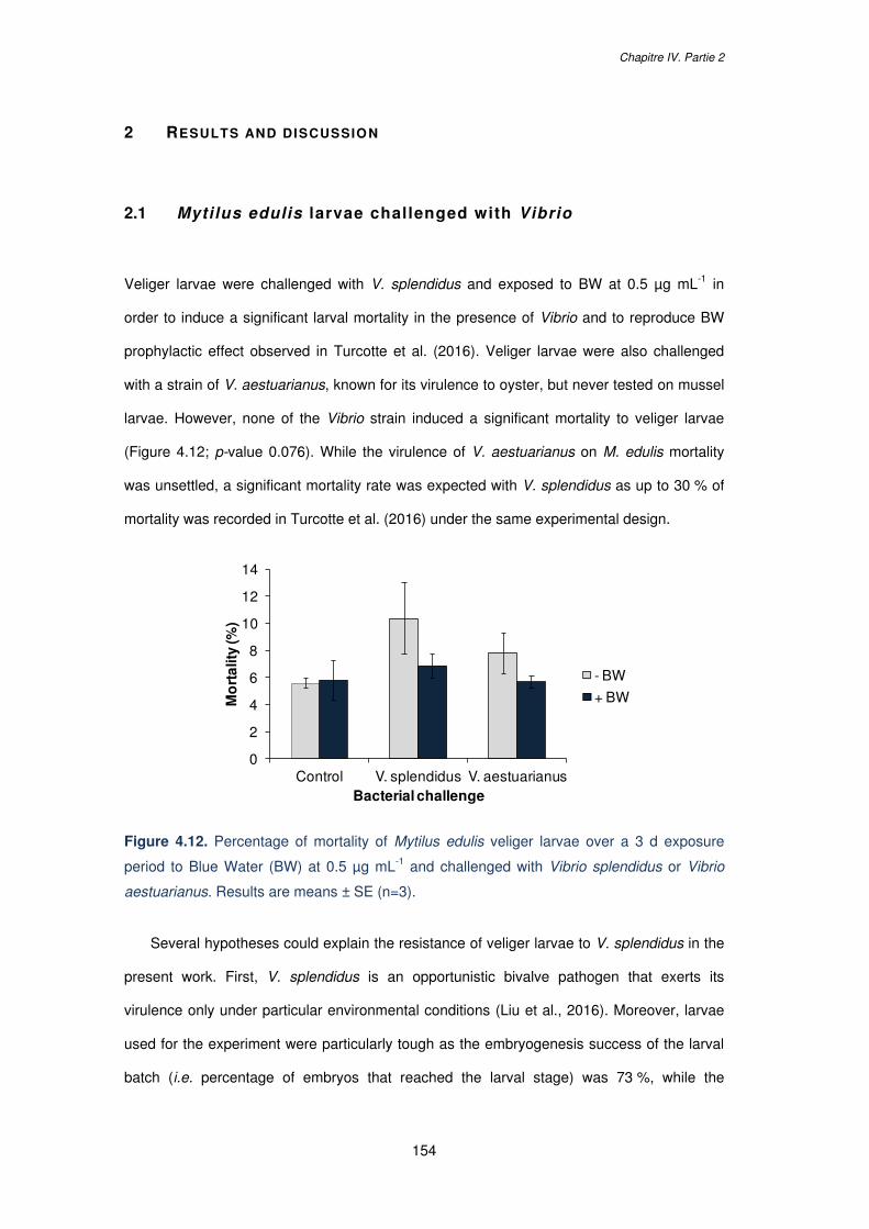
Chapitre IV. Partie 2
154
2 RESULTS AND DISCUSSION
2.1 Mytilus edulis larvae challenged with Vibrio
Veliger larvae were challenged with V. splendidus and exposed to BW at 0.5 µg mL-1 in
order to induce a significant larval mortality in the presence of Vibrio and to reproduce BW
prophylactic effect observed in Turcotte et al. (2016). Veliger larvae were also challenged
with a strain of V. aestuarianus, known for its virulence to oyster, but never tested on mussel
larvae. However, none of the Vibrio strain induced a significant mortality to veliger larvae
(Figure 4.12; p-value 0.076). While the virulence of V. aestuarianus on M. edulis mortality
was unsettled, a significant mortality rate was expected with V. splendidus as up to 30 % of
mortality was recorded in Turcotte et al. (2016) under the same experimental design.
Figure 4.12. Percentage of mortality of Mytilus edulis veliger larvae over a 3 d exposure
period to Blue Water (BW) at 0.5 µg mL-1 and challenged with Vibrio splendidus or Vibrio
aestuarianus. Results are means ± SE (n=3).
Several hypotheses could explain the resistance of veliger larvae to V. splendidus in the
present work. First, V. splendidus is an opportunistic bivalve pathogen that exerts its
virulence only under particular environmental conditions (Liu et al., 2016). Moreover, larvae
used for the experiment were particularly tough as the embryogenesis success of the larval
batch (i.e. percentage of embryos that reached the larval stage) was 73 %, while the
0
2
4
6
8
10
12
14
Control V. splendidus V. aestuarianus
Mo
rta
lity
(%)
Bacterial challenge
- BW
+ BW
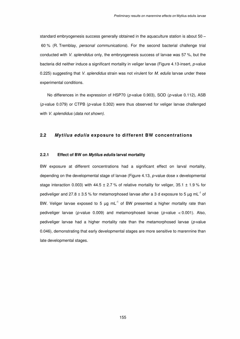
Preliminary results on marennine effects on Mytilus edulis larvae
155
standard embryogenesis success generally obtained in the aquaculture station is about 50 –
60 % (R. Tremblay, personal communications). For the second bacterial challenge trial
conducted with V. splendidus only, the embryogenesis success of larvae was 57 %, but the
bacteria did neither induce a significant mortality in veliger larvae (Figure 4.13-insert, p-value
0.225) suggesting that V. splendidus strain was not virulent for M. edulis larvae under these
experimental conditions.
No differences in the expression of HSP70 (p-value 0.903), SOD (p-value 0.112), ASB
(p-value 0.079) or CTPB (p-value 0.302) were thus observed for veliger larvae challenged
with V. splendidus (data not shown).
2.2 Mytilus edulis exposure to different BW concentrations
2.2.1 Effect of BW on Mytilus edulis larval mortality
BW exposure at different concentrations had a significant effect on larval mortality,
depending on the developmental stage of larvae (Figure 4.13, p-value dose x developmental
stage interaction 0.003) with 44.5 ± 2.7 % of relative mortality for veliger, 35.1 ± 1.9 % for
pediveliger and 27.8 ± 3.5 % for metamorphosed larvae after a 3 d exposure to 5 µg mL-1 of
BW. Veliger larvae exposed to 5 µg mL-1 of BW presented a higher mortality rate than
pediveliger larvae (p-value 0.009) and metamorphosed larvae (p-value < 0.001). Also,
pediveliger larvae had a higher mortality rate than the metamorphosed larvae (p-value
0.046), demonstrating that early developmental stages are more sensitive to marennine than
late developmental stages.
Chapitre IV. Partie 2
156
Figure 4.13. Percentage of relative mortality of Mytilus edulis veliger, pediveliger and
metamorphosed larvae over a 3 d exposure period to different Blue Water (BW)
concentrations (µg mL-1) and of veliger larvae challenged with Vibrio splendidus (insert).
Results are means ± SE (n=3). Error bars with different lower case letters in are significantly
different and asterisks indicate significant difference with the control (p-value < 0.05).
2.2.2 Molecular response of Mytilus edulis larvae under BW exposure
BW concentration enhanced the expression levels of the HSP70 gene (Figure 4.14A, p-value
0.004). Also, a tendency for early developmental stage to express more HSP70 in
comparison with late developmental stages was observed but did not appear significant
because of the high variability between replicates (p-value 0.128). A higher HSP70
expression was also observed in the sea urchin Paracentrotus lividus embryos and larvae
after exposure to diatom-derived oxilipins, with differential expression level depending on the
developmental stage (Varrella et al., 2016b). Also, a higher expression of HSP70 genes was
previously observed in two different Mytilus species under thermal stress (Lockwood et al.,
2010). The up-regulation of HSP70 in the present work confirmed that larvae exposed to BW
were subjected to stress, activating this gene as a first defense system.
The relative expression of SOD varied significantly depending on the BW
concentration and the larval developmental stage (Figure 4.14B, p-value dose x
aa
*b
a a
*c
aa
*d
-10
-5
0
5
10
15
20
25
30
35
40
45
50
0.5 1 5
Rel
ativ
e m
ort
alit
y (%
)
BW concentration (µg mL-1)
Veliger
Pediveliger
Metamorphosed
aa
-5
0
5
10
V. splendidus BW [0.5] + V. splendidus
Preliminary results on marennine effects on Mytilus edulis larvae
157
developmental stage interaction < 0.001). The expression of SOD was significantly higher in
veliger larvae exposed to BW at 5 µg mL-1 in comparison with pediveliger and
metamorphosed larvae (p-values < 0.001 and < 0.001 respectively). However, no difference
in SOD expression was found between pediveliger and metamorphosed larvae (p-value
0.900). The higher activity levels of SOD suggest the activation of antioxidant defense that
occurs before mortality events as it was previously observed on oyster larvae.
A dose effect of BW was also observed for the CTPB gene (Figure 4.14C) with an
up-regulation under high BW exposure (p-value 0.003), but no differences were found
between larval stages (p-value 0.527). CTPB plays crucial roles in host defense system
against pathogen infection in molluscs and the overexpression of this gene was previously
demonstrated in oysters infected with a parasite (Morga et al., 2009).
The expression of ASB was also activated under high BW exposure (Figure 4.14D;
p-value 0.013) with no related effect of the larval developmental stage (p-values 0.848). ASB
(or ATPB) is part of the large family of genes coding for different enzymes that participate in
formation of ATP. In molluscs, immune stimulation results in an increased demand of energy
to sustain cell defense response (e.g. phagocytosis, cytotoxicity; Coyne, 2011). The
activation of ATPB expression would reflect the extra demand for ATP required by cells to
sustain the immune defense under BW exposure. Expression of ATPB was also shown to be
up-regulated in oyster exposed to pesticides (Tanguy et al., 2005).
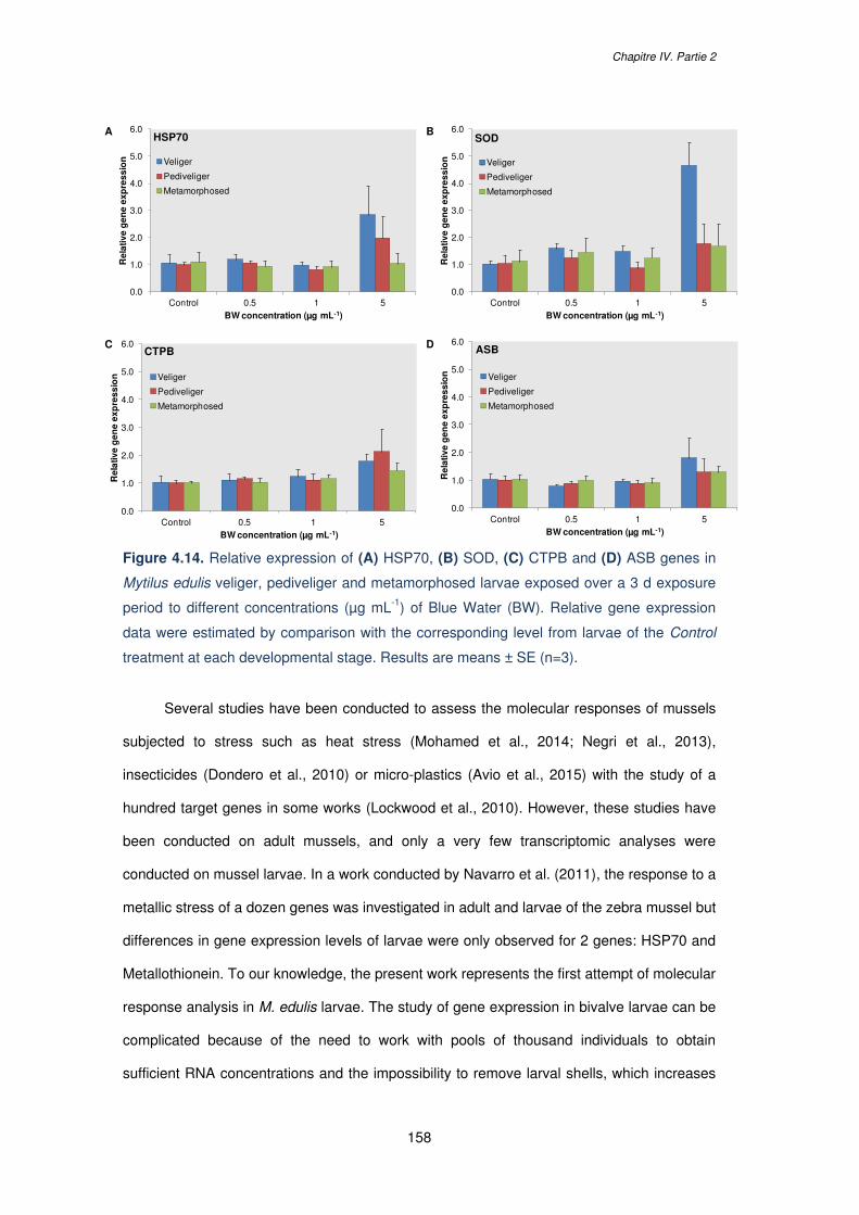
Chapitre IV. Partie 2
158
Figure 4.14. Relative expression of (A) HSP70, (B) SOD, (C) CTPB and (D) ASB genes in
Mytilus edulis veliger, pediveliger and metamorphosed larvae exposed over a 3 d exposure
period to different concentrations (µg mL-1) of Blue Water (BW). Relative gene expression
data were estimated by comparison with the corresponding level from larvae of the Control
treatment at each developmental stage. Results are means ± SE (n=3).
Several studies have been conducted to assess the molecular responses of mussels
subjected to stress such as heat stress (Mohamed et al., 2014; Negri et al., 2013),
insecticides (Dondero et al., 2010) or micro-plastics (Avio et al., 2015) with the study of a
hundred target genes in some works (Lockwood et al., 2010). However, these studies have
been conducted on adult mussels, and only a very few transcriptomic analyses were
conducted on mussel larvae. In a work conducted by Navarro et al. (2011), the response to a
metallic stress of a dozen genes was investigated in adult and larvae of the zebra mussel but
differences in gene expression levels of larvae were only observed for 2 genes: HSP70 and
Metallothionein. To our knowledge, the present work represents the first attempt of molecular
response analysis in M. edulis larvae. The study of gene expression in bivalve larvae can be
complicated because of the need to work with pools of thousand individuals to obtain
sufficient RNA concentrations and the impossibility to remove larval shells, which increases
0.0
1.0
2.0
3.0
4.0
5.0
6.0
Control 0.5 1 5
Rel
ativ
e g
ene
exp
ress
ion
BW concentration (µg mL-1)
HSP70
Veliger
Pediveliger
Metamorphosed
0.0
1.0
2.0
3.0
4.0
5.0
6.0
Control 0.5 1 5
Rel
ativ
e g
ene
exp
ress
ion
BW concentration (µg mL-1)
SOD
Veliger
Pediveliger
Metamorphosed
0.0
1.0
2.0
3.0
4.0
5.0
6.0
Control 0.5 1 5
Rel
ativ
e g
ene
exp
ress
ion
BW concentration (µg mL-1)
CTPB
Veliger
Pediveliger
Metamorphosed
0.0
1.0
2.0
3.0
4.0
5.0
6.0
Control 0.5 1 5
Rel
ativ
e g
ene
exp
ress
ion
BW concentration (µg mL-1)
ASB
Veliger
Pediveliger
Metamorphosed
A B
C D
Preliminary results on marennine effects on Mytilus edulis larvae
159
the risk to obtain poor RNA quality extractions. Also, only a few gene sequences specific to
M. edulis larvae are available on gene data banks. Moreover, several genes are only
punctually expressed in larvae depending on the developmental stage. Indeed, some genes
involved in muscular and neuronal development or in biomineralization are only expressed in
veliger and pediveliger larvae but not in metamorphosed ones and vice versa (Bassim et al.,
2014). Also, stable reference genes have to be defined to obtain reliable results, which can
be challenging as the stability of housekeeping gene can vary between developmental
stages, as demonstrated in Cubero-Leon et al. (2012) during the different stages of
gametogenesis in M. edulis. In the present work, we proposed two housekeeping genes with
stable expressions over the three different developmental stages studied (i.e. TUB and EF1).
These two housekeeping genes could therefore be good reference gene candidates for
further transcriptomic studies on M. edulis larvae.
Chapitre IV. Partie 2
160
3 CONCLUSION
Our results demonstrate that BW from 5 µg mL-1 induces a significant mortality in M. edulis
larvae at each of the three developmental stages tested, with veliger larvae particularly
sensitive in comparison with pediveliger and metamorphosed larvae. The effects of BW were
evidenced at a molecular level with the up-regulation of genes involved in cytoprotection
(HSP70), oxidative stress (SOD), energy metabolism (ASB) and pathogen recognition
(CTPB). These molecular effects were mainly demonstrated for veliger larvae while no
differences in gene expression were observed in metamorphosed larvae, suggesting that
different stress response and immune defense are activated in larvae depending on the
developmental stage. Although several other primers for potential target genes were
designed for the present study (e.g. glutathione S-transferase for the detoxification response,
P53 for the apoptotic response, or catalase for the antioxidative response) but they did not
present a satisfactory efficiency and could not be tested. The analysis of an extensive
number of target genes in future studies should allow to further identifying marennine
molecular targets and the defense systems activated by larvae. Indeed, in recent works
several dozens of target genes were investigated in the sea urchin P. lividus after exposure
to diatom-derived PUAs and oxylipins and allowed identifying cellular mechanisms and part
of the chemical defensome (e.g. network of genes that afford protection) activated by the sea
urchin at different developmental stages (Marrone et al., 2012; Varrella et al., 2014, 2016a).
So far, this work represents the first molecular evidence of marennine effects on the
mussel M. edulis larvae. Four target genes were found to be up-regulated in veliger larvae
under BW exposure but no molecular target of the blue pigment was defined in
metamorphosed larvae. Additional studies on a larger number of target genes may provide
some clues about BW mechanisms of action and about the differential defense systems
activated by larvae depending on the developmental stage.
Preliminary results on marennine effects on Mytilus edulis larvae
161
4 MATERIALS AND METHODS
4.1 Experimental design
4.1.1 Mytilus edulis rearing procedure and exposure to BW
Details about M. edulis spawning conditioning, spawning and larval rearing are given in the
Chapiter IV Part 1 of this manuscript. Briefly, this work was conducted in the Station aquicole
de Pointe-au-Père (ISMER-UQAR; Québec, Canada) in 2017. After fertilization, larvae were
reared in 60 L filtered sea water (FSW) tanks at densities of 8 D-larvae µL-1. Tanks were
washed and sterilized every 2-3 d and the larvae fed with a mixture of Pavlova lutherii,
Isochrisis galbana and Chaetoceros gracilis. Veliger larvae were collected in the rearing
thanks 17 dpf, pediveliger larvae 23 dpf and metamorphosed larvae 30 dpf. Larvae of each
developmental stage were collected on a 53 μm square mesh filter and re-suspended in 2 L
of FSW. Larval concentration and size was estimated under light microscope (Olympus
BX41; W. Carsen Co., Ltd., Don Mills, ON, Canada) coupled to a digital camera (Evolution
VF Color, MediaCybernetics, Silver Spring, MD, USA), and the lengths of at least 30 larvae
were measured to ensure size homogeneity with Image Pro-Express (Media Cybernetics)
after addition of 2 µL of formaldehyde 37 % in 50 µL sample. Larvae were transferred in a
2.5 L Erlenmeyer filled with FSW (9 larvae mL-1) and exposed to BW at the final
concentrations of 0, 0.5, 1 or 5 µg mL-1 over a 3 d period in a 20°C controlled temperature
room (n=3 Erlenmeyers per treatment). BW was prepared as described in the Chapter IV.
Part 1 of this manuscript, stored in the dark at 4°C and syringe-filtered through 0.2 µm before
use.
4.1.2 Mytilus edulis challenged with Vibrio species
A challenge with two Vibrio strains was also conducted with veliger larvae only. Veliger
larvae were exposed to 0.5 µg mL-1 of BW and to V. splendidus and V. aestuarianus.
Challenges were conducted as described above in triplicates with six treatments: Control,
Chapitre IV. Partie 2
162
BW, V. splendidus, V. splendidus + BW, V. aestuarianus and V. aestuarianus + BW. The
strain V. splendidus 7SHRW was isolated from sediments of Hillsborough River, Prince
Edward Island (Gulf of Saint Lawrence, Canada) and was shown to induce significant
mortality of blue mussel and scallop larvae. The strain V. aestuarianus 02/041 was isolated
from oyster tissues and provided by the Laboratoire de Génétique et de Pathologie des
Mollusques Marins (LGPMM; La Tremblade, France). Vibrio strains were kept at - 80°C,
streaked onto Marine Agar plates to ensure purity and cultured in 20 mL Marine Broth
medium (Difco, Lawrence, KS, USA) at 25°C for 24 h prior to use. Bacterial cultures were
centrifuged, the supernatant discarded and the pellets re-suspended in 10 mL of sterile
physiologic water (9 ‰ NaCl; 3x5 min at 5000 g). Optical density (OD600) of the bacterial
suspension was measured to determine the bacterial concentration and Erlenmeyers were
inoculated at an initial concentration of 1.106 bacteria mL-1 or with sterile physiologic water
only for treatments without Vibrio. Another trial with V. splendidus only was conducted on
veliger larvae at the same time as the BW exposure experiment presented in section 3.1.1.
Veliger larvae were challenged with V. splendidus at a final concentration of 2.106 bacteria
mL-1 and exposed to 0.5 µg mL-1 of BW and the mortality rate and gene expression in the
different treatments were assessed.
4.2 Sample collection
After a 72 h period, larvae were collected by filtration using a 52 µm mesh screen, rinsed
with FSW and re-suspended in 200 mL of FSW. To estimate the mortality and larval size, the
larval suspension was homogenized; 3x1 mL samples per treatment were transferred in
Eppendorf micro-tubes containing 100 µL of formaldehyde 37 % and the samples were
subsequently observed under light microscopy. The larval suspension was then filtered on a
0.35 µm filter with filtration system connected to a manual pump. Larvae were collected on
the filter with a scalpel, transferred to a cryotube and frozen in liquid nitrogen before storage
at - 80°C. Larvae were grinded in the mixer Mill MM400 (Retsch) for 15 s at 30 freq s-1 and
the obtained powder transferred in Eppendorf microtubes, shortly plunged in liquid nitrogen
Preliminary results on marennine effects on Mytilus edulis larvae
163
and stored at - 80 °C before RNA extraction. Total RNA was extracted for each replicate
using E.Z.N.A® Mollusc RNA Kit Protocol (OMEGA bio-tek) according to the manufacturer’s
protocol. RNA was re-suspended in sterile Diethyl pyrocarbonate (DEPC) water and both
quality and quantity were assessed (ratio 260/280 for quantity and 260/230 for quality) with a
NanoVue™ Plus (GE Healthcare) spectrophotometer. Total RNA samples were then diluted
to a final concentration of 200 ng µL-1 in RNase-free water. RNA quality was further
estimated with migration on a 2 % agarose gel containing the InvitrogenTM SYBERTM SafeTM
DNA gel stain (Fisher). Migration bands were observed after 1 h migration at 100 V on
Alphamger HP under UV and fluorescence SYBER gold filters. Ten µg of total RNA
extracted for each treatment were reverse transcribed using the Quantitect® Reverse
Transcription Kit (Qiagen) and following the manufacturer’s protocol.
4.3 Studied genes and RT-qPCR
Mytilus edulis nucleotide sequences for reference and target genes were obtained from the
GenBank database, designed on Primer3 (v.0.4.0) online software and the primers quality
checked with Oligo Calc (v.3.27) online software. Three genes were selected as putative
housekeeping genes: tubulin (TUB), elongation factor 1 (EF1) and the ribosomal protein
subunit 28S (28S), and four target genes: cathepsin B (CTPB), superoxide dismutase
(SOD), ATP Synthase Beta (ASB) and Heat Shock protein 70 (HSP70) chosen for their role
in pathogen recognition, oxidative stress, energy metabolism and cytoprotection,
respectively (Table 4.3). The specificity of PCR products was checked for the 7 genes by
DNA sequencing of fragments amplified with AmpliTaq Gold® 360 Master Mix (Applied
Biosystems). Real-time amplification reactions were carried out in a final volume of 20 μL
containing 10 μL of Fast SYBR™ Green Master Mix (Fisher), 8 μL of diluted cDNA (1/200)
and 500 nM of each primer. Reactions were run in triplicates and a control lacking cDNA
template was included in qPCR analysis to determine the specificity of target cDNA
amplification. Amplification was detected with a 7900 HT Fast Real Time PCR System
(Applied Biosystems). Thermocycling was performed using the following conditions:
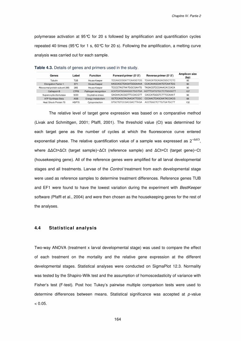
Chapitre IV. Partie 2
164
polymerase activation at 95°C for 20 s followed by amplification and quantification cycles
repeated 40 times (95°C for 1 s, 60°C for 20 s). Following the amplification, a melting curve
analysis was carried out for each sample.
Table 4.3. Details of genes and primers used in the study.
The relative level of target gene expression was based on a comparative method
(Livak and Schmittgen, 2001; Pfaffl, 2001). The threshold value (Ct) was determined for
each target gene as the number of cycles at which the fluorescence curve entered
exponential phase. The relative quantification value of a sample was expressed as 2−ΔΔCt,
where ΔΔCt=ΔCt (target sample)−ΔCt (reference sample) and ΔCt=Ct (target gene)−Ct
(housekeeping gene). All of the reference genes were amplified for all larval developmental
stages and all treatments. Larvae of the Control treatment from each developmental stage
were used as reference samples to determine treatment differences. Reference genes TUB
and EF1 were found to have the lowest variation during the experiment with BestKeeper
software (Pfaffl et al., 2004) and were then chosen as the housekeeping genes for the rest of
the analyses.
4.4 Statistical analysis
Two-way ANOVA (treatment x larval developmental stage) was used to compare the effect
of each treatment on the mortality and the relative gene expression at the different
developmental stages. Statistical analyses were conducted on SigmaPlot 12.3. Normality
was tested by the Shapiro-Wilk test and the assumption of homoscedasticity of variance with
Fisher’s test (F-test). Post hoc Tukey’s pairwise multiple comparison tests were used to
determine differences between means. Statistical significance was accepted at p-value
< 0.05.
Genes Label Function Forward primer (5'-3') Reverse primer (5'-3')Amplicon size
(bp)Tubulin TUB House Keeper TCCAACCGGATTCAAGGTCG TCAACATGCAGACGGCTCTC 95
Elongation Factor 1 EF1 House Keeper AAGCAGCTGAGATGGGAAAA CCACAAAGCAATGTCAATGG 90
Ribosomal protein subunit 28S 28S House Keeper TCCCCTAGTAATGGCGAATG TAGACGTCCCAAACACCACA 90
Cathepsin B CTPB Pathogen recognition ACATCATGGAAGGCTGGTGA AGTTTCATGTGCTCTGGGGTT 107
Superoxyde dismutase SOD Oxydative stress GAGAACACGGTTTCCACGTT CACCATGGGTCTTTCCAAAT 96
ATP Synthase Beta ASB Energy metabolism GCTCCAGCTACAACATTCGC CCCAACTCAGCAATACCACG 68
Heat Shock Protein 70 HSP70 Cytoprotection GTGCTGTCCGACGACTTAGA ACCTGGCTCTTGTGATGCTT 132
165
CHAPITRE V.
DISCUSSION GÉNÉRALE ET
PERSPECTIVES

166
Discussion générale et perspectives
167
1 CONTEXTE DES TRAVAUX DE RECHERCHE
Les travaux de ce doctorat s’inscrivent dans le cadre du projet européen GHaNA (The
Genus Haslea, New marine resources for blue biotechnology and Aquaculture, H2020-
MSCA-RISE-2016) et tout particulièrement dans l’axe de recherche visant à explorer les
applications potentielles des diatomées du genre Haslea et de leur pigment bleu dans le
secteur de l’aquaculture, et notamment de la conchyliculture.
L’aquaculture est un secteur en pleine expansion dont la croissance annuelle
mondiale moyennait 6 % entre 2000 et 2016 (FAO, 2018a). Alors que la proportion
d’animaux issus de la pêche en milieu naturel tend à diminuer, la production d’animaux
d’élevage en fermes aquacoles ne cesse d’augmenter depuis ces dix dernières années en
réponse à la hausse de la demande de la consommation mondiale (FAO, 2018b). Le
tonnage annuel des productions aquacoles est estimé à 80 millions en 2016 (contre 90
millions pour les captures en milieu naturel), dont 17,1 millions de tonnes pour les
mollusques. L’exploitation des mollusques, comprenant majoritairement les bivalves, est le
3ème secteur d’élevage le plus rentable (29,2 milliards US$ de revenus en 2016) après ceux
des poissons et de crustacés (FAO, 2018b). Cependant, l’essor de l’aquaculture et
l’intensification des pratiques d’élevage ont été accompagnés par la propagation
d’épidémies dans les zones d’exploitation (d’origine endémique ou bien exotique) ainsi qu’à
l’émergence de nouveaux agents infectieux. De plus, les conditions de culture parfois
intensives induisent des stress aux animaux et affaiblissent leur résistance face aux
infections (Eissa et Wang, 2016 ; Lin et al., 2018). Ces maladies, causées essentiellement
par des bactéries, des virus et des champignons, peuvent affecter les productions en
écloseries et en bassins d’élevage larvaire mais également s’épandre dans les installations
extérieures et engendrent alors des pertes économiques importantes pour les éleveurs
(FAO, 2018b).
Le recourt aux antibiotiques a été rapidement un moyen communément utilisé pour
éradiquer ou limiter les propagations d’épidémies dans les exploitations aquacoles
(Henriksson et al., 2015 ; Romero et al., 2012). Cependant, les utilisations récurrentes et
Chapitre V.
168
parfois massives de ces produits ont conduit à une diminution de leur efficacité de par le
développement de résistances des microorganismes ciblés (Santos et Ramos, 2018 ; Watts
et al., 2017). De plus, l’utilisation d’antibiotiques chimiques soulève des enjeux de santé
publique avec une préoccupation des consommateurs concernant la présence de résidus de
xénobiotiques dans leurs aliments (Okocha et al., 2018).
Afin de renforcer le développement d’une aquaculture durable, plus respectueuse de
l’environnement et du consommateur, des alternatives aux antibiotiques chimiques telles
que l’utilisation d’additifs alimentaires ou d’antibiotiques naturels sont développées (Dawood
et al., 2018). Les additifs alimentaires (e.g. probiotiques, plantes médicinales), sont utilisés
depuis plus de trois décennies en aquaculture mais connaissent une utilisation accrue
depuis le début des années 2000 (Zorriehzahra et al., 2016). Ces additifs peuvent être
administrés par ajout direct dans la colonne d’eau, ou délivrés directement aux animaux en
les nourrissant avec des organismes ayant reçu, eux-mêmes, une alimentation
supplémentée, ou encore par l’intermédiaire de granulés contenant la substance d’intérêt
(Jahangiri et Esteban, 2018).
Les additifs alimentaires ont démontré de nombreux bénéfices sur les animaux
d’élevage ainsi que sur la qualité du milieu, et permettraient notamment de préserver la
santé générale des animaux en exerçant des propriétés immunostimulantes et antistress
(Dawood et al., 2018 ; Jahangiri et Esteban, 2018 ; Van Hai, 2015). Parmi les additifs
alimentaires connaissant une utilisation accrue on retrouve les probiotiques (i.e.
microorganismes, morts ou vivants, agissant selon différents modes d’actions et conférant
des effets bénéfiques à l’hôte et à son environnement), représentés majoritairement par des
bactéries mais également par des levures et des microalgues (Banerjee et Ray, 2017 ;
Zorriehzahra et al., 2016).
Discussion générale et perspectives
169
2 B ILAN DES EFFETS BIOLOGIQUES DE LA MARENNINE
Le Chapitre II de ce manuscrit développe ainsi les potentielles utilisations des microalgues
comme probiotiques en aquaculture en présentant leurs propriétés antibactériennes,
antifongiques ou immunostimulantes. Les microalgues sont essentielles aux élevages
aquacoles car elles représentent une source alimentaire pour les animaux des stades
larvaires à adulte (Guedes et Malcata, 2012 ; Roy et Pal, 2015). Ainsi, l’utilisation dans les
élevages de certaines microalgues ayant des effets biologiques peuvent apporter des
bénéfices multiples en 1) contribuant à l’apport d’une nourriture de qualité, 2) en permettant
d’améliorer les conditions physiologiques et la croissance des animaux, et enfin 3) en
limitant les infections microbiennes.
Parmi les microalgues aux propriétés bioactives présentées dans cette revue de
littérature, un intérêt tout particulier a été apporté aux diatomées du genre Haslea. Le
pigment bleu produit par certaines espèces, et spécialement la marennine produite par
H. ostrearia, représente d’ores et déjà un intérêt économique dans le secteur de
l’ostréiculture. La couleur bleue-verte que prennent les branchies des huîtres suite à une
exposition au pigment augmente en effet la valeur marchande des bivalves. Différentes
études ont démontré in vitro les activités biologiques de la marennine et des pigments « de
type marennine » produits par d’autres espèce d’Haslea bleues avec des effets inhibiteurs
sur la croissance de bactéries (Gastineau et al., 2012b, 2014b ; Pouvreau, 2006) ou de
champignons (Gastineau et al., 2012c) nuisibles en aquaculture, ainsi que des activités
antivirales (Bergé et al., 1999 ; Gastineau et al., 2012c), antiprolifératives (Carbonnelle et al.,
1998) et également antioxydantes (Pouvreau et al., 2008). Une étude conduite par Turcotte
et al. (2016) démontrant clairement les effets prophylactiques du pigment à faible dose sur
des larves de moule et de pétoncle exposées à une souche de Vibrio pathogène a conduit à
considérer l’utilisation potentielle de la marennine dans le secteur de l’aquaculture et à
développer cette approche dans mon travail de thèse. Des résultats originaux présentés
dans le Chapitre II de mon mémoire démontrent effectivement une action inhibitrice de la
marennine extracellulaire purifiée (EMn) sur la croissance de diverses souches de Vibrio
avec des effets espèces-dépendants mais également souches-dépendants.
Chapitre V.
170
Dans le Chapitre III de ce manuscrit les effets de la marennine sur la croissance de
souches bactériennes du genre Vibrio ont davantage été étudiés. Les vibrions sont les
bactéries les plus fréquemment impliquées lors d’épisodes de mortalité associés à des
infections bactériennes chez les bivalves (Travers et al., 2015). Un panel de 30 souches de
Vibrio appartenant à 10 espèces différentes a été testé pour sa sensibilité à la marennine,
certaines d’entre elles (e.g. V. crassostreae) reconnues comme virulentes pour les huîtres.
La croissance des souches exposées à la marennine sous forme de Blue Water (BW,
surnageant de culture d’ Haslea concentré) a été suivie par mesures spectrophotométriques
pendant 24 h. Ce screening a permis de mettre en évidence la variabilité de réponses entre
les différentes souches exposées à 10 et 20 µg mL-1 de BW. Une exposition à la BW a
modifié la croissance de 80 % des souches testées : 67 % des souches ont eu une
croissance significativement inhibée par la BW et 13 % des souches ont présenté une
stimulation de croissance. Les effets de la BW sont donc apparus dépendants des espèces
testées, mais également des souches au sein d’une même espèce, confirmant ainsi les
résultats préliminaires présentés dans le Chapitre II. Dans une seconde série
d’expérimentations, une sélection de 10 souches a été exposée à une gamme de
concentration de BW allant de 0 à 80 µg mL-1. Les courbes dose-réponse obtenues ont mis
en évidence des effets non linéaires de la marennine : l’augmentation des concentrations
d’exposition n’a pas systématiquement conduit à une augmentation de l’effet inhibiteur. En
effet, des courbes dose-réponse en forme de cloche inversée (ou en forme de « U ») ont été
observées pour certaines souches. Un phénomène d’hormèse a également pu être mis en
évidence chez la souche V. fortis #9, caractérisé par une stimulation de la croissance à la
plus faible concentration testée (croissance de la souche stimulée à plus de 50 %), et par
une inhibition de la croissance aux plus fortes concentrations d’exposition. La réponse des
Vibrio à la marennine est apparue complexe et imprévisible, étant dépendante des souches
étudiées ainsi que des concentrations testées. Bien qu’ayant exercé un effet sur la
croissance de la majorité des souches testées, la BW n’a pas présenté d’effets
antibactériens puissants. En effet, l’inhibition de la croissance relative des souches se situait
aux alentours de 10 à 30 %, à l’exception de la souche V. tasmaniensis #112 dont la
Discussion générale et perspectives
171
croissance a été totalement inhibée. De plus, pour considérer une application de la
marennine en aquaculture pour limiter les infections microbiennes, il faudrait au préalable
s’assurer de l’innocuité du pigment, voire de ses effets bénéfiques, sur les animaux exposés.
Enfin, très peu de données concernant les effets de la marennine in vivo sur des
animaux marins sont disponibles dans la littérature. Turcotte et al. (2016) ont montré les
effets prophylactiques de la BW à faible doses (0,5 µg mL-1) chez les larves de la moule
Mytilus edulis et du pétoncle Placopecten magellanicus exposées durant 3 jours à une
souche virulente de V. splendidus (Turcotte et al., 2016). La même étude a également
montré un meilleur taux de survie des larves exposées à 0,1 µg mL-1 de BW
comparativement au contrôle sur une période de 20 jours. Cependant, une exposition à
1 µg mL-1 de BW a conduit à la mortalité de la totalité des larves, démontrant les effets ou
bien bénéfiques (≤ 0,5 µg mL-1), ou bien nocifs (≥ 1µg mL-1) de la marennine en fonction de
la dose considérée. Par la suite, Prasetiya et al. (2017) ont démontré qu’une brève
exposition à 2 µg mL-1 de EMn (forme du pigment externalisée) sur des juvéniles de moule
M. edulis et d’huître Crassostrea virginica induisait sur le long terme (8 semaines) des
modifications comportementales (diminution de la fréquence d’ouverture des valves) ou
encore physiologiques (diminution de la croissance).
Ainsi, le Chapitre IV explore les effets de la marennine sur divers organismes
marins, d’intérêt en aquaculture ou représentant des modèles écotoxicologiques pertinents.
Les essais conduits sur les oursins Paracentrotus lividus et Sphaerechinus granularis ont
montré des effets de la marennine concentrée (CS) sur le taux de fécondation à partir de
10 µg mL-1 et également des effets sur le développement embryonnaire chez S. granularis
exposé à des concentrations ≥ 1 µg mL-1. Des concentrations de BW ≥ 1 µg mL-1 ont
également induit une forte mortalité et un retard de développement des embryons de la
moule M. edulis ainsi qu’une diminution de la motilité des larves. De même, la sensibilité des
larves de moule à la BW est apparue dépendante de leur stade de développement avec les
larves véligères (larves âgées de 17 jours) significativement plus sensibles que les larves
pédivéligères (23 j), elles-mêmes plus sensibles que les larves métamorphosées (30 j).
Aucun effet nocif de la BW n’a cependant été observé sur les embryons et larves de moule
Chapitre V.
172
aux concentrations 0,1 et 0,5 µg mL-1. Ces faibles concentrations n’ont également pas eu
d’impact sur la survie des larves du pétoncle Chthamalus bisinuatus ni sur le développement
des larves de la plie Pseudopleuronectes americanus. L’état physiologique des larves de
plie exposées à 1 µg mL-1 de BW a même été amélioré (augmentation des réserves
énergétiques), rejoignant les résultats obtenus par Turcotte et al. (2016) sur les larves de
moule. La marennine purifiée (EMn) à 1 µg mL-1 n’a pas non plus eu d’effet sur les
hémocytes de l’huître adulte Crassostrea gigas ni sur la viabilité de lignées cellulaires
branchiales de poissons jusqu’à des concentrations ≥ 50 µg mL-1. Les résultats montrent
donc que la marennine peut, selon la dose considérée, présenter des effets nocifs sur les
stades de développement précoces et fragiles (embryons, larves) alors que les stades
adultes exposés aux mêmes concentrations ou à des concentrations supérieures ne
sembleraient pas affectés.
Pour une application de la marennine en aquaculture, les doses de pigment à délivrer
devront donc être estimées en fonction des organismes exposés et également en fonction
de leur stade de développement. La maîtrise des doses administrées ne peut pas
s’envisager par ajout direct de cultures d’Haslea bleues dans les bassins d’élevages mais
plutôt par l’ajout de marennine sous forme de solutions, de gélules ou de granules de
concentrations connues. Le développement d’additifs alimentaires à base de marennine
destinés aux élevages aquacoles pourrait être envisagé à échelle industrielle grâce au
procédé de récolte du pigment développé au cours de ce doctorat (brevet n°1 872 316), qui
présente un meilleur rendement et un faible coût comparativement à ceux précédemment
développés (Pouvreau, 2006 ; Turcotte et al., 2016). Il a été montré dans le Chapitre IV de
ce manuscrit que la marennine concentrée (CS) obtenue avec ce procédé conserve ses
activités biologiques avec des effets comparables à ceux de la BW ou de la EMn.
Cependant, les effets prophylactiques de la marennine concentrée (CS) resteraient à
démontrer.
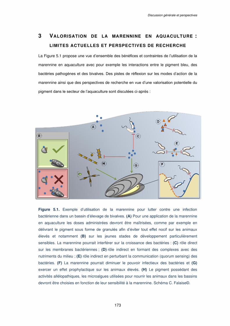
Discussion générale et perspectives
173
3 VALORISATION DE LA MARENNINE EN AQUACULTURE :
LIMITES ACTUELLES ET PERSPECTIVES DE RECHERCHE
La Figure 5.1 propose une vue d’ensemble des bénéfices et contraintes de l’utilisation de la
marennine en aquaculture avec pour exemple les interactions entre le pigment bleu, des
bactéries pathogènes et des bivalves. Des pistes de réflexion sur les modes d’action de la
marennine ainsi que des perspectives de recherche en vue d’une valorisation potentielle du
pigment dans le secteur de l’aquaculture sont discutées ci-après :
Figure 5.1. Exemple d’utilisation de la marennine pour lutter contre une infection
bactérienne dans un bassin d’élevage de bivalves. (A) Pour une application de la marennine
en aquaculture les doses administrées devront être maîtrisées, comme par exemple en
délivrant le pigment sous forme de granulés afin d’éviter tout effet nocif sur les animaux
élevés et notamment (B) sur les jeunes stades de développement particulièrement
sensibles. La marennine pourrait interférer sur la croissance des bactéries : (C) rôle direct
sur les membranes bactériennes ; (D) rôle indirect en formant des complexes avec des
nutriments du milieu ; (E) rôle indirect en perturbant la communication (quorum sensing) des
bactéries. (F) La marennine pourrait diminuer le pouvoir infectieux des bactéries et (G)
exercer un effet prophylactique sur les animaux élevés. (H) Le pigment possédant des
activités allélopathiques, les microalgues utilisées pour nourrir les animaux dans les bassins
devront être choisies en fonction de leur sensibilité à la marennine. Schéma C. Falaise©.
Chapitre V.
174
Afin de maîtriser les concentrations administrées dans les bassins d’élevages, les
doses de marennine pourraient être délivrées par granulés (Figure 5.1A) et devraient être
ajustées en fonction des animaux élevés et de leur stade de développement. Les stades de
développement précoces (larves, juvéniles), étant particulièrement sensibles, ne devraient
pas être exposés à des concentrations ≥ 0,5 µg mL-1 pour éviter tout effet adverse sur leur
survie et leur développement (Figure 5.1B).
La marennine pourrait exercer un effet direct sur la croissance des bactéries et
pourrait ainsi limiter la prolifération bactérienne (Figure 5.1C). Les mécanismes d’actions
exactes du pigment sur les bactéries ne sont pas encore déterminés mais, étant donné le
poids moléculaire estimé de la marennine extracellulaire (environ 10 kDa ; Pouvreau et al.,
2006), il paraît plus vraisemblable que la molécule ne pénètre pas les cellules mais agisse
plutôt au niveau de la surface membranaire des bactéries. Des travaux conduits avec le
pigment de type marennine purifié produit par Haslea provincialis ont d’ailleurs permis de
mettre en évidence une interaction du pigment avec la membrane externe de la bactérie
Gram (-) Escherichia coli (Tardy-Laporte et al., 2013). Récemment, une étude conduite avec
de la marennine purifiée et une souche de V. splendidus a démontré un effet du pigment sur
la fluidité de la membrane bactérienne. Il semblerait ainsi que la marennine induise une
rigidification des membranes de la souche de V. splendidus testée en interagissant avec des
lipopolysaccharides présents sur la membrane externe (Bouhlel et al., article soumis). La
marennine agirait donc au niveau de la surface membranaire des Vibrio, pourrait se lier
spécifiquement à certains récepteurs et déclencher des réponses de stress ou des signaux
apoptotiques en induisant des modifications du métabolisme cellulaire. La marennine
pourrait donc cibler des récepteurs membranaires particuliers, dont la présence et la
fréquence pourraient varier entre les différentes espèces de Vibrio et également entre
souches d’une même espèce (Biosca et al., 1993 ; Wang et al., 2015a ; Zhu et al., 2013), ce
qui pourrait expliquer la variabilité de réponses des souches de Vibrio exposées à la
marennine observée dans le Chapitre III de ce manuscrit.
La marennine pourrait également avoir un effet indirect sur la croissance des
bactéries en limitant leur accès à certains nutriments (Figure 5.1D). Bien que sa structure
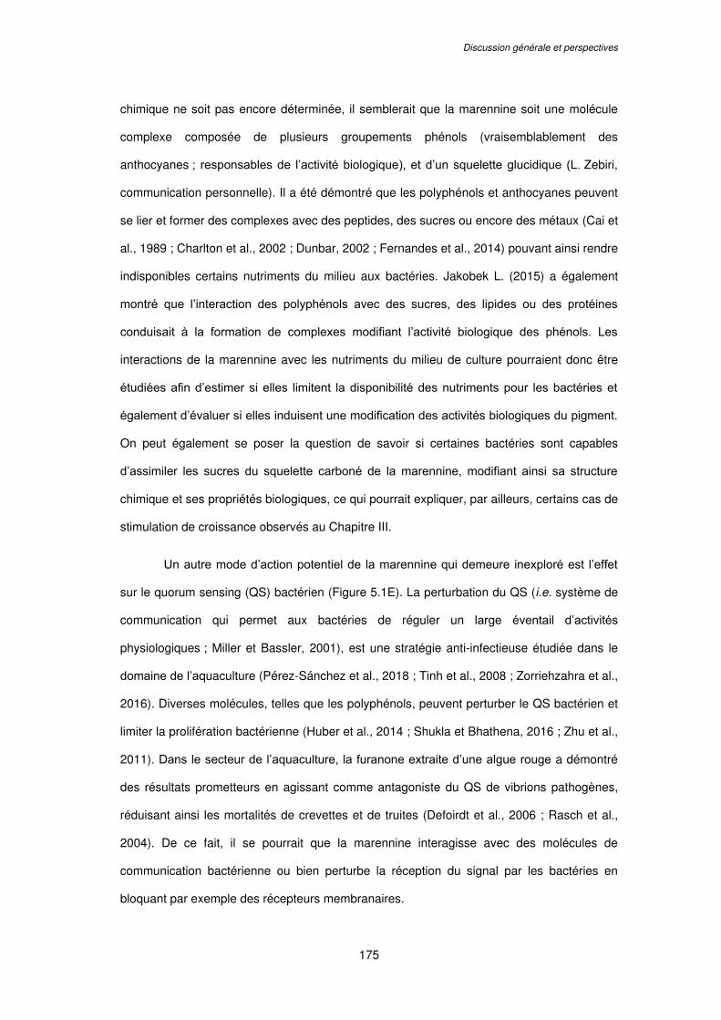
Discussion générale et perspectives
175
chimique ne soit pas encore déterminée, il semblerait que la marennine soit une molécule
complexe composée de plusieurs groupements phénols (vraisemblablement des
anthocyanes ; responsables de l’activité biologique), et d’un squelette glucidique (L. Zebiri,
communication personnelle). Il a été démontré que les polyphénols et anthocyanes peuvent
se lier et former des complexes avec des peptides, des sucres ou encore des métaux (Cai et
al., 1989 ; Charlton et al., 2002 ; Dunbar, 2002 ; Fernandes et al., 2014) pouvant ainsi rendre
indisponibles certains nutriments du milieu aux bactéries. Jakobek L. (2015) a également
montré que l’interaction des polyphénols avec des sucres, des lipides ou des protéines
conduisait à la formation de complexes modifiant l’activité biologique des phénols. Les
interactions de la marennine avec les nutriments du milieu de culture pourraient donc être
étudiées afin d’estimer si elles limitent la disponibilité des nutriments pour les bactéries et
également d’évaluer si elles induisent une modification des activités biologiques du pigment.
On peut également se poser la question de savoir si certaines bactéries sont capables
d’assimiler les sucres du squelette carboné de la marennine, modifiant ainsi sa structure
chimique et ses propriétés biologiques, ce qui pourrait expliquer, par ailleurs, certains cas de
stimulation de croissance observés au Chapitre III.
Un autre mode d’action potentiel de la marennine qui demeure inexploré est l’effet
sur le quorum sensing (QS) bactérien (Figure 5.1E). La perturbation du QS (i.e. système de
communication qui permet aux bactéries de réguler un large éventail d’activités
physiologiques ; Miller et Bassler, 2001), est une stratégie anti-infectieuse étudiée dans le
domaine de l’aquaculture (Pérez-Sánchez et al., 2018 ; Tinh et al., 2008 ; Zorriehzahra et al.,
2016). Diverses molécules, telles que les polyphénols, peuvent perturber le QS bactérien et
limiter la prolifération bactérienne (Huber et al., 2014 ; Shukla et Bhathena, 2016 ; Zhu et al.,
2011). Dans le secteur de l’aquaculture, la furanone extraite d’une algue rouge a démontré
des résultats prometteurs en agissant comme antagoniste du QS de vibrions pathogènes,
réduisant ainsi les mortalités de crevettes et de truites (Defoirdt et al., 2006 ; Rasch et al.,
2004). De ce fait, il se pourrait que la marennine interagisse avec des molécules de
communication bactérienne ou bien perturbe la réception du signal par les bactéries en
bloquant par exemple des récepteurs membranaires.
Chapitre V.
176
Une hypothèse sur les effets biologiques de la marennine actuellement mise à
l’essai est la capacité du pigment à diminuer le pouvoir infectieux des bactéries
(Figure 5.1F). Pour provoquer une maladie, un organisme pathogène doit être capable de
coloniser les surfaces de l’hôte et de pénétrer dans ses tissus (Casadevall et Pirofski, 1999).
La marennine fixée sur les membranes bactériennes ou sur les tissus (e.g. branchies) des
bivalves pourrait ainsi représenter une barrière aux infections bactériennes. Des tests ont
récemment été mis en place afin d’évaluer si des huîtres aux branchies préalablement
verdies seraient moins sensibles aux infections bactériennes que les animaux aux branchies
non verdies.
Ainsi, les effets prophylactiques de la marennine représentent sans doute la facette
la plus prometteuse du pigment pour une valorisation en aquaculture (Figure 5.1G). La BW à
faible concentration (0,5 µg mL-1) permet une amélioration de l’état physiologique (i.e.
augmentation des réserves énergétiques) des larves de moule et de pétoncle (Turcotte et
al., 2016) et de la plie (Chapitre IV. de ce manuscrit). Cette faible concentration de BW n’a
pas eu d’effet sur la concentration bactérienne dans les bassins d’élevage mais a procuré
une meilleure résistance des larves de moule et de pétoncle face à une souche pathogène
de V. splendidus (Turcotte et al., 2016). Le même protocole expérimental a récemment
permis de démontrer l’effet protecteur de la marennine sur les larves de moules en modifiant
la composition de leur microbiote intestinal (Latour et al., 2018). Il semblerait donc
intéressant d’étudier davantage les effets prophylactiques et immunostimulants de le
marennine. Les bivalves, et les invertébrés de façon générale, ne possèdent pas de système
immunitaire stricto sensu mais possèdent une mémoire immunitaire et peuvent mettre en
place des réponses cellulaires (hémocytaires) et humorales pour faire face à des
microorganismes envahisseurs (e.g. limitation de l’entrée du microbe, résistance ou
tolérance à la maladie ; Rowley et Powell, 2007). En plus d’améliorer l’état physiologique
des animaux (Turcotte et al., 2016) et de prévenir les infections en modifiant la composition
du microbiote intestinal (Latour et al., 2018), la marennine pourrait également stimuler la
mise en place de processus tels que la libération de peptides antimicrobiens ou autres
Discussion générale et perspectives
177
mécanismes cellulaires de défense (e.g. phagocytose, encapsulation) permettant à l’animal
de se protéger des agents infectieux (Smith, 2016 ; Soudant et al., 2013).
Enfin, les activités allélopatiques de la marennine devront également être prises en
considération pour une application en aquaculture (Figure 5.1H). En effet, il a été montré
que le pigment réduisait la croissance de microalgues fréquemment utilisées pour nourrir les
bivalves telles que Skeletonema costatum ou Chaetoceros calcitrans (Prasetiya et al., 2016),
et donc la co-culture de ces espèces avec une Haslea bleue, ou en présence de marennine
en solution dans le milieu, devrait être évitée. En revanche, d’autres espèces comme
Phaeodactylum tricornutum, Tetraselmis suecica ou Isochrysis galbana (Prasetiya et al.,
2016 ; Turcotte et al., 2016) semblent insensibles à la présence de marennine et devraient
donc être privilégiées.
En conclusion principale de ce travail de thèse, nous pouvons affirmer que la
marennine présente des effets biologiques significatifs qui encouragent son utilisation dans
le secteur de l’aquaculture. Administré à faibles doses, le pigment ne permettrait pas de
réduire la prolifération des bactéries dans les bassins d’élevage mais pourrait conférer aux
animaux exposés une meilleure résistance aux agents infectieux. Cependant, les doses
d’exposition devront être maîtrisées pour ne pas nuire au développement ou à la survie des
animaux, et particulièrement ceux des stades larvaires ou juvéniles. La définition de la
structure chimique de la molécule permettra d’explorer davantage les mécanismes d’action
du pigment et également d’étendre les secteurs de valorisation potentiels de la marennine.

178

179
RÉFÉRENCES
BIBLIOGRAPHIQUES

180
Références bibliographiques
181
- A -
Abedin, R., and Taha, H. (2008). Antibacterial and antifungal activity of cyanobacteria and green microalgae. Evaluation of medium components by Plackett-Burman design for antimicrobial activity of Spirulina platensis. Glob. J. Biotechnol. Biochem. 3, 22–31.
Accoceberry, I., and Noël, T. (2006). Antifongiques : cibles cellulaires et mécanismes de résistance. Thérapie 61, 195–199.
Ackermann, M. (2015). A functional perspective on phenotypic heterogeneity in microorganisms. Nat. Rev. Microbiol. 13, 497–508.
Adolph, S., Bach, S., Blondel, M., Cueff, A., Moreau, M., Pohnert, G., Poulet, S.A., Wichard, T., and Zuccaro, A. (2004). Cytotoxicity of diatom-derived oxylipins in organisms belonging to different phyla. J. Exp. Biol. 207, 2935–2946.
Aguirre-Guzmán, G., Mejia Ruíz, H., and Ascencio, F. (2004). A review of extracellular virulence product of Vibrio species important in diseases of cultivated shrimp. Aquac. Res. 35, 1395–1404.
Ahmad, A.L., Yasin, N.H.M., Derek, C.J.C., and Lim, J.K. (2011). Microalgae as a sustainable energy source for biodiesel production: A review. Renew. Sustain. Energy Rev. 15, 584–593.
Ahmadi, A., Zorofchian Moghadamtousi, S., Abubakar, S., and Zandi, K. (2015). Antiviral potential of algae polysaccharides isolated from marine sources: A review,. BioMed Res. Int. 2015.
Ahne, W. (1994). Viral infections of aquatic animals with special reference to Asian aquaculture. Annu. Rev. Fish Dis. 4, 375–388.
Alangaden, G.J. (2011). Nosocomial fungal infections: epidemiology, infection control, and prevention. Infect. Dis. Clin. North Am. 25, 201–225.
AlgaeBase (2018). World-wide electronic publication, National University of Ireland, Galway.
Allard, W.G., Belt, S.T., Massé, G., Naumann, R., Robert, J.M., and Rowland, S. (2001). Tetra-unsaturated sesterterpenoids (Haslenes) from Haslea ostrearia and related species. Phytochemistry 56, 795–800.
Amaro, H.M., Guedes, A.C., and Malcata, F.X. (2011). Science against microbial pathogens: communicating current research and technological advances (A. Mendez-Vilas).
Amin, S.A., Parker, M.S., and Armbrust, E.V. (2012). Interactions between diatoms and bacteria. Microbiol. Mol. Biol. Rev. 76, 667–684.
Anaissie, E., and Bodey, G.P. (1989). Nosocomial fungal infections. Old problems and new challenges. Infect. Dis. Clin. North Am. 3, 867–882.
Anater, A., Manyes, L., Meca, G., Ferrer, E., Luciano, F.B., Pimpão, C.T., and Font, G. (2016). Mycotoxins and their consequences in aquaculture: A review. Aquaculture 451, 1–10.
Références bibliographiques
182
Arif, J.M., Farooqui, A., Siddiqui, M.H., Al-Karrawi, M., Al-Hazmi, A., and Al-Sagair, O.A. (2012). Chapter 5. Novel bioactive peptides from cyanobacteria: Functional, biochemical, and biomedical significance. In Studies in Natural Products Chemistry, Atta-ur-Rahman, ed. (Elsevier), pp. 111–161.
Armbrust, E.V. (2009). The life of diatoms in the world’s oceans. Nature 459, 185–192.
Arzul, I., and Renault, T. (2002). Herpèsvirus et bivalves marins. Virologie 6, 169–174.
Arzul, I., Renault, T., Lipart, C., and Davison, A.J. (2001). Evidence for interspecies transmission of oyster herpesvirus in marine bivalves. J. Gen. Virol. 82, 865–870.
Auffret, M., and Oubella, R. (1997). Hemocyte Aggregation in the Oyster Crassostrea gigas: In Vitro Measurement and Experimental Modulation by Xenobiotics. Comp. Biochem. Physiol. -- Part Physiol. 3, 705–712.
Austin, B. (2012). Infectious Disease in Aquaculture: Prevention and Control (Elsevier).
Austin, B., and Austin, D.A. (2007). Bacterial fish pathogens: disease of farmed and wild fish (Springer Science & Business Media).
Austin, B., and Day, J.G. (1990). Inhibition of prawn pathogenic Vibrio spp. by a commercial spray-dried preparation of Tetraselmis suecica. Aquaculture 90, 389–392.
Austin, B., Baudet, E., and Stobie, M. (1992). Inhibition of bacterial fish pathogens by Tetraselmis suecica. J. Fish Dis. 15, 55–61.
Avio, C.G., Gorbi, S., Milan, M., Benedetti, M., Fattorini, D., d’Errico, G., Pauletto, M., Bargelloni, L., and Regoli, F. (2015). Pollutants bioavailability and toxicological risk from microplastics to marine mussels. Environ. Pollut. 198, 211–222.
- B -
Bachrach, E., and Simonet, M. (1936). Le phénomène du bleuissement chez les diatomées. Rev. Trav. Inst. Pêch. Marit. 9, 113–116.
Bag, J. (1974). Glucose inhibition of the transport and phosphoenolpyruvate-dependent phosphorylation of galactose and fructose in Vibrio cholerae. J. Bacteriol. 118, 764–767.
Balasubramanian, G., Sarathi, M., Venkatesan, C., Thomas, J., and Hameed, A.S.S. (2008). Studies on the immunomodulatory effect of extract of Cyanodon dactylon in shrimp, Penaeus monodon, and its efficacy to protect the shrimp from white spot syndrome virus (WSSV). Fish Shellfish Immunol. 25, 820–828.
Ban, S., Burns, C., Castel, J., Chaudron, Y., Christou, E., Escribano, R., Umani, S.F., Gasparini, S., Ruiz, F.G., Hoffmeyer, M., et al. (1997). The paradox of diatom-copepod interactions. Mar. Ecol. Prog. Ser. 157, 287–293.
Banerjee, G., and Ray, A.K. (2017). The advancement of probiotics research and its application in fish farming industries. Res. Vet. Sci. 115, 66–77.
Références bibliographiques
183
Banerjee, S., Kim, L.M., Shariff, M., Khatoon, H., and Yusoff, F.M. (2013). Antibacterial activity of neem (Azadirachta indica) leaves on Vibrio spp. isolated from cultured shrimp. Asian J. Anim. Vet. Adv. 8, 355–361.
Barra, L., Chandrasekaran, R., Corato, F., and Brunet, C. (2014). The challenge of ecophysiological biodiversity for biotechnological applications of marine microalgae. Mar. Drugs 12, 1641–1675.
Bassim, S., Tanguy, A., Genard, B., Moraga, D., and Tremblay, R. (2014). Identification of Mytilus edulis genetic regulators during early development. Gene 551, 65–78.
Bavestrello, G., Arillo, A., Calcinai, B., Cattaneo-Vietti, R., Cerrano, C., Gaino, E., Penna, A., and Sarà, M. (2000). Parasitic diatoms inside antarctic sponges. Biol. Bull. 198, 29–33.
Bellou, S., Triantaphyllidou, I.-E., Aggeli, D., Elazzazy, A.M., Baeshen, M.N., and Aggelis, G. (2016). Microbial oils as food additives: recent approaches for improving microbial oil production and its polyunsaturated fatty acid content. Curr. Opin. Biotechnol. 37, 24–35.
Belz, R.G., and Duke, S.O. (2014). Herbicides and plant hormesis. Pest Manag. Sci. 70, 698–707.
Bennett, A.G. (1920). On the occurrence of diatoms on the skin of whales. Proc R Soc Lond B 91, 352–357.
Benoiston, A.-S., Ibarbalz, F.M., Bittner, L., Guidi, L., Jahn, O., Dutkiewicz, S., and Bowler, C. (2017). The evolution of diatoms and their biogeochemical functions. Philos. Trans. R. Soc. B Biol. Sci. 372.
Bergé, J.-P., Bourgougnon, N., Alban, S., Pojer, F., Billaudel, S., Chermann, J.-C., Robert, J.M., and Franz, G. (1999). Antiviral and anticoagulant activities of a water-soluble fraction of the marine diatom Haslea ostrearia. Planta Med. 65, 604–609.
Berland, B.R., Bonin, D.J., Cornu, A.L., Maestrini, S.Y., and Marino, J.-P. (1972). The antibacterial substances of the marine alga Stichochrysis immobilis (chrysophyta). J. Phycol. 8, 383–392.
Bhagavathy, S., Sumathi, P., and Jancy Sherene Bell, I. (2011). Green algae Chlorococcum humicola-a new source of bioactive compounds with antimicrobial activity. Asian Pac. J. Trop. Biomed. 1, S1–S7.
Bhatnagar, I., and Kim, S.-K. (2010). Immense essence of excellence: marine microbial bioactive compounds. Mar. Drugs 8, 2673–2701.
Bhattacharya, D., Ghosh, D., Bhattacharya, S., Sarkar, S., Karmakar, P., Koley, H., and Gachhui, R. (2018). Antibacterial activity of polyphenolic fraction of Kombucha against Vibrio cholerae: targeting cell membrane. Lett. Appl. Microbiol. 66, 145–152.
Biosca, E.G., Garay, E., Toranzo, A.E., and Amaro, C. (1993). Comparison of outer membrane protein profiles of Vibrio vulnificus biotypes 1 and 2. FEMS Microbiol. Lett. 107, 217–222.
Références bibliographiques
184
Bocat, L. (1907). Sur la marennine de la diatomée bleue; comparaison avec la phycocyanine. Comptes Rendus Seances Société Biol. Ses Fil. 62, 1073–1075.
Boon, D.D. (1977). Coloration in bivalves: A review. J Food Sci 42, 1008–1012.
Borowitzka, M.A. (1995). Microalgae as sources of pharmaceuticals and other biologically active compounds. J. Appl. Phycol. 7, 3–15.
Borowitzka, M.A. (1997). Microalgae for aquaculture: Opportunities and constraints. J. Appl. Phycol. 9, 393–401.
Bory de Saint-Vincent, J.B. (1826). Dictionnaire Classique d’Histoire Naturelle (Paris, France: Rey et Gravier).
Bosc, G., and Lair, P.A. (1817). Huître. In Nouveau Dictionnaire d’Histoire Naturelle Appliquée Aux Arts, à l’agriculture, à l’économie Rurale et Domestique, à La Médecine, Etc. Tome XV, (Paris, France: Deterville), p.
Bouhlal Rhimou, R.H. (2010). Antiviral activity of the extracts of Rhodophyceae from Morocco. Afr. J. Biotechnol. 9, 7968–7975.
Bouhlel, Z., Deschênes, J.-S., Mouget, J.-L., Tremblay, R., and Marcotte, I. (article soumis). Insight into the interaction mechanism of marennine with Vibrio splendidus as seen by in vivo 2H solid-state NMR.
Bouhlel, Z., Turcotte, F., Warschawski, D.E., Arnold, A., Lemarchand, K., Deschênes, J.-S., Marcotte, I., and Tremblay, R. (2018). Marennine, a potential alternative antibiotic to polymyxine in aquaculture. (Québec (Canada)), p.
Bower, S.M., Sharon E. McGladdery, and Iola M. Price (1994). Synopsis of infectious diseases and parasites of commercially exploited shellfish. Annu. Rev. Fish Dis. 4, 1–199.
Boyce, R.W., and Herdman, W.A. (1896). The possible infectivity of the oyster, and upon the green disease in oysters. Comm Br Assoc Adv Sci 66, 663–669.
Brauch, J.E., Zapata-Porras, S.P., Buchweitz, M., Aschoff, J.K., and Carle, R. (2016). Jagua blue derived from Genipa americana L. fruit: A natural alternative to commonly used blue food colorants? Food Res. Int.
Brown, M.R., Jeffrey, S.W., Volkman, J.K., and Dunstan, G.A. (1997). Nutritional properties of microalgae for mariculture. Aquaculture 151, 315–331.
Bruce, D.L., Duff, D.C., and Antia, N.J. (1967). The identification of two antibacterial products of the marine planktonic alga Isochrysis galbana. J. Gen. Microbiol. 48, 293–298.
Bruto, M., James, A., Petton, B., Labreuche, Y., Chenivesse, S., Alunno-Bruscia, M., Polz, M.F., and Le Roux, F. (2017). Vibrio crassostreae, a benign oyster colonizer turned into a pathogen after plasmid acquisition. ISME J. 11, 1043–1052.
Buchweitz, M. (2016). Chapter 17. Natural Solutions for Blue Colors in Food. In Handbook on Natural Pigments in Food and Beverages, (Woodhead Publishing), pp. 355–384.
Références bibliographiques
185
Burja, A.M., Banaigs, B., Abou-Mansour, E., Grant Burgess, J., and Wright, P.C. (2001). Marine cyanobacteria—a prolific source of natural products. Tetrahedron 57, 9347–9377.
Buttino, I., Miralto, A., Ianora, A., Romano, G., and Poulet, S.A. (1999). Water-soluble extracts of the diatom Thalassiosira rotula induce aberrations in embryonic tubulin organisation of the sea urchin Paracentrotus lividus. Mar. Biol. 134, 147–154.
Buttino, I., Hwang, J.-S., Romano, G., Sun, C.-K., Liu, T.-M., Pellegrini, D., Gaion, A., and Sartori, D. (2016). Detection of malformations in sea urchin plutei exposed to mercuric chloride using different fluorescent techniques. Ecotoxicol. Environ. Saf. 123, 72–80.
- C -
Cadoret, J.-P., Garnier, M., and Saint-Jean, B. (2012). Microalgae, functional genomics and biotechnology. Adv. Bot. Res. 64, 285–341.
Cahu, C.L., Zambonino Infante, J.L., Péres, A., Quazuguel, P., and Le Gall, M.M. (1998). Algal addition in sea bass (Dicentrarchus labrax) larvae rearing: effect on digestive enzymes. Aquaculture 161, 479–489.
Cai, Y., Gaffney, S.H., Lilley, T.H., and Haslam, E. (1989). Carbohydrate - Polyphenol Complexation. In Chemistry and Significance of Condensed Tannins, R.W. Hemingway, J.J. Karchesy, and S.J. Branham, eds. (Boston, MA: Springer US), pp. 307–322.
Calabrese, E.J. (2013). Hormetic mechanisms. Crit. Rev. Toxicol. 43, 580–606.
Calabrese, E.J., and Baldwin, L.A. (1997). A quantitatively-based methodology for the evaluation of chemical hormesis. Hum. Ecol. Risk Assess. Int. J. 3.
Calabrese, E.J., and Baldwin, L.A. (2001). Hormesis: A Generalizable and Unifying Hypothesis. Crit. Rev. Toxicol. 31, 353–424.
Calabrese, E.J., and Baldwin, L.A. (2002). Defining hormesis. Hum. Exp. Toxicol. 21, 91–97.
Caldwell, G.S. (2009). The influence of bioactive oxylipins from marine diatoms on invertebrate reproduction and development. Mar. Drugs 7, 367–400.
Caldwell, G.S., Olive, P.J.W., and Bentley, M.G. (2002). Inhibition of embryonic development and fertilization in broadcast spawning marine invertebrates by water soluble diatom extracts and the diatom toxin 2-trans,4-trans decadienal. Aquat. Toxicol. Amst. Neth. 60, 123–137.
Cannell, R.J.P., Owsianka, A.M., and Walker, J.M. (1988). Results of a large-scale screening programme to detect antibacterial activity from freshwater algae. Br. Phycol. J. 23, 41–44.
Carbonnelle, D., Pondaven, P., Morancais, M., Masse, G., Bosch, S., Jacquot, C., Briand, G., Robert, J., and Roussakis, C. (1998). Antitumor and antiproliferative effects of an aqueous extract from the marine diatom Haslea ostrearia (Simonsen) against solid tumors: lung carcinoma (NSCLC-N6), kidney carcinoma (E39) and melanoma (M96) cell lines. Anticancer Res. 19, 621–624.
Casadevall, A., and Pirofski, L.A. (1999). Host-pathogen interactions: redefining the basic concepts of virulence and pathogenicity. Infect. Immun. 67, 3703–3713.

Références bibliographiques
186
Cavallo, R., Acquaviva, M., Stabili, L., Cecere, E., Petrocelli, A., and Narracci, M. (2013). Antibacterial activity of marine macroalgae against fish pathogenic Vibrio species. Open Life Sci. 8, 646–653.
Cedergreen, N., Streibig, J.C., Kudsk, P., Mathiassen, S.K., and Duke, S.O. (2006). The occurrence of hormesis in plants and algae. Dose-Response Publ. Int. Hormesis Soc. 5, 150–162.
Chang, T., Ohta, S., Ikegami, N., Miyata, H., Kashimoto, T., and Kondo, M. (1993). Antibiotic substances produced by a marine green alga, Dunaliella primolecta. Bioresour. Technol. 44, 149–153.
Charlton, A.J., Baxter, N.J., Khan, M.L., Moir, A.J.G., Haslam, E., Davies, A.P., and Williamson, M.P. (2002). Polyphenol/peptide binding and precipitation. J. Agric. Food Chem. 50, 1593–1601.
Chatterjee, T., Chatterjee, B.K., and Chakrabarti, P. (2017). Modelling of growth kinetics of Vibrio cholerae in presence of gold nanoparticles: effect of size and morphology. Sci. Rep. 7, 9671.
Cheung, R.C.F., Wong, J.H., Pan, W.L., Chan, Y.S., Yin, C.M., Dan, X.L., Wang, H.X., Fang, E.F., Lam, S.K., Ngai, P.H.K., et al. (2014). Antifungal and antiviral products of marine organisms. Appl. Microbiol. Biotechnol. 98, 3475–3494.
Citarasu, T. (2012). Natural antimicrobial compounds for use in aquaculture. In Infectious Disease in Aquaculture: Prevention and Control, (Brian Austin), pp. 419–457.
Clinical and Laboratory Standards Institute (CLSI) (2012). Methods for Dilution Antimicrobial Susceptibility Tests for Bacteria That Grow Aerobically. Document M07-A9. Approved Standard—Ninth Edition (Wayne, PA, USA).
Cooper, S., Battat, A., Marsot, P., and Sylvestre, M. (1983). Production of antibacterial activities by two Bacillariophyceae grown in dialysis culture. Can. J. Microbiol. 29, 338–341.
Costa, M., Costa-Rodrigues, J., Fernandes, M.H., Barros, P., Vasconcelos, V., and Martins, R. (2012). Marine cyanobacteria compounds with anticancer properties: A review on the implication of apoptosis. Mar. Drugs 10, 2181–2207.
Coyne, V.E. (2011). The importance of ATP in the immune system of molluscs. Invertebr. Surviv. J.
Crane, M., and Hyatt, A. (2011). Viruses of Fish: An Overview of Significant Pathogens. Viruses 3, 2025–2046.
Croll, D.A., and Holmes, R.W. (1982). A note on the occurence of diatoms on the feathers of diving seabirds. The Auk 99, 765–766.
- D -
Darwin, C. (1859). On the Origin of Species by Means of Natural Selection (London, England: J. Murray).
Références bibliographiques
187
Das, B.K., Pradhan, J., Pattnaik, P., Samantaray, B.R., and Samal, S.K. (2005). Production of antibacterials from the freshwater alga Euglena viridis (Ehren). World J. Microbiol. Biotechnol. 21, 45–50.
Davey, M.R., Anthony, P., Power, J.B., and Lowe, K.C. (2005). Plant protoplasts: status and biotechnological perspectives. Biotechnol. Adv. 23, 131–171.
Davidovich, N.A., Mouget, J.-L., and Gaudin, P. (2009). Heterothallism in the pennate diatom Haslea ostrearia (Bacillariophyta). Eur. J. Phycol. 44, 251–261.
Dawood, M.A.O., Koshio, S., and Esteban, M.Á. (2018). Beneficial roles of feed additives as immunostimulants in aquaculture: A review. Rev. Aquac. 10, 950–974.
Defoirdt, T., Crab, R., Wood, T.K., Sorgeloos, P., Verstraete, W., and Bossier, P. (2006). Quorum sensing-disrupting brominated furanones protect the gnotobiotic brine shrimp Artemia franciscana from pathogenic Vibrio harveyi, Vibrio campbellii, and Vibrio parahaemolyticus isolates. Appl Env. Microbiol 72, 6419–6423.
del Pilar Sanchez-Saavedra, M., Licea-Navarro, A., and Bernaldez-Sarabia, J. (2010). Evaluation of the antibacterial activity of different species of phytoplankton.
Desbois, A.P., Lebl, T., Yan, L., and Smith, V. (2008). Isolation and structure characterization of two antibacterial free fatty acids from marine diatom, Phaeodactylum tricornutum. Appl. Microbiol. Biotechnol. 81, 755–764.
Desbois, A.P., Mearns-Spragg, A., and Smith, V.J. (2009). A fatty acid from the diatom Phaeodactylum tricornutum is antibacterial against diverse bacteria including multi-resistant Staphylococcus aureus (MRSA). Mar. Biotechnol. 11, 45–52.
Desbois, A.P., Walton, M., and Smith, V.J. (2010). Differential antibacterial activities of fusiform and oval morphotypes of Phaeodactylum tricornutum (Bacillariophyceae). J. Mar. Biol. Assoc. U. K. 90, 769–774.
Deschamps, P., and Moreira, D. (2012). Reevaluating the green contribution to diatom genomes. Genome Biol. Evol. 4, 683–688.
Dondero, F., Negri, A., Boatti, L., Marsano, F., Mignone, F., and Viarengo, A. (2010). Transcriptomic and proteomic effects of a neonicotinoid insecticide mixture in the marine mussel (Mytilus galloprovincialis, Lam.). Sci. Total Environ. 408, 3775–3786.
Dorantes-Aranda, J.J., Waite, T.D., Godrant, A., Rose, A.L., Tovar, C.D., Woods, G.M., and Hallegraeff, G.M. (2011). Novel application of a fish gill cell line assay to assess ichthyotoxicity of harmful marine microalgae. Harmful Algae 10, 366–373.
Duff, D.C.B., Bruce, D.L., and Antia, N.J. (1966). The Antibacterial Activity of Marine
Planktonic Algae. Can. J. Microbiol. 12, 877–884.
Dunbar, R.C. (2002). Metal cation binding to phenol: DFT comparison of the competing sites. J. Phys. Chem. A 106, 7328–7337.
Références bibliographiques
188
Dyshlovoy, S.A., and Honecker, F. (2015). Marine compounds and cancer: where do we stand? Mar. Drugs 13, 5657–5665.
- E -
Ehresmann, D.W., Deig, E.F., Hatch, M.T., DiSalvo, L.H., and Vedros, N.A. (1977). Antiviral Substances from California Marine Algae1. J. Phycol. 13, 37–40.
Eissa, N., and Wang, H.-P. (2016). Transcriptional stress responses to environmental and husbandry stressors in aquaculture species. Rev. Aquac. 8, 61–88.
El Amraoui, B., El Amraoui, M., Cohen, N., and Fassouane, A. (2014). Anti-Candida and anti-Cryptococcus antifungal produced by marine microorganisms. J. Mycol. Médicale J. Med. Mycol. 24, 149–153.
Elston, R. (1997). Special topic review : bivalve mollusc viruses. World J Microbiol Biotechnol 13, 393–403.
Encarnacao, T., Pais, A.A.C.C., Campos, M.G., and Burrows, H.D. (2015). Cyanobacteria and microalgae: A renewable source of bioactive compounds and other chemicals. Sci. Prog. 98(2), 145–168.
Eriksen, N.T. (2008). Production of phycocyanin - a pigment with applications in biology, biotechnology, foods and medicine. Appl. Microbiol. Biotechnol. 80, 1–14.
- F -
Fabregas, J., García, D., Fernandez-Alonso, M., Rocha, A.I., Gómez-Puertas, P., Escribano, J.M., Otero, A., and Coll, J.M. (1999). In vitro inhibition of the replication of haemorrhagic septicaemia virus (VHSV) and African swine fever virus (ASFV) by extracts from marine microalgae. Antiviral Res. 44, 67–73.
Falaise, C., Gastineau, R., Poulin, M., Leignel, V., Hardivillier, Y., Widowati, I., Syakti, A.D., Lemieux, C., Turmel, M., and Mouget, J.-L. (2016a). Haslea nusantara sp. nov., a novel species of blue diatom from Indonesia (24th International Diatom Symposium, Québec (Canada)).
Falaise, C., François, C., Travers, M.-A., Morga, B., Haure, J., Tremblay, R., Turcotte, F., Pasetto, P., Gastineau, R., Hardivillier, Y., et al. (2016b). Antimicrobial compounds from eukaryotic microalgae against human pathogens and diseases in aquaculture. Mar. Drugs 14, 159.
Falaise, C., Dewi, I.C., Hellio, C., Bourgougnon, N., and Mouget, J.-L. (2018). Chapter 12. Anticancer, antiviral, antibacterial and antifungal properties in microalgae. In Levine I., Fleurence J. (Eds). Microalgae in Health and Disease Prevention, (Elsevier Science, Amsterdam, Netherlands), p.
Falasco, E., Bona, F., Badino, G., Hoffmann, L., and Ector, L. (2009). Diatom teratological forms and environmental alterations: a review. Hydrobiologia 623, 1–35.
FAO (2018a). La situation mondiale des pêches et de l’aquaculture 2018. Atteindre les objectifs de développement durable (Rome: Licence: CC BY-NC-SA 3.0 IGO.).
Références bibliographiques
189
FAO (2018b). FAO Aquaculture Newsletter (FAN) April 2018.
FAO/WHO (2001). Health and nutritional properties of probiotics in food including powder milk with liver lactic acid bacteria. (Food and Agriculture Organization and World Health Organization Joint report).
Farley, C. (1978). Viruses and virus like lesions in marine molluscs. Mar Fish Rev 40, 18–20.
FDA (2016). http://www.fda.gov.
Fernandes, A., Brás, N.F., Mateus, N., and de Freitas, V. (2014). Understanding the molecular mechanism of anthocyanin binding to pectin. Langmuir 30, 8516–8527.
Fernández-Alarcón, C., Miranda, C.D., Singer, R.S., López, Y., Rojas, R., Bello, H., Domínguez, M., and González-Rocha, G. (2010). Detection of the floR gene in a diversity of florfenicol resistant Gram-negative bacilli from freshwater salmon farms in Chile. Zoonoses Public Health 57, 181–188.
Field, C.B., Behrenfeld, M.J., Randerson, J.T., and Falkowski, P. (1998). Primary production of the biosphere: integrating terrestrial and oceanic components. Science 281, 237–240.
Findlay, J.A., and Patil, A.D. (1984). Antibacterial constituents of the diatom Navicula delognei. J. Nat. Prod. 47, 815–818.
Folch, J., Lees, M., and Stanley, G.H.S. (1957). A Simple Method for the Isolation and Purification of Total Lipides from Animal Tissues. J. Biol. Chem. 226, 497–509.
Fraboulet, E., Lambert, Y., Tremblay, R., and Audet, C. (2010). Assessment of paternal effect and physiological cost of metamorphosis on growth of young winter flounder Pseudopleuronectes americanus juveniles in a cold environment. J. Fish Biol. 76, 930–948.
Frick, D. (2003). The coloration of food. Rev. Prog. Color. Relat. Top. 33, 15–32.
Fuentes, J.L., Garbayo, I., Cuaresma, M., Montero, Z., González-del-Valle, M., and Vílchez, C. (2016). Impact of microalgae-bacteria interactions on the production of algal biomass and associated compounds. Mar. Drugs 14.
- G -
Gagneux-Moreaux, S., Moreau, C., Gonzalez, J.-L., and Cosson, R.P. (2007). Diatom artificial medium (DAM): a new artificial medium for the diatom Haslea ostrearia and other marine microalgae. J. Appl. Phycol. 19, 549–556.
Gaillon, B. (1820). Des huîtres vertes, et des causes de cette coloration. J. Phys. Chim. D’histoire Nat. Arts 91, 222–225.
Galbraith, H., and Miller, T.B. (1973). Physicochemical effects of long chain fatty acids on bacterial cells and their protoplasts. J. Appl. Bacteriol. 36, 647–658.
Gallager, S.M., Mann, R., and Sasaki, G.C. (1986). Lipid as an index of growth and viability in three species of bivalve larvae. Aquaculture 56, 81–103.
Références bibliographiques
190
Garnier, M., Labreuche, Y., Garcia, C., Robert, M., and Nicolas, J.-L. (2007). Evidence for the involvement of pathogenic bacteria in summer mortalities of the pacific oyster Crassostrea gigas. Microb. Ecol. 53, 187–196.
Garnier, M., Labreuche, Y., and Nicolas, J.-L. (2008). Molecular and phenotypic characterization of Vibrio aestuarianus subsp. francensis subsp. nov., a pathogen of the oyster Crassostrea gigas. Syst. Appl. Microbiol. 31, 358–365.
Gastineau, R. (2011). Biodiversité, reproduction et phylogénie des diatomées bleues du genre Haslea et valorisation de leurs pigments de type marennine. Université du Maine.
Gastineau, R., Davidovich, N.A., Bardeau, J.-F., Caruso, A., Leignel, V., Hardivillier, Y., Jacquette, B., Davidovich, O.I., Rincé, Y., Gaudin, P., et al. (2012a). Haslea karadagensis (Bacillariophyta): a second blue diatom, recorded from the Black Sea and producing a novel blue pigment. Eur. J. Phycol. 47, 469–479.
Gastineau, R., Pouvreau, J.-B., Hellio, C., Morançais, M., Fleurence, J., Gaudin, P., Bourgougnon, N., and Mouget, J.-L. (2012b). Biological activities of purified marennine, the blue pigment responsible for the greening of oysters. J. Agric. Food Chem. 60, 3599–3605.
Gastineau, R., Hardivillier, Y., Leignel, V., Tekaya, N., Morançais, M., Fleurence, J., Davidovich, N., Jacquette, B., Gaudin, P., Hellio, C., et al. (2012c). Greening effect on oysters and biological activities of the blue pigments produced by the diatom Haslea karadagensis (Naviculaceae). Aquaculture 368–369, 61–67.
Gastineau, R., Davidovich, N., Hansen, G., Rines, J., Wulff, A., Kaczmarska, I., Ehrman, J., Hermann, D., Maumus, F., Hardivillier, Y., et al. (2014a). Chapter 15. - Haslea ostrearia-like Diatoms: Biodiversity out of the Blue. In Advances in Botanical Research, N. Bourgougnon, ed. (Academic Press), pp. 441–465.
Gastineau, R., Turcotte, F., Pouvreau, J.-B., Morançais, M., Fleurence, J., Windarto, E., Prasetiya, F.S., Arsad, S., Jaouen, P., Babin, M., et al. (2014b). Marennine, promising blue pigments from a widespread Haslea diatom species complex. Mar. Drugs 12, 3161–3189.
Gastineau, R., Hansen, G., Davidovich, N.A., Davidovich, O., Bardeau, J.-F., Kaczmarska, I., Ehrman, J.M., Leignel, V., Hardivillier, Y., Jacquette, B., et al. (2016). A new blue-pigmented hasleoid diatom, Haslea provincialis, from the Mediterranean Sea. Eur. J. Phycol. 51, 156–170.
Gastineau, R., Prasetiya, F.S., Falaise, C., Cognie, B., Decottignies, P., Morancais, M., Méléder, V., Davidovich, N.A., Turcotte, F., Tremblay, R., et al. (2018). Chapter 16. Marennine-Like Pigments: Blue Diatom of Green Oyster Cult? In Blue Biotechnology. Production and Use of Marine Molecules., (Wiley-VCH, Weinheim, Germany: Stéphane La Barre and Stephen S. Bates), pp. 529–551.
Gaudin, P., Lebeau, T., and Robert, J.-M. (2006). Microalgal cell immobilization for the long-term storage of the marine diatom Haslea ostrearia. J. Appl. Phycol. 18, 175–184.
Gauthier, M.J. (1969). Activité antibactérienne d’une Diatomée marine : Asterionella notata (Grun). Rev Intern Océan. Méd 25–26, 103–165.
Références bibliographiques
191
Genard, B., Moraga, D., Pernet, F., David, E., Boudry, P., and Tremblay, R. (2012). Expression of candidate genes related to metabolism, immunity and cellular stress during massive mortality in the American oyster Crassostrea virginica larvae in relation to biochemical and physiological parameters. Gene 499, 70–75.
Geneves, L., Choussy, M., Barbier, M., Neuville, D., and Daste, P. (1976). Ultrastructure et composition pigmentaire comparées des chromatophores de la diatomee Navicula ostrearia (Gaillon) Bory normale et bleue. Comptes Rendus Hebd. Seances Ser. Sci. Nat.
Gerber, P., Dutcher, J.D., Adams, E.V., and Sherman, J.H. (1958). Protective Effect of Seaweed Extracts for Chicken Embryos Infected with Influenza B or Mumps Virus. Exp. Biol. Med. 99, 590–593.
Ghannoum, M.A., and Rice, L.B. (1999). Antifungal agents: mode of action, mechanisms of resistance, and correlation of these mechanisms with bacterial resistance. Clin. Microbiol. Rev. 12, 501–517.
Ghasemi, Y., Moradian, A., Mohagheghzadeh, A., Shokravi, S., and Morowvat, M.H. (2007). Antifungal and antibacterial activity of the microalgae collected from paddy fields of Iran: Characterization of antimicrobial activity of Chroococcus dispersus. J. Biol. Sci. 7 (6), 904–910.
Ghasemi, Y., Rasoul-Amini, S., Naseri, A.T., Montazeri-Najafabady, N., Mobasher, M.A., and Dabbagh, F. (2012). Microalgae biofuel potentials (Review). Appl. Biochem. Microbiol. 48, 126–144.
Gomez-Casado, E., Estepa, A., and Coll, J.M. (2011). A comparative review on European-farmed finfish RNA viruses and their vaccines. Vaccine 29, 2657–2671.
González-Davis, O., Ponce-Rivas, E., Sánchez-Saavedra, M.D.P., Muñoz-Márquez, M.-E., and Gerwick, W.H. (2012). Bioprospection of microalgae and cyanobacteria as biocontrol agents against Vibrio campbellii and their use in white shrimp Litopenaeus vannamei culture. J. World Aquac. Soc. 43, 387–399.
Grelon, M. (1978). Saintonge, Pays des Huîtres Vertes (La Rochelle, France: Rupella).
Grimbergen, A.J., Siebring, J., Solopova, A., and Kuipers, O.P. (2015). Microbial bet-hedging: the power of being different. Curr. Opin. Microbiol. 25, 67–72.
Grovel, O., Pouchus, Y.F., and Verbist, J.-F. (2003). Accumulation of gliotoxin, a cytotoxic mycotoxin from Aspergillus fumigatus, in blue mussel (Mytilus edulis). Toxicon Off. J. Int. Soc. Toxinology 42, 297–300.
Gudimova, E., Eilertsen, H.C., Jørgensen, T.Ø., and Hansen, E. (2016). In vivo exposure to northern diatoms arrests sea urchin embryonic development. Toxicon 109, 63–69.
Guedes, A.C., and Malcata, F.X. (2012). Nutritional value and uses of microalgae in aquaculture. Aquaculture 10, 59–78.
Guedes, A.C., Barbosa, C.R., Amaro, H.M., Pereira, C.I., and Malcata, F.X. (2011). Microalgal and cyanobacterial cell extracts for use as natural antibacterial additives against food pathogens. Int. J. Food Sci. Technol. 46, 862–870.
Références bibliographiques
192
Gueho, E., Pesando, D., and Barelli, M. (1977). Propriétés antifongiques d’une diatomeé Chaetoceros lauderi ralfs C C. Mycopathologia 60, 105–107.
Guiry, M.D. (2012). How many species of algae are there? J. Phycol. 48, 1057–1063.
Gupta, V., Ratha, S.K., Sood, A., Chaudhary, V., and Prasanna, R. (2013). New insights into the biodiversity and applications of cyanobacteria (blue-green algae)—Prospects and challenges. Algal Res. 2, 79–97.
- H -
Halim, R., Danquah, M.K., and Webley, P.A. (2012). Extraction of oil from microalgae for biodiesel production: A review. Biotechnol. Adv. 30, 709–732.
Hallegraeff, G.M., Bolch, C.J.S., Hill, D.R.A., Jameson, I., LeRoy, J.-M., McMinn, A., Murray, S., de Salas, M.F., and Saunders, K. (2010). Algae of Australia: Phytoplankton of Temperate Coastal Waters (Melbourne: CSIRO Publishing).
Hamm, C.E., Merkel, R., Springer, O., Jurkojc, P., Maier, C., Prechtel, K., and Smetacek, V. (2003). Architecture and material properties of diatom shells provide effective mechanical protection. Nature 421, 841–843.
Hanic, L.A., Sekimoto, S., and Bates, S.S. (2009). Oomycete and chytrid infections of the marine diatom Pseudo-nitzschia pungens (Bacillariophyceae) from Prince Edward Island, Canada. Botany 87, 1096–1105.
Harrison, P.J., Waters, R.E., and Taylor, F.J.R. (1980). A broad spectrum artificial sea water medium for coastal and open ocean phytoplankton. J. Phycol. 16, 28–35.
Hasui, M., Matsuda, M., Okutani, K., and Shigeta, S. (1995). In vitro antiviral activities of sulfated polysaccharides from a marine microalga (Cochlodinium polykrikoides) against human immunodeficiency virus and other enveloped viruses. Int. J. Biol. Macromol. 17, 293–297.
Hayashi, T., Hayashi, K., Maeda, M., and Kojima, I. (1996). Calcium spirulan, an inhibitor of enveloped virus replication, from a blue-green alga Spirulina platensis. J. Nat. Prod. 59, 83–87.
Hennebicq, R., Fabra, G., Pellerin, C., Marcotte, I., Myrand, B., and Tremblay, R. (2013). The effect of spawning of cultured mussels (Mytilus edulis) on mechanical properties, chemical and biochemical composition of byssal threads. Aquaculture 410–411, 11–17.
Henriksson, P.J.G., Troell, M., and Rico, A. (2015). Antimicrobial use in aquaculture: Some complementing facts. Proc. Natl. Acad. Sci. 112, E3317–E3317.
Herrero, M., Ibáñez, E., Cifuentes, A., Reglero, G., and Santoyo, S. (2006). Dunaliella salina microalga pressurized liquid extracts as potential antimicrobials. J. Food Prot. 69, 2471–2477.
Herrero, M., Mendiola, J.A., Plaza, M., and Ibañez, E. (2013). Chapter 35. Screening for Bioactive Compounds from Algae. In Advanced Biofuels and Bioproducts, J.W. Lee, ed. (Springer New York), pp. 833–872.
Références bibliographiques
193
Hjörleifsson, E., and Klein-MacPhee, G. (1992). Estimation of live standard length of winter flounder Pleuronectes americanus larvae from formalin-preserved, ethanol-preserved and frozen specimens. Mar. Ecol. Prog. Ser. 82, 13–19.
Hoagland, K.D., Roemer, S.C., and Rosowski, J.R. (1982). Colonization and community structure of two periphyton assemblages, with emphasis on the diatoms (Bacillariophyceae). Am. J. Bot. 69, 188–213.
Hoek, C., Hoeck, H.V. den, Mann, D., and Jahns, H.M. (1995). Algae: An Introduction to Phycology (Cambridge University Press).
Hong, J.-H., Jang, S., Heo, Y.M., Min, M., Lee, H., Lee, Y.M., Lee, H., and Kim, J.-J. (2015). Investigation of marine-derived fungal diversity and their exploitable biological activities. Mar. Drugs 13, 4137–4155.
Houdai, T., Matsuoka, S., Matsumori, N., and Murata, M. (2004). Membrane-permeabilizing activities of amphidinol 3, polyene-polyhydroxy antifungal from a marine dinoflagellate. Biochim. Biophys. Acta BBA - Biomembr. 1667, 91–100.
Huber, B., Eberl, L., Feucht, W., and Polster, J. (2014). Influence of polyphenols on bacterial biofilm formation and quorum-sensing. Z. Für Naturforschung C 58, 879–884.
Huleihel, M., Ishanu, V., Tal, J., and Arad, S. (2001). Antiviral effect of red microalgal polysaccharides on Herpes simplex and Varicella zoster viruses. J. Appl. Phycol. 13, 127–134.
Huleihel, M., Ishanu, V., Tal, J., and Arad, S. (2002). Activity of Porphyridium sp. polysaccharide against Herpes simplex viruses in vitro and in vivo. J. Biochem. Biophys. Methods 50, 189–200.
Hussein, M.M.A., Wada, S., Hatai, K., and Yamamoto, A. (2000). Antimycotic activity of eugenol against selected water molds. J. Aquat. Anim. Health 12, 224–229.
Hussein, M.M.A., El-Feki, M.A., Hatai, K., and Yamamoto, A. (2002). Inhibitory effects of thymoquinone from Nigella sativa on pathogenic Saprolegnia in fish. Biocontrol Sci. 7, 31–35.
- I -
Ianora, A., and Miralto, A. (2010). Toxigenic effects of diatoms on grazers, phytoplankton and other microbes: a review. Ecotoxicol. Lond. Engl. 19, 493–511.
Ingalls, A.E., Whitehead, K., and Bridoux, M.C. (2010). Tinted windows: The presence of the UV absorbing compounds called mycosporine-like amino acids embedded in the frustules of marine diatoms. Geochim. Cosmochim. Acta 74, 104–115.
Ingebrigtsen, R.A., Hansen, E., Andersen, J.H., and Eilertsen, H.C. (2015). Light and temperature effects on bioactivity in diatoms. J. Appl. Phycol. 28, 939–950.
- J -
Jahangiri, L., and Esteban, M.Á. (2018). Administration of probiotics in the water in finfish aquaculture systems: A review. Fishes 3, 33.
Références bibliographiques
194
Jakobek, L. (2015). Interactions of polyphenols with carbohydrates, lipids and proteins. Food Chem. 175, 556–567.
Jha, R.K., and Zi-rong, X. (2004). Biomedical compounds from marine organisms. Mar. Drugs 2, 123–146.
Johnson, K.N., van Hulten, M.C.W., and Barnes, A.C. (2008). “Vaccination” of shrimp against viral pathogens: Phenomenology and underlying mechanisms. Vaccine 26, 4885–4892.
Jørgensen, E.G. (1962). Antibiotic substances from cells and culture solutions of unicellular algae with special reference to some chlorophyll derivatives. Physiol. Plant. 15, 530–545.
Jorgensen, J.H., Hindler, J.F., Reller, L.B., and Weinstein, M.P. (2007). New Consensus Guidelines from the Clinical and Laboratory Standards Institute for Antimicrobial Susceptibility Testing of Infrequently Isolated or Fastidious Bacteria. Clin. Infect. Dis. 44, 280–286.
Jüttner, F. (2001). Liberation of 5,8,11,14,17-Eicosapentaenoic Acid and Other Polyunsaturated Fatty Acids from Lipids as a Grazer Defense Reaction in Epilithic Diatom Biofilms. J. Phycol. 37, 744–755.
- K -
Kabara, J.J., Swieczkowski, D.M., Conley, A.J., and Truant, J.P. (1972). Fatty acids and derivatives as antimicrobial agents. Antimicrob. Agents Chemother. 2, 23–28.
Kaczmarska, I., Poulíčková, A., Sato, S., Edlund, M.B., Idei, M., Watanabe, T., and Mann, D.G. (2013). Proposals for a terminology for diatom sexual reproduction, auxospores and resting stages. Diatom Res. 28, 263–294.
Kang, H.K., Seo, C.H., and Park, Y. (2015). Marine peptides and their anti-infective activities. Mar. Drugs 13, 618–654.
Karthikeyan, V., and Gopalakrishnan, A. (2014). A novel report of phytopathogenic fungi Gilbertella persicaria infection on Penaeus monodon. Aquaculture 430, 224–229.
Karthikeyan, V., Selvakumar, P., and Gopalakrishnan, A. (2015). A novel report of fungal pathogen Aspergillus awamori causing black gill infection on Litopenaeus vannamei (pacific white shrimp). Aquaculture 444, 36–40.
Kasten, P., and Flores, A.A.V. (2013). Disruption of endogenous tidal rhythms of larval release linked to food supply and heat stress in an intertidal barnacle. Mar. Ecol. Prog. Ser. 472, 185–198.
Katharios, P., Papadakis, I.., Prapas, A., Dermon, C.., Ampatzis, K., and Divanach, P. (2005). Mortality control of viral encephalopathy and retinopathy in 0+ grouper Epinephelus marginatus after prolonged bath in dense Chlorella minutissima culture. Bull. Eur. Assoc. Fish Pathol. 25, 28–31.
Katircioglu, H., Beyatli, Y., Aslim, B., Yüksekdag, Z., and Atici, T. (2005). Screening for antimicrobial agent production of some microalgae in freshwater. Internet J. Microbiol. 2.
Références bibliographiques
195
Kellam, S.J., and Walker, J.M. (1989). Antibacterial activity from marine microalgae in laboratory culture. Br. Phycol. J. 24, 191–194.
Kellam, S.J., Cannell, R.J.P., Owsianka, A.M., and Walker, J.M. (1988). Results of a large-scale screening programme to detect antifungal activity from marine and freshwater microalgae in laboratory culture. Br. Phycol. J. 23, 45–47.
Kendig, E.L., Le, H.H., and Belcher, S.M. (2010). Defining Hormesis: Evaluation of a Complex Concentration Response Phenomenon. Int. J. Toxicol. 29, 235–246.
Khemis, I.B., Audet, C., Fournier, R., and Noüe, J.D.L. (2003). Early weaning of winter flounder (Pseudopleuronectes americanus Walbaum) larvae on a commercial microencapsulated diet. Aquac. Res. 34, 445–452.
Kim, M., Yim, J.H., Kim, S.-Y., Kim, H.S., Lee, W.G., Kim, S.J., Kang, P.-S., and Lee, C.-K. (2012). In vitro inhibition of influenza A virus infection by marine microalga-derived sulfated polysaccharide p-KG03. Antiviral Res. 93, 253–259.
Kitazato K, Wang Y, and Kobayashi N (2007). Viral infectious disease and natural products with antiviral activity. Drug Discov. Ther. 1, 14–22.
Kociolek, J.P., and Hamsher, S.E. (2017). Chapter 12. Diatoms: By, with and as Endosymbionts. In Algal and Cyanobacteria Symbioses, (Martin Grube, Joseph Seckbach, Lucia Muggia), pp. 371–397.
Kociolek, J.P., Spaulding, S.A., and Lowe, R.L. (2015a). Chapter 16. Bacillariophyceae: The Raphid Diatoms. In Freshwater Algae of North America (Second Edition), J.D. Wehr, R.G. Sheath, and J.P. Kociolek, eds. (Boston: Academic Press), pp. 709–772.
Kociolek, J.P., Theriot, Edward C., Williams, David M., Julius, Matthew, Stoermer, Eugene F., and Kingston, J.C. (2015b). Chapter 15. Centric and Araphid Diatoms. In Freshwater Algae of North America (Second Edition), J.D. Wehr, R.G. Sheath, and J.P. Kociolek, eds. (Boston: Academic Press), pp. 653–708.
Kogure, K., Simidu, U., and Taga, N. (1979). Effect of Skeletonema costatum (Grev.) Cleve on the growth of marine bacteria. J. Exp. Mar. Biol. Ecol. 36, 201–215.
Kokou, F., Makridis, P., Kentouri, M., and Divanach, P. (2012). Antibacterial activity in microalgae cultures. Aquac. Res. 43, 1520–1527.
Kooistra, W.H., De Stefano, M., Mann, D.G., and Medlin, L.K. (2003). The phylogeny of the diatoms. Prog. Mol. Subcell. Biol. 33, 59–97.
Kusumaningrum, H.P., and Zainuri, M. (2015). Detection of bacteria and fungi associated with Penaeus monodon postlarvae mortality. Procedia Environ. Sci. 23, 329–337.
- L -
La Barre, S., and Bates, S.. (2018). Chapter 27. Marine Biominerals with a Biotechnological Future. In Blue Biotechnology. Production and Use of Marine Molecules, (Weinheim, Germany: Stéphane La Barre and Stephen S. Bates), pp. 855–912.
Références bibliographiques
196
Lankester, E.R. (1886). On green oysters. Q J Microsc Sci 26, 71–94.
Latour, J.C.-T., Bernier, S.-B., Doiron, K., Tremblay, R., and Lemarchand, K. (2018). Protective effect of marennine as probiotic against pathogen-challenge of Mytilus edulis larvae in aquaculture: role of the larvae microbiota (Oregon, USA).
Lauritano, C., Romano, G., Roncalli, V., Amoresano, A., Fontanarosa, C., Bastianini, M., Braga, F., Carotenuto, Y., and Ianora, A. (2016). New oxylipins produced at the end of a diatom bloom and their effects on copepod reproductive success and gene expression levels. Harmful Algae 55, 221–229.
Lavilla-Pitogo, C.R., Albright, L.J., and Paner, M.G. (1998). Will microbial manipulation sustain the ecological balance in shrimp (Penaeus monodon) hatcheries? In In: Flegel, TW (Ed.). Advances in Shrimp Biotechnology. Proceedings to the Special Session on Shrimp Biotechnology, 5th Asian Fisheries Forum, 11-14 November 1998, Chiengmai, Thailand. Bangkok, Thailand: National Center for Genetic Engineering and Biotechnology. Pp. 185-192, (National Center for Genetic Engineering and Biotechnology), p.
Lee, J.J. (2011). Diatoms as Endosymbionts. In The Diatom World, J. Seckbach, and P. Kociolek, eds. (Dordrecht: Springer Netherlands), pp. 437–464.
Lee, J.-B., Hayashi, K., Hirata, M., Kuroda, E., Suzuki, E., Kubo, Y., and Hayashi, T. (2006). Antiviral sulfated polysaccharide from Navicula directa, a diatom collected from deep-sea water in Toyama bay. Biol. Pharm. Bull. 29, 2135–2139.
Lefebvre, K.A., and Robertson, A. (2010). Domoic acid and human exposure risks: A review. Toxicon 56, 218–230.
Lépinay, A., Capiaux, H., Turpin, V., Mondeguer, F., and Lebeau, T. (2016). Bacterial community structure of the marine diatom Haslea ostrearia. Algal Res. 16, 418–426.
Li, S.-S., and Tsai, H.-J. (2009). Transgenic microalgae as a non-antibiotic bactericide producer to defend against bacterial pathogen infection in the fish digestive tract. Fish Shellfish Immunol. 26, 316–325.
Lin, W., Li, L., Chen, J., Li, D., Hou, J., Guo, H., and Shen, J. (2018). Long-term crowding stress causes compromised nonspecific immunity and increases apoptosis of spleen in grass carp (Ctenopharyngodon idella). Fish Shellfish Immunol. 80, 540–545.
Lio-Po, G.D., Leaño, E.M., Peñaranda, M.M.D., Villa-Franco, A.U., Sombito, C.D., and Guanzon Jr., N.G. (2005). Anti-luminous Vibrio factors associated with the ‘green water’ grow-out culture of the tiger shrimp Penaeus monodon. Aquaculture 250, 1–7.
Liu, R., Chen, H., Zhang, R., Zhou, Z., Hou, Z., Gao, D., Zhang, H., Wang, L., and Song, L. (2016). Comparative transcriptome analysis of Vibrio splendidus JZ6 reveals the mechanism of its pathogenicity at low temperatures. Appl. Environ. Microbiol. 82, 2050–2061.
Livak, K.J., and Schmittgen, T.D. (2001). Analysis of relative gene expression data using real-time quantitative PCR and the 2(-Delta Delta C(T)) Method. Methods San Diego Calif 25, 402–408.
Références bibliographiques
197
Lockwood, B.L., Sanders, J.G., and Somero, G.N. (2010). Transcriptomic responses to heat stress in invasive and native blue mussels (genus Mytilus): molecular correlates of invasive success. J. Exp. Biol. 213, 3548–3558.
Loker, E.S., Adema, C.M., Zhang, S.-M., and Kepler, T.B. (2004). Invertebrate immune systems – not homogeneous, not simple, not well understood. Immunol. Rev. 198, 10–24.
Lorenz, R.T., and Cysewski, G.R. (2000). Commercial potential for Haematococcus microalgae as a natural source of astaxanthin. Trends Biotechnol. 18, 160–167.
Losic, D., Mitchell, J.G., and Voelcker, N.H. (2009). Diatomaceous Lessons in Nanotechnology and Advanced Materials. Adv. Mater. 21, 2947–2958.
Luescher-Mattli, M. (2003). Algae, a possible source for new drugs in the treatment of HIV and other viral diseases. Curr. Med. Chem. - Anti-Infect. Agents 2, 219–225.
Lushchak, V.I. (2014). Dissection of the Hormetic Curve: Analysis of Components and Mechanisms. Dose-Response 12, 466–479.
Lustigman, B. (1988). Comparison of antibiotic production from four ecotypes of the marine alga, Dunaliella. Bull. Environ. Contam. Toxicol. 40, 18–22.
- M -
Mackay, A.W., Jones, V.J., and Battarbee, R.W. (2003). Chapter 20. Approaches to Holocene Climate Reconstruction using Diatoms. In Global Change in the Holocene, (London), pp. 294–309.
Makridis, P., Costa, R.A., and Dinis, M.T. (2006). Microbial conditions and antimicrobial activity in cultures of two microalgae species, Tetraselmis chuii and Chlorella minutissima, and effect on bacterial load of enriched Artemia metanauplii. Aquaculture 255, 76–81.
Mann, D.G., and Vanormelingen, P. (2013). An inordinate fondness? The number, distributions, and origins of diatom species. J. Eukaryot. Microbiol. 60, 414–420.
Manuel, R.J., and Kibbler, C.C. (1998). The epidemiology and prevention of invasive aspergillosis. J. Hosp. Infect. 39, 95–109.
Marques, A., Thanh, T.H., Sorgeloos, P., and Bossier, P. (2006). Use of microalgae and bacteria to enhance protection of gnotobiotic Artemia against different pathogens. Aquaculture 258, 116–126.
Marrone, V., Piscopo, M., Romano, G., Ianora, A., Palumbo, A., and Costantini, M. (2012). Defensome against toxic diatom aldehydes in the sea urchin Paracentrotus lividus. PLOS ONE 7, e31750.
Martins, B.M., and Locke, J.C. (2015). Microbial individuality: how single-cell heterogeneity enables population level strategies. Curr. Opin. Microbiol. 24, 104–112.
Matallah-Boutiba, A., Ruiz, N., Sallenave-Namont, C., Grovel, O., Amiard, J.-C., Pouchus, Y.F., and Boutiba, Z. (2012). Screening for toxigenic marine-derived fungi in Algerian mussels and their immediate environment. Aquaculture 342–343, 75–79.
Références bibliographiques
198
Mayer, A.M.S., Rodríguez, A.D., Taglialatela-Scafati, O., and Fusetani, N. (2013). Marine Pharmacology in 2009–2011: Marine compounds with antibacterial, antidiabetic, antifungal, anti-inflammatory, antiprotozoal, antituberculosis, and antiviral activities; affecting the immune and nervous systems, and other miscellaneous mechanisms of action. Mar. Drugs 11, 2510–2573.
Medlin, L.K. (2011). A Review of the Evolution of the Diatoms from the Origin of the Lineage to Their Populations. In The Diatom World, J. Seckbach, and P. Kociolek, eds. (Dordrecht: Springer Netherlands), pp. 93–118.
Medlin, L.K. (2016). Evolution of the diatoms: major steps in their evolution and a review of the supporting molecular and morphological evidence. Phycologia 55, 79–103.
Medlin, L.K. (2018). Mini review: Diatom species as seen through a molecular window. Braz. J. Bot. 41, 457–469.
Mendiola, J.A., Torres, C.F., Toré, A., Martín-Álvarez, P.J., Santoyo, S., Arredondo, B.O., Señoráns, F.J., Cifuentes, A., and Ibáñez, E. (2007). Use of supercritical CO2 to obtain extracts with antimicrobial activity from Chaetoceros muelleri microalga. A correlation with their lipidic content. Eur. Food Res. Technol. 224, 505–510.
Mendiola, J.A., Santoyo, S., Cifuentes, A., Reglero, G., Ibáñez, E., and Señoráns, F.J. (2008). Antimicrobial activity of sub- and supercritical CO2 extracts of the green alga Dunaliella salina. J. Food Prot. 71, 2138–2143.
Miller, M.B., and Bassler, B.L. (2001). Quorum sensing in bacteria. Annu. Rev. Microbiol. 55, 165–199.
Mimouni, V., Ulmann, L., Tremblin, G., and Robert, J.-M. (2003). Desaturation of linoleic acid in the marine diatom Haslea ostrearia simonsen (Bacillariophyceae). Cryptogam. Algol. 24, 269–276.
Miranda, C.D., and Zemelman, R. (2002). Bacterial resistance to oxytetracycline in Chilean salmon farming. Aquaculture 212, 31–47.
Mitchell, P.H., and Barney, R.L. (1917). The occurrence in Virginia of green-gilled oysters similar to those of Marennes. (Washington,: Govt. print. off.,).
Mitchell, P.H., and Barney, R.L. (1918). The occurrence in Virginia of green-gilled oysters similar to those of Marennes. Bull. U. S. Bur. Fish. 35, 135–149.
Mohamed, B., Hajer, A., Susanna, S., Caterina, O., Flavio, M., Hamadi, B., and Aldo, V. (2014). Transcriptomic responses to heat stress and nickel in the mussel Mytilus galloprovincialis. Aquat. Toxicol. 148, 104–112.
Molina-Cárdenas, C.A., and Sánchez-Saavedra, M. del P. (2017). Inhibitory effect of benthic diatom species on three aquaculture pathogenic vibrios. Algal Res. 27, 131–139.
Molina-Cárdenas, C.A., Sánchez-Saavedra, M. del P., and Lizárraga-Partida, M.L. (2014). Inhibition of pathogenic Vibrio by the microalgae Isochrysis galbana. J. Appl. Phycol. 26, 2347–2355.
Références bibliographiques
199
Molisch, H. (1903). Notiz über eine blaue Diatomee. Berichte Dtsch. Bot. Ges. 21, 23–26.
Monod, J. (1949). The Growth of Bacterial Cultures. Annu. Rev. Microbiol. 3, 371–394.
Morga, B., Arzul, I., Chollet, B., and Renault, T. (2009). Infection with the protozoan parasite Bonamia ostreae modifies in vitro haemocyte activities of flat oyster Ostrea edulis. Fish Shellfish Immunol. 26, 836–842.
Mori, T., Hirose, H., Hanjavanit, C., and Hatai, K. (2002). Antifungal activities of plant extracts against some aquatic fungi. Biocontrol Sci. 7, 187–191.
Mouget, J.-L., Tremblin, G., Morant-Manceau, A., Morançais, M., and Robert, J.-M. (1999). Long-term photoacclimation of Haslea ostrearia (Bacillariophyta): effect of irradiance on growth rates, pigment content and photosynthesis. Eur. J. Phycol. 34, 109–115.
Mouget, J.-L., Rosa, P., Vachoux, C., and Tremblin, G. (2005). Enhancement of marennine production by blue light in the diatom Haslea ostrearia. J. Appl. Phycol. 17, 437–445.
Mouget, J.-L., Gastineau, R., Davidovich, O., Gaudin, P., and Davidovich, N.A. (2009). Light is a key factor in triggering sexual reproduction in the pennate diatom Haslea ostrearia. FEMS Microbiol. Ecol. 69, 194–201.
Mousavi, S.., Mirzargar, S.., Ebrahim Zadeh Mousavi, H., Omid Baigi, R., Khosravi, A., Bahonar, A., and Ahmadi, M.. (2009). Evaluation of antifungal activity of new combined essential oils in comparaison with malachite green on hatching rate in rainbow trout (Oncorhynchus mykiss) eggs. J. Fish. Aquat. Sci. 4, 103–110.
Moussa, H.S., Ibrahem, A.B.B., Sayed, A.F.M.E.-, and Mohammed, F.A. (2015). In vitro evaluation of anti-microbial activities of marine streptomyces against viral models, bacterial and fungal Strains. Int. J. Virol. 11, 20–31.
Mudimu, O., Rybalka, N., Bauersachs, T., Born, J., Friedl, T., and Schulz, R. (2014). Biotechnological screening of microalgal and cyanobacterial strains for biogas production and antibacterial and antifungal effects. Metabolites 4, 373–393.
Muller-Feuga, A. (2000). The role of microalgae in aquaculture: situation and trends. J. Appl. Phycol. 12, 527–534.
Murakami, M., Makabe, K., Yamaguchi, K., Konosu, S., and Wälchli, M.R. (1988). Goniodomin a, a novel polyether macrolide from the dinoflagellate Goniodoma pseudogoniaulax. Tetrahedron Lett. 29, 1149–1152.
Myrand, B., Tremblay, R., and Sévigny, J.-M. (2009). Impact of suspension culture using mesh sleeves on genetic characteristics of Mytilus edulis L. in Canada. Aquaculture 291, 147–153.
- N -
Naas, K.E., N ss, T., and Harboe, T. (1992). Enhanced first feeding of halibut larvae (Hippoglossus hippoglossus L.) in green water. Aquaculture 105, 143–156.
Références bibliographiques
200
Nagai, H., Satake, M., and Yasumoto, T. (1990). Antimicrobial activities of polyether compounds of dinoflagellate origins. J. Appl. Phycol. 2, 305–308.
Nagai, H., Mikami, Y., Yazawa, K., Gonoi, T., and Yasumoto, T. (1993). Biological activities of novel polyether antifungals, gambieric acids A and B from a marine dinoflagellate Gambierdiscus toxicus. J. Antibiot. (Tokyo) 46, 520–522.
Najdenski, H.M., Gigova, L.G., Iliev, I.I., Pilarski, P.S., Lukavský, J., Tsvetkova, I.V., Ninova, M.S., and Kussovski, V.K. (2013). Antibacterial and antifungal activities of selected microalgae and cyanobacteria. Int. J. Food Sci. Technol. 48, 1533–1540.
Nassiri, Y., Robert, J.-M., Rincé, Y., and Ginsburger-Vogel, T. (1998). The cytoplasmic fine structure of the diatom Haslea ostrearia (Bacillariophyceae) in relation to marennine production. Phycologia 37, 84–91.
Navarro, A., Faria, M., Barata, C., and Piña, B. (2011). Transcriptional response of stress genes to metal exposure in zebra mussel larvae and adults. Environ. Pollut. 159, 100–107.
Naviner, M., Bergé, J.-P., Durand, P., and Le Bris, H. (1999). Antibacterial activity of the marine diatom Skeletonema costatum against aquacultural pathogens. Aquaculture 174, 15–24.
Negri, A., Oliveri, C., Sforzini, S., Mignione, F., Viarengo, A., and Banni, M. (2013). Transcriptional response of the mussel Mytilus galloprovincialis (Lam.) following exposure to heat stress and copper. PLOS ONE 8, e66802.
Neori, A. (2010). “Green water” microalgae: the leading sector in world aquaculture. J. Appl. Phycol. 23, 143–149.
Neuville, D., and Daste, P. (1978). Research on the determinative conditions of the production of blue pigment by Navicula astrearia (Gaillon) Bory maintained in culture [application to oyster industry]. Rev. Gen. Bot.
Newaj-Fyzul, A., Al-Harbi, A.H., and Austin, B. (2014). Review: Developments in the use of probiotics for disease control in aquaculture. Aquaculture 431, 1–11.
Newsome, A.G., Culver, C.A., and van Breemen, R.B. (2014). Nature’s palette: the search for natural blue colorants. J. Agric. Food Chem. 62, 6498–6511.
Noga, E.J. (2011). Fish Disease: Diagnosis and Treatment (John Wiley & Sons).
Norme AFNOR (NF T90-354) (2007). Qualité de l’eau - Détermination de l’Indice Biologique Diatomées (IBD).
Norme AFNOR (NF T90-354) (2016). Qualité de l’eau - Échantillonnage, traitement et analyse de diatomées benthiques en cours d’eau et canaux.
- O -
Ohta, S., Chang, T., Ikegami, N., Kondo, M., and Miyata, H. (1993). Antibiotic substance produced by a newly isolated marine microalga, Chlorococcum HS-101. Bull. Environ. Contam. Toxicol. 50, 171–178.
Références bibliographiques
201
Ohta, S., Shiomi, Y., Kawashima, A., Aozasa, O., Nakao, T., Nagate, T., Kitamura, K., and Miyata, H. (1995). Antibiotic effect of linolenic acid from Chlorococcum strain HS-101 and Dunaliella primolecta on methicillin-resistant Staphylococcus aureus. J. Appl. Phycol. 7, 121–127.
Ohta, S., Ono, F., Shiomi, Y., Nakao, T., Aozasa, O., Nagate, T., Kitamura, K., Yamaguchi, S., Nishi, M., and Miyata, H. (1998). Anti-Herpes Simplex Virus substances produced by the marine green alga, Dunaliella primolecta. J. Appl. Phycol. 10, 349–356.
Øie, G., Reitan, K.I., and Olsen, Y. (1994). Comparison of rotifer culture quality with yeast plus oil and algal-based cultivation diets. Aquac. Int. 2, 225–238.
Okocha, R.C., Olatoye, I.O., and Adedeji, O.B. (2018). Food safety impacts of antimicrobial use and their residues in aquaculture. Public Health Rev. 39.
Olsen, A.I., Olsen, Y., Attramadal, Y., Christie, K., Birkbeck, T.H., Skjermo, J., and Vadstein, O. (2000). Effects of short term feeding of microalgae on the bacterial flora associated with juvenile Artemia franciscana. Aquaculture 190, 11–25.
Ördög, V., Stirk, W.A., Lenobel, R., Bancířová, M., Strnad, M., Staden, J. van, Szigeti, J., and Németh, L. (2004). Screening microalgae for some potentially useful agricultural and pharmaceutical secondary metabolites. J. Appl. Phycol. 16, 309–314.
- P -
Palmer, P.J., Burke, M.J., Palmer, C.J., and Burke, J.B. (2007). Developments in controlled green-water larval culture technologies for estuarine fishes in Queensland, Australia and elsewhere. Aquaculture 272, 1–21.
Pandiyan, P., Balaraman, D., Thirunavukkarasu, R., George, E.G.J., Subaramaniyan, K., Manikkam, S., and Sadayappan, B. (2013). Probiotics in aquaculture. Drug Invent. Today 5, 55–59.
Pane, G., Cacciola, G., Giacco, E., Mariottini, G.L., and Coppo, E. (2015). Assessment of the antimicrobial activity of algae extracts on bacteria responsible of external otitis. Mar. Drugs 13, 6440–6452.
Papandroulakis, N., Divanach, P., Anastasiadis, P., and Kentouri, M. (2001). The pseudo-green water technique for intensive rearing of sea bream (Sparus aurata) larvae. Aquac. Int. 9, 205–216.
Parrish, C.C. (1987). Separation of Aquatic Lipid Classes by Chromarod Thin-Layer Chromatography with Measurement by latroscan Flame Ionization Detection. Can. J. Fish. Aquat. Sci. 44, 722–731.
Pasquier, A. (1818). Essai Médical sur les Huîtres (Paris, France).
Pérez-Sánchez, T., Mora-Sánchez, B., and Balcázar, J.L. (2018). Biological approaches for disease control in aquaculture: advantages, limitations and challenges. Trends Microbiol. 26, 896–903.
Références bibliographiques
202
Pesando, D., Gnassia-Barelli, M., and Gueho, E. (1979a). Partial characterization of a specific antibiotic, antifungal substance isolated from the marine diatom Chaetoceros lauderi Ralfs. In Marine Algae in Pharmaceutical Science, (Walter de Gruyter. Berlin-New York), pp. 447–459.
Pesando, D., Gnassia-Barelli, M., and Gueho, E. (1979b). Antifungal properties of some marine algae. In Marine Algae in Pharmaceutical Science, (Walter de Gruyter Berlin-New York), pp. 461–472.
Petschow, B.W., Batema, R.P., and Ford, L.L. (1996). Susceptibility of Helicobacter pylori to bactericidal properties of medium-chain monoglycerides and free fatty acids. Antimicrob. Agents Chemother. 40, 302–306.
Pfaffl, M.W. (2001). A new mathematical model for relative quantification in real-time RT-PCR. Nucleic Acids Res. 29, e45.
Pfaffl, M.W., Tichopad, A., Prgomet, C., and Neuvians, T.P. (2004). Determination of stable housekeeping genes, differentially regulated target genes and sample integrity: BestKeeper – Excel-based tool using pair-wise correlations. Biotechnol. Lett. 26, 509–515.
Pinsino, A., Matranga, V., Trinchella, F., and Roccheri, M.C. (2010). Sea urchin embryos as an in vivo model for the assessment of manganese toxicity: developmental and stress response effects. Ecotoxicology 19, 555–562.
Pohnert, G. (2005). Diatom/Copepod Interactions in Plankton: The Indirect Chemical Defense of Unicellular Algae. ChemBioChem 6, 946–959.
Poulet, S.A., Wichard, T., Ledoux, J.B., Lebreton, B., Marchetti, J., Dancie, C., Bonnet, D., Cueff, A., Morin, L.P., Pohnert, G., et al. (2006). Influence of diatoms on copepod reproduction. I. Field and laboratory observations related to Calanus helgolandicus egg production.
Pouvreau, J.-B. (2006). Purification et caractérisation du pigment bleu-vert “marennine” synthétisé par la diatomée marine Haslea ostrearia (Gaillon/Bory) Simonsen : propriétés physico-chimiques et activités biologiques. PhD Thesis, Université de Nantes.
Pouvreau, J.-B., Morançais, M., Fleury, F., Rosa, P., Thion, L., Cahingt, B., Zal, F., Fleurence, J., and Pondaven, P. (2006a). Preliminary characterisation of the blue-green pigment “marennine” from the marine tychopelagic diatom Haslea ostrearia (Gaillon/Bory) Simonsen. J. Appl. Phycol. 18, 757–767.
Pouvreau, J.-B., Morançais, M., Massé, G., Rosa, P., Robert, J.-M., Fleurence, J., and Pondaven, P. (2006b). Purification of the blue-green pigment “marennine” from the marine tychopelagic diatom Haslea ostrearia (Gaillon/Bory) Simonsen. J. Appl. Phycol. 18, 769–781.
Pouvreau, J.-B., Morançais, M., Fleurence, J., and Pondaven, P. (2006c). Method for the quantification of the blue-green pigment “marennine” synthesized by the marine diatom Haslea ostrearia (Gaillon/Bory) Simonsen using HPLC gel-filtration and photodiode-array detection. J. Appl. Phycol. 19, 263–270.
Pouvreau, J.-B., Housson, E., Tallec, L.L., Morançais, M., Rincé, Y., Fleurence, J., and Pondaven, P. (2007). Growth inhibition of several marine diatom species induced by the
Références bibliographiques
203
shading effect and allelopathic activity of marennine, a blue-green polyphenolic pigment of the diatom Haslea ostrearia (Gaillon/Bory) Simonsen. J. Exp. Mar. Biol. Ecol. 352, 212–225.
Pouvreau, J.-B., Michèle Morançais, Taran, F., Rosa, P., Laurent Dufossé, Fabienne Guérard, Pin, S., Fleurence, J., and Pondaven, P. (2008). Antioxidant and free radical scavenging properties of marennine, a blue-green polyphenolic pigment from the diatom Haslea ostrearia (Gaillon/Bory) Simonsen responsible for the natural greening of cultured oysters. J. Agric. Food Chem. 56, 6278–6286.
Pradhan, J., Das, S., and Das, B.K. (2014). Antibacterial activity of freshwater microalgae: A review. Afr. J. Pharm. Pharmacol. 8, 809–818.
Prasetiya, F.S., Gastineau, R., Poulin, M., Lemieux, C., Turmel, M., Syakti, A.D., Hardivillier, Y., Widowati, I., Risjani, Y., Iskandar, I., et al. (in press). Haslea nusantara, a new blue diatom from the Java Sea, Indonesia: Morphology, biometry and molecular characterization. Plant Ecol. Evol.
Prasetiya, F.S., Safitri, I., Widowati, I., Cognie, B., Decottignies, P., Gastineau, R., Morançais, M., Windarto, E., Tremblay, R., and Mouget, J.-L. (2016). Does allelopathy affect co-culturing Haslea ostrearia with other microalgae relevant to aquaculture? J. Appl. Phycol. 28, 2241–2254.
Prasetiya, F.S., Comeau, L.A., Gastineau, R., Decottignies, P., Cognie, B., Morançais, M., Turcotte, F., Mouget, J.-L., and Tremblay, R. (2017). Effect of marennine produced by the blue diatom Haslea ostrearia on behavioral, physiological and biochemical traits of juvenile Mytilus edulis and Crassostrea virginica. Aquaculture 467, 138–148.
Pratt, R., Daniels, T.C., Eiler, J.J., Gunnison, J.B., Kumler, W.D., Oneto, J.F., Strait, L.A., Spoehr, H.A., Hardin, G.J., Milner, H.W., et al. (1944). Chlorellin, an antibacterial substance from Chlorella. Science 99, 351–352.
Priyadarshani, I., and Biswajit, R. (2012). Commercial and industrial applications of micro algae - A review. J. Algal Biomass Util. 3, 89–100.
Provasoli, L., Mclaughlin, J.J., and Droop, M.R. (1957). The development of artificial media for marine algae. Arch. Mikrobiol. 25, 392–428.
- Q -
Quinn, G.P., and Keough, M.J. (2002). Experimental design and data analysis for biologists (Cambridge).
- R -
Radonić, A., Thulke, S., Achenbach, J., Kurth, A., Vreemann, A., König, T., Walter, C., Possinger, K., and Nitsche, A. (2011). Anionic polysaccharides from phototrophic microorganisms exhibit antiviral activities to Vaccinia virus.
Ranson, G. (1927). L’absorption de matières organiques dissoutes par la surface extérieure du corps chez les animaux aquatiques. Ann. Inst. Océan. 4, 174.
Ranson, G. (1937). Le verdissement des huîtres. Sciences 8, 13–24.
Références bibliographiques
204
Raposo, M.F. de J., de Morais, R.M.S.C., and de Morais, A.M.M.B. (2013a). Health applications of bioactive compounds from marine microalgae. Life Sci. 93, 479–486.
Raposo, M.F. de J., de Morais, R.M.S.C., and Bernardo de Morais, A.M.M. (2013b). Bioactivity and applications of sulphated polysaccharides from marine microalgae. Mar. Drugs 11, 233–252.
Raposo, M.F. de J., de Morais, A.M.M.B., and de Morais, R.M.S.C. (2014). Influence of sulphate on the composition and antibacterial and antiviral properties of the exopolysaccharide from Porphyridium cruentum. Life Sci. 101, 56–63.
Raposo, M.F. de J., de Morais, A.M.B., and de Morais, R.M.S.C. (2015a). Marine polysaccharides from algae with potential biomedical applications. Mar. Drugs 13, 2967–3028.
Raposo, M.F. de J., de Morais, A.M.M.B., and de Morais, R.M.S.C. (2015b). Carotenoids from marine microalgae: A valuable natural source for the prevention of chronic diseases. Mar. Drugs 13, 5128–5155.
Raposo, M.F. de J., de Morais, A.M.M.B., and de Morais, R.M.S.C. (2016). Emergent sources of prebiotics: Seaweeds and microalgae. Mar. Drugs 14, 27.
Rasch, M., Buch, C., Austin, B., Slierendrecht, W.J., Ekmann, K.S., Larsen, J.L., Johansen, C., Riedel, K., Eberl, L., Givskov, M., et al. (2004). An inhibitor of bacterial quorum sensing reduces mortalities caused by Vibriosis in rainbow trout (Oncorhynchus mykiss, Walbaum). Syst. Appl. Microbiol. 27, 350–359.
Raven, J.A., and Waite, A.M. (2004). The evolution of silicification in diatoms: inescapable sinking and sinking as escape? New Phytol. 162, 45–61.
Rech, M., Mouget, J.-L., Morant-Manceau, A., Philippe, R., and Tremblin, G. (2005). Long-term acclimation to UV radiation: effects on growth, photosynthesis and carbonic anhydrase activity in marine diatoms. Bot. Mar. 48, 407–420.
Rech, M., Morant-Manceau, A., and Tremblin, G. (2008). Carbon fixation and carbonic anhydrase activity in Haslea ostrearia (Bacillariophyceae) in relation to growth irradiance. Photosynthetica 46, 56–62.
Rechter, S., König, T., Auerochs, S., Thulke, S., Walter, H., Dörnenburg, H., Walter, C., and Marschall, M. (2006). Antiviral activity of Arthrospira-derived spirulan-like substances. Antiviral Res. 72, 197–206.
Regunathan C., and Wesley S.G. (2004). Control of Vibrio spp. in shrimp hatcheries using the green algae Tetraselmis suecica. Asian Fish. Sci. 17, 147–158.
Reitan, K.I., Rainuzzo, J.R., Øie, G., and Olsen, Y. (1997). A review of the nutritional effects of algae in marine fish larvae. Aquaculture 155, 207–221.
Rhoobunjongde, W., Hatai, K., Wada, S., and Kubota, S.S. (1991). Fusarium moniliforme (Sheldon) isolated from gills of kuruma prawn Penaeus japonicus (Bate) with black gill disease. Nippon Suisan Gakkaishi 57, 629–635.
Références bibliographiques
205
Robert, J.M. (1973). La diatomée Navicula ostrearia Bory en baie de Bourgneuf. Rev. Trav. Inst. Pêch. Marit. 37, 363–368.
Robert, J.-M. (1978). Variations biométriques de l’algue Navicula ostrearia Bory (Diatomée Pennée) en culture. Bull. - Société Phycol. Fr. 23, 38–44.
Robert, J.-M. (1983). Fertilité des eaux des claires ostréicoles et verdissement: Utilisation de l’azote par les diatomées dominantes. University of Nantes.
Robert, J.-M., Maestrini, S.Y., and Zanette, Y. (1982). Production des micro-algues des claires ostréicoles en relation avec l’azote organique dissous excrété par les huîtres. Oceanol. Acta Sp 389–395.
Robert, J.-M., Morançais, M., Pradier, E., Mouget, J.-L., and Tremblin, G. (2002). Extraction and quantitative analysis of the blue-green pigment “marennine” synthesized by the diatom Haslea ostrearia. J. Appl. Phycol. 14, 299–305.
Rodríguez-Meizoso, I., Jaime, L., Santoyo, S., Señoráns, F.J., Cifuentes, A., and Ibáñez, E. (2010). Subcritical water extraction and characterization of bioactive compounds from Haematococcus pluvialis microalga. J. Pharm. Biomed. Anal. 51, 456–463.
Romano, G., Miralto, A., and Ianora, A. (2010). Teratogenic effects of diatom metabolites on sea urchin Paracentrotus lividus embryos. Mar. Drugs 8, 950–967.
Romero, J., Feijoo, C.G., and Navarrete, P. (2012). Antibiotics in Aquaculture – Use, Abuse and Alternatives. In Health and Environment in Aquaculture, (Edmir Daniel Carvalho, Gianmarco Silva David and Reinaldo J. Silva), p.
Rossignol, N., Jaouen, P., Robert, J.-M., and Quéméneur, F. (2000). Production of exocellular pigment by the marine diatom Haslea ostrearia Simonsen in a photobioreactor equipped with immersed ultrafiltration membranes. Bioresour. Technol. 73, 197–200.
Round, F.E., Crawford, R.M., and Mann, D.G. (1990). Diatoms: Biology and Morphology of the Genera (Cambridge University Press).
Rowley, A.F., and Pope, E.C. (2012). Vaccines and crustacean aquaculture—A mechanistic exploration. Aquaculture 334–337, 1–11.
Rowley, A.F., and Powell, A. (2007). Invertebrate immune systems specific, quasi-specific, or nonspecific? J. Immunol. Baltim. Md 1950 179, 7209–7214.
Roy, S.S., and Pal, R. (2015). Microalgae in aquaculture: A review with special references to nutritional value and fish dietetics. Proc. Zool. Soc. 68, 1–8.
Ruck, E.C., and Theriot, E.C. (2011). Origin and evolution of the canal raphe system in diatoms. Protist 162, 723–737.
- S -
Safonova, E., and Reisser, W. (2005). Growth promoting and inhibiting effects of extracellular substances of soil microalgae and cyanobacteria on Escherichia coli and Micrococcus luteus. Phycol. Res. 53, 189–193.
Références bibliographiques
206
Salvesen, I., Skjermo, J., and Vadstein, O. (1999). Growth of turbot (Scophthalmus maximus L.) during first feeding in relation to the proportion of r/K-strategists in the bacterial community of the rearing water. Aquaculture 175, 337–350.
Sánchez-Saavedra, M. del P., Licea-Navarro, A., and Bernáldez-Sarabia, J. (2010). Evaluation of the antibacterial activity of different species of phytoplankton. Rev. Biol. Mar. Oceanogr. 45, 531–536.
Santos, L., and Ramos, F. (2018). Antimicrobial resistance in aquaculture: Current knowledge and alternatives to tackle the problem. Int. J. Antimicrob. Agents 52, 135–143.
Santoyo, S., Rodríguez-Meizoso, I., Cifuentes, A., Jaime, L., García-Blairsy Reina, G., Señorans, F.J., and Ibáñez, E. (2009). Green processes based on the extraction with pressurized fluids to obtain potent antimicrobials from Haematococcus pluvialis microalgae. LWT - Food Sci. Technol. 42, 1213–1218.
Santoyo, S., Plaza, M., Jaime, L., Ibañez, E., Reglero, G., and Señorans, F.J. (2010). Pressurized liquid extraction as an alternative process to obtain antiviral agents from the edible microalga Chlorella vulgaris. J. Agric. Food Chem. 58, 8522–8527.
Santoyo, S., Jaime, L., Plaza, M., Herrero, M., Rodriguez-Meizoso, I., Ibañez, E., and Reglero, G. (2011). Antiviral compounds obtained from microalgae commonly used as carotenoid sources. J. Appl. Phycol. 24, 731–741.
Sarthou, G., Timmermans, K.R., Blain, S., and Tréguer, P. (2005). Growth physiology and fate of diatoms in the ocean: a review. J. Sea Res. 53, 25–42.
Sarwar, S., Chakraborti, S., Bera, S., Sheikh, I.A., Hoque, K.M., and Chakrabarti, P. (2016). The antimicrobial activity of ZnO nanoparticles against Vibrio cholerae: Variation in response depends on biotype. Nanomedicine Nanotechnol. Biol. Med. 12, 1499–1509.
Satake, M., Murata, M., Yasumoto, T., Fujita, T., and Naoki, H. (1991). Amphidinol, a polyhydroxy-polyene antifungal agent with an unprecedented structure, from a marine dinoflagellate, Amphidinium klebsii. J. Am. Chem. Soc. 113, 9859–9861.
Sauvageau, C. (1906). A propos de la présence de la diatomée bleue dans la Méditerranée. Trav. Lab. Arcachon 6, 46–59.
Schaeffer, D.J., and Krylov, V.S. (2000). Anti-HIV activity of extracts and compounds from algae and cyanobacteria. Ecotoxicol. Environ. Saf. 45, 208–227.
Schubert, H., Tremblin, G., Robert, J.-M., Sagert, S., and Rincé, Y. (1995). In-vivo fluorescence measurement of photosynthesis of Haslea ostrearia Simonsen in relation to marennine content. Diatom Res. 10, 341–349.
Semba Prasetiya, F. (2015). Greening phenomenon in bivalve by marennine produced from Haslea ostrearia and its consequences on bivalve’s integrated response. Le Mans.
Seychelles, L.H., Audet, C., Tremblay, R., Lemarchand, K., and Pernet, F. (2011). Bacterial colonization of winter flounder Pseudopleuronectes americanus fed live feed enriched with three different commercial diets. Aquac. Nutr. 17, e196–e206.
Références bibliographiques
207
Sharma, G.M., Michaels, L., and Burkholder, P.R. (1968). Goniodomin, a new antibiotic from a dinoflagellate. J. Antibiot. (Tokyo) 21, 659–664.
Sharma, V., McKone, H.T., and Markow, P.G. (2011). A global perspective on the history, use, and identification of synthetic food dyes. J. Chem. Educ. 88, 24–28.
Shimizu, Y. (2003). Microalgal metabolites. Curr. Opin. Microbiol. 6, 236–243.
Shishido, T.K., Humisto, A., Jokela, J., Liu, L., Wahlsten, M., Tamrakar, A., Fewer, D.P., Permi, P., Andreote, A.P.D., Fiore, M.F., et al. (2015). Antifungal compounds from cyanobacteria. Mar. Drugs 13, 2124–2140.
Shukla, V., and Bhathena, Z. (2016). Broad Spectrum Anti-Quorum Sensing Activity of Tannin-Rich Crude Extracts of Indian Medicinal Plants. Scientifica 2016.
Simonsen, R. (1974). The diatom plankton of the Indian Ocean expedition of RV “Meteor.” Hrsg. Von Dtsch. Forschungsgemeinschaft 19, 46–49.
Singh, S., Kate, B.N., and Banerjee, U.C. (2005). Bioactive compounds from cyanobacteria and microalgae: an overview. Crit. Rev. Biotechnol. 25, 73–95.
Sivasankar, P., Anix Vivek Santhiya, A., and Kanaga, V. (2015). A review on plants and herbal extracts against viral diseases in aquaculture. J. Med. Plants Stud. 3, 75–79.
Smetacek, V. (2000). Oceanography: The giant diatom dump. Nature 406, 574–575.
Smith, V.J. (2016). Immunology of Invertebrates: Cellular. In ELS, (American Cancer Society), pp. 1–13.
Smith, V.J., Desbois, A.P., and Dyrynda, E.A. (2010). Conventional and unconventional antimicrobials from fish, marine invertebrates and micro-algae. Mar. Drugs 8, 1213–1262.
Smol, J.P., and Stoermer, E.F. (2010). The Diatoms: Applications for the Environmental and Earth Sciences (Cambridge University Press).
Snoeijs, P., and Kasperoviciene, J. (1996). Intercalibration and distribution of diatom species in the Baltic Sea. Balt. Mar. Biol. Publ. 16, 62.
Soares, A.R., Robaina, M.C.S., Mendes, G.S., Silva, T.S.L., Gestinari, L.M.S., Pamplona, O.S., Yoneshigue-Valentin, Y., Kaiser, C.R., and Romanos, M.T.V. (2012). Antiviral activity of extracts from Brazilian seaweeds against herpes simplex virus. Rev. Bras. Farmacogn. 22, 714–723.
Solopova, A., Gestel, J. van, Weissing, F.J., Bachmann, H., Teusink, B., Kok, J., and Kuipers, O.P. (2014). Bet-hedging during bacterial diauxic shift. Proc. Natl. Acad. Sci. 111, 7427–7432.
Sommerset, I., Krossøy, B., Biering, E., and Frost, P. (2005). Vaccines for fish in aquaculture. Expert Rev. Vaccines 4, 89–101.
Soudant, P., E. Chu, F.-L., and Volety, A. (2013). Host–parasite interactions: Marine bivalve molluscs and protozoan parasites, Perkinsus species. J. Invertebr. Pathol. 114, 196–216.
Références bibliographiques
208
Spolaore, P., Joannis-Cassan, C., Duran, E., and Isambert, A. (2006). Commercial applications of microalgae. J. Biosci. Bioeng. 101, 87–96.
Sprat, T. (1667). History of the Royal Society (Martyn: London, UK).
Sprouffske, K., and Wagner, A. (2016). Growthcurver: an R package for obtaining interpretable metrics from microbial growth curves. BMC Bioinformatics 17.
Srinivasakumar, K.P., and Rajashekhar, M. (2009). In vitro studies on bactericidal activity and sensitivity pattern of isolated marine microalgae against selective human bacterial pathogens. Indian J. Sci. Technol. 2, 16–23.
Steinhardt, R.A., and Epel, D. (1974). Activation of Sea-Urchin Eggs by a Calcium Ionophore. Proc. Natl. Acad. Sci. 71, 1915–1919.
Stewart, W.D.P., Rowell, P., and Rai, A.N. (1983). Cyanobacteria-eukaryotic plant symbioses. Ann. Inst. Pasteur Microbiol. 134, 205–228.
Suleria, H.A.R., Osborne, S., Masci, P., and Gobe, G. (2015). Marine-based nutraceuticals: An innovative trend in the food and supplement industries. Mar. Drugs 13, 6336–6351.
Sun, C.Q., O’Connor, C.J., and Roberton, A.M. (2003). Antibacterial actions of fatty acids and monoglycerides against Helicobacter pylori. FEMS Immunol. Med. Microbiol. 36, 9–17.
Supamattaya, K., Kiriratnikom, S., Boonyaratpalin, M., and Borowitzka, L. (2005). Effect of a Dunaliella extract on growth performance, health condition, immune response and disease resistance in black tiger shrimp (Penaeus monodon). Aquaculture 248, 207–216.
- T -
Talero, E., García-Mauriño, S., Ávila-Román, J., Rodríguez-Luna, A., Alcaide, A., and Motilva, V. (2015). Bioactive compounds isolated from microalgae in chronic inflammation and cancer. Mar. Drugs 13, 6152–6209.
Talyshinsky, M.M., Souprun, Y.Y., and Huleihel, M.M. (2002). Anti-viral activity of red microalgal polysaccharides against retroviruses. Cancer Cell Int. 2, 8.
Tanguy, A., Boutet, I., Laroche, J., and Moraga, D. (2005). Molecular identification and expression study of differentially regulated genes in the Pacific oyster Crassostrea gigas in response to pesticide exposure. FEBS J. 272, 390–403.
Tanniou, A., Turpin, V., and Lebeau, T. (2012). Comparison of cryopreservation methods for the long term storage of the marine diatom Haslea ostrearia (simonsen). Cryobiology 65, 45–50.
Tardy-Laporte, C., Arnold, A.A., Genard, B., Gastineau, R., Morançais, M., Mouget, J.-L., Tremblay, R., and Marcotte, I. (2013). A 2H solid-state NMR study of the effect of antimicrobial agents on intact Escherichia coli without mutating. Biochim. Biophys. Acta BBA - Biomembr. 1828, 614–622.
Références bibliographiques
209
Tendencia, E.A., and dela Peña, M. (2003). Investigation of some components of the greenwater system which makes it effective in the initial control of luminous bacteria. Aquaculture 218, 115–119.
Terekhova, V.E., Aizdaicher, N.A., Buzoleva, L.S., and Somov, G.P. (2009). Influence of extrametabolites of marine microalgae on the reproduction of the bacterium Listeria monocytogenes. Russ. J. Mar. Biol. 35, 355–358.
Tinh, N.T.N., Dierckens, K., Sorgeloos, P., and Bossier, P. (2008). A review of the functionality of probiotics in the larviculture food chain. Mar. Biotechnol. N. Y. N 10, 1–12.
Tolosa, J., Font, G., Mañes, J., and Ferrer, E. (2014). Natural occurrence of emerging Fusarium mycotoxins in feed and fish from aquaculture. J. Agric. Food Chem. 62, 12462–12470.
Trainer, V.L., Bates, S.S., Lundholm, N., Thessen, A.E., Cochlan, W.P., Adams, N.G., and Trick, C.G. (2012). Pseudo-nitzschia physiological ecology, phylogeny, toxicity, monitoring and impacts on ecosystem health. Harmful Algae 14, 271–300.
Trakranrungsie, N. (2011). Plant derived antifungals- trends and potential applications in veterinary medicine: A mini-review (Science against microbial pathogens: communicating current research and technological advances A. Méndez-Vilas (Ed.)).
Travers, M.-A., Boettcher Miller, K., Roque, A., and Friedman, C.S. (2015). Bacterial diseases in marine bivalves. J. Invertebr. Pathol. 131, 11–31.
Tréguer, P.J., and De La Rocha, C.L. (2013). The World Ocean Silica Cycle. Annu. Rev. Mar. Sci. 5, 477–501.
Tréguer, P., Bowler, C., Moriceau, B., Dutkiewicz, S., Gehlen, M., Aumont, O., Bittner, L., Dugdale, R., Finkel, Z., Iudicone, D., et al. (2018). Influence of diatom diversity on the ocean biological carbon pump. Nat. Geosci. 11, 27.
Tremblin, G., and Robert, J.-M. (2001). Carbon Fixation by the Peculiar Marine Diatom Haslea ostrearia. Photosynthetica 39, 215–220.
Tremblin, G., Cannuel, R., Mouget, J.-L., Rech, M., and Robert, J.-M. (2000). Change in light quality due to a blue-green pigment, marennine, released in oyster-ponds: effect on growth and photosynthesis in two diatoms, Haslea ostrearia and Skeletonema costatum. J. Appl. Phycol. 12, 557–566.
Turcotte, F., Mouget, J.-L., Genard, B., Lemarchand, K., Deschênes, J.-S., and Tremblay, R. (2016). Prophylactic effect of Haslea ostrearia culture supernatant containing the pigment marennine to stabilize bivalve hatchery production. Aquat. Living Resour. 29, 401.
Turpin, V. (1999). Limiting nutrients of oyster pond seawaters in the Marennes-Oléron region for Haslea ostrearia: applications to the mass production of the diatom in mesocosm experiments. Aquat. Living Resour. 12, 335–342.
Turpin, V., Robert, J.-M., Goulletquer, P., Massé, G., and Rosa, P. (2001). Oyster greening by outdoor mass culture of the diatom Haslea ostrearia Simonsen in enriched seawater. Aquac. Res. 32, 801–809.
Références bibliographiques
210
- U -
Udomkusonsri, P., Trongvanichnam, K., Limpoka, M., Klangkaew, N., and Kusucharit, N. (2007). In vitro efficacy of the antifungal activity of some Thai medicinal-plants on the pathogenic fungus, Saprolegnia parasitica H2, in fish. Kasetsart J Nat Sci 41, 56–61.
Uitz, J., Claustre, H.¤, Gentili, B., and Stramski, D. (2010). Phytoplankton class-specific primary production in the world’s oceans: Seasonal and interannual variability from satellite observations. Glob. Biogeochem. Cycles 24.
US Report of the Secretary of Commerce and Reports of Bureaus (1916). Reports of the Department of Commerce. Report of the Secretary of Commerce and reports of bureaus.
- V -
Valenciennes, A. (1841). Sur les causes de la coloration en vert de certaines huîtres. Comptes Rendus Hebd. Séances Académie Sci. 12, 345–346.
Van der Meeren, T., Mangor-Jensen, A., and Pickova, J. (2007). The effect of green water and light intensity on survival, growth and lipid composition in Atlantic cod (Gadus morhua) during intensive larval rearing. Aquaculture 265, 206–217.
Van Hai, N. (2015). The use of medicinal plants as immunostimulants in aquaculture: A review. Aquaculture 446, 88–96.
Vandenberghe, J., Thompson, F.L., Gomez-Gil, B., and Swings, J. (2003). Phenotypic diversity amongst Vibrio isolates from marine aquaculture systems. Aquaculture 219, 9–20.
Varrella, S., Romano, G., Ianora, A., Bentley, M.G., Ruocco, N., and Costantini, M. (2014). Molecular response to toxic diatom-derived aldehydes in the sea urchin Paracentrotus lividus. Mar. Drugs 12, 2089–2113.
Varrella, S., Romano, G., Costantini, S., Ruocco, N., Ianora, A., Bentley, M.G., and Costantini, M. (2016a). Toxic diatom aldehydes affect defence gene networks in sea urchins. PLoS ONE 11.
Varrella, S., Romano, G., Ruocco, N., Ianora, A., Bentley, M.G., and Costantini, M. (2016b). First morphological and molecular evidence of the negative impact of diatom-derived hydroxyacids on the sea urchin Paracentrotus lividus. Toxicol. Sci. 151, 419–433.
Venegas, C.A., Nonaka, L., Mushiake, K., Nishizawa, T., and Murog, K. (2000). Quasi-immune response of Penaeus japonicus to penaeid rod-shaped DNA virus (PRDV). Dis. Aquat. Organ. 42, 83–89.
Venkatesan, R., Karthikayen, R., Periyanayagi, R., Sasikala, V., and Balasubramanian, T. (2007). Antibacterial activity of the marine diatom, Rhizosolenia alata (Brightwell, 1858) against human pathogens. Res. J. Microbiol. 2, 98–100.
Vijayakumar, S., and Menakha, M. (2015). Pharmaceutical applications of cyanobacteria—A review. J. Acute Med. 5, 15–23.
Références bibliographiques
211
Vine, N.G., Leukes, W.D., and Kaiser, H. (2004). In vitro growth characteristics of five candidate aquaculture probiotics and two fish pathogens grown in fish intestinal mucus. FEMS Microbiol. Lett. 231, 145–152.
Viso, A.C., Pesando, D., and Baby, C. (1987). Antibacterial and antifungal properties of some marine diatoms in culture. Bot. Mar. 30, 41–46.
Vo, T.-S., Ngo, D.-H., Ta, Q.V., and Kim, S.-K. (2011). Marine organisms as a therapeutic source against herpes simplex virus infection. Eur. J. Pharm. Sci. Off. J. Eur. Fed. Pharm. Sci. 44, 11–20.
- W -
Walter, C.S, and Mahesh, R (2000). Antibacterial and antifungal activities of some marine diatoms in culture. Indian J. Mar. Sci. 29, 238–242.
Wang, D., Lin, Z., Wang, T., Ding, X., and Liu, Y. (2017). An analogous wood barrel theory to explain the occurrence of hormesis: A case study of sulfonamides and erythromycin on Escherichia coli growth. PLOS ONE 12, e0181321.
Wang, R., Zhong, Y., Gu, X., Yuan, J., Saeed, A.F., and Wang, S. (2015a). The pathogenesis, detection, and prevention of Vibrio parahaemolyticus. Front. Microbiol. 6.
Wang, Y.-T., Xue, Y.-R., and Liu, C.-H. (2015b). A brief review of bioactive metabolites derived from deep-sea fungi. Mar. Drugs 13, 4594–4616.
Washida, K., Koyama, T., Yamada, K., Kita, M., and Uemura, D. (2006). Karatungiols A and B, two novel antimicrobial polyol compounds, from the symbiotic marine dinoflagellate Amphidinium sp. Tetrahedron Lett. 47, 2521–2525.
Watts, J.E.M., Schreier, H.J., Lanska, L., and Hale, M.S. (2017). The rising tide of antimicrobial resistance in aquaculture: Sources, sinks and solutions. Mar. Drugs 15, 158.
Wetzel, C.E., Van de Vijver, B., and Ector, L. (2010). Tursiocola podocnemicola sp. nov., a new epizoic freshwater diatom species from the Rio Negro in the Brazilian Amazon Basin. Vie Milieu 60, 177–184.
Wey SB, Mori M, Pfaller MA, Woolson RF, and Wenzel RP (1988). Hospital-acquired candidemia: The attributable mortality and excess length of stay. Arch. Intern. Med. 148, 2642–2645.
Wichard, T., Poulet, S.A., Boulesteix, A.L., Ledoux, J.B., Lebreton, B., Marchetti, J., Pohnert, G., and Pohnert, G. (2008). Influence of diatoms on copepod reproduction. Ii. Uncorrelated effects of diatom-derived alpha,beta,gamma,delta-unsaturated aldehydes and polyunsaturated fatty acids on Calanus helgolandicus in the field.
Wijesekara, I., Pangestuti, R., and Kim, S.-K. (2011). Biological activities and potential health benefits of sulfated polysaccharides derived from marine algae. Carbohydr. Polym. 84, 14–21.
Références bibliographiques
212
Wraige, E.J., Belt, S.T., Massé, G., Robert, J.-M., and Rowland, S.J. (1998). Variations in distributions of C25 highly branched isoprenoid (HBI) alkenes in the diatom, Haslea ostrearia: influence of salinity. Org. Geochem. 28, 855–859.
Wrolstad, R.E., and Culver, C.A. (2012). Alternatives to those artificial FD&C food colorants. Annu. Rev. Food Sci. Technol. 3, 59–77.
- X -
Xu, L., Meng, W., Cao, C., Wang, J., Shan, W., and Wang, Q. (2015). Antibacterial and antifungal compounds from marine fungi. Mar. Drugs 13, 3479–3513.
- Y -
Yaakob, Z., Ali, E., Zainal, A., Mohamad, M., Takriff, M.S., and others (2014). An overview: biomolecules from microalgae for animal feed and aquaculture. J Biol Res 21, 1–10.
Yanong, R.P.E. (2003). Fungal diseases of fish. Veterinary Clin. North Am. Exot. Anim. Pract. 6, 377–400.
Yasuhara-Bell, J., and Lu, Y. (2010). Marine compounds and their antiviral activities. Antiviral Res. 86, 231–240.
Yim, J.H., Kim, S.J., Ahn, S.H., Lee, C.K., Rhie, K.T., and Lee, H.K. (2004). Antiviral effects of sulfated exopolysaccharide from the marine microalga Gyrodinium impudicum strain KG03. Mar. Biotechnol. 6, 17–25.
- Z -
Zheng, C.J., Yoo, J.-S., Lee, T.-G., Cho, H.-Y., Kim, Y.-H., and Kim, W.-G. (2005). Fatty acid synthesis is a target for antibacterial activity of unsaturated fatty acids. FEBS Lett. 579, 5157–5162.
Zhu, H., He, C.-C., and Chu, Q.-H. (2011). Inhibition of quorum sensing in Chromobacterium violaceum by pigments extracted from Auricularia auricular. Lett. Appl. Microbiol. 52, 269–274.
Zhu, S., Kojima, S., and Homma, M. (2013). Structure, gene regulation and environmental response of flagella in Vibrio. Front. Microbiol. 4.
Zorriehzahra, M.J., Delshad, S.T., Adel, M., Tiwari, R., Karthik, K., Dhama, K., and Lazado, C.C. (2016). Probiotics as beneficial microbes in aquaculture: an update on their multiple modes of action: A review. Vet. Q. 36, 228–241

213
ANNEXES

ANNEXE 1 : Publication
Falaise et al. (2016), Marine Drugs [Texte Complet]
Falaise, C., François, C., Travers, M.-A., Morga, B., Haure, J., Tremblay, R., Turcotte, F., Pasetto, P.,
Gastineau, R., Hardivillier, Y., Leignel, V., Mouget, J.-L., 2016. Antimicrobial compounds from
eukaryotic microalgae against human pathogens and diseases in aquaculture. Marine Drugs 14,
159-186.
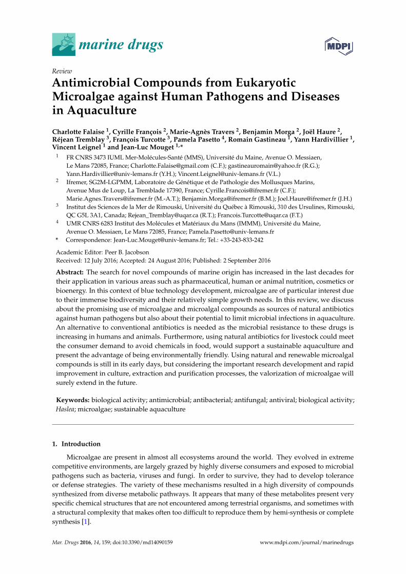
marine drugs
Review
Antimicrobial Compounds from EukaryoticMicroalgae against Human Pathogens and Diseasesin Aquaculture
Charlotte Falaise 1, Cyrille François 2, Marie-Agnès Travers 2, Benjamin Morga 2, Joël Haure 2,Réjean Tremblay 3, François Turcotte 3, Pamela Pasetto 4, Romain Gastineau 1, Yann Hardivillier 1,Vincent Leignel 1 and Jean-Luc Mouget 1,*
1 FR CNRS 3473 IUML Mer-Molécules-Santé (MMS), Université du Maine, Avenue O. Messiaen,Le Mans 72085, France; [email protected] (C.F.); [email protected] (R.G.);[email protected] (Y.H.); [email protected] (V.L.)
2 Ifremer, SG2M-LGPMM, Laboratoire de Génétique et de Pathologie des Mollusques Marins,Avenue Mus de Loup, La Tremblade 17390, France; [email protected] (C.F.);[email protected] (M.-A.T.); [email protected] (B.M.); [email protected] (J.H.)
3 Institut des Sciences de la Mer de Rimouski, Université du Québec à Rimouski, 310 des Ursulines, Rimouski,QC G5L 3A1, Canada; [email protected] (R.T.); [email protected] (F.T.)
4 UMR CNRS 6283 Institut des Molécules et Matériaux du Mans (IMMM), Université du Maine,Avenue O. Messiaen, Le Mans 72085, France; [email protected]
* Correspondence: [email protected]; Tel.: +33-243-833-242
Academic Editor: Peer B. JacobsonReceived: 12 July 2016; Accepted: 24 August 2016; Published: 2 September 2016
Abstract: The search for novel compounds of marine origin has increased in the last decades fortheir application in various areas such as pharmaceutical, human or animal nutrition, cosmetics orbioenergy. In this context of blue technology development, microalgae are of particular interest dueto their immense biodiversity and their relatively simple growth needs. In this review, we discussabout the promising use of microalgae and microalgal compounds as sources of natural antibioticsagainst human pathogens but also about their potential to limit microbial infections in aquaculture.An alternative to conventional antibiotics is needed as the microbial resistance to these drugs isincreasing in humans and animals. Furthermore, using natural antibiotics for livestock could meetthe consumer demand to avoid chemicals in food, would support a sustainable aquaculture andpresent the advantage of being environmentally friendly. Using natural and renewable microalgalcompounds is still in its early days, but considering the important research development and rapidimprovement in culture, extraction and purification processes, the valorization of microalgae willsurely extend in the future.
Keywords: biological activity; antimicrobial; antibacterial; antifungal; antiviral; biological activity;Haslea; microalgae; sustainable aquaculture
1. Introduction
Microalgae are present in almost all ecosystems around the world. They evolved in extremecompetitive environments, are largely grazed by highly diverse consumers and exposed to microbialpathogens such as bacteria, viruses and fungi. In order to survive, they had to develop toleranceor defense strategies. The variety of these mechanisms resulted in a high diversity of compoundssynthesized from diverse metabolic pathways. It appears that many of these metabolites present veryspecific chemical structures that are not encountered among terrestrial organisms, and sometimes witha structural complexity that makes often too difficult to reproduce them by hemi-synthesis or completesynthesis [1].
Mar. Drugs 2016, 14, 159; doi:10.3390/md14090159 www.mdpi.com/journal/marinedrugs
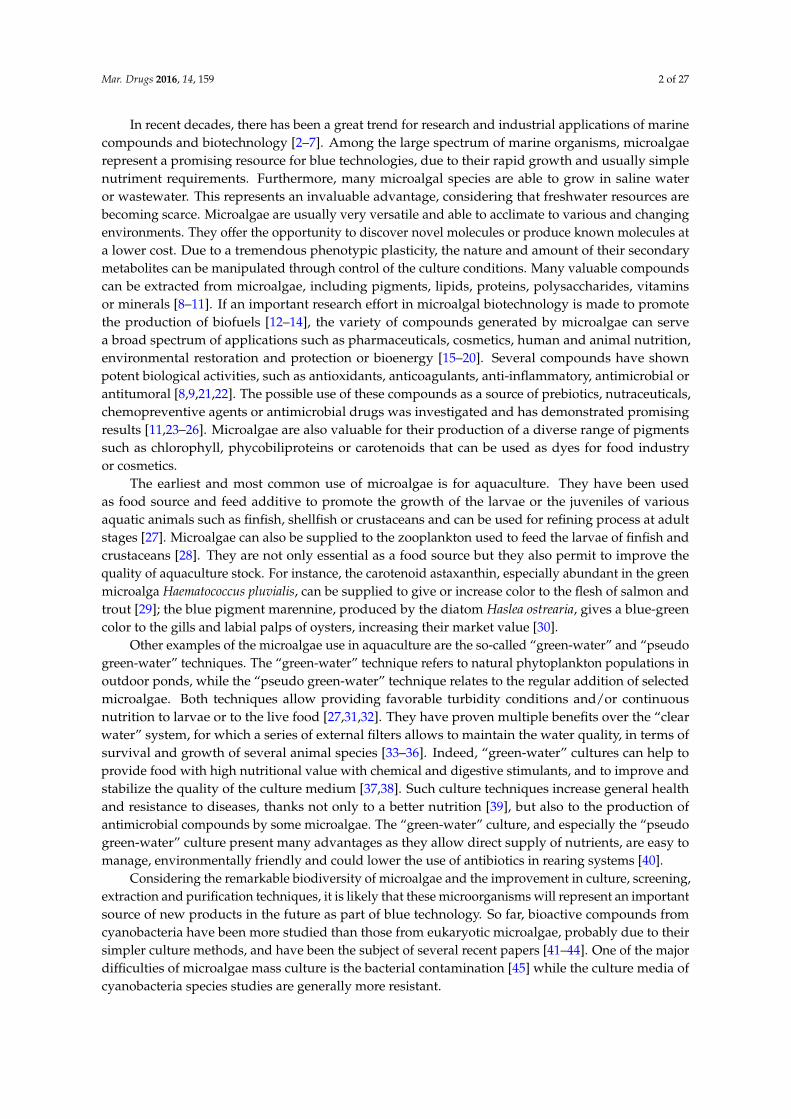
Mar. Drugs 2016, 14, 159 2 of 27
In recent decades, there has been a great trend for research and industrial applications of marinecompounds and biotechnology [2–7]. Among the large spectrum of marine organisms, microalgaerepresent a promising resource for blue technologies, due to their rapid growth and usually simplenutriment requirements. Furthermore, many microalgal species are able to grow in saline wateror wastewater. This represents an invaluable advantage, considering that freshwater resources arebecoming scarce. Microalgae are usually very versatile and able to acclimate to various and changingenvironments. They offer the opportunity to discover novel molecules or produce known molecules ata lower cost. Due to a tremendous phenotypic plasticity, the nature and amount of their secondarymetabolites can be manipulated through control of the culture conditions. Many valuable compoundscan be extracted from microalgae, including pigments, lipids, proteins, polysaccharides, vitaminsor minerals [8–11]. If an important research effort in microalgal biotechnology is made to promotethe production of biofuels [12–14], the variety of compounds generated by microalgae can servea broad spectrum of applications such as pharmaceuticals, cosmetics, human and animal nutrition,environmental restoration and protection or bioenergy [15–20]. Several compounds have shownpotent biological activities, such as antioxidants, anticoagulants, anti-inflammatory, antimicrobial orantitumoral [8,9,21,22]. The possible use of these compounds as a source of prebiotics, nutraceuticals,chemopreventive agents or antimicrobial drugs was investigated and has demonstrated promisingresults [11,23–26]. Microalgae are also valuable for their production of a diverse range of pigmentssuch as chlorophyll, phycobiliproteins or carotenoids that can be used as dyes for food industryor cosmetics.
The earliest and most common use of microalgae is for aquaculture. They have been usedas food source and feed additive to promote the growth of the larvae or the juveniles of variousaquatic animals such as finfish, shellfish or crustaceans and can be used for refining process at adultstages [27]. Microalgae can also be supplied to the zooplankton used to feed the larvae of finfish andcrustaceans [28]. They are not only essential as a food source but they also permit to improve thequality of aquaculture stock. For instance, the carotenoid astaxanthin, especially abundant in the greenmicroalga Haematococcus pluvialis, can be supplied to give or increase color to the flesh of salmon andtrout [29]; the blue pigment marennine, produced by the diatom Haslea ostrearia, gives a blue-greencolor to the gills and labial palps of oysters, increasing their market value [30].
Other examples of the microalgae use in aquaculture are the so-called “green-water” and “pseudogreen-water” techniques. The “green-water” technique refers to natural phytoplankton populations inoutdoor ponds, while the “pseudo green-water” technique relates to the regular addition of selectedmicroalgae. Both techniques allow providing favorable turbidity conditions and/or continuousnutrition to larvae or to the live food [27,31,32]. They have proven multiple benefits over the “clearwater” system, for which a series of external filters allows to maintain the water quality, in terms ofsurvival and growth of several animal species [33–36]. Indeed, “green-water” cultures can help toprovide food with high nutritional value with chemical and digestive stimulants, and to improve andstabilize the quality of the culture medium [37,38]. Such culture techniques increase general healthand resistance to diseases, thanks not only to a better nutrition [39], but also to the production ofantimicrobial compounds by some microalgae. The “green-water” culture, and especially the “pseudogreen-water” culture present many advantages as they allow direct supply of nutrients, are easy tomanage, environmentally friendly and could lower the use of antibiotics in rearing systems [40].
Considering the remarkable biodiversity of microalgae and the improvement in culture, screening,extraction and purification techniques, it is likely that these microorganisms will represent an importantsource of new products in the future as part of blue technology. So far, bioactive compounds fromcyanobacteria have been more studied than those from eukaryotic microalgae, probably due to theirsimpler culture methods, and have been the subject of several recent papers [41–44]. One of the majordifficulties of microalgae mass culture is the bacterial contamination [45] while the culture media ofcyanobacteria species studies are generally more resistant.
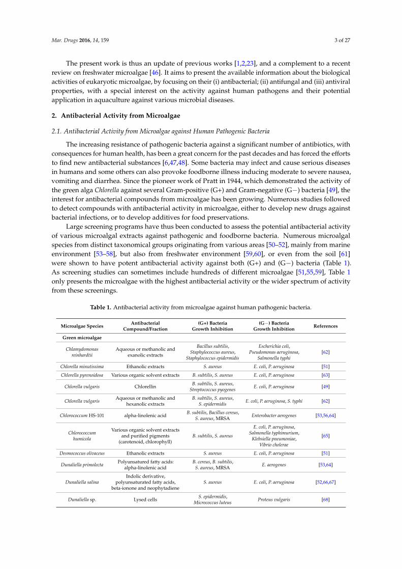
Mar. Drugs 2016, 14, 159 3 of 27
The present work is thus an update of previous works [1,2,23], and a complement to a recentreview on freshwater microalgae [46]. It aims to present the available information about the biologicalactivities of eukaryotic microalgae, by focusing on their (i) antibacterial; (ii) antifungal and (iii) antiviralproperties, with a special interest on the activity against human pathogens and their potentialapplication in aquaculture against various microbial diseases.
2. Antibacterial Activity from Microalgae
2.1. Antibacterial Activity from Microalgae against Human Pathogenic Bacteria
The increasing resistance of pathogenic bacteria against a significant number of antibiotics, withconsequences for human health, has been a great concern for the past decades and has forced the effortsto find new antibacterial substances [6,47,48]. Some bacteria may infect and cause serious diseasesin humans and some others can also provoke foodborne illness inducing moderate to severe nausea,vomiting and diarrhea. Since the pioneer work of Pratt in 1944, which demonstrated the activity ofthe green alga Chlorella against several Gram-positive (G+) and Gram-negative (G−) bacteria [49], theinterest for antibacterial compounds from microalgae has been growing. Numerous studies followedto detect compounds with antibacterial activity in microalgae, either to develop new drugs againstbacterial infections, or to develop additives for food preservations.
Large screening programs have thus been conducted to assess the potential antibacterial activityof various microalgal extracts against pathogenic and foodborne bacteria. Numerous microalgalspecies from distinct taxonomical groups originating from various areas [50–52], mainly from marineenvironment [53–58], but also from freshwater environment [59,60], or even from the soil [61]were shown to have potent antibacterial activity against both (G+) and (G−) bacteria (Table 1).As screening studies can sometimes include hundreds of different microalgae [51,55,59], Table 1only presents the microalgae with the highest antibacterial activity or the wider spectrum of activityfrom these screenings.
Table 1. Antibacterial activity from microalgae against human pathogenic bacteria.
Microalgae SpeciesAntibacterial
Compound/Fraction(G+) Bacteria
Growth Inhibition(G−) Bacteria
Growth InhibitionReferences
Green microalgae
Chlamydomonasreinhardtii
Aqueous or methanolic andexanolic extracts
Bacillus subtilis,Staphylococcus aureus,
Staphylococcus epidermidis
Escherichia coli,Pseudomonas aeruginosa,
Salmonella typhi[62]
Chlorella minutissima Ethanolic extracts S. aureus E. coli, P. aeruginosa [51]
Chlorella pyrenoidosa Various organic solvent extracts B. subtilis, S. aureus E. coli, P. aeruginosa [63]
Chlorella vulgaris ChlorellinB. subtilis, S. aureus,
Streptococcus pyogenesE. coli, P. aeruginosa [49]
Chlorella vulgarisAqueous or methanolic and
hexanolic extractsB. subtilis, S. aureus,
S. epidermidisE. coli, P. aeruginosa, S. typhi [62]
Chlorococcum HS-101 alpha-linolenic acidB. subtilis, Bacillus cereus,
S. aureus, MRSAEnterobacter aerogenes [53,56,64]
Chlorococcumhumicola
Various organic solvent extractsand purified pigments
(carotenoid, chlorophyll)B. subtilis, S. aureus
E. coli, P. aeruginosa,Salmonella typhimurium,
Klebsiella pneumoniae,Vibrio cholerae
[65]
Desmococcus olivaceus Ethanolic extracts S. aureus E. coli, P. aeruginosa [51]
Dunaliella primolectaPolyunsatured fatty acids:
alpha-linolenic acidB. cereus, B. subtilis,
S. aureus, MRSAE. aerogenes [53,64]
Dunaliella salinaIndolic derivative,
polyunsaturated fatty acids,beta-ionone and neophytadiene
S. aureus E. coli, P. aeruginosa [52,66,67]
Dunaliella sp. Lysed cellsS. epidermidis,
Micrococcus luteusProteus vulgaris [68]
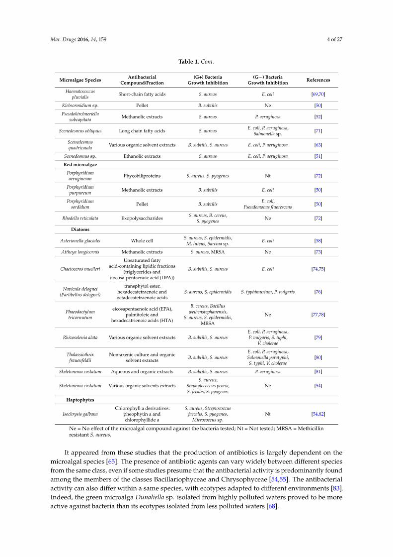
Mar. Drugs 2016, 14, 159 4 of 27
Table 1. Cont.
Microalgae SpeciesAntibacterial
Compound/Fraction(G+) Bacteria
Growth Inhibition(G−) Bacteria
Growth InhibitionReferences
Haematococcuspluvialis
Short-chain fatty acids S. aureus E. coli [69,70]
Klebsormidium sp. Pellet B. subtilis Ne [50]
Pseudokirchneriellasubcapitata
Methanolic extracts S. aureus P. aeruginosa [52]
Scenedesmus obliquus Long chain fatty acids S. aureusE. coli, P. aeruginosa,
Salmonella sp.[71]
Scenedesmusquadricauda
Various organic solvent extracts B. subtilis, S. aureus E. coli, P. aeruginosa [63]
Scenedesmus sp. Ethanolic extracts S. aureus E. coli, P. aeruginosa [51]
Red microalgae
Porphyridiumaerugineum
Phycobiliproteins S. aureus, S. pyogenes Nt [72]
Porphyridiumpurpureum
Methanolic extracts B. subtilis E. coli [50]
Porphyridiumsordidum
Pellet B. subtilisE. coli,
Pseudomonas fluorescens[50]
Rhodella reticulata ExopolysaccharidesS. aureus, B. cereus,
S. pyogenesNe [72]
Diatoms
Asterionella glacialis Whole cellS. aureus, S. epidermidis,
M. luteus, Sarcina sp.E. coli [58]
Attheya longicornis Methanolic extracts S. aureus, MRSA Ne [73]
Chaetoceros muelleri
Unsaturated fattyacid-containing lipidic fractions
(triglycerides anddocosa-pentaenoic acid (DPA))
B. subtilis, S. aureus E. coli [74,75]
Navicula delognei(Parlibellus delognei)
transphytol ester,hexadecatetraenoic andoctadecatetraenoic acids
S. aureus, S. epidermidis S. typhimurium, P. vulgaris [76]
Phaeodactylumtricornutum
eicosapentaenoic acid (EPA),palmitoleic and
hexadecatrienoic acids (HTA)
B. cereus, Bacillusweihenstephanensis,
S. aureus, S. epidermidis,MRSA
Ne [77,78]
Rhizosolenia alata Various organic solvent extracts B. subtilis, S. aureusE. coli, P. aeruginosa,P. vulgaris, S. typhi,
V. cholerae[79]
Thalassiothrixfrauenfeldii
Non-axenic culture and organicsolvent extracts
B. subtilis, S. aureusE. coli, P. aeruginosa,Salmonella paratyphi,S. typhi, V. cholerae
[80]
Skeletonema costatum Aqueous and organic extracts B. subtilis, S. aureus P. aeruginosa [81]
Skeletonema costatum Various organic solvents extractsS. aureus,
Staphylococcus peoria,S. fecalis, S. pyogenes
Ne [54]
Haptophytes
Isochrysis galbanaChlorophyll a derivatives:
pheophytin a andchlorophyllide a
S. aureus, Streptococcusfaecalis, S. pyogenes,
Micrococcus sp.Nt [54,82]
Ne = No effect of the microalgal compound against the bacteria tested; Nt = Not tested; MRSA = Methicillinresistant S. aureus.
It appeared from these studies that the production of antibiotics is largely dependent on themicroalgal species [65]. The presence of antibiotic agents can vary widely between different speciesfrom the same class, even if some studies presume that the antibacterial activity is predominantly foundamong the members of the classes Bacillariophyceae and Chrysophyceae [54,55]. The antibacterialactivity can also differ within a same species, with ecotypes adapted to different environments [83].Indeed, the green microalga Dunaliella sp. isolated from highly polluted waters proved to be moreactive against bacteria than its ecotypes isolated from less polluted waters [68].
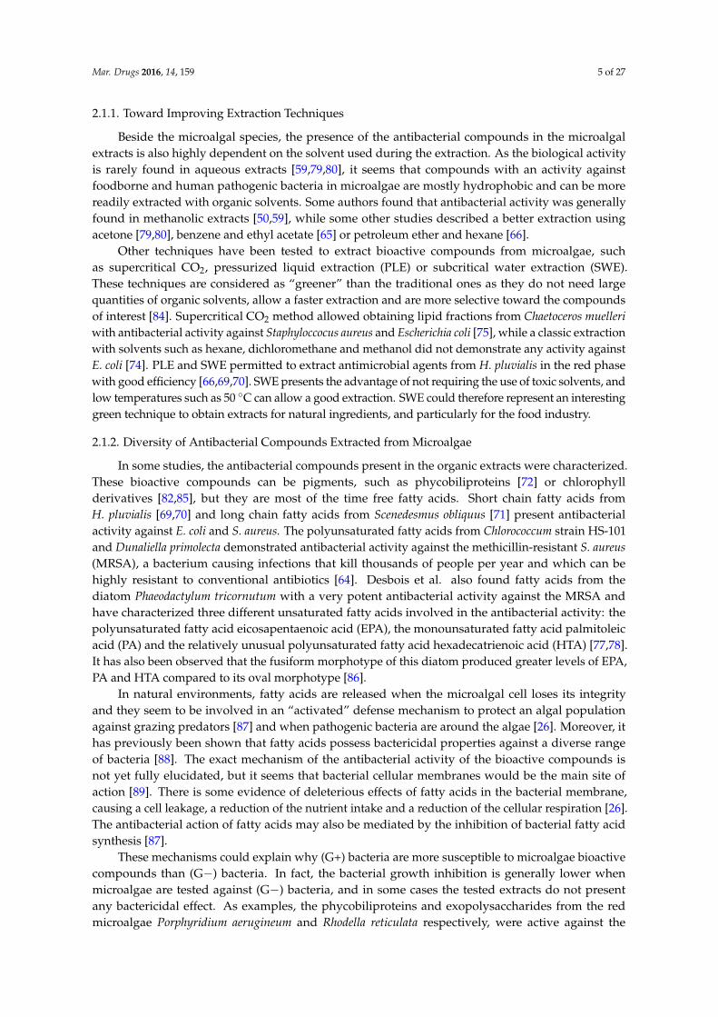
Mar. Drugs 2016, 14, 159 5 of 27
2.1.1. Toward Improving Extraction Techniques
Beside the microalgal species, the presence of the antibacterial compounds in the microalgalextracts is also highly dependent on the solvent used during the extraction. As the biological activityis rarely found in aqueous extracts [59,79,80], it seems that compounds with an activity againstfoodborne and human pathogenic bacteria in microalgae are mostly hydrophobic and can be morereadily extracted with organic solvents. Some authors found that antibacterial activity was generallyfound in methanolic extracts [50,59], while some other studies described a better extraction usingacetone [79,80], benzene and ethyl acetate [65] or petroleum ether and hexane [66].
Other techniques have been tested to extract bioactive compounds from microalgae, suchas supercritical CO2, pressurized liquid extraction (PLE) or subcritical water extraction (SWE).These techniques are considered as “greener” than the traditional ones as they do not need largequantities of organic solvents, allow a faster extraction and are more selective toward the compoundsof interest [84]. Supercritical CO2 method allowed obtaining lipid fractions from Chaetoceros muelleri
with antibacterial activity against Staphyloccocus aureus and Escherichia coli [75], while a classic extractionwith solvents such as hexane, dichloromethane and methanol did not demonstrate any activity againstE. coli [74]. PLE and SWE permitted to extract antimicrobial agents from H. pluvialis in the red phasewith good efficiency [66,69,70]. SWE presents the advantage of not requiring the use of toxic solvents, andlow temperatures such as 50 ◦C can allow a good extraction. SWE could therefore represent an interestinggreen technique to obtain extracts for natural ingredients, and particularly for the food industry.
2.1.2. Diversity of Antibacterial Compounds Extracted from Microalgae
In some studies, the antibacterial compounds present in the organic extracts were characterized.These bioactive compounds can be pigments, such as phycobiliproteins [72] or chlorophyllderivatives [82,85], but they are most of the time free fatty acids. Short chain fatty acids fromH. pluvialis [69,70] and long chain fatty acids from Scenedesmus obliquus [71] present antibacterialactivity against E. coli and S. aureus. The polyunsaturated fatty acids from Chlorococcum strain HS-101and Dunaliella primolecta demonstrated antibacterial activity against the methicillin-resistant S. aureus
(MRSA), a bacterium causing infections that kill thousands of people per year and which can behighly resistant to conventional antibiotics [64]. Desbois et al. also found fatty acids from thediatom Phaeodactylum tricornutum with a very potent antibacterial activity against the MRSA andhave characterized three different unsaturated fatty acids involved in the antibacterial activity: thepolyunsaturated fatty acid eicosapentaenoic acid (EPA), the monounsaturated fatty acid palmitoleicacid (PA) and the relatively unusual polyunsaturated fatty acid hexadecatrienoic acid (HTA) [77,78].It has also been observed that the fusiform morphotype of this diatom produced greater levels of EPA,PA and HTA compared to its oval morphotype [86].
In natural environments, fatty acids are released when the microalgal cell loses its integrityand they seem to be involved in an “activated” defense mechanism to protect an algal populationagainst grazing predators [87] and when pathogenic bacteria are around the algae [26]. Moreover, ithas previously been shown that fatty acids possess bactericidal properties against a diverse rangeof bacteria [88]. The exact mechanism of the antibacterial activity of the bioactive compounds isnot yet fully elucidated, but it seems that bacterial cellular membranes would be the main site ofaction [89]. There is some evidence of deleterious effects of fatty acids in the bacterial membrane,causing a cell leakage, a reduction of the nutrient intake and a reduction of the cellular respiration [26].The antibacterial action of fatty acids may also be mediated by the inhibition of bacterial fatty acidsynthesis [87].
These mechanisms could explain why (G+) bacteria are more susceptible to microalgae bioactivecompounds than (G−) bacteria. In fact, the bacterial growth inhibition is generally lower whenmicroalgae are tested against (G−) bacteria, and in some cases the tested extracts do not presentany bactericidal effect. As examples, the phycobiliproteins and exopolysaccharides from the redmicroalgae Porphyridium aerugineum and Rhodella reticulata respectively, were active against the
Mar. Drugs 2016, 14, 159 6 of 27
(G+) bacteria S. aureus and Streptoccocus pyogenes but presented no effect against the (G−) bacteriaE. coli and Pseudomonas aeruginosa [72]. The diatom P. tricornutum did not demonstrate antibacterialeffect against these two (G−) bacteria either, whereas a good antibacterial activity against the (G+)MRSA was observed [78]. Thus, the difference in sensitivities between bacteria may be due to theircomplex membrane permeability, making the penetration and the bactericidal action of the compoundmore difficult.
The potent activity of microalgal compounds, especially free fatty acids, against various bacteriastraightens further development in the search for drugs and food preservatives from microalgae.Bacterial resistance to free fatty acids has not been encountered yet [90,91], so their exploitation inmedicine deserves to be further investigated [78]. Furthermore, as consumers tend to avoid syntheticadditives, microalgae could be good candidates as natural sources against food-borne pathogens.
2.2. Use of Microalgae against Pathogenic Bacteria in Aquaculture
Bacteria are nowadays considered as main causes of infections in intensive aquaculture of finfishand shellfish worldwide [92], occurring as well in hatcheries and nurseries as in rear and grow-outponds. These infections can lead to serious mass mortalities and imply considerable economiclosses. A wide range of bacteria are known to infect farmed and wild species with minor to severeconsequences on health and survival. Among the important bacterial diseases in finfish we can citethose caused by Aeromonas sp. and Pseudomonas sp., inducing hemorrhages in many fresh-waterspecies, or Vibrio sp. inducing vibrioses in marine fish species [93]. Vibrio are able to infect a widevariety of hosts [94] and are also the most common and harmful shrimp pathogenic bacteria [95],with luminous species such as Vibrio harveyi involved in mass mortalities in shrimp hatcheries [96].Vibrio sp. have also been demonstrated to be involved in a large number of massive mortalities inbivalve hatcheries [97–101].
Antibiotics have been largely used in intensive aquaculture, as well for finfish [102,103],shrimp [104–107] or shellfish cultures [108,109]. However, as for humans and terrestrial animals,bacterial resistance in aquaculture is increasing and most antibiotics are less effective [110–112].The presence of drug residues in tissues of aquatic animals and the risk of transferring resistantbacteria to humans have led to a great concern about the use of antibiotics for public health [113].A few antibiotics are now licensed in aquaculture due to the establishment of strict regulations by theEuropean Conformity (EC) or the Food and Drug Administration (FDA), such as the regulation (EC)No 470/2009 of the European Parliament and the Council of 6 May 2009 laying down Communityprocedures for the establishment of residue limits of pharmacologically active substances in foodstuffsof animal origin. Various alternative and natural compounds are available today to control aquaticpathogenic bacteria, mainly derived from plants. They present the advantages of decreasing theside effects observed with synthetic antibiotics, being less expensive [114]. In this context, severalmicroalgae species have been investigated for their antibacterial activity in vitro, and in co-culturewith pathogenic bacteria, and some studies have also been conducted in vivo with the “green water”technique and using microalgae as food supplements (Table 2).
Table 2. Antibacterial activity from microalgae against diseases in aquaculture.
Microalgae Species Compound/Fraction Tested Target Bacteria/Antibacterial Effect References
In vitro experiments
Chaetoceros lauderi Whole cell Vibrio anguillarum, Aeromonas salmonicida [58]
Dunaliella tertiolecta Aqueous extract Vibrio campbellii [115]
Euglena viridis Organic solvent extracts
Aeromonas hydrophila, Edwardsiella tarda, Pseudomonasaeruginosa, Pseudomonas fluorescens, Pseudomonasputida Vibrio alginolyticus, V. anguillarum, Vibriofluvialis, Vibrio harveyi, Vibrio parahaemolyticus
[116]
Haslea karadagensisPurified pigment (intra- and
extracellular forms)Polaribacter irgensii, Pseudoalteromonas elyakowii,
Vibrio aestuarianus[117]
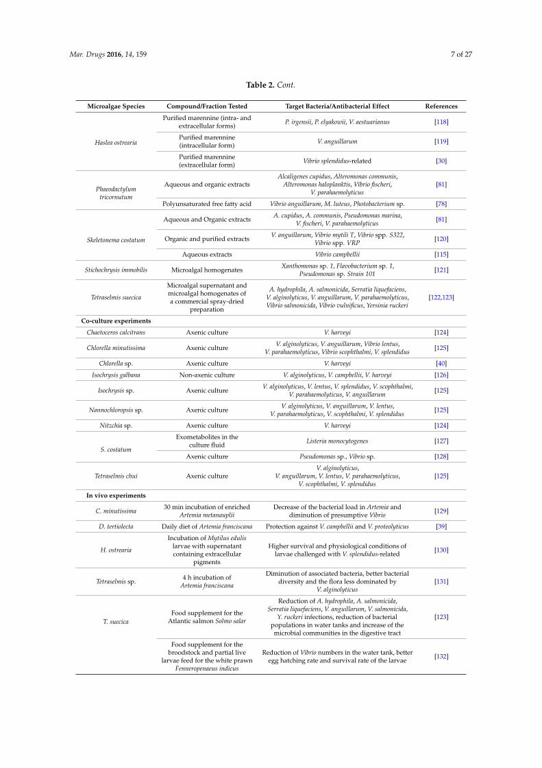
Mar. Drugs 2016, 14, 159 7 of 27
Table 2. Cont.
Microalgae Species Compound/Fraction Tested Target Bacteria/Antibacterial Effect References
Haslea ostrearia
Purified marennine (intra- andextracellular forms)
P. irgensii, P. elyakowii, V. aestuarianus [118]
Purified marennine(intracellular form)
V. anguillarum [119]
Purified marennine(extracellular form)
Vibrio splendidus-related [30]
Phaeodactylumtricornutum
Aqueous and organic extractsAlcaligenes cupidus, Alteromonas communis,
Alteromonas haloplanktis, Vibrio fischeri,V. parahaemolyticus
[81]
Polyunsaturated free fatty acid Vibrio anguillarum, M. luteus, Photobacterium sp. [78]
Skeletonema costatum
Aqueous and Organic extractsA. cupidus, A. communis, Pseudomonas marina,
V. fischeri, V. parahaemolyticus[81]
Organic and purified extractsV. anguillarum, Vibrio mytili T, Vibrio spp. S322,
Vibrio spp. VRP[120]
Aqueous extracts Vibrio campbellii [115]
Stichochrysis immobilis Microalgal homogenatesXanthomonas sp. 1, Flavobacterium sp. 1,
Pseudomonas sp. Strain 101[121]
Tetraselmis suecica
Microalgal supernatant andmicroalgal homogenates ofa commercial spray-dried
preparation
A. hydrophila, A. salmonicida, Serratia liquefaciens,V. alginolyticus, V. anguillarum, V. parahaemolyticus,Vibrio salmonicida, Vibrio vulnificus, Yersinia ruckeri
[122,123]
Co-culture experiments
Chaetoceros calcitrans Axenic culture V. harveyi [124]
Chlorella minutissima Axenic cultureV. alginolyticus, V. anguillarum, Vibrio lentus,
V. parahaemolyticus, Vibrio scophthalmi, V. splendidus[125]
Chlorella sp. Axenic culture V. harveyi [40]
Isochrysis galbana Non-axenic culture V. alginolyticus, V. campbellii, V. harveyi [126]
Isochrysis sp. Axenic cultureV. alginolyticus, V. lentus, V. splendidus, V. scophthalmi,
V. parahaemolyticus, V. anguillarum[125]
Nannochloropsis sp. Axenic cultureV. alginolyticus, V. anguillarum, V. lentus,
V. parahaemolyticus, V. scophthalmi, V. splendidus[125]
Nitzchia sp. Axenic culture V. harveyi [124]
S. costatum
Exometabolites in theculture fluid
Listeria monocytogenes [127]
Axenic culture Pseudomonas sp., Vibrio sp. [128]
Tetraselmis chui Axenic cultureV. alginolyticus,
V. anguillarum, V. lentus, V. parahaemolyticus,V. scophthalmi, V. splendidus
[125]
In vivo experiments
C. minutissima30 min incubation of enriched
Artemia metanaupliiDecrease of the bacterial load in Artemia and
diminution of presumptive Vibrio[129]
D. tertiolecta Daily diet of Artemia franciscana Protection against V. campbellii and V. proteolyticus [39]
H. ostrearia
Incubation of Mytilus edulislarvae with supernatantcontaining extracellular
pigments
Higher survival and physiological conditions oflarvae challenged with V. splendidus-related
[130]
Tetraselmis sp.4 h incubation of
Artemia franciscana
Diminution of associated bacteria, better bacterialdiversity and the flora less dominated by
V. alginolyticus[131]
T. suecica
Food supplement for theAtlantic salmon Solmo salar
Reduction of A. hydrophila, A. salmonicida,Serratia liquefaciens, V. anguillarum, V. salmonicida,
Y. ruckeri infections, reduction of bacterialpopulations in water tanks and increase of themicrobial communities in the digestive tract
[123]
Food supplement for thebroodstock and partial live
larvae feed for the white prawnFenneropenaeus indicus
Reduction of Vibrio numbers in the water tank, betteregg hatching rate and survival rate of the larvae
[132]
Mar. Drugs 2016, 14, 159 8 of 27
2.2.1. Benefits of the “Green-Water” Technique against Bacterial Diseases in Aquaculture
The “green water” technique has demonstrated beneficial effects on health, survival rates andresistance of different organisms [32–37,39]. Addition of the microalga Isochrysis galbana alloweda better viability and a faster grow of the turbot Scophthalmus maximus larvae as well as a lowerproliferation of opportunistic bacteria [133]. The green microalga Tetraselmis suecica first showed goodantibacterial activity in vitro against several Vibrio species [122] and then proved to reduce in vivothe number of various bacterial species in the water tank of the Atlantic salmon Salmo salar [123].Adding T. suecica also reduced the number of Vibrio species in broodstock gut, eggs and larvae ofthe white prawn Fenneropenaeus indicus, resulting in improved egg hatching and larval survival [132].A better survival and physiological conditions of the blue mussel Mytilus edulis larvae and the scallopPlacopecten magellanicus larvae were observed when incubated with supernatant containing marennine,the extracellular blue pigment of the diatom H. ostrearia, at concentrations as low as 0.1 mg/L [130].The use of the diatoms Skeletonema costatum and Chaetoceros calcitrans to feed Penaeus monodon larvaeallowed a growth inhibition of the luminous bacteria Vibrio harveyi [134]. However, this seemed mainlydue to the microbial flora associated with the diatoms rather than their metabolic products. Indeed,the effectiveness of the “green water” culture in preventing bacterial infections and outbreaks canalso be attributed to the presence of antibacterial factors in the bacterial, fungal and phytoplanktonmicrobiota associated with this culture technique [124].
Yet, several co-culture experiments with axenic microalgae demonstrated the ability of somespecies to produce and release compounds with potent activity against pathogenic bacteria.Axenic cultures of Chlorella minutissima, Tetraselmis chui, Isochrysis sp. and Nannochloropsis sp. limitedthe growth of various Vibrio species [125], and growth of the luminous bacteria V. harveyi highlydecreased when co-cultured with pure Chlorella sp. [40], C. calcitrans or Nitzschia sp. [124].
Finally, genetically modified microalgae were also used in vivo. In a recent study, better growth,resistance to bacteria and survival rate were observed in the shrimp postlarvae P. monodon fed with“fusant” Chlorella and Dunaliella [135]. The so-called “fusant” microalgae resulted from the generation ofa unique cell through somatic hybridization by fusion of the two protoplasts. This fusion technology isespecially used to transfer agronomically useful traits to plants [136] and would allow an improvementof valuable metabolites production from these two microalgae. Another genetically modified microalgatested in “green water” systems is the transgenic line of Nannochloropsis oculata, able to producethe bovine antimicrobial lactoferricin (LFB) peptide. These LFB-containing transgenic microalgaewere developed and tested as food supplement for the medaka fish Oryzias latipes infected withVibrio parahaemolyticus, which displayed a significantly better survival rate after 24 h [137].
2.2.2. Microalgae to Improve the Live-Food Quality
Microalgae also show advantages for the live-food quality [39,138], by reducing the numberof associated pathogenic bacteria such as Vibrio and allowing a lower risk of transmission to fishlarvae. Daily addition of Dunaliella tertiolecta to feed the brine shrimp Artemia franciscana, consideredas an essential part of the live food chain for the culture of fish, conferred a full protection againstVibrio campbellii and Vibrio proteolyticus [39]. A 4 h incubation of A. franciscana with the microalgaeTetraselmis sp. resulted in a diminution by 75% of associated bacteria, with a better bacterial diversityand the flora less dominated by Vibrio alginolyticus [131]. Similar observations were made byMakridis et al. with incubation of Artemia with T. chui and C. minutissima [129]. These authors proposedthat the reduction of Vibrio cells in Artemia cultures could either be due to compounds released bymicroalgae or to (G+) associated bacteria.
2.2.3. In Vitro Efficiency of Microalgal Compounds against Marine Bacteria
Several in vitro studies demonstrated the ability of various microalgae to produce antibacterialcompounds effective against relevant marine pathogenic bacteria. The whole cells of Chaetoceros
Mar. Drugs 2016, 14, 159 9 of 27
lauderi [58], supernatant and homogenates of T. suecica [122,123] or homogenates from Stichochrysis
immobilis [121] induced a growth inhibition of various marine bacteria. However, Berland et al. notedthat limited attention should be paid to antibacterial activity obtained with broken cells, as in naturalconditions substances synthesized have to be released into the water to target another organism [121].Microalgae producing antibacterial compounds that are not released in the medium cannot indeedbe considered gainful in “green water” techniques, but these compounds can be highly useful in thedesign of novel drugs.
Only a few compounds with activity against marine bacteria have been characterized, suchas the polyunsaturated free fatty acid in P. tricornutum, identified as eicosapentaenoic acid [78].An antibacterial activity was also demonstrated in vitro with the blue pigment of the two diatomsH. ostrearia and Haslea karadagensis. These blue pigments can be observed in the apex of the microalgae(intracellular form) but can also be released in the medium (extracellular form). The purified forms ofthese pigments [139] have been shown to inhibit the growth of several marine bacteria including Vibrio
such bacteria belonging to Vibrio splendidus clade or Vibrio aestuarianus species [30,117–119], involvedin oysters mass mortality [140–142].
A series of experiments were conducted to assess the spectrum of activity of purified extracellularmarennine, the pigment produced by H. ostrearia, against pathogenic Vibrio species. The growth ofV. splendidus-related strain (Vibrio tasmaniensis LGP32 (CIP107715)) was slowed down when exposed toa concentration of marennine up to 10 µg per mL and seemed totally inhibited with a concentration of1 mg per mL (Figure 1).
Figure 1. Growth inhibition of Vibrio tasmaniensis CIP 107715 by purified marennine, the blue pigmentproduced by Haslea ostrearia. (a) V. tasmaniensis was grown over night at 25 ◦C, cells were then washedwith sterile water 2 times and adjusted to an OD600 = 0.5. Cells were added to wells with marennine inthe following concentrations: 0, 0.1, 1, 10, 100 and 1000 µg per mL. Kinetics were run at OD600 for 48 h,with measurements taken every 30 min (n = 3); (b) Relative values were graphed in order to account forthe absorbance differences due to the pigment. The effective concentration reducing bacteria growthrate by 50%, EC50, was estimated at 19.14 µg per mL (Standard error: 6.73) (original results).
In another series of experiments, a comparative approach combining several species and strainswas chosen to reflect the diversity of strains within species. The sensitivity of various strains recognizedfor their virulence, of V. aestuarianus [143], Vibrio coralliilyticus [144] and Vibrio tubiashii [145] toextracellular marennine purified as previously described [139] was assessed by exposing the bacteriato marennine concentration ranging from 1 to 100 µg per mL. For all the three species, after 48 h, thehigher the marennine concentration, the higher was the bacterial growth inhibition (data not shown),confirming the antibacterial activity of the pigment produced by the diatom H. ostrearia. Moreover,when comparing different ecotypes within a same Vibrio species, the sensitivity of the strains exposedto marennine could be significantly different (Figure 2). It can be noted that the strains coming fromcollections, used to describe the species and often isolated besides mortality events, seemed moresensitive to marennine comparing to virulent strains. Complementary experiments are under way in
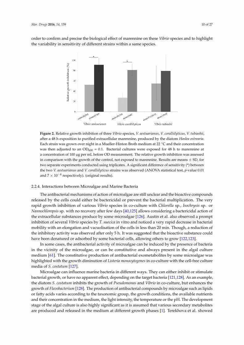
Mar. Drugs 2016, 14, 159 10 of 27
order to confirm and precise the biological effect of marennine on these Vibrio species and to highlightthe variability in sensitivity of different strains within a same species.
Figure 2. Relative growth inhibition of three Vibrio species, V. aestuarianus, V. coralliilyticus, V. tubiashii,after a 48 h exposition to purified extracellular marennine, produced by the diatom Haslea ostrearia.
Each strain was grown over night in a Mueller-Hinton Broth medium at 22 ◦C and their concentrationwas then adjusted to an OD600 = 0.1. Bacterial cultures were exposed for 48 h to marennine ata concentration of 100 µg per mL before OD measurement. The relative growth inhibition was assessedin comparison with the growth of the control, not exposed to marennine. Results are means ± SD, fortwo separate experiments conducted using triplicates. A significant difference of sensitivity (*) betweenthe two V. aestuarianus and V. coralliilyticus strains was observed (ANOVA statistical test, p-value 0.01and 7 × 10⁻⁴ respectively). (original results).
2.2.4. Interactions between Microalgae and Marine Bacteria
The antibacterial mechanisms of action of microalgae are still unclear and the bioactive compoundsreleased by the cells could either be bactericidal or prevent the bacterial multiplication. The veryrapid growth inhibition of various Vibrio species in co-culture with Chlorella sp., Isochrysis sp. orNannochloropsis sp. with no recovery after few days [40,125] allows considering a bactericidal action ofthe extracellular substances produce by some microalgae [126]. Austin et al. also observed a promptinhibition of several Vibrio species by T. suecica in vitro and noticed a very rapid decrease in bacterialmobility with an elongation and vacuolisation of the cells in less than 20 min. Though, a reduction ofthe inhibitory activity was observed after only 5 h. It was suggested that the bioactive substance couldhave been denatured or adsorbed by some bacterial cells, allowing others to grow [122,123].
In some cases, the antibacterial activity of microalgae can be induced by the presence of bacteriain the vicinity of the microalgae, or can be constitutive and always present in the algal culturemedium [61]. The constitutive production of antibacterial exometabolites by some microalgae washighlighted with the growth diminution of Listeria monocytogenes in co-culture with the cell-free culturemedia of S. costatum [127].
Microalgae can influence marine bacteria in different ways. They can either inhibit or stimulatebacterial growth, or have no apparent effect, depending on the target bacteria [121,128]. As an example,the diatom S. costatum inhibits the growth of Pseudomonas and Vibrio in co-culture, but enhances thegrowth of Flavobacterium [128]. The production of antibacterial compounds by microalgae such as lipidsor fatty acids varies according to the taxonomic group, the growth conditions, the available nutrientsand their concentration in the medium, the light intensity, the temperature or the pH. The developmentstage of the algal culture is also highly significant as it is assumed that various secondary metabolitesare produced and released in the medium at different growth phases [1]. Terekhova et al. showed
Mar. Drugs 2016, 14, 159 11 of 27
that only the exometabolites produced by S. costatum during the middle steady-state growth phasepresented an antibacterial activity against L. monocytogenes while compounds released during theexponential growth phase had no effect on these bacteria [127]. Cooper et al. have also demonstratedthe direct relation between cell growth phase and antibacterial activity, and showed that P. tricornutum
presented a better activity against a wide spectrum of marine bacteria during the exponential growthphase compared to the stationary phase, while the reverse relationship was found for S. costatum [81].
3. Antifungal Activity from Microalgae
3.1. Antifungal Activity from Microalgae against Human Pathogens
The search for antifungal compounds from microalgae started much later than screening forantibacterial activity. As a matter of fact, fungi have been considered as harmful human pathogenssince the 1970s, when mortality induced by fungal infections and the frequency of nosocomialmycoses increased in hospitalized patients. Increase of fungal infections was mainly due to therapiesthat depress patients’ immune system such as the use of intensive and aggressive chemotherapyregimens, the expansion of organ transplant programs and the spread of the AIDS epidemic. [146,147].The incidence of invasive aspergillosis (induced by Aspergillus species), and the number of cases ofcandidemia (an infection and a disease caused by Candida species), have been rising inexorablyfrom that time [148,149]. The growing use of antifungal agents in recent years has led to thedevelopment of drug resistance [150,151]. Thus, there is a need for novel drugs and several studieswere recently conducted to find fungicide activity from natural marine products against humanpathogenic fungi [152–157], including antifungal agents from microalgae (Table 3).
There are fewer screening activities for microalgal fungicides than for bactericides, andmost of the studies focus not only on the antifungal activity but also on antibacterialactivities [50,51,58,60,63,65,72,80,117,158,159]. As for antibacterial activity, antifungal activity varieswidely depending on microalgal species, type of solvent used to extract the compound and themicroorganism tested. It does not seem to be a taxonomic trend for the antifungal activity, and thecapability to produce antifungal compounds would have evolved independently of phylogeneticrelationship in microalgae [50]. However, Pesando et al. noticed a significant activity of the genusChaetoceros [160] and Kellam et al. also indicated that marine microalgae showed more potential in thesearch for new antifungal agents than freshwater species [161].
Table 3. Antifungal activity from microalgae.
Microalgae Species Antifungal Compounds/Fraction Target Microorganims References
Green microalgae
Chlorella vulgaris Microalgal supernatantYeast: Candida kefyr
Mold: Aspergillus fumigatus,Aspergillus niger
[62]
Chlorococcum humicolaOrganic solvent extracts and
pigments: beta carotene,Chlorophyll a and Chlorophyll b
Yeast: C. albicansMold: A. flavus, A. niger
[65]
Heterochlorella luteoviridis Microalgal supernatant Yeast: C. albicans [50]
Haematococcus pluvialis Short-chain fatty acids Yeast: C. albicans [70]
Scenedesmus quadricauda Organic solvent extracts
Yeast: C. albicans, S. cerevisiaeMold: A. flavus, A. niger, P. herqueiOther: A. brassicae, F. moniliforme,
Helminthosporium sp.
[63]
Red microalgae
Porphyridium aerugineum Phycobiliproteins Yeast: C. albicans [72]
Porphyridium purpureum Microalgal supernatant Yeast: C. albicans [50]
Rhodella reticulata Exopolysaccharides Yeast: C. albicans [72]
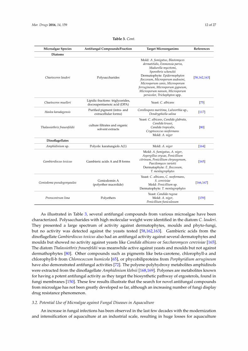
Mar. Drugs 2016, 14, 159 12 of 27
Table 3. Cont.
Microalgae Species Antifungal Compounds/Fraction Target Microorganims References
Diatoms
Chaetoceros lauderi Polysaccharides
Mold: A. fumigatus, Blastomycesdermatitidis, Emmonsia parva,
Madurella mycetomi,Sporothrix schenckii
Dermatophyte: Epidermophytonfloccosum, Microsporum audouini,Microsporum canis, Microsporum
ferrugineum, Microsporum gypseum,Microsporum nanum, Microsporum
persicolor, Trichophyton spp.
[58,162,163]
Chaetoceros muelleriLipidic fractions: triglycerides,docosapentaenoic acid (DPA)
Yeast: C. albicans [75]
Haslea karadagensisPurified pigment (intra- and
extracellular forms)Corollospora maritima, Lulworthia sp.,
Dendryphiella salina[117]
Thalassiothrix frauenfeldiiculture filtrates and organic
solvent extracts
Yeast: C. albicans, Candida glabrata,Candida krusei,
Candida tropicalis,Cryptococcus neoformans
Mold: A. niger
[80]
Dinoflagellates
Amphidinium sp. Polyols: karatungiols A(1) Mold: A. niger [164]
Gambierdiscus toxicus Gambieric acids A and B forms
Mold: A. fumigatus, A. niger,Aspergillus oryzae, Penicillium
citrinum, Penicillium chrysogenum,Paecilomyces variotii
Dermatophyte: E. floccosum,T. mentagrophytes
[165]
Goniodoma pseudogonyaulaxGoniodomin A
(polyether macrolide)
Yeast: C. albicans, C. neoformans,S. cerevisiae
Mold: Penicillium sp.Dermatophyte: T. mentagrophytes
[166,167]
Prorocentrum lima PolyethersYeast: Candida rugosa
Mold: A. niger,Penicillium funiculosum
[159]
As illustrated in Table 3, several antifungal compounds from various microalgae have beencharacterized. Polysaccharides with high molecular weight were identified in the diatom C. lauderi.They presented a large spectrum of activity against dermatophytes, moulds and phyto-fungi,but no activity was detected against the yeasts tested [58,162,163]. Gambieric acids from thedinoflagellate Gambierdiscus toxicus also had an antifungal activity against several dermatophytes andmoulds but showed no activity against yeasts like Candida albicans or Saccharomyces cerevisiae [165].The diatom Thalassiothrix frauenfeldii was meanwhile active against yeasts and moulds but not againstdermathophytes [80]. Other compounds such as pigments like beta-carotene, chlorophyll-a andchlorophyll-b from Chlorococcum humicola [65], or phycobiliproteins from Porphyridium aerugineum
have also demonstrated antifungal activities [72]. The polyene-polyhydroxy metabolites amphidinolswere extracted from the dinoflagellate Amphidinium klebsii [168,169]. Polyenes are metabolites knownfor having a potent antifungal activity as they target the biosynthetic pathway of ergosterols, found infungi membranes [150]. These few results illustrate that the search for novel antifungal compoundsfrom microalgae has not been greatly developed so far, although an increasing number of fungi displaydrug resistance phenomenon.
3.2. Potential Use of Microalgae against Fungal Diseases in Aquaculture
An increase in fungal infections has been observed in the last few decades with the modernizationand intensification of aquaculture at an industrial scale, resulting in huge losses for aquaculture
Mar. Drugs 2016, 14, 159 13 of 27
industries. Fungal infections in aquaculture may cause severe diseases and mortality events leadingto economic losses. They are often considered secondary to other factors or pathogens such asconsequences of water quality problems, fish trauma by rough handling or temperature shock, bacterialdiseases or parasites [170]. Several fungi are known to induce diseases by developing in the skin andthe gills of the infected fish or in eggs, or by producing toxins [171]. Indeed, mycotoxins can provokemany disorders and can accumulate in fish tissues, representing a risk for public health [172].
Chemicals used to treat infected animals are limited and, due to the increasing resistance offungi against conventional drugs, environmental legislations and consumer's safety, the alternative“herbal formulations” and alternative safe and cheap methods have become of renewed interest [173].Studies have been conducted in order to find new antimycotics of natural origin such as plantextracts [174–176] or essential oils [177,178], which should have no harmful effect on fish, fish eggs,human health and ecosystems.
So far, very few studies have been conducted to assess the antifungal activity from microalgaeagainst pathogenic fungi in aquaculture systems (Table 3). Gastineau et al. have demonstrated in vitrothe antifungal activity of the pigment produced by the diatom H. karadagensis against three marinefungal species Corollospora maritima, Dendryphiella salina, Lulworthia sp., which can be involved inthe phenomenon of biofouling [117]. Organic extracts of the green microalgae Chlorella pyrenoidosa
and Scenedesmus quadricauda have demonstrated antifungal activity against Fusarium monofiliform [63]reported of causing black gill disease in shrimps [179]. This result is of great interest as Fusarium sp.were recently shown to produce toxins that accumulate in fish [180]. Aspergillus fumigatus is alsoa fungus susceptible to produce toxins and it thus represents a potential contaminant for seafood,particularly for marine bivalves [181]. It can be inhibited by compounds produced by C. lauderi [58],G. toxicus [165], and Chlorella vulgaris [62].
More generally, fungi from the taxa Fusarium and Aspergillus are found in many countries inwater, sediments and marine invertebrates, and their presence in shellfish farming areas evidencesthe necessity to pay attention to shellfish contamination by such fungi [182]. Furthermore, reports offungi causing deleterious effects are frequently related [183,184], which encourage a larger screeningof microalgae for the production of novel antifungal compounds.
4. Antiviral Activity from Microalgae
4.1. Antiviral Activity from Microalgae against Pathogenic Human Viruses
Viral pathogens are the leading cause of human diseases and mortality worldwide. Treatments toblock the entry of the virus or its replication directly are difficult to design, as they can have adverseeffects on the infected host cells [185]. Thus, treatments against diseases caused by viruses arelimited, and resistance to these available treatments demonstrate the need for new medicines [186].Many drugs exhibiting selective inhibition of mammalian originate from synthetic organic chemicals orfrom natural products, for instance secondary metabolites in plants [185]. Along with the developmentof “blue” technology and extraction improvement, there is a growing interest in marine-derivedantiviral compounds. Thus, thousands of compounds from various marine organisms such as algae,bacteria, fungi, marine invertebrates or sponges have been screened [153,186–188] and some of themhave demonstrated antiviral activities and are commercially available.
Potential antiviral activity from algal compounds has been first demonstrated in the 1950sby Gerber et al. who observed that the polysaccharides extracted from Gelidium cartilagenium
afforded protection for embryonated eggs against influenza B and mumps viruses [189], and the firstbrood-based studies of seaweeds for their antiviral substances started in the 1970s [190]. Screenings forantiviral compounds from macroalgae are still predominant [191–195], but the interest for antiviralcompounds from microalgae and cyanobacteria rapidly increased as they also present relevantantiviral activities and are easier to culture. Cyanobacteria are promising sources of antiviralcompounds, and their simple growth needs make them good candidates for the production of antiviral
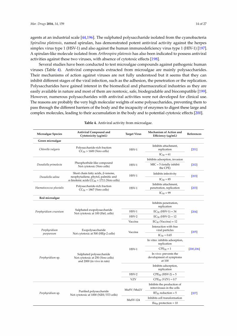
Mar. Drugs 2016, 14, 159 14 of 27
agents at an industrial scale [44,196]. The sulphated polysaccharide isolated from the cyanobacteriaSpirulina platensis, named spirulan, has demonstrated potent antiviral activity against the herpessimplex virus type 1 (HSV-1) and also against the human immunodeficiency virus type 1 (HIV-1) [197].A spirulan-like molecule isolated from Arthrospira platensis has also been indicated to possess antiviralactivities against these two viruses, with absence of cytotoxic effects [198].
Several studies have been conducted to test microalgae compounds against pathogenic humanviruses (Table 4). Antiviral compounds extracted from microalgae are mainly polysaccharides.Their mechanisms of action against viruses are not fully understood but it seems that they caninhibit different stages of the viral infection, such as the adhesion, the penetration or the replication.Polysaccharides have gained interest in the biomedical and pharmaceutical industries as they areeasily available in nature and most of them are nontoxic, safe, biodegradable and biocompatible [199].However, numerous polysaccharides with antiviral activities were not developed for clinical use.The reasons are probably the very high molecular weights of some polysaccharides, preventing them topass through the different barriers of the body and the incapacity of enzymes to digest these large andcomplex molecules, leading to their accumulation in the body and to potential cytotoxic effects [200].
Table 4. Antiviral activity from microalgae.
Microalgae SpeciesAntiviral Compound and
Cytotoxicity (µg/mL)Target Virus
Mechanism of Action andEfficiency (µg/mL)
References
Green microalgae
Chlorella vulgarisPolysaccharide-rich fraction
CC50 > 1600 (Vero cells) HSV-1Inhibits attachment,
replication [201]
IC50 = 61
Dunaliella primolectaPheophorbide-like compound
Not cytotoxic (Vero cells) HSV-1
Inhibits adsorption, invasion
[202]MIC = 5 (totally inhibitthe CPE)
Dunaliella salina
Short chain fatty acids, β-ionone,neophytadiene, phytol, palmitic and
α-linolenic acids CC50 = 1711 (Vero cells)
HSV-1Inhibits infectivity
[203]IC50 = 85
Haematococcus pluvialisPolysaccharide-rich fraction
CC50 = 1867 (Vero cells) HSV-1Inhibits attachment,
penetration, replication [203]
IC50 = 99
Red microalgae
Porphyridium cruentumSulphated exopolysaccharide
Not cytotoxic at 100 (HeL cells)
Inhibits penetration,replication
[204]HSV-1 EC50 (HSV-1) = 34
HSV-2 EC50 (HSV-2) = 12
Vaccina EC50 (Vaccina) = 12
Porphyridiumpurpureum
ExopolysaccharideNot cytotoxic at 500 (HEp-2 cells) Vaccina
Interaction with freeviral particles
[205]IC50 = 0.65
Porphyridium sp.Sulphated polysaccharide
Not cytotoxic at 250 (Vero cells)and 2000 (in vivo in rats)
HSV-1
In vitro: inhibits adsorption,replication
[200,206]CPE50 = 1
In vivo: prevents thedevelopment of symptoms
at 100
Inhibits adsorption,replication
HSV-2 CPE50 (HSV-2) = 5
VZV CPE50 (VZV) = 0.7
Porphyridium sp.Purified polysaccharide
Not cytotoxic at 1000 (NIH/3T3 cells)
MuSV/MuLV
Inhibits the production ofretroviruses in the cells
[207]RT50 reduction = 5
MuSV-124Inhibits cell transformation
ffu50 protection = 10
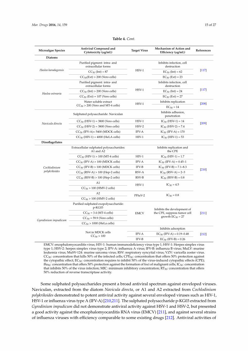
Mar. Drugs 2016, 14, 159 15 of 27
Table 4. Cont.
Microalgae SpeciesAntiviral Compound and
Cytotoxicity (µg/mL)Target Virus
Mechanism of Action andEfficiency (µg/mL)
References
Diatoms
Haslea karadagensis
Purified pigment: intra- andextracellular forms
HSV-1
Inhibits infection, celldestruction
[117]CC50 (Int) = 87 EC50 (Int) = 62
CC50(Ext) > 200 (Vero cells) EC50 (Ext) = 23
Haslea ostrearia
Purified pigment: intra- andextracellular forms
HSV-1
Inhibits infection, celldestruction
[117]CC50 (Int) > 200 (Vero cells) EC50 (Int) = 24
CC50 (Ext) = 107 (Vero cells) EC50 (Ext) = 27
Water soluble extractCC50 > 200 (Vero and MT-4 cells) HSV-1
Inhibits replication[208]
EC50 = 14
Navicula directa
Sulphated polysaccharide: NaviculanInhibits adhesion,
penetration
[209]CC50 (HSV-1) = 3800 (Vero cells) HSV-1 IC50 (HSV-1) = 14
CC50 (HSV-2) = 3800 (Vero cells) HSV-2 IC50 (HSV-2) = 7.4
CC50 (IFV-A)= 5400 (MDCK cells) IFV-A IC50 (IFV-A) = 170
CC50 (HIV-1) = 4000 (HeLA cells) HIV-1 IC50 (HIV-1) = 53
Dinoflagellates
Cochlodiniumpolykrikoides
Extracellular sulphated polysaccharides:A1 and A2
Inhibits replication andthe CPE
[210]
CC50 (HIV-1) > 100 (MT-4 cells) HIV-1 IC50 (HIV-1) = 1.7
CC50 (IFV-A) > 100 (MDCK cells) IFV-A IC50 (IFV-A) = 0.45–1
CC50 (IFV-B) > 100 (MDCK cells) IFV-B IC50 (IFV-B) = 7.1–8.3
CC50 (RSV-A) > 100 (Hep-2 cells) RSV-A IC50 (RSV-A) = 2–3
CC50 (RSV-B) > 100 (Hep-2 cells) RSV-B IC50 (RSV-B) = 0.8
A1HSV-1 IC50 = 4.5
CC50 > 100 (HMV-2 cells)
A2PFluV-2 IC50 = 0.8
CC50 > 100 (HMV-2 cells)
Gyrodinium impudicum
Purified sulphated exopolysaccharide:p-KG03
EMCVInhibits the development ofthe CPE, suppress tumor cell
growth EC50 = 27[211]CC50 = 3.4 (MT-4 cells)
CC50 = 59.9 (Vero cells)
CC50 > 1000 (HeLa cells)
Not in MDCK cellsCC50 > 100
Inhibits adsorption
[212]IFV-A EC50 (IFV-A) = 0.19–0.48
IFV-B EC50 (IFV-B) = 0.26
EMCV: encephalomyocarditis virus; HIV-1: human immunodeficiency virus type 1; HSV-1: Herpes simplex virustype 1; HSV-2: herpes simplex virus type 2; IFV-A: influenza A virus; IFV-B: influenza B virus; MuLV: murineleukemia virus; MuSV-124: murine sarcoma virus; RSV: respiratory syncytial virus; VZV: varicella zoster virus.CC50: concentration that kills 50% of the infected cells; CPE50: concentration that offers 50% protection againstthe cytopathic effect; EC50: concentration requires to inhibit 50% of the virus-induced cytopathic effects (CPE);ffu50: concentration that offers 50% protection against the formation of foci of malignant cells; IC50: concentrationthat inhibits 50% of the virus infection; MIC: minimum inhibitory concentration; RT50: concentration that offers50% reduction of reverse transcriptase activity.
Some sulphated polysaccharides present a broad antiviral spectrum against enveloped viruses.Naviculan, extracted from the diatom Navicula directa, or A1 and A2 extracted from Cochlodinium
polykrikoides demonstrated to potent antiviral activity against several enveloped viruses such as HIV-1,HSV-1 or influenza virus type A (IFV-A) [210,211]. The sulphated polysaccharide p-KG03 extracted fromGyrodinium impudicum did not demonstrate antiviral activity against HSV-1 and HSV-2, but presenteda good activity against the encephalomyocarditis RNA virus (EMCV) [211], and against several strainsof influenza viruses with efficiency comparable to some existing drugs [212]. Antiviral activities of

Mar. Drugs 2016, 14, 159 16 of 27
microalgae against HSV type 1 and 2 are the most studied [117,118,200–203,206,208–210]. More thanone third of the world population is affected by HSV-1 or HSV-2, infections that cause contagiousdiseases such as oral and genital herpes [187]. The efficiency and the low toxicity of some of themicroalgal compounds tested attest their advantageous use as antiviral agents. They could help tocontrol viral diseases occurring in humans, but also in animal species with economic value.
4.2. Potential Use of Microalgae against Viruses in Aquaculture
Aquaculture production undergoes numerous viral diseases, which can affect organism healthand survival rates, and can sometimes lead to mass mortality. Viral diseases are spreading and areconsequently important limiting factors for the expansion of aquaculture. Many different viruses,from various virus families, are known to infect farmed species, such as finfish, crustaceans ormolluscs [213–215]. As a few relevant examples, we can cite the infectious pancreatic necrosis virus(IPNV), isolated from a very wide host range among finfish [215], the yellow head virus (YHV)and the white spot syndrome virus (WSSV) causing important losses in shrimp culture, or theostreid herpesvirus-1 (OsHV-1) leading to high mortality in marine bivalves [216] and which can betransmitted between different bivalve species [217]. It appears that diseases induced by RNA virusesare the highest cause of ecological and socio-economic impacts in European farmed finfish [218].Viruses in aquaculture species are either established for decades or are newly emerging because of theintensification of farming practices that facilitates rapid transmission of diseases.
Viral diseases in aquaculture are challenging to manage. They are difficult to treat directly anda few, if any, efficacious treatments are available other than destroying all organisms in infected farmsand avoiding their movements to and from infected areas. In some particular cases, vaccination isused in farmed finfish, mainly to treat trout and salmon [218,219]. The vaccination issue did notarise in invertebrates, as it was widely assumed that, unlike vertebrates, they do not possess thecapacity to develop long-term acquired immunity. Nevertheless, there are evidences for specificimmune memory in some invertebrates [220] such as crustaceans [221], and several studies havedemonstrated the antiviral protection of shrimp by “vaccination” [222,223]. A recent work has alsodemonstrated the presence of an antiviral system in oysters after injection of a synthetic viral analogue(Poly I:C) against OsHV-1. This immune response showed similarity with the vertebrate interferonresponse pathway [224]. The lack of marine mollusc cell culture not only restricts virus isolationcapacities and subsequent characterization work, but also limits investigations on host-virus interaction.However, recent progress has been made with the development of stem cells in the cupped oystersCrassostrea gigas [225].
Plant and herbal extracts with activity against viral diseases in aquaculture production haverecently been reviewed by Sivasankar et al. [226]. Some extracts have already been successfullytested in vivo, such as the plant extract of Cyanodon dactylon against the WSS virus of the shrimpP. monodon [227]. Plant extracts, acting as immunostimulants, have the advantage of being easilydelivered by oral administration and may be eco-friendly as they are biodegradable.
In contrast, very few studies have been conducted to assess the antiviral activity of microalgaeagainst viruses in aquaculture. Some polysaccharide extracts of various microalgae have been testedagainst the viral hemorrhagic septicaemia virus (VHSV) [228], a virus of economic importance afflictingover 50 species of fresh water and marine fish including salmonid fish [215]. Endocellular extractsof Porphyridium cruentum, D. tertiolecta, Ellipsoidon sp., Isochrysis galbana var. Tiso and Chlorella
autotrophica inhibited the viral infection of VHSV in vitro in epithelioma papulosum cyprinid (EPC)cells. Concentrations lower than 2 µg of extracts per mL of P. cruentum and D. tertiolecta were sufficientto detect an antiviral activity. Exocellular extracts of these algal species were also able to inhibit the viralinfection, except for I. galbana var. Tiso. This study has also demonstrated that there is no correlationbetween the content of sulphated polysaccharides of each microalga and its capacity to inhibit the viralreplication. Thus, the observed antiviral effects would be due to different polysaccharide molecularspecies with differences in molecular size.
Mar. Drugs 2016, 14, 159 17 of 27
A higher resistance to the WSSV was observed in the tiger shrimp P. menodon reared in a “greenwater” system using commercially available extracts of Dunaliella salina [35]. Culture bath treatmentswith the microalga C. minutissima significantly reduced the mortality of Epinephelus marginatus showingsigns of Viral Encephalopathy and Retinopathy (VER) [229]. These results indicate that the control ofthe disease is probably due to the antiviral effect of C. minutissima cultures, which thus needs to befurther investigated using in vitro testing of water-soluble algal extracts.
Viral diseases are increasingly spreading, causing great economic losses for aquaculture industries.No effective treatment has yet been developed, as vaccination possibility is limited and chemicaldrugs are gradually avoided because of their toxicity and potential residue accumulation. Plant andherbal extracts have shown potent antiviral activity against several pathogenic viruses in aquaculture.They could be promising candidates for antiviral agents as part of an environmentally friendly andsustainable aquaculture, although their production and delivery processes could be limiting factors.In this context, the use of microalgae as a source of antiviral agents should be further studied, aswater-soluble compounds present in algal supernatants could be a valuable alternative.
5. Conclusions and Final Remarks
The diversity of microalgae is immense, with species, genera or even classes being discoveredevery year. On the estimated millions of existing species, about 30,000 have been described, but onlya dozen are cultivated in a large scale for biotechnological applications. The main obstacle for theircommercial exploitation remains the production cost, but it should be bypassed by the optimizationof mass culturing conditions. Microalgae are promising source of high-value products, and theirapplication as antimicrobials is only in its onset. The development of novel drugs with no microbialresistance and the use of environmentally friendly antibiotics in a context of sustainable aquacultureare needed. The efficiency of various microalgal compounds against human or aquatic pathogens isvery encouraging and there is no doubt that their exploitation and application will expand.
Acknowledgments: The authors acknowledge H.T. Badawy for her help with Vibrio splendidus experiments, theRegion Pays de la Loire and Le Mans Centre for Technology Transfer for facilities. This publication benefitedfrom funding from the European Commission under the Community’s Seventh Framework Program BIOVADIA(contract No. FP7-PEOPLE-2010-IRSES-269294, Biodiversity and Valorisation of Blue Diatoms).
Conflicts of Interest: The authors declare no conflict of interest.
References
1. Borowitzka, M.A. Microalgae as sources of pharmaceuticals and other biologically active compounds.J. Appl. Phycol. 1995, 7, 3–15. [CrossRef]
2. Bhatnagar, I.; Kim, S.-K. Immense essence of excellence: Marine microbial bioactive compounds. Mar. Drugs
2010, 8, 2673–2701. [CrossRef] [PubMed]3. Dyshlovoy, S.A.; Honecker, F. Marine compounds and cancer: Where do we stand? Mar. Drugs 2015, 13,
5657–5665. [CrossRef] [PubMed]4. Jha, R.K.; Zi-rong, X. Biomedical compounds from marine organisms. Mar. Drugs 2004, 2, 123–146.5. Kang, H.K.; Seo, C.H.; Park, Y. Marine peptides and their anti-infective activities. Mar. Drugs 2015, 13,
618–654. [CrossRef] [PubMed]6. Mayer, A.M.S.; Rodríguez, A.D.; Taglialatela-Scafati, O.; Fusetani, N. Marine Pharmacology in
2009–2011: Marine compounds with antibacterial, antidiabetic, antifungal, anti-inflammatory, antiprotozoal,antituberculosis, and antiviral activities; affecting the immune and nervous systems, and other miscellaneousmechanisms of action. Mar. Drugs 2013, 11, 2510–2573. [PubMed]
7. Suleria, H.A.R.; Osborne, S.; Masci, P.; Gobe, G. Marine-based nutraceuticals: An innovative trend in thefood and supplement industries. Mar. Drugs 2015, 13, 6336–6351. [CrossRef] [PubMed]
8. Encarnacao, T.; Pais, A.A.; Campos, M.G.; Burrows, H.D. Cyanobacteria and microalgae: A renewable sourceof bioactive compounds and other chemicals. Sci. Prog. 2015, 98, 145–168. [CrossRef] [PubMed]
9. Singh, S.; Kate, B.N.; Banerjee, U.C. Bioactive compounds from cyanobacteria and microalgae: An overview.Crit. Rev. Biotechnol. 2005, 25, 73–95. [CrossRef] [PubMed]
Mar. Drugs 2016, 14, 159 18 of 27
10. Shimizu, Y. Microalgal metabolites. Curr. Opin. Microbiol. 2003, 6, 236–243. [CrossRef]11. Talero, E.; García-Mauriño, S.; Ávila-Román, J.; Rodríguez-Luna, A.; Alcaide, A.; Motilva, V. Bioactive compounds
isolated from microalgae in chronic inflammation and cancer. Mar. Drugs 2015, 13, 6152–6209. [CrossRef][PubMed]
12. Ahmad, A.L.; Yasin, N.H.M.; Derek, C.J.C.; Lim, J.K. Microalgae as a sustainable energy source for biodieselproduction: A review. Renew. Sustain. Energy Rev. 2011, 15, 584–593. [CrossRef]
13. Ghasemi, Y.; Rasoul-Amini, S.; Naseri, A.T.; Montazeri-Najafabady, N.; Mobasher, M.A.; Dabbagh, F.Microalgae biofuel potentials (Review). Appl. Biochem. Microbiol. 2012, 48, 126–144. [CrossRef]
14. Halim, R.; Danquah, M.K.; Webley, P.A. Extraction of oil from microalgae for biodiesel production: A review.Biotechnol. Adv. 2012, 30, 709–732. [CrossRef] [PubMed]
15. Barra, L.; Chandrasekaran, R.; Corato, F.; Brunet, C. The challenge of ecophysiological biodiversity forbiotechnological applications of marine microalgae. Mar. Drugs 2014, 12, 1641–1675. [CrossRef] [PubMed]
16. Cadoret, J.-P.; Garnier, M.; Saint-Jean, B. Microalgae, functional genomics and biotechnology. Adv. Bot. Res.
2012, 64, 285–341.17. Priyadarshani, I.; Biswajit, R. Commercial and industrial applications of micro algae—A review. J. Algal
Biomass Utln. 2012, 3, 89–100.18. De Jesus Raposo, M.F.; de Morais, R.M.S.C.; de Morais, A.M.M.B. Health applications of bioactive compounds
from marine microalgae. Life Sci. 2013, 93, 479–486. [CrossRef] [PubMed]19. Spolaore, P.; Joannis-Cassan, C.; Duran, E.; Isambert, A. Commercial applications of microalgae.
J. Biosci. Bioeng. 2006, 101, 87–96. [CrossRef] [PubMed]20. Yaakob, Z.; Ali, E.; Zainal, A.; Mohamad, M.; Takriff, M.S. An overview: Biomolecules from microalgae for
animal feed and aquaculture. J. Biol. Res. 2014, 21, 1–10. [CrossRef] [PubMed]21. De Jesus Raposo, M.F.; de Morais, R.M.S.C.; Bernardo de Morais, A.M.M. Bioactivity and applications of
sulphated polysaccharides from marine microalgae. Mar. Drugs 2013, 11, 233–252. [CrossRef] [PubMed]22. De Jesus Raposo, M.F.; de Morais, A.M.B.; de Morais, R.M.S.C. Marine polysaccharides from algae with
potential biomedical applications. Mar. Drugs 2015, 13, 2967–3028. [CrossRef] [PubMed]23. Amaro, H.M.; Guedes, A.C.; Malcata, F.X. Chapter: Antimicrobial activities of microalgae: An invited review.
In Science against Microbial Pathogens: Communicating Current Research and Technological Advances; FormatexMicrobiology Book Series; Mendez-Vilas, A., Ed.; Formatex Research Center: Badajoz, Spain, 2011; Volume 2.
24. De Jesus Raposo, M.F.; de Morais, A.M.M.B.; de Morais, R.M.S.C. Emergent sources of prebiotics: Seaweedsand microalgae. Mar. Drugs 2016, 14, 27. [CrossRef] [PubMed]
25. De Jesus Raposo, M.F.; de Morais, A.M.M.B.; de Morais, R.M.S.C. Carotenoids from marine microalgae:A valuable natural source for the prevention of chronic diseases. Mar. Drugs 2015, 13, 5128–5155. [CrossRef][PubMed]
26. Smith, V.J.; Desbois, A.P.; Dyrynda, E.A. Conventional and unconventional antimicrobials from fish, marineinvertebrates and micro-algae. Mar. Drugs 2010, 8, 1213–1262. [CrossRef] [PubMed]
27. Muller-Feuga, A. The role of microalgae in aquaculture: Situation and trends. J. Appl. Phycol. 2000, 12,527–534. [CrossRef]
28. Borowitzka, M.A. Microalgae for aquaculture: Opportunities and constraints. J. Appl. Phycol. 1997, 9,393–401. [CrossRef]
29. Lorenz, R.T.; Cysewski, G.R. Commercial potential for Haematococcus microalgae as a natural sourceof astaxanthin. Trends Biotechnol. 2000, 18, 160–167. [CrossRef]
30. Gastineau, R.; Turcotte, F.; Pouvreau, J.-B.; Morançais, M.; Fleurence, J.; Windarto, E.; Prasetiya, F.S.;Arsad, S.; Jaouen, P.; Babin, M.; et al. Marennine, promising blue pigments from a widespread Haslea diatomspecies complex. Mar. Drugs 2014, 12, 3161–3189. [CrossRef] [PubMed]
31. Neori, A. “Green water” microalgae: The leading sector in world aquaculture. J. Appl. Phycol. 2010, 23,143–149. [CrossRef]
32. Papandroulakis, N.; Divanach, P.; Anastasiadis, P.; Kentouri, M. The pseudo-green water technique forintensive rearing of sea bream (Sparus aurata) larvae. Aquac. Int. 2001, 9, 205–216. [CrossRef]
33. Naas, K.E.; N˦sss, T.; Harboe, T. Enhanced first feeding of halibut larvae (Hippoglossus hippoglossus L.) ingreen water. Aquaculture 1992, 105, 143–156. [CrossRef]
34. Reitan, K.I.; Rainuzzo, J.R.; Øie, G.; Olsen, Y. A review of the nutritional effects of algae in marine fish larvae.Aquaculture 1997, 155, 207–221. [CrossRef]
Mar. Drugs 2016, 14, 159 19 of 27
35. Supamattaya, K.; Kiriratnikom, S.; Boonyaratpalin, M.; Borowitzka, L. Effect of a Dunaliella extract ongrowth performance, health condition, immune response and disease resistance in black tiger shrimp(Penaeus monodon). Aquaculture 2005, 248, 207–216. [CrossRef]
36. Van der Meeren, T.; Mangor-Jensen, A.; Pickova, J. The effect of green water and light intensity on survival,growth and lipid composition in Atlantic cod (Gadus morhua) during intensive larval rearing. Aquaculture
2007, 265, 206–217. [CrossRef]37. Cahu, C.L.; Zambonino Infante, J.L.; Péres, A.; Quazuguel, P.; Le Gall, M.M. Algal addition in sea bass
(Dicentrarchus labrax) larvae rearing: Effect on digestive enzymes. Aquaculture 1998, 161, 479–489. [CrossRef]38. Palmer, P.J.; Burke, M.J.; Palmer, C.J.; Burke, J.B. Developments in controlled green-water larval culture
technologies for estuarine fishes in Queensland, Australia and elsewhere. Aquaculture 2007, 272, 1–21.[CrossRef]
39. Marques, A.; Thanh, T.H.; Sorgeloos, P.; Bossier, P. Use of microalgae and bacteria to enhance protection ofgnotobiotic Artemia against different pathogens. Aquaculture 2006, 258, 116–126. [CrossRef]
40. Tendencia, E.A.; dela Peña, M. Investigation of some components of the greenwater system which makes iteffective in the initial control of luminous bacteria. Aquaculture 2003, 218, 115–119. [CrossRef]
41. Burja, A.M.; Banaigs, B.; Abou-Mansour, E.; Grant Burgess, J.; Wright, P.C. Marine cyanobacteria—A prolificsource of natural products. Tetrahedron 2001, 57, 9347–9377. [CrossRef]
42. Costa, M.; Costa-Rodrigues, J.; Fernandes, M.H.; Barros, P.; Vasconcelos, V.; Martins, R. Marine cyanobacteriacompounds with anticancer properties: A review on the implication of apoptosis. Mar. Drugs 2012, 10,2181–2207. [CrossRef] [PubMed]
43. Gupta, V.; Ratha, S.K.; Sood, A.; Chaudhary, V.; Prasanna, R. New insights into the biodiversity andapplications of cyanobacteria (blue-green algae)—Prospects and challenges. Algal Res. 2013, 2, 79–97. [CrossRef]
44. Vijayakumar, S.; Menakha, M. Pharmaceutical applications of cyanobacteria—A review. J. Acute Med. 2015,5, 15–23. [CrossRef]
45. Deschênes, J.-S.; Boudreau, A.; Tremblay, R. Mixotrophic production of microalgae in pilot-scalephotobioreactors: Practicability and process considerations. Algal Res. 2015, 10, 80–86. [CrossRef]
46. Pradhan, J.; Das, S.; Das, B.K. Antibacterial activity of freshwater microalgae: A review. Afr. J. Pharm. Pharmacol.
2014, 8, 809–818.47. Jin, L.; Quan, C.; Hou, X.; Fan, S. Potential Pharmacological Resources: Natural Bioactive Compounds from
Marine-Derived Fungi. Mar. Drugs 2016, 14, 76. [CrossRef] [PubMed]48. Shannon, E.; Abu-Ghannam, N. Antibacterial Derivatives of Marine Algae: An Overview of Pharmacological
Mechanisms and Applications. Mar. Drugs 2016, 14, 81. [CrossRef] [PubMed]49. Pratt, R.; Daniels, T.C.; Eiler, J.J.; Gunnison, J.B.; Kumler, W.D.; Oneto, J.F.; Strait, L.A.; Spoehr, H.A.;
Hardin, G.J.; Milner, H.W.; et al. Chlorellin, an antibacterial substance from Chlorella. Science 1944, 99,351–352. [CrossRef] [PubMed]
50. Mudimu, O.; Rybalka, N.; Bauersachs, T.; Born, J.; Friedl, T.; Schulz, R. Biotechnological screening ofmicroalgal and cyanobacterial strains for biogas production and antibacterial and antifungal effects.Metabolites 2014, 4, 373–393. [CrossRef] [PubMed]
51. Ördög, V.; Stirk, W.A.; Lenobel, R.; Bancírová, M.; Strnad, M.; van Staden, J.; Szigeti, J.; Németh, L.Screening microalgae for some potentially useful agricultural and pharmaceutical secondary metabolites.J. Appl. Phycol. 2004, 16, 309–314. [CrossRef]
52. Pane, G.; Cacciola, G.; Giacco, E.; Mariottini, G.L.; Coppo, E. Assessment of the antimicrobial activity ofalgae extracts on bacteria responsible of external otitis. Mar. Drugs 2015, 13, 6440–6452. [CrossRef] [PubMed]
53. Chang, T.; Ohta, S.; Ikegami, N.; Miyata, H.; Kashimoto, T.; Kondo, M. Antibiotic substances produced bya marine green alga, Dunaliella primolecta. Bioresour. Technol. 1993, 44, 149–153. [CrossRef]
54. Duff, D.C.B.; Bruce, D.L.; Antia, N.J. The Antibacterial Activity of Marine Planktonic Algae. Can. J. Microbiol.
1966, 12, 877–884. [CrossRef] [PubMed]55. Kellam, S.J.; Walker, J.M. Antibacterial activity from marine microalgae in laboratory culture. Br. Phycol. J.
1989, 24, 191–194. [CrossRef]56. Ohta, S.; Chang, T.; Ikegami, N.; Kondo, M.; Miyata, H. Antibiotic substance produced by a newly isolated
marine microalga, Chlorococcum HS-101. Bull. Environ. Contam. Toxicol. 1993, 50, 171–178. [CrossRef] [PubMed]57. Srinivasakumar, K.P.; Rajashekhar, M. In vitro studies on bactericidal activity and sensitivity pattern of
isolated marine microalgae against selective human bacterial pathogens. Indian J. Sci. Technol. 2009, 2, 16–23.
Mar. Drugs 2016, 14, 159 20 of 27
58. Viso, A.C.; Pesando, D.; Baby, C. Antibacterial and antifungal properties of some marine diatoms in culture.Bot. Mar. 1987, 30, 41–46. [CrossRef]
59. Cannell, R.J.P.; Owsianka, A.M.; Walker, J.M. Results of a large-scale screening programme to detectantibacterial activity from freshwater algae. Br. Phycol. J. 1988, 23, 41–44. [CrossRef]
60. Katircioglu, H.; Beyatli, Y.; Aslim, B.; Yüksekdag, Z.; Atici, T. Screening for antimicrobial agent production ofsome microalgae in freshwater. Internet J. Microbiol. 2005, 2, 1–5.
61. Safonova, E.; Reisser, W. Growth promoting and inhibiting effects of extracellular substances of soilmicroalgae and cyanobacteria on Escherichia coli and Micrococcus luteus. Phycol. Res. 2005, 53, 189–193. [CrossRef]
62. Ghasemi, Y.; Moradian, A.; Mohagheghzadeh, A.; Shokravi, S.; Morowvat, M.H. Antifungal and antibacterialactivity of the microalgae collected from paddy fields of Iran: Characterization of antimicrobial activity ofChroococcus dispersus. J. Biol. Sci. 2007, 7, 904–910.
63. Abedin, R.; Taha, H. Antibacterial and antifungal activity of cyanobacteria and green microalgae. Evaluationof medium components by Plackett-Burman design for antimicrobial activity of Spirulina platensis. Glob. J.
Biotechnol. Biochem. 2008, 3, 22–31.64. Ohta, S.; Shiomi, Y.; Kawashima, A.; Aozasa, O.; Nakao, T.; Nagate, T.; Kitamura, K.; Miyata, H. Antibiotic effect
of linolenic acid from Chlorococcum strain HS-101 and Dunaliella primolecta on methicillin-resistantStaphylococcus aureus. J. Appl. Phycol. 1995, 7, 121–127. [CrossRef]
65. Bhagavathy, S.; Sumathi, P.; Jancy Sherene Bell, I. Green algae Chlorococcum humicola—A new source ofbioactive compounds with antimicrobial activity. Asian Pac. J. Trop. Biomed. 2011, 1, S1–S7. [CrossRef]
66. Herrero, M.; Ibáñez, E.; Cifuentes, A.; Reglero, G.; Santoyo, S. Dunaliella salina microalga pressurized liquidextracts as potential antimicrobials. J. Food Prot. 2006, 69, 2471–2477. [PubMed]
67. Mendiola, J.A.; Santoyo, S.; Cifuentes, A.; Reglero, G.; Ibáñez, E.; Señoráns, F.J. Antimicrobial activity of sub-and supercritical CO2 extracts of the green alga Dunaliella salina. J. Food Prot. 2008, 71, 2138–2143. [PubMed]
68. Lustigman, B. Comparison of antibiotic production from four ecotypes of the marine alga, Dunaliella.Bull. Environ. Contam. Toxicol. 1988, 40, 18–22. [CrossRef] [PubMed]
69. Rodríguez-Meizoso, I.; Jaime, L.; Santoyo, S.; Señoráns, F.J.; Cifuentes, A.; Ibáñez, E. Subcritical waterextraction and characterization of bioactive compounds from Haematococcus pluvialis microalga. J. Pharm.
Biomed. Anal. 2010, 51, 456–463. [CrossRef] [PubMed]70. Santoyo, S.; Rodríguez-Meizoso, I.; Cifuentes, A.; Jaime, L.; García-Blairsy Reina, G.; Señorans, F.J.; Ibáñez, E.
Green processes based on the extraction with pressurized fluids to obtain potent antimicrobials fromHaematococcus pluvialis microalgae. LWT—Food Sci. Technol. 2009, 42, 1213–1218. [CrossRef]
71. Guedes, A.C.; Barbosa, C.R.; Amaro, H.M.; Pereira, C.I.; Malcata, F.X. Microalgal and cyanobacterial cellextracts for use as natural antibacterial additives against food pathogens. Int. J. Food Sci. Technol. 2011, 46,862–870. [CrossRef]
72. Najdenski, H.M.; Gigova, L.G.; Iliev, I.I.; Pilarski, P.S.; Lukavský, J.; Tsvetkova, I.V.; Ninova, M.S.;Kussovski, V.K. Antibacterial and antifungal activities of selected microalgae and cyanobacteria. Int. J. Food
Sci. Technol. 2013, 48, 1533–1540. [CrossRef]73. Ingebrigtsen, R.A.; Hansen, E.; Andersen, J.H.; Eilertsen, H.C. Light and temperature effects on bioactivity
in diatoms. J. Appl. Phycol. 2015, 28, 939–950. [CrossRef] [PubMed]74. Del Pilar Sánchez-Saavedra, M.; Licea-Navarro, A.; Bernáldez-Sarabia, J. Evaluation of the antibacterial
activity of different species of phytoplankton. Rev. Biol. Mar. Oceanogr. 2010, 45, 531–536. [CrossRef]75. Mendiola, J.A.; Torres, C.F.; Toré, A.; Martín-Álvarez, P.J.; Santoyo, S.; Arredondo, B.O.; Señoráns, F.J.;
Cifuentes, A.; Ibáñez, E. Use of supercritical CO2 to obtain extracts with antimicrobial activity fromChaetoceros muelleri microalga. A correlation with their lipidic content. Eur. Food Res. Technol. 2007,224, 505–510. [CrossRef]
76. Findlay, J.A.; Patil, A.D. Antibacterial constituents of the diatom Navicula delognei. J. Nat. Prod. 1984, 47,815–818. [CrossRef] [PubMed]
77. Desbois, A.P.; Lebl, T.; Yan, L.; Smith, V. Isolation and structure characterization of two antibacterial freefatty acids from marine diatom, Phaeodactylum tricornutum. Appl. Microbiol. Biotechnol. 2008, 81, 755–764.[CrossRef] [PubMed]
78. Desbois, A.P.; Mearns-Spragg, A.; Smith, V.J. A fatty acid from the diatom Phaeodactylum tricornutum isantibacterial against diverse bacteria including multi-resistant Staphylococcus aureus (MRSA). Mar. Biotechnol.
2008, 11, 45–52. [CrossRef] [PubMed]
Mar. Drugs 2016, 14, 159 21 of 27
79. Venkatesan, R.; Karthikayen, R.; Periyanayagi, R.; Sasikala, V.; Balasubramanian, T. Antibacterial activity ofthe marine diatom, Rhizosolenia alata (Brightwell, 1858) against human pathogens. Res. J. Microbiol. 2007, 2,98–100.
80. Walter, C.S.; Mahesh, R. Antibacterial and antifungal activities of some marine diatoms in culture. Indian J.
Mar. Sci. 2000, 29, 238–242.81. Cooper, S.; Battat, A.; Marsot, P.; Sylvestre, M. Production of antibacterial activities by two Bacillariophyceae
grown in dialysis culture. Can. J. Microbiol. 1983, 29, 338–341. [CrossRef]82. Bruce, D.L.; Duff, D.C.; Antia, N.J. The identification of two antibacterial products of the marine planktonic
alga Isochrysis galbana. Microbiology 1967, 48, 293–298. [CrossRef] [PubMed]83. Pawlik-Skowronska, B. Resistance, accumulation and allocation of zinc in two ecotypes of the green alga
Stigeoclonium tenue Kütz. coming from habitats of different heavy metal concentrations. Aquat. Bot. 2003,75, 189–198. [CrossRef]
84. Herrero, M.; Mendiola, J.A.; Plaza, M.; Ibañez, E. Screening for Bioactive Compounds from Algae. In Advanced
Biofuels and Bioproducts; Lee, J.W., Ed.; Springer: New York, NY, USA, 2013; pp. 833–872.85. Jørgensen, E.G. Antibiotic substances from cells and culture solutions of unicellular algae with special
reference to some chlorophyll derivatives. Physiol. Plant. 1962, 15, 530–545. [CrossRef]86. Desbois, A.P.; Walton, M.; Smith, V.J. Differential antibacterial activities of fusiform and oval morphotypes of
Phaeodactylum tricornutum (Bacillariophyceae). J. Mar. Biol. Assoc. UK 2010, 90, 769–774. [CrossRef]87. Jüttner, F. Liberation of 5,8,11,14,17-Eicosapentaenoic Acid and Other Polyunsaturated Fatty Acids from
Lipids as a Grazer Defense Reaction in Epilithic Diatom Biofilms. J. Phycol. 2001, 37, 744–755. [CrossRef]88. Kabara, J.J.; Swieczkowski, D.M.; Conley, A.J.; Truant, J.P. Fatty acids and derivatives as antimicrobial agents.
Antimicrob. Agents Chemother. 1972, 2, 23–28. [CrossRef] [PubMed]89. Galbraith, H.; Miller, T.B. Physicochemical effects of long chain fatty acids on bacterial cells and
their protoplasts. J. Appl. Bacteriol. 1973, 36, 647–658. [CrossRef] [PubMed]90. Sun, C.Q.; O’Connor, C.J.; Roberton, A.M. Antibacterial actions of fatty acids and monoglycerides against
Helicobacter pylori. FEMS Immunol. Med. Microbiol. 2003, 36, 9–17. [CrossRef]91. Petschow, B.W.; Batema, R.P.; Ford, L.L. Susceptibility of Helicobacter pylori to bactericidal properties of
medium-chain monoglycerides and free fatty acids. Antimicrob. Agents Chemother. 1996, 40, 302–306. [PubMed]92. Austin, B. Infectious Disease in Aquaculture: Prevention and Control; Woodhead Publishing: Cambridge, UK, 2012.93. Austin, B.; Austin, D.A. Bacterial Fish Pathogens: Disease of Farmed and Wild Fish; Springer Science &
Business Media: Berlin, Germany, 2007.94. Vandenberghe, J.; Thompson, F.L.; Gomez-Gil, B.; Swings, J. Phenotypic diversity amongst Vibrio isolates
from marine aquaculture systems. Aquaculture 2003, 219, 9–20. [CrossRef]95. Aguirre-Guzmán, G.; Mejia Ruíz, H.; Ascencio, F. A review of extracellular virulence product of Vibrio
species important in diseases of cultivated shrimp. Aquac. Res. 2004, 35, 1395–1404. [CrossRef]96. Austin, B.; Zhang, X.-H. Vibrio harveyi: A significant pathogen of marine vertebrates and invertebrates.
Lett. Appl. Microbiol. 2006, 43, 119–124. [CrossRef] [PubMed]97. Beaz-Hidalgo, R.; Diéguez, A.L.; Cleenwerck, I.; Balboa, S.; Doce, A.; de Vos, P.; Romalde, J.L. Vibrio celticus sp. nov.,
a new Vibrio species belonging to the Splendidus clade with pathogenic potential for clams. Syst. Appl. Microbiol.
2010, 33, 311–315. [CrossRef] [PubMed]98. Kesarcodi-Watson, A.; Kaspar, H.; Lategan, M.J.; Gibson, L. Two pathogens of Greenshell mussel larvae,
Perna canaliculus: Vibrio splendidus and a V. coralliilyticus/neptunius-like isolate. J. Fish Dis. 2009, 32, 499–507.[CrossRef] [PubMed]
99. Lambert, C.; Nicolas, J.L.; Cilia, V.; Corre, S. Vibrio pectenicida sp. nov., a pathogen of scallop (Pectenmaximus) larvae. Int. J. Syst. Bacteriol. 1998, 48, 481–487. [CrossRef] [PubMed]
100. Prado, S.; Romalde, J.L.; Montes, J.; Barja, J.L. Pathogenic bacteria isolated from disease outbreaks in shellfishhatcheries. First description of Vibrio neptunius as an oyster pathogen. Dis. Aquat. Org. 2005, 67, 209–215.[CrossRef] [PubMed]
101. Travers, M.-A.; Boettcher Miller, K.; Roque, A.; Friedman, C.S. Bacterial diseases in marine bivalves.J. Invertebr. Pathol. 2015, 131, 11–31. [CrossRef] [PubMed]
102. Armstrong, S.M.; Hargrave, B.T.; Haya, K. Antibiotic Use in Finfish Aquaculture: Modes of Action,Environmental Fate, and Microbial Resistance. In Environmental Effects of Marine Finfish Aquaculture;Hargrave, B.T., Ed.; Handbook of Environmental Chemistry; Springer: Berlin, Germany, 2005; pp. 341–357.
Mar. Drugs 2016, 14, 159 22 of 27
103. Cabello, F.C. Heavy use of prophylactic antibiotics in aquaculture: A growing problem for human andanimal health and for the environment. Environ. Microbiol. 2006, 8, 1137–1144. [CrossRef] [PubMed]
104. Bermdez-Almada, M.C.; Espinosa-Plascenci, A. The Use of Antibiotics in Shrimp Farming. In Health and
Environment in Aquaculture; Carvalho, E., Ed.; InTech: Rijeka, Croatia, 2012.105. Gräslund, S.; Bengtsson, B.E. Chemicals and biological products used in south-east Asian shrimp farming,
and their potential impact on the environment—A review. Sci. Total Environ. 2001, 280, 93–131. [CrossRef]106. Primavera, J.H.; Lavilla-Pitogo, C.R.; Ladja, J.M.; Dela Peña, M.R. A survey of chemical and biological
products used in intensive prawn farms in the Philippines. Mar. Pollut. Bull. 1993, 26, 35–40. [CrossRef]107. Silva, E.F.; Soares, M.A.; Calazans, N.F.; Vogeley, J.L.; do Valle, B.C.; Soares, R.; Peixoto, S. Effect of probiotic
(Bacillus spp.) addition during larvae and postlarvae culture of the white shrimp Litopenaeus vannamei.Aquac. Res. 2012, 44, 13–21. [CrossRef]
108. Holbach, M.; Robert, R.; Boudry, P.; Petton, B.; Archambault, P.; Tremblay, R. Scallop larval survival fromerythromycin treated broodstock after conditioning without sediment. Aquaculture 2015, 437, 312–317. [CrossRef]
109. Prado, S.; Romalde, J.L.; Barja, J.L. Review of probiotics for use in bivalve hatcheries. Vet. Microbiol. 2010,145, 187–197. [CrossRef] [PubMed]
110. Albuquerque Costa, R.; Araújo, R.L.; Souza, O.V.; Vieira, R.H.S.D.F. Antibiotic-Resistant Vibrios inFarmed Shrimp. BioMed Res. Int. 2015, 2015, 505914. [CrossRef] [PubMed]
111. Fernández-Alarcón, C.; Miranda, C.D.; Singer, R.S.; López, Y.; Rojas, R.; Bello, H.; Domínguez, M.;González-Rocha, G. Detection of the floR gene in a diversity of florfenicol resistant Gram-negative bacillifrom freshwater salmon farms in Chile. Zoonoses Public Health 2010, 57, 181–188. [CrossRef] [PubMed]
112. Miranda, C.D.; Zemelman, R. Bacterial resistance to oxytetracycline in Chilean salmon farming. Aquaculture
2002, 212, 31–47. [CrossRef]113. Sapkota, A.; Sapkota, A.R.; Kucharski, M.; Burke, J.; McKenzie, S.; Walker, P.; Lawrence, R. Aquaculture
practices and potential human health risks: Current knowledge and future priorities. Environ. Int. 2008, 34,1215–1226. [CrossRef] [PubMed]
114. Citarasu, T. Natural antimicrobial compounds for use in aquaculture. In Infectious Disease in Aquaculture:
Prevention and Control; Austin, B., Ed.; Woodhead Publishing: Cambridge, UK, 2012; pp. 419–457.115. González-Davis, O.; Ponce-Rivas, E.; Sánchez-Saavedra, M.D.P.; Muñoz-Márquez, M.-E.; Gerwick, W.H.
Bioprospection of microalgae and cyanobacteria as biocontrol agents against Vibrio campbellii and their use inwhite shrimp Litopenaeus vannamei culture. J. World Aquac. Soc. 2012, 43, 387–399. [CrossRef]
116. Das, B.K.; Pradhan, J.; Pattnaik, P.; Samantaray, B.R.; Samal, S.K. Production of antibacterials from thefreshwater alga Euglena viridis (Ehren). World J. Microbiol. Biotechnol. 2005, 21, 45–50. [CrossRef]
117. Gastineau, R.; Hardivillier, Y.; Leignel, V.; Tekaya, N.; Morançais, M.; Fleurence, J.; Davidovich, N.;Jacquette, B.; Gaudin, P.; Hellio, C.; et al. Greening effect on oysters and biological activities of the bluepigments produced by the diatom Haslea karadagensis (Naviculaceae). Aquaculture 2012, 368, 61–67. [CrossRef]
118. Gastineau, R.; Pouvreau, J.-B.; Hellio, C.; Morançais, M.; Fleurence, J.; Gaudin, P.; Bourgougnon, N.;Mouget, J.-L. Biological activities of purified marennine, the blue pigment responsible for the greeningof oysters. J. Agric. Food Chem. 2012, 60, 3599–3605. [CrossRef] [PubMed]
119. Pouvreau, J.-B. Purification et Caractérisation du Pigment Bleu-Vert “Marennine” Synthétisé par la DiatoméeMarine Haslea ostrearia (Gaillon/Bory) Simonsen: Propriétés Physico-Chimiques et Activités Biologiques.Ph.D. Thesis, Université de Nantes, Nantes, France, 2006.
120. Naviner, M.; Bergé, J.-P.; Durand, P.; Le Bris, H. Antibacterial activity of the marine diatomSkeletonema costatum against aquacultural pathogens. Aquaculture 1999, 174, 15–24. [CrossRef]
121. Berland, B.R.; Bonin, D.J.; Cornu, A.L.; Maestrini, S.Y.; Marino, J.-P. The antibacterial substances of the marinealga Stichochrysis immobilis (chrysophyta). J. Phycol. 1972, 8, 383–392. [CrossRef]
122. Austin, B.; Day, J.G. Inhibition of prawn pathogenic Vibrio spp. by a commercial spray-dried preparation ofTetraselmis suecica. Aquaculture 1990, 90, 389–392. [CrossRef]
123. Austin, B.; Baudet, E.; Stobie, M. Inhibition of bacterial fish pathogens by Tetraselmis suecica. J. Fish Dis. 1992,15, 55–61. [CrossRef]
124. Lio-Po, G.D.; Leaño, E.M.; Peñaranda, M.M.D.; Villa-Franco, A.U.; Sombito, C.D.; Guanzon, N.G., Jr.Anti-luminous Vibrio factors associated with the “green water” grow-out culture of the tiger shrimpPenaeus monodon. Aquaculture 2005, 250, 1–7. [CrossRef]
Mar. Drugs 2016, 14, 159 23 of 27
125. Kokou, F.; Makridis, P.; Kentouri, M.; Divanach, P. Antibacterial activity in microalgae cultures. Aquac. Res.
2012, 43, 1520–1527. [CrossRef]126. Molina-Cárdenas, C.A.; del Pilar Sánchez-Saavedra, M.; Lizárraga-Partida, M.L. Inhibition of pathogenic
Vibrio by the microalgae Isochrysis galbana. J. Appl. Phycol. 2014, 26, 2347–2355. [CrossRef]127. Terekhova, V.E.; Aizdaicher, N.A.; Buzoleva, L.S.; Somov, G.P. Influence of extrametabolites of marine
microalgae on the reproduction of the bacterium Listeria monocytogenes. Russ. J. Mar. Biol. 2009, 35, 355–358.[CrossRef]
128. Kogure, K.; Simidu, U.; Taga, N. Effect of Skeletonema costatum (Grev.) Cleve on the growth of marine bacteria.J. Exp. Mar. Biol. Ecol. 1979, 36, 201–215. [CrossRef]
129. Makridis, P.; Costa, R.A.; Dinis, M.T. Microbial conditions and antimicrobial activity in cultures oftwo microalgae species, Tetraselmis chuii and Chlorella minutissima, and effect on bacterial load of enrichedArtemia metanauplii. Aquaculture 2006, 255, 76–81. [CrossRef]
130. Turcotte, F. Effet Protecteur d’un Pigment (Marennine) de Diatomée Bleue Contre la Bactérie Pathogène Vibrio
splendidus chez les Larves de Moule Bleue Mytilus edulis: Utilisation Potentielle en Ecloseries de Vivalves.Master’s Thesis, Université du Québec à Rimouski, Rimouski, QC, Canada, 2014.
131. Olsen, A.I.; Olsen, Y.; Attramadal, Y.; Christie, K.; Birkbeck, T.H.; Skjermo, J.; Vadstein, O. Effects of shortterm feeding of microalgae on the bacterial flora associated with juvenile Artemia franciscana. Aquaculture
2000, 190, 11–25. [CrossRef]132. Regunathan, C.; Wesley, S.G. Control of Vibrio spp. in shrimp hatcheries using the green algae
Tetraselmis suecica. Asian Fish. Sci. 2004, 17, 147–158.133. Salvesen, I.; Skjermo, J.; Vadstein, O. Growth of turbot (Scophthalmus maximus L.) during first feeding in
relation to the proportion of r/K-strategists in the bacterial community of the rearing water. Aquaculture
1999, 175, 337–350. [CrossRef]134. Lavilla-Pitogo, C.R.; Albright, L.J.; Paner, M.G. Will microbial manipulation sustain the ecological balance
in shrimp (Penaeus monodon) hatcheries? In Advances in Shrimp Biotechnology, Proceedings of the 5th AsianFisheries Forum on Special Session on Shrimp Biotechnology, Chiengmai, Thailand, 11–14 November 1998;Flegel, T.W., Ed.; National Center for Genetic Engineering and Biotechnology: Bangkok, Thailand, 1998;pp. 185–192.
135. Kusumaningrum, H.P.; Zainuri, M. Detection of bacteria and fungi associated with Penaeus monodon
postlarvae mortality. Procedia Environ. Sci. 2015, 23, 329–337. [CrossRef]136. Davey, M.R.; Anthony, P.; Power, J.B.; Lowe, K.C. Plant protoplasts: Status and biotechnological perspectives.
Biotechnol. Adv. 2005, 23, 131–171. [CrossRef] [PubMed]137. Li, S.-S.; Tsai, H.-J. Transgenic microalgae as a non-antibiotic bactericide producer to defend against bacterial
pathogen infection in the fish digestive tract. Fish Shellfish Immunol. 2009, 26, 316–325. [CrossRef] [PubMed]138. Øie, G.; Reitan, K.I.; Olsen, Y. Comparison of rotifer culture quality with yeast plus oil and algal-based
cultivation diets. Aquac. Int. 1994, 2, 225–238.139. Pouvreau, J.-B.; Morançais, M.; Massé, G.; Rosa, P.; Robert, J.-M.; Fleurence, J.; Pondaven, P.
Purification of the blue-green pigment “marennine” from the marine tychopelagic diatom Haslea ostrearia
(Gaillon/Bory) Simonsen. J. Appl. Phycol. 2006, 18, 769–781. [CrossRef]140. Garnier, M.; Labreuche, Y.; Garcia, C.; Robert, M.; Nicolas, J.-L. Evidence for the Involvement of Pathogenic
Bacteria in Summer Mortalities of the Pacific Oyster Crassostrea gigas. Microb. Ecol. 2007, 53, 187–196.[CrossRef] [PubMed]
141. Garnier, M.; Labreuche, Y.; Nicolas, J.-L. Molecular and phenotypic characterization of Vibrio aestuarianus
subsp. francensis subsp. nov., a pathogen of the oyster Crassostrea gigas. Syst. Appl. Microbiol. 2008, 31,358–365. [PubMed]
142. Gay, M.; Renault, T.; Pons, A.-M.; Le Roux, F. Two Vibrio splendidus related strains collaborate to killCrassostrea gigas: Taxonomy and host alterations. Dis. Aquat. Org. 2004, 62, 65–74. [CrossRef] [PubMed]
143. Goudenège, D.; Travers, M.A.; Lemire, A.; Petton, B.; Haffner, P.; Labreuche, Y.; Tourbiez, D.; Mangenot, S.;Calteau, A.; Mazel, D.; et al. A single regulatory gene is sufficient to alter Vibrio aestuarianus pathogenicityin oysters. Environ. Microbiol. 2015, 17, 4189–4199. [CrossRef] [PubMed]
144. Genard, B.; Miner, P.; Nicolas, J.-L.; Moraga, D.; Boudry, P.; Pernet, F.; Tremblay, R. Integrative Study ofPhysiological Changes Associated with Bacterial Infection in Pacific Oyster Larvae. PLoS ONE 2013, 8,e64534. [CrossRef] [PubMed]
Mar. Drugs 2016, 14, 159 24 of 27
145. Travers, M.-A.; Mersni Achour, R.; Haffner, P.; Tourbiez, D.; Cassone, A.-L.; Morga, B.; Doghri, I.; Garcia, C.;Renault, T.; Fruitier-Arnaudin, I.; et al. First description of French V. tubiashii strains pathogenic to mollusk:I. Characterization of isolates and detection during mortality events. J. Invertebr. Pathol. 2014, 123, 38–48.[CrossRef] [PubMed]
146. Anaissie, E.; Bodey, G.P. Nosocomial fungal infections. Old problems and new challenges. Infect. Dis. Clin.
N. Am. 1989, 3, 867–882.147. Alangaden, G.J. Nosocomial fungal infections: Epidemiology, infection control, and prevention. Infect. Dis.
Clin. N. Am. 2011, 25, 201–225. [CrossRef] [PubMed]148. Manuel, R.J.; Kibbler, C.C. The epidemiology and prevention of invasive aspergillosis. J. Hosp. Infect. 1998,
39, 95–109. [CrossRef]149. Wey, S.B.; Mori, M.; Pfaller, M.A.; Woolson, R.F.; Wenzel, R.P. Hospital-acquired candidemia: The attributable
mortality and excess length of stay. Arch. Intern. Med. 1988, 148, 2642–2645. [CrossRef] [PubMed]150. Ghannoum, M.A.; Rice, L.B. Antifungal agents: Mode of action, mechanisms of resistance, and correlation of
these mechanisms with bacterial resistance. Clin. Microbiol. Rev. 1999, 12, 501–517. [PubMed]151. Accoceberry, I.; Noël, T. Antifongiques: Cibles cellulaires et mécanismes de résistance. Thérapie 2006, 61,
195–199. [CrossRef] [PubMed]152. El Amraoui, B.; El Amraoui, M.; Cohen, N.; Fassouane, A. Anti-Candida and anti-Cryptococcus antifungal
produced by marine microorganisms. J. Med. Mycol. 2014, 24, 149–153. [CrossRef] [PubMed]153. Cheung, R.C.F.; Wong, J.H.; Pan, W.L.; Chan, Y.S.; Yin, C.M.; Dan, X.L.; Wang, H.X.; Fang, E.F.; Lam, S.K.;
Ngai, P.H.K.; et al. Antifungal and antiviral products of marine organisms. Appl. Microbiol. Biotechnol. 2014,98, 3475–3494. [CrossRef] [PubMed]
154. Hong, J.-H.; Jang, S.; Heo, Y.M.; Min, M.; Lee, H.; Lee, Y.M.; Lee, H.; Kim, J.-J. Investigation of marine-derivedfungal diversity and their exploitable biological activities. Mar. Drugs 2015, 13, 4137–4155. [CrossRef] [PubMed]
155. Shishido, T.K.; Humisto, A.; Jokela, J.; Liu, L.; Wahlsten, M.; Tamrakar, A.; Fewer, D.P.; Permi, P.; Andreote, A.P.D.;Fiore, M.F.; et al. Antifungal compounds from cyanobacteria. Mar. Drugs 2015, 13, 2124–2140. [CrossRef][PubMed]
156. Wang, Y.-T.; Xue, Y.-R.; Liu, C.-H. A brief review of bioactive metabolites derived from deep-sea fungi.Mar. Drugs 2015, 13, 4594–4616. [CrossRef] [PubMed]
157. Xu, L.; Meng, W.; Cao, C.; Wang, J.; Shan, W.; Wang, Q. Antibacterial and antifungal compounds frommarine fungi. Mar. Drugs 2015, 13, 3479–3513. [CrossRef] [PubMed]
158. Gauthier, M.J. Activité antibactérienne d’une Diatomée marine: Asterionella notata (Grun). Rev. Intern.
Oceanogr. Med. 1969, 25, 103–165.159. Nagai, H.; Satake, M.; Yasumoto, T. Antimicrobial activities of polyether compounds of dinoflagellate origins.
J. Appl. Phycol. 1990, 2, 305–308. [CrossRef]160. Pesando, D.; Gnassia-Barelli, M.; Gueho, E. Antifungal properties of some marine algae. In Marine Algae in
Pharmaceutical Science; Walter de Gruyter: Berlin, Germany; New York, NY, USA, 1979; pp. 461–472.161. Kellam, S.J.; Cannell, R.J.P.; Owsianka, A.M.; Walker, J.M. Results of a large-scale screening programme to
detect antifungal activity from marine and freshwater microalgae in laboratory culture. Br. Phycol. J. 1988,23, 45–47. [CrossRef]
162. Gueho, E.; Pesando, D.; Barelli, M. Propriétés antifongiques d’une diatomeé Chaetoceros lauderi ralfs C C.Mycopathologia 1977, 60, 105–107. [CrossRef] [PubMed]
163. Pesando, D.; Gnassia-Barelli, M.; Gueho, E. Partial characterization of a specific antibiotic, antifungalsubstance isolated from the marine diatom Chaetoceros lauderi Ralfs. In Marine Algae in Pharmaceutical Science;Walter de Gruyter: Berlin, Germany; New York, NY, USA, 1979; Volume 1, pp. 447–459.
164. Washida, K.; Koyama, T.; Yamada, K.; Kita, M.; Uemura, D. Karatungiols A and B, two novel antimicrobialpolyol compounds, from the symbiotic marine dinoflagellate Amphidinium sp. Tetrahedron Lett. 2006, 47,2521–2525. [CrossRef]
165. Nagai, H.; Mikami, Y.; Yazawa, K.; Gonoi, T.; Yasumoto, T. Biological activities of novel polyether antifungals,gambieric acids A and B from a marine dinoflagellate Gambierdiscus toxicus. J. Antibiot. 1993, 46, 520–522.[CrossRef] [PubMed]
166. Sharma, G.M.; Michaels, L.; Burkholder, P.R. Goniodomin, a new antibiotic from a dinoflagellate. J. Antibiot.
1968, 21, 659–664. [CrossRef] [PubMed]
Mar. Drugs 2016, 14, 159 25 of 27
167. Murakami, M.; Makabe, K.; Yamaguchi, K.; Konosu, S.; Wälchli, M.R. Goniodomin a, a novel polyethermacrolide from the dinoflagellate Goniodoma pseudogoniaulax. Tetrahedron Lett. 1988, 29, 1149–1152. [CrossRef]
168. Houdai, T.; Matsuoka, S.; Matsumori, N.; Murata, M. Membrane-permeabilizing activities of amphidinol3, polyene-polyhydroxy antifungal from a marine dinoflagellate. Biochim. Biophys. Acta 2004, 1667, 91–100.[CrossRef] [PubMed]
169. Satake, M.; Murata, M.; Yasumoto, T.; Fujita, T.; Naoki, H. Amphidinol, a polyhydroxy-polyene antifungalagent with an unprecedented structure, from a marine dinoflagellate, Amphidinium klebsii. J. Am. Chem. Soc.
1991, 113, 9859–9861. [CrossRef]170. Yanong, R.P.E. Fungal diseases of fish. Vet. Clin. N. Am. Exot. Anim. Pract. 2003, 6, 377–400. [CrossRef]171. Noga, E.J. Fish Disease: Diagnosis and Treatment; John Wiley & Sons: New York, NY, USA, 2011.172. Anater, A.; Manyes, L.; Meca, G.; Ferrer, E.; Luciano, F.B.; Pimpão, C.T.; Font, G. Mycotoxins and their
consequences in aquaculture: A review. Aquaculture 2016, 451, 1–10. [CrossRef]173. Trakranrungsie, N. Plant derived antifungals-trends and potential applications in veterinary medicine:
A mini-review. In Science against Microbial Pathogens: Communicating Current Research and Technological Advances;Mendez-Vilas, A., Ed.; Formatex Research Center: Badajoz, Spain, 2011.
174. Hussein, M.M.A.; El-Feki, M.A.; Hatai, K.; Yamamoto, A. Inhibitory effects of thymoquinone fromNigella sativa on pathogenic Saprolegnia in fish. Biocontrol Sci. 2002, 7, 31–35. [CrossRef]
175. Mori, T.; Hirose, H.; Hanjavanit, C.; Hatai, K. Antifungal activities of plant extracts against some aquatic fungi.Biocontrol Sci. 2002, 7, 187–191. [CrossRef]
176. Udomkusonsri, P.; Trongvanichnam, K.; Limpoka, M.; Klangkaew, N.; Kusucharit, N. In vitro efficacy of theantifungal activity of some Thai medicinal-plants on the pathogenic fungus, Saprolegnia parasitica H2, in fish.Kasetsart J. Nat. Sci. 2007, 41, 56–61.
177. Hussein, M.M.A.; Wada, S.; Hatai, K.; Yamamoto, A. Antimycotic activity of eugenol against selectedwater molds. J. Aquat. Anim. Health 2000, 12, 224–229. [CrossRef]
178. Mousavi, S.M.; Mirzargar, S.S.; Ebrahim Zadeh Mousavi, H.; Omid Baigi, R.; Khosravi, A.; Bahonar, A.;Ahmadi, M. Evaluation of antifungal activity of new combined essential oils in comparaison with malachitegreen on hatching rate in rainbow trout (Oncorhynchus mykiss) eggs. J. Fish. Aquat. Sci. 2009, 4, 103–110.
179. Rhoobunjongde, W.; Hatai, K.; Wada, S.; Kubota, S.S. Fusarium moniliforme (Sheldon) isolated from gills ofkuruma prawn Penaeus japonicus (Bate) with black gill disease. Nippon Suisan Gakkaishi 1991, 57, 629–635.[CrossRef]
180. Tolosa, J.; Font, G.; Mañes, J.; Ferrer, E. Natural occurrence of emerging Fusarium mycotoxins in feed and fishfrom aquaculture. J. Agric. Food Chem. 2014, 62, 12462–12470. [CrossRef] [PubMed]
181. Grovel, O.; Pouchus, Y.F.; Verbist, J.-F. Accumulation of gliotoxin, a cytotoxic mycotoxin fromAspergillus fumigatus, in blue mussel (Mytilus edulis). Toxicon 2003, 42, 297–300. [CrossRef]
182. Matallah-Boutiba, A.; Ruiz, N.; Sallenave-Namont, C.; Grovel, O.; Amiard, J.-C.; Pouchus, Y.F.; Boutiba, Z.Screening for toxigenic marine-derived fungi in Algerian mussels and their immediate environment.Aquaculture 2012, 342–343, 75–79. [CrossRef]
183. Karthikeyan, V.; Gopalakrishnan, A. A novel report of phytopathogenic fungi Gilbertella persicaria infectionon Penaeus monodon. Aquaculture 2014, 430, 224–229. [CrossRef]
184. Karthikeyan, V.; Selvakumar, P.; Gopalakrishnan, A. A novel report of fungal pathogen Aspergillus awamori
causing black gill infection on Litopenaeus vannamei (pacific white shrimp). Aquaculture 2015, 444, 36–40.[CrossRef]
185. Kitazato, K.; Wang, Y.; Kobayashi, N. Viral infectious disease and natural products with antiviral activity.Drug Discov. Ther. 2007, 1, 14–22. [PubMed]
186. Yasuhara-Bell, J.; Lu, Y. Marine compounds and their antiviral activities. Antivir. Res. 2010, 86, 231–240.[CrossRef] [PubMed]
187. Husseiny, S.; Ibrahem, A.B.B.; El Sayed, A.F.M.; Mohammed, F.A. In vitro evaluation of anti-microbialactivities of marine streptomyces against viral models, bacterial and fungal Strains. Int. J. Virol. 2015, 11,20–31.
188. Vo, T.-S.; Ngo, D.-H.; Ta, Q.V.; Kim, S.-K. Marine organisms as a therapeutic source against herpes simplexvirus infection. Eur. J. Pharm. Sci. 2011, 44, 11–20. [CrossRef]
189. Gerber, P.; Dutcher, J.D.; Adams, E.V.; Sherman, J.H. Protective Effect of Seaweed Extracts for ChickenEmbryos Infected with Influenza B or Mumps Virus. Exp. Biol. Med. 1958, 99, 590–593. [CrossRef]
Mar. Drugs 2016, 14, 159 26 of 27
190. Ehresmann, D.W.; Deig, E.F.; Hatch, M.T.; DiSalvo, L.H.; Vedros, N.A. Antiviral Substances from CaliforniaMarine Algae1. J. Phycol. 1977, 13, 37–40. [CrossRef]
191. Bouhlal Rhimou, R.H. Antiviral activity of the extracts of Rhodophyceae from Morocco. Afr. J. Biotechnol. 2010,9, 7968–7975. [CrossRef]
192. Luescher-Mattli, M. Algae, a possible source for new drugs in the treatment of HIV and other viral diseases.Curr. Med. Chem.—Anti-Infect. Agents 2003, 2, 219–225. [CrossRef]
193. Schaeffer, D.J.; Krylov, V.S. Anti-HIV activity of extracts and compounds from algae and cyanobacteria.Ecotoxicol. Environ. Saf. 2000, 45, 208–227. [CrossRef] [PubMed]
194. Soares, A.R.; Robaina, M.C.S.; Mendes, G.S.; Silva, T.S.L.; Gestinari, L.M.S.; Pamplona, O.S.;Yoneshigue-Valentin, Y.; Kaiser, C.R.; Romanos, M.T.V. Antiviral activity of extracts from Brazilian seaweedsagainst herpes simplex virus. Rev. Bras. Farmacogn. 2012, 22, 714–723. [CrossRef]
195. Wijesekara, I.; Pangestuti, R.; Kim, S.-K. Biological activities and potential health benefits of sulfatedpolysaccharides derived from marine algae. Carbohydr. Polym. 2011, 84, 14–21. [CrossRef]
196. Arif, J.M.; Farooqui, A.; Siddiqui, M.H.; Al-Karrawi, M.; Al-Hazmi, A.; Al-Sagair, O.A. Chapter 5: Novelbioactive peptides from cyanobacteria: Functional, biochemical, and biomedical significance. In Studies in
Natural Products Chemistry; Bioactive Natural Products; Atta-ur-Rahman, Ed.; Elsevier: Amsterdam,The Netherlands, 2012; Volume 36, pp. 111–161.
197. Hayashi, T.; Hayashi, K.; Maeda, M.; Kojima, I. Calcium spirulan, an inhibitor of enveloped virus replication,from a blue-green alga Spirulina platensis. J. Nat. Prod. 1996, 59, 83–87. [CrossRef] [PubMed]
198. Rechter, S.; König, T.; Auerochs, S.; Thulke, S.; Walter, H.; Dörnenburg, H.; Walter, C.; Marschall, M.Antiviral activity of Arthrospira-derived spirulan-like substances. Antivir. Res. 2006, 72, 197–206. [CrossRef][PubMed]
199. Ahmadi, A.; Zorofchian Moghadamtousi, S.; Abubakar, S.; Zandi, K. Antiviral potential of algae polysaccharidesisolated from marine sources: A review. BioMed Res. Int. 2015, 2015, 825203. [CrossRef] [PubMed]
200. Huleihel, M.; Ishanu, V.; Tal, J.; Arad, S. Antiviral effect of red microalgal polysaccharides on Herpes simplexand Varicella zoster viruses. J. Appl. Phycol. 2001, 13, 127–134. [CrossRef]
201. Santoyo, S.; Plaza, M.; Jaime, L.; Ibañez, E.; Reglero, G.; Señorans, F.J. Pressurized liquid extraction asan alternative process to obtain antiviral agents from the edible microalga Chlorella vulgaris. J. Agric.
Food Chem. 2010, 58, 8522–8527. [CrossRef] [PubMed]202. Ohta, S.; Ono, F.; Shiomi, Y.; Nakao, T.; Aozasa, O.; Nagate, T.; Kitamura, K.; Yamaguchi, S.; Nishi, M.;
Miyata, H. Anti-Herpes Simplex Virus substances produced by the marine green alga, Dunaliella primolecta.J. Appl. Phycol. 1998, 10, 349–356. [CrossRef]
203. Santoyo, S.; Jaime, L.; Plaza, M.; Herrero, M.; Rodriguez-Meizoso, I.; Ibañez, E.; Reglero, G. Antiviral compoundsobtained from microalgae commonly used as carotenoid sources. J. Appl. Phycol. 2011, 24, 731–741.[CrossRef]
204. De Jesus Raposo, M.F.; de Morais, A.M.M.B.; de Morais, R.M.S.C. Influence of sulphate on the compositionand antibacterial and antiviral properties of the exopolysaccharide from Porphyridium cruentum. Life Sci.
2014, 101, 56–63. [CrossRef] [PubMed]205. Radonic, A.; Thulke, S.; Achenbach, J.; Kurth, A.; Vreemann, A.; König, T.; Walter, C.; Possinger, K.; Nitsche, A.
Anionic polysaccharides from phototrophic microorganisms exhibit antiviral activities to Vaccinia virus.J. Antivir. Antiretrovir. 2010, 2, 51–55. [CrossRef]
206. Huleihel, M.; Ishanu, V.; Tal, J.; Arad, S. Activity of Porphyridium sp. polysaccharide against Herpes simplexviruses in vitro and in vivo. J. Biochem. Biophys. Methods 2002, 50, 189–200. [CrossRef]
207. Talyshinsky, M.M.; Souprun, Y.Y.; Huleihel, M.M. Anti-viral activity of red microalgal polysaccharidesagainst retroviruses. Cancer Cell Int. 2002, 2. [CrossRef]
208. Bergé, J.P.; Bourgougnon, N.; Alban, S.; Pojer, F.; Billaudel, S.; Chermann, J.C.; Robert, J.M.; Franz, G.Antiviral and anticoagulant activities of a water-soluble fraction of the marine diatom Haslea ostrearia.Planta Med. 1999, 65, 604–609. [CrossRef] [PubMed]
209. Lee, J.-B.; Hayashi, K.; Hirata, M.; Kuroda, E.; Suzuki, E.; Kubo, Y.; Hayashi, T. Antiviral sulfatedpolysaccharide from Navicula directa, a diatom collected from deep-sea water in Toyama bay. Biol. Pharm. Bull.
2006, 29, 2135–2139. [CrossRef] [PubMed]
Mar. Drugs 2016, 14, 159 27 of 27
210. Hasui, M.; Matsuda, M.; Okutani, K.; Shigeta, S. In vitro antiviral activities of sulfated polysaccharidesfrom a marine microalga (Cochlodinium polykrikoides) against human immunodeficiency virus and otherenveloped viruses. Int. J. Biol. Macromol. 1995, 17, 293–297. [CrossRef]
211. Yim, J.H.; Kim, S.J.; Ahn, S.H.; Lee, C.K.; Rhie, K.T.; Lee, H.K. Antiviral effects of sulfated exopolysaccharidefrom the marine microalga Gyrodinium impudicum strain KG03. Mar. Biotechnol. 2004, 6, 17–25. [CrossRef][PubMed]
212. Kim, M.; Yim, J.H.; Kim, S.-Y.; Kim, H.S.; Lee, W.G.; Kim, S.J.; Kang, P.-S.; Lee, C.-K. In vitro inhibition ofinfluenza A virus infection by marine microalga-derived sulfated polysaccharide p-KG03. Antivir. Res. 2012,93, 253–259. [CrossRef]
213. Ahne, W. Viral infections of aquatic animals with special reference to Asian aquaculture. Annu. Rev. Fish Dis.
1994, 4, 375–388. [CrossRef]214. Arzul, I.; Corbeil, S.; Morga, B.; Renault, T. Viruses infecting marine molluscs. J. Invertebr. Pathol. 2016,
under review.215. Crane, M.; Hyatt, A. Viruses of Fish: An Overview of Significant Pathogens. Viruses 2011, 3, 2025–2046.
[CrossRef] [PubMed]216. Segarra, A.; Pépin, J.F.; Arzul, I.; Morga, B.; Faury, N.; Renault, T. Detection and description of a particular
Ostreid herpesvirus 1 genotype associated with massive mortality outbreaks of Pacific oysters, Crassostrea gigas,in France in 2008. Virus Res. 2010, 153, 92–99. [CrossRef] [PubMed]
217. Arzul, I.; Renault, T.; Lipart, C.; Davison, A.J. Evidence for interspecies transmission of oyster herpesvirus inmarine bivalves. J. Gen. Virol. 2001, 82, 865–870. [CrossRef] [PubMed]
218. Gomez-Casado, E.; Estepa, A.; Coll, J.M. A comparative review on European-farmed finfish RNA virusesand their vaccines. Vaccine 2011, 29, 2657–2671. [CrossRef] [PubMed]
219. Sommerset, I.; Krossøy, B.; Biering, E.; Frost, P. Vaccines for fish in aquaculture. Expert Rev. Vaccines 2005, 4,89–101. [CrossRef] [PubMed]
220. Loker, E.S.; Adema, C.M.; Zhang, S.-M.; Kepler, T.B. Invertebrate immune systems—Not homogeneous, notsimple, not well understood. Immunol. Rev. 2004, 198, 10–24. [CrossRef] [PubMed]
221. Rowley, A.F.; Pope, E.C. Vaccines and crustacean aquaculture—A mechanistic exploration. Aquaculture 2012,334–337, 1–11. [CrossRef]
222. Johnson, K.N.; van Hulten, M.C.W.; Barnes, A.C. “Vaccination” of shrimp against viral pathogens:Phenomenology and underlying mechanisms. Vaccine 2008, 26, 4885–4892. [CrossRef] [PubMed]
223. Venegas, C.A.; Nonaka, L.; Mushiake, K.; Nishizawa, T.; Murog, K. Quasi-immune response of Penaeus
japonicus to penaeid rod-shaped DNA virus (PRDV). Dis. Aquat. Org. 2000, 42, 83–89. [CrossRef] [PubMed]224. Green, T.J.; Montagnani, C. Poly I: C induces a protective antiviral immune response in the Pacific oyster
(Crassostrea gigas) against subsequent challenge with Ostreid herpesvirus (OsHV-1 µvar). Fish Shellfish Immunol.
2013, 35, 382–388. [CrossRef] [PubMed]225. Jemaà, M.; Morin, N.; Cavelier, P.; Cau, J.; Strub, J.M.; Delsert, C. Adult somatic progenitor cells and
hematopoiesis in oysters. J. Exp. Biol. 2014, 217, 3067–3077. [CrossRef] [PubMed]226. Sivasankar, P.; Anix Vivek Santhiya, A.; Kanaga, V. A review on plants and herbal extracts against viral
diseases in aquaculture. J. Med. Plants Stud. 2015, 3, 75–79.227. Balasubramanian, G.; Sarathi, M.; Venkatesan, C.; Thomas, J.; Hameed, A.S.S. Studies on the
immunomodulatory effect of extract of Cyanodon dactylon in shrimp, Penaeus monodon, and its efficacyto protect the shrimp from white spot syndrome virus (WSSV). Fish Shellfish Immunol. 2008, 25, 820–828.[CrossRef] [PubMed]
228. Fabregas, J.; García, D.; Fernandez-Alonso, M.; Rocha, A.I.; Gómez-Puertas, P.; Escribano, J.M.; Otero, A.;Coll, J.M. In vitro inhibition of the replication of haemorrhagic septicaemia virus (VHSV) and African swinefever virus (ASFV) by extracts from marine microalgae. Antivir. Res. 1999, 44, 67–73. [CrossRef]
229. Katharios, P.; Papadakis, I.; Prapas, A.; Dermon, C.; Ampatzis, K.; Divanach, P. Mortality control ofviral encephalopathy and retinopathy in 0+ grouper Epinephelus marginatus after prolonged bath in denseChlorella minutissima culture. Bull. Eur. Assoc. Fish Pathol. 2005, 25, 28–31.
© 2016 by the authors; licensee MDPI, Basel, Switzerland. This article is an open accessarticle distributed under the terms and conditions of the Creative Commons Attribution(CC-BY) license (http://creativecommons.org/licenses/by/4.0/).
Annexes
ANNEXE 2 : Publication
Falaise et al. (2019), Aquatic Toxicology [Texte Complet]
Falaise, C., Cormier, P., Tremblay, R., Audet, C., Deschênes, J.-S., Turcotte, F., François, C., Seger,
A., Hallegraeff, G., Lindquist, N., Sirjacobs, D., Gobert, S., Lejeune, P., Demoulin, V., Mouget, J.-L.,
2019. Harmful or harmless: Biological effects of marennine on marine organisms. Aquatic
Toxicology 209, 13–25.
Contents lists available at ScienceDirect
Aquatic Toxicology
journal homepage: www.elsevier.com/locate/aqtox
Harmful or harmless: Biological effects of marennine on marine organisms
Charlotte Falaisea, Patrick Cormierb, Réjean Tremblayc, Céline Audetc,
Jean-Sébastien Deschênesc, François Turcottec, Cyrille Françoisd, Andreas Segere,f,
Gustaaf Hallegraeffe, Niels Lindquistg, Damien Sirjacobsh, Sylvie Gobertj,i, Pierre Lejeunej,
Vincent Demoulinh, Jean-Luc Mougeta,⁎
a Laboratoire Mer Molécule Santé (EA 2160, FR CNRS 3473 IUML), Le Mans Université, Le Mans, Franceb Sorbonne Universités (UPMC Paris 06, CNRS, UMR 8227) Biologie Intégrative des Modèles Marins, Station Biologique de Roscoff, Roscoff, Francec Institut des sciences de la mer, Université du Québec à Rimouski, Rimouski, Canadad Ifremer, RBE-SG2M-LGPMM, Laboratoire de Génétique et de Pathologie des Mollusques Marins, Station La Tremblade, Avenue Mus Loup, F-17390, La Tremblade,
Francee Institute for Marine and Antarctic Studies (IMAS), University of Tasmania, Hobart, Australiaf South Australian Research and Development Institute (SARDI), Urrbrae, South Australia, Australiag Institute of Marine Sciences, University of North Carolina, Chapel Hill, United Statesh INBIOS - PHYTOSYSTEMS, Eukaryotic Phylogenomics, Liège University, Liège, BelgiumiUniversité de Liège, MARE, Focus, Océanologie Biologique, Liège, Belgiumj Station de Recherche Sous-marines et Océanographiques (STARESO), 20260, Calvi, France
A R T I C L E I N F O
Keywords:
Diatom
Haslea ostrearia
Marennine
Marine organisms
Natural bioactive compound
A B S T R A C T
Marennine is a water-soluble blue-green pigment produced by the marine diatom Haslea ostrearia. The diatom
and its pigment are well known from oyster farming areas as the source of the greening of oyster gills, a natural
process increasing their market value in Western France. Blooms of blue Haslea are also present outside oyster
ponds and hence marine organisms can be exposed, periodically and locally, to significant amounts of marennine
in natural environments. Due to its demonstrated antibacterial activities against marine pathogenic bacteria (e.g.
Vibrio) and possible prophylactic effects toward bivalve larvae, marennine is of special interest for the aqua-
culture industry, especially bivalve hatcheries. The present study aimed to provide new insights into the effects
of marennine on a large spectrum of marine organisms belonging to different phyla, including species of
aquaculture interest and organisms frequently employed in standardised ecotoxicological assays. Different active
solutions containing marennine were tested: partially purified Extracellular Marennine (EMn), and concentrated
solutions of marennine present in H. ostrearia culture supernatant; the Blue Water (BW) and a new process called
Concentrated Supernatant (CS). Biological effects were meanwhile demonstrated in invertebrate species for the
three marennine-based solutions at the highest concentrations tested (e.g., decrease of fertilization success, delay
of embryonic developmental stages or larval mortality). Exposure to low concentrations did not impact larval
survival or development and even tended to enhance larval physiological state. Furthermore, no effects of
marennine were observed on the fish gill cell line tested. Marennine could be viewed as a Jekyll and Hyde
molecule, which possibly affects the earliest stages of development of some organisms but with no direct impacts
on adults. Our results emphasize the need to determine dosages that optimize beneficial effects and critical
concentrations not to be exceeded before considering the use of marennine in bivalve or fish hatcheries.
1. Introduction
Haslea ostrearia is a cosmopolitan marine pennate diatom that
synthesizes and releases a water-soluble blue-green pigment called
marennine. This species can bloom erratically in oyster ponds in
Western France (Baie de Marennes-Oléron, Baie de Bourgneuf), a
phenomenon that has long been known to be responsible for the
greening of oyster gills. This phenomenon is of economic interest for
the French oyster farming industry as the market value of green oyster
is significantly increased (by 20 to 30%) due to changes in the orga-
noleptic properties of the oysters and the scarcity of the product,
blooms in ponds being an erratic phenomenon. In the last decade, new
https://doi.org/10.1016/j.aquatox.2019.01.016
Received 12 November 2018; Received in revised form 16 January 2019; Accepted 17 January 2019
⁎ Corresponding author.
E-mail address: [email protected] (J.-L. Mouget).
Aquatic Toxicology 209 (2019) 13–25
Available online 21 January 20190166-445X/ © 2019 Elsevier B.V. All rights reserved.
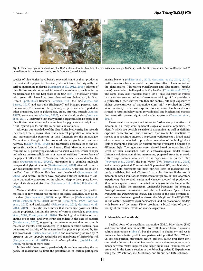
species of blue Haslea have been discovered, some of them producing
marennine-like pigments chemically distinct from the originally de-
scribed marennine molecule (Gastineau et al., 2012, 2016). Blooms of
blue Haslea are also observed in natural environments, such as in the
Mediterranean Sea and East coast of the USA (Fig. 1). Moreover, oysters
with green gills have long been observed worldwide, e.g., in Great
Britain (Sprat, 1667), Denmark (Petersen, 1916), the USA (Mitchell and
Barney, 1917) and Australia (Hallegraeff and Mouget, personal com-
munication). Furthermore, the greening of gills has been reported in
other organisms, such as polychaetes, crabs, littorina, mussels (Ranson,
1927), sea-anemones (Gaillon, 1820), scallops and cockles (Gastineau
et al., 2018), illustrating that many marine organisms can be exposed to
blue Haslea populations and marennine-like pigments not only in arti-
ficial (oyster) ponds, but also in natural environments.
Although our knowledge of the blue Haslea biodiversity has recently
increased, little is known about the chemical properties of marennine
and marennine-like pigments or their functions for the microalgae.
Marennine is thought to be produced via a cytoplasmic synthesis
pathway (Nassiri et al., 1998) and transiently accumulates at the cell
apices (intracellular form of the pigment, IMn). Marennine is excreted
from the cells, possibly by exocytosis via small vesicles, which collapse
and release an extracellular form of marennine (EMn). The two forms of
the pigment differ in their UV–vis spectral characteristics and molecular
mass (Pouvreau et al., 2006b). Marennine is a complex molecule
composed of glycosidic units (Gastineau et al., 2014) attached to one or
various aromatic rings (Pouvreau et al., 2006b). A protocol to obtain a
purified form of EMn or IMn has been developed (Pouvreau et al.,
2006c) and several authors have proposed different methods to esti-
mate marennine concentration in solution, despite incomplete knowl-
edge of its chemical structure (Pouvreau et al., 2006a; Robert et al.,
2002).
Various studies have demonstrated that marennine (as purified
molecule or raw extract) has multiple biological activities, such as an-
tioxidant (Pouvreau et al., 2008), antiproliferative (Carbonnelle et al.,
1998; Gastineau et al., 2012), antiviral (Bergé et al., 1999; Gastineau
et al., 2012) and antibacterial (Falaise et al., 2016; Gastineau et al.,
2014, 2012). It has also been shown that marennine possesses allelo-
pathic properties, limiting the growth of various microalgae (Pouvreau
et al., 2007; Prasetiya et al., 2016). The biological activities of mar-
ennine are species- and even strain-dependent in the case of bacteria
(Falaise et al., 2016), suggesting that marennine could act on specific
molecular targets. Tests conducted with Gram-negative bacteria have
demonstrated activity of the marennine-like pigment produced by Ha-
slea provincialis (Gastineau et al., 2016) and marennine produced by H.
ostrearia, on the lipopolysaccharidic cell membrane of Escherichia coli
(Tardy-Laporte et al., 2013) and of Vibrio splendidus (Bouhlel et al.,
2018), rendering it more rigid.
In line with these results, particularly those demonstrating the ca-
pacity of marennine to limit the proliferation of certain pathogenic
marine bacteria (Falaise et al., 2016; Gastineau et al., 2012, 2014),
further research has confirmed the protective effect of marennine on
the giant scallop (Placopecten magellanicus) and blue mussel (Mytilus
edulis) larvae when challenged with V. splendidus (Turcotte et al., 2016).
The same study also revealed that a 20 d (day) exposure of mussel
larvae to low concentrations of marennine (0.1 μg mL−1) provided a
significantly higher survival rate than the control, although exposure to
higher concentrations of marennine (1 μg mL−1) resulted in 100%
larval mortality. Even brief exposure to marennine has been demon-
strated to result in behavioural, physiological and biochemical changes
that were still present eight weeks after exposure (Prasetiya et al.,
2017).
These results underpin the interest to further study the effects of
marennine on early developmental stages of marine organisms, to
identify which are possibly sensitive to marennine, as well as defining
exposure concentrations and durations that would be beneficial to
species of aquaculture interest. The present work presents a broad panel
of experiments conducted to provide an overview of the biological ef-
fects of marennine solutions on various marine organisms belonging to
different phyla. The organisms were selected based on aquaculture in-
terest or their established role in ecotoxicological model assays.
Different solutions containing marennine, prepared from H. ostrearia
culture supernatants, were used in the exposures: the purified EMn
(Pouvreau et al., 2006c), the Blue Water (BW; (Turcotte et al., 2016)
and a newly patented Concentrated Supernatant (CS; see Materials).
Although EMn represents the most purified form of marennine cur-
rently available, BW and CS are of particular interest if the use of
marennine-based solutions is considered at larger scales than laboratory
experiments due to their easier and cheaper method of production.
Marennine exposures were conducted on embryos and/or larvae of the
mollusc M. edulis, the crustacean Chthamalus bisinuatus, the chordate
Pseudopleuronectes americanus and the echinoderms Sphaerechinus
granularis and Paracentrotus lividus. The effects of the blue pigment so-
lutions were also investigated in vitro on the fish gill cell line RTgill-W1,
on the oyster Crassostrea gigas haemocytes, and on prokaryotic models
with bacteria of the genus Vibrio, providing a broad view of the di-
versity of marennine effects on marine organisms.
2. Materials and methods
Purified form of extracellular marennine (EMn), Blue Water (BW)
and Concentrated Supernatant (CS) were all obtained from H. ostrearia
culture supernatant (Table 1), but the process to obtain BW and CS is
faster and has a better yield in comparison with purified EMn. BW and
CS are not purified marennine per se, but they allow preparing con-
centrated solutions of marennine needed to run dose-response experi-
ments between Haslea pigment and target organisms. Experiments are
presented in the Results section in the following order: 1) Experiments
using the BW solution, 2) CS solution, and 3) purified EMn solution.
Fig. 1. Underwater pictures of natural blue Haslea blooms forming biofilms observed A) in macro-algae Padina sp. in the Mediterranean sea, Corsica (France) and B)
on sediments in the Beaufort Strait, North Carolina (United States).
C. Falaise et al. Aquatic Toxicology 209 (2019) 13–25
14
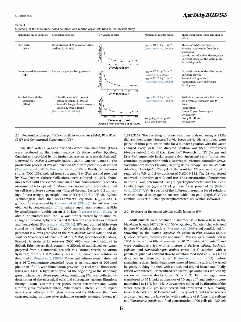
2.1. Preparation of the purified extracellular marennine (EMn), Blue Water
(BW) and Concentrated Supernatant (CS)
The Blue Water (BW) and purified extracellular marennine (EMn)
were produced at the Station aquicole de Pointe-au-Père (Québec,
Canada) and provided by the Institut des sciences de la mer de Rimouski-
Université du Québec à Rimouski (ISMER-UQAR; Québec, Canada). The
production process of BW and purified EMn were previously described
(Turcotte et al., 2016; Pouvreau et al., 2006c). Briefly, H. ostrearia
strains (NCC 136), isolated from Bourgneuf Bay (France) and provided
by NCC (Nantes Culture Collection), were cultured in 100 L photo-
bioreactors until the extracellular marennine concentration reached a
maximum of 6 to 8 μg mL−1. Marennine concentration was determined
on cell-free culture supernatant (filtered through Sarstedt 0.2 μm syr-
inge filters) using a spectrophotometer (Cary 100 Bio UV–vis, Agilent
Technologies) and the Beer-Lambert’s equation (ε677=12.13 L
g−1 cm−1) as proposed by Pouvreau et al. (2006c). The BW was then
obtained by concentration of the culture supernatant containing EMn
by ultrafiltration (double cut off 3–30 kDa; (Turcotte et al., 2016). To
obtain the purified EMn, the BW was further treated by an anion-ex-
change chromatography process and the fraction collected was dialyzed
and freeze-dried (Pouvreau et al., 2006c). BW and purified EMn were
stored in the dark at 4 °C and −20 °C respectively. Concentrated Su-
pernatant (CS) was produced in the Mer Molécule Santé (MMS) and In-
stitut des Molécules et Matériaux du Mans (IMMM) laboratories (Le Mans,
France). A strain of H. ostrearia (NCC 495) was batch cultured in
500mL Erlenmeyers flask containing 250mL of autoclaved sea water
prepared from a commercial sea salt mix (Instant Ocean, Aquarium
Systems®; pH 7.6 ± 0.2; salinity 32) with an enrichment solution as
described in Mouget et al. (2009). Microalgal cultures were maintained
in a 16 °C temperature-controlled room at an irradiance of 200 μmol
photon m−2 s−1, with illumination provided by cool-white fluorescent
tubes in a 14/10 h light/dark cycle. At the beginning of the stationary
growth phase the culture supernatant containing EMn was collected by
decantation of the microalgal cells and subsequent vacuum filtrations
through 15 μm (150mm Filter paper, Fisher Scientific®) and 1.2 μm(37mm glass microfiber filters, Whatman®). Filtered culture super-
natant was collected in 1 L glass bottles and the EMn was then con-
centrated using an innovative technique recently patented (patent n°:
1,872,316). The resulting solution was then dialyzed using a 2 kDa
dialysis membrane (Spectra/Por®6, Spectrum®). Dialysis tubes were
placed in ultra-pure water tanks for 3 d under agitation with the water
changed every 24 h. The dialyzed solution was then ultra-filtered
(double cut-off 1 kD-30 kDa; Kros Flo® Research IIi TFF System and
Kros Flo® Automatic Backpressure valve, Spectrum®) and further con-
centrated by evaporation with a Rotavapor (Vacuum controller CVC2,
Vacuubrand®; Rotary Elevator, Heating Bath Hei-Vap, Base Hei-Vap ML
Adv/Pre, Heidolph®). The pH of the resulting CS was neutralized if
required to 7.5 ± 0.2 by addition of NaOH 0.1M. The CS was stored
one week in the dark at 4 °C until use. The concentration of marennine
in the CS was determined using a spectrophotometer and the Beer-
Lambert equation (ε669=17.2 L g−1 cm−1) as proposed by (Robert
et al., 2002). UV–vis spectra of the different marennine based solutions
were conducted using quartz cuvettes with 1 cm path length (UV/Vis
Lambda 25 Perkin Elmer spectrophotometer, UV Winlab software).
2.2. Exposure of the mussel Mytilus edulis larvae to BW
Adult mussels were obtained in summer 2017 from a farm in the
Magdalen Islands (47° 25′N, 61° 50′W, Quebec, Canada), characterized
by pure M. edulis populations (Myrand et al., 2009) and conditioned for
spawning in the Station aquicole de Pointe-au-Père (ISMER-UQAR;
Québec, Canada) facilities for one month. Mussels were maintained in
180 L tanks in 1 μm filtered seawater at 20 °C flowing at 1 L min−1 and
were continuously fed with a mixture of Pavlova lutherii, Isochrysis
galbana, and Nannochloropsis oculata (ratio 1:1:1) supplied with a
peristaltic pump at constant flow to maintain food load at 0.5mg L-1 as
described in Hennebicq et al. (Hennebicq et al., 2013). Before
spawning, a dozen individuals were removed from the tank and washed
by gently rubbing the shell with a brush and diluted bleach and finally
rinsed with filtered, UV sterilized sea water. Spawning was induced by
successive thermal shocks from 10 to 25 °C. Fertilized eggs were
transferred to 60 L tanks at densities of 10 eggs μL-1 and embryos were
maintained at 18 °C for 48 h. D-larvae were collected by filtration of the
water through a 20 μm mesh screen and transferred to 60 L rearing
tanks at densities of 10 D-larvae mL-1. Every 2–3 d, tanks were washed
and sterilized and the larvae fed with a mixture of P. lutherii, I. galbana
and Chaetoceros gracilis at a final concentration of 90 cells μL-1 (30 cell
Table 1
Summary of the marennine based solutions and marine organisms used in the present study.
Marennine based solutions Production process UV-visible spectra Method of quantification Marine organisms tested and studied
effect
Blue Water
(BW)
Ultrafiltration of H. ostrearia culture
medium (3-30 kDa)
ε677=12.13 L g−1 cm-1
(Pouvreau et al., 2006c)
Mussel M. edulis, barnacle C.
bisinuatus and winter flounder P.
americanus:
Larval survival and/or development
Bacterial species of the Vibrio genus:
Bacterial growth
Concentrated Supernatant
(CS)
Innovative process being patented ε669=17.2 L g−1 cm−1
(Robert et al., 2002)
Bacterial species of the Vibrio genus:
Bacterial growth
ε677=12.13 L g−1 cm-1
(Pouvreau et al., 2006c)
Sea urchin S. granularis:
Fertilization, early embryonic
development
Purified Extracellular
Marennine
(EMn)
- Ultrafiltration of H. ostrearia
culture medium (3-30 kDa)
- Anion-Exchange chromatography
- Dialysis & freeze-drying
(Pouvreau et al., 2006c)
Adapted from Pouvreau et al. (2006)
ε677=12.13 L g−1 cm-1
(Pouvreau et al., 2006c)
Preliminary assays with EMn on the
sea urchins S. granularis and P.
lividus:
Fertilization
Oyster C. gigas haemocytes:
Cytotoxicity
Weighing of the purified
EMn dried powder
Fish gill cell line:
Cytotoxicity
C. Falaise et al. Aquatic Toxicology 209 (2019) 13–25
15
μL−1 for each component of the diet; Turcotte et al., 2016).
M. edulis embryos were collected prior to the transfer to the em-
bryogenesis tank, and D-larvae prior to the transfer to the rearing tank
(48 h post fertilization). Veliger larvae were collected in the rearing
tanks (14 d post fertilization) by filtration of the water through a mesh
screen. Embryos and larvae were exposed to BW in 12-well flat bottom
sterile microplates with each well containing 3mL of diluted BW and
10–15 embryos or 10–15 larvae per well. Five concentrations of mar-
ennine from BW were tested: 0, 0.1, 0.5, 1 and 5 μg mL−1. Marennine
concentration from BW was estimated as described in Section 2.1 and
Table 1. BW was syringe-filtered through 0.2 μm and diluted in sterile
sea water. Microplates were kept in a controlled temperature room at
18 °C without addition of food during the time of the experiment. The
mortality of embryos and veliger larvae was assessed using a light
microscope (Olympus BX41; W. Carsen Co., Ltd., Don Mills, ON, Ca-
nada) coupled to a digital camera (Evolution VF Color, MediaCyber-
netics, Silver Spring, MD, USA). The percentages of swimming and
motionless D-larvae were assessed by observation of the microplates
using an inverted microscope (Axiovert 100, Zeiss). Larval size was
measured with Image Pro-Express (Media Cybernetics) after addition of
formaldehyde to the wells. A minimum of 10 larvae were counted per
well with at least 3 wells per concentration tested.
2.3. Exposure of the barnacle Chthamalus bisinuatus larvae to BW
Experiments were conducted in June 2015 in the Center for Marine
Biology of Sao Paulo University (CEBIMar/USP; Sao Paulo, Brazil).
Spawners were harvested by collecting rock fragments in the intertidal
zone of Calhetas Beach (23°49′ 28″S, 45°25′11″W). The fragments were
transferred to the laboratory and placed in seawater pumped from the
adjacent bay, as described in Kasten and Flores (Kasten and Flores,
2013). A light source was placed above the tanks containing the bree-
ders to recreate the natural photoperiod. Broodstock was submerged for
30min every 12.4 h to simulate the natural effect of the tide and sti-
mulate spawning. After a first submersion, the water used to submerge
the broodstock was siphoned and filtered to harvest the larvae. The
larvae were placed in filtered seawater and isolated using a dissecting
microscope to obtain a number of larvae sufficient to run the experi-
ment. Larvae were placed individually in the wells of a 96-well mi-
croplate in 2mL of seawater containing 0, 0.05 or 0.1 μg mL−1 of
marennine from BW without food to test only the potential toxicity of
BW. The BW was prepared and the concentration estimated as de-
scribed in Section 2.1 (Table 1). Four larvae were tested at each con-
centration. The microplate was placed on a stirring plate and no food
was provided during the experiment. Larvae were observed once a day
and survival recorded over 9 d. The experiment was repeated once with
spat from a different production.
2.4. Exposure of the winter flounder Pseudopleuronectes americanus larvae
to BW
Larvae were reared as described by (Khemis et al., 2003) in 57 L
cylindro-conical polyethylene tanks and fed rotifers from d 4 to d 26
(Fraboulet et al., 2010). Rotifers were fed a cocktail of microalgal
concentrates (1:1:1 N. oculata: I. galbana: P. lutherii, Instant algae, REED
Mariculture) and a SELCO food supplement (1 g M−1 rotifers, INVE
Aquaculture Nutrition, Gransville, UT, USA). The larvae were kept in
green water (addition of N. oculata culture directly to basins). From d 2
to d 14, two concentrations of marennine from BW were tested on the
larvae, 0.05 and 0.1 μg mL−1, with three tanks per treatment (n=3).
BW preparation, as described in Section 2.1 (Table 1), was added to the
tanks in the morning, when water flow was cut to feed the larvae. Water
flow was restarted at the end of the day (for a total of about 8 h of
exposure) and BW was gradually evacuated from the tanks with the
flow of water outlet. A dose of BW was given every two days, for a total
of seven treatments per tank for the duration of the experiment.
Procedures were the same for the control treatments, without the ad-
dition of BW in the control tanks. The effect of BW treatment on bac-
terial load and larval size was evaluated on d 2, d 6, d 10, and d 14, and
the size and physiological condition (assessment of energy reserves) of
the larvae were determined at the end of the experiment, i.e. at d 14 (12
d of treatment). Standard length was measured on formaldehyde-pre-
served larval pictures as described in (Hjörleifsson and Klein-MacPhee,
1992) using a dissecting microscope (Olympus SZ61) coupled to a di-
gital camera (Evolution VF; Media Cybernetics) and Image Pro-Plus
measurement software 5.0 (Media Cybernetics). Lipids were extracted
in a 2:1 mixture of dichloromethane: methanol according to (Folch
et al., 1957), the lipid classes (triglyceride [TAG], sterol [ST], acetone
mobile polar lipids [AMPL], and phospholipids [PL]) concentrations
were quantified by TLC-FID as described by (Parrish, 1987), and
chromatograms analyzed using PeakSimple v3.21 software (SRI Inc.).
To estimate the potential effect of BW on bacterial load in rearing
water, bacterial analyzes were carried out using a flow cytometer on
water samples taken from the tanks and frozen with glutaraldehyde
according to (Seychelles et al., 2011).
2.5. Exposure of the sea urchins Sphaerechinus granularis and
Paracentrotus lividus to EMn and CS
A first series of assays were conducted in May 2016 with purified
EMn on S. granularis and P. lividus, model animals frequently used for in
vivo ecotoxicological bioassays (Buttino et al., 2016; Pinsino et al.,
2010). Sea urchins were collected in the Brest area (France) and ob-
tained from the Centre de Ressources Biologiques Marines (CRBM) at the
Roscoff Biological Station (Roscoff, France). Other experiments on sea
urchins were run in March 2017 using CS rather than purified EMn due
to its limited production, and the species S. granularis because it is
available throughout the year.
Marennine-based solutions were prepared by weighing purified
EMn freeze-dried powder or after the estimation of CS concentration as
described in Section 2.1 (Table 1). Solutions were diluted in 0.22 μmMillipore-filtered seawater (FSW) and the final solutions were syringe
filtered through 0.2 μm. Sea urchin spawning was induced by in-
tracoelomic injection of 0.1 M acetylcholine. Eggs were collected in
FSW, rinsed twice by centrifugation (2.000 g, 2 min) and re-suspended
in FSW for a final 2% (v/v) egg solution containing 0.1% (v/v) glycine.
Sperm was kept dry at 4 °C until use. Experiments took place in a
temperature-controlled room at 16 °C.
Effects of purified EMn and CS were first assessed on fertilization.
Eggs were transferred to a 24-well culture plate and incubated during
10min in marennine-based solutions prior to addition of sperm.
Solutions at different concentrations were added in wells containing
1mL of the egg suspension. Only FSW was added for the control con-
dition. pH strips were used to ensure that the pH did not vary between
wells of the different concentrations tested. For fertilization, 50 μL of
dry sperm was diluted in 1mL of FSW shortly before use and 4 μL of
diluted sperm added per mL of egg suspension. Observation of the
culture plate under phase contrast inverted microscopy allowed the
determination of the fertilization rate by counting at least 60 eggs per
well (n= 3 wells per concentration tested). For experiments conducted
with CS, unfertilized eggs exposed to the highest CS concentrations
were rinsed 3 times in FSW using a benchtop centrifuge for 5–10 sec-
onds and exposed to sperm for fertilization as described above. Two
hours post fertilization, eggs exposed to CS were fixed on a DNA
fluorescent stain (Hoechst fixative: Bisbenzymide 0.1 μg mL−1; me-
thanol 75%, glycerol 25%) and observed under a fluorescence micro-
scope (ApoTome, Zeiss).
The effects of CS were also assessed post fertilization (kinetics of
first cleavage and early embryonic development). For fertilization, di-
luted sperm was added in a 50mL tube containing 25mL of eggs sus-
pended in FSW (2% (v/v) egg suspension). When egg batches exhibited
greater than 90% fertilization, sperm was removed by centrifugation in
C. Falaise et al. Aquatic Toxicology 209 (2019) 13–25
16
Heraeus Labofuge centrifuge (2.000 g, swinging bucket rotor 2min).
Fertilized eggs were transferred to a 24-well plate and CS solutions at 1;
10; 50 and 100 μg mL−1 added 10min post fertilization. The percen-
tage of dividing eggs was determined under phase contrast inverted
light microscope; at least 60 eggs per well were counted. Three different
sea urchin couples were used to replicate the experiment. For the
“washed” condition, embryos from 3 wells per concentration tested
were rinsed 6 h post fertilization as described earlier. Culture plates
were maintained under constant agitation at 16 °C and embryos were
observed 8, 48 and 72 h post fertilization with a Leica DMi8 inverted
microscope and pictures acquisition done with LASX software. For each
observation time, a 75 μL sample of each well was transferred to a glass
slide and pictures of the developmental stages predominantly observed
were taken. For post hatching developmental stages, a 1 μL drop of
Janus green was added to the samples. Bright Field microscopy tech-
nique (BF) was applied for embryos 8 h post fertilization and a
Differential Interference Contrast technique (DIC) for embryos 48 h and
72 h post fertilization.
2.6. Exposure of Vibrio species to BW and CS
Experiments were conducted during spring 2018 in MMS laboratory
(Le Mans, France). BW and CS were prepared as described in Section
2.1 (Table 1), and the concentration of marennine was determined with
spectrophotometric measurements using the Beer-Lambert law with the
specific extinction coefficient (ε677=12.13 L g−1 cm−1) proposed by
(Pouvreau et al., 2006b). CS and BW were diluted with sterile ultra-
pure water. Salinity and pH were adjusted by addition of NaCl and
0.1 M of NaOH or HCl (pH 7.5 ± 0.2; salinity 32) and the solutions
were then syringe-filtered through 0.22 μm (Sarstedt). The three Vibrio
strains tested, Vibrio chagasii (strain 8T3_5), Vibrio crassostreae (strain
8T2_1) and Vibrio sp. (strain 7G1_11) were previously identified (Bruto
et al., 2017) and kindly provided by the Laboratory of Integrative Biology
of Marine Models (CRBM/Roscoff, France). Bacterial strains were kept at
−80 °C in 25% glycerol, inoculated in Mueller Hinton Broth (MHB) +
1% NaCl (pH 7.5 ± 0.2; salinity 32) and incubated overnight at 25 °C.
Isolations were done on Petri dishes containing agar prepared with
Nutrient Agar (Biokar) + 2.3% NaCl (pH 7.5 ± 0.2; salinity 32). The
plates were incubated at 25 °C for one day. The antibacterial activity of
BW and CS was assessed according to a method described in the Clinical
and Laboratory Standards Institute (CLSI) antimicrobial microdilution
guidelines (Clinical and Laboratory Standards Institute (Clinical and
Laboratory Standards Institute (CLSI, 2012). The day prior to the ex-
periment, three different colonies per Petri dish were inoculated in
broth media and grown overnight at ambient temperature. Wells of a
96-well sterile microplate with cover and flat bottom were filled with
50 μL of either CS or BW at a final concentration of 10 μg mL−1 (or
sterile saline water for the controls) and 50 μL of a bacterial inoculate. A
negative control was also run with sterile saline water and broth media
without bacteria to ensure that no contamination occurred during the
experiment. To prepare the bacterial inoculate the optical density (OD)
of the broth culture was measured at 630 nm (V-10 Plus Onda Spec-
trophotometer), the OD was adjusted to 0.1 by dilution in MHB+1%
NaCl and the solution was then further diluted by 1/100 as re-
commended by the CLSI guidelines (Clinical and Laboratory Standards
Institute (Clinical and Laboratory Standards Institute (CLSI, 2012).
Microplates were inserted in a microplate spectrophotometer (xMark
Bio-Rad) for a 20 h run at ambient temperature. Growth was managed
with microplate Manager 6 Software by taking the OD in each well
every 30min at 600 nm.
2.7. Exposure of fish gill cell lines to EMn
Experiments took place in August 2017 at the Institute for Marine
and Antarctic Studies (IMAS; Hobart, Australia). The gill epithelium cell
line RTgill-W1 was obtained from the American Type Culture
Collection (ATTC; and originally isolated from the Rainbow trout
Oncorhyncus mykiss). The cell line was maintained and exposures to
purified EMn conducted as described in Dorantes-Aranda et al. (2011)
in conventional 96-well plates. Gill cells were seeded into a flat-bottom
96-well plate (655180, Greiner) at 2.5× 10⁵ cells mL−1 in L-15
medium (L-1518, Sigma) and allowed to attach for 48 h in the dark.
Confluence of cell cultures was verified 12 h before experimental ex-
posure and the L-15 medium replaced by L-15/ex (Schirmer et al.,
1997). The concentration of the original purified EMn solution was
estimated (540 μg mL−1 in 50% methanol) as described in Section 2.1
(Table 1), and the solution was diluted in L-15/ex by factors of
1× 10⁻¹, 5× 10⁻², 1× 10⁻², 5× 10⁻³, 1× 10⁻³, 5× 10⁻⁴ and 1×10-
4, yielding final exposure concentrations of 0–54 μg mL−1. Methanol
was added to all dilutions to achieve a constant final MeOH con-
centration of 5% across all treatments, including the non-toxic control
(L-15/ex). Gill cells were exposed to these solutions for 2 h at
20 ± 1 °C in the dark (quadruplicate wells per concentration). After
the exposure, wells were rinsed twice with saline phosphate buffer
(100 μL per well) and incubated for a further 2 h in the dark with 100 μLof 5% resazurin viability stain in L-15/ex medium. Metabolic reduction
of resazurin to resorufin by the gill cells was measured in a microplate
reader (Fluostar Omega, BMG Labtech) at excitation and emission
wavelengths of 540 and 590 nm, respectively. Results are expressed as
percentage viability of the nontoxic control (5% MeOH in L-15/ex).
2.8. Exposure of the oyster Crassostrea gigas haemocytes to EMn
Pacific oysters C. gigas were harvested in 2015–2016 in the hatchery
of Laboratoire de Génétique et de Pathologie des Mollusques Marins
(LGPMM; La Tremblade, France) in raceways supplied with a constant
flow of seawater enriched with phytoplankton (Skeletonema costatum,
Tetraselmis suecica, I. galbana and C. gracilis). They were maintained in
safe conditions, free of known infectious pathogens. The shell of 20
adult oysters was broken with metal clamps and haemolymph with-
drawn from the adductor muscle sinus (1 to 1.5mL of haemolymph per
oyster) using 1mL sterile plastic syringes equipped with a needle
(0.90 mm x 25mm). The haemolymph of the different individuals was
pooled, filtered through 60 μm nylon mesh and held on ice to prevent
haemocyte aggregation (Auffret and Oubella, 1997). Haemocytes were
observed under light microscopy using a Malassez-cell. Haemocytes
were then exposed to different concentrations of purified EMn: 0, 1, 50
and 100 μg mL−1. The purified EMn was obtained as described in
Section 2.1 (Table 1) and the solutions were prepared by weighing of
the EMn dried powder on an analytical balance (Sartorius Entris®) and
by dilution on sterile sea water. Purified EMn solutions were added to
the haemolymph (1:1 ratio) for a final haemocyte concentration of
2.104 cells mL−1. Haemocytes were exposed to EMn during 1, 3 or 6 h
and mortality was quantified using 200 μL of cell suspension. Cells were
incubated in the dark for 30min on ice with 50 μL of Propidium Iodide
(PI, 1.0 g L−1, Interchim), a fluorescent DNA/RNA-specific dye that
only permeates through the membranes of dead cells and stains the
nucleic acids. Haemocyte samples were analyzed with flow cytometry
using an EPICS XL 4 (Beckman Coulter) and red fluorescence following
the protocol of (Morga et al., 2009). Based on size discrimination, only
haemocytes were taken into account with 5.000 events counted per
sample. Results were depicted as cell cytograms and reported as log
scale fluorescence levels. Data were analyzed with Flowing Software 2.
2.9. Statistics
Statistical analyses were run using SigmaPlot 12.3 software for
Windows. Differences between treatments were assessed with One-Way
or Two-Ways ANOVAs. Normality was tested by the Shapiro-Wilk test
and the assumption of homoscedasticity of variance with Fisher’s test
(F-test) and/or verified visually by the spread of residuals, as suggested
by (Quinn and Keough, 2002). Post hoc Tukey’s pairwise multiple
C. Falaise et al. Aquatic Toxicology 209 (2019) 13–25
17
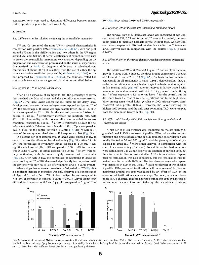
comparison tests were used to determine differences between means.
Unless specified, alpha value used was 0.05.
3. Results
3.1. Differences in the solutions containing the extracellular marennine
BW and CS presented the same UV–vis spectral characteristics in
comparison with purified EMn (Pouvreau et al., 2006b), with one peak
around 670 nm in the visible region and two others in the UV region
around 250 and 320 nm. Different coefficients of extinction were used
to assess the extracellular marennine concentration depending on the
preparation and concentration process and on the series of experiments
(summarized in Table 1). Despite a difference in calculated con-
centrations of about 30–40 % resulting from the use of either the ap-
parent extinction coefficient proposed by (Robert et al., 2002) or the
one proposed by (Pouvreau et al., 2006a), the solutions tested had
comparable concentration ranges and activities as described below.
3.2. Effects of BW on Mytilus edulis larvae
After a 48 h exposure of embryos to BW, the percentage of larvae
that reached the D-larval stage and the mortality rate were assessed
(Fig. 2A). The three lowest concentrations tested did not delay larval
development; however, when embryos were exposed to 1 μg mL−1 of
BW, the percentage of D-larvae was significantly lower (22 ± 1% of D-
larvae compared to 52 ± 2% for the control; p-value= 0.026). Ex-
posure to 1 μg mL−1 significantly increased the mortality rate, with
47 ± 1% of mortality while no mortality was recorded in control
condition. Exposure to 1 μg mL−1 of BW significantly delayed the de-
velopment with a D-larvae mean length of 88 ± 7 μm compared to
122 ± 1 μm for the control (p-value<0.001; Fig. 2B). At 5 μg mL-1,
none of the embryos survived after a 48 h exposure to BW (Fig. 2A).
In a second series of experiments, D-larvae were exposed to BW in
order to assess the effects on larval swimming (Fig. 3A). After 24 h in
BW, the percentage of swimming larvae exposed to 1 μg mL−1 was
significantly lowered (80 ± 9% compared to 100 ± 0% for the con-
trol; p-value< 0.001). D-larvae exposed to 5 μg mL−1 of BW were to-
tally motionless, with the velum highly coloured with marennine
(Fig. 3B). After 72 h in BW, the percentage of swimming D-larvae ex-
posed to 1 μg mL−1 of BW decreased significantly in comparison with
the day one with only 45 ± 2% of swimming larvae (p-value 0.012).
When veliger larvae were exposed over a 5 d period to BW (Fig. 4A),
a significant increase in mortality was only observed at a concentration
of 5 μg mL−1, with 64 ± 7% of dead veliger larvae compared to
7 ± 6% of mortality in control (p-value<0.001). Larval length only
differed for treatments of 0.5 and 1 μg mL-1 compared to 5 μg mL−1 of
BW (Fig. 4B; p-values 0.036 and 0.030 respectively).
3.3. Effect of BW on the barnacle Chthamalus bisinuatus larvae
The survival rate of C. bisinuatus larvae was measured at two con-
centrations of BW, 0.05 and 0.1 μg mL−1 over a 9 d period, the max-
imum period to maintain barnacle larvae without food. At both con-
centrations, exposure to BW had no significant effect on C. bisinuatus
larval survival rate in comparison with the control (Fig. 5; p-value
0.1427).
3.4. Effect of BW on the winter flounder Pseudopleuronectes americanus
larvae
The addition of BW at 0.05 and 0.1 μg mL−1 had no effect on larval
growth (p-value 0.287). Indeed, the three groups experienced a growth
of 0.1 mm d−1 from d 2 to d 14 (Fig. 6A). The bacterial load remained
comparable in all treatments (p-value 0.868), demonstrating that, at
such concentration, marennine had no impact on bacterial development
in fish rearing tanks (Fig. 6B). Energy reserves in larvae treated with
marennine seemed to increase with 4.6 ± 0.7 μg larva−1 under 0.1 μgmL−1 of BW exposure vs 3.9 ± 0.7 μg larva−1 for the control, but the
difference from the control was not significant due to the high varia-
bility among tanks (total lipids, p-value 0.1092; triacyglycerol/sterol
(TAG/ST) ratio, p-value 0.0767). However, the larvae showing the
highest lipid content, and the only ones containing TAG, were sampled
from the marennine treated tanks (Fig. 6C).
3.5. Effects of CS and purified EMn on Sphaerechinus granularis and
Paracentrotus lividus
A first series of experiments was conducted on the sea urchins S.
granularis and P. lividus to assess if purified EMn had an effect on fer-
tilization and first cleavage of the egg. In both species, fertilization was
totally blocked at 50 and 100 μg mL−1 and the phenotypes of embryos
exposed to 10 μg mL−1 were either delayed in comparison with the
control or abnormal (e.g., flattened). Four different incubation periods
were tested, from 0 to 20min prior to the addition of purified EMn, but
the effects on fertilization were similar. A 10min incubation of sperm
prior to fertilization was also conducted, but the fertilization rate re-
mained unaffected with 100% fertilization observed even when sperm
was incubated in EMn at 100 μg mL−1 (data not shown). It was checked
if purified EMn prevented fertilization or if the absence of fertilization
membrane around the eggs was caused by an effect of EMn on the
elevation of fertilization membrane steps. To do so, a calcium iono-
phore (i.e., a chemical that can activate echinoderm eggs by a release of
intracellular calcium ions and inducing the membrane elevation
Fig. 2. Exposure of the mussel Mytilus edulis embryos to different concentrations (μg mL−1) of Blue Water (BW) over a 48 h period. A) Percentage of embryos that
reached the D-larval stage (grey bars) and percentage of mortality (black bars). B) Length of the larvae that reached the D stage (μm). Values are means ± SE
(n=3). Error bars with different lower case letters are significantly different.
C. Falaise et al. Aquatic Toxicology 209 (2019) 13–25
18
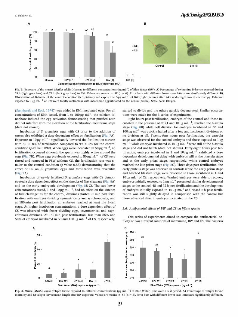
(Steinhardt and Epel, 1974)) was added in EMn incubated eggs. For all
concentrations of EMn tested, from 1 to 100 μg mL-1, the calcium io-
nophore induced the egg activation demonstrating that purified EMn
did not interfere with the elevation of the fertilization membrane steps
(data not shown).
Incubation of S. granularis eggs with CS prior to the addition of
sperm also exhibited a dose-dependent effect on fertilization (Fig. 7A).
Exposure to 10 μg mL−1 significantly lowered the fertilization success
with 85 ± 8% of fertilization compared to 99 ± 2% for the control
condition (p-value 0.032). When eggs were incubated in 50 μg mL-1, no
fertilization occurred although the sperm was highly active around the
eggs (Fig. 7B). When eggs previously exposed to 50 μg mL−1 of CS were
rinsed and removed in FSW without CS, the fertilization rate was si-
milar to the control condition (p-value 0.58) demonstrating that the
effect of CS on S. granularis eggs and fertilization was reversible
(Fig. 7A).
Incubation of newly fertilized S. granularis eggs with CS demon-
strated a dose dependent effect on the kinetics of first cleavage (Fig. 8A)
and on the early embryonic development (Fig. 8B-C). The two lower
concentrations tested, 1 and 10 μg mL−1, had no effect on the kinetics
of first cleavage: as for the control, divisions started 95min post ferti-
lization with embryos dividing symmetrically and synchronously, and
at 180min post fertilization all embryos reached at least the 2-cell
stage. At higher incubation concentrations, a dose-dependent effect of
CS was observed with fewer dividing eggs, asymmetrical and asyn-
chronous divisions. At 180min post fertilization, less than 85% and
50% of embryos incubated in 50 and 100 μg mL−1 of CS, respectively,
started to divide and the others quickly degenerated. Similar observa-
tions were made for the 3 series of experiments.
Eight hours post fertilization, embryos of the control and those in-
cubated in the presence of CS (1 and 10 μg mL−1) reached the blastula
stage (Fig. 8B) while cell division for embryos incubated in 50 and
100 μg mL-1 was quickly halted after a few and incoherent divisions or
no division at all. Twenty-four hours post fertilization, the gastrula
stage was observed for the control embryos and those exposed to 1 μgmL−1 while embryos incubated in 10 μg mL−1 were still at the blastula
stage and did not hatch (data not shown). Forty-eight hours post fer-
tilization, embryos incubated in 1 and 10 μg mL−1 exhibited a dose
dependent developmental delay with embryos still at the blastula stage
and at the early prism stage, respectively, while control embryos
reached the late prism stage (Fig. 8C). Three days post fertilization, the
early pluteus stage was observed in controls while the early prism stage
and hatched blastula stage were observed in those incubated in 1 and
10 μg mL-1 of CS, respectively. Washed embryos were able to recover;
embryos initially exposed to 1 μg mL-1 presented similar developmental
stages to the control, 48 and 72 h post fertilization and the development
of embryos initially exposed to 10 μg mL-1 and rinsed 6 h post fertili-
zation was still slightly delayed in comparison with the control but
more advanced than in embryos incubated in the CS.
3.6. Antibacterial effects of BW and CS on Vibrio species
This series of experiments aimed to compare the antibacterial ac-
tivity of two different solutions of marennine, BW and CS. The bacteria
Fig. 3. Exposure of the mussel Mytilus edulis D-larvae to different concentrations (μg mL-1) of Blue Water (BW). A) Percentage of swimming D-larvae exposed during
24 h (light grey bars) and 72 h (dark grey bars) to BW. Values are means ± SE (n=6). Error bars with different lower case letters are significantly different. B)
Observation of D-larvae of the control condition (left picture) and exposed to 5 μg mL−1 of BW (right picture) after 24 h under light invert microscopy. D-larvae
exposed to 5 μg mL−1 of BW were totally motionless with marennine agglutinated on the velum (arrow). Scale bars: 100 μm.
Fig. 4. Mussel Mytilus edulis veliger larvae exposed to different concentrations (μg mL−1) of Blue Water (BW) over a 5 d period. A) Percentage of veliger larvae
mortality and B) veliger larvae mean length after BW exposure. Values are means ± SE (n=3). Error bars with different lower case letters are significantly different.
C. Falaise et al. Aquatic Toxicology 209 (2019) 13–25
19
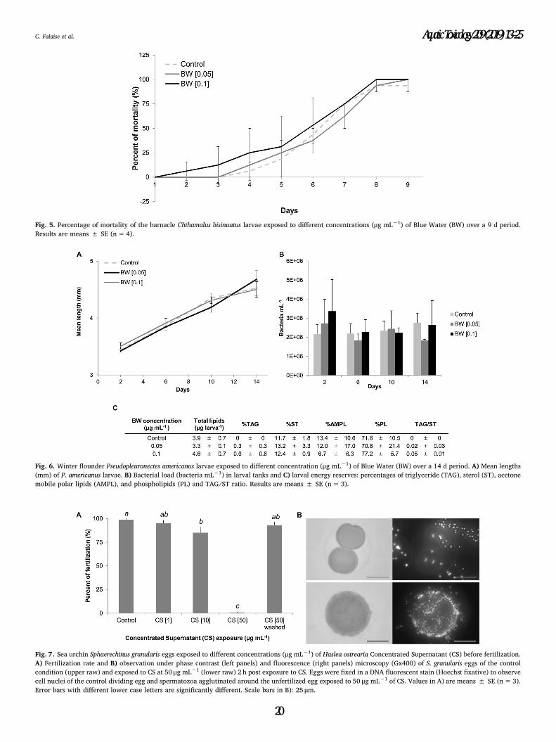
Fig. 5. Percentage of mortality of the barnacle Chthamalus bisinuatus larvae exposed to different concentrations (μg mL−1) of Blue Water (BW) over a 9 d period.
Results are means ± SE (n=4).
Fig. 6. Winter flounder Pseudopleuronectes americanus larvae exposed to different concentration (μg mL−1) of Blue Water (BW) over a 14 d period. A) Mean lengths
(mm) of P. americanus larvae. B) Bacterial load (bacteria mL−1) in larval tanks and C) larval energy reserves: percentages of triglyceride (TAG), sterol (ST), acetone
mobile polar lipids (AMPL), and phospholipids (PL) and TAG/ST ratio. Results are means ± SE (n= 3).
Fig. 7. Sea urchin Sphaerechinus granularis eggs exposed to different concentrations (μg mL−1) of Haslea ostrearia Concentrated Supernatant (CS) before fertilization.
A) Fertilization rate and B) observation under phase contrast (left panels) and fluorescence (right panels) microscopy (Gx400) of S. granularis eggs of the control
condition (upper raw) and exposed to CS at 50 μg mL−1 (lower raw) 2 h post exposure to CS. Eggs were fixed in a DNA fluorescent stain (Hoechst fixative) to observe
cell nuclei of the control dividing egg and spermatozoa agglutinated around the unfertilized egg exposed to 50 μg mL−1 of CS. Values in A) are means ± SE (n=3).
Error bars with different lower case letters are significantly different. Scale bars in B): 25 μm.
C. Falaise et al. Aquatic Toxicology 209 (2019) 13–25
20
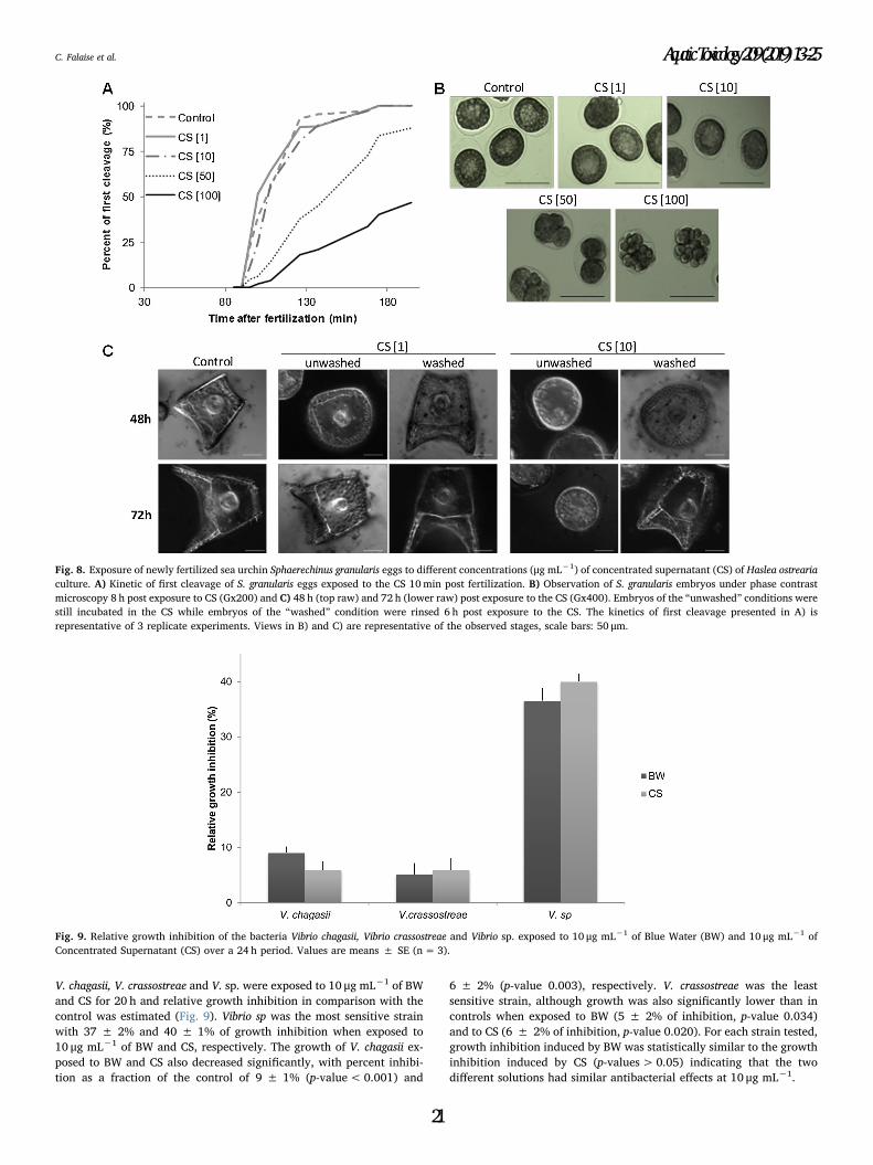
V. chagasii, V. crassostreae and V. sp. were exposed to 10 μg mL−1 of BW
and CS for 20 h and relative growth inhibition in comparison with the
control was estimated (Fig. 9). Vibrio sp was the most sensitive strain
with 37 ± 2% and 40 ± 1% of growth inhibition when exposed to
10 μg mL−1 of BW and CS, respectively. The growth of V. chagasii ex-
posed to BW and CS also decreased significantly, with percent inhibi-
tion as a fraction of the control of 9 ± 1% (p-value<0.001) and
6 ± 2% (p-value 0.003), respectively. V. crassostreae was the least
sensitive strain, although growth was also significantly lower than in
controls when exposed to BW (5 ± 2% of inhibition, p-value 0.034)
and to CS (6 ± 2% of inhibition, p-value 0.020). For each strain tested,
growth inhibition induced by BW was statistically similar to the growth
inhibition induced by CS (p-values> 0.05) indicating that the two
different solutions had similar antibacterial effects at 10 μg mL−1.
Fig. 8. Exposure of newly fertilized sea urchin Sphaerechinus granularis eggs to different concentrations (μg mL−1) of concentrated supernatant (CS) of Haslea ostrearia
culture. A) Kinetic of first cleavage of S. granularis eggs exposed to the CS 10min post fertilization. B) Observation of S. granularis embryos under phase contrast
microscopy 8 h post exposure to CS (Gx200) and C) 48 h (top raw) and 72 h (lower raw) post exposure to the CS (Gx400). Embryos of the “unwashed” conditions were
still incubated in the CS while embryos of the “washed” condition were rinsed 6 h post exposure to the CS. The kinetics of first cleavage presented in A) is
representative of 3 replicate experiments. Views in B) and C) are representative of the observed stages, scale bars: 50 μm.
Fig. 9. Relative growth inhibition of the bacteria Vibrio chagasii, Vibrio crassostreae and Vibrio sp. exposed to 10 μg mL−1 of Blue Water (BW) and 10 μg mL−1 of
Concentrated Supernatant (CS) over a 24 h period. Values are means ± SE (n=3).
C. Falaise et al. Aquatic Toxicology 209 (2019) 13–25
21
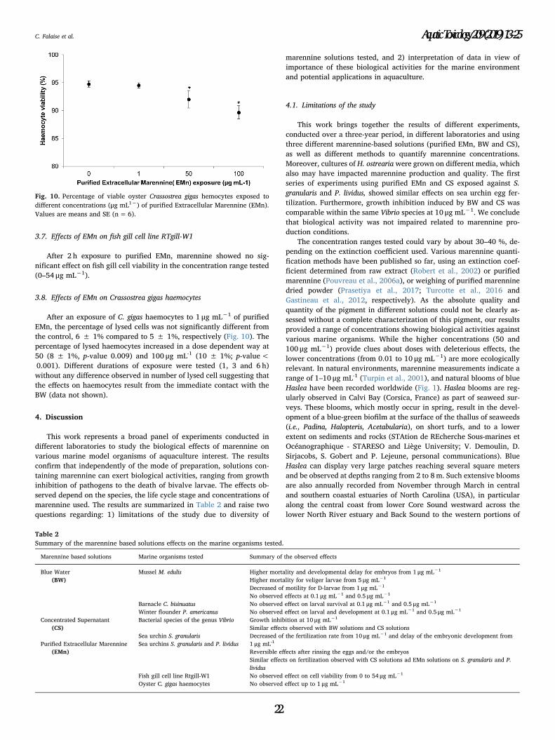
3.7. Effects of EMn on fish gill cell line RTgill-W1
After 2 h exposure to purified EMn, marennine showed no sig-
nificant effect on fish gill cell viability in the concentration range tested
(0–54 μg mL−1).
3.8. Effects of EMn on Crassostrea gigas haemocytes
After an exposure of C. gigas haemocytes to 1 μg mL−1 of purified
EMn, the percentage of lysed cells was not significantly different from
the control, 6 ± 1% compared to 5 ± 1%, respectively (Fig. 10). The
percentage of lysed haemocytes increased in a dose dependent way at
50 (8 ± 1%, p-value 0.009) and 100 μg mL-1 (10 ± 1%; p-value<
0.001). Different durations of exposure were tested (1, 3 and 6 h)
without any difference observed in number of lysed cell suggesting that
the effects on haemocytes result from the immediate contact with the
BW (data not shown).
4. Discussion
This work represents a broad panel of experiments conducted in
different laboratories to study the biological effects of marennine on
various marine model organisms of aquaculture interest. The results
confirm that independently of the mode of preparation, solutions con-
taining marennine can exert biological activities, ranging from growth
inhibition of pathogens to the death of bivalve larvae. The effects ob-
served depend on the species, the life cycle stage and concentrations of
marennine used. The results are summarized in Table 2 and raise two
questions regarding: 1) limitations of the study due to diversity of
marennine solutions tested, and 2) interpretation of data in view of
importance of these biological activities for the marine environment
and potential applications in aquaculture.
4.1. Limitations of the study
This work brings together the results of different experiments,
conducted over a three-year period, in different laboratories and using
three different marennine-based solutions (purified EMn, BW and CS),
as well as different methods to quantify marennine concentrations.
Moreover, cultures of H. ostrearia were grown on different media, which
also may have impacted marennine production and quality. The first
series of experiments using purified EMn and CS exposed against S.
granularis and P. lividus, showed similar effects on sea urchin egg fer-
tilization. Furthermore, growth inhibition induced by BW and CS was
comparable within the same Vibrio species at 10 μg mL−1. We conclude
that biological activity was not impaired related to marennine pro-
duction conditions.
The concentration ranges tested could vary by about 30–40 %, de-
pending on the extinction coefficient used. Various marennine quanti-
fication methods have been published so far, using an extinction coef-
ficient determined from raw extract (Robert et al., 2002) or purified
marennine (Pouvreau et al., 2006a), or weighing of purified marennine
dried powder (Prasetiya et al., 2017; Turcotte et al., 2016 and
Gastineau et al., 2012, respectively). As the absolute quality and
quantity of the pigment in different solutions could not be clearly as-
sessed without a complete characterization of this pigment, our results
provided a range of concentrations showing biological activities against
various marine organisms. While the higher concentrations (50 and
100 μg mL−1) provide clues about doses with deleterious effects, the
lower concentrations (from 0.01 to 10 μg mL−1) are more ecologically
relevant. In natural environments, marennine measurements indicate a
range of 1–10 μg mL-1 (Turpin et al., 2001), and natural blooms of blue
Haslea have been recorded worldwide (Fig. 1). Haslea blooms are reg-
ularly observed in Calvi Bay (Corsica, France) as part of seaweed sur-
veys. These blooms, which mostly occur in spring, result in the devel-
opment of a blue-green biofilm at the surface of the thallus of seaweeds
(i.e., Padina, Halopteris, Acetabularia), on short turfs, and to a lower
extent on sediments and rocks (STAtion de REcherche Sous-marines et
Océanographique - STARESO and Liège University; V. Demoulin, D.
Sirjacobs, S. Gobert and P. Lejeune, personal communications). Blue
Haslea can display very large patches reaching several square meters
and be observed at depths ranging from 2 to 8m. Such extensive blooms
are also annually recorded from November through March in central
and southern coastal estuaries of North Carolina (USA), in particular
along the central coast from lower Core Sound westward across the
lower North River estuary and Back Sound to the western portions of
Fig. 10. Percentage of viable oyster Crassostrea gigas hemocytes exposed to
different concentrations (μg mL1−) of purified Extracellular Marennine (EMn).
Values are means and SE (n= 6).
Table 2
Summary of the marennine based solutions effects on the marine organisms tested.
Marennine based solutions Marine organisms tested Summary of the observed effects
Blue Water
(BW)
Mussel M. edulis Higher mortality and developmental delay for embryos from 1 μg mL−1
Higher mortality for veliger larvae from 5 μg mL−1
Decreased of motility for D-larvae from 1 μg mL−1
No observed effects at 0.1 μg mL−1 and 0.5 μg mL−1
Barnacle C. bisinuatus No observed effect on larval survival at 0.1 μg mL−1 and 0.5 μg mL−1
Winter flounder P. americanus No observed effect on larval and development at 0.1 μg mL−1 and 0.5 μg mL−1
Bacterial species of the genus Vibrio Growth inhibition at 10 μg mL−1
Similar effects observed with BW solutions and CS solutions
Concentrated Supernatant
(CS)
Sea urchin S. granularis Decreased of the fertilization rate from 10 μg mL−1 and delay of the embryonic development from
1 μg mL-1
Reversible effects after rinsing the eggs and/or the embryos
Similar effects on fertilization observed with CS solutions ad EMn solutions on S. granularis and P.
lividus
Purified Extracellular Marennine
(EMn)
Sea urchins S. granularis and P. lividus
Fish gill cell line Rtgill-W1 No observed effect on cell viability from 0 to 54 μg mL−1
Oyster C. gigas haemocytes No observed effect up to 1 μg mL−1
C. Falaise et al. Aquatic Toxicology 209 (2019) 13–25
22
Bogue Sound. These blooms of a blue Haslea possibly different from H.
ostrearia do not seem to be associated with the development of biofilms
on Padina sp. thalli, however they cause a wide-spread greening of
oysters, which could underpin emerging green oyster industries in the
USA (N. Lindquist, personal communications).
Thus it can be inferred that, at the microenvironment scale, high
concentrations of marennine could be encountered, and that many
marine organisms can periodically and locally face acute marennine
exposure. All laboratory experiments to study biological activity of
marennine lasted hours or days, and the cumulated effect of long-
lasting exposure to low concentrations of marennine has not yet been
tested. Greening of oyster gills can be viewed as a cumulative, longer
term effect. According to green oyster producers, the greening in oyster
ponds can occur in a matter of days, depending on Haslea growth and
density. In the laboratory, EMn rather than IMn was preferentially re-
sponsible for greening, which proved to be time dependent and long-
lasting (Gastineau et al., 2018, 2014). However, green oysters have also
been observed in natural open environments, in absence of any record
of Haslea blooms, for instance in oyster leases in Australia (New South
Wales and Tasmania). This means that marennine could have biological
activities in the long-term, from subacute and chronic exposure, and
further work is required to better assess the impact of this pigment in
the natural environment.
4.2. The unpredictable “Jekyll and Hyde”, good and bad nature of
marennine
On the one hand, our results showed that exposure to the tested
marennine solutions could lead to adverse effects against many marine
animals, depending on the exposure dose. Concentrations from 1 μgmL−1 significantly lowered the survival rate of M. edulis embryos and
locomotion of the D-larvae. Significantly higher mortality rates were
observed for veliger larvae exposed to 5 μg mL-1 of BW while no mor-
tality was recorded at 1 μg mL−1, which contrasts with results of a
previous study where exposure of veliger larvae to 1 μg mL-1 of BW led
to 100% mortality, but the duration of that experiment was 4 times
longer, 20 d vs 5 d (Turcotte et al., 2016). Significant dose-response
effects of marennine were also observed with the sea urchin model.
While high concentrations (50 and 100 μg mL−1) resulted in a total
blockage of fertilization and embryonic development, lower con-
centrations (1 and 10 μg mL−1) of CS induced a developmental delay of
S. granularis embryos. Previous works also have demonstrated the ef-
fects of a water-soluble extract of the diatom Thalassiosira rotula on cell
division of the sea urchin P. lividus, with a blockage of cell division at
the higher doses tested and a delay and abnormal development of
embryos at lower doses (Buttino et al., 1999). Also, intact diatom cells
of six different species could inhibit the egg first cleavage of the sea
urchins Strongylocentrotus droebachiensis and Echinus acutus (Gudimova
et al., 2016). Most interestingly, our results indicated that even at high
concentrations, the effects of CS on fertilization and embryonic devel-
opment were reversible after rinsing the eggs or embryos. This confirms
previous observations of the effects of water-soluble diatom extracts
against various marine invertebrates, including the sea urchin Psam-
mechinus miliaris (Caldwell et al., 2002).
The present results could suggest an inability of marennine to bind
tightly at the cell surface if considering the reversible effects observed
in sea urchins, and to penetrate the cell and exert irreversible damages
given the high molecular weight of the molecule (> 10 kDa; Pouvreau
et al., 2006b). Marennine could however link to cell surface or mem-
brane receptors and trigger stress responses or even apoptotic signals by
inducing changes in cell metabolism. Furthermore, the variability of
BW and CS effects observed at both the interspecific and intraspecific
levels on the three Vibrio species tested, but also those previously shown
with V. aestuarianus, V. coralliilyticus and V. tubiashii strains (Falaise
et al., 2016) could reflect differences in membrane receptor affinities
and in mechanisms of action. Such variability should be considered
before application of marennine in aquaculture as an antibacterial
agent. However, more data is necessary to confirm these hypotheses.
On the other hand, despite observed deleterious effect at the higher
concentrations tested, very low concentrations (0.05 and 0.1 μg mL−1)
of BW did not exhibit a negative effect on the barnacle C. bisinuatus
survival nor on the winter flounder P. americanus. Both flounder growth
rate and bacterial load in the water remained unaffected throughout the
marennine exposure (0.05 and 0.1 μg mL−1), and physiological condi-
tion (energetic reserves) improved in response to marennine treatment.
These results compare with no observable effect of marennine (0–54 μgL−1) on the viability (metabolic activity) of the rainbow trout gill cell
line RTgill-W1. Although the gill cell line exposure was conducted for
only 2 h, results provide a strong indication that marennine does not
impair fish gill function at the concentration ranges tested here. These
results are further supported by (Turcotte et al., 2016) who demon-
strated that low exposure to BW (0.1 μg mL−1) enhanced survival and
physiological condition of M. edulis and P. magellanicus larvae. In ad-
dition, our results showed that low concentrations of EMn (1 μg mL−1)
did not present a cytotoxic effect against haemocytes of adult oysters.
Even though cytotoxic effects were evidenced at the highest con-
centrations (50 and 100 μg mL−1), the point of inflection could not be
determined with the concentration range tested. However, it should be
emphasized that even after centuries of production of green oysters, no
negative impacts on adult oysters, nor green oyster consumers have
been reported.
Despite the fact that diatoms are traditionally considered a high
quality food source enhancing growth and survival of many marine
organisms, they can produce secondary metabolites that function as
grazing deterrents (Ianora and Miralto, 2010). Most of these secondary
metabolites are polyunsaturated fatty acids (PUFAs) and poly-
unsaturated aldehydes (PUAs) with a special focus on oxylipins
(Caldwell, 2009). Effects of diatom aldehydes on reproductive and
fertilization success or embryonic development of various organisms
such as crustaceans, echinoderms or arthropods have been widely stu-
died (Caldwell, 2009; Ianora and Miralto, 2010). Aldehyde production
is a diatom chemical defense strategy to limit growth of their grazers
(Ban et al., 1997; Pohnert, 2005), but other studies reported the ab-
sence of a correlation between diatom aldehyde and reproductive
biology and early development of marine invertebrates, suggesting the
involvement of other unidentified diatom metabolites (Poulet et al.,
2006; Wichard et al., 2008). Unlike aldehydes, that are likely to exert
their activity following diatom cell destruction and ingestion, the
water-soluble marennine may target a larger spectrum of marine or-
ganisms and not only diatom grazers.
In conclusion, the present work demonstrates that H. ostrearia su-
pernatant containing marennine represents a biologically active water-
soluble solution with potential effects on various marine organisms.
More studies are required to better estimate the long term impacts of
blue Haslea blooms in natural environments, facilitated by laboratory
studies, investigating chronic exposure to low concentrations of mar-
ennine. If the use of H. ostrearia supernatant is considered in hatcheries
as a preventive or curative anti-infectious agent, concentrated solutions
such as BW or CS could be of interest in order to control the delivered
marennine doses. It is likely that the adverse effects of marennine only
target the early and more succeptable fragile developmental stages such
as embryos and larvae, depending on the exposure concentration,
whereas adults remain unaffected.
Acknowledgements
We thank the reviewers for their valuable comments that allowed us
improving this work. This publication benefited from funding from the
research program on the valorization of marine biomass for larval
feeding (ISMER/UQAR) (RT), from the RFI Food for Tomorrow (project
Maliblue), from the Territorial Collectivity of Corsica and the Rhone-
Mediterranean and Corsica Water Agency (STARE-CAPMED - Station of
C. Falaise et al. Aquatic Toxicology 209 (2019) 13–25
23
reference and research on change of local and global anthropogenic
pressures on Mediterranean ecosystem drifts), and also from the
European Commission under the Horizon 2020 Research and
Innovation Program GHaNA (The Genus Haslea, New marine resources
for blue biotechnology and Aquaculture, grant agreement No [734708/
GHANA/H2020-MSCA-RISE- 2016]) (JLM).
References
Auffret, M., Oubella, R., 1997. Hemocyte aggregation in the oyster Crassostrea gigas: In
Vitro measurement and experimental modulation by xenobiotics. Comp. Biochem.
Physiol. – Part Physiol. 3, 705–712.
Ban, S., Burns, C., Castel, J., Chaudron, Y., Christou, E., Escribano, R., Umani, S.F.,
Gasparini, S., Ruiz, F.G., Hoffmeyer, M., Ianora, A., Kang, H., Laabir, M., Lacoste, A.,
Miralto, A., Ning, X., Poulet, S., Rodriguez, V., Runge, J., Shi, J., Starr, M., Uye, S.,
Wang, Y., 1997. The paradox of diatom-copepod interactions. Mar. Ecol. Prog. Ser.
157, 287–293. https://doi.org/10.3354/meps157287.
Bergé, J.P., Bourgougnon, N., Alban, S., Pojer, F., Billaudel, S., Chermann, J.C., Robert,
J.M., Franz, G., 1999. Antiviral and anticoagulant activities of a water-soluble frac-
tion of the marine diatom Haslea ostrearia. Planta Med. 65, 604–609.
Bouhlel, Z., Turcotte, F., Warschawski, D.E., Arnold, A., Lemarchand, K., Deschênes, J.-S.,
Marcotte, I., Tremblay, R., 2018. Marennine, a potential alternative antibiotic to
polymyxine in aquaculture. Presented at the Aquaculture Canada 2018.
Bruto, M., James, A., Petton, B., Labreuche, Y., Chenivesse, S., Alunno-Bruscia, M., Polz,
M.F., Le Roux, F., 2017. Vibrio crassostreae, a benign oyster colonizer turned into a
pathogen after plasmid acquisition. ISME J. 11, 1043–1052. https://doi.org/10.
1038/ismej.2016.162.
Buttino, I., Miralto, A., Ianora, A., Romano, G., Poulet, S.A., 1999. Water-soluble extracts
of the diatom Thalassiosira rotula induce aberrations in embryonic tubulin organisa-
tion of the sea urchin Paracentrotus lividus. Mar. Biol. 134, 147–154. https://doi.org/
10.1007/s002270050533.
Buttino, I., Hwang, J.-S., Romano, G., Sun, C.-K., Liu, T.-M., Pellegrini, D., Gaion, A.,
Sartori, D., 2016. Detection of malformations in sea urchin plutei exposed to mercuric
chloride using different fluorescent techniques. Ecotoxicol. Environ. Saf., 6th
Biannual Ecotoxicology Meeting (BECOME 2014) - Environmental Emergencies:
Ecotoxicology as a Management Tool 123, 72–80. https://doi.org/10.1016/j.ecoenv.
2015.07.027.
Caldwell, G.S., 2009. The influence of bioactive oxylipins from marine diatoms on in-
vertebrate reproduction and development. Mar. Drugs 7, 367–400. https://doi.org/
10.3390/md7030367.
Caldwell, G.S., Olive, P.J.W., Bentley, M.G., 2002. Inhibition of embryonic development
and fertilization in broadcast spawning marine invertebrates by water soluble diatom
extracts and the diatom toxin 2-trans,4-trans decadienal. Aquat. Toxicol. Amst. Neth.
60, 123–137.
Carbonnelle, D., Pondaven, P., Morancais, M., Masse, G., Bosch, S., Jacquot, C., Briand,
G., Robert, J., Roussakis, C., 1998. Antitumor and antiproliferative effects of an
aqueous extract from the marine diatom Haslea ostrearia (Simonsen) against solid
tumors: lung carcinoma (NSCLC-N6), kidney carcinoma (E39) and melanoma (M96)
cell lines. Anticancer Res. 19, 621–624.
Clinical and Laboratory Standards Institute (CLSI), 2012. Methods for Dilution
Antimicrobial Susceptibility Tests for Bacteria That Grow Aerobically. Document
M07-A9. Approved Standard—Ninth Edition. Wayne, PA, USA. .
Falaise, C., François, C., Travers, M.-A., Morga, B., Haure, J., Tremblay, R., Turcotte, F.,
Pasetto, P., Gastineau, R., Hardivillier, Y., Leignel, V., Mouget, J.-L., 2016.
Antimicrobial compounds from eukaryotic microalgae against human pathogens and
diseases in aquaculture. Mar. Drugs 14, 159. https://doi.org/10.3390/md14090159.
Folch, J., Lees, M., Stanley, G.H.S., 1957. A simple method for the isolation and pur-
ification of total lipides from animal tissues. J. Biol. Chem. 226, 497–509.
Fraboulet, E., Lambert, Y., Tremblay, R., Audet, C., 2010. Assessment of paternal effect
and physiological cost of metamorphosis on growth of young winter flounder
Pseudopleuronectes americanus juveniles in a cold environment. J. Fish Biol. 76,
930–948. https://doi.org/10.1111/j.1095-8649.2010.02538.x.
Gaillon, B., 1820. Des huîtres vertes, et des causes de cette coloration. J. Phys. Chim.
D’histoire Nat. Arts 91, 222–225.
Gastineau, R., Pouvreau, J.-B., Hellio, C., Morançais, M., Fleurence, J., Gaudin, P.,
Bourgougnon, N., Mouget, J.-L., 2012. Biological activities of purified marennine, the
blue pigment responsible for the greening of oysters. J. Agric. Food Chem. 60,
3599–3605. https://doi.org/10.1021/jf205004x.
Gastineau, R., Turcotte, F., Pouvreau, J.-B., Morançais, M., Fleurence, J., Windarto, E.,
Prasetiya, F.S., Arsad, S., Jaouen, P., Babin, M., Coiffard, L., Couteau, C., Bardeau, J.-
F., Jacquette, B., Leignel, V., Hardivillier, Y., Marcotte, I., Bourgougnon, N.,
Tremblay, R., Deschênes, J.-S., Badawy, H., Pasetto, P., Davidovich, N., Hansen, G.,
Dittmer, J., Mouget, J.-L., 2014. Marennine, promising blue pigments from a wide-
spread Haslea diatom species complex. Mar. Drugs 12, 3161–3189. https://doi.org/
10.3390/md12063161.
Gastineau, R., Hansen, G., Davidovich, N.A., Davidovich, O., Bardeau, J.-F., Kaczmarska,
I., Ehrman, J.M., Leignel, V., Hardivillier, Y., Jacquette, B., Poulin, M., Morançais, M.,
Fleurence, J., Mouget, J.-L., 2016. A new blue-pigmented hasleoid diatom, Haslea
provincialis, from the Mediterranean Sea. Eur. J. Phycol. 51, 156–170. https://doi.
org/10.1080/09670262.2015.1110861.
Gastineau, R., Prasetiya, F.S., Falaise, C., Cognie, B., Decottignies, P., Morancais, M.,
Méléder, V., Davidovich, N.A., Turcotte, F., Tremblay, R., Pasetto, P., Dittmer, J.,
Bardeau, J.-F., Pouvreau, J.-B., Mouget, J.-L., 2018. Part II: marine molecules for
disease treatment / prevention and for biological research, Chap16: marennine-like
pigments: Blue diatom of Green oyster cult? In: La Barre, S., Bates, S., Qian, P. (Eds.),
Blue Biotechnologies: Production and Uses of Marine Molecules. Wiley VCH,
Weinheim, Germany.
Gudimova, E., Eilertsen, H.C., Jørgensen, T.Ø., Hansen, E., 2016. In vivo exposure to
northern diatoms arrests sea urchin embryonic development. Toxicon 109, 63–69.
https://doi.org/10.1016/j.toxicon.2015.11.001.
Hennebicq, R., Fabra, G., Pellerin, C., Marcotte, I., Myrand, B., Tremblay, R., 2013. The
effect of spawning of cultured mussels (Mytilus edulis) on mechanical properties,
chemical and biochemical composition of byssal threads. Aquaculture 410–411,
11–17. https://doi.org/10.1016/j.aquaculture.2013.06.011.
Hjörleifsson, E., Klein-MacPhee, G., 1992. Estimation of live standard length of winter
flounder Pleuronectes americanus larvae from formalin-preserved, ethanol-preserved
and frozen specimens. Mar. Ecol. Prog. Ser. 82, 13–19.
Ianora, A., Miralto, A., 2010. Toxigenic effects of diatoms on grazers, phytoplankton and
other microbes: a review. Ecotoxicol. Lond. Engl. 19, 493–511. https://doi.org/10.
1007/s10646-009-0434-y.
Kasten, P., Flores, A.A.V., 2013. Disruption of endogenous tidal rhythms of larval release
linked to food supply and heat stress in an intertidal barnacle. Mar. Ecol. Prog. Ser.
472, 185–198. https://doi.org/10.3354/meps10005.
Khemis, I.B., Audet, C., Fournier, R., Noüe, J.D.L., 2003. Early weaning of winter flounder
(Pseudopleuronectes americanus Walbaum) larvae on a commercial microencapsulated
diet. Aquac. Res. 34, 445–452. https://doi.org/10.1046/j.1365-2109.2003.00828.x.
Mitchell, P.H., Barney, R.L., 1917. The Occurrence in Virginia of Green-gilled Oysters
Similar to Those of Marennes. Govt. print. off., Washington.
Morga, B., Arzul, I., Chollet, B., Renault, T., 2009. Infection with the protozoan parasite
Bonamia ostreae modifies in vitro haemocyte activities of flat oyster Ostrea edulis. Fish
Shellfish Immunol. 26, 836–842. https://doi.org/10.1016/j.fsi.2009.03.018.
Mouget, J.-L., Gastineau, R., Davidovich, O., Gaudin, P., Davidovich, N.A., 2009. Light is
a key factor in triggering sexual reproduction in the pennate diatom Haslea ostrearia.
FEMS Microbiol. Ecol. 69, 194–201. https://doi.org/10.1111/j.1574-6941.2009.
00700.x.
Myrand, B., Tremblay, R., Sévigny, J.-M., 2009. Impact of suspension culture using mesh
sleeves on genetic characteristics of Mytilus edulis L. in Canada. Aquaculture 291,
147–153. https://doi.org/10.1016/j.aquaculture.2009.03.015.
Nassiri, Y., Robert, J.-M., Rincé, Y., Ginsburger-Vogel, T., 1998. The cytoplasmic fine
structure of the diatom Haslea ostrearia (Bacillariophyceae) in relation to marennine
production. Phycologia 37, 84–91. https://doi.org/10.2216/i0031-8884-37-2-84.1.
Parrish, C.C., 1987. Separation of aquatic lipid classes by chromarod thin-layer chro-
matography with measurement by latroscan flame ionization detection. Can. J. Fish.
Aquat. Sci. 44, 722–731. https://doi.org/10.1139/f87-087.
Petersen, C.G., 1916. “Grüne Austern” in Dänemark in 1911–1912. Int. Rev. Gesamten
Hydrobiol. Hydrogr. 7, 39–41.
Pinsino, A., Matranga, V., Trinchella, F., Roccheri, M.C., 2010. Sea urchin embryos as an
in vivo model for the assessment of manganese toxicity: developmental and stress
response effects. Ecotoxicology 19, 555–562. https://doi.org/10.1007/s10646-009-
0432-0.
Pohnert, G., 2005. Diatom/Copepod interactions in plankton: the indirect chemical de-
fense of unicellular algae. ChemBioChem 6, 946–959. https://doi.org/10.1002/cbic.
200400348.
Poulet, S.A., Wichard, T., Ledoux, J.B., Lebreton, B., Marchetti, J., Dancie, C., Bonnet, D.,
Cueff, A., Morin, L.P., Pohnert, G., Poulet, S.A., Ledoux, J.B., Dancie, C., Morin, L.P.,
Poulet, S.A., Cueff, C., Pohnert, G., 2006. Influence of Diatoms on Copepod
Reproduction. I. Field and Laboratory Observations Related to Calanus helgolandicus
Egg Production. https://doi.org/10.3354/meps308129.
Pouvreau, J.-B., Morançais, M., Fleurence, J., Pondaven, P., 2006a. Method for the
quantification of the blue-green pigment “marennine” synthesized by the marine
diatom Haslea ostrearia (Gaillon/Bory) Simonsen using HPLC gel-filtration and pho-
todiode-array detection. J. Appl. Phycol. 19, 263–270. https://doi.org/10.1007/
s10811-006-9133-8.
Pouvreau, J.-B., Morançais, M., Fleury, F., Rosa, P., Thion, L., Cahingt, B., Zal, F.,
Fleurence, J., Pondaven, P., 2006b. Preliminary characterisation of the blue-green
pigment “marennine” from the marine tychopelagic diatom Haslea ostrearia (Gaillon/
Bory) Simonsen. J. Appl. Phycol. 18, 757–767. https://doi.org/10.1007/s10811-006-
9087-x.
Pouvreau, J.-B., Morançais, M., Massé, G., Rosa, P., Robert, J.-M., Fleurence, J.,
Pondaven, P., 2006c. Purification of the blue-green pigment “marennine” from the
marine tychopelagic diatom Haslea ostrearia (Gaillon/Bory) Simonsen. J. Appl.
Phycol. 18, 769–781. https://doi.org/10.1007/s10811-006-9088-9.
Pouvreau, J.-B., Housson, E., Tallec, L.L., Morançais, M., Rincé, Y., Fleurence, J.,
Pondaven, P., 2007. Growth inhibition of several marine diatom species induced by
the shading effect and allelopathic activity of marennine, a blue-green polyphenolic
pigment of the diatom Haslea ostrearia (Gaillon/Bory) Simonsen. J. Exp. Mar. Biol.
Ecol. 352, 212–225. https://doi.org/10.1016/j.jembe.2007.07.011.
Pouvreau, J.-B., Morançais, Michèle, Taran, F., Rosa, P., Dufossé, Laurent, Guérard,
Fabienne, Pin, S., Fleurence, J., Pondaven, P., 2008. Antioxidant and free radical
scavenging properties of Marennine, a blue-green polyphenolic pigment from the
diatom Haslea ostrearia (Gaillon/Bory) simonsen responsible for the natural greening
of cultured oysters. J. Agric. Food Chem. 56, 6278–6286. https://doi.org/10.1021/
jf073187n.
Prasetiya, F.S., Safitri, I., Widowati, I., Cognie, B., Decottignies, P., Gastineau, R.,
Morançais, M., Windarto, E., Tremblay, R., Mouget, J.-L., 2016. Does allelopathy
affect co-culturing Haslea ostrearia with other microalgae relevant to aquaculture? J.
Appl. Phycol. 28, 2241–2254. https://doi.org/10.1007/s10811-015-0779-y.
Prasetiya, F.S., Comeau, L.A., Gastineau, R., Decottignies, P., Cognie, B., Morançais, M.,
Turcotte, F., Mouget, J.-L., Tremblay, R., 2017. Effect of marennine produced by the
C. Falaise et al. Aquatic Toxicology 209 (2019) 13–25
24
blue diatom Haslea ostrearia on behavioral, physiological and biochemical traits of
juvenile Mytilus edulis and Crassostrea virginica. Aquaculture 467, 138–148. https://
doi.org/10.1016/j.aquaculture.2016.08.029.
Quinn, G.P., Keough, M.J., 2002. Experimental Design and Data Analysis for Biologists.
Cambridge University Press, Cambridge.
Ranson, G., 1927. L’absorption de matières organiques dissoutes par la surface extérieure
du corps chez les animaux aquatiques. Ann. Inst. Océan. 4, 174.
Robert, J.-M., Morançais, M., Pradier, E., Mouget, J.-L., Tremblin, G., 2002. Extraction
and quantitative analysis of the blue-green pigment “marennine” synthesized by the
diatom Haslea ostrearia. J. Appl. Phycol. 14, 299–305. https://doi.org/10.1023/
A:1021184532581.
Seychelles, L.H., Audet, C., Tremblay, R., Lemarchand, K., Pernet, F., 2011. Bacterial
colonization of winter flounder Pseudopleuronectes americanus fed live feed enriched
with three different commercial diets. Aquac. Nutr. 17, e196–e206. https://doi.org/
10.1111/j.1365-2095.2009.00750.x.
Sprat, T., 1667. History of the Royal Society. Martyn, London, UK.
Steinhardt, R.A., Epel, D., 1974. Activation of sea-urchin eggs by a calcium ionophore.
Proc. Natl. Acad. Sci. 71, 1915–1919. https://doi.org/10.1073/pnas.71.5.1915.
Tardy-Laporte, C., Arnold, A.A., Genard, B., Gastineau, R., Morançais, M., Mouget, J.-L.,
Tremblay, R., Marcotte, I., 2013. A 2H solid-state NMR study of the effect of anti-
microbial agents on intact Escherichia coli without mutating. Biochim. Biophys. Acta
BBA - Biomembr. 1828, 614–622. https://doi.org/10.1016/j.bbamem.2012.09.011.
Turcotte, F., Mouget, J.-L., Genard, B., Lemarchand, K., Deschênes, J.-S., Tremblay, R.,
2016. Prophylactic effect of Haslea ostrearia culture supernatant containing the pig-
ment marennine to stabilize bivalve hatchery production. Aquat. Living Resour. 29,
401. https://doi.org/10.1051/alr/2016032.
Turpin, V., Robert, J.-M., Goulletquer, P., Massé, G., Rosa, P., 2001. Oyster greening by
outdoor mass culture of the diatom Haslea ostrearia Simonsen in enriched seawater.
Aquac. Res. 32, 801–809. https://doi.org/10.1046/j.1365-2109.2001.00615.x.
Wichard, T., Poulet, S.A., Boulesteix, A.L., Ledoux, J.B., Lebreton, B., Marchetti, J.,
Pohnert, G., Pohnert, G., 2008. Influence of Diatoms on Copepod Reproduction. Ii.
Uncorrelated Effects of Diatom-derived alpha,beta,gamma,delta-unsaturated
Aldehydes and Polyunsaturated Fatty Acids on Calanus helgolandicus in the Field.
C. Falaise et al. Aquatic Toxicology 209 (2019) 13–25
25
Annexes
ANNEXE 3 : Publication
Falaise et al. (2019), Marine Drugs [Texte Complet]
Falaise, C., James A., Travers M-A., Zanella M., Badawi M., Mouget, J.-L., 2019. Complex
relationships between the blue pigment marennine and marine bacteria of the genus Vibrio.
Marine Drugs, 17(3), 160-172.
marine drugs
Article
Complex Relationships between the Blue PigmentMarennine and Marine Bacteria of the Genus Vibrio
Charlotte Falaise 1, Adèle James 2,3, Marie-Agnès Travers 4, Marie Zanella 1, Myriam Badawi 1
and Jean-Luc Mouget 1,*
1 Laboratoire Mer Molécule Santé (EA 2160, FR CNRS 3473 IUML), Le Mans Université,
72000 Le Mans, France; [email protected] (C.F.); [email protected] (M.Z.);
[email protected] (M.B.)2 Ifremer, Unité Physiologie Fonctionnelle des Organismes Marins, ZI de la Pointe du Diable,
29280 Plouzané, France; [email protected] Sorbonne Université (UPMC Paris 06, CNRS, UMR 8227) Laboratoire de Biologie Intégrative des Modèles
Marins, Station Biologique de Roscoff, 29680 Roscoff, France4 Ifremer (RBE-SG2M-LGPMM) Laboratoire de Génétique et de Pathologie des Mollusques Marins,
Station La Tremblade, Avenue Mus Loup, F-17390 La Tremblade, France; [email protected]
* Correspondence: [email protected]; Tel.: +33-243-833-242
Received: 14 February 2019; Accepted: 4 March 2019; Published: 8 March 2019�����������������
Abstract: Marennine, the water-soluble blue pigment produced by the marine diatom Haslea ostrearia,
is known to display antibacterial activities. Previous studies have demonstrated a prophylactic
effect of marennine on bivalve larvae challenged with a pathogenic Vibrio splendidus, suggesting
that the blue Haslea is a good candidate for applications in aquaculture as a source of a natural
antimicrobial agent. Indeed, the genus Vibrio is ubiquitous in aquaculture ecosystems, and regular
events of pathogenic invasion cause some of the biggest losses worldwide. To better characterize
the effects of marennine on Vibrios, a panel of 30 Vibrio strains belonging to 10 different species
was tested, including bivalve pathogenic species (e.g., Vibrio crassostreae and Vibrio harveyi). Vibrio
strains were first exposed to 10 and 25 µg mL−1 of Blue Water (BW), a concentrated culture
supernatant of H. ostrearia containing marennine. This screening evidenced a great diversity in
responses, from growth stimulation to a total inhibition, at both the interspecific or intraspecific level.
In a second series of experiments, 10 Vibrio strains were exposed to BW at concentrations ranging from
5 to 80 µg mL−1. The highest concentrations of BW did not systematically result in the highest growth
inhibition as hormetic responses—opposite effects regarding the concentration—were occasionally
evidenced. The relationships between marennine and Vibrio strains appear more complex than
expected and justify further study—in particular, on the mechanisms of action—before considering
applications as a natural prophylactic or antibiotic agent in aquaculture.
Keywords: antibacterial activity; diauxie; Haslea; hormesis; marennine; Vibrio
1. Introduction
The marine diatom Haslea ostrearia is characterized by the production of a specific blue-green
pigment, named marennine. This water-soluble pigment accumulates at the apices of the cells before
its release into the surrounding environment [1]. H. ostrearia is a ubiquitous diatom and is of special
interest in the Atlantic French coast (e.g., Marennes Bay, Bourgneuf Bay), where blooms in oyster ponds
induce the greening of oyster gills that increases the market value of bivalves. Blue diatoms other
than H. ostrearia have been identified in the last decade, such as Haslea karadagensis (Black Sea; [2]),
Haslea provincialis (Mediterranean Sea; [3]), and more recently, Haslea nusantara (Java Sea; [4]). All these
species produce blue pigments whose spectral characteristics slightly differ from marennine, and are
Mar. Drugs 2019, 17, 160; doi:10.3390/md17030160 www.mdpi.com/journal/marinedrugs
Mar. Drugs 2019, 17, 160 2 of 12
named marennine-like pigments in the absence of more specific determination (e.g., [2]). Despite
an increasing knowledge on blue Haslea biodiversity and distribution, questions still remain about
these blue pigments and their functions for the algae. Indeed, marennine or marennine-like pigments
are highly complex molecules, and their chemical structure remains undetermined. Some glycosidic
units attached to one or various aromatic rings have been evidenced, but the exact nature of the
chromophore is still unknown yet [5].
Regarding the function of the pigment for the microalga, the significant release of blue pigments
by Haslea species in seawater (in the range of 1–15 µg mL−1 in oyster ponds; [6]), combined
with an increasing amount of evidence that marennine interacts with different marine organisms,
could advocate for a protective or a competitive role. Indeed, allelopathic effects were demonstrated
toward various microalgal species [7,8], as well as antimicrobial effects against several marine
bacteria and fungi (reviewed in [9]). More particularly, in vitro experiments have demonstrated
antibacterial effects of marennine against various marine bacteria, including strains from the Vibrio
genus, such as Vibrio anguillarium [10], Vibrio aestuarianus [11], or Vibrio splendidus [12]. The Vibrio
genus is genetically and metabolically highly diverse, and several species have been described as
pathogenic for shellfish [13]. Major pathogens found in hatcheries or in fields belong to Splendidus,
Coralliilyticus, Harveyi clades, or to V. aestuarianus and V. tapetis species. However, it is important to
consider the ecological populations, as all strains of a same species do not share colonization and
toxicity characteristics, and thus are not pathogenic. Furthermore, different strains of a same Vibrio
species present distinct sensitivities toward marennine [9]. Hence, bacterial response to marennine
exposure can be species- and strain-dependent. This biological activity seems to be intrinsic to blue
Haslea species, as antibacterial activities were also demonstrated with the marennine-like pigment
produced by H. karadagensis against V. aestuarianus and other species of interest in aquaculture [14].
At the sight of such results, the use of blue Haslea and marennine has been considered for
aquaculture applications. Marennine biological activities have thus been investigated in vivo, using
blue mussel and giant scallop larvae exposed to a concentrated supernatant of H. ostrearia culture
enriched in extracellular marennine [15]. Low concentrations of this Blue Water (BW) solution
significantly increased larval survival when challenged with a pathogenic V. splendidus strain [15].
This result is very promising in aquaculture for shellfish and fish larval health, but a better
characterization of the interactions between marennine and pathogenic bacteria is needed. Thus,
the present work aims to increase further our knowledge about the antimicrobial activity of H. ostrearia
blue pigment, by assessing the effects of marennine on different species and strains of the genus Vibrio
that are threatening aquaculture sustainability.
2. Materials and Methods
2.1. Vibrio Strains
Thirty Vibrio strains belonging to 10 species were tested for their sensitivity toward Blue Water
(BW), a concentrated supernatant of H. ostrearia culture containing the extracellular marennine. Vibrio
chagasii (strain #11, #12, #13), Vibrio crassostreae (#51, #52, #53), Vibrio fortis (#7, #8, #9), Vibrio harveyi
(#21, #22, #23), Vibrio orientalis (#1, #2, #3), Vibrio splendidus (#90, #91, #93), V. tasmaniensis (#112, #113,
#114), Vibrio sp. (isolated from oyster tissues; #36, #37, #38), and V sp. (isolated from sea water; #90,
#91, #33) strains were provided by the Genomics of Vibrio team (Laboratoire de Biologie Intégrative
des Modèles Marins (LBI2M), station biologique de Roscoff, France) and were previously described
in [16]. Vibrio aestuarianus strains (#07/115, #12/016, #03/008) were provided by the Laboratoire de
Génétique et de Pathologie des Mollusques Marins (LGPMM) of the Institut Français de Recherche
pour l’Exploitation de la Mer (IFREMER; La Tremblade, France).
Mar. Drugs 2019, 17, 160 3 of 12
2.2. Vibrio Exposure to Blue Water Solutions
The susceptibility of the Vibrio strains to BW was assessed with the method described in
the Clinical and Laboratory Standards Institute (CLSI) antimicrobial microdilution guidelines [17].
Bacterial inocula at a defined concentration were exposed to different BW concentrations in a 96-well
microplate with a flat bottom and cover (BrandTechTMBRANDplatesTMpureGradeTM S 96-well
Microplates, Thermo Fisher Scientific, Waltham, MA, USA). Bacterial growth was monitored by
Optical Density (OD) measurements with a microplate spectrophotometer (xMark, Bio-Rad, Hercules,
CA, USA). Bacterial growth was then recorded using Microplate Manager 6 Software (MPM6, Bio-Rad,
Hercules, CA, USA) with OD measurement (at 600 nm, to avoid the absorbance peak of marennine
around 677 nm; [5]) of each well every 30 min for 24 h at ambient temperature.
2.2.1. Preparation of Bacterial Inocula
Vibrio strains were kept at −80 ◦C in 25% glycerol. Broth cultures were prepared with an autoclaved,
cation-adjusted, Mueller–Hinton broth media (CaMHB; Biokar, Solabia Group, Pantin, France) by
the addition of 1% NaCl (pH 7.5 ± 0.2; salinity = 32) and agar media, prepared with autoclaved,
cation-adjusted Nutrient Agar (CaNA; Biokar) by the addition of 2.3% NaCl (final pH 7.5 ± 0.2;
salinity = 32). Prior to the antibacterial assays, each Vibrio strain was inoculated in CaMHB from
the −80 ◦C sample, incubated overnight at 25 ◦C under moderate agitation (130 rpm), and isolated
on CaNA Petri dishes. After 1 day of incubation at 25 ◦C, plates containing the isolated colonies
were kept at 4 ◦C for no more than a week. Three different colonies per Petri dish were inoculated
in CaMHB (biological replicates, n = 3) and grown overnight at ambient temperature. The next day,
the OD (630 nm) of bacteria in the broth culture was measured (V-10 Plus Humeau Spectrophotometer,
La-Chapelle-sur-Erdre, France) and the absorbance was adjusted at 0.1 by dilution in CaMHB. To obtain
the bacterial inoculum, the solution was further diluted by 1/100 in CaMHB, as recommended by the
CLSI guidelines [17]. The bacterial inocula were exposed to BW in the microplates within 15 min after
the dilution.
2.2.2. Blue Water (BW) Production
The growth of the 30 Vibrio strains exposed to Blue Water (BW) solutions was studied over a 24 h
period. BW was prepared from a concentrated supernatant of H. ostrearia culture containing the
extracellular marennine, and was produced at the Station aquicole de Pointe-au-Père, Institut des
Sciences de la Mer à Rimouski-Université du Québec à Rimouski (ISMER-UQAR; Québec, Canada)
during the spring of 2017. H. ostrearia was cultured in 100 L circular and flat bottom photobioreactors
with filtered sea water (temperature: 20 ◦C; salinity 28) at high irradiance (180 µmol photons m−2 s−1),
in a 14/10 h light/dark cycle for 3 weeks, until marennine concentration reached around 6–7 µg mL−1,
as described in [15]. The supernatant was then collected and concentrated ca. 20 times by ultrafiltration
(double cut-off at 3–30 kDa) as described in [18], for a final estimated concentration of ca. 120 µg mL−1
(pH 7.7 ± 0.2; salinity = 0), and stored in the dark at 4 ◦C. The BW concentration was assessed with
spectrophotometric measurements (UV/Vis Lambda 25 Perkin Elmer spectrophotometer and UV
Winlab Perkin Elmer software (version 6.0.4 2011), Waltham, MA, USA) on a syringe-filtered BW
solution (0.2 µm; Sarstedt) and 1 cm path-length quartz cuvettes, using the Beer–Lambert equation
(ε677 = 12.13 L g−1 cm−1) as proposed by [18]. Prior to the antibacterial experiments, the BW stock
solution was syringe-filtered on 0.4 µm, and the salinity and the pH were adjusted to be similar to
the CaMHB at 32 and 7.5 ± 0.2, respectively, by addition of NaCl and HCl 0.1 M. BW dilutions were
prepared with sterile, ultra-pure water plus NaCl (pH 7.5 ± 0.2; salinity = 32). The BW solutions at
different concentrations were then syringe-filtered through 0.2 µm and kept at 4 ◦C.
Mar. Drugs 2019, 17, 160 4 of 12
2.2.3. Antibacterial Essay
In a first series of experiments, the 30 Vibrio strains corresponding to 10 different species were
screened and exposed to three BW concentrations: 0 µg mL−1 (control), 10 µg mL−1, and 25 µg mL−1.
In a second series of experiments, 10 Vibrio strains presenting different patterns of sensitivity to
marennine were exposed to a dilution range of BW: 0 µg mL−1, 5 µg mL−1, 10 µg mL−1, 25 µg mL−1,
50 µg mL−1, 70 µg mL−1, and 85 µg mL−1. For the screening experiment, the final volume in each well
of the microplates was 100 µL, with a final ratio of 1:1 (v/v) bacterial inoculum: BW. In the dilution
range experiment, to reach concentrations as high as 70 and 85 µg mL−1, with a BW stock solution
of 117 µg mL−1, the final volume in each well was adjusted to 200 µL, with a final ratio of 1:4 (v/v)
bacterial inoculum: BW. Microplates were first filled with the BW solutions, and bacterial inocula
were then added to each well using a single channel electronic micropipette (Eppendorf Research
Pro 50–1000 µL, Eppendorf, Hamburg, Germany). After being completed, microplates were sealed
with parafilm and placed in the microplate spectrophotometer for the 24 h run. The experiments were
conducted in triplicate, with technical triplicates for each condition. A negative control was also run
per microplate for only BW and CaMHB.
2.3. Growth Curve Analyses and Statistics
Bacterial growth kinetics were analyzed using R 3.5.1 software. For the screening experiment,
the OD (600 nm) data obtained over the 24 h run were fitted with a bi-phasic logistic growth equation,
defined as below:
f(x) =k1
1 + e−r1(x−x1)+
k2
1 + e−r2(x−x2).
Interpretable metric parameters were then obtained, such as the maximum possible population
size in a particular environment for the first phase of growth (k1 parameter) and for the second phase
of growth (k2 parameter), or the growth rate (r1 and r2 parameters). The k2 parameter was chosen in
the screening experiment to study the effects of BW on the Vibrio strains.
For the concentration range experiments, the k parameter was also studied, but growth curves
were fitted with a simple logistic growth equation, as it was not possible to fit growth curves of a same
strain under the different BW exposures with the same bi-phasic logistic growth equation. Growth
curves were analyzed with the R package “Growth Curver” [19].
R software was also used for statistical analyses. A Shapiro–Wilkinson test was used to verify
data normality, and a Fisher test for the homogeneity of variance. There was no need to perform data
transformation. The differences between treatments were assessed with one-way ANOVA, and post-hoc
Tukey’s pairwise multiple comparison tests were used to determine differences between pairs. Unless
specified, data are expressed as mean ± standard error (SE).
3. Results
3.1. Different Patterns of Vibrio Growth Curves Evidenced by the Screening Experiment
The growth curves of the 30 Vibrio strains exposed to BW at 0 µg mL−1, 10 µg mL−1, or 25 µg mL−1
were recorded for 24 h (Supplementary Figure S1). Typical growth patterns selected from the 30 strains
tested are presented in Figure 1. Vibrio strains in the CaMHB growth media presented a diauxic growth
characterized by two distinct exponential phases [20]. A diauxic growth is typically observed when
bacteria grow in a medium containing two different sources of nutrients (e.g., sugars). The diauxic lag
phase was particularly marked for the three V. harveyi strains tested (Figure 1c) and to a lesser extent to
the other tested strains of V. orientalis, V. fortis, V. chagasii, and V. crassostreae (Supplementary Figure S1).
Exposure to BW at 10 and 25 µg mL−1 affected the growth of 80% of the Vibrio strains tested
over a 24 h period, and had varying effects on the maximum bacterial population size (Figure 2).
An inhibiting dose-dependent effect was observed for 10 strains: V. orientalis #3; V. chagasii #12 and #13;
V. harveyi #23; V. sp. #36, #37, and #43; V. crassostreae #52; V. tasmaniensis #114; and V. aestuarianus #02/041.
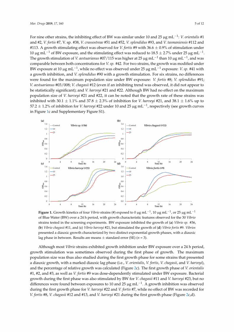
Mar. Drugs 2019, 17, 160 5 of 12
For nine other strains, the inhibiting effect of BW was similar under 10 and 25 µg mL−1: V. orientalis #1
and #2, V. fortis #7, V. sp. #38, V. crassostreae #51 and #52, V. splendidus #93, and V. tasmaniensis #112 and
#113. A growth stimulating effect was observed for V. fortis #9 with 36.6 ± 0.9% of stimulation under
10 µg mL−1 of BW exposure, and the stimulating effect was reduced to 18.5 ± 2.7% under 25 µg mL−1.
The growth stimulation of V. aestuarianus #07/115 was higher at 25 µg mL−1 than 10 µg mL−1, and was
comparable between both concentrations for V. sp. #42. For two strains, the growth was modified under
BW exposure at 10 µg mL−1, while no effect was observed under 25 µg mL−1 exposure: V. sp. #41 with
a growth inhibition, and V. splendidus #90 with a growth stimulation. For six strains, no differences
were found for the maximum population size under BW exposure: V. fortis #8; V. splendidus #91;
V. aestuarianus #03/008; V. chagasii #12 (even if an inhibiting trend was observed, it did not appear to
be statistically significant); and V. harveyi #21 and #22. Although BW had no effect on the maximum
population size of V. harveyi #21 and #22, it can be noted that the growth rate of these strains was
inhibited with 30.1 ± 1.1% and 37.8 ± 2.3% of inhibition for V. harveyi #21, and 38.1 ± 1.6% up to
57.2 ± 1.2% of inhibition for V. harveyi #22 under 10 and 25 µg mL−1, respectively (see growth curves
in Figure 1c and Supplementary Figure S1).
0.0
0.2
0.4
0.6
0.8
1.0
1.2
1.4
0 4 8 12 16 20 24
OD600
Time (h)
Vibrio fortis (#9)Control
10
25
0
0.2
0.4
0.6
0.8
1
1.2
1.4
0 4 8 12 16 20 24
OD600
Time (h)
Vibrio harveyi (#21)Control
10
25
0
0.2
0.4
0.6
0.8
1
1.2
1.4
0 4 8 12 16 20 24
OD600
Time (h)
Vibrio chagasii (#11)Control
10
25
0
0.2
0.4
0.6
0.8
1
1.2
1.4
0 4 8 12 16 20 24
OD600
Time (h)
Vibrio sp. (#36)Control
10
25
(a)
(c)
(b)
(d)
Figure 1. Growth kinetics of four Vibrio strains (#) exposed to 0 µg mL−1, 10 µg mL−1, or 25 µg mL−1
of Blue Water (BW) over a 24 h period, with growth characteristic features observed for the 30 Vibrio
strains tested in the screening experiments. BW exposure inhibited the growth of (a) Vibrio sp. #36,
(b) Vibrio chagasii #11, and (c) Vibrio harveyi #21, but stimulated the growth of (d) Vibrio fortis #9. Vibrios
presented a diauxic growth characterized by two distinct exponential growth phases, with a diauxic
lag phase in between. Results are means ± standard error (SE) (n = 3).
Although most Vibrio strains exhibited growth inhibition under BW exposure over a 24 h period,
growth stimulation was sometimes observed during the first phase of growth. The maximum
population size was thus also studied during the first growth phase for some strains that presented
a diauxic growth, with a marked diauxic lag phase (i.e., V. orientalis, V. fortis, V. chagasii, and V. harveyi),
and the percentage of relative growth was calculated (Figure 2c). The first growth phase of V. orientalis
#1, #2, and #3, as well as V. fortis #9 was dose-dependently stimulated under BW exposure. Bacterial
growth during the first phase was also stimulated by BW for V. chagasii #11 and V. harveyi #23, but no
differences were found between exposures to 10 and 25 µg mL−1. A growth inhibition was observed
during the first growth phase for V. harveyi #22 and V. fortis #7, while no effect of BW was recorded for
V. fortis #8, V. chagasii #12 and #13, and V. harveyi #21 during the first growth phase (Figure 2c,d).
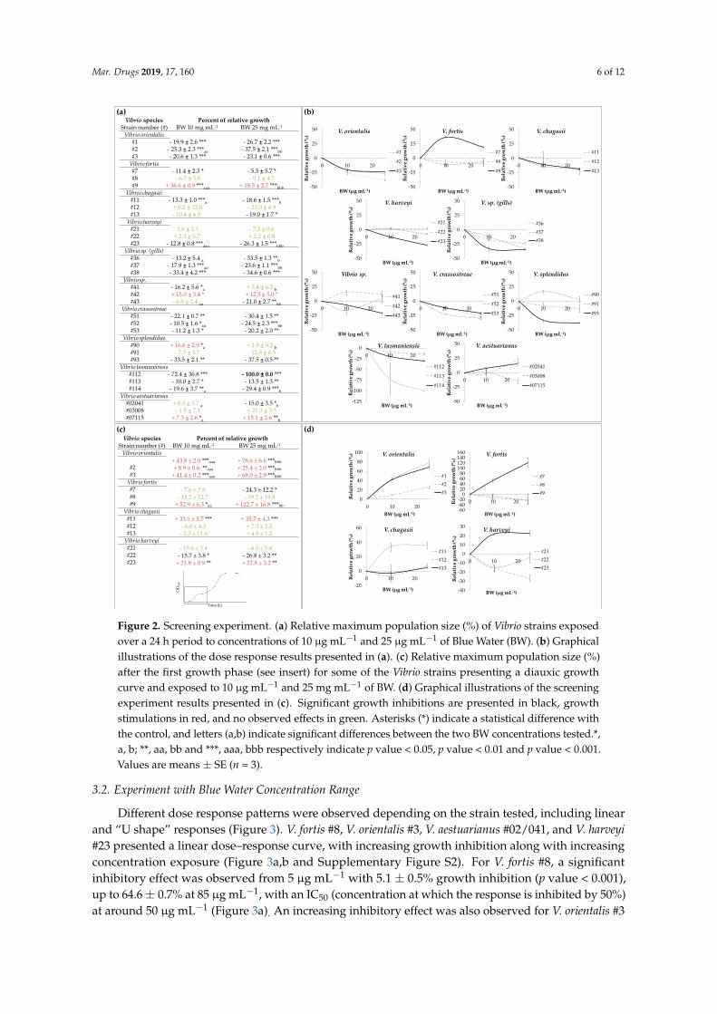
Mar. Drugs 2019, 17, 160 6 of 12
Vibrio species Percent of relative growthStrain number (#) BW 10 mg mL1 BW 25 mg mL1
Vibrio orientalis#1 19.9 2.6 *** 26.7 2.2 ***#2 25.3 2.3 ***aa 37.5 2.1 ***bb#3 20.6 1.3 *** 23.1 0.6 ***Vibrio fortis#7 11.4 2.3 * 5.5 5.7 *#8 6.7 3.8 9.1 4.7#9 + 36.6 0.9 ***aaa + 18.5 2.7 ***bbb
Vibrio chagasii#11 13.3 1.0 ***a 18.6 1.5 ***b#12 + 0.2 12.0 23.0 4.9#13 10.4 4.9 19.0 1.7 *Vibrio harveyi#21 1.6 2.1 7.2 0.6#22 + 2.3 0.7 + 2.2 0.8#23 12.8 0.8 ***aaa 26.3 1.5 ***bbb
Vibrio sp. (gills)#36 13.2 5.4 a 33.5 1.3 **b#37 17.9 1.3 ***aa 23.6 1.1 ***bb#38 33.4 4.2 *** 34.6 0.6 ***
Vibrio sp.#41 16.2 5.6 *a + 5.4 6.2 b
#42 + 15.0 3.4 * + 12.5 5.0 *#43 6.8 2.4 aa 21.0 2.7 **bb
Vibrio crassostreae#51 22.1 0.7 ** 30.4 1.5 **#52 10.5 1.6 *aa 24.5 2.3 ***bb#53 11.2 1.3 * 20.2 2.0 **
Vibrio splendidus#90 + 16.6 2.9 *a + 2.8 5.2 b
#91 7.5 3.7 12.8 6.5#93 33.5 2.1 ** 37.5 0.5 **
Vibrio tasmaniensis#112 72.4 36.8 *** 100.0 0.0 ***#113 10.0 2.7 * 13.5 1.3 **#114 19.6 3.7 **a 29.4 0.9 ***b
Vibrio aestuarianus#02041 + 0.5 3.7 a 15.0 3.5 *b#03008 1.5 7.1 + 21.3 3.5#07115 + 7.3 2.6 *a + 15.1 2.6 **b
50
25
0
25
50
0 10 20
Relative growth (%)
BW (µg mL1)
V. orientalis
#1
#2
#3
50
25
0
25
50
0 10 20
Relative growth (%)
BW (µg mL1)
V. fortis
#7
#8
#9
50
25
0
25
50
0 10 20
Relative growth (%)
BW (µg mL1)
V. chagasii
#11
#12
#13
50
25
0
25
50
0 10 20
Relative growth (%)
BW (µg mL1)
V. harveyi
#21
#22
#23
50
25
0
25
50
0 10 20
Relative growth (%)
BW (µg mL1)
V. crassostreae
#51
#52
#53
50
25
0
25
50
0 10 20
Relative growth (%)
BW (µg mL1)
V. sp. (gills)
#36
#37
#38
50
25
0
25
50
0 10 20Relative growth (%)
BW (µg mL1)
Vibrio sp.
#41
#42
#43
50
25
0
25
50
0 10 20
Relative growth (%)
BW (µg mL1)
V. splendidus
#90
#91
#93
125
100
75
50
25
0
0 10 20
Relative growth (%)
BW (µg mL1)
V. tasmaniensis
#112
#113
#114
50
25
0
25
50
0 10 20
Relative growth (%)
BW (µg mL1)
V. aestuarianus
#02041
#03008
#07115
(a) (b)
Vibrio species Percent of relative growthStrain number (#) BW 10 mg mL1 BW 25 mg mL1
Vibrio orientalis+ 43.8 ± 2.0 ***aaa + 78.6 ± 6.6 ***bbb
#2 + 8.9 ± 0.6 **aaa + 25.4 ± 2.0 ***bbb#3 + 41.4 ± 0.2 ***aaa + 68.0 ± 2.9 ***bbbVibrio fortis#7 7.6 ± 7.8 24.3 ± 12.2 *#8 13.2 ± 12.7 19.2 ± 19.8#9 + 52.9 ± 6.3 *aa + 122.7 ± 16.8 ***bb
Vibrio chagasii#11 + 33.1 ± 5.7 *** + 35.7 ± 4.3 ***#12 6.0 ± 4.3 + 7.3 ± 2.5#13 3.3 ± 11.6 + 4.8 ± 1.2Vibrio harveyi#21 15.6 ± 3.4 4.0 ± 5.8#22 15.7 ± 3.8 * 26.8 ± 3.2 **#23 + 21.8 ± 0.9 ** + 22.8 ± 3.2 **
Time (h)
OD
600
0
20
40
60
80
100
0 10 20
Relative growth (%)
BW (µg mL1)
V. orientalis
#1
#2
#3
604020020406080100120140160
0 10 20Relative growth (%)
BW (µg mL1)
V. fortis
#7
#8
#9
20
0
20
40
60
0 10 20Relative growth (%)
BW (µg mL1)
V. chagasii
#11
#12
#13
40
30
20
10
0
10
20
30
0 10 20
Relative growth (%)
BW (µg mL1)
V. harveyi
#21
#22
#23
(c) (d)
Figure 2. Screening experiment. (a) Relative maximum population size (%) of Vibrio strains exposed
over a 24 h period to concentrations of 10 µg mL−1 and 25 µg mL−1 of Blue Water (BW). (b) Graphical
illustrations of the dose response results presented in (a). (c) Relative maximum population size (%)
after the first growth phase (see insert) for some of the Vibrio strains presenting a diauxic growth
curve and exposed to 10 µg mL−1 and 25 mg mL−1 of BW. (d) Graphical illustrations of the screening
experiment results presented in (c). Significant growth inhibitions are presented in black, growth
stimulations in red, and no observed effects in green. Asterisks (*) indicate a statistical difference with
the control, and letters (a,b) indicate significant differences between the two BW concentrations tested.*,
a, b; **, aa, bb and ***, aaa, bbb respectively indicate p value < 0.05, p value < 0.01 and p value < 0.001.
Values are means ± SE (n = 3).
3.2. Experiment with Blue Water Concentration Range
Different dose response patterns were observed depending on the strain tested, including linear
and “U shape” responses (Figure 3). V. fortis #8, V. orientalis #3, V. aestuarianus #02/041, and V. harveyi
#23 presented a linear dose–response curve, with increasing growth inhibition along with increasing
concentration exposure (Figure 3a,b and Supplementary Figure S2). For V. fortis #8, a significant
inhibitory effect was observed from 5 µg mL−1 with 5.1 ± 0.5% growth inhibition (p value < 0.001),
up to 64.6 ± 0.7% at 85 µg mL−1, with an IC50 (concentration at which the response is inhibited by 50%)
at around 50 µg mL−1 (Figure 3a). An increasing inhibitory effect was also observed for V. orientalis #3
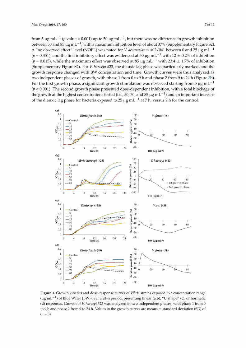
Mar. Drugs 2019, 17, 160 7 of 12
from 5 µg mL−1 (p value < 0.001) up to 50 µg mL−1, but there was no difference in growth inhibition
between 50 and 85 µg mL−1, with a maximum inhibition level of about 37% (Supplementary Figure S2).
A “no observed effect” level (NOEL) was noted for V. aestuarianus #02/041 between 0 and 25 µg mL−1
(p = 0.351), and the first inhibitory effect was evidenced at 50 µg mL−1 with 12 ± 0.2% of inhibition
(p = 0.015), while the maximum effect was observed at 85 µg mL−1 with 23.4 ± 1.7% of inhibition
(Supplementary Figure S2). For V. harveyi #23, the diauxic lag phase was particularly marked, and the
growth response changed with BW concentration and time. Growth curves were thus analyzed as
two independent phases of growth, with phase 1 from 0 to 9 h and phase 2 from 9 to 24 h (Figure 3b).
For the first growth phase, a significant growth stimulation was observed starting from 5 µg mL−1
(p < 0.001). The second growth phase presented dose-dependent inhibition, with a total blockage of
the growth at the highest concentrations tested (i.e., 50, 70, and 85 µg mL−1) and an important increase
of the diauxic lag phase for bacteria exposed to 25 µg mL−1 at 7 h, versus 2 h for the control.
0
0.2
0.4
0.6
0.8
1
1.2
0 4 8 12 16 20 24
OD600
Time (h)
Vibrio sp. (#38)Control51025507085
70
50
30
10
10
30
50
70
0 20 40 60 80
Relative growth (%)
BW (µg mL1)
V. sp. (#38)
0
0.2
0.4
0.6
0.8
1
1.2
0 4 8 12 16 20 24
OD600
Time (h)
Vibrio fortis (#9)Control51025507085
70
50
30
10
10
30
50
70
0 20 40 60 80
Relative growth (%)
BW (µg mL1)
V. fortis (#9)
0
0.2
0.4
0.6
0.8
1
1.2
0 4 8 12 16 20 24
OD600
Time (h)
Vibrio fortis (#8)Control51025507085
70
50
30
10
10
30
50
70
0 20 40 60 80
Relative growth (%)
BW (µg mL1)
V. fortis (#8)
0
0.2
0.4
0.6
0.8
1
1.2
0 4 8 12 16 20 24
OD600
Time (h)
Vibrio harveyi (#23)Control51025507085
1007550250
25
50
75
100
0 20 40 60 80
Relative growth (%)
BW (µg mL1)
V. harveyi (#23)
1st growth phase2nd growth phase
(a)
(b)
(c)
(d)
Figure 3. Growth kinetics and dose–response curves of Vibrio strains exposed to a concentration range
(µg mL−1) of Blue Water (BW) over a 24-h period, presenting linear (a,b), “U shape” (c), or hormetic
(d) responses. Growth of V. harveyi #23 was analyzed in two independent phases, with phase 1 from 0
to 9 h and phase 2 from 9 to 24 h. Values in the growth curves are means ± standard deviation (SD) of
(n = 3).
Mar. Drugs 2019, 17, 160 8 of 12
The growth of V. sp. 38, V. chagasii #13, V. crassostreae #53, and V. splendidus #90 was also
inhibited by the BW concentrations tested, but the dose–response curves expressed in function of the
control presented a “U” shape, with the growth inhibitory effect first increasing, then decreasing with
increasing BW concentrations (Figure 3c and Supplementary Figure S2). For V. sp. #38, the maximum
effect was observed at the concentration 50 µg mL−1, with 51.7 ± 0.6% of growth inhibition (Figure 3c).
However, the relative growth inhibition decreased to 37.1 ± 1.1% at 70 µg mL−1, and was similar to
the inhibition obtained at 25 µg mL−1 (p value 0.971). Same observations were made for V. chagasii #13
and V. crassostreae #53, with the inhibitory effect significantly higher at 50 µg mL−1 than at 70 µg mL−1
(p < 0.005). For V. splendidus #90, the percentage of maximum inhibition was 33.3 ± 1.5% at 70 µg mL−1,
and the inhibition was significantly lower, at 23.0 ± 0.7% at 85 µg mL−1 (p = 0.004; Supplementary
Figure S2).
The strains V. tasmaniensis #114 and V. fortis #9 presented a hormetic dose–response curve,
with opposite effects depending on the BW concentration. For V. tasmaniensis #114 the maximum
inhibitory effect was observed between 5 and 10 µg mL−1, with about 23% of inhibition, and decreased
until reaching the NOEL between 50 and 70 µg mL−1 (Supplementary Figure S2). V. fortis #9 presented
a growth stimulation at the lower concentrations tested, with a maximum growth stimulation of
53.0 ± 0.1% at 5 µg mL−1, then a decrease with increasing BW concentration. The NOEL was reached
at 50 µg mL−1, and the growth inhibition increased dose-dependently, with 31.4 ± 0.6% of inhibition
at 85 µg mL−1 (Figure 3d).
4. Discussion
The effects of marennine on several Vibrio strains with different ecological and virulence properties
have been investigated, by monitoring bacterial growth over a 24 h period after addition of BW into
the culture medium. The general trend for all the experiments is that BW affected most of the Vibrio
strains tested (80%), but the results are contrasting. Indeed, BW either inhibited or stimulated the
growth of the Vibrio strains.
As BW solution (i.e., the concentrated supernatant of H. ostrearia containing marennine) was used
in the present work, and not a purified form of the pigment, it could be questioned if the observed
effects were induced by marennine or by other molecules possibly present in the non-axenic culture
medium (e.g., exopolysaccharides (EPS) from Haslea or epibiont bacteria). However, previous studies
conducted with purified extracellular marennine (EMn) on marine bacteria, including Vibrio species,
confirmed that the pigment itself did exert an antibacterial activity [9–12]. Moreover, a recent study
has demonstrated the biological activities of marennine solutions, including BW and EMn, on the
development or survival of various marine organisms, and comparable effects were observed between
the different solutions tested [21]. Also, if blue Haslea species should be cultured at large scale to ensure
the production of marennine solutions for applications as antimicrobials in aquaculture, BW represents
the easiest and cheapest production process, compared to the purified pigment.
The observed effect of BW on vibrios was species- but also strain-dependent, as for a same
species, the sensitivity of distinct strains could significantly differ. This is in accordance with previous
observations of V. aestuarianus, V. coralliilyticus, and V. tubiashii exposed to purified marennine [9].
Moreover, it seemed difficult to perceive a correlation between the effects of BW and the strain
ecological characteristics, such as sampling season, distribution (sea water fractions and oyster tissues),
or the virulence of the strain toward bivalves (Supplementary Table S1).
However, the present work considerably extends our understanding of the variability of
marennine’s effect on bacteria of the Vibrio genus. The diversity observed in growth responses was
especially noteworthy in two species: V. fortis on the one hand, with a growth inhibition for strains
#7 and #8, and important growth stimulation for strain #9; and on the other hand, V. tasmaniensis,
with a total inhibition of strain #112, but no effect observed on the strain #113.
In addition, diauxic growth was evidenced for all the Vibrio strains tested, a phenomenon
commonly observed when bacteria are grown in a medium containing two types of carbon sources
Mar. Drugs 2019, 17, 160 9 of 12
(e.g., sugars), and characterized by two distinct exponential phases, with a diauxic lag phase in
between [20]. Vibrio diauxic growth is not extensively evidenced in the literature, and was mainly
illustrated using V. cholerae [22–24] but also V. alginolyticus [25]. It is worth noting that complete
growth kinetics are rarely recorded for antibacterial assays, as most experiments conducted to screen
bioactive compounds against marine Vibrio species used the disc diffusion method [26–28], as the
ones conducted with marennine and marennine-like pigments on V. aestuarianus [11,14]. The effect of
purified marennine was previously tested with the microdilution method on another V. tasmaniensis
strain [9,12] and no obvious diauxic growth was observed then, which could be explained by the use
of a different growth media (marine broth media versus CaMHB in the present study), a different time
scale, or a different treatment of data.
Bacterial diauxic growth has long been considered as a bacterial phase of enzymatic acclimation
to metabolize a different type of sugar; however, there is now growing evidence that this phenomenon
could actually correspond to the presence of two bacterial subpopulations in the isogenic culture,
with different phenotypic adaptation and growth strategies [29]. Based on this new hypothesis, diauxic
growth curves in the present study could indicate the existence of different subpopulations of Vibrio
strains: Type 1, which can divide quickly by using the most suitable carbon source to sustain its
growth; and Type 2, which grows more slowly, but which would be able to metabolize a different
carbon source. According to Solopova et al. [29], the Type 1 subpopulation, which cannot switch to an
alternative metabolic pathway, will stop dividing, possibly because of the low energy state of cells [29].
This phenotype heterogeneity has been recently demonstrated in V. cholerae [24], and would result
from the bet-hedging strategy, an evolutionary strategy that allows colonies to cope with fluctuating
environments [30–32]. In the present work, although bacterial growth was globally inhibited over the
24 h of the essay, a growth stimulation of the subpopulation Type 1 was sometimes observed under
BW exposure (e.g., V. orientalis, V. chagasii, and V. harveyi strains). It can be hypothesized that the
subpopulation Type 1 would be able to metabolize part of the sugars constituting the carbon skeleton
of the marennine molecule—or, if any, other EPS present in BW solution to sustain its growth—or
that the stimulation would result from overcompensation mechanisms by the bacteria under BW
exposure, a mechanism that can be observed when cells undergo a disruption in homeostasis [33].
However, other experiments would be needed to confirm these hypotheses. It may also be argued
that, when exposed to BW, it is the growth of subpopulation Type 2 that was mainly affected by the
BW, as illustrated by the total absence of growth of V. harveyi #23 at the highest concentrations tested
(from 50 to 80 µg mL−1).
A global observation from the results shows that the dose–response curves of most Vibrio
strains did not follow a linear or threshold model, with growth inhibition increasing with
concentrations. Indeed, some curves presented a “U-shape” response regarding the antibacterial
effect, which decreased with increasing BW exposure (e.g., V. chagasii #13 or V. sp #38). On top of this,
opposite effects were observed at low and high concentrations, a phenomenon known as hormesis [34].
Indeed, the lowest BW concentration tested (5 µg mL−1) highly stimulated the growth of V. fortis
#9, up to 50%, while the growth was significantly inhibited at higher concentrations (from 50 to
80 µg mL−1). Hormetic dose–response relationships have raised a growing awareness in toxicological
and ecotoxicological studies, and have been extensively documented over the past two decades
in different models, as plants, algae, or fungi [35–37]. The mechanisms of hormesis are not yet
clearly understood, and it seems that only a subset of compounds with specific cellular mechanisms
would mediate hormetic responses [38,39]. Moreover, hormetic effects are challenging to observe in
laboratory conditions, being dependent on various factors, such as endpoint measurements or growth
conditions [40]. In the present study, hormetic response of vibrios to BW was observed in the CaMHB
medium, a culture medium that was previously reported to promote the observation of hormesis in
Escherichia coli exposed to antibiotics, while the hormetic effect was not observed in Luria–Bertani (LB)
culture media [41].

Mar. Drugs 2019, 17, 160 10 of 12
The mechanisms of action responsible for the antibacterial activity of marennine remain little
explored. So far, the Haslea provincialis purified blue pigment was demonstrated to interact with the outer
membrane of the gram-negative bacteria E. coli, rendering it more rigid [42]. Recent studies conducted
on V. cholerae also demonstrate a disruption of the bacterial membrane integrity and a deformation of
the cell architecture by antibacterial agents, such as polyphenols [23] or nanoparticles [43]. For now,
it is still unclear how marennine and Vibrio interact in vivo, and whether the pigment has a direct
effect on Vibrio growth or if it could decrease its pathogenicity by fixing on cell membranes, both of
which could explain the better survival of bivalve larvae exposed to marennine and challenged with
V. splendidus [15].
In conclusion, the present work indicates much more complex interactions between marennine
and vibrios than a standard linear correlation between dose and effect. Moreover, the antibacterial
activity of marennine is dependent on the species, the strain, and possibly the population of bacteria,
suggesting that the blue pigment would act on specific targets. Marennine antibacterial mechanisms
and low-dose stimulation phenomenon will have to be better understood before considering any
application of Haslea and marennine as antimicrobials in aquaculture.
Supplementary Materials: The following are available online at http://www.mdpi.com/1660-3397/17/3/160/s1,Figure S1: Growth curves of 30 Vibrio strains (#) from 10 different species exposed over a 24 h period to 0, 10 or
25 µg mL−1 of Blue Water (BW), the concentrated supernatant of Haslea ostrearia containing the extracellularmarennine. Results are means ± SE (n = 3), Figure S2: Growth kinetics and dose-response curves of Vibrio strains
exposed to a concentration range (µg mL−1) of Blue Water (BW) over a 24 h period. Values are means ± SD (n = 3),Table S1: Preferential distribution, virulence to oysters and Blue Water (BW) effect over a 24 h period and over thefirst phase of growth of the 30 Vibrio strains tested.
Author Contributions: Conceptualization, C.F. and J.-L.M.; Data curation, C.F.; Formal analysis, C.F. and M.B.;Funding acquisition, J.-L.M.; Methodology, C.F. and M.Z.; Resources, A.J. and M.-A.T.; Software, C.F. and M.B.;Supervision, J.-L.M.; Validation, M.-A.T. and J.-L.M.; Writing—original draft, C.F.; Writing—review & editing, A.J.,M.-A.T., M.B., M.Z. and J.-L.M.
Funding: This publication benefited from funding from the Horizon 2020 Research and Innovation ProgramGHaNA (The Genus Haslea, New marine resources for blue biotechnology and Aquaculture) under GrantAgreement No 734708/GHANA/H2020-MSCA-RISE-2016 (JLM).
Acknowledgments: The authors want to thank Frédérique Le Roux from the Biologie Intégrative des ModèlesMarins laboratory, Génomique des Vibrios team (Station Biologique de Roscoff, France) for having kindly providedmost of the Vibrio strains necessary for the present study.
Conflicts of Interest: The authors declare no conflict of interest.
References
1. Nassiri, Y.; Robert, J.-M.; Rincé, Y.; Ginsburger-Vogel, T. The cytoplasmic fine structure of the diatom Haslea
ostrearia (Bacillariophyceae) in relation to marennine production. Phycologia 1998, 37, 84–91. [CrossRef]
2. Gastineau, R.; Davidovich, N.A.; Bardeau, J.-F.; Caruso, A.; Leignel, V.; Hardivillier, Y.; Jacquette, B.;
Davidovich, O.I.; Rincé, Y.; Gaudin, P.; et al. Haslea karadagensis (Bacillariophyta): A second blue diatom,
recorded from the Black Sea and producing a novel blue pigment. Eur. J. Phycol. 2012, 47, 469–479. [CrossRef]
3. Gastineau, R.; Hansen, G.; Davidovich, N.A.; Davidovich, O.; Bardeau, J.-F.; Kaczmarska, I.; Ehrman, J.M.;
Leignel, V.; Hardivillier, Y.; Jacquette, B.; et al. A new blue-pigmented hasleoid diatom, Haslea provincialis,
from the Mediterranean Sea. Eur. J. Phycol. 2016, 51, 156–170. [CrossRef]
4. Prasetiya, F.S.; Gastineau, R.; Poulin, M.; Lemieux, C.; Turmel, M.; Syakti, A.D.; Hardivillier, Y.; Widowati, I.;
Risjani, Y.; Iskandar, I.; et al. Haslea nusantara, a new blue diatom from the Java Sea, Indonesia: Morphology,
biometry and molecular characterization. Plant. Ecol. Evol. 2019, 151. (in press).
5. Pouvreau, J.-B.; Morançais, M.; Fleury, F.; Rosa, P.; Thion, L.; Cahingt, B.; Zal, F.; Fleurence, J.; Pondaven, P.
Preliminary characterisation of the blue-green pigment “marennine” from the marine tychopelagic diatom
Haslea ostrearia (Gaillon/Bory) Simonsen. J. Appl. Phycol. 2006, 18, 757–767. [CrossRef]
6. Turpin, V.; Robert, J.-M.; Goulletquer, P.; Massé, G.; Rosa, P. Oyster greening by outdoor mass culture of the
diatom Haslea ostrearia Simonsen in enriched seawater. Aquac. Res. 2001, 32, 801–809. [CrossRef]
Mar. Drugs 2019, 17, 160 11 of 12
7. Pouvreau, J.-B.; Housson, E.; Tallec, L.L.; Morançais, M.; Rincé, Y.; Fleurence, J.; Pondaven, P. Growth
inhibition of several marine diatom species induced by the shading effect and allelopathic activity of
marennine, a blue-green polyphenolic pigment of the diatom Haslea ostrearia (Gaillon/Bory) Simonsen.
J. Exp. Mar. Biol. Ecol. 2007, 352, 212–225. [CrossRef]
8. Prasetiya, F.S.; Safitri, I.; Widowati, I.; Cognie, B.; Decottignies, P.; Gastineau, R.; Morançais, M.; Windarto, E.;
Tremblay, R.; Mouget, J.-L. Does allelopathy affect co-culturing Haslea ostrearia with other microalgae relevant
to aquaculture? J. Appl. Phycol. 2016, 28, 2241–2254. [CrossRef]
9. Falaise, C.; François, C.; Travers, M.-A.; Morga, B.; Haure, J.; Tremblay, R.; Turcotte, F.; Pasetto, P.;
Gastineau, R.; Hardivillier, Y.; et al. Antimicrobial compounds from eukaryotic microalgae against human
pathogens and diseases in aquaculture. Mar. Drugs 2016, 14, 159. [CrossRef]
10. Pouvreau, J.-B. Purification et Caractérisation du Pigment Bleu-Vert “Marennine” Synthétisé par la Diatomee
Marine Haslea ostrearia (Gaillon/Bory) Simonsen: Propriétés Physico-Chimiques et Activités Biologiques.
Ph.D. Thesis, Université de Nantes, Nantes, France, July 2006.
11. Gastineau, R.; Pouvreau, J.-B.; Hellio, C.; Morançais, M.; Fleurence, J.; Gaudin, P.; Bourgougnon, N.;
Mouget, J.-L. Biological activities of purified marennine, the blue pigment responsible for the greening of
oysters. J. Agric. Food Chem. 2012, 60, 3599–3605. [CrossRef]
12. Gastineau, R.; Turcotte, F.; Pouvreau, J.-B.; Morançais, M.; Fleurence, J.; Windarto, E.; Prasetiya, F.S.; Arsad, S.;
Jaouen, P.; Babin, M.; et al. Marennine, promising blue pigments from a widespread Haslea diatom species
complex. Mar. Drugs 2014, 12, 3161–3189. [CrossRef] [PubMed]
13. Travers, M.-A.; Boettcher Miller, K.; Roque, A.; Friedman, C.S. Bacterial diseases in marine bivalves.
J. Invertebr. Pathol. 2015, 131, 11–31. [CrossRef] [PubMed]
14. Gastineau, R.; Hardivillier, Y.; Leignel, V.; Tekaya, N.; Morançais, M.; Fleurence, J.; Davidovich, N.;
Jacquette, B.; Gaudin, P.; Hellio, C.; et al. Greening effect on oysters and biological activities of the blue
pigments produced by the diatom Haslea karadagensis (Naviculaceae). Aquaculture 2012, 368–369, 61–67.
[CrossRef]
15. Turcotte, F.; Mouget, J.-L.; Genard, B.; Lemarchand, K.; Deschênes, J.-S.; Tremblay, R. Prophylactic effect
of Haslea ostrearia culture supernatant containing the pigment marennine to stabilize bivalve hatchery
production. Aquat. Living Resour. 2016, 29, 401. [CrossRef]
16. Bruto, M.; James, A.; Petton, B.; Labreuche, Y.; Chenivesse, S.; Alunno-Bruscia, M.; Polz, M.F.; Le Roux, F.
Vibrio crassostreae, a benign oyster colonizer turned into a pathogen after plasmid acquisition. ISME J. 2017,
11, 1043–1052. [CrossRef] [PubMed]
17. Clinical and Laboratory Standards Institute (CLSI). Methods for Dilution Antimicrobial Susceptibility Tests for
Bacteria That Grow Aerobically. Document M07-A9. Approved Standard—Ninth Edition; Clinical and Laboratory
Institute: Wayne, PA, USA, 2012; Volume 32.
18. Pouvreau, J.-B.; Morançais, M.; Massé, G.; Rosa, P.; Robert, J.-M.; Fleurence, J.; Pondaven, P. Purification of
the blue-green pigment “marennine” from the marine tychopelagic diatom Haslea ostrearia (Gaillon/Bory)
Simonsen. J. Appl. Phycol. 2006, 18, 769–781. [CrossRef]
19. Sprouffske, K.; Wagner, A. Growthcurver: An R package for obtaining interpretable metrics from microbial
growth curves. BMC Bioinform. 2016, 17, 1–4. [CrossRef]
20. Monod, J. The Growth of Bacterial Cultures. Annu. Rev. Microbiol. 1949, 3, 371–394. [CrossRef]
21. Falaise, C.; Cormier, P.; Tremblay, R.; Audet, C.; Deschênes, J.-S.; Turcotte, F.; François, C.; Seger, A.;
Hallegraeff, G.; Lindquist, N.; et al. Harmful or harmless: Biological effects of marennine on marine
organisms. Aquatic Toxicol. 2019, 209, 13–25. [CrossRef]
22. Bag, J. Glucose inhibition of the transport and phosphoenolpyruvate-dependent phosphorylation of galactose
and fructose in Vibrio cholerae. J. Bacteriol. 1974, 118, 764–767.
23. Bhattacharya, D.; Ghosh, D.; Bhattacharya, S.; Sarkar, S.; Karmakar, P.; Koley, H.; Gachhui, R. Antibacterial
activity of polyphenolic fraction of Kombucha against Vibrio cholerae: Targeting cell membrane. Lett. Appl.
Microbiol. 2018, 66, 145–152. [CrossRef]
24. Chatterjee, T.; Chatterjee, B.K.; Chakrabarti, P. Modelling of growth kinetics of Vibrio cholerae in presence of
gold nanoparticles: Effect of size and morphology. Sci. Rep. 2017, 7, 9671. [CrossRef] [PubMed]
25. Vine, N.G.; Leukes, W.D.; Kaiser, H. In vitro growth characteristics of five candidate aquaculture probiotics
and two fish pathogens grown in fish intestinal mucus. FEMS Microbiol. Lett. 2004, 231, 145–152. [CrossRef]
Mar. Drugs 2019, 17, 160 12 of 12
26. Banerjee, S.; Kim, L.M.; Shariff, M.; Khatoon, H.; Yusoff, F.M. Antibacterial activity of neem (Azadirachta
indica) leaves on Vibrio spp. isolated from cultured shrimp. Asian J. Anim. Vet. Adv. 2013, 8, 355–361.
27. Cavallo, R.; Acquaviva, M.; Stabili, L.; Cecere, E.; Petrocelli, A.; Narracci, M. Antibacterial activity of marine
macroalgae against fish pathogenic Vibrio species. Open Life Sci. 2013, 8, 646–653.
28. Das, B.K.; Pradhan, J.; Pattnaik, P.; Samantaray, B.R.; Samal, S.K. Production of antibacterials from the
freshwater alga Euglena viridis (Ehren). World J. Microbiol. Biotechnol. 2005, 21, 45–50. [CrossRef]
29. Solopova, A.; van Gestel, J.; Weissing, F.J.; Bachmann, H.; Teusink, B.; Kok, J.; Kuipers, O.P. Bet-hedging
during bacterial diauxic shift. PNAS 2014, 111, 7427–7432. [CrossRef] [PubMed]
30. Ackermann, M. A functional perspective on phenotypic heterogeneity in microorganisms. Nat. Rev. Microbiol.
2015, 13, 497–508. [CrossRef]
31. Grimbergen, A.J.; Siebring, J.; Solopova, A.; Kuipers, O.P. Microbial bet-hedging: The power of being
different. Curr. Opin.Microbiol. 2015, 25, 67–72. [CrossRef]
32. Martins, B.M.; Locke, J.C. Microbial individuality: How single-cell heterogeneity enables population level
strategies. Curr. Opin. Microbiol. 2015, 24, 104–112. [CrossRef]
33. Calabrese, E.J. Hormetic mechanisms. Crit. Rev. Toxicol. 2013, 43, 580–606. [CrossRef] [PubMed]
34. Calabrese, E.J.; Baldwin, L.A. Defining hormesis. Hum. Exp. Toxicol 2002, 21, 91–97. [CrossRef] [PubMed]
35. Belz, R.G.; Duke, S.O. Herbicides and plant hormesis. Pest. Manag. Sci. 2014, 70, 698–707. [CrossRef]
[PubMed]
36. Calabrese, E.J.; Baldwin, L.A. Hormesis: A Generalizable and Unifying Hypothesis. Crit. Rev. Toxicol. 2001,
31, 353–424. [CrossRef] [PubMed]
37. Cedergreen, N.; Streibig, J.C.; Kudsk, P.; Mathiassen, S.K.; Duke, S.O. The occurrence of hormesis in plants
and algae. Dose Response 2006, 5, 150–162. [CrossRef] [PubMed]
38. Kendig, E.L.; Le, H.H.; Belcher, S.M. Defining Hormesis: Evaluation of a Complex Concentration Response
Phenomenon. Int. J. Toxicol. 2010, 29, 235–246. [CrossRef]
39. Lushchak, V.I. Dissection of the Hormetic Curve: Analysis of Components and Mechanisms. Dose Response
2014, 12, 466–479. [CrossRef] [PubMed]
40. Calabrese, E.J.; Baldwin, L.A. A quantitatively-based methodology for the evaluation of chemical hormesis.
Hum. Ecolog. Risk Assess. An. Int. J. 1997, 3, 545–554. [CrossRef]
41. Wang, D.; Lin, Z.; Wang, T.; Ding, X.; Liu, Y. An analogous wood barrel theory to explain the occurrence
of hormesis: A case study of sulfonamides and erythromycin on Escherichia coli growth. PLoS ONE 2017,
12, e0181321. [CrossRef]
42. Tardy-Laporte, C.; Arnold, A.A.; Genard, B.; Gastineau, R.; Morançais, M.; Mouget, J.-L.; Tremblay, R.;
Marcotte, I. A 2H solid-state NMR study of the effect of antimicrobial agents on intact Escherichia coli without
mutating. Biochim. Biophys. Acta (BBA)—Biomembr. 2013, 1828, 614–622. [CrossRef]
43. Sarwar, S.; Chakraborti, S.; Bera, S.; Sheikh, I.A.; Hoque, K.M.; Chakrabarti, P. The antimicrobial activity of
ZnO nanoparticles against Vibrio cholerae: Variation in response depends on biotype. Nanomed. Nanotechnol.
Biol. Med. 2016, 12, 1499–1509. [CrossRef] [PubMed]
© 2019 by the authors. Licensee MDPI, Basel, Switzerland. This article is an open access
article distributed under the terms and conditions of the Creative Commons Attribution
(CC BY) license (http://creativecommons.org/licenses/by/4.0/).
Annexes
ANNEXE 4 : Publication
Prasetiya et al. (sous presse), Plant Ecology & Evolution [Texte Complet]
Prasetiya, F.S., Gastineau, R., Poulin, M., Lemieux, C., Turmel, M., Syakti, A.D., Hardivillier, Y.,
Widowati, I., Risjani, Y., Iskandar, I., Subroto, T., Falaise, C., Mouget, J.-L., Leignel, V., in press.
Haslea nusantara, a new blue diatom from the Java Sea, Indonesia: Morphology, biometry and
molecular characterization. Plant Ecology and Evolution.
1
Haslea nusantara, a new blue diatom from the Java Sea, Indonesia:
Morphology, biometry and molecular characterization Fiddy Semba PRASETIYA1,7,8*, Romain GASTINEAU1, Michel POULIN2, Claude LEMIEUX3, Monique TURMEL3, Agung Dhamar SYAKTI4, Yann HARDIVILLIER1, Ita WIDOWATI5, Yenny RISJANI6, Iskandar ISKANDAR7, Toto SUBROTO8, Charlotte FALAISE1, Jean-Luc MOUGET1$ & Vincent LEIGNEL1$ 1FR CNRS 3473 IUML, Mer-Molécules-Santé (MMS), Le Mans Université, Avenue Olivier Messiaen, 72085 Le Mans CEDEX 9, France
2Division Recherche et collections, Musée canadien de la nature, C.P. 3443, Succursale D, Ottawa, Ontario, K1P 6P4, Canada 3Institut de Biologie Intégrative et des Systèmes, Département de Biochimie, de Microbiologie et de Bio-Informatique, Université Laval, Québec, Canada 4Fisheries and Marine Sciences Department, Jenderal Soedirman University, Kampus Perikanan Unsoed Karangwangkal, Jl dr. Suparno, Purwokerto 53123, Indonesia 5Faculty of Fisheries and Marine Sciences, Diponegoro University, Jl. Prof. H. Soedharto,SH, Tembalang, Semarang 50275, Indonesia 6Faculty of Fisheries and Marine Sciences, University of Brawijaya, Jalan Veteran, Ketawanggede, Malang 65145, Indonesia 7Marine Science Department, Faculty of Fisheries and Marine Sciences, Universitas Padjadjaran, Jl. Raya Bandung Sumedang KM.21, Jatinangor 45363, Indonesia 8Research Center of Biotechnology and Bioinformatics, Jl. Raya Bandung Sumedang KM.21, Jatinangor 45363, Indonesia ($ co-contribution)
*Author for correspondence *: Dr Fiddy Semba PRASETIYA. [email protected]
2
Background and aims – The present study aims to describe a new species of pennate blue diatom from the genus Haslea, H. nusantara sp. nov., collected from Semak Daun Island, the Seribu Archipelago, in Indonesian marine waters. Methods – Assessment for species identification was conducted using light microscopical (LM), Scanning Electron Microscopical (SEM) and molecular techniques. The morphological characteristics of H. nusantara sp. nov. has been described, illustrated and compared to other morphologically similar Haslea taxa, while molecular characteristics known worldwide. Additionally, the molecular characteristic (rbcL chloroplast gene and cox1 mitochondrial gene) was also analyzed. Key results – This new species, named Haslea nusantara, is characterized by specific morphological (stria density) and molecular criteria (rbcL chloroplast gene and cox1 mitochondrial gene). It differentiates from other blue Haslea species by the presence of a thin central bar, which has been previously reported in non-blue species like H. pseudostrearia Massé, Rincé et E.J. Cox. The complete mitochondrion (36,288 basepairs, bp) and plastid (120,448 bp) genomes of H. nusantara were sequenced and the gene arrangements were compared with other diatom genomes. Phylogeny analyses established using rbcL indicated that H. nusantara sp. nov. is included in the blue Haslea cluster and close to a blue Haslea sp. found in Canary Islands (H. silbo sp. inedit.). Conclusions – All investigations carried out in this study show that H. nusantara is a new blue-pigmented species, which belongs to the blue Haslea clade, with an exceptional geographic distribution in the Southern Hemisphere. Key words – Indonesia, Bacillariophyceae / blue diatoms / Haslea / molecular phylogeny, genomic study
INTRODUCTION
The pennate diatom, Haslea ostrearia (Gaillon) Simonsen, has long been known as the only blue diatom ever described, and it is also the type species of the genus Haslea Simonsen (Simonsen, 1974; Poulin et al., in press, this journal’s issue). This microalga produces a water-soluble blue pigment, marennine (Lankester 1886), which accumulates at cell apices and is released into the seawater. This pigment is responsible for the greening of oyster’s gills in the Atlantic coast of France (Gaillon 1820). This phenomenon gives a significant economic added value to the French oyster industry, since green oysters, with specific flavor and emerald color, are rarer and more expensive than ordinary oysters. Furthermore, this blue pigment has biological properties like antioxidant (Pouvreau et al. 2008), antibacterial and antiviral (Gastineau et al. 2012a) and allelopathic effect against other diatoms (Pouvreau et al. 2007a, Prasetiya et al. 2016). For decades, any record worldwide of a blue diatom was assigned to H. ostrearia. However, recent works on the blue Haslea using scanning electron microscopy (SEM) observation and molecular approaches have enlightened an unsuspected biodiversity of this taxon, with two new species, H. karadagensis Davidovich, Gastineau et Mouget (Gastineau et al. 2012b) collected in the Black Sea, and H. provincialis Gastineau, Hansen et Mouget
3
(Gastineau et al. 2016) in the Mediterranean Sea. Gastineau et al. (2014) also mentioned one undescribed species yet in the Canary Islands (Haslea silbo sp. inedit.). Our knowledge about the diversity of non-blue Haslea also increased, with H. sigma Talgatti, Sar et Carvalho Torgan found in salt marshes in Southern Brazil (Talgatti et al. 2014), and seven species more from different collections worldwide (Sterrenburg et al. 2015). To date, the genus Haslea encompasses 35 taxa as listed in AlgaeBase (Guiry & Guiry 2018) (see http://www.algaebase.org/), but recently, Li et al. (2017) revisited the Haslea phylogeny, using SSU rDNA and rbcL data, demonstrating that two species must be transferred to the genus Navicula Bory, H. tsukamotoi Sterrenburg et Hinz and H. avium Tiffany, Herwig et Sterrenburg. Most of Haslea species have been encountered in the Northern Hemisphere. The present study aims to describe a new species of pennate blue diatom from the genus Haslea, H. nusantara sp. nov., collected from Semak Daun Island, the Seribu Archipelago, in Indonesian marine waters. Morphological, biometry (using SEM), molecular investigations (chloroplast and mitochondrial genomes) were carried out and spectral analysis of the pigment in the culture supernatant was achieved. The specificity of this Indonesian new species was established as compared with other known blue Haslea species.
MATERIAL AND METHODS
Diatom sampling, isolation and culture
Specimens of H. nusantara were sampled using 50 mL conical centrifuge tubes (Thermo Fisher Scientific, France), on 12 March 2015 by scraping the biofilm covering Padina sp. thalli growing in the subtidal zone of Semak Daun Island (5°43'49.27'' S, 106°34'20.27'' E) in the Seribu Archipelago (“Kepulauan Seribu”), Indonesia (fig. 1). A few days after the sampling, six single cells (IND1 to IND6) were isolated from one tube at Le Mans Université (France), using micropipettes and grown in Petri dishes containing modified artificial seawater (Mouget et al. 2009). The H. nusantara cultures were grown in Erlenmeyer flasks in a controlled temperature room at 16 ± 1 °C and an irradiance of 50-100 µmol photons m–2 s–1, provided by fluorescent tubes (Philips TLD 36W/965), during a 14:10 h light:dark cycle. Among the six cells initially isolated, only one (IND6) survived and the strain was maintained in culture for several months. Microscopic observations
The preparation of samples was performed as described in Kaczmarska et al. (2005). Samples were prepared using a Millipore vacuum filtration apparatus with 25 mm diameter and 3 μm pore size polytetrafluoroethylene (PTFE) membrane (General Electric Osmonics, Minnetonka, USA). Samples were washed with distilled water, re-suspended in 5 mL distilled water and cleaned by adding 10 mL of concentrated sulfuric and nitric acid in a water bath at 100 °C for 60 min. Samples were then re-washed using distilled water in the filtration apparatus and re-suspended in 5 mL distilled water. Cleaned samples were then mounted on glass slides using Naphrax and examined using an Olympus BX51 light microscope with a DP72 digital camera, equipped with phase contrast and differential interference contrast (DIC) optics.
4
For scanning electron microscopic (SEM) examination, cleaned diatom frustules and isolated valves were air-dried onto a circular coverslip, mounted on an aluminum stub prior to coating with platinum or palladium-platinum using a JEOL JUC-5000 sputter coater or JEOL JFC-2300 HR high-resolution fine coater, respectively. Genome sequencing, annotation and analysis
Total DNA was extracted as described in Doyle & Doyle (1990). An Illumina library of 250 base pairs (bp) DNA inserts was prepared and sequenced on the HiSeq 4000 platform by the Beijing Genomic Institute (Shenzhen). A total of 11 million paired-end reads of 150 bp were assembled using Ray 2.3.0 (Boisvert et al. 2012) and a k-mer value of 45. Contigs of chloroplast and mitochondrial origins were identified by BLAST searches against a local database of organelle genomes and then merged using an overlap-layout consensus approach as implemented in Sequencher 5.4.1 (Gene Codes Corporation, Ann Arbor, USA). Organelle genomes were annotated using a custom set of tools (Turmel et al. 2015) and circular genome maps were drawn using OGDraw (Lohse et al. 2007). Alignments of chloroplast and mitochondrial genomes from diatom taxa belonging to the Naviculales were carried out using the ProgressiveMauve algorithm of Mauve 2.3.1 (Darling et al. 2010)
Phylogenetic trees were built, based on rbcL chloroplast gene and cox1 mitochondrial gene to establish the evolutionary process inside the genus Haslea. Our choice focused on rbcL (ribulose-bisphosphate carboxylase, large subunit gene) and cox1 (cytochrome oxidase subunit I) because they are characterized in many Haslea species (sequences available in Genbank™). The multiple alignments were carried out using clustalO (https://www.ebi.ac.uk/Tools/msa/clustalo/). The best evolution model was defined using MEGA7 (Kumar et al. 2016). Maximum likelihood (ML) analysis was generated using RaxML (https://embnet.vital-it.ch/raxml-bb, Stamatakis et al. 2008) and MEGA7 with 1000 bootstrap and maximum parsimony (MP) was performed using PAUP (Swofford 1998) and MEGA7 with 1000 bootstrap too.
Pigment characterization with UV-visible spectrophotometry
The marennine-like pigment produced by H. nusantara was characterized on cell-free culture supernatant (syringe-filtered on 0.22 µm, Thermo Fisher Scientific) using the Beer-Lambert law. The spectrum was scanned from 200 to 800 nm, in a 5 cm cell using a spectrophotometer (Perkin Elmer, Lambda 25 scan mode). Marennine-like concentration was estimated using the specific extinction coefficient for the extracellular form of marennine (EMn) at the peak wavelength 677 nm (ε677 = 12.13 L g−1 cm−1), according to Pouvreau et al. (2007b). Spectral characteristics from H. nusantara extracellular marennine-like pigment was compared to EMn as control.
RESULTS Haslea nusantara Mouget, Gastineau et Syakti sp. nov.
DIAGNOSIS: Cells with two parietal, narrow band-like chloroplasts lying appressed on each side of the valve margin and not reaching the apex. Cell apices characteristically blue
5
colored. Valves narrow and lanceolate with acute apices. The maximum and minimum length of H. nusantara was 91.1 and 54.1 μm, respectively (average, 73.9 ± 1.7 μm, n = 33), while the maximum and minimum width was 8.2 and 5.9 μm (average 6.8 ± 0.1 μm, n = 33). The equation of the fitting curve to describe the relationship between the length to width measurements in H. nusantara is:
y = 0.023 x + 5.06, R2 = 0.164, n = 33 (1) (Fig 2.) Axial and central areae are indistinct. Raphe is straight with approximate central endings. Internally, the raphe is bordered on one side by an axial costa, while at center on opposite side, a thin and short central bar can be observed. Striation consists of a transapical pattern (34-37 striae in 10 µm) crossed at right angle by a longitudinal pattern (51-53 striae in 10 µm). HOLOTYPE (here designated): The embedded strain IND6 was acid cleaned and mounted on a glass slide and a SEM stub that have been deposited in the Muséum National d’Histoire Naturelle of Paris, France (PC) under accession numbers PC0576262 and PC0576265, respectively. The cell representative of the type is presented in Figure 3. ISOTYPES: A slide and a SEM stub with acid cleaned valves of strain IND6 have been deposited at the Bremerhaven Hustedt collection in Bremerhaven, Germany (BRM) under the accession numbers BRM Zu11/18 and BRM R1280, respectively.
TYPE LOCALITY: Haslea nusantara was found on a sandy beach in Semak Daun Island, Seribu Archipelago (“Kepulauan Seribu”), Indonesia (5°43'49.27'' S, 106°34'20.27'' E), associated with Padina sp. at depths not exceeding 5 m.
ETYMOLOGY: The species name refers to the Sanskrit word ‘nusantara’, which is a contemporary Malay term for the Malay Archipelago and Indonesian Archipelago. The word ‘nusantara’ itself originated from two Sanskrit words, ‘nusa’ that means ‘island’, and ‘antara’ that means ‘in between’ or ‘including’ (Evers 2016). Today, Nusantara covers all Indonesian Archipelago or the national territory of Indonesia, where the species H. nusantara has been isolated.
Description
Living cells show the typical colored areas associated with blue Haslea species, which entirely occupy the space immediately beyond the two parietal chloroplasts lying on both valve margins at both apices (fig. 3). Very few details can be seen regarding the valve features in LM (fig. 3). Our SEM observation showed the full view of the internal valve (fig. 4A). The valve appears as narrow and fusiform or lanceolate with acute apices (fig. 4A): 54.1-91.1 µm in length and 5.9-8.2 µm in width. The ornamentation is hardly seen while a median line holding the raphe system runs from one apex to the other (fig. 4A). In SEM, the external valve surface shows distinct linear, fissure-like openings that run continuously throughout the entire length of the valve from one apex to the other (figs. 4A, B & D). Each of these longitudinal fissures merge with a peripheral stria at the valve margin, which both fuse together at the apex beyond the terminal area (fig. 4D). Most of morphological features
6
previously observed on the valve of two other species of blue Haslea (Gastineau et al. 2012b, 2016) are present in H. nusantara, i.e., lanceolate valves, acute apices and rectangular frustules in girdle view. The true nature of the striation can be best appreciated internally. It consists of a criss-cross pattern with slightly rectangular areolae forming transapical rows that are crossed at right angle by longitudinal rows. There are 34-37 transapical striae in 10 µm (36.0 ± 1.0 in 10 µm, n = 36) and 51-53 longitudinal striae in 10 µm (52.0 ± 0.2 in 10 µm, n = 33). The internal raphe system is composed of two straight branches, which terminate at center in approximate co-axial endings and at the apex in slightly elongated helictoglossa (fig. 4E). There is a narrow and raised up axial costa on one side of the raphe system running almost continuously but stopping shortly before reaching the apices (fig. 4C). In addition, a short (ca. 2 µm long) and narrow central bar is present opposite to the axial costa, which is reported for the first time in a blue Haslea (fig. 4C).
The comparative analysis of morphological characteristics between H. nusantara and other species of the genus Haslea described elsewhere (H. pseudostrearia Massé, Rincé et E.J. Cox and H. crucigera [W. Smith] Simonsen) is presented in Table 3. It shows that, biometrically and morphologically, H. nusantara resembles H. ostrearia, for instance regarding the mean length and width of the cell, as well as the number of striae in both transapical and longitudinal axis. Marennine-like pigment
Measurements using UV-visible spectrophotometry revealed that the extracellular form of H.
nusantara blue pigment demonstrated spectral characteristics similar to marennine produced by H. ostrearia, and to marennine-like pigments produced by other blue Haslea species, with two peaks, one in the UV and the other in the red part of the spectrum. The maximum spectral absorbance for H. nusantara and H. ostrearia at pH 7 in the visible part of the spectra was observed at 663 and 666 nm, respectively (fig. 5). It is very similar to the extracellular form of marennine (EMn) produced by H. ostrearia. Structure and organization of organelle genomes
We have sequenced both the mitochondrial and chloroplast genomes of H. nusantara and compared them with those of previously examined diatoms. The H. nusantara mitochondrial genome was assembled as a circular DNA molecule of 36,288 bp that encodes 36 proteins, 22 transfer RNAs (tRNAs) and two ribosomal RNAs (rRNAs) (Table 1 & fig. 6). The accession number of this mitogenome is MH681882. It is among the smallest diatom mitochondrial genomes that have been completely sequenced to date but is in the same size range as other naviculalean genomes (Pogoda et al. 2018). At 29.24 %, its G+C content is similar to those of several other diatom mitochondrial genomes (Pogoda et al. 2018). Comparative analyses of gene order using MAUVE revealed that the H. nusantara and Berkeleya fennica mitochondrial genomes are colinear, except for a single sequence inversion (fig. 7). In contrast, the gene arrangement of H. nusantara is highly scrambled with respect to those of all other naviculaleans.
The 120,448-bp chloroplast genome (accession number: MH681881) of H. nusantara features the typical quadripartite architecture observed in many algae (Turmel & Lemieux 2018, Yu et al. 2018); two copies of a large inverted repeat (IR) sequence of 7,241 bp divide
7
the H. nusantara genome into a large (63,119 bp) and a small (42,847 bp) single-copy regions (fig. 8). The circular chloroplast genome encodes 132 proteins, 27 tRNAs and three rRNAs, with the IR including the ycf89, psbY and trnP(ugg) genes in addition to the rRNA operon (fig. 8). Currently available diatom chloroplast genomes all feature the quadripartite architecture (Yu et al. 2018) and tend to be more conserved in size than their mitochondrial counterparts (Tables 1 & 2). MAUVE alignments of the chloroplast genomes of H. nusantara and two other naviculaleans showed that the gene order of H. nusantara more closely resembles that of Phaeodactylum tricornutum Bohlin than that of Fistulifera solaris Mayama, Matsumoto, Nemoto et Tanaka (fig. 9). Phylogeny analysis
A phylogeny analysis was carried out to establish the relationship between H. nusantara with other Haslea species. The best model selected by MEGA7 to rbcL fragment (1336 bp) was GTR+G+I (BIC: 7055.021, AIC: 6751.877, lnL: -3336.850, +G parameter: 0.5359, +I: 60.2 % evolutionarily invariable sites). The base frequencies calculated were respectively: f(A)= 0.296, f(T)= 0.321, f(C)=0.177 and f(G)=0.206. The phylogenies (ML/MP), generated using RaxML (ML), PAUP (MP) and MEGA7 (ML/MP) (to compare the typologies generated) showed that H. nusantara is included in the blue-pigmented Haslea species clade (robust node: 99/92), and it is close to H. silbo sp. inedit. (node: 60/73) (fig. 10).
On the other hand, phylogenetic analysis (ML, MP) carried out using RaxML, PAUP and MEGA7 (to compare the results) on mitochondrial cox1 fragment (696 bp), was based on best model HKY+G (BIC: 6089.902, AIC: 5942.063, lnL: -2948.949, +G parameter: 0.3229). The base frequencies were f(A)= 0.252, f(T)= 0.411, f(C)=0.162 and f(G)=0.176, respectively. The typologies obtained were similar whatever the software used, showing a minor resolution for relationship between the blue Haslea, when compared to rbcL analysis. Haslea nusantara seems genetically closer to H. ostrearia, and only another clade including H. provincialis and H. silbo sp. inedit. appeared (node: 98/100) (fig. 11).
8
DISCUSSION
Considered as one of the largest archipelago in the tropical region, Indonesia is one of the marine biodiversity hotspots (Roberts et al. 2006). Although some works describe Indonesian diatom flora, most of them only concentrated on freshwater species (Bramburger et al. 2008, 2017; Bramburger & Hamilton 2014) and only a few on marine environment (Sterrenburg et al. 1995; Hendrarto & Nitisuparjo 2011). The diversity of marine diatoms from Indonesia thus remains underexplored. Furthermore, up to now, most of the blue Haslea species have been mainly discovered in the Northern Hemisphere, with the exception of the type species H. ostrearia, with specimens observed in the Indian Ocean (Simonsen 1974), but for there are neither SEM pictures nor genetic data, and populations of H. ostrearia living in the Australian waters (Gastineau et al. 2014). The discovery of a blue Haslea species in the Java Sea is thus a novelty.
Characteristics of the new taxon Haslea nusantara
Cells of H. nusantara were collected in biofilms associated to Padina thalli, and when in culture, they formed aggregates and biofilm on the bottom of the flasks. In light microscopy, as for other blue Haslea, H. nusantara cells appeared lanceolate and free living, with apices filled with a typical blue pigment. The blue color in H. nusantara was similar to those of H.
ostrearia and H. provincialis but different from that of H. karadagensis, which is more blue-greyish (Gastineau et al. 2012b). The UV-visible spectrophotometry revealed that at pH 7, the extracellular form of the pigment produced by H. nusantara displays a maximum absorbance in the red region with little difference when compared to marennine (663 vs 666 nm, respectively). The peak of maximum absorbance in the red region measured in this study for marennine is comparable to those observed in previous works (Pouvreau et al. 2006a, 2006b, 2007b). The spectra being pH-dependent, in absence of more material, however, it cannot be assessed if the differences between pigments of both Haslea species are significant. Furthermore, the H. nusantara strain having been lost before running further experiments, we were not able to characterize its intracellular form. Therefore, we could not compare this measurement with the intracellular form of marennine (IMn) from H. ostrearia.
Observation of cleaned frustule using light microscope revealed that the striation in H.
nusantara is almost invisible, confirming that this method is of limited use for Haslea species identification. In SEM, however, H. nusantara is biometrically and morphologically similar to the previously described blue Haslea species, for instance H. ostrearia, H. karadagensis and H. provincialis, with lanceolate valves, acute apices in valve view and rectangular frustules in girdle view (Gastineau et al. 2012b, 2016). If the fitting equation for the length to width ratio in H. nusantara cells differs from the ones in H. ostrearia, H. karadagensis and H. provincialis (Table 3), the stria density is very similar for both H. nusantara and H.
ostrearia. From these results, it can be inferred that biometrically H. nusantara is closely related to H. ostrearia, however, using molecular markers, there is no doubt that H.
nusantara is a new species of blue Haslea. Indeed, according to the genetic markers sequenced and the phylogenetic trees, H. nusantara is included in the blue-pigmented Haslea spp. cluster, and this new species seems distinct from H. ostrearia but close to H. silbo sp. inedit.
9
The Indonesian blue diatom inside the genus Haslea
A global consideration of the 35 species of Haslea identified so far allows distinguishing different features regarding their morphology, ecology and physiology. For instance, H.
gigantea Simonsen has been identified as a mesoplanktonic species with a cell length that may reach 500 μm. In contrast to most Haslea species that are free benthic organisms, H.
crucigera (W. Smith) Simonsen is known to be a tube-dwelling species, and H. wawrikae (Hustedt) Simonsen is planktonic. The diversity in the frustule morphology can also be viewed within Haslea species. For example a pseudostauros (thickening of the transapical central virgae into thick ribs) is present in H. crucigera, H. crucigeroides (Hustedt) Simonsen and var. densestriata Cardinal, Poulin et Bérard-Therriault, H. salstonica Massé, Rincé et E.J. Cox and H. spicula (Hickie) Bukhtiyarova (Cardinal et al. 1984; Massé et al. 2001). A molecular phylogeny approach using 16S rDNA in several species suggested the division of the genus Haslea into sub-genera, discriminating between taxa with a pseudostauros (H.
crucigera and H. salstonica) on the one hand, the sigmoid H. nipkowii (Meister) Poulin et Massé (Poulin et al. 2004), the non-blue H. pseudostrearia and the blue H. ostrearia that do not have a pseudostauros on the other hand, and a third group composed of the planktonic H.
wawrikae (Hustedt) Simonsen (1974) (Pillet et al. 2011). From the present work, it can be assessed that the new species H. nusantara refers to benthic pennate diatoms, with a marennine-like pigment accumulating at cell apices, and a frustule devoid of a pseudostauros but with a short and narrow central bar at the valve center.
Last, the new species H. nusantara has been discovered in a geographical region different from the previous ones (H. karadagensis, H. provincialis and H. ostrearia), which all were from the Northern Hemisphere. This suggests that the diversity of this peculiar taxon is undoubtedly underestimated, and that there are probably more species of blue Haslea to discover
ACKNOWLEDGMENTS
This publication benefited from funding from the International research collaboration-scientific publication (Indonesia – France) ‘Sustainable valorization of Indonesian phytoplankton in aquaculture: new approaches to control infectious disease’ (SALINA, 2015-2017), the Partenariat Hubert Curien (PHC) Nusantara ‘Valorisation of algae for sustainable aquaculture and bioactive compounds’ (Vasabi) (2016-2017), and also from the Horizon 2020 Research and Innovation Programme GHaNA (The Genus Haslea, New marine resources for blue biotechnology and Aquaculture) under Grant Agreement No 734708/GHANA/H2020-MSCA-RISE-2016 (JLM). REFERENCES
AN S.M., NOH J.H., CHOI D.H., LEE J.H. & YANG E.C., 2016 – Repeat region absent in mitochondrial genome of tube-dwelling diatom Berkeleya fennica (Naviculales, Bacillariophyceae). Mitochondrial
DNA Part A 27: 2137-2138.
10
AN S.M., KIM S.Y., NOH J.H. & YANG E.C., 2017 – Complete mitochondrial genome of Skeletonema
marinoi (Mediophyceae, Bacillariophyta), a clonal chain forming diatom in the west coast of Korea. Mitochondrial DNA Part A 28: 19-20.
BOISVERT S., RAYMOND F., GODZARIDIS É., LAVIOLETTE F. & CORBEIL J., 2012 – Ray Meta: scalable de novo metagenome assembly and profiling. Genome Biology 13, R122: 1-13.
BRAMBURGER A. & HAMILTON P.B., 2014 – Effects of a simulated upwelling event on the littoral epilithic diatom community of an ancient tropical lake (Lake Matano, Sulawesi Island, Indonesia). Hydrobiologia 739: 133-143.
BRAMBURGER A.J., HAMILTON P.B., HEHANUSSA P.E. & HAFFNER G.D., 2008 – Processes regulating the community composition and relative abundance of taxa in the diatom communities of the Malili Lakes, Sulawesi Island, Indonesia. Hydrobiologia 615: 215-224.
CAO M., YUAN X-L. & BI G., 2016 – Complete sequence and analysis of plastid genomes of Pseudo-nitzschia multiseries (Bacillariophyta). Mitochondrial DNA Part A 27: 2897-2898.
CARDINAL A., POULIN M. & BÉRARD-THERRIAULT L., 1984 – Les diatomées benthiques de substrats durs des eaux marines et saumâtres du Québec. 4. Naviculales, Naviculaceae (à l’exclusion des genres Navicula, Donkinia, Gyrosigma et Pleurosigma). Naturaliste canadien 111: 369-394.
DARLING A.E., MAU B. & PERNA N. T., 2010 – ProgressiveMauve: multiple genome alignment with gene gain, loss and rearrangement. PLoS One 5(6): e11147.
DOYLE J.J. & DOYLE J.L., 1990 – Isolation of plant DNA from fresh tissue. Focus 12: 13-15. EVERS H., 2016 - Nusantara: history of a concept. Journal of Malaysian Branch of the Royal Asiatic Society
89: 3-14. FALKOWSKI P.G., KATZ M.E., KNOLL A.H., QUIGG A., RAVEN J.A., SCHOFIELD O. & TAYLOR
F.J.R., 2004 – The evolution of modern eukaryotic phytoplankton. Science 305: 354-360. GAGNON J., 2004 – Création d'outils pour l'automatisation d'analyses phylogénétiques de génomes d'organites.
M. Sc. Thesis, Université Laval, Quebec City, 211 p. GAILLON B., 1820 – Des huîtres vertes, et des causes de cette coloration. Journal de Physique, de Chimie,
d'Histoire Naturelle et des Arts 91: 222-225. GALACHYANTS Y.P., MOROZOV A.A., MARDANOV A.V., BELETSKY A.V., RAVIN N.V., PETROVA
D.P. & LIKHOSHWAY Y.V., 2012 – Complete chloroplast genome sequence of freshwater araphid pennate diatom alga Synedra acus from Lake Baikal. International Journal of Biology 4: 27-35.
GASTINEAU R., 2011 – Biodiversité, reproduction et phylogénie des diatomées bleues du genre Haslea et valorisation de leurs pigments de type marennine. Ph.D. Thesis, Université du Maine, Le Mans, 328 p.
GASTINEAU R., POUVREAU J.B., HELLIO C., MORANÇAIS M., FLEURENCE J., GAUDI, P., BOURGOUGNON N. & MOUGET J.-L., 2012a – Biological activities of purified marennine, the blue pigment responsible for the greening of oysters. Journal of Agricultural and Food Chemistry 60: 3599-3605.
GASTINEAU R., DAVIDOVICH N.A., BARDEAU J-F., CARUSO A., LEIGNEL V., HARDIVILLIER Y., JACQUETTE B., DAVIDOVICH O. I., RINCÉ Y., GAUDIN P., COX E.J. & MOUGET J-L., 2012b – Haslea karadagensis (Bacillariophyta): a second blue diatom, recorded from the Black Sea and producing a novel blue pigment. European Journal of Phycology 47: 469-479.
GASTINEAU R., TURCOTTE F., POUVREAU J.B., MORANÇAIS M., FLEURENCE J., WINDARTO E., PRASETYIA F.S., ARSAD S., JAOUEN P., BABIN M., COIFFARD L., COUTEAU C., BARDEAU J-F., JACQUETTE B., LEIGNEL V., HARDIVILLIER Y., MARCOTTE I., BOURGOUGNON N., TREMBLAY R., DESCHÊNES J-S., BADAWY H., PASETTO P., DAVIDOVICH N., HANSEN G., DITTMER J. & MOUGET J-L., 2014a – Marennine, promising blue pigments from a widespread Haslea diatom species complex. Marine Drugs 12: 3161-3189.
GASTINEAU R., HARDIVILLIER Y., LEIGNEL V., TEKAYA N., MORANÇAIS M., FLEURENCE J., DAVIDOVICH N., JACQUETTE B., GAUDIN P., HELLIO C., BOURGOUGNON N. & MOUGET J-L., 2014b – Greening effect on oysters and biological activities of the bleu pigments produced by the diatom Haslea karadagensis (Naviculaceae). Aquaculture 368/369: 61-67.
GASTINEAU R., HANSEN G., DAVIDOVICH N.A., DAVIDOVICH O., BARDEAU J-F., KACZMARSKA I., EHRMAN J.M., LEIGNEL V., HARDIVILLIER Y., JACQUETTE B., POULIN M., MORANÇAIS M., FLEURENCE J., MOUGET J-L. & 2016 – A new blue-pigmented hasleoid diatom, Haslea
provincialis, from the Mediterranean Sea. European Journal of Phycology 51: 156-170. GUIRY M.D. & GUIRY G.M., 2018 ‒ AlgaeBase. World-wide electronic publication, National University of
Ireland, Galway. http://www.algaebase.org. HENDRARTO B. & NITISUPARJO M., 2011 – Biodiversity of benthic diatom and primary productivity of
benthic micro-flora in mangrove forest on central Java. Journal of Coastal Development 14: 131-140. KACZMARSKA I., LEGRESLEY M.M., MARTIN J.L. & EHRMAN J., 2005 – Diversity of the diatom genus
Pseudo-nitzschia Peragallo in the Quoddy region of the Bay of Fundy, Canada. Harmful Algae 4: 1-19.
11
KUMAR S., STECHER G. & TAMURA K., 2016 – MEGA7: Molecular evolutionary genetics analysis version 7.0 for bigger datasets. Molecular Biology and Evolution 33: 1870-1874.
LANKESTER E.R., 1886 - On green oysters. Quarterly Journal of Microscopical Science 26: 1-94. LI Y., CHEN X., SUN Z. & XU K., 2017 – Taxonomy and molecular phylogeny of three marine benthic
species of Haslea (Bacillariophyceae), with transfer of two species to Navicula. Diatom Research 451463pp.DOI:10.1080/0269249X.2017.1401008.
LOHSE M., DRECHSEL O. & BOCK R., 2007 – OrganellarGenomeDRAW (OGDRAW): a tool for the easy generation of high-quality custom graphical maps of plastid and mitochondrial genomes. Current
Genetics 52: 267-274. MASSÉ G., RINCÉ Y., COX E.J., ALLARD G., BELT S.T. & ROWLAND S.J., 2001 – Haslea salstonica sp.
nov. and Haslea pseudostrearia sp. nov. (Bacillariophyta), two new epibenthic diatoms from the Kingsbridge Estuary, United Kingdom. Sciences de la vie/Life Sciences 324: 617-626.
MOUGET J-L., GASTINEAU R., DAVIDOVICH O., GAUDIN P. & DAVIDOVICH N.A., 2009 – Light is a key factor in triggering sexual reproduction in the pennate diatom Haslea ostrearia. FEMS Microbiology
and Ecology 69: 194-201. OUDOT-LE SECQ M.P., GRIMWOOD J., SHAPIRO H., ARMBRUST E.V., BOWLER C. & GREEN B.R.,
2007 – Chloroplast genomes of the diatoms Phaeodactylum tricornutum and Thalassiosira pseudonana: comparison with other plastid genomes of the red lineage. Molecular Genetics and Genomics 277: 427-439.
OUDOT-LE SECQ M.P. & GREEN B.R., 2011 – Complex repeat structures and novel features in the mitochondrial genomes of the diatoms Phaeodactylum tricornutum and Thalassiosira pseudonana. Gene 476: 20-26.
PALINSKA K.A. & SUROSZ W., 2018 – A bleu-pigmented hasleoid diatom, Haslea sp., from the Adriatic Sea. Oceanological and Hydrobiological Studies 47: 60-66.
PILLET L., VARGAS C. DE, PAWLOWSKI J., 2011 – Molecular Identification of Sequestered Diatom Chloroplasts and Kleptoplastidy in Foraminifera. Protist 162: 394-404.
POGODA C.S., KEEPERS K.G., HAMSHER S.E., STEPANEK J.G., KANE N.C. & KOCIOLEK J.P., 2018 – Comparative analysis of the mitochondrial genomes of six newly sequenced diatoms reveals group II introns in the barcoding region of cox1. Mitochondrial DNA Part A 11: 1-9.
POULIN M., MASSÉ G., BELT S.T., DELAVAULT P., ROUSSEAU F., ROBERT J-M. & ROWLAND S.J., ‒ Morphological, biochemical and molecular evidence for the transfer of Gyrosigma nipkowii Meister tot he genus Haslea (Bacillariophyta). European Journal of Phycology 39: 181-195.
POUVREAU J.B., MORANÇAIS M., MASSÉ G., ROSA P., ROBERT J-M., FLEURENCE J. & PONDAVEN P., 2006a – Purification of the blue-green pigment “marennine” from the marine tychopelagic diatom Haslea ostrearia (Gaillon/Bory) Simonsen. Journal of Applied Phycology 18: 769-781.
POUVREAU J.B., MORANÇAIS M., FLEURY F., ROSA P., THION L., CAHINGT B., ZAL F., FLEURENCE J. & PONDAVEN P., 2006b – Preliminary characterization of the blue-green pigment “marennine” from the marine tychopelagic diatom Haslea ostrearia (Gaillon/Bory) Simonsen. Journal of
Applied Phycology 18: 757-767. POUVREAU J.B., HOUSSON E., LE TALLEC L., MORANÇAIS M., RINCÉ Y., FLEURENCE J. &
PONDAVEN P., 2007a – Growth inhibition of several marine diatom species induced by the shading effect and allelopathic activity of marennine, a blue-green polyphenolic pigment of the diatom Haslea
ostrearia (Gaillon/Bory) Simonsen. Journal of Experimental Marine Biology and Ecology 352: 212-225. POUVREAU J.B., MORANÇAIS M., FLEURENCE J. & PONDAVEN P., 2007b – Method for the
quantification of the blue-green pigment “marennine” synthesized by the marine diatom Haslea ostrearia (Gaillon/Bory) Simonsen using HPLC gel-filtration and photodiode-array detection. Journal of Applied
Phycology 19: 263-270. POUVREAU J.B., MORANÇAIS M., TARAN F., ROSA P., DUFOSSÉ L., GUÉRARD F., PIN S.,
FLEURENCE J. & PONDAVEN P., 2008 – Antioxidant and free radical scavenging properties of marennine, a blue-green polyphenolic pigment from the diatom Haslea ostrearia (Gaillon/Bory) Simonsen responsible for the natural greening of cultured oysters. Journal of Agricultural and Food
Chemistry 56: 6278-6286. PRASETIYA F.S., SAFITRI I., WIDOWATI I., COGNIE B., DECOTTIGNIES P., GASTINEAU R.,
MORANÇAIS M., WINDARTO E., TREMBLAY R. & MOUGET J-L., 2016 – Does allelopathy affect co-culturing Haslea ostrearia with other microalgae relevant to aquaculture? Journal of Applied
Phycology 28: 2241-2254. RAVIN N.V., GALAHYANTS Y.P., MARDANOV A.V., BELETSKY A.V., PETROVA D.P.,
SHERBAKOVA T.A., ZAKHAROVA Y.R., LIKHOSHWAY Y.V., SKYABIN K.G. & GRACHEV M.A., 2010 – Complete sequence of the mitochondrial genome of a diatom alga Synedra acus and comparative analysis of diatom mitochondrial genomes. Current Genetics 56: 215-223.

12
ROBERTS C.M., MITTERMEIER C.G. & SCHUELER F.W., 2006 – Marine biodiversity hotspots and conservation priorities for tropical reefs. Science 295: 1280-1284.
RUCK E.C., NAKOV T., JANSEN R.K., THERIOT E.C. & ALVERSON A.J., 2014 – Serial gene losses and foreign DNA underlie size and sequence variation in the plastid genomes of diatoms. Genome Biology
and Evolution 6: 644-654. SIMONSEN R., 1974 – The diatom plankton of the India Ocean Expedition of RV Meteor 1964–1965. ’Meteor’
Forschungsergebnisse Rehe D 19: 1-107. SPRAT T., 1669 – The history of the generation and ordering of green oysters, commonly called Colchester
oysters. History of Royal Society of London. 307–319. IN The history of the Royal-Society of London for the improving of natural knowledge by Tho. Sprat. http://name.umdl.umich.edu/A61158.0001.001.
STAMATAKIS A., HOOVER P. & ROUGEMONT J., 2008 – A rapid bootstrap algorithm for the RAxML web-servers. Systematic Biology 75: 758-771.
STERRENBURG F.A.S., ERFTEMEIJER P.L.A. & NIENHUIS P.H., 1995 – Diatoms as epiphytes on seagrasses in South Sulawesi (Indonesia) comparison with growth on inert substrata. Botanica Marina 38: 1-7.
SWOFFORD D.L., 1998 – PAUP. Phylogenetic analysis using parsimony (and other methods). Version 4. Sinauer Associates. Smithsonian Institution, Sunderland, Massachusetts.
TALGATTI D., SAR E.A. & TORGAN L.C., 2014 – Haslea sigma (Naviculaceae, Bacillariophyta) a new sigmoid benthic species from salt marshes of Southern Brazil. Phytotaxa 177: 231-238.
TANAKA T., FUKUDA Y., YOSHINO T., MAEDA Y., MUTO M., MATSUMOTO M., MAYAMA S. & MATSUNAGA T., 2011 – High-throughput pyrosequencing of the chloroplast genome of a high neutral-lipid-producing marine pennate diatom, Fistulifera sp. strain JPCC DA0580. Photosynthesis Research
109: 223-229. TANG X. & BI G., 2016 – Complete mitochondrial genome of Fistulifera solaris (Bacillariophycidae).
Mitochondrial DNA Part A 27: 4405-4406. TURMEL M. & LEMIEUX C., 2018 –Evolution of the plastid genome in green algae. Advances in Botanical
Research 85: 157-193. TURMEL M., OTIS C. & LEMIEUX C., 2015 – Dynamic evolution of the chloroplast genome in the green
algal classes Pedinophyceae and Trebouxiophyceae. Genome Biololgy and Evolution 7: 2062-2082. VILLAIN A., KOJADINOVIC M., PUPPO C., PRIORETTI L., HUBERT P., ZHANG Y., GRÉGORI G.,
ROULET A., ROQUES C., CLAVERIE J-M., GONTERO B. & BLANC G., 2017 – Complete mitochondrial genome sequence of the freshwater diatom Asterionella formosa. Mitochondrial DNA Part
B 2: 97-98. YUAN X-L., CAO M. & BI G-Q., 2016 – The complete mitochondrial genome of Pseudo-nitzschia multiseries
(Bacillariophyta). Mitochondrial DNA Part A 27: 2777-2778. YU M., ASHWORTH M.P., HAJRAH N.H., KHIYAMI M.A., SABIR M.J., ALHEBSHI A.M. & JANSEN
R.K., 2018 –Evolution of the plastid genomes in diatoms. Advances in Botanical Research 85: 129-155.
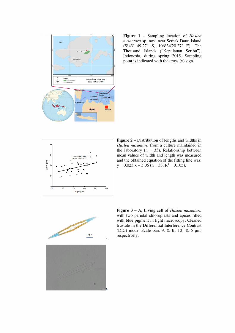
Figure 1 – Sampling location of Haslea
nusantara sp. nov. near Semak Daun Island (5°43' 49.27'' S, 106°34'20.27'' E), The Thousand Islands (“Kepulauan Seribu”), Indonesia, during spring 2015. Sampling point is indicated with the cross (x) sign.
Figure 2 – Distribution of lengths and widths inHaslea nusantara from a culture maintained in the laboratory (n = 33). Relationship between mean values of width and length was measured and the obtained equation of the fitting line was: y = 0.023 x + 5.06 (n = 33, R2 = 0.165).
Figure 3 – A, Living cell of Haslea nusantara
with two parietal chloroplasts and apices filled with blue pigment in light microscopy; Cleaned frustule in the Differential Interference Contrast (DIC) mode. Scale bars A & B: 10 & 5 μm, respectively.
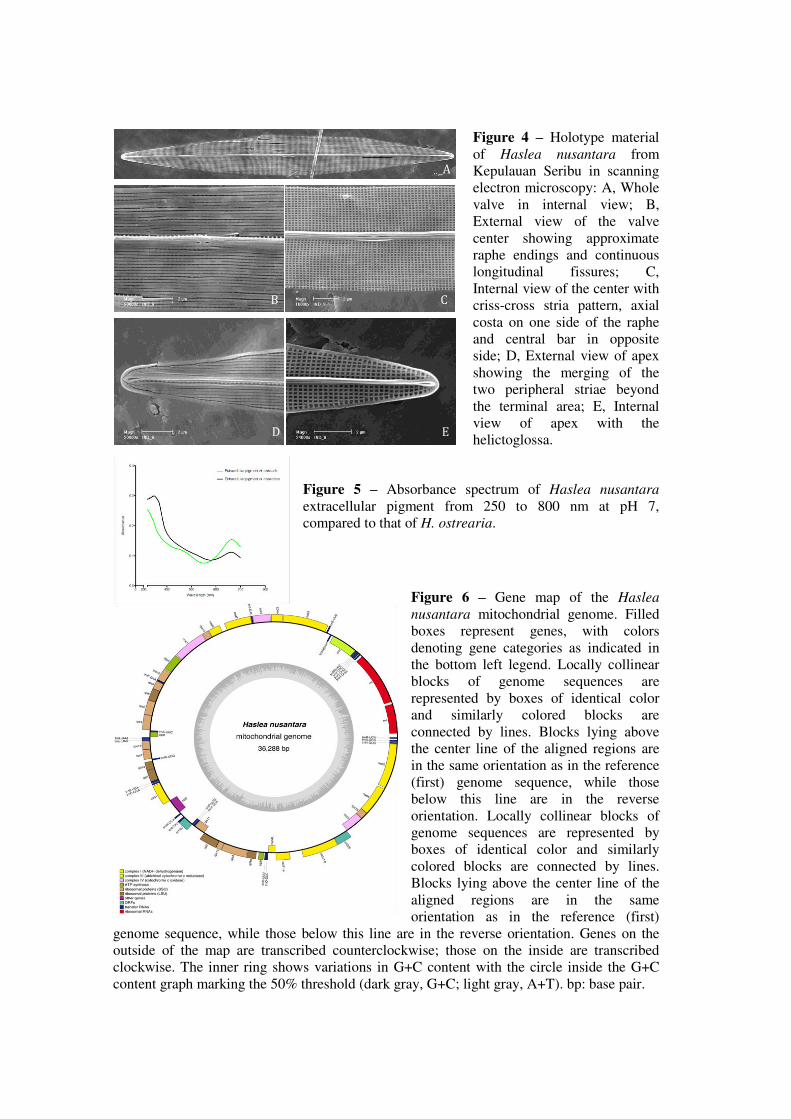
Figure 4 – Holotype material of Haslea nusantara from Kepulauan Seribu in scanning electron microscopy: A, Whole valve in internal view; B, External view of the valve center showing approximate raphe endings and continuous longitudinal fissures; C, Internal view of the center with criss-cross stria pattern, axial costa on one side of the raphe and central bar in opposite side; D, External view of apex showing the merging of the two peripheral striae beyond the terminal area; E, Internal view of apex with the helictoglossa.
Figure 5 – Absorbance spectrum of Haslea nusantara extracellular pigment from 250 to 800 nm at pH 7, compared to that of H. ostrearia.
Figure 6 – Gene map of the Haslea
nusantara mitochondrial genome. Filled boxes represent genes, with colors denoting gene categories as indicated in the bottom left legend. Locally collinear blocks of genome sequences arerepresented by boxes of identical color and similarly colored blocks are connected by lines. Blocks lying above the center line of the aligned regions are in the same orientation as in the reference (first) genome sequence, while those below this line are in the reverse orientation. Locally collinear blocks of genome sequences are represented by boxes of identical color and similarly colored blocks are connected by lines. Blocks lying above the center line of the aligned regions are in the same orientation as in the reference (first)
genome sequence, while those below this line are in the reverse orientation. Genes on the outside of the map are transcribed counterclockwise; those on the inside are transcribed clockwise. The inner ring shows variations in G+C content with the circle inside the G+C content graph marking the 50% threshold (dark gray, G+C; light gray, A+T). bp: base pair.
A
B C
D E
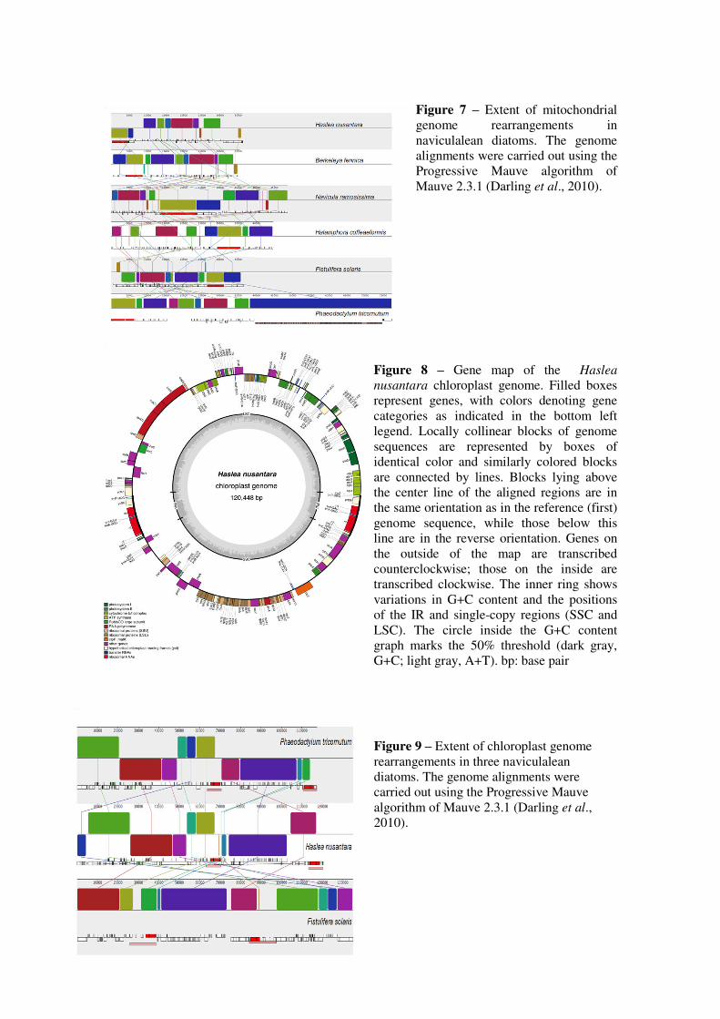
Figure 7 – Extent of mitochondrial genome rearrangements in naviculalean diatoms. The genome alignments were carried out using the Progressive Mauve algorithm of Mauve 2.3.1 (Darling et al., 2010).
Figure 8 – Gene map of the Haslea
nusantara chloroplast genome. Filled boxes represent genes, with colors denoting gene categories as indicated in the bottom left legend. Locally collinear blocks of genome sequences are represented by boxes of identical color and similarly colored blocks are connected by lines. Blocks lying above the center line of the aligned regions are in the same orientation as in the reference (first) genome sequence, while those below this line are in the reverse orientation. Genes on the outside of the map are transcribed counterclockwise; those on the inside are transcribed clockwise. The inner ring shows variations in G+C content and the positions of the IR and single-copy regions (SSC and LSC). The circle inside the G+C content graph marks the 50% threshold (dark gray, G+C; light gray, A+T). bp: base pair
Figure 9 – Extent of chloroplast genome rearrangements in three naviculalean diatoms. The genome alignments were carried out using the Progressive Mauve algorithm of Mauve 2.3.1 (Darling et al., 2010).
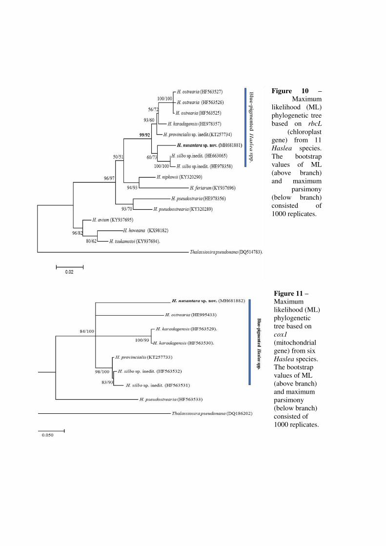
Figure 10 – Maximum
likelihood (ML) phylogenetic tree based on rbcL
(chloroplast gene) from 11 Haslea species. The bootstrap values of ML (above branch) and maximum
parsimony (below branch) consisted of 1000 replicates.
Figure 11 – Maximum likelihood (ML)phylogenetic tree based on cox1 (mitochondrial gene) from six Haslea species. The bootstrap values of ML (above branch) and maximum parsimony (below branch) consisted of 1000 replicates.
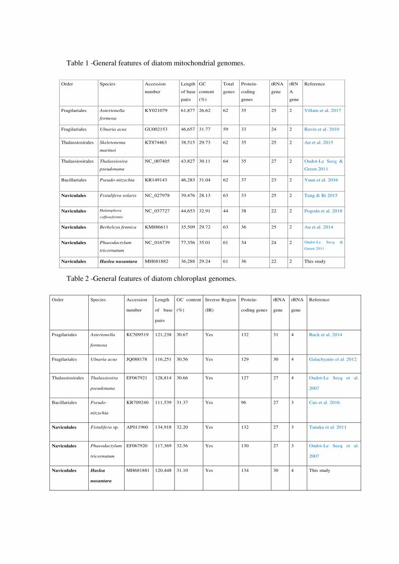
Table 1 -General features of diatom mitochondrial genomes.
Table 2 -General features of diatom chloroplast genomes.
Order Species Accession
number
Length
of base
pairs
GC
content
(%)
Total
genes
Protein-
coding
genes
tRNA
gene
rRN
A
gene
Reference
Fragilariales Asterionella
formosa
KY021079 61,877 26.62 62 35 25 2 Villain et al. 2017
Fragilariales Ulnaria acus GU002153 46,657 31.77 59 33 24 2 Ravin et al. 2010
Thalassiosirales Skeletonema
marinoi
KT874463 38,515 29.73 62 35 25 2 An et al. 2015
Thalassiosirales Thalassiosira
pseudonana
NC_007405 43,827 30.11 64 35 27 2 Oudot-Le Secq &
Green 2011
Bacillariales Pseudo-nitzschia KR149143 46,283 31.04 62 37 23 2 Yuan et al. 2016
Naviculales Fistulifera solaris NC_027978 39,476 28.13 63 33 25 2 Tang & Bi 2015
Naviculales Halamphora
coffeaeformis NC_037727 44,653 32.91 44 38 22 2 Pogoda et al. 2018
Naviculales Berkeleya fennica KM886611 35,509 29.72 63 36 25 2 An et al. 2014
Naviculales Phaeodactylum
tricornutum
NC_016739 77,356 35.01 61 34 24 2 Oudot-Le Secq &
Green 2011
Naviculales Haslea nusantara MH681882 36,288 29.24 61 36 22 2 This study
Order Species Accession
number
Length
of base
pairs
GC content
(%)
Inverse Region
(IR)
Protein-
coding genes
tRNA
gene
rRNA
gene
Reference
Fragilariales Asterionella
formosa
KC509519 121,238 30.67 Yes 132 31 4 Ruck et al. 2014
Fragilariales Ulnaria acus JQ088178 116,251 30.56 Yes 129 30 4 Galachyants et al. 2012
Thalassiosirales Thalassiosira
pseudonana
EF067921 128,814 30.66 Yes 127 27 4 Oudot-Le Secq et al.
2007
Bacillariales Pseudo-
nitzschia
KR709240 111,539 31.37 Yes 96 27 3 Cao et al. 2016
Naviculales Fistulifera sp. AP011960 134,918 32.20 Yes 132 27 3 Tanaka et al. 2011
Naviculales Phaeodactylum
tricornutum
EF067920 117,369 32.56 Yes 130 27 3 Oudot-Le Secq et al.
2007
Naviculales Haslea
nusantara
MH681881 120,448 31.10 Yes 134 30 4 This study
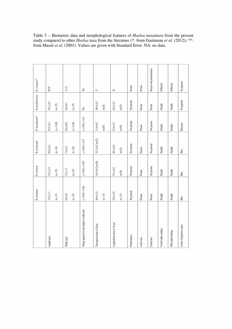
Table 3 -- Biometric data and morphological features of Haslea nusantara from the present study compared to other Haslea taxa from the literature (*: from Gastineau et al. (2012); **: from Massé et al. (2001). Values are given with Standard Error. NA: no data.
H
. nus
anta
ra
H. o
stre
aria
H
. pro
vinc
ialis
H
. kar
adag
ensi
s*
H. p
seud
ostr
eari
a H
. cru
cige
ra**
Leng
th (μm)
73.9
± 1.7
(n =
33)
71.6
± 5.5
(n =
25)
65.8
± 0.1
(n =
26)
52.5
± 0.1
(n =
1148
)
55.5
± 0.2
(n =
25)
95-9
7
Widt
h (μm)
6.8 ±
0.1
(n =
33)
7.5 ±
1.2
(n =
25)
7.4 ±
0.1
(n =
26)
8.0 ±
0.03
(n =
1148
)
8.8 ±
0.1
(n =
25)
11-1
2
Fitti
ng eq
uatio
n for
the l
ength
to w
idth r
atio
y = 0.
02x +
5.06
y =
0.03
x + 4.
28*
y = 0.
02x +
6.33
y =
0.01
x + 8.
21
NA
NA
Tran
sapic
al str
iae in
10 μm
36
.0 ±
1.0
(n =
33)
34.8
± 0.2
(n=3
0)
32.7
± 0.2
(n=2
5)
31.4
± 0.2
(n=3
0)
38.6
± 0.2
(n=2
5)
17
Long
itudin
al str
iae in
10 µ
m 52
.0 ±
2.0
(n =
33)
53.3
± 0.2
(n=3
0)
60.2
± 0.2
(n=2
5)
57.8
± 0.3
(n=3
0)
42.8
± 0.2
(n=2
5)
20
Pseu
dosta
uros
No
t pres
ent
Not p
resen
t No
t pres
ent
Not p
resen
t No
t pres
ent
Pres
ent
Axial
costa
Pr
esen
t Pr
esen
t Pr
esen
t Pr
esen
t Pr
esen
t Pr
esen
t
Centr
al ba
r Pr
esen
t No
t pres
ent
Not p
resen
t No
t pres
ent
Pres
ent
Pres
ent w
ith ps
eudo
staur
os
Centr
al rap
he en
dings
St
raigh
t St
raigh
t St
raigh
t St
raigh
t St
raigh
t De
flecte
d
Polar
raph
e end
ings
Strai
ght
Strai
ght
Strai
ght
Strai
ght
Strai
ght
Defle
cted
Colou
r of p
igmen
t at a
pex
Blue
Bl
ue
Blue
Bl
ue-g
rey
No pi
gmen
t No
pigm
ent
Annexes
ANNEXE 5 : Chapitres de Livres
Falaise, Dewi et al. (2018) ; Gastineau et al. (2018) [Prévisualisations]
Falaise, C., Dewi, I.C., Hellio, C., Bourgougnon, N., Mouget, J.-L., 2018. Chapter 12. Anticancer,
antiviral, antibacterial and antifungal properties in microalgae, in: Microalgae in Health and Disease
Prevention. Levine I. and Fleurence J. (Eds.). Elsevier Science, Amsterdam, Netherlands.
et
Gastineau, R., Prasetiya, F.S., Falaise, C., Cognie, B., Decottignies, P., Morancais, M., Méléder, V.,
Davidovich, N.A., Turcotte, F., Tremblay, R., Pasetto, P., Dittmer, J., Bardeau, J.-F., Pouvreau, J.-B.,
Mouget, J.-L., 2018. Chapter 16. Marennine-Like Pigments: Blue Diatom of Green Oyster Cult?, in:
Blue Biotechnology, Production and Use of Marine Molecules. Stéphane La Barre and Stephen S.
Bates (Eds.). Wiley-VCH, Weinheim, Germany.
235Microalgae in Health and Disease Prevention. https://doi.org/10.1016/B978-0-12-811405-6.00012-8
Copyright © 2018 Elsevier Inc. All rights reserved.
Chapter 12
Anticancer, Antiviral, Antibacterial, and Antifungal Properties in Microalgae
Intan C. Dewi1,a, Charlotte Falaise1,a, Claire Hellio2, Nathalie Bourgougnon3, Jean-Luc Mouget1
1Le Mans Université, Le Mans, France; 2Universitaire Européen de la Mer (IUEM) - UBO – UEB Technopôle Brest-Iroise, Plouzané, France;
3Université Bretagne Sud, Vannes, France
1. INTRODUCTION
This chapter mainly focuses on antiproliferative and antimicrobial compounds produced by microalgae (eukaryotic, photo-
trophic microorganisms) and cyanobacteria, with special emphasis on health and disease prevention. To seek for new bio-
active compounds as antiproliferative, antiviral, or antibacterial agents, the pharmaceutical industry has shown a growing
interest for a vast marine bioresource, underexplored yet, the aquatic phototrophic microorganisms. Indeed, many strains
of microalgae and cyanobacteria are reported to produce intracellular as well as extracellular metabolites with various
biological activities, and screening studies have been recurrently carried out with the aim to discover new bioactive metabo-
lites. Illustrating the global interest for microalgae and cyanobacteria as new sources of bioactive agents, several reviews
illustrate the increasing number of compounds identified so far (e.g., Ahmadi et al., 2015; Borowitzka, 1995; Encarnacao
et al., 2015; Falaise et al., 2016; Raposo et al., 2015; Shimizu, 1996, 2003; Singh et al., 2005). Physicochemical and phar-
macological studies have paid special attention to proteins, pigments, terpenoids, and steroids (Abdo et al., 2012; Gastineau
et al., 2012a,b; Gustafson et al., 1989; Rechter et al., 2006). In fact, secondary metabolites produced by microalgae and
cyanobacteria represent a very chemically diverse reservoir, including nearly every chemical class of natural products, from
amino and fatty acids to alkaloids and polyphenolic compounds.
Chapter Outline1. Introduction 235
2. Anticancer Activity 236
2.1 Purified Molecules 236
2.1.1 Microalgae 236
2.1.2 Cyanobacteria 237
2.2 Methods for Assessment of Anticancer Activity 239
3. Antiviral Activity 240
3.1 Screening and Extracts 240
3.2 Purified Molecules 241
3.2.1 Polysaccharides 241
3.2.2 Proteins, Lipids, Pigments 243
3.3 Methods to Assess the Antiviral Activity of a Compound 244
4. Antibacterial Activity 244
4.1 Antibacterial Activities of Microalgae 245
4.1.1 Green Microalgae 245
4.1.2 Red Microalgae 247
4.1.3 Diatoms 247
4.2 Antibacterial Activities of Cyanobacteria 248
4.3 Methods to Assess the Antibacterial Activity
of a Compound 248
4.4 Antibacterial Activity of Microalgae and Cyanobacteria
Against Animal Diseases 249
5. Antifungal Activity 249
5.1 Antifungal Activities From Eukaryotic Microalgae 250
5.1.1 Green and Red Microalgae 250
5.1.2 Diatoms and Dinoflagellates 251
5.2 Antifungal Activities From Cyanobacteria 252
6. Conclusion and Prospects 252
Acknowledgment 253
References 253
a Intan Chandra Dewi and Charlotte Falaise contributed equally to this work.
529
16
Marennine-Like Pigments: Blue Diatom or Green
Oyster Cult?
Romain Gastineau1, Fiddy S. Prasetiya1,2,3, Charlotte Falaise1, Bruno Cognie4,Priscilla Decottignies4, Michèle Morançais4, VonaMéléder4,Nikolai Davidovich5, François Turcotte6, Réjean Tremblay6, Pamela Pasetto7,Jens Dittmer7, Jean-François Bardeau7, Jean-Bernard Pouvreau8, andJean-LucMouget1
1LeMans Université, LUNAM,Mer Molecule Santé (MMS), Faculté des Sciences et des Techniques,
Mer-Molécules-Santé (MMS) EA 2160, Avenue Olivier Messiaen, 72085 LeMans Cedex 9, France2Universitas Padjadjaran, Marine Science Department, Jl. Raya Bandung Sumedang KM 21, Jatinangor 40600,
Indonesia3Postgraduate school Universitas Padjadjaran, Program Study of Biotechnology, Research center of
Biomolecular and Bioinformatics, Jl. Dipatiukur No. 35 Bandung Gedung Sekolah Pascasarjana, Bandung
40132, Indonesia4Université de Nantes, LUNAM,Mer Molecule Santé (MMS), Faculté des Sciences et des Techniques,
Mer-Molécules-Santé (MMS) EA 2160, 2 rue de la Houssinière, 44322 Nantes, France5Karadag Scientific Station – Nature Reserve of the Russian Academy of Sciences, Laboratory of Algae and
Microbiota, p/o Kurortnoe, 98188 Feodosiya, Republic of Crimea, Russia6Université du Québec à Rimouski, Institut des Sciences de laMer à Rimouski, Allée des Ursulines, Québec
G5L2Z9, Canada7LeMans Université, Faculté des Sciences et des Techniques, Institut des Molécules et Matériaux duMans
(IMMM), UMR CNRS 6283, Avenue Olivier Messiaen, 72085 LeMans Cedex 9, France8Université de Nantes, Faculté des Sciences et des Techniques, Laboratoire de Biologie et Pathologie Végétales,
SFR 4207 QUASAV, 2 rue de la Houssinière, 44322 Nantes, France
Abstract
his chapter presents new insights into “blue” diatom species and
marennine-like pigments in particular, concerning the biodiversity of these
microalgae, their pigments, their interactions with bivalves, and their potential
use in biotechnology, in particular as natural blue pigments. Historically, the
term “blue diatom” refers to the marine diatom Haslea ostrearia, known for
its specific ability to produce a water-soluble blue pigment, marennine, but
it applies now to several new species recently described, all producing blue
pigments. Genetically, these species form a well-defined clade inside the genus
Haslea, and their pigments seem to possess similar spectral characteristics and
biological activities, for instance, antimicrobial, especially against some oyster
pathogens (e.g., Vibrio splendidus). Marennine is the main factor responsible
Blue Biotechnology: Production and Use of Marine Molecules,
First Edition. Edited by Stéphane La Barre and Stephen S. Bates.
© 2018Wiley-VCH Verlag GmbH & Co. KGaA. Published 2018 byWiley-VCH Verlag GmbH & Co. KGaA.
530 16 Marennine-Like Pigments: Blue Diatom or Green Oyster Cult?
for the greening of oyster gills in western France, where oyster farming is
a phenomenon that has a noticeable economic impact. he green bivalves
are a must for connoisseurs and a way to boost the marketability of regional
oyster products. Although the coloration of the gills by marennine has not
been correlated with an increase of oyster palatability, the biological activities
of marennine could represent a new avenue of valorization of this “old”
pigment, especially considering oyster mortality events and the sustainability
of molluscan shellfish industry.
16.1 Introduction
Diatoms are silicified brown microalgae of high ecological importance [1, 2],with ever-extending applications in blue biotechnologies (see Chapter 27).hey can be used in aquaculture, bioremediation, biofuel production, nan-otechnologies, and the production of various high added-value metabolites [3].he characteristic brown color of diatoms results from their pigment content,chlorophylls a and c, masked by carotenoids diatoxanthin, diadinoxanthin,and mainly fucoxanthin. In addition to these photosynthetic pigments, somespecies belonging to the genus Haslea synthesize water-soluble blue pigments,known as marennine-like pigments, and are therefore often referred to as “bluediatoms” or “blue Haslea” [4–7]. he chemical structure of these pigments isnot yet determined, although clues advocate for a polyphenolic [8] or glycosidicnature [6]. Some of these blue pigments, like marennine produced by Haslea
ostrearia (Figure 16.1), seem to exist under two different forms, one intracel-lular marennine (IMn) and one extracellular marennine (EMn), with differentmolecular weights and UV–visible spectral characteristics [8]. Furthermore,their color is pH dependent, reversibly shifting from purple blue in acidicmedia to green in neutral to alkaline conditions [8–11] (Figure 16.2). In vivo,the pigment appears intense blue, especially when accumulated at cell apices,but it can turn the surrounding water green when released from the cells.Furthermore, supernatants of water-soluble extracts of H. ostrearia cultures,or purified marennine-like pigments, are known to display several biologicalactivities, for example, antibacterial, antiviral, antifungal, antiproliferative, andantioxidant properties [6, 12–17]. Purified marennine [18] and H. ostrearia
culture supernatants [19, 20] have also demonstrated allelopathic activitiesagainst other species of microalgae. hese various activities make blue Haslea
and their marennine-like pigments a promising model for blue biotechnolo-gies. After a brief presentation of the biodiversity of blue Haslea species andtheir pigments, this chapter will present new insights into the structure andpossible applications of marennine-like pigments, mainly as a coloring agentand probiotic in aquaculture or a natural blue colorant in the food industry andcosmetology.
Annexes
ANNEXE 6 : Essais préliminaires
1) Inactivation d’une suspension du virus OsHV-1 par exposition à la marennine purifiée (EMn) et bio-
essai par administration de la suspension traitée à des huîtres creuses
2) Effet de la marennine purifiée (EMn) sur les premières divisions embryonnaires des oursins
Paracentrotus lividus et Sphaerechinus granularis : effets sur la synthèse protéique globale et sur
l’activité de CDK1
Les expérimentations présentées ci-après ont été respectivement conduites 1) en Avril 2016 au Laboratoire de Génétique et de Pathologie des Mollusques Marins (RBE/SG2M/LGPMM/Ifremer), Station La Tremblade sous l’encadrement de Cyrille François et 2) en juillet 2016 au Laboratoire de Biologie Intégrative des Modèles Marins, Sorbonne Universités (UPMC Paris 06/CNRS/UMR 8227),Station Biologique de Roscoff, sous l’encadrement de Patrick Cormier.
1) Lors de cet accueil scientifique de 3 semaines au LGPMM, nous avons développé 3 axes de recherches en étudiant les effets de la EMn i) in vitro sur la croissance de bactéries du genre Vibrio isolées de bivalves ; ii) sur les hémocytes d’huîtres en conduisant des essais de cytotoxicité avec des mesures en cytométrie en flux ; et enfin iii) sur l’inactivation d’une suspension du virus OsHV-1 en menant un bio-essai par administration de la suspension traitée à des huîtres creuses. Les résultats des essais i) et ii) ont pu être valorisés (Falaise et al. (2016), Annexe 1 et Falaise et al. (2019), Annexe 2), de plus les effets observés sur la croissance des souches de Vibrio ont pu être davantage étudiés en poursuivant les expérimentations au laboratoire MMS du Mans. Des biais expérimentaux n’ont pas permis d’exploiter les résultats de l’axe de recherche iii), néanmoins la méthodologie employée est présentée dans cette annexe.
2) Lors d’un premier séjour d’une semaine à la station biologique de Roscoff en mai 2016, nous avions réalisé des essais exploratoires pour évaluer si la EMn présentait des effets sur la fécondation et la cinétique de première division embryonnaire chez les oursins P. lividus et S. granularis. Lesrésultats préliminaires obtenus ont encouragé la planification d’une seconde série d’expérimentations lors d’un nouvel accueil scientifique de 2 semaines en juillet 2016. Pour ces expérimentations, des tests biochimiques ont été conduits sur P. lividus et S. granularis pour évaluer si la EMn impactait la synthèse globale de protéines dans l’œuf nouvellement fécondé ou si elle avait un effet sur l’activité du complexe CDK-1 Cycline B lors de la mitose, expliquant ainsi l’absence de division des œufs exposés aux fortes concentrations de pigment. La méthodologie employée pour ces essais et les résultats préliminaires obtenus sont présentés dans cette annexe. Bien que ces tests aient donné des premiers résultats intéressants, il a été décidé, lors d’un 3
ème séjour scientifique de 2 semaines en mars 2017, de finaliser les essais concernant les effets de la marennine sur la fécondation et le développement embryonnaire en vue d’une publication. Les expérimentations ont été conduites sur S. granularis uniquement car l’espèce P. lividus n’était pas disponible en cette période de l’année. Les résultats obtenus lors de ce 3ème séjour scientifique ainsi que certains des résultats préliminaires ont été valorisés dans la publication Falaise et al. (2019), Annexe 2.
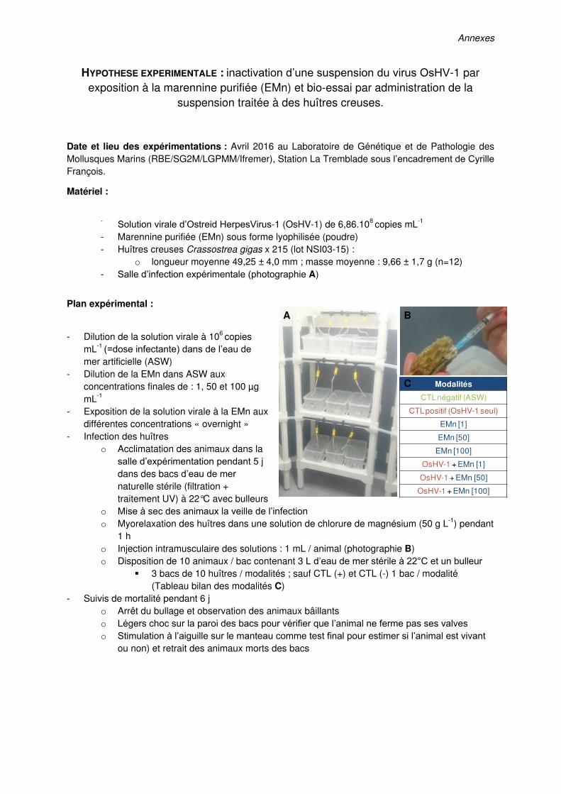
Annexes
HYPOTHESE EXPERIMENTALE : inactivation d’une suspension du virus OsHV-1 par exposition à la marennine purifiée (EMn) et bio-essai par administration de la
suspension traitée à des huîtres creuses.
Date et lieu des expérimentations : Avril 2016 au Laboratoire de Génétique et de Pathologie des Mollusques Marins (RBE/SG2M/LGPMM/Ifremer), Station La Tremblade sous l’encadrement de Cyrille
François.
Matériel :
- Solution virale d’Ostreid HerpesVirus-1 (OsHV-1) de 6,86.108 copies mL-1
- Marennine purifiée (EMn) sous forme lyophilisée (poudre) - Huîtres creuses Crassostrea gigas x 215 (lot NSI03-15) :
o longueur moyenne 49,25 ± 4,0 mm ; masse moyenne : 9,66 ± 1,7 g (n=12) - Salle d’infection expérimentale (photographie A)
Plan expérimental :
- Dilution de la solution virale à 106 copies mL-1
(=dose infectante) dans de l’eau de
mer artificielle (ASW) - Dilution de la EMn dans ASW aux
concentrations finales de : 1, 50 et 100 µg mL-1
- Exposition de la solution virale à la EMn aux différentes concentrations « overnight »
- Infection des huîtres o Acclimatation des animaux dans la
salle d’expérimentation pendant 5 j dans des bacs d’eau de mer
naturelle stérile (filtration + traitement UV) à 22°C avec bulleurs
o Mise à sec des animaux la veille de l’infection o Myorelaxation des huîtres dans une solution de chlorure de magnésium (50 g L-1) pendant
1 h o Injection intramusculaire des solutions : 1 mL / animal (photographie B) o Disposition de 10 animaux / bac contenant 3 L d’eau de mer stérile à 22°C et un bulleur
3 bacs de 10 huîtres / modalités ; sauf CTL (+) et CTL (-) 1 bac / modalité (Tableau bilan des modalités C)
- Suivis de mortalité pendant 6 j o Arrêt du bullage et observation des animaux bâillants o Légers choc sur la paroi des bacs pour vérifier que l’animal ne ferme pas ses valves o Stimulation à l’aiguille sur le manteau comme test final pour estimer si l’animal est vivant
ou non) et retrait des animaux morts des bacs
Modalités
CTLnégatif (ASW)
CTL positif (OsHV-1 seul)
EMn [1]
EMn [50]
EMn [100]
OsHV-1 + EMn [1]
OsHV-1 + EMn [50]
OsHV-1 + EMn [100]
A B
C
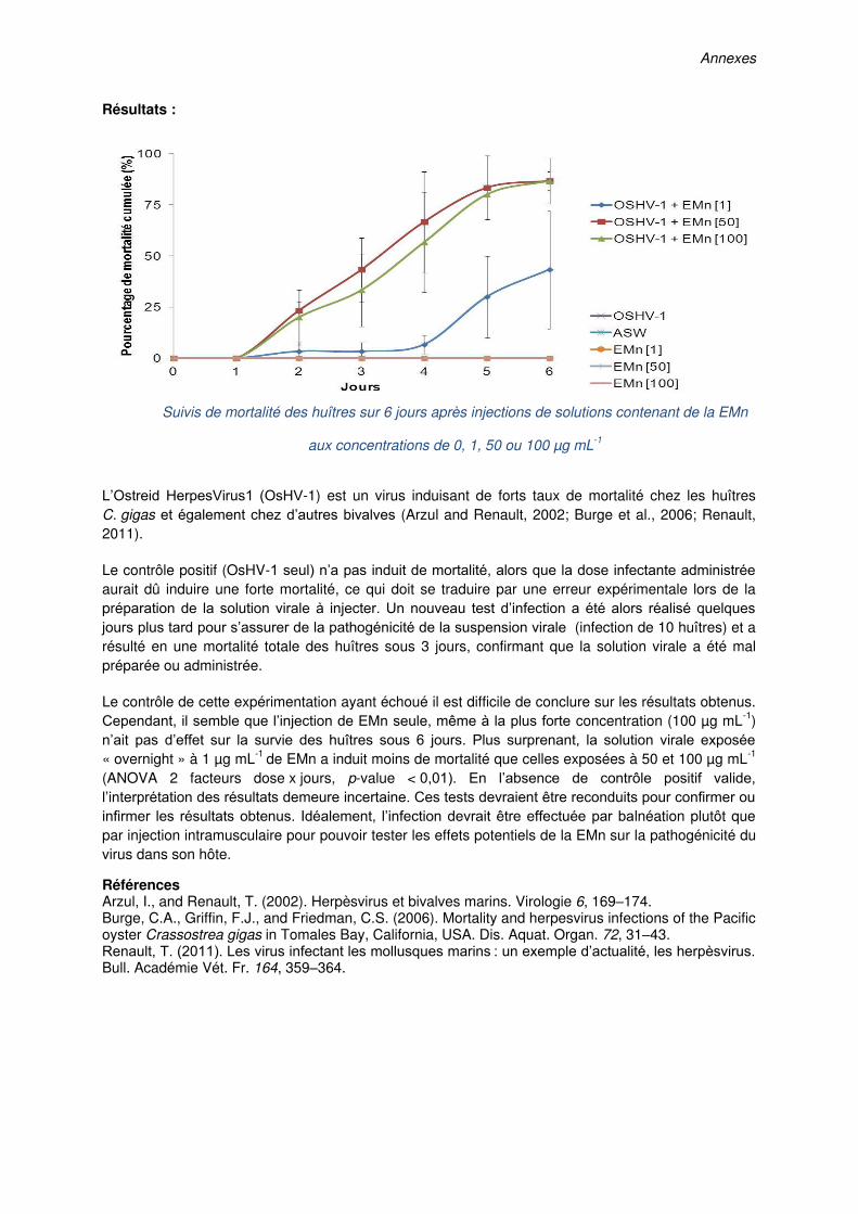
Annexes
Résultats :
Suivis de mortalité des huîtres sur 6 jours après injections de solutions contenant de la EMn
aux concentrations de 0, 1, 50 ou 100 µg mL-1
L’Ostreid HerpesVirus1 (OsHV-1) est un virus induisant de forts taux de mortalité chez les huîtres C. gigas et également chez d’autres bivalves (Arzul and Renault, 2002; Burge et al., 2006; Renault, 2011).
Le contrôle positif (OsHV-1 seul) n’a pas induit de mortalité, alors que la dose infectante administrée
aurait dû induire une forte mortalité, ce qui doit se traduire par une erreur expérimentale lors de la préparation de la solution virale à injecter. Un nouveau test d’infection a été alors réalisé quelques
jours plus tard pour s’assurer de la pathogénicité de la suspension virale (infection de 10 huîtres) et a résulté en une mortalité totale des huîtres sous 3 jours, confirmant que la solution virale a été mal préparée ou administrée.
Le contrôle de cette expérimentation ayant échoué il est difficile de conclure sur les résultats obtenus. Cependant, il semble que l’injection de EMn seule, même à la plus forte concentration (100 µg mL-1) n’ait pas d’effet sur la survie des huîtres sous 6 jours. Plus surprenant, la solution virale exposée
« overnight » à 1 µg mL-1 de EMn a induit moins de mortalité que celles exposées à 50 et 100 µg mL-1
(ANOVA 2 facteurs dose x jours, p-value < 0,01). En l’absence de contrôle positif valide,
l’interprétation des résultats demeure incertaine. Ces tests devraient être reconduits pour confirmer ou
infirmer les résultats obtenus. Idéalement, l’infection devrait être effectuée par balnéation plutôt que par injection intramusculaire pour pouvoir tester les effets potentiels de la EMn sur la pathogénicité du virus dans son hôte.
Références Arzul, I., and Renault, T. (2002). Herpèsvirus et bivalves marins. Virologie 6, 169–174. Burge, C.A., Griffin, F.J., and Friedman, C.S. (2006). Mortality and herpesvirus infections of the Pacific oyster Crassostrea gigas in Tomales Bay, California, USA. Dis. Aquat. Organ. 72, 31–43. Renault, T. (2011). Les virus infectant les mollusques marins : un exemple d’actualité, les herpèsvirus. Bull. Académie Vét. Fr. 164, 359–364.
Annexes
HYPOTHESE EXPERIMENTALE : effet de la marennine purifiée (EMn) sur les premières divisions embryonnaires des oursins Paracentrotus lividus et Sphaerechinus granularis : effets sur la synthèse protéique globale et sur l’activité de CDK1
Date et lieu des expérimentations : Juillet 2016 au Laboratoire de Biologie Intégrative des Modèles Marins, Sorbonne Universités (UPMC Paris 06/CNRS/UMR 8227), Station Biologique de Roscoff, sous l’encadrement de Patrick Cormier.
Préambule : lors de précédentes expérimentations préliminaires, nous avions remarqué l’effet de la
marennine purifiée (EMn) ajoutée 5 minutes après fécondation sur les premières étapes du cycle cellulaire de l’embryon d’oursin Paracentrotus lividus. Les concentrations de 100 et 50 µg mL-1 de EMn avaient eu des effets très nets : observation de divisions asymétriques et asynchrones, puis arrêt total du développement. La concentration 10 µg mL-1 avait induit un léger décalage de la cinétique de division cellulaire par rapport au CTL. Il a ensuite été remarqué pour les œufs exposés à 10 µg m-1 de EMn un développement anormal des embryons (formes aplatie, absence de spicules). La concentration 1 µg mL-1 de EMn n’a pas semblé avoir eu d’effet sur le cycle cellulaire et le
développement embryonnaire.
Plan expérimental :
Deux tests biochimiques sont réalisés à T0 (avant fécondation), T30 (30 min post fécondation), T60, T90, T120 et T150 :
1) évaluation de la synthèse protéique globale en réponse à la fécondation par la méthode SUnSET (Schmidt et al., 2009) Ajout de puromycine à faible concentration : ce composé mime un acide aminé et se fixe sur le peptide -> un anticorps sera dirigé vers la puromycine ; le marquage à la puromycine est relatif à la quantité de peptides produits.
2) évaluation de l’activité de CDK1 en dirigeant un anticorps vers le site phosphorylé de la phosphatase protein 1 (PP1) CDK1 (kinase mitotique) est activée par la CyclineB. L’activation de la kinase CDK1 induit la
phosphorylation de PP1 : un anticorps sera dirigé vers le site phosphorylé de PP1. Pour sortir de la métaphase, la CyclineB est inactivée, la quantité de CDK1 diminue.
Principales étapes expérimentales : -Prélèvement des œufs après fécondation à T0, T30, T60, T90, T120 et T150 -Western Blot -Exposition aux Ac I et Ac II
-Révélation en ECL (enhanced chemiluminescence) -Traitement des résultats des Western Blot sur Image J La ponte des oursins et la fécondation sont réalisées selon le protocole décrit dans le Chapitre IV. Partie 2 de ce manuscrit.
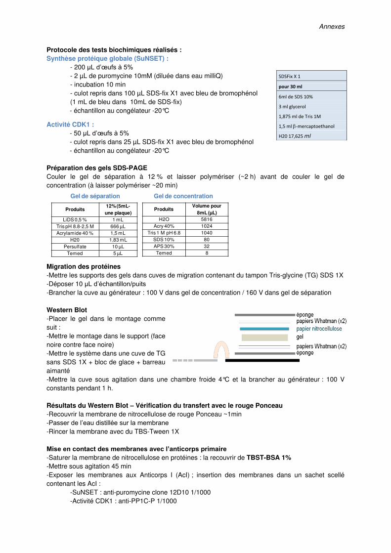
Annexes
Protocole des tests biochimiques réalisés : Synthèse protéique globale (SuNSET) :
- 200 µL d’œufs à 5% - 2 µL de puromycine 10mM (diluée dans eau milliQ) - incubation 10 min - culot repris dans 100 µL SDS-fix X1 avec bleu de bromophénol (1 mL de bleu dans 10mL de SDS-fix) - échantillon au congélateur -20°C
Activité CDK1 : - 50 µL d’œufs à 5% - culot repris dans 25 µL SDS-fix X1 avec bleu de bromophénol - échantillon au congélateur -20°C
Préparation des gels SDS-PAGE Couler le gel de séparation à 12 % et laisser polymériser (~2 h) avant de couler le gel de concentration (à laisser polymériser ~20 min)
Migration des protéines -Mettre les supports des gels dans cuves de migration contenant du tampon Tris-glycine (TG) SDS 1X -Déposer 10 µL d’échantillon/puits -Brancher la cuve au générateur : 100 V dans gel de concentration / 160 V dans gel de séparation Western Blot -Placer le gel dans le montage comme suit : -Mettre le montage dans le support (face noire contre face noire) -Mettre le système dans une cuve de TG sans SDS 1X + bloc de glace + barreau aimanté -Mettre la cuve sous agitation dans une chambre froide 4°C et la brancher au générateur : 100 V constants pendant 1 h. Résultats du Western Blot – Vérification du transfert avec le rouge Ponceau -Recouvrir la membrane de nitrocellulose de rouge Ponceau ~1min -Passer de l’eau distillée sur la membrane -Rincer la membrane avec du TBS-Tween 1X Mise en contact des membranes avec l’anticorps primaire -Saturer la membrane de nitrocellulose en protéines : la recouvrir de TBST-BSA 1% -Mettre sous agitation 45 min -Exposer les membranes aux Anticorps I (AcI) ; insertion des membranes dans un sachet scellé contenant les AcI : -SuNSET : anti-puromycine clone 12D10 1/1000 -Activité CDK1 : anti-PP1C-P 1/1000
ProduitsVolume pour
8mL(µL)H2O 5816
Acry 40% 1024Tris 1 M pH 6.8 1040
SDS 10% 80APS 30% 32Temed 8
Produits12% (5mL-une plaque)
LiDS 0,5 % 1 mLTris pH 8.8-2,5 M 666 µLAcrylamide 40 % 1,5 mL
H20 1,83 mLPersulfate 10 µL
Temed 5 µL
Gel de séparation Gel de concentration
SDSFix X 1
pour 30 ml
6ml de SDS 10%
3 ml glycerol
1,875 ml de Tris 1M
1,5 ml mercaptoethanol
H20 17,625 ml
Annexes
-Mettre les sachets sous agitation à 4°C over night Mise en contact des membranes avec l’anticorps secondaire -Retirer les AcI et rincer les membranes au TBST (3x10min sous agitation) -Recouvrir la membrane d’AcII (dilution dans TBST-BSA 1%) : -SuNSET : anti-mouse au 1/5000 -Activité CDK1 : anti-rabbit au 1/4000 -Mettre sous agitation pendant 45 min -Rincer les membranes au TBST (2x5 min sous agitation) Révélation – chimioluminescence -SuNSET : ECL+ _scanner Typhoon : -Activité CDK1 : ECL_révélation sur films photographiques
Traitement des données sous image J Les images scanners des révélations sont analysées sous ImageJ par comparaison des niveaux de gris entre les bandes des différents traitements. Les niveaux de gris de chaque bande sont normalisés avec un contrôle de charge (PSTAIR).
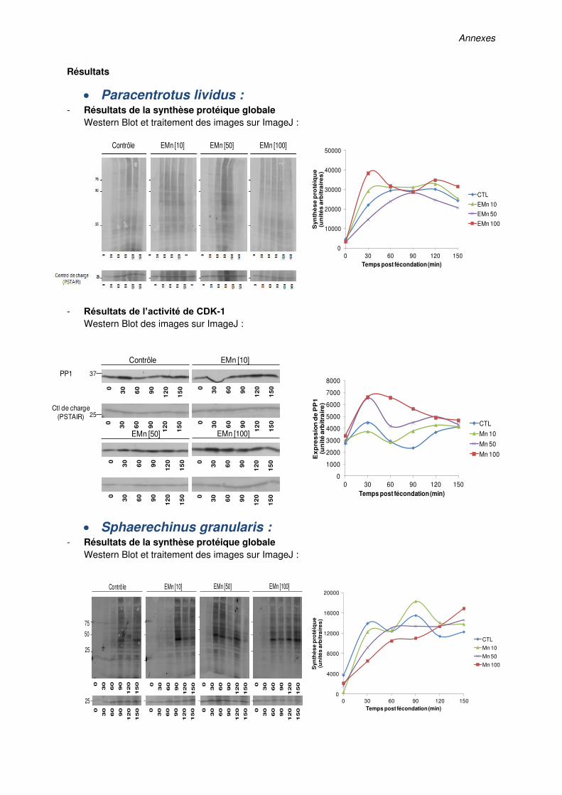
Annexes
Résultats
Paracentrotus lividus : - Résultats de la synthèse protéique globale
Western Blot et traitement des images sur ImageJ :
- Résultats de l’activité de CDK-1
Western Blot des images sur ImageJ :
Sphaerechinus granularis : - Résultats de la synthèse protéique globale
Western Blot et traitement des images sur ImageJ :
Contrôle EMn [10] EMn [50] EMn [100]
0
1000
2000
3000
4000
5000
6000
7000
8000
0 30 60 90 120 150
Ex
pre
ss
ion
de
PP
1
(un
ité
arb
itra
ire
)
Temps post fécondation (min)
CTL
Mn 10
Mn 50
Mn 100
0
30
60
90
120
150
25
50
75
Contrôle
25
EMn [100]EMn [10] EMn [50]
0
30
60
90
120
150
0
30
60
90
120
150
0
30
60
90
120
150
0
30
60
90
120
150
0
30
60
90
120
150
0
30
60
90
120
150
0
30
60
90
120
150
0
10000
20000
30000
40000
50000
0 30 60 90 120 150
Syn
thè
se
pro
téiq
ue
(un
ité
sa
rbit
rair
es
)
Temps post fécondation (min)
CTL
EMn 10
EMn 50
EMn 100
37
0
30
60
90
12
0
15
0
Contrôle
25
0
30
60
90
12
0
15
0
EMn [100]
0
30
60
90
12
0
15
0
0
30
60
90
12
0
15
0
0
30
60
90
12
0
15
0
0
30
60
90
12
0
15
0
EMn [50]
0
30
60
90
12
0
15
0
0
30
60
90
12
0
15
0
EMn [10]
PP1
Ctl de charge(PSTAIR)
0
4000
8000
12000
16000
20000
0 30 60 90 120 150
Syn
thè
se
pro
téiq
ue
(un
ité
sa
rbit
rair
es
)
Temps post fécondation (min)
CTL
Mn 10
Mn 50
Mn 100

Annexes
- Résultats de l’activité de CDK-1
Western Blot des images sur ImageJ :
Discussion des résultats
Des œufs d’oursins nouvellement fécondés ont été exposés à de la EMn purifiée. Pour ces tests,
préliminaires, un seul réplicat a été effectué par modalité testée.
Chez P. lividus, la EMn à 50 et 100 µg mL-1 a induit un blocage de la première division mitotique alors que la concentration 10 µg mL-1 n’a pas eu d’effet sur la cinétique de 1
ère division (données non montrées). Chez S. granularis, contrairement à ce qui était attendu, l’exposition des œufs à la EMn n’a
pas eu d’effet sur la cinétique de 1ère division, même à 100 µg mL-1 (données non montrées). Ces
résultats sont à l’encontre de tests menés précédemment montrant une sensibilité de S. granularis à la EMn à 50 et 100 µg mL-1.
Avec la méthode de purification de la EMn développée par (Pouvreau et al., 2006), la pureté de la EMn obtenue (sous forme de poudre lyophilisée) à l’Université du Québec à Rimouski (UQAR) varie en fonction des fractions sortantes de la colonne de chromatographie par échanges d’ions. Il s’est
avéré que la fraction d’EMn utilisée pour ces tests présentait une pureté de seulement 35 % (contre 80 % de pureté pour la fraction utilisée pour les précédents tests), mais ce biais expérimental n’a été
déterminé qu’a posteriori. Ainsi, la plus forte dose de EMn testée dans les tests présentés ci-dessus ne correspond finalement pas à une concentration réelle de 100 µg mL-1 mais à une concentration avoisinant les 35 µg mL-1, ce qui pourrait expliquer l’absence d’effet observé sur la première division mitotique chez S. granularis.
Il apparait par ailleurs que l’oursin P. lividus est beaucoup plus sensible à la EMn que S. granularis puisque les concentrations testées ont induit un blocage des divisions cellulaires chez P. lividus mais pas chez S. granularis.
Des tests biochimiques ont été conduits pour rendre compte de la synthèse protéique globale ainsi que de l’activité CDK1-CyclineB (en étudiant l’expression de PP1, phosphorylé par CDK1) dans l’œuf
d’oursin suite à la fécondation.
Le schéma suivant illustre les événements ayant lieu dans l’œuf suite à la fécondation chez l’oursin :
0
2000
4000
6000
8000
10000
0 30 60 90 120 150
Ex
pre
ss
ion
de
PP
1
(un
ité
arb
itra
ire
)
Temps post fécondation (min)
CTL
Mn 10
Mn 50
Mn 100
37
0
30
60
90
12
0
15
0
Contrôle
25
0
30
60
90
12
0
15
0
EMn [100]
0
30
60
90
12
0
15
0
0
30
60
90
12
0
15
0
0
30
60
90
12
0
15
0
0
30
60
90
12
0
15
0
EMn [50]
0
30
60
90
12
0
15
0
0
30
60
90
12
0
15
0
EMn [10]
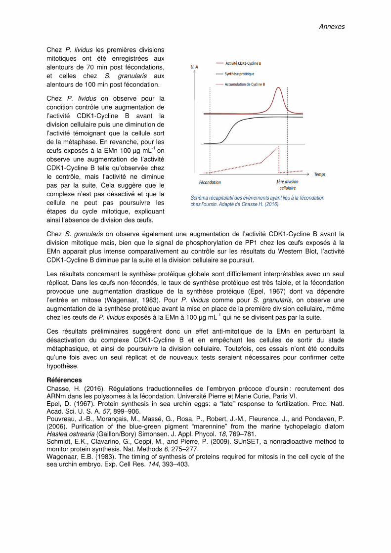
Annexes
Chez P. lividus les premières divisions mitotiques ont été enregistrées aux alentours de 70 min post fécondations, et celles chez S. granularis aux alentours de 100 min post fécondation.
Chez P. lividus on observe pour la condition contrôle une augmentation de l’activité CDK1-Cycline B avant la division cellulaire puis une diminution de l’activité témoignant que la cellule sort
de la métaphase. En revanche, pour les œufs exposés à la EMn 100 µg mL-1 on observe une augmentation de l’activité
CDK1-Cycline B telle qu’observée chez
le contrôle, mais l’activité ne diminue
pas par la suite. Cela suggère que le complexe n’est pas désactivé et que la
cellule ne peut pas poursuivre les étapes du cycle mitotique, expliquant ainsi l’absence de division des œufs.
Chez S. granularis on observe également une augmentation de l’activité CDK1-Cycline B avant la division mitotique mais, bien que le signal de phosphorylation de PP1 chez les œufs exposés à la
EMn apparait plus intense comparativement au contrôle sur les résultats du Western Blot, l’activité
CDK1-Cycline B diminue par la suite et la division cellulaire se poursuit.
Les résultats concernant la synthèse protéique globale sont difficilement interprétables avec un seul réplicat. Dans les œufs non-fécondés, le taux de synthèse protéique est très faible, et la fécondation provoque une augmentation drastique de la synthèse protéique (Epel, 1967) dont va dépendre l’entrée en mitose (Wagenaar, 1983). Pour P. lividus comme pour S. granularis, on observe une augmentation de la synthèse protéique avant la mise en place de la première division cellulaire, même chez les œufs de P. lividus exposés à la EMn à 100 µg mL-1 qui ne se divisent pas par la suite.
Ces résultats préliminaires suggèrent donc un effet anti-mitotique de la EMn en perturbant la désactivation du complexe CDK1-Cycline B et en empêchant les cellules de sortir du stade métaphasique, et ainsi de poursuivre la division cellulaire. Toutefois, ces essais n’ont été conduits
qu’une fois avec un seul réplicat et de nouveaux tests seraient nécessaires pour confirmer cette hypothèse.
Références Chasse, H. (2016). Régulations traductionnelles de l’embryon précoce d’oursin : recrutement des ARNm dans les polysomes à la fécondation. Université Pierre et Marie Curie, Paris VI. Epel, D. (1967). Protein synthesis in sea urchin eggs: a “late” response to fertilization. Proc. Natl. Acad. Sci. U. S. A. 57, 899–906. Pouvreau, J.-B., Morançais, M., Massé, G., Rosa, P., Robert, J.-M., Fleurence, J., and Pondaven, P. (2006). Purification of the blue-green pigment “marennine” from the marine tychopelagic diatom Haslea ostrearia (Gaillon/Bory) Simonsen. J. Appl. Phycol. 18, 769–781. Schmidt, E.K., Clavarino, G., Ceppi, M., and Pierre, P. (2009). SUnSET, a nonradioactive method to monitor protein synthesis. Nat. Methods 6, 275–277. Wagenaar, E.B. (1983). The timing of synthesis of proteins required for mitosis in the cell cycle of the sea urchin embryo. Exp. Cell Res. 144, 393–403.
Schéma récapitulatif des évènements ayant lieu à la fécondation chez l’oursin. Adapté de Chasse H. (2016)


Titre : Valorisation des activités biologiques de la diatomée marine Haslea ostrearia
Mots clés : aquaculture ; diatomée ; Haslea ; marennine ; composé naturel bioactif
Résumé : Haslea ostrearia est une diatomée cosmopolite pouvant proliférer en milieu naturel et dont les efflorescences sont particulièrement fréquentes dans les claires à huîtres de la côte Atlantique française. Cette diatomée produit la marennine, un pigment bleu-vert hydrosoluble connu pour induire le verdissement des branchies des huîtres, augmentant ainsi leur valeur marchande. La marennine présente des activités allélopathiques, antioxydantes ou encore antibactériennes in vitro et son utilisation en aquaculture a été envisagée suite à la mise en évidence d’effets prophylactiques chez des bivalves. Les objectifs de ce doctorat étaient 1) d’identifier les effets de ce pigment sur la croissance des bactéries du genre Vibrio, fréquemment impliquées dans la mortalité massive de bivalves et 2)de s’assurer de son innocuité sur des organismes marins (e.g. mollusques, crustacés, échinodermes).
Nos résultats sur la croissance bactérienne ont montré que les relations dose-effet ne présentaient pas systématiquement une tendance linéaire et également que la marennine pouvait totalement inhiber ou bien stimuler la croissance de Vibrio en fonction de la souche testée. De plus, des concentrations écologiquement pertinentes ont induit des effets négatifs sur la survie et le développement d’animaux marins. Ces effets indésirables n’ont cependant été observés que chez des stades fragiles de développement tels que les embryons et les larves. Enfin, nos résultats questionnent l’utilisation de H. ostrearia en aquaculture et soulignent la nécessité d’établir des doses seuils d'exposition afin de prévenir tout effet indésirable, tout en bénéficiant de la stratégie prophylactiquerésultant de l'utilisation de la marennine.
Title: Valorization of the biological activities of the diatom Haslea ostrearia
Keywords: aquaculture; diatom; Haslea; marennine; natural bioactive compound
Abstract: Blooms of the diatom Haslea ostrearia are recorded worldwide in marine environments and occur frequently in oyster ponds in the Western French coast. This diatom produces a blue-green water soluble pigment named marennine that is known to turn oyster gills green, which provides a higher market value to the bivalves. Although H. ostrearia has been identified centuries ago, little is known about the ecological significance of the blue pigment. Marennine displays allelopathic, antioxidant or antibacterial activities in vitro and its use in aquaculture was considered as some prophylactic effects were demonstrated on farmed shellfish. The aims of this thesis were thus 1) to further identify the effects of this pigment on the growth of bacteria from the genus Vibrio that are frequently involved in bivalve mass mortality and 2) to ensure its safety on marine organisms (e.g. molluscs, crustaceans, echinoderms).
Our results on bacterial growth demonstrated that the dose-response curves did not systematically present a linear pattern (e.g. “U shape”, hormetic responses) and that marennine could either totally inhibit the growth of Vibrio or stimulate it depending on the strain tested. Also, ecologically relevant doses could impair the survival and the development of the marine organisms tested. Such noxious effects seemed to only target early and fragile developmental stages as embryos and larvae, while adults appeared unaffected. Finally, our results question the exploitation of H. ostrearia in aquaculture and highlight the need to set exposure threshold doses to prevent any adverse effects but to benefit from the prophylactic strategy resulting from the use of marennine.


![[tel-00495610, v1] Les déterminismes biologiques. Analyse](https://img.pdfslide.us/doc/110x75/61e18eb8453f5571915978a8/tel-00495610-v1-les-dterminismes-biologiques-analyse-.jpg)
