Embed Size (px)
Citation preview
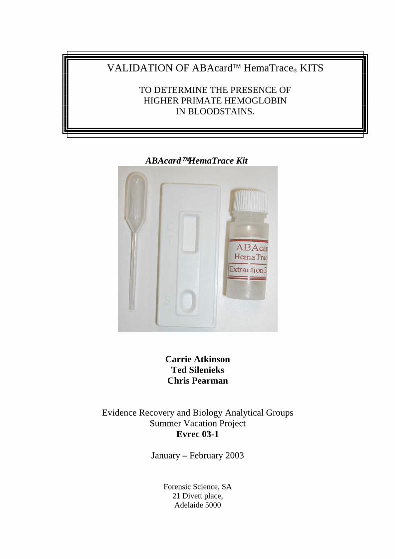
VALIDATION OF ABAcard™ HemaTrace® KITS
TO DETERMINE THE PRESENCE OF HIGHER PRIMATE HEMOGLOBIN
IN BLOODSTAINS.
ABAcard™HemaTrace Kit
Carrie Atkinson Ted Silenieks
Chris Pearman
Evidence Recovery and Biology Analytical Groups Summer Vacation Project
Evrec 03-1
January – February 2003
Forensic Science, SA 21 Divett place, Adelaide 5000
CONTENTS
1. Introduction
2. Materials and Methods
3. ABAcard™ HemaTrace® Test Mechanism
4. Validation Studies
4.1 Sensitivity
♦ Serial Dilutions
♦ Sample Size Comparison
♦ Washed Samples
♦ Aged Bloodstain Extraction
♦ Supernatant Sensitivity
4.2 Specificity
♦ Species Cross Reactivity
♦ Ouchterlony Comparison
♦ Forensic Casework Samples
5. Results
6. Discussion
7. Conclusion
8. Recommended Protocols
9. Images
10. Tables
11. References
12. Appendix 1 - HemaTrace® Technical Information Sheet
1. Introduction .
DNA amplification technology enables forensic scientists to profile a victim or
suspect from small amounts of biological tissues found on evidence or at crime
scenes. Blood is one of the sample types utilised for profiling, however if these
samples fail to provide a DNA profile it can be important to determine that the sample
was in fact human blood.
The current screening test for blood in the Evidence Recovery Section is the
Hemastix Test Strip. Hemastix are used in Evidence Recovery as an indication that
visible stains are the result of bloodstaining. However, Hemastix is not a human or
blood specific test and other oxidising agents can provide false positives. Therefore
this method can only suggest the presence of blood, particularly in the instance of
weak Hemastix positive stains.
There are occasions where a sample is Hemastix positive, but no human DNA
has been detected. In these cases it may be pertinent to determine if the sample is of
human origin. To determine the species of origin of the sample an Ouchterlony
Immunodiffusion test may be performed. These assays are carried out against a
number of species anti-serums, including anti-human hemoglobin, to determine the
species of origin of the suspected bloodstains.
This paper studies the validity and practicality for the use of the ABAcard™
HemaTrace test to complement the current presumptive Hemastix test, and as an
alternative to the Ouchterlony method to confirm the presence of human (higher
primate) hemoglobin. For most sample types the HemaTrace test can be carried out
simply and efficiently in less than 30min compared to the time consuming and labour
intensive Ouchterlony test. This validation also compares the HemaTrace
Recommended Protocols with methods designed to optimise Evidence Recovery
requirements.
#2. Materials and Methods .
1. ABAcard™ Hematrace® Test Kits
• Kits contain 25 Test cards and one transfer pipette (sealed and desiccated in a
foil pouch) and 25 tubes containing 2mls of Buffer.
2. Blood
• Human blood Provided by laboratory donors (ouch) from staff at the
Adelaide Forensic Science Centre.
• Animal Blood: From Forensic Science –80C° freezer storage, Dr David
Schultz of Adelaide Zoo Veterinary Department: and
local Veterinary practices.
• Bloodstains: Bloodstains were prepared by depositing freshly taken
blood on washed cotton cloth and air dried for 24hours.
• Aged bloodstains These were accessed from laboratory storage kept at
room temperature.
3. Sample Extraction Procedure
Standard Extraction Procedures as detailed in the Technical Information Sheet (see
Appendix 1) were generally adhered to, with some variation as listed in the following
Validation Studies. All extractions were carried out at room temperature with the time
period being the incubation time of the sample in the extraction solution.
4. Sample Test Methods
Standard Testing Procedures as detailed in the Technical Information Sheet (see
Appendix 1) were adhered to.
♦ 150µl or 4drops were added to the test well as indicated
♦ Results were recorded at various intervals up to the maximum 10 minutes.
#3. ABAcard™ HemaTrace Test .
The HemaTrace test exploits antigen/antibody reactions and protein
chromatography methods, targeting human hemoglobin (hHb) that is specific to red
blood cells. The cards are compact, disposable, self-contained units that are
individually packaged with disposable pipettes. The test has an optimum pH range of
pH1 to pH9.
They have a stationary phase that is flanked top and bottom by absorbent
membrane material. The bottom membrane is located in the sample well and allows
the sample solution to migrate through the stationary phase. The stationary phase has
been impregnated with mobile dye-tagged antibodies located near the sample well and
immobilised antibodies located in the regions designated “T”(Test area) and
“C”(Control area).
The mobile antibodies are monoclonal, anti-hHb conjugated-dye antibodies
that to bind to human (higher primate) hemoglobin molecules forming a soluble,
mobile complex.
The immobilised antibodies located in the “T”area are polyclonal anti-hHb-
antibody antibodies that capture the mobile hHb-complexes as they migrate in the
solution. Mobile monoclonal antibodies that are not bound to hemoglobin are not
caught at this point and continue to move up the test screen.
The immobilised antibodies located in the “C” area are polyclonal anti-
immunoglobulin antibodies that will bind the excess mobile dye-tagged antibodies.
The concentration of these molecules causes the appearance of the characteristic pink
line in the control region to indicate that the test was successfully executed. If a line
fails to appear in this region, the ABAcard HemaTrace test is faulty and should be
recorded as invalid.
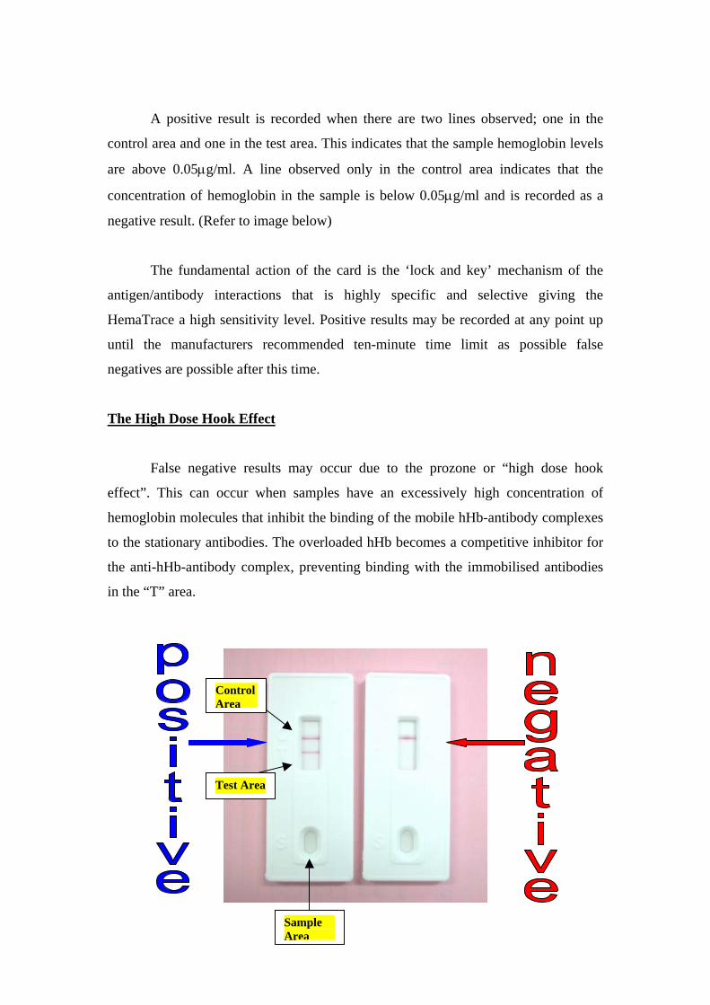
A positive result is recorded when there are two lines observed; one in the
control area and one in the test area. This indicates that the sample hemoglobin levels
are above 0.05μg/ml. A line observed only in the control area indicates that the
concentration of hemoglobin in the sample is below 0.05μg/ml and is recorded as a
negative result. (Refer to image below)
The fundamental action of the card is the ‘lock and key’ mechanism of the
antigen/antibody interactions that is highly specific and selective giving the
HemaTrace a high sensitivity level. Positive results may be recorded at any point up
until the manufacturers recommended ten-minute time limit as possible false
negatives are possible after this time.
The High Dose Hook Effect
False negative results may occur due to the prozone or “high dose hook
effect”. This can occur when samples have an excessively high concentration of
hemoglobin molecules that inhibit the binding of the mobile hHb-antibody complexes
to the stationary antibodies. The overloaded hHb becomes a competitive inhibitor for
the anti-hHb-antibody complex, preventing binding with the immobilised antibodies
in the “T” area.
Control Area
Sample Area
Test Area
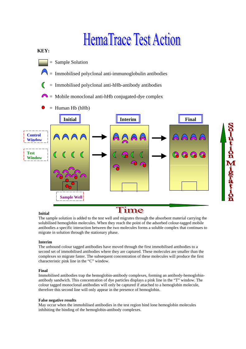
KEY: = Sample Solution = Immobilised polyclonal anti-immunoglobulin antibodies = Immobilised polyclonal anti-hHb-antibody antibodies = Mobile monoclonal anti-hHb conjugated-dye complex = Human Hb (hHb)
Initial Interim Final
Control Window
Test Window
Sample Well Initial The sample solution is added to the test well and migrates through the absorbent material carrying the solubilised hemoglobin molecules. When they reach the point of the adsorbed colour-tagged mobile antibodies a specific interaction between the two molecules forms a soluble complex that continues to migrate in solution through the stationary phase. Interim The unbound colour tagged antibodies have moved through the first immobilised antibodies to a second set of immobilised antibodies where they are captured. These molecules are smaller than the complexes so migrate faster. The subsequent concentration of these molecules will produce the first characteristic pink line in the “C” window. Final Immobilised antibodies trap the hemoglobin-antibody complexes, forming an antibody-hemoglobin-antibody sandwich. This concentration of dye particles displays a pink line in the “T” window. The colour tagged monoclonal antibodies will only be captured if attached to a hemoglobin molecule, therefore this second line will only appear in the presence of hemoglobin. False negative results May occur when the immobilised antibodies in the test region bind lone hemoglobin molecules inhibiting the binding of the hemoglobin-antibody complexes.
4. Validation Studies .
44..11 SSEENNSSIITTIIVVIITTYY SSEERRIIAALL DDIILLUUTTIIOONNSS:: LLiiqquuiidd aanndd SSttaaiinnss
#1 Stain Samples Aim: To test the sensitivity of the HemaTrace extraction procedure using
known stain dilutions. Variable: Dilution Factor Method:
• Serial dilution of neat fresh blood was made using an initial volume of 750μl each of distilled water and blood. A 750μl aliquot of the sample was then added to 750μl of distilled water, this was repeated until the desired dilution factor was reached.
• 100μl aliquot of each dilution was pipetted onto washed cotton cloth and air dried for 48hrs.
• Each entire stain sample was extracted in 2mls of HemaTrace buffer for 5 minutes.
• Some dilutions re-extracted in 300μl of HemaTrace buffer for 5 minutes.
• 4 drops of sample extract solution was applied to test well. • HemaTrace results were recorded at 2min and 10min intervals. • Samples were Hemastix tested for comparison.
#2 Liquid Samples
Aim: To test the sensitivity of the HemaTrace test card using liquid samples.
Variable: Dilution factor
Method:
• Serial dilution of neat fresh blood was made using an initial volume of 750μl each of distilled water and blood. A 750μl aliquot of the sample was then added to 750μl of distilled water, this was repeated until the desired dilution factor was reached.
• The dilutions were placed directly into the test well, no buffer or further liquid was added.
• HemaTrace Results were recorded at 2min and 10min intervals. • The dilutions were Hemastix tested for comparison.
SSAAMMPPLLEE SSIIZZEE CCOOMMPPAARRIISSOONN
Aim: To test the sensitivity of the HemaTrace cards in respect to sample sizes that may be encountered in routine casework.
Variable: Sample size Method:
• Fresh human blood was dropped onto washed cotton cloth and air-dried at room temperature for 48hrs.
• Sample sizes tested were as follows: 10mm², 5mm², 2.5mm², 1mm², 2mm thread and a 1mm thread.
• Samples were extracted in the supplied 2ml HemaTrace Buffer for 5mins. No centrifugation was necessary.
• 4 drops of sample extract solution was applied to test well. • HemaTrace test observations were recorded at 45secs,
2minutes and 10minutes.
WWAASSHHEEDD SSAAMMPPLLEESS
Aim: To test the ability of the HemaTrace cards to detect hemaglobin in bloodstains after washing.
Variable: 2 different washing methods were used on control bloodstains,
which were then HemaTrace tested.
Method: • Fresh bloodstains, which had been air dried at room temperature
for 24 hours and one year old bloodstains which had been stored at –20C were used in this trial. All stains were prepared on washed cotton cloth.
• 2 washing methods were used. 1. A hot water wash where stains were agitated under running hot
water for 2min. 2. A tepid detergent wash where stains were soaked in 200ml of
20% laboratory detergent (Decon 90) solution for 30min. • A 5mm2 sample size was extracted for 5 minutes in 2mls of
HemaTrace buffer. • 4 drops of sample extract solution was applied to test well. • HemaTrace results were recorded at 2minutes and 10minutes. • All extract solutions were Hemastix tested.
AAGGEEDD BBLLOOOODDSSTTAAIINNSS
Aim: To test the viability of different extraction solvents for aged bloodstains.
Variable: Extraction method
Method:
• Samples consisted of bloodstains on cotton cloth stored at room temperature in laboratory conditions. A 12year old bloodstain and a 26year old bloodstain were tested.
• Hemastix tests were performed on each stain sample. • 2mm2 sample size was used for all tests. • HemaTrace aged stain extraction protocol:
1. 2-5 min soak in 2-3drops of 5% ammonia solution.2. Allow ammonia to evaporate. 3. Add 300µl Hematrace buffer and apply this
solution to the test well • All other extractions were soaked for 30minutes in the supplied 2ml
Hematrace buffer.
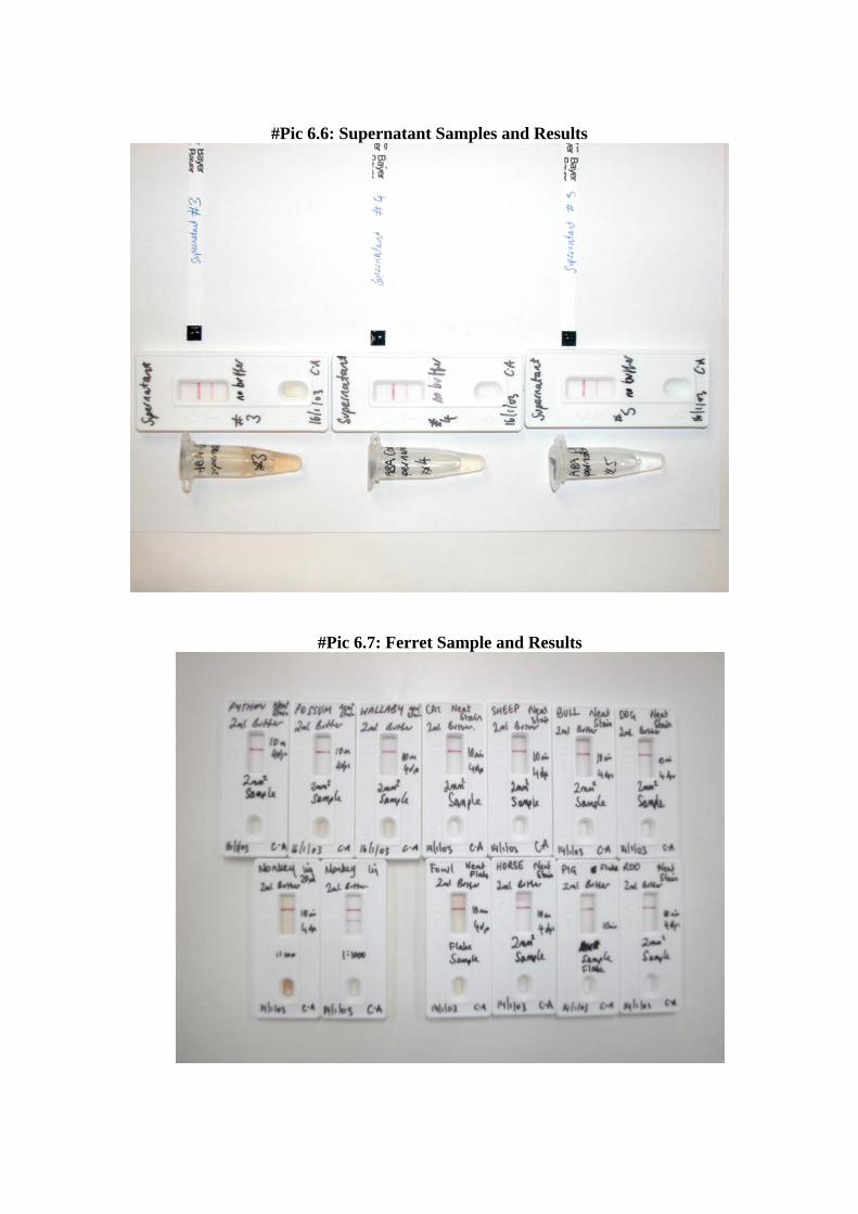
SSUUPPEERRNNAATTAANNTT SSEENNSSIITTIIVVIITTYY
Aim: To test the viability of using the discarded supernatant from DNA
blood extraction samples.
Variable: Sample (hemoglobin) Concentration
Method: • Samples 1 and 2 were derived from extractions of fresh bloodstains
on FTA paper using 10mM NaOH. • Samples 3 to 8 were derived from actual casework extractions
using distilled water. • Volume of all samples consistent with initial wash for DNA
extraction supernatant excess, approximately 950µl. • All supernatants were Hemastix tested. • Hematrace results were recorded at 2minutes and 10minutes. • The supernatant colour was recorded. • In each test, 4 drops of neat supernatant were placed directly in the
Hematrace test well.
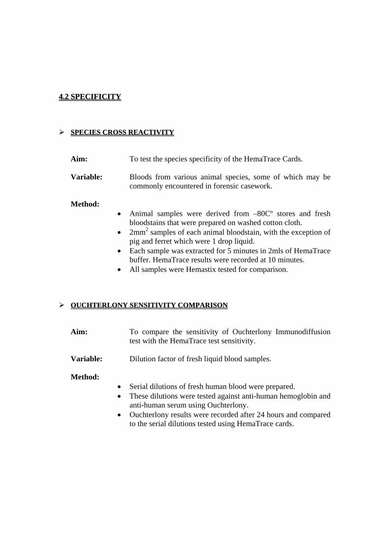
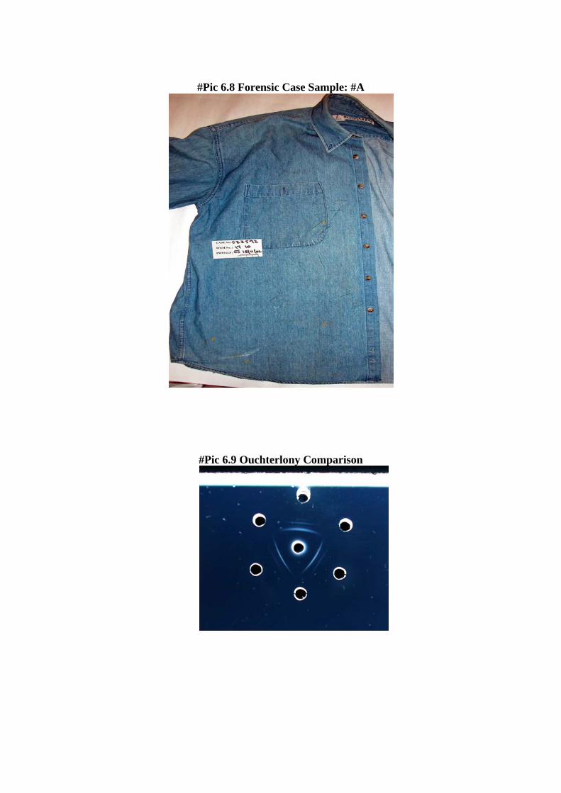
44..22 SSPPEECCIIFFIICCIITTYY SSPPEECCIIEESS CCRROOSSSS RREEAACCTTIIVVIITTYY
Aim: To test the species specificity of the HemaTrace Cards.
Variable: Bloods from various animal species, some of which may be commonly encountered in forensic casework.
Method:
• Animal samples were derived from –80Cº stores and fresh bloodstains that were prepared on washed cotton cloth.
• 2mm2 samples of each animal bloodstain, with the exception of pig and ferret which were 1 drop liquid.
• Each sample was extracted for 5 minutes in 2mls of HemaTrace buffer. HemaTrace results were recorded at 10 minutes.
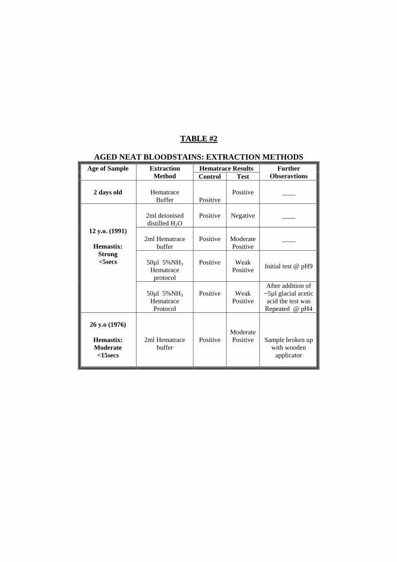
• All samples were Hemastix tested for comparison. OOUUCCHHTTEERRLLOONNYY SSEENNSSIITTIIVVIITTYY CCOOMMPPAARRIISSOONN
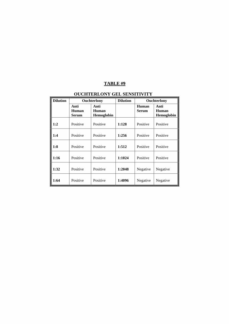
Aim: To compare the sensitivity of Ouchterlony Immunodiffusion test with the HemaTrace test sensitivity.
Variable: Dilution factor of fresh liquid blood samples.
Method:
• Serial dilutions of fresh human blood were prepared. • These dilutions were tested against anti-human hemoglobin and
anti-human serum using Ouchterlony. • Ouchterlony results were recorded after 24 hours and compared
to the serial dilutions tested using HemaTrace cards.
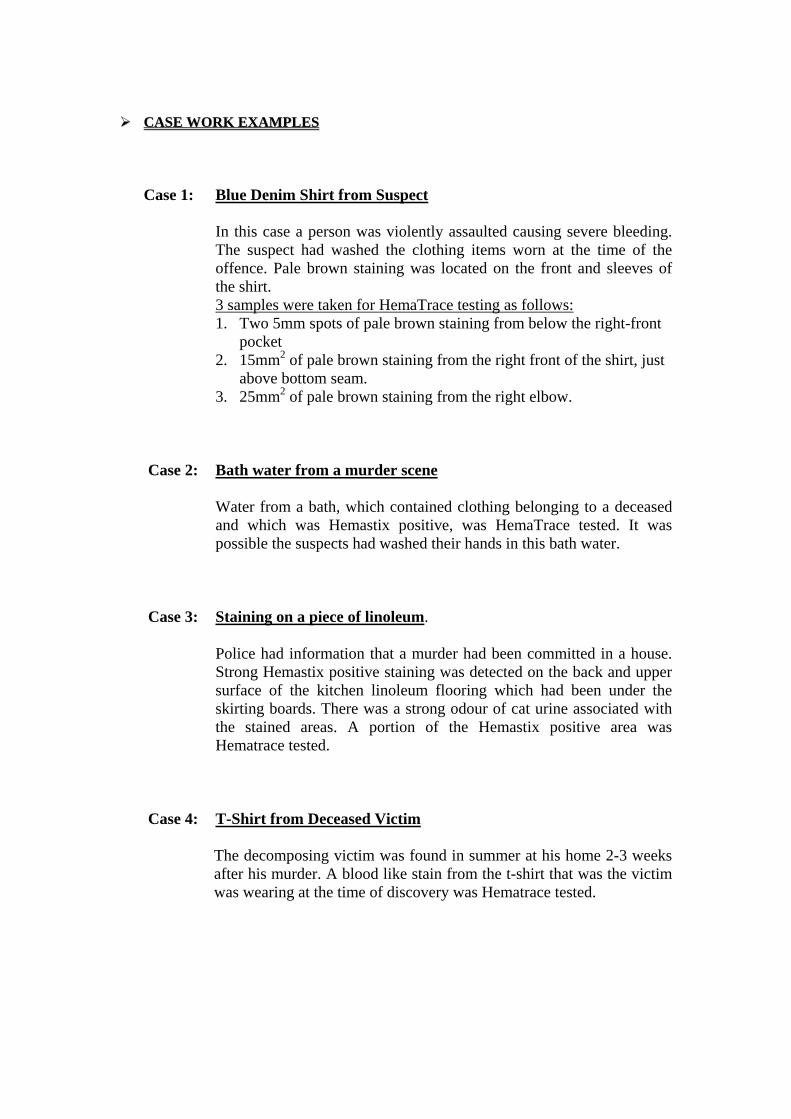
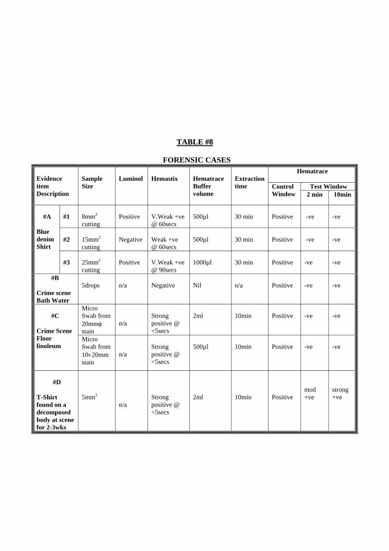
CCAASSEE WWOORRKK EEXXAAMMPPLLEESS
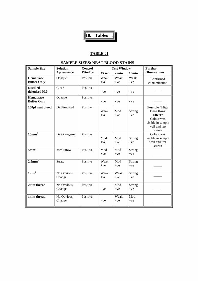
Case 1: Blue Denim Shirt from Suspect
In this case a person was violently assaulted causing severe bleeding. The suspect had washed the clothing items worn at the time of the offence. Pale brown staining was located on the front and sleeves of the shirt. 3 samples were taken for HemaTrace testing as follows: 1. Two 5mm spots of pale brown staining from below the right-front
pocket 2. 15mm2 of pale brown staining from the right front of the shirt, just
above bottom seam. 3. 25mm2 of pale brown staining from the right elbow.
Case 2: Bath water from a murder scene
Water from a bath, which contained clothing belonging to a deceased and which was Hemastix positive, was HemaTrace tested. It was possible the suspects had washed their hands in this bath water.
Case 3: Staining on a piece of linoleum. Police had information that a murder had been committed in a house. Strong Hemastix positive staining was detected on the back and upper surface of the kitchen linoleum flooring which had been under the skirting boards. There was a strong odour of cat urine associated with the stained areas. A portion of the Hemastix positive area was Hematrace tested.
Case 4: T-Shirt from Deceased Victim The decomposing victim was found in summer at his home 2-3 weeks after his murder. A blood like stain from the t-shirt that was the victim was wearing at the time of discovery was Hematrace tested.
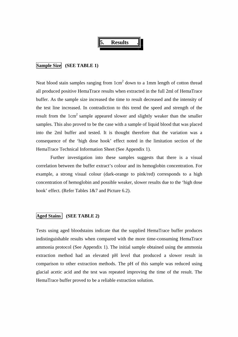
5. Results .
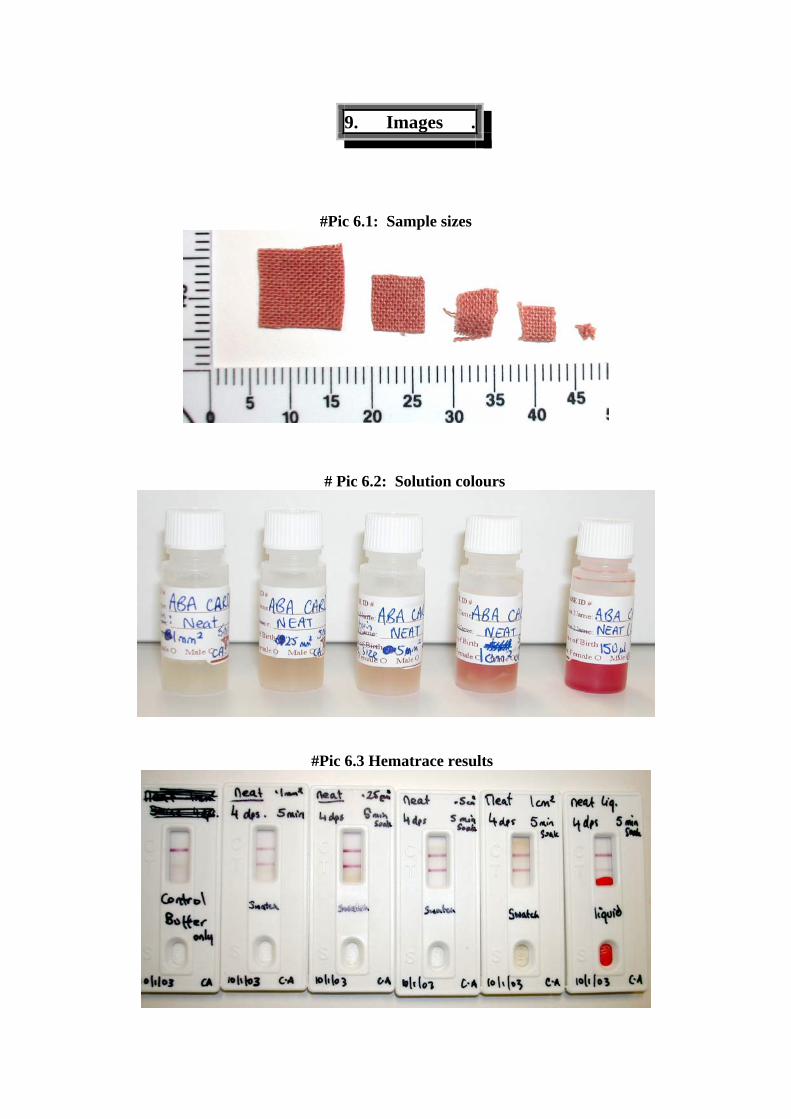
Sample Size (SEE TABLE 1)
Neat blood stain samples ranging from 1cm2 down to a 1mm length of cotton thread
all produced positive HemaTrace results when extracted in the full 2ml of HemaTrace
buffer. As the sample size increased the time to result decreased and the intensity of
the test line increased. In contradiction to this trend the speed and strength of the
result from the 1cm2 sample appeared slower and slightly weaker than the smaller
samples. This also proved to be the case with a sample of liquid blood that was placed
into the 2ml buffer and tested. It is thought therefore that the variation was a
consequence of the ‘high dose hook’ effect noted in the limitation section of the
HemaTrace Technical Information Sheet (See Appendix 1).
Further investigation into these samples suggests that there is a visual
correlation between the buffer extract’s colour and its hemoglobin concentration. For
example, a strong visual colour (dark-orange to pink/red) corresponds to a high
concentration of hemoglobin and possible weaker, slower results due to the ‘high dose
hook’ effect. (Refer Tables 1&7 and Picture 6.2).
Aged Stains (SEE TABLE 2)
Tests using aged bloodstains indicate that the supplied HemaTrace buffer produces
indistinguishable results when compared with the more time-consuming HemaTrace
ammonia protocol (See Appendix 1). The initial sample obtained using the ammonia
extraction method had an elevated pH level that produced a slower result in
comparison to other extraction methods. The pH of this sample was reduced using
glacial acetic acid and the test was repeated improving the time of the result. The
HemaTrace buffer proved to be a reliable extraction solution.
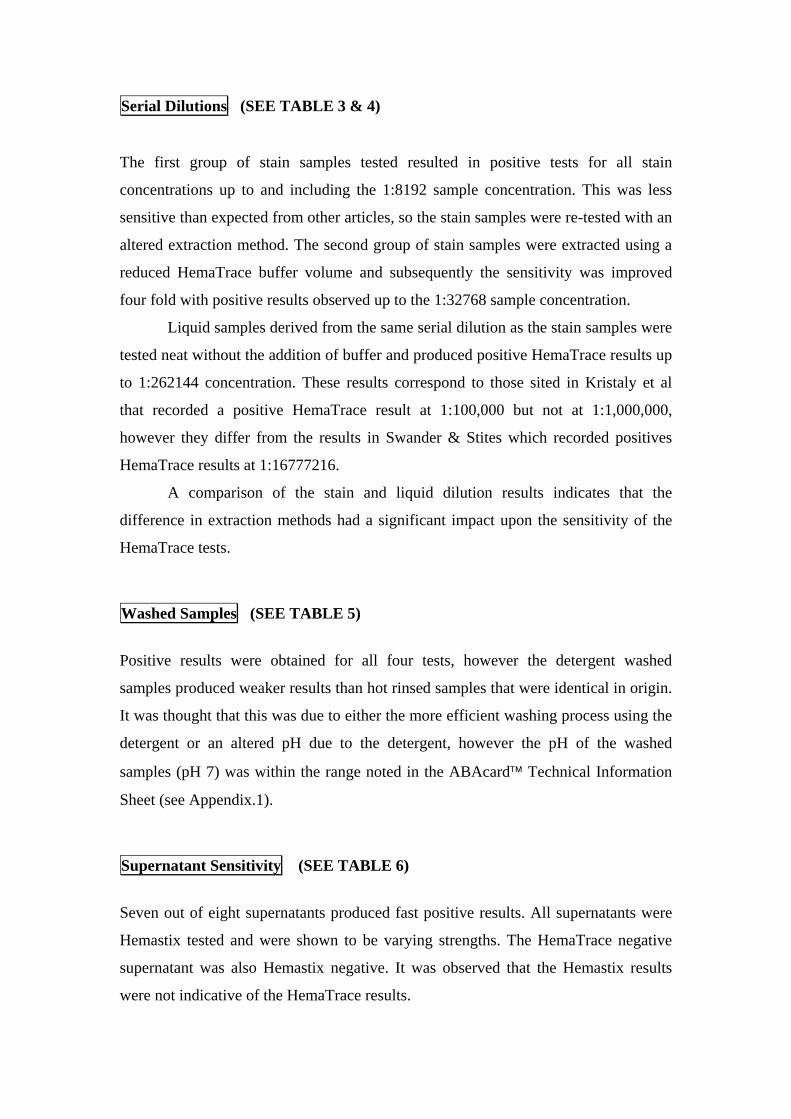
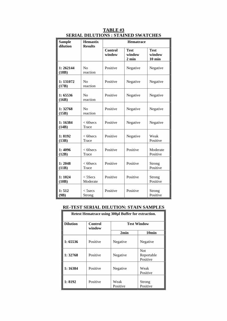
Serial Dilutions (SEE TABLE 3 & 4) The first group of stain samples tested resulted in positive tests for all stain
concentrations up to and including the 1:8192 sample concentration. This was less
sensitive than expected from other articles, so the stain samples were re-tested with an
altered extraction method. The second group of stain samples were extracted using a
reduced HemaTrace buffer volume and subsequently the sensitivity was improved
four fold with positive results observed up to the 1:32768 sample concentration.
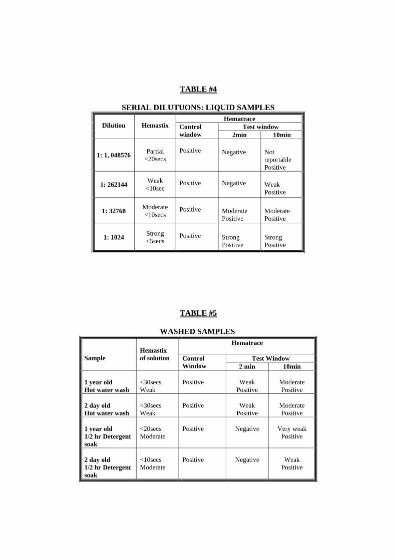
Liquid samples derived from the same serial dilution as the stain samples were
tested neat without the addition of buffer and produced positive HemaTrace results up
to 1:262144 concentration. These results correspond to those sited in Kristaly et al
that recorded a positive HemaTrace result at 1:100,000 but not at 1:1,000,000,
however they differ from the results in Swander & Stites which recorded positives
HemaTrace results at 1:16777216.
A comparison of the stain and liquid dilution results indicates that the
difference in extraction methods had a significant impact upon the sensitivity of the
HemaTrace tests.
Washed Samples (SEE TABLE 5)
Positive results were obtained for all four tests, however the detergent washed
samples produced weaker results than hot rinsed samples that were identical in origin.
It was thought that this was due to either the more efficient washing process using the
detergent or an altered pH due to the detergent, however the pH of the washed
samples (pH 7) was within the range noted in the ABAcard™ Technical Information
Sheet (see Appendix.1). Supernatant Sensitivity (SEE TABLE 6)
Seven out of eight supernatants produced fast positive results. All supernatants were
Hemastix tested and were shown to be varying strengths. The HemaTrace negative
supernatant was also Hemastix negative. It was observed that the Hemastix results
were not indicative of the HemaTrace results.
Species Specificity (SEE TABLE 7) The Ferret samples (neat and diluted) produced a false positive HemaTrace result as
expected from the ABAcard™ Technical Information Sheet: Limitations (see
Appendix.1). As the HemaTrace test is Higher Primate specific not human specific
the monkey sample also produced an expected positive result. The monkey solution
appeared slower and weaker than expected from its solution colour that had a dark
pink visual appearance. It was concluded that this was due to a slight ‘high dose hook’
effect (See Sample Size Study).
All other species tested produced negative results, which is consistent with the papers
read for this study (See References) that tested a number of species similar to those
used in this validation as well as other novel species samples.
Forensic Cases (SEE TABLE 8)
The HemaTrace tests were used to analyse forensic cases that were suitable
candidates for the study. Case 1 did not produce a positive result as expected from the
Hemastix results, although the best samples had been taken for casework testing. Case
2 was not expected to give a positive result as any possible sample would have been
significantly diluted by the volume of bath water. Case 3 was tested a number of times
but did not produce a positive which was in line with observations made by evidence
recovery analysts. Case 4 was expected to produce a positive as the sample was
known to be concentrated neat blood and tissue fluid although the quality of the
sample was in question to body decomposition processes and possible hot
environmental exposure.
Ouchterlony Comparison (SEE TABLE 9)
Ouchterlony gels were run as a sensitivity comparison starting with the initial dilution
of 1:2 descending to 1:4096. Both the Anti Human Serum and the Anti Human Hb
Ouchterlony gel series dropped out at 1:2048 with the both serums giving a positive
result for 1:1024 dilution. These results correspond with the sensitivity noted in
Swander & Stites.
6. Discussion .
The HemaTrace® ABAcards™ are highly sensitive one-step sandwich
immunoassays that are compact, disposable and user friendly. This one-step detection
method has been used for a variety of commercial tests, such as pregnancy tests, and
in this case has been adapted to detect human hemoglobin in bloodstain samples. The
HemaTrace tests produce reliable results from a range of bloodstains encountered in
Evidence Recovery for example samples ranging from 1cm2 to 1mm thread, aged
stains, DNA extraction supernatant and denatured samples. They are an expedient
way to confirm that doubtful stains found to be Hemastix positive are derived from
human blood.
One of the more significant limitations of the HemaTrace test that is noted in the
ABAcard™ Technical Information Sheet (see Appendix.1) is the “High Dose Hook
Effect”. Excessive quantities of human hemoglobin molecules can bind non-
specifically to the solid phase antibodies. This steric hindrance obstructs the desired
specific binding of hHb-antibody complexes. Two step immunoassays prevent this by
washing excess molecules away in-between binding steps ( Fernando & Wilson).
However with the one-step immunoassays both the mobile molecules are mixed
together thus causing a competitive binding environment.
Hemastix tests are based upon the oxidising properties of the Heme group in
Hemoglobin, subsequently other oxidising agents such as bleach and compounds that
have peroxidase-like properties can also produce false positives. The SANGUR
validation study (Russell Cook) which tested the Hemastix sensitivity produced
positive results for liquid dilutions of whole blood up to and including 1/500000
(HemaTrace 1/250000) and 1/10000 (HemaTrace 1/32000)in similar stain dilutions.
Although the sensitivities of the two tests are similar the Hemastix is not always
indicative of the HemaTrace result. Negative Hemastix results did not always
correspond to negative HemaTrace was results as observed in the supernatant
study.(See Table 6)
A sample size study was carried out to determine an optimum sample size in
relation to casework items encountered in Evidence Recovery. This study considered
neat bloodstains and found that samples ranging from 1cm2 to a 1mm thread gave
strong reliable results. During the casework study, Case 1 did not give the expected
positive result though it was known that the victim had lost a large volume of blood
and visible washed stains produced weak Hemastix positive results(See Table 8).
Further to this the first group of stain samples in the serial dilution study produced
significantly less sensitive results with positives only observed up to 1:8092
concentration. A reduction in extraction buffer volume improved these results four
fold. (See Table 3 & 4)
As a part of this validation, efforts were made to induce the “High Dose Hook
Effect” to determine its impact on forensic casework. All HemaTrace tests from the
highly concentrated samples gave genuine positive results. When these samples were
diluted and re-tested the results were observed to be faster and stronger than the
original tests. This was confirmed by further diluting other suspect samples such as
the monkey and re-testing the extracts, which again improved the time and strength of
the results.
The high concentration samples indicated a visual correlation between their
buffer colour (dark orange to pink/red) and the strength and speed of their HemaTrace
results. Therefore buffer colour should be considered along with other case
observations when analysing the HemaTrace test results to ensure that results are
reliable (for example: a negative HemaTrace result from an obviously bloodied
solution). It should be noted though that a colourless sample solution is not an
indication that the sample is hemoglobin negative.
Various extraction methods, including the recommended protocol noted in the
HemaTrace Technical Information sheet (See Appendix1), were tested using neat
aged bloodstains to determine the optimum extraction protocol for poor quality
samples. Results indicated that the supplied HemaTrace buffer sufficiently extracted
the hemoglobin from these stains with an extended extraction time.
The results of the neat samples indicated that the HemaTrace tests were
remarkably sensitive and it was expected that this would prove to be the same in the
serial dilution study, however the initial stain samples were disappointing with
positive results only being recorded up to 1:8192. The stain samples were re-tested
with an altered extraction method that used less HemaTrace buffer and subsequently
this increased the HemaTrace sensitivity for the stain samples to 1:32768.
Liquid samples derived from the same serial dilution as the stain samples were
tested neat without the addition of buffer and produced positive HemaTrace results up
to 1:262144 concentration. The results of liquid dilution were up to ten times more
sensitive than the stain dilution samples therefore the variation in the extraction
method, ie buffer volume, had a significant impact upon the sensitivity of the
HemaTrace tests. Theses results highlighted the buffer volume as a factor in protocol
design (See Tables 3& 4).
Further evidence of the HemaTrace sensitivity was observed through the
accidental contamination of a negative control. The supplied pipette was placed upon
the sampling area and the trace blood dust was sufficient to produce a positive result
from the neat buffer sample. This result was confirmed by testing the sampling area
with a Hemastix that produced a strong positive result.
A Species specific study was carried out using a range of animal samples both
domestic and novel. The specificity of the HemaTrace cards is adequate for real
forensic casework as in the majority of cases it is not necessary to know which
species the blood originated from but merely if it is human (higher primate) in origin.
Tests were carried out on washed stains to study the specificity and sensitivity
of the HemaTrace test under these conditions. This validation was limited and did not
include more extensive tests such as washing machine cleaning or bleaching. Whilst
the positive results are promising, a comparison to Forensic Case Sample 1(see table
8) which did not produce a positive HemaTrace result, indicates that protocols for
washed samples need to be independent of protocols for neat stain samples.
In cases that yield very little physical evidence, DNA testing must be given
priority over all other evidence recovery methods. It is useful then to determine if the
supernatant from the initial washing of the sample is suitable for HemaTrace testing.
This supernatant is routinely tested in Biology with Hemastix. The Hemastix results
from the corresponding supernatant’s were not a reliable indication of the possible
HemaTrace results.
As a basis for our HemaTrace protocols we compared those recommended in
the Technical Information Sheet with those used in Swander & Stites and Kristaky et
al. All extractions were carried out at room temperature with our initial sample sizes
under 1cm2. The methods were then refined with each validation according to sample
type and the results observed when altering sample size, extraction time and buffer
volume.
7. Conclusion .
The HemaTrace test is a viable and effective presumptive test method suitable for
use in the Evidence Recovery department of the Adelaide Forensic Science Centre to
confirm the presence of human (higher primate) hemoglobin in samples. The High
Dose Hook effect will not be a limitation in the use of the HemaTrace test for
evidence recovery purposes, as no false negatives were observed due to this
limitation, despite efforts to induce the effect. However, negative test results should
be considered in conjunction with observations regarding the visual colour of the
sample solution (strong colour indicates high hemoglobin concentration) and
background case information.
Positive HemaTrace results conclusively indicate the presence of human
hemoglobin in the tested sample irrespective of the strength of the result. False
positives will not be a serious consideration as ferret and higher primates will not be
common considerations to the majority of crime scenes or evidence and will generally
be referred to in case notes.
There is a strong indication from the serial dilution study that the buffer decreases
the concentration of the hemoglobin in the sample and therefore decreases the
HemaTrace test sensitivity. In the instance of forensic casework, care must be taken in
the consideration of buffer volume when extracting weak stains (eg: small or washed).
False negatives from these types of samples that produce weak sample solutions, will
be the main concern in the use of HemaTrace test, therefore careful attention must be
given to the sample type and the optimum testing procedure, to minimise the
possibility of false negative results.
The HemaTrace cards can exclude a sample from consideration but cannot shed
light on blood sample origins if they prove to be negative for human hemoglobin.
In these cases, where necessary, the Ouchterlony test will still be required to
determine the non-human species of origin.
The protocols for the use of the HemaTrace ABAcards in Evidence Recovery
have been designed based upon the results and observations of the validations with
specific consideration to the type of evidence encountered and the information
required.
In most real forensic cases it is sufficient to include or exclude samples based
upon the results of the HemaTrace tests. This test should reduce the non-human stains
sent to biology for DNA profiling reducing costs and time. As the HemaTrace test is
up to ten times more sensitive than the current Ouchterlony test it will be a valuable
addition to the Evidence Recovery apparatus.
8. Recommended Protocols .
Neat Stains, FREDS & Aged Stains (Neat Stains: Sample size <5mm2 in 2ml buffer for 5min @ room Temp) (FREDS: Sample size <5mm length in 2ml buffer for 5min @ room Temp) (Aged Stains: Sample size <5mm2 in 2ml buffer for 1hr @ room Temp) 1. Cut out a sample using sterile scissors or scalpel on a clean surface. The sample
size should be between 5mm2 and 2mm2, however smaller fragments are sufficient to produce a result. ♦ FREDS: use 2-5mm length of thread
2. Place the sample into the provided container of 2ml HemaTrace buffer allowing the sample to extract for 5minutes at room temperature. ♦ AGED STAINS: extract for 1hr @ room temperature
3. If the sample is not to be tested immediately store the solution at 4ºC until required.
4. Check the buffer’s visual appearance ensuring the colour is no darker than a light orange/pink colour to avoid the “high dose hook effect”. Colourless solutions will still result in positive results if the sample contains sufficient hHb.
5. If the solution is deemed too strong dilute the sample 1:4 with de-ionised water to a minimum total volume of 200µl in an eppendorf tube before testing.
6. Using a sterile pipette extract 150µl of sample solution and place it into the small sample well at the bottom of the test marked “S”.
7. Begin recording the time allowing a maximum of 10minutes to observe a result. 8. Record positive if two separate pink lines appear in the window next to the “T”
and “C” labelled areas respectively. If a single line appears in the uppermost area of the window labelled “C” record a negative result. In the absence of any lines in the window the test must be deemed invalid. Any line regardless of colour strength can be interpreted as valid.
NB: A Decrease in quality or size of a sample should relate to a decrease in volume used (Minimum Buffer volume 300µl).
8. Recommended Protocols .
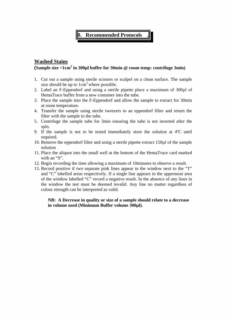
Washed Stains (Sample size <1cm2 in 300µl buffer for 30min @ room temp: centrifuge 3min) 1. Cut out a sample using sterile scissors or scalpel on a clean surface. The sample
size should be up to 1cm2 where possible. 2. Label an F-Eppendorf and using a sterile pipette place a maximum of 300µl of
HemaTrace buffer from a new container into the tube. 3. Place the sample into the F-Eppendorf and allow the sample to extract for 30min
at room temperature. 4. Transfer the sample using sterile tweezers to an eppendorf filter and return the
filter with the sample to the tube. 5. Centrifuge the sample tube for 3min ensuring the tube is not inverted after the
spin. 9. If the sample is not to be tested immediately store the solution at 4ºC until
required. 10. Remove the eppendorf filter and using a sterile pipette extract 150µl of the sample
solution 11. Place the aliquot into the small well at the bottom of the HemaTrace card marked
with an “S”. 12. Begin recording the time allowing a maximum of 10minutes to observe a result. 13. Record positive if two separate pink lines appear in the window next to the “T”
and “C” labelled areas respectively. If a single line appears in the uppermost area of the window labelled “C” record a negative result. In the absence of any lines in the window the test must be deemed invalid. Any line no matter regardless of colour strength can be interpreted as valid.
NB: A Decrease in quality or size of a sample should relate to a decrease in volume used (Minimum Buffer volume 300µl).
8. Recommended Protocols .
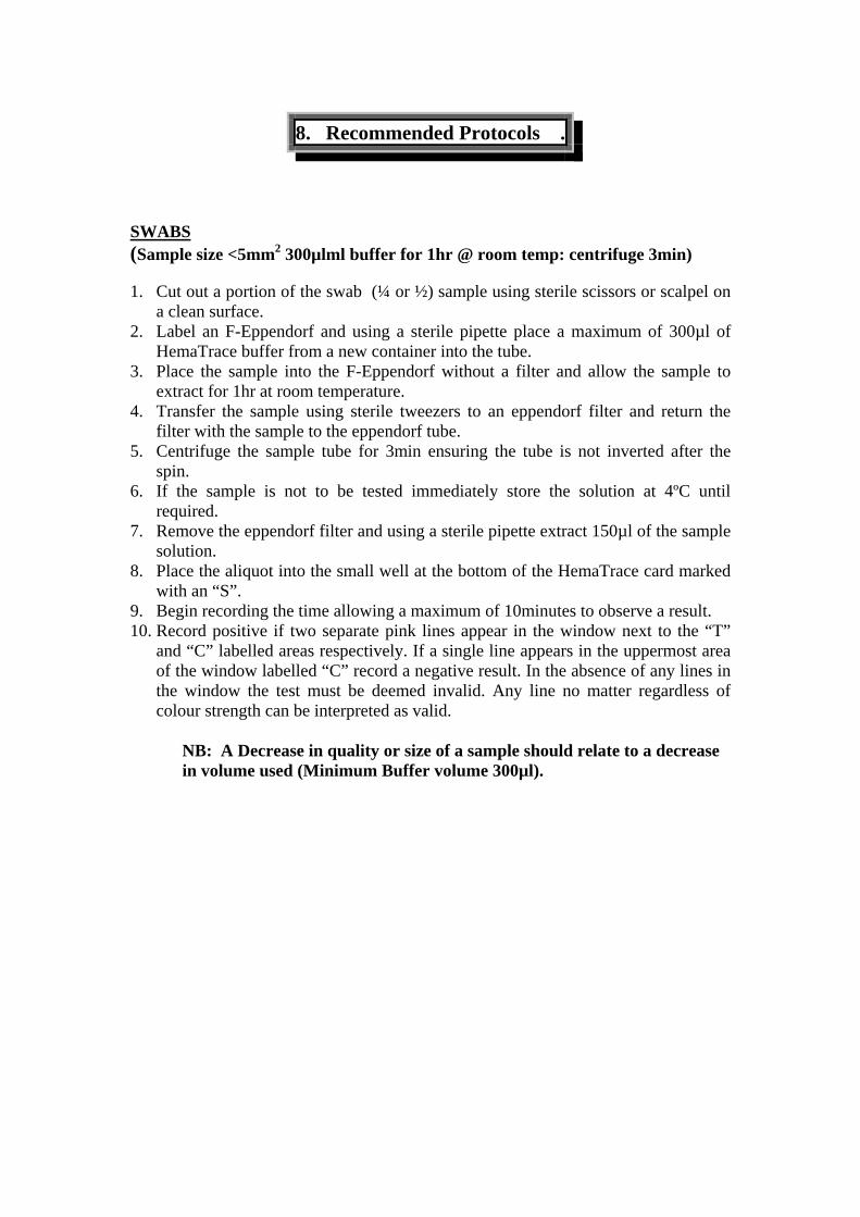
SWABS (Sample size <5mm2 300µlml buffer for 1hr @ room temp: centrifuge 3min) 1. Cut out a portion of the swab (¼ or ½) sample using sterile scissors or scalpel on
a clean surface. 2. Label an F-Eppendorf and using a sterile pipette place a maximum of 300µl of
HemaTrace buffer from a new container into the tube. 3. Place the sample into the F-Eppendorf without a filter and allow the sample to
extract for 1hr at room temperature. 4. Transfer the sample using sterile tweezers to an eppendorf filter and return the
filter with the sample to the eppendorf tube. 5. Centrifuge the sample tube for 3min ensuring the tube is not inverted after the
spin. 6. If the sample is not to be tested immediately store the solution at 4ºC until
required. 7. Remove the eppendorf filter and using a sterile pipette extract 150µl of the sample
solution. 8. Place the aliquot into the small well at the bottom of the HemaTrace card marked
with an “S”. 9. Begin recording the time allowing a maximum of 10minutes to observe a result. 10. Record positive if two separate pink lines appear in the window next to the “T”
and “C” labelled areas respectively. If a single line appears in the uppermost area of the window labelled “C” record a negative result. In the absence of any lines in the window the test must be deemed invalid. Any line no matter regardless of colour strength can be interpreted as valid.
NB: A Decrease in quality or size of a sample should relate to a decrease in volume used (Minimum Buffer volume 300µl).
9. Images .
#Pic 6.1: Sample sizes
# Pic 6.2: Solution colours
#Pic 6.3 Hematrace results
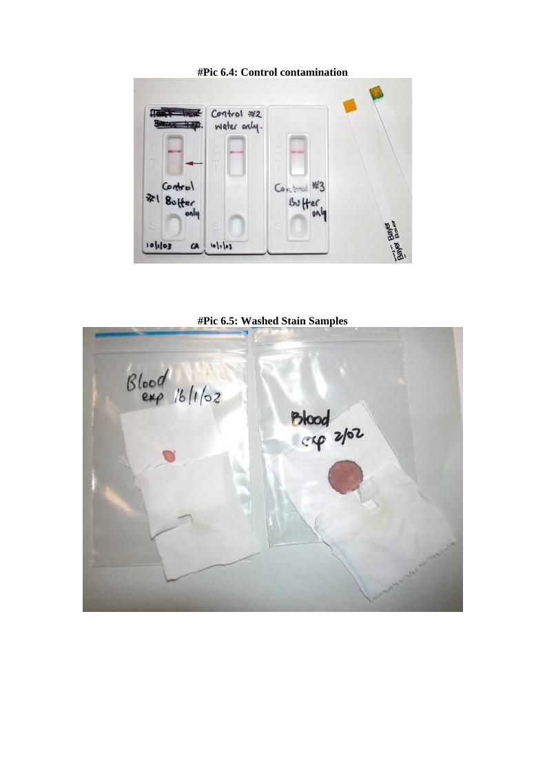
#Pic 6.4: Control contamination
#Pic 6.5: Washed Stain Samples
#Pic 6.6: Supernatant Samples and Results
#Pic 6.7: Ferret Sample and Results
#Pic 6.8 Forensic Case Sample: #A
#Pic 6.9 Ouchterlony Comparison
10. Tables .
TTAABBLLEE ##11
SAMPLE SIZES: NEAT BLOOD STAINS Test Window Sample Size Solution
Appearance Control Window 45 sec 2 min 10min
Further Observations
Hematrace Buffer Only
Opaque Positive
Weak +ve
Weak +ve
Weak +ve
Confirmed contamination
Distilled deionised H20
Clear Positive - ve
- ve
- ve ____
Hematrace Buffer Only
Opaque Positive - ve
- ve
- ve _____
150µl neat blood Dk Pink/Red Positive Weak +ve
Mod +ve
Strong +ve
Possible “High Dose Hook
Effect” Colour was
visible in sample well and test
screen 10mm2 Dk Orange/red Positive
Mod +ve
Mod +ve
Strong +ve
Colour was visible in sample
well and test screen
5mm2 Med Straw Positive Mod +ve
Mod +ve
Strong +ve
_____
2.5mm2 Straw Positive Weak +ve
Mod +ve
Strong +ve _____
1mm2 No Obvious Change
Positive Weak +ve
Weak +ve
Strong +ve
_____
2mm thread No Obvious Change
Positive - ve
Mod +ve
Strong +ve
_____
1mm thread No Obvious Change
Positive - ve
Weak +ve
Mod +ve
_____
TTAABBLLEE ##22
AGED NEAT BLOODSTAINS: EXTRACTION METHODS Hematrace Results Age of Sample Extraction
Method Control Test Further
Obseravtions
2 days old
Hematrace
Buffer
Positive
Positive ____
2ml deionised distilled H2O
Positive
Negative ____
2ml Hematrace
buffer
Positive
Moderate Positive
____
50µl 5%NH3
Hematrace protocol
Positive
Weak
Positive Initial test @ pH9
12 y.o. (1991)
Hemastix: Strong <5secs
50µl 5%NH3
Hematrace Protocol
Positive
Weak
Positive
After addition of ~5µl glacial acetic acid the test was
Repeated @ pH4
26 y.o (1976)
Hemastix: Moderate <15secs
2ml Hematrace buffer
Positive
Moderate Positive
Sample broken up with wooden
applicator
TTAABBLLEE ##33 SERIAL DILUTIONS : STAINED SWATCHES
Hematrace Sample dilution
Hemastix Results
Control window
Test window 2 min
Test window 10 min
1: 262144 (18B)
No reaction
Positive
Negative
Negative
1: 131072 (17B)
No reaction
Positive
Negative
Negative
1: 65536 (16B)
No reaction
Positive
Negative
Negative
1: 32768 (15B)
No reaction
Positive
Negative
Negative
1: 16384 (14B)
< 60secs Trace
Positive
Negative
Negative
1: 8192 (13B)
< 60secs Trace
Positive
Negative
Weak Positive
1: 4096 (12B)
< 60secs Trace
Positive
Positive
Moderate Positive
1: 2048 (11B)
< 60secs Trace
Positive
Positive
Strong Positive
1: 1024 (10B)
< 5Secs Moderate
Positive
Positive
Strong Positive
1: 512 (9B)
< 5secs Strong
Positive
Positive
Strong Positive
RE-TEST SERIAL DILUTION: STAIN SAMPLES
Retest Hematrace using 300µl Buffer for extraction.
Test Window
Dilution Control window
2min 10min 1: 65536
Positive
Negative
Negative
1: 32768
Positive
Negative
Not Reportable Positive
1: 16384
Positive
Negative
Weak Positive
1: 8192
Positive
Weak Positive
Strong Positive
TTAABBLLEE ##44
SERIAL DILUTUONS: LIQUID SAMPLES Hematrace
Test window Dilution Hemastix Control window 2min 10min
1: 1, 048576
Partial <20secs
Positive
Negative
Not reportable Positive
1: 262144
Weak
<10sec Positive
Negative
Weak Positive
1: 32768
Moderate <10secs
Positive
Moderate Positive
Moderate Positive
1: 1024
Strong <5secs
Positive
Strong Positive
Strong Positive
TTAABBLLEE ##55
WASHED SAMPLES Hematrace
Test Window
Sample
Hemastix of solution Control
Window 2 min 10min 1 year old Hot water wash
<30secs Weak
Positive
Weak
Positive
Moderate Positive
2 day old Hot water wash
<30secs Weak
Positive
Weak
Positive
Moderate Positive
1 year old 1/2 hr Detergent soak
<20secs Moderate
Positive
Negative
Very weak
Positive
2 day old 1/2 hr Detergent soak
<10secs Moderate
Positive
Negative
Weak
Positive
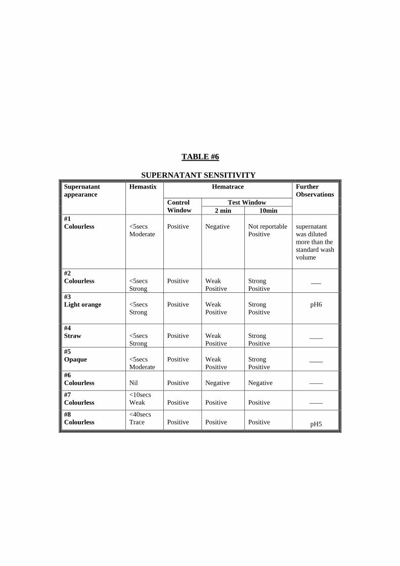
TTAABBLLEE ##66
SUPERNATANT SENSITIVITY Hematrace
Test Window
Supernatant appearance
Hemastix
Control Window 2 min 10min
Further Observations
#1 Colourless
<5secs Moderate
Positive
Negative
Not reportable Positive
supernatant was diluted more than the standard wash volume
#2 Colourless
<5secs Strong
Positive
Weak Positive
Strong Positive
___
#3 Light orange
<5secs Strong
Positive
Weak Positive
Strong Positive
pH6
#4 Straw
<5secs Strong
Positive
Weak Positive
Strong Positive
____
#5 Opaque
<5secs Moderate
Positive
Weak Positive
Strong Positive
____
#6 Colourless
Nil
Positive
Negative
Negative ____
#7 Colourless
<10secs Weak
Positive
Positive
Positive ____
#8 Colourless
<40secs Trace
Positive
Positive
Positive
pH5
TTAABBLLEE ##77
SPECIES CROSS REACTIVITY
Hematrace Species
Hemastix
Solution appearance
Control Window
Test Window 10min
Further Observations
Cat
<5secs Strong
Straw
Positive
Negative ____
Kangaroo
<5secs Strong
Straw
Positive
Negative ____
Horse
<5secs Strong
Straw
Positive
Negative ____
Dog
<5secs Strong
Straw
Positive
Negative ____
Bull
<5secs Strong
Dark Straw
Positive
Negative ____
Sheep
<5secs Strong
Dark Straw
Positive
Negative ____
Pig
<5secs Strong
Orange
Positive
Negative ____
Fowl
<5secs Strong
Dark Orange
Positive
Negative ____
Python
<5secs Strong
Dark Straw
Positive
Negative ____
Possum
<5secs Strong
Dark Orange
Positive
Negative ____
Wallaby
<5secs Strong
Dark Orange
Positive
Negative
____
<5secs Strong
Dark Red
Positive
Not reportable Positive
Appeared to give a slight “High Dose Hook effect”
Monkey
<5secs Strong
Straw
Positive
Weak Positive
Original sample further diluted and retested
<5secs Strong
Dark Red
Positive
Weak Positive
4dps of neat blood into test well followed by 2dps of buffer
Ferret
<5secs Strong
Dark Pink
Positive
Weak Positive
1dp of neat blood into 2ml buffer
TTAABBLLEE ##88
FORENSIC CASES Hematrace
Test Window
Evidence item Description
Sample Size
Luminol
Hemastix
Hematrace Buffer volume
Extraction time Control
Window 2 min 10min
#1
8mm2
cutting
Positive
V.Weak +ve @ 60secs
500µl
30 min
Positive
-ve
-ve
#2
15mm2
cutting
Negative
Weak +ve @ 60secs
500µl
30 min
Positive
-ve
-ve
#A Blue denim Shirt
#3 25mm2
cutting
Positive
V.Weak +ve @ 90secs
1000µl
30 min
Positive
-ve
-ve
#B Crime scene Bath Water
5drops
n/a
Negative
Nil
n/a
Positive
-ve
-ve
Micro Swab from 20mmφ stain
n/a
Strong positive @ <5secs
2ml
10min
Positive
-ve
-ve
#C Crime Scene Floor linoleum
Micro Swab from 10×20mm stain
n/a
Strong positive @ <5secs
500µl
10min
Positive
-ve
-ve
#D T-Shirt found on a decomposed body at scene for 2-3wks
5mm2
n/a
Strong positive @ <5secs
2ml
10min
Positive
mod +ve
strong +ve
TTAABBLLEE ##99
OUCHTERLONY GEL SENSITIVITY Ouchterlony Dilution Ouchterlony Dilution
Anti Human Serum
Anti Human Hemoglobin
Human Serum
Anti Human Hemoglobin
1:2
Positive
Positive
1:128
Positive
Positive
1:4
Positive
Positive
1:256
Positive
Positive
1:8
Positive
Positive
1:512
Positive
Positive
1:16
Positive
Positive
1:1024
Positive
Positive
1:32
Positive
Positive
1:2048
Negative
Negative
1:64
Positive
Positive
1:4096
Negative
Negative
8. References .
ABAcard Hematrace Onestep Test Kit – Technical Information Sheet. ABACUC Diagnostics Inc USA Swander, C.J and Stites, J.G., Michigan State Police, Forensic Laboratory. The Evaluation of the ABAcard HemaTrace for the Forensic Identification of Human Blood. Reynolds, M. The Abacard HemaTrace: Confirmatory Identification of Human Blood located at Crime Scenes. The Forensic Bulletin, December 2002. Kristaly, A and Smith, D.A.S, Forensic Biology Section, Crime Laboratory Bureau, Miami-Dade Police Department, Miami, Florida, USA. Validation of the OneStep ABAcard HemaTrace for the rapid forensic detection of human blood.

9. Appendix 1 .