Embed Size (px)
Citation preview

Generalintroductionandoutlineofthethesis
Chapter
1
Chapter 1
10
Content
1. Neuroinflammation,ashortintroduction 1.1Microglia 1.2Astrocytes 1.3Pro-andanti-inflammatorycytokines
2. MultipleSclerosis 2.1Clinicalcharacteristics 2.2Neuropathologicalcharacteristics 2.2.1 Cellular infiltration into the CNS
2.2.2 Demyelination and remyelination: a role for oligodendrocytes
2.2.3 The glial scar and CNS repair
2.3AnimalmodelsforMultipleSclerosis 2.3.1 Experimental Autoimmune Encephalomyelitis (EAE)
2.3.2 The cuprizone model
2.4Treatment
3. TissueTransglutaminase 3.1Transglutaminases,whatarethey? 3.2Thetransglutaminaseproteinfamily 3.3StructureofTG2 3.4BiochemicalfunctionsofTG2 3.4.1 Calcium dependent functions of TG2
3.4.2 Calcium independent functions of TG2
3.5CellularlocalizationandfunctionofTG2 3.5.1 TG2 and apoptosis
3.5.2 TG2 and cell differentiation
3.5.3 TG2 and cell adhesion and migration
3.6TG2andinflammation
4. Aimsandoutlineofthethesis
General introduction
11
1.Neuroinflammation,ashortintroduction
Inflammation is a complex reaction of living cells to pathogens and tissue injury. This response may exist in any vascularized compartment of the body, including the central nervous system (CNS).65 Inflammation of the CNS, or neuroinflammation, is of particular interest since the healthy CNS is considered ‘immunoprotected’ with relatively few resident immune cells and a highly specific blood brain barrier (BBB), that hinders entry of unwanted compounds and cells into the CNS.428 Neuroinflammatory symptoms present in disorders such as Multiple Sclerosis (MS),191, 396 HIV-associated dementia46 and encephalitis77 or with a neuroinflammatory component, such as stroke28 and brain trauma,235 are associated with BBB disruption and infiltration of inflammatory cells (monocytes and lymphocytes) into the CNS. During a neuroinflammatory state, local microglia, astrocytes, and infiltrated monocytes are the main effectors of the innate immune response in the CNS.29 These cells produce numerous inflammatory mediators to recruit other glial cells and peripheral immune cells to the site of injury.141 Although inflammatory mediators are of importance for their restorative actions under restricted inflammatory conditions in the brain,12 they become detrimental in a situation of uncontrolled production by activated glial cells and infiltrated monocytes.146
1.1MicrogliaMicroglia are the resident macrophages of the CNS and therefore considered to be the primary immune effector cells in the brain and spinal cord. They are present in the entire CNS and represent approximately 15% of the total CNS cell population.61 Microglia are of haematopoietic (mesenchymal) origin183 and are derived from bone marrow precursors that enter the CNS during early CNS development.151 Resting microglia have long multiple processes with high motility with which they can directly contact astrocytes, neurons and blood vessels. In the healthy CNS, microglia are constantly monitoring for damaged neurons and infectious agents.129 In response to inflammation, microglial cells undergo activation resulting in a characteristic phenotypic switch, evolving from resting ramified cells to motile amoeboid microglia.197 These activated microglia are phenotypically and functionally similar to peripheral macrophages and perform several innate immune functions.61, 134 Microglia can be classical or alternatively activated, which also has been described for macrophages.74 Microglia/macrophage activation depends on the products of of activated T helper 1 (Th1) or natural killer cells that induce classical activation or Th2 cells that induce alternative activation.135 At the molecular level, a variety of genes are upregulated, including the major histocompatibility complex (MHC) class I and II, pattern recognition receptors and complement receptors.218 Activated microglia release inflammatory mediators, including nitric oxide (NO), reactive oxygen species (ROS) and pro- and anti-inflammatory cytokines.
1.2AstrocytesAstrocytes represent nearly 35% of the total CNS cell population and, like microglia, are found in all regions of the CNS.399 Astrocytes are of neuroectodermal origin275 and the most abundant type of glial cells in the CNS. Two types are known, protoplasmic and
Chapter 1
12
fibrous astrocytes. Protoplasmic astrocytes are found in the grey matter and extend numerous ramified branches that contact neuronal surfaces and blood vessels with their end-feet.262 Conversely, fibrous astrocytes occupy areas adjacent to axon bundles in white matter tracts with processes longer and thinner than protoplasmic astrocytes.53 In the healthy CNS, astrocytes play many different roles. Astrocytes contribute to the formation of the BBB by projecting their end-feet on the basal lamina.3, 169 Astrocytes regulate cerebral blood flow in response to neuronal activity,161, 364 provide metabolic substrates for neurons,45 uptake of neurotransmitters such as glutamate,312 and regulate oxidative balance in the brain.411 Furthermore, astrocytes are a source of various neurotrophic factors including brain-derived neurotrophic factor (BDNF) and nerve growth factor (NGF). Upon inflammation, astrocytes increase the expression of BDNF and NGF, ultimately promoting cell survival and neurogenesis.265, 333 Astrocytes also express platelet-derived growth factor (PDGF), basic fibroblast growth factor (bFGF) and leukemia inhibitory factor (LIF) which stimulate the survival of oligodendrocytes.121, 126, 216
During neuroinflammation, astrocytes undergo a phenotypic change known as ‘reactive astrogliosis’, a process characterized by cellular hypertrophy and hyperplasia, cytoplasmic enlargement, elongated cytoplasmic processes, elevated metabolic activity and increased expression of the glial-specific intermediate filament protein, glial fibrillary acidic protein (GFAP).21 Reactive astrogliosis has been used as a marker of pathology in a variety of inflammatory CNS disorders, including MS.97
Similar to microglia, activated or reactive astrocytes are involved in neuroinflammation, however, they play a dual role, providing both defense and destruction of damaged neural tissue.268 Astrocytes produce inflammatory cytokines and chemokines thereby facilitating leukocyte extravasation from the blood stream into the CNS parenchyma,132, 301 and maintain local inflammatory reactions.210 On the other hand, astrogliosis is a defensive reaction, limiting the area of brain damage by formation of a glial scar.289
1.3Pro-andanti-inflammatorycytokinesCytokines are pleiotropic factors that coordinate innate and adaptive immune responses. The majority of cytokines are secreted proteins or glycoproteins. Cytokines and their receptors are expressed physiologically in CNS cells and are important for development and function of the brain.320 Under normal conditions there is a fine balance between pro-inflammatory and anti-inflammatory cytokine molecules. Pro-inflammatory cytokines, such as tumor necrosis factor-a (TNFa), interleukin-1 (IL-1) and IL-6, primarily facilitate the inflammatory response.320 In contrast, anti-inflammatory cytokines, such as IL-10, IL-4 and transforming growth factor-b (TGF-b), limit or inhibit the inflammatory response.251,
267 The pro-inflammatory cytokines TNFa, IL-6 and IL-1 share the ability to stimulate T and B lymphocytes, augment cell proliferation, and to initiate or suppress gene expression for several proteins.91 IL-1 is the term for two polypeptides, IL-1a and IL-1b that both recognize the same receptors.91 The effect of IL-1 can be limited by IL-1 receptor antagonist (IL-1ra), which is a natural competitive inhibitor that limits the reaction of IL-1 by blocking IL-1/IL-1 receptor interaction or by binding to the type II IL-1 receptor that acts as a decoy.92 The anti-inflammatory cytokines TGF-b, IL-10 and IL-4 have been found to be increased during CNS inflammation and may provide a negative feedback mechanism to limit the production of pro-inflammatory cytokines.206, 267
General introduction
13
In the pathogenesis of neuroinflammatory disorders, the balance between pro- and anti-inflammatory cytokines is disturbed.48, 165, 387 Lymphocytes, monocytes/macrophages and activated glial cells such as astrocytes and microglia can secrete pro- and anti-inflammatory cytokines. In particular IL-1b, IL-6 and TNFa released by microglia and astrocytes are key mediators of local inflammatory responses in the CNS.380 Astrocytes close to the BBB release IL-1b, IL-6 and TNFa leading to increased BBB permeability.89,
186, 332 However, astrocytes can also tighten the BBB via TGF-b secretion.4 Thus, different cytokines secreted by different cell types in the CNS have multiple functions during neuroinflammation.
2.MultipleSclerosis
Multiple Sclerosis (MS) is a chronic, inflammatory, and demyelinating disease of the CNS. Most of the patients are between 20 and 40 years old when the first symptoms appear and in general more females than males are affected (2:1).75 MS is the most prevalent neurological disorder in young adults with an incidence of one per 1000 in Europe and North America.354 The cause of MS is still unknown.
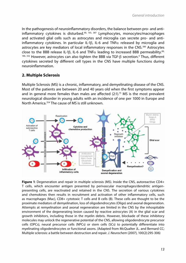
Figure 1: Degeneration and repair in multiple sclerosis (MS). Inside the CNS, autoreactive CD4+ T cells, which encounter antigen presented by perivascular macrophages/dendritic antigen-presenting cells, are reactivated and retained in the CNS. The secretion of various cytokines and chemokines then results in recruitment and activation of other inflammatory cells, such as macrophages (Mac), CD8+ cytotoxic T cells and B cells (B). These cells are thought to be the proximate mediators of demyelination, loss of oligodendrocytes (Oligo) and axonal degeneration. Attempts at remyelination and axonal regeneration are limited in the CNS by the inhospitable environment of the degenerating lesion caused by reactive astrocytes (A) in the glial scar and growth inhibitors, including those in the myelin debris. However, blockade of these inhibitory molecules may unlock the regenerative potential of the CNS, allowing oligodendrocyte precursor cells (OPCs), neural precursor cells (NPCs) or stem cells (SCs) to potentially differentiate into myelinating oligodendrocytes or functional axons. (Adapted from McQualter JL. and Bernard CC; Multiple sclerosis: a battle between destruction and repair. J. Neurochem (2007), 100(2):295-306)
Chapter 1
14
2.1ClinicalcharacteristicsClinical symptoms of MS include motor deficits, such as spasms, tremor and muscle weakness, finally leading to paralysis, and progressive sensory deficits, like impaired vision due to optic neuritis and also cognitive impairment has been recognized. MS is divided into 4 different clinical subgroups depending on clinical features.225 Relapsing-remitting MS (RR-MS) is the most common form. The majority of MS patients, approximately 70%, show a RR pattern during onset of the disease.76 RR-MS is characterized by episodes of clinical illness, followed by periods of clinical improvement. The clinical condition of the majority of the RR-MS patients gradually worsens over time and enters a secondary-progressive phase (SP-MS) at later stages. SP-MS is characterized by progressive neurological deterioration.282 Approximately 15-20% of MS patients suffer from primary-progressive MS (PP-MS) from disease initiation onwards.249, 370 These patients experience progressive disease without relapses. Less than 5% of the patients are diagnosed with progressive-relapsing MS (PR-MS), which is characterized by a continuous progressive disease course from the onset with occasional relapses.378
2.2NeuropathologicalcharacteristicsStudying post-mortem CNS material of MS patients gives insight into the neuropathological features characterizing MS at the tissue/cellular level. A major pathological hallmark of MS is the presence of demyelinated white matter lesions in the CNS195 and staging of these lesions is very important in defining the lesion under discussion.389 White matter MS lesions can be classified as pre-active, active, chronic-active and chronic-inactive lesions, based on the presence of MHC class II positive inflammatory cells (macrophages and lymphocytes), human leukocyte antigen-DR (HLA-DR) expression on leukocytes and resident microglial cells and the degree of myelin loss.42, 375, 389 Pre-active lesions are marked by clusters of activated microglial cells/macrophages that have increased HLA-DR expression without apparent loss of myelin. Active lesions are characterized by a demyelinated area and abundant phagocytic, perivascular and parenchymal macrophage infiltration throughout the lesion area, whereas chronic active lesions have a demyelinated gliotic center with a hypercellular rim containing macrophages. Inactive lesions are hypocellular and demyelinated lesions containing widened extracellular spaces occupied by gliotic scar tissue. Inactive lesions have only few infiltrating inflammatory cells throughout the lesion.Pathological features of MS which occur to a certain extent in all the different forms of lesions are the presence of inflammatory infiltrates that consist mainly of macrophages and lymphocytes, destructed myelin sheets, dying oligodendrocytes, damaged axons and glial scar tissue. These features and their putative relations are summarized in Fig. 1 and described more at length in the following paragraphs.
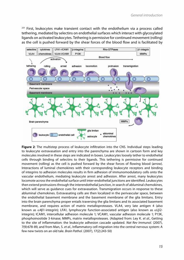
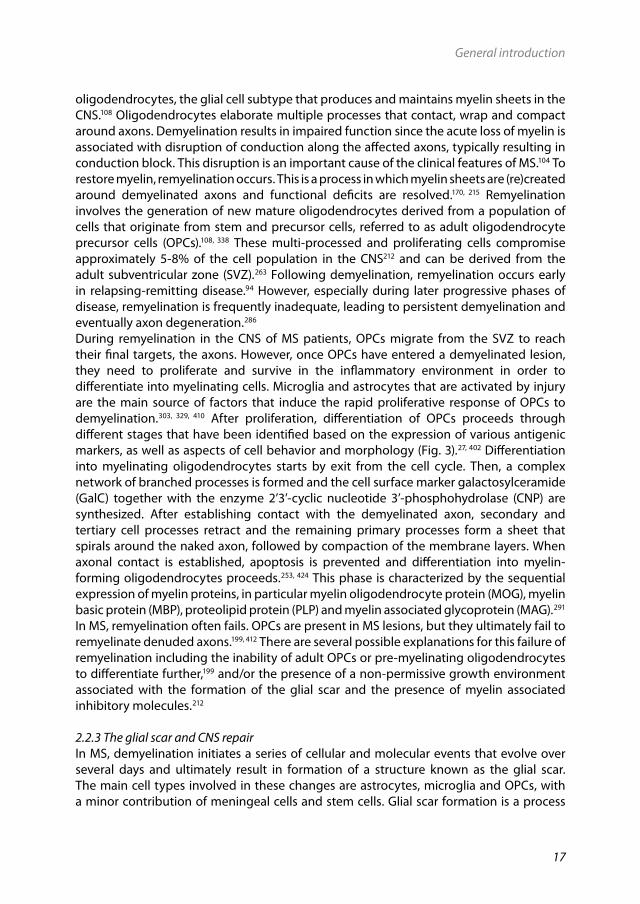
2.2.1 Cellular infiltration into the CNSThe BBB prevents entry of circulating immune cells and molecules into the CNS. However, in neuroinflammatory diseases such as MS, there is a massive cellular migration across the BBB. The migration of leukocytes (lymphocytes and monocytes) across the BBB occurs according to a multistep process that includes rolling across vascular endothelium, arrest and initial adhesion to endothelium, strong adhesion and transmigration of leukocytes across the endothelial monolayer as well as invasion into underlying tissues (Fig. 2).213,
General introduction
15
231 First, leukocytes make transient contact with the endothelium via a process called tethering, mediated by selectins on endothelial surfaces which interact with glycosylated ligands on activated leukocytes. Tethering is permissive for continued movement (rolling) as the cell is pushed forward by the shear forces of the blood flow and is facilitated by
Figure 2: The multistep process of leukocyte infiltration into the CNS. Individual steps leading to leukocyte extravasation and entry into the parenchyma are shown in cartoon form and key molecules involved in these steps are indicated in boxes. Leukocytes loosely tether to endothelial cells through binding of selectins to their ligands. This tethering is permissive for continued movement (rolling) as the cell is pushed forward by the shear forces of flowing blood (arrow). Interactions of luminal chemokines with their corresponding leukocyte receptors and binding of integrins to adhesion molecules results in firm adhesion of immunomodulatory cells onto the vascular endothelium, mediating leukocyte arrest and adhesion. After arrest, many leukocytes locomote across the endothelial surface until inter-endothelial junctions are identified. Leukocytes then extend protrusions through the interendothelial junction, in search of abluminal chemokines, which will serve as guidance cues for extravasation. Transmigration occurs in response to these abluminal chemokines. Extravasating cells are then localized in the perivascular space, between the endothelial basement membrane and the basement membrane of the glia limitans. Entry into the brain parenchyma proper entails traversing the glia limitans and its associated basement membrane, and requires action of matrix metalloproteases. VLA4, very late antigen 4 (also known as a4b1-integrin); LFA1, lymphocyte function-associated antigen (also known as aLb2-integrin); ICAM1, intercellular adhesion molecule 1; VCAM1, vascular adhesion molecule 1; P13K, phosphoinositide 3-kinase; MMPs, matrix metalloproteases. (Adapted from: Ley K. et al., Getting to the site of inflammation: the leukocyte adhesion cascade updated. Nat Rev Immunol. (2007), 7(9):678-89, and from Man, S. et al., Inflammatory cell migration into the central nervous system: A few new twists on an old tale. Brain Pathol. (2007), 17(2):243-50)
Chapter 1
16
cytokine-mediated endothelial activation.62 Rolling enables travelling leukocytes to scan endothelial surfaces for luminal chemokines, which are immobilized on endothelial surfaces. Interaction of luminal chemokines with their corresponding leukocyte receptors139 and binding of integrins (e.g. a4b1) present on the surface of leukocytes to adhesion molecules (e.g. vascular adhesion molecule 1 (VCAM1) and intercellular adhesion molecule 1 (ICAM1))355 present on endothelial cells results in firm adhesion of immunomodulatory cells onto the vascular endothelium.416 After adhesion, many leukocytes locomote across the endothelial surface to identify inter-endothelial junctions.327 Leukocytes then extend protrusions through the inter-endothelial junction, in search of abluminal chemokines, which will serve as guidance cues for extravasation.330
To facilitate extravasation, membrane protrusions and formation of new adhesive contacts at the leading edge of a migrating cell must be coordinated with down-regulation of adhesion and retraction at the rear of the cell.205 A crucial factor in this highly dynamic and tightly regulated process appears to be the actin cytoskeleton which is malleable, responsive to diverse stimuli and capable of translating extracellular signals into changes in cell shape and adhesive properties. The Rho family of small GTPases is a potent regulator of actin dynamics and regulates cell migration in response to extracellular stimuli.328 For instance, RhoA GTPase activity is required for transendothelial migration of monocytes but at the same time down-regulates integrin-mediated adhesion processes in monocytes.413 Following locomotion and process extension, cells migrate across the endothelial basement membrane into the perivascular space, prior to movement across astrocyte end-feet, the glia limitans. Subsequently, they migrate into the brain parenchyma to the sites of inflammation, a process that is facilitated by local tissue destruction via e.g. activation of matrix metalloproteases (MMPs).405
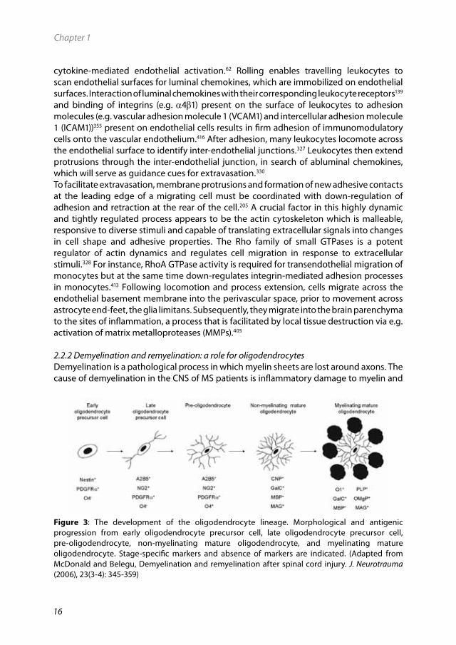
2.2.2 Demyelination and remyelination: a role for oligodendrocytesDemyelination is a pathological process in which myelin sheets are lost around axons. The cause of demyelination in the CNS of MS patients is inflammatory damage to myelin and
Figure 3: The development of the oligodendrocyte lineage. Morphological and antigenic progression from early oligodendrocyte precursor cell, late oligodendrocyte precursor cell, pre-oligodendrocyte, non-myelinating mature oligodendrocyte, and myelinating mature oligodendrocyte. Stage-specific markers and absence of markers are indicated. (Adapted from McDonald and Belegu, Demyelination and remyelination after spinal cord injury. J. Neurotrauma (2006), 23(3-4): 345-359)
General introduction
17
oligodendrocytes, the glial cell subtype that produces and maintains myelin sheets in the CNS.108 Oligodendrocytes elaborate multiple processes that contact, wrap and compact around axons. Demyelination results in impaired function since the acute loss of myelin is associated with disruption of conduction along the affected axons, typically resulting in conduction block. This disruption is an important cause of the clinical features of MS.104 To restore myelin, remyelination occurs. This is a process in which myelin sheets are (re)created around demyelinated axons and functional deficits are resolved.170, 215 Remyelination involves the generation of new mature oligodendrocytes derived from a population of cells that originate from stem and precursor cells, referred to as adult oligodendrocyte precursor cells (OPCs).108, 338 These multi-processed and proliferating cells compromise approximately 5-8% of the cell population in the CNS212 and can be derived from the adult subventricular zone (SVZ).263 Following demyelination, remyelination occurs early in relapsing-remitting disease.94 However, especially during later progressive phases of disease, remyelination is frequently inadequate, leading to persistent demyelination and eventually axon degeneration.286
During remyelination in the CNS of MS patients, OPCs migrate from the SVZ to reach their final targets, the axons. However, once OPCs have entered a demyelinated lesion, they need to proliferate and survive in the inflammatory environment in order to differentiate into myelinating cells. Microglia and astrocytes that are activated by injury are the main source of factors that induce the rapid proliferative response of OPCs to demyelination.303, 329, 410 After proliferation, differentiation of OPCs proceeds through different stages that have been identified based on the expression of various antigenic markers, as well as aspects of cell behavior and morphology (Fig. 3).27, 402 Differentiation into myelinating oligodendrocytes starts by exit from the cell cycle. Then, a complex network of branched processes is formed and the cell surface marker galactosylceramide (GalC) together with the enzyme 2’3’-cyclic nucleotide 3’-phosphohydrolase (CNP) are synthesized. After establishing contact with the demyelinated axon, secondary and tertiary cell processes retract and the remaining primary processes form a sheet that spirals around the naked axon, followed by compaction of the membrane layers. When axonal contact is established, apoptosis is prevented and differentiation into myelin-forming oligodendrocytes proceeds.253, 424 This phase is characterized by the sequential expression of myelin proteins, in particular myelin oligodendrocyte protein (MOG), myelin basic protein (MBP), proteolipid protein (PLP) and myelin associated glycoprotein (MAG).291 In MS, remyelination often fails. OPCs are present in MS lesions, but they ultimately fail to remyelinate denuded axons.199, 412 There are several possible explanations for this failure of remyelination including the inability of adult OPCs or pre-myelinating oligodendrocytes to differentiate further,199 and/or the presence of a non-permissive growth environment associated with the formation of the glial scar and the presence of myelin associated inhibitory molecules.212
2.2.3 The glial scar and CNS repairIn MS, demyelination initiates a series of cellular and molecular events that evolve over several days and ultimately result in formation of a structure known as the glial scar. The main cell types involved in these changes are astrocytes, microglia and OPCs, with a minor contribution of meningeal cells and stem cells. Glial scar formation is a process
Chapter 1
18
characterized by various cell types arriving and participating at different times.103 Macrophages derived from blood-stream monocytes and microglia arrive at sites of CNS injury within hours. After 3-5 days, large numbers of OPCs are recruited from the surrounding tissue.103 At the same time, astrocytes undergo morphological changes and increase the synthesis of GFAP. GFAP is an important intermediate filament protein that allows the astrocytes to synthesize more cytoskeletal supportive structures.173 As a consequence, in its final form, the glial scar consists of a fine meshwork of astrocyte processes tightly interwoven and bound together by tight and gap junctions, surrounded by extracellular matrix.36, 103, 309 Most likely, the glial scar serves to establish a barrier to isolate inflammation in the CNS and induces remodeling of the extracellular matrix409 which can be beneficial. However, astrogliosis may result in a physical and biochemical barrier to OPC migration and/or axon-oligodendrocyte interaction.103 Moreover, myelin debris trapped in scar tissue contains several axon growth inhibitory molecules, including MAG243, oligodendrocyte myelin glycoprotein (OMgp)400 and Nogo-A.51 Since there is compelling evidence indicating that glial scars strongly inhibit both axon growth and (re)myelination, for treatment purposes, it is vitally important to prevent glial scarring.
2.3AnimalmodelsforMultipleSclerosisTo study the disease mechanism(s) of MS, a number of animal models have been developed based on myelin mutants, chemically induced lesions and viral and auto-immune induced lesions.318 The experimental models that are currently used for MS can be divided roughly into two groups respectively: 1) disease models which attempt to replicate the disease process as accurately as possible and 2) models that provide a reductionist approach to study a specific aspect of the complex pathology.93 Experimental autoimmune encephalomyelitis (EAE) is the most frequently used example of the first category of disease models for MS, while toxin-induced models, such as the cuprizone model, belong to the second category offering an in vivo system to specifically study the process of demyelination and subsequent remyelination.
2.3.1 Experimental Autoimmune Encephalomyelitis (EAE)EAE is an experimentally induced neuroinflammatory and cell mediated autoimmune demyelinating disease that is characterized clinically by neurological deficits, in particular paralysis, and weight loss. Neuropathological features of EAE include mononuclear cell infiltration into the CNS, an early increase in BBB permeability and, depending on the type of EAE model used, demyelination.124 Based on shared similarities in disease pathology, EAE is a widely used animal model to study pathological mechanisms and experimental treatments in MS. It was first induced accidentally in humans during vaccination against rabies, using viruses grown on rabbit spinal cords. Residues of spinal cord injected together with the inactivated virus induced the CNS disease. Following these observations, a first model of EAE was described in non-human primates immunized with a CNS homogenate.315 EAE has since been generated in a variety of species and can follow different courses depending on the species/strain and immunizing antigen used.423
EAE can be induced in rodents and non-human primates by active immunization with myelin (components) including MOG, MBP, PLP and MAG together with a strong adjuvant such as complete Freunds’ adjuvant (CFA) or incomplete Freunds’ adjuvant (IFA).1 These
General introduction
19
myelin proteins, when systemically administered, induce an autoimmune response, leading to an attack on CNS myelin. Depending on the immunization protocol, EAE can be either acute or chronic. Acute EAE is characterized by a highly reproducible monophasic and transient disease course from which most animals recover. Usually, acute EAE is induced in the Lewis rat using MBP in combination with CFA in the footpath resulting in clinical signs.408 The clinical symptoms of acute EAE start about 10 days after immunization. First signs of disease are loss of tail tonus progressing to paralysis of the hind limbs. This may proceed into paralysis up to the lower back. Occasionally, an animal dies due to severe acute EAE. On average, the peak of clinical scores is reached 14 days after immunization and animals will recover spontaneously and permanently after 17 days.407, 408 The value of the acute EAE model in Lewis rats for the elucidation of the pathogenetic processes in MS is limited by the lack of spontaneous relapses and also by the absence of primary demyelinated lesions. Therefore, an alternative model like chronic relapsing EAE (cr-EAE) may be more appropriate to study mechanisms of axonal damage and demyelination. Cr-EAE is usually induced with MOG in combination with IFA.359 Initially, the course of cr-EAE is similar to that of acute EAE. However, in contrast to acute EAE, cr-EAE exhibits a chronic-relapsing course. In fact, after recovery from the acute phase (remission), animals will experience a relapse with increased severity of clinical signs. Chronic relapsing and demyelinating variants of EAE have been induced by active immunization in different species and strains, including guinea pigs,307 rabbits,302 mice strains like C57Bl/6 and SJL,224 rat strains like Dark Agouti223 and non-human primates.2
2.3.2 The cuprizone model Oral administration of the copper chelator cuprizone (bis-cyclohexanone-oxaldehydrazone) in mice results in demyelination of white matter in the thalamus, internal capsule, arterior commissure and cerebellar peduncles.39 In addition, cerebellar cortical demyelination has been observed in this model.344 However, for practical reasons, demyelination of the corpus callosum represents the most frequently investigated white matter lesion in this animal model.194, 358 The first experiments using cuprizone were performed in the late 1960s.59, 60 It was reported that oxalic biscyclohexylidenehydarzide, a chelator used as a reagent for copper analysis, induces microscopic lesions in the brain accompanied by demyelination, astrogliosis and edema.60 When 8 week old C57Bl/6 mice are fed with 0.2% cuprizone, mature oligodendrocytes are specifically ablated, a process believed to be closely followed by a rapid proliferation and accumulation of microglia and macrophages in the corpus callosum, prior to actual demyelination.153 After 5-6 weeks of cuprizone treatment, the corpus callosum is almost completely demyelinated, a process called ‘acute demyelination’. In the acute cuprizone model, the toxicity of cuprizone is reversible, in such that removal of the toxin from the diet results in spontaneous remyelination.148 The precise mechanism of demyelination and remyelination remains unexplained. Although, it has been presumed that copper deficiency was primarily responsible for the lesions, copper administration failed to reduce cuprizone toxicity.240 Since is well known that cuprizone treatment results in mitochondrial dysfunction,190 the most likely hypothesis is that disturbance in energy metabolism leads to oligodendrocyte cell death, which causes demyelination.373
Chapter 1
20
2.4Treatment Although MS is still not curable, several therapeutic treatment regimens are in clinical use for MS. The most widely used drugs during relapses are glucocorticoids (GCs), such as methylprednisolone, which act both immunosuppressive and anti-inflammatory. The mechanism(s) by which GCs exert their therapeutic effect is not completely understood, but include altered gene transcription via GC response elements in the promoter and enhancer regions of genes and interference with signaling via other factors such as nuclear factor kappa B (NF-kB) or phosphoinositide 3-kinase (PI3K).372 GCs are used in short courses of a high dose to reduce the duration and severity of acute relapses.300 However, use of GCs is plagued by severe metabolic side effects and they have no proven beneficial effect on long-term disease progression.300 Therefore, presently, RR-MS patients are usually treated with interferon-g (IFN-g), which has been shown to have a modest effect on reducing the number of relapses and slowing the progression of disability.167 The exact beneficial mechanism of IFN-g action in MS is still not known but there are several possible explanations. IFN-g may mediate a shift from Th1 (stimulatory) towards a Th2 (inhibitory/regulatory) response by reducing pro-inflammatory cytokines and increasing the expression of anti-inflammatory cytokines.321, 419 In addition, IFN-g may reduce adhesion and migration of monocytes and T-cells into the CNS since IFN-g reduces the expression of molecules involved in cell adhesion and migration.111, 361 Despite the beneficial effect for some patients, a large proportion of patients does not respond well to IFN-g404 and the therapy has multiple side effects via interference with the immune system, including flu-like symptoms, skin reactions and fatique.269 Moreover, during treatment with IFN-g, neutralizing antibodies can emerge resulting in possible loss of efficacy. In contrast, formation of antibodies does not play a role in treatment with glatiramer acetate, which is another compound used for treatment of RR-MS patients. Although its mode of action is still not exactly known, two models have been suggested.40 First, glatiramer acetate can bind MHC class II molecules of antigen presenting cells (e.g. macrophages) and competes with MBP, thereby preventing the activation of MBP reactive T-cells.122 Secondly, T-cells reacting to glatiramer acetate may shift from a Th1 to a Th2 response, which has been shown to be beneficial for MS patients.270 Unfortunately, however, although effective in reducing relapse rate, the benefit of glatiramer acetate use on sustained disability progression was shown to be very low.176, 177
Mitoxantrone is an immunosuppressive agent that is used for patients with RR-MS and SP-MS.248 It has been shown that treatment with mitoxantrone results in reduced demyelination.403 At the cellular level, mitoxantrone treatment impedes the ability of T-cells to induce an immune response and B-cells to proliferate.109 For RR-MS patients, it has been shown to have significant beneficial effects on sustained disability progression, relapses and new lesions.248 In contrast, the actual efficacy of mitoxantrone in SP-MS is unclear. More importantly, however, major drawbacks of mitoxantrone therapy are the risk of developing serious side effects, such as acute myeloid leukemia and cardiotoxicity, which seriously restricts its use.131 One of the more promising forms of therapy currently available is natalizumab (Tysabri). Natalizumab is an antibody directed against the integrin very late antigen 4 (VLA4, a4b1) that is expressed on activated lymphocytes and monocytes involved in transendothelial migration.311 Natalizumab successfully reduces relapse rates and slows disease progression
General introduction
21
in RR-MS patients.299 At the same time, however, adverse effects of natalizumab have been reported and natalizumab treatment was temporarily halted after several patients developed progressive multifocal leukoencephalopathy (PML), a fatal demyelinating disease affecting the central nervous system.192, 202, 388 After a review of safety information and no further cases of PML, natalizumab was re-approved, although with a restricted indication. Despite the fact that new PML cases were described recently217, 230 the drug is still available for the treatment of MS. Although natalizumab was shown to be more effective than glucocorticoids or IFN-g, it is also not able to stop progression of MS. The failure of current forms of therapy to stop progression of the disease and their lack of safety therefore provides strong compelling impulse to continue the search for more effective and less toxic therapies.
3.TissueTransglutaminase
3.1Transglutaminases,whatarethey?The term transglutaminases (TGs, EC 2.3.2.13) was introduced by Clarke et al. in 1957 to describe the transamidating activity observed in guinea-pig liver.72 TGs are a widely distributed group of enzymes that catalyze a variety of calcium- and thiol-dependent posttranslational protein modifications. This may occur either through protein cross-linking via formation of e-(g-glutamyl)lysine bonds or through incorporation of primary amines at the level of peptide bound glutamine residues.115 The cross-linked protein products are highly resistant to mechanical challenge and proteolytic degradation, and their accumulation is found in a number of tissues, including hair, skin and blood cloths.221 All TG family members are comprised of four sequential and structurally distinct domains (an NH2-terminal b-sandwich, an a/b catalytic core, and 2 COOH-terminal b-barrel domains) that assume a compact conformation in the absence of calcium.31
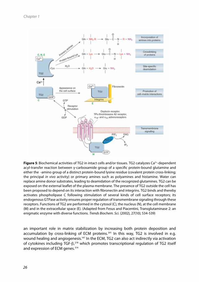
3.2ThetransglutaminaseproteinfamilyIn mammals, at least eight catalytically active isoforms have been identified at the genomic level, including TG1-7 and Factor XIIIa (FXIIIa) (Table 1). The best characterized isotypes include FXIIIa, TG1, TG2, TG3 and TG5, while the function of TG4, TG6 and TG7 is still largely unclear.163 FXIIIa, as a zymogen, has both extracellular and intracellular functions. It is soluble and expressed mostly in circulating blood cells. FXIII is converted by thrombin-dependent proteolysis into 1) the active TG FXIIIa, (plasma TG, 83 kDa) involved in stabilization of fibrin clots and in wound healing, and 2) the regulatory B subunit (80 kDa) which does not belong to the TG family since it has no clear enzymatic activity.261 Besides blood cells, FXIIIa can be detected in monocytes associated with wound healing and tumor progression374 and in a number of other cell types such as chondrocytes and osteoblasts.279, 280, 374 TG1 (106 kDa) and TG3 (77 kDa) exist in membrane-bound and soluble forms and are activated by proteolysis. They are both involved in epidermal terminal differentiation. TG5 (81 kDa) may require proteolytic processing like TG1 and TG3. TG5 appears to associate with the vimentin intermediate filament network in cultured epithelial cells that are undergoing epithelial mesenchymal transition, a mechanism that provides cells with increased cell mobility and loss of adhesion.57, 295 The ubiquitous TG2 (also known as tissue Transglutaminase, 78 kDa) is without doubt the most diverse and
Chapter 1
22
best characterized member of the transglutaminase family of enzymes.66, 106, 140, 245 TG2 is expressed throughout the human body,163 including the CNS369 where it is expressed in different cell types including astrocytes and neurons.31, 221
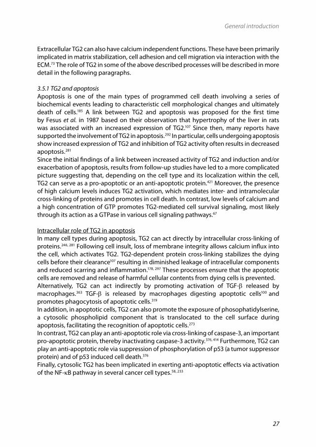
3.3StructureofTG2The molecular structure of TG2 was reported in 2002.219 As shown in Fig. 4, TG2 has four distinct molecular domains including: an N-terminal β-sandwich, a catalytic core, a C-terminal β-barrel 1 domain and a β-barrel 2 domain.159 The N-terminal region of TG2 is responsible for binding of fibronectin and integrins172 whereas the C-terminal segment of β-barrel 2 interacts with phospholipase C.254 In addition, a unique guanidine nucleotide (GDP) binding site, which has not been found in any other protein, is located between the catalytic core and the first β-barrel219 coded by exon 10 of the TG2 gene (Fig. 4A).
Table1: The transglutaminase protein family
Protein Synonyms Molecular Mass (kDa) Tissue expression
TG1 Keratinocyte TG 106 Keratinocytes
TG2 Tissue TG (tTG) 78 Ubiquitous
TG3 Epidermal TG 77 Squamous epithelium
TG4 Prostate TG 77 Prostate
TG5 TGx 81 Lymphatic system, epithelial cells
TG6 TGy 80 Ubiquitous
TG7 TGz 80 Ubiquitous
FXIIIa Fibrin-stabilizing factor 83 Dermal dendritic cells, platelets, placenta, monocytes, chondrocytes, osteoblasts
TG = transglutaminase
In human TG2, domains 1-4 span amino acids 1-139, 140-454, 479-585 and 586-687 respectively (Fig. 4A). These domains have different secondary structure arrangements since domains 1, 3 and 4 are folded in b-structures and domain 2 presents prevalently a-helical secondary structures.219 TG2 exhibits two distinct catalytic activities, which are differentially regulated by calcium and GDP/GTP. TG2 is only active as a transamidase when bound to calcium and inactive when bound to GDP.30 Investigation of the structural basis for the activation of TG2 by calcium by the use of several techniques, including small-angle scattering, protein dynamics, site-directed mutagenesis and crystallography, suggested that switching on the transamidation (i.e. cross-linking) activity of TG2 involves movement of protein domains, which influences the reactivity of the active site and its accessibility to the substrates.63, 219, 234, 260 Recently, the crystal structures of the different conformations of TG2 including the GDP-bound and calcium activated conformer were published.296 These different conformers were identified by using an active site-directed irreversible TG2 inhibitor and studying of the crystal structure of the enzyme bound to the inhibitor or GDP.296
Based on these studies, TG2 is proposed to have open and closed conformers (Fig. 4B,C), with notably distinct features.30, 296 The active site of GDP-bound TG2 is in a closed conformation as a result of GDP binding between the first β-barrel domain and the

General introduction
23
Figure4: Domains and functional elements of human (TG2) and overall structures of GDP-bound and inhibitor-bound TG2. A) The four structural domains are indicated by arrows with amino acid positions (top). Exon boundaries of the gene encoding TG2 are indicated by arrowheads with numbers corresponding to the last amino acids of each exon-encoded region. Exon 10, which contains residues forming the GDP-binding site, is marked. Functional regions and amino acid positions indicated are as follows; FN, fibronectin binding site (cyan); integrin-binding region, N-terminal 28-kDa state stabilization, forming an inhibitory H-bond with C277 and potential inhibition of activity by disulfide bonding of C277, respectively; Ca2+-binding site predicted from the FXIIIa structure (red); non-proline cis peptide bonds 273KY274, 387KY388 (yellow); GTP, GDP-binding and GTPase catalytic site residues (orange); PLCδ1, interaction site for phospholipase Cδ1 (magenta). Putative sites are labeled by an asterisk. (B and C) The crystal structures are shown as ribbons, and simplified cartoons are included for clarity. The N-terminal β-sandwich is shown in blue (N), the catalytic domain (Core) in green, and the C-terminal β-barrels (β1 and β2) in yellow and red, respectively. B) GDP-bound TG2. C) TG2 inhibited with the active-site inhibitor Ac-P(DON)LPF-NH2. (Adapted from Fesus and Piacentini, Transglutaminase 2: an enigmatic enzyme with diverse functions, Trends Biochem. Sci. (2002), 27(10); 534-539 and Pinkas et al., Transglutaminase 2 undergoes a large conformational change upon activation. PloS Biology (2007), 5(12); e327)
Chapter 1
24
catalytic domain.219 In the GDP-bound form of TG2, access to the transamidation active site is blocked by two loops, and the active site cysteine is hydrogen-bonded to a tyrosine residue at position 516 (Y516).219 TG2 becomes activated to transamidate upon calcium binding.140 It has been suggested that the structure of calcium activated TG2 is similar to that observed in the open inhibitor-bound conformation.296 During activation, interactions between domain 2 (the active-site domain) and domains 3 and 4 break down. Calcium-binding residues are not known with certainty, although structure prediction and site directed mutagenesis studies have suggested at least 3 different calcium-binding sites.63,
80 The main calcium binding site is located in domain 2 and this site becomes available during activation of the enzyme, following binding of calcium,140, 296 when the interactions between domains 2, 3 and 4 break down. Upon calcium binding at this site, or others14 a conformational change in the protein is induced, creating an open access to the active site thereby inducing the transamidating activity (Fig. 4C).The transamidating activity of TG2 depends on the catalytic domain that contains the catalytic triad; cysteine 277 (C277), histidine 335 (H335) and aspartate 358 (D358)288 and the nucleotide-binding residues.162 It has been well established that C277 is the essential nucleophile for transamidation417 and therefore, cysteine to serine mutation at position 277 (C277S) has been extensively used to inactivate the transamidation function of TG2.377 In addition to the catalytic triad, a conserved tryptophan residue (W241) is also critical for the transamidating activity.260 The mechanism of activation of TG2 by calcium can be counteracted by the allosteric inhibitor GTP. GTP binds at lysine 173 and is finally hydrolyzed leading to a reversible, GTPase dependent regulatory mechanism.260 It has been suggested that binding of GTP involves replacement of GDP, which is normally bound to the protein.33 There are only subtle differences in the conformations induced by GDP versus GTP.140 The function of GTP binding will be discussed later in paragraph 3.4.2.
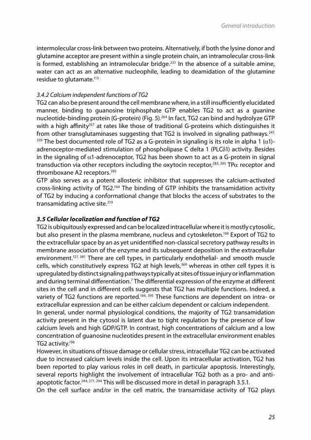
3.4BiochemicalfunctionsofTG2TG2 has diverse biochemical functions which can be roughly divided into calcium dependent and calcium independent functions,264 as shown in Fig. 5 and described more in detail in the following paragraphs.
3.4.1 Calcium dependent functions of TG2In intact cells and/or tissues, several posttranslational modification reactions are catalyzed by TG2. As described earlier, TG2 is activated by the binding of calcium. The conformational change resulting from this interaction allows for the acylation of the active site cysteine residue (C277) by a protein-bound glutamine residue to form a thioester intermediate between TG2 and the glutamine containing protein substrate, with ammonia being released as a by-product. This initial step is followed by the binding of an amine resulting in the formation of a stable isopeptide or glutamylamine bond. In this manner, the so-called transamidation reaction results in incorporation of either a primary amine onto a glutamine side chain of the acceptor protein, or in protein cross-linking in which a glutamine residue is cross-linked to an e-amino group of a protein-bound lysine residue.221 The outcome of this reaction is a bridge (cross-link) between a lysine donor residue on one protein with an acceptor glutamine residue of another protein, creating a covalent
General introduction
25
intermolecular cross-link between two proteins. Alternatively, if both the lysine donor and glutamine acceptor are present within a single protein chain, an intramolecular cross-link is formed, establishing an intramolecular bridge.221 In the absence of a suitable amine, water can act as an alternative nucleophile, leading to deamidation of the glutamine residue to glutamate.113
3.4.2 Calcium independent functions of TG2TG2 can also be present around the cell membrane where, in a still insufficiently elucidated manner, binding to guanosine triphosphate GTP enables TG2 to act as a guanine nucleotide-binding protein (G-protein) (Fig. 5).264 In fact, TG2 can bind and hydrolyze GTP with a high affinity357 at rates like those of traditional G-proteins which distinguishes it from other transglutaminases suggesting that TG2 is involved in signaling pathways.247,
339 The best documented role of TG2 as a G-protein in signaling is its role in alpha 1 (a1)-adrenoceptor-mediated stimulation of phospholipase C delta 1 (PLCδ1) activity. Besides in the signaling of a1-adrenoceptor, TG2 has been shown to act as a G-protein in signal transduction via other receptors including the oxytocin receptor,283, 395 TPa receptor and thromboxane A2 receptors.395
GTP also serves as a potent allosteric inhibitor that suppresses the calcium-activated cross-linking activity of TG2.164 The binding of GTP inhibits the transamidation activity of TG2 by inducing a conformational change that blocks the access of substrates to the transamidating active site.219
3.5CellularlocalizationandfunctionofTG2TG2 is ubiquitously expressed and can be localized intracellular where it is mostly cytosolic, but also present in the plasma membrane, nucleus and cytoskeleton.106 Export of TG2 to the extracellular space by an as yet unidentified non-classical secretory pathway results in membrane association of the enzyme and its subsequent deposition in the extracellular environment.127, 381 There are cell types, in particularly endothelial- and smooth muscle cells, which constitutively express TG2 at high levels,369 whereas in other cell types it is upregulated by distinct signaling pathways typically at sites of tissue injury or inflammation and during terminal differentiation.7 The differential expression of the enzyme at different sites in the cell and in different cells suggests that TG2 has multiple functions. Indeed, a variety of TG2 functions are reported.106, 395 These functions are dependent on intra- or extracellular expression and can be either calcium dependent or calcium independent. In general, under normal physiological conditions, the majority of TG2 transamidation activity present in the cytosol is latent due to tight regulation by the presence of low calcium levels and high GDP/GTP. In contrast, high concentrations of calcium and a low concentration of guanosine nucleotides present in the extracellular environment enables TG2 activity.196 However, in situations of tissue damage or cellular stress, intracellular TG2 can be activated due to increased calcium levels inside the cell. Upon its intracellular activation, TG2 has been reported to play various roles in cell death, in particular apoptosis. Interestingly, several reports highlight the involvement of intracellular TG2 both as a pro- and anti-apoptotic factor.244, 271, 294 This will be discussed more in detail in paragraph 3.5.1. On the cell surface and/or in the cell matrix, the transamidase activity of TG2 plays
Chapter 1
26
an important role in matrix stabilization by increasing both protein deposition and accumulation by cross-linking of ECM proteins.391 In this way, TG2 is involved in e.g. wound healing and angiogenesis.147 In the ECM, TG2 can also act indirectly via activation of cytokines including TGF-b,278 which promotes transcriptional regulation of TG2 itself and expression of ECM genes.314
Figure5: Biochemical activities of TG2 in intact cells and/or tissues. TG2 catalyzes Ca2+-dependent acyl-transfer reaction between γ-carboxamide group of a specific protein-bound glutamine and either the -amino group of a distinct protein-bound lysine residue (covalent protein cross-linking; the principal in vivo activity) or primary amines such as polyamines and histamine. Water can replace amine donor substrates, leading to deamidation of the recognized glutamines. TG2 can be exposed on the external leaflet of the plasma membrane. The presence of TG2 outside the cell has been proposed to depend on its interaction with fibronectin and integrins. TG2 binds and thereby activates phospholipase C following stimulation of several kinds of cell surface receptors; its endogenous GTPase activity ensures proper regulation of transmembrane signaling through these receptors. Functions of TG2 are performed in the cytosol (C), the nucleus (N), at the cell membrane (M) and in the extracellular space (E). (Adapted from Fesus and Piacentini, Transglutaminase 2: an enigmatic enzyme with diverse functions. Trends Biochem. Sci. (2002), 27(10); 534-539)
General introduction
27
Extracellular TG2 can also have calcium independent functions. These have been primarily implicated in matrix stabilization, cell adhesion and cell migration via interaction with the ECM.73 The role of TG2 in some of the above described processes will be described in more detail in the following paragraphs.
3.5.1 TG2 and apoptosisApoptosis is one of the main types of programmed cell death involving a series of biochemical events leading to characteristic cell morphological changes and ultimately death of cells.185 A link between TG2 and apoptosis was proposed for the first time by Fesus et al. in 1987 based on their observation that hypertrophy of the liver in rats was associated with an increased expression of TG2.107 Since then, many reports have supported the involvement of TG2 in apoptosis.292 In particular, cells undergoing apoptosis show increased expression of TG2 and inhibition of TG2 activity often results in decreased apoptosis.281 Since the initial findings of a link between increased activity of TG2 and induction and/or exacerbation of apoptosis, results from follow-up studies have led to a more complicated picture suggesting that, depending on the cell type and its localization within the cell, TG2 can serve as a pro-apoptotic or an anti-apoptotic protein.421 Moreover, the presence of high calcium levels induces TG2 activation, which mediates inter- and intramolecular cross-linking of proteins and promotes in cell death. In contrast, low levels of calcium and a high concentration of GTP promotes TG2-mediated cell survival signaling, most likely through its action as a GTPase in various cell signaling pathways.67
Intracellular role of TG2 in apoptosisIn many cell types during apoptosis, TG2 can act directly by intracellular cross-linking of proteins.246, 281 Following cell insult, loss of membrane integrity allows calcium influx into the cell, which activates TG2. TG2-dependent protein cross-linking stabilizes the dying cells before their clearance107 resulting in diminished leakage of intracellular components and reduced scarring and inflammation.178, 297 These processes ensure that the apoptotic cells are removed and release of harmful cellular contents from dying cells is prevented.Alternatively, TG2 can act indirectly by promoting activation of TGF-b released by macrophages.363 TGF-b is released by macrophages digesting apoptotic cells100 and promotes phagocytosis of apoptotic cells.319 In addition, in apoptotic cells, TG2 can also promote the exposure of phosophatidylserine, a cytosolic phospholipid component that is translocated to the cell surface during apoptosis, facilitating the recognition of apoptotic cells.273
In contrast, TG2 can play an anti-apoptotic role via cross-linking of caspase-3, an important pro-apoptotic protein, thereby inactivating caspase-3 activity.376, 414 Furthermore, TG2 can play an anti-apoptotic role via suppression of phosphorylation of p53 (a tumor suppressor protein) and of p53 induced cell death.376 Finally, cytosolic TG2 has been implicated in exerting anti-apoptotic effects via activation of the NF-kB pathway in several cancer cell types.58, 233
Chapter 1
28
Extracellular role of TG2 in apoptosisAt the cell surface, TG2 plays mainly an anti-apoptotic role. Thus, TG2 can rescue apoptotic cells via adhesion dependent survival signaling. When TG2 is deposited in the ECM, this pathway is mediated by binding of TG2 to fibronectin and cell surface heparin sulphate proteoglycans (HSPGs). HSPGs bind ECM ligands through the heparin sulphate chains, influencing their biological activity, trafficking, and secretion. Among the HSPG subfamilies, the syndecans, such as syndecan-4, act as co-receptors for both ECM components and soluble ligands.257 The binding of TG2 to fibronectin and syndecan-4 requires the function of protein kinase Cδ and leads to activation of Rho and focal adhesion kinase (FAK).394 A comparable survival signaling pathway can also be induced when TG2 is present on the cell membrane and associates with integrins, thereby inducing activation of Bcl-2, an anti-apoptotic protein, which in its turn is also able to activate FAK signal transduction pathways.142
3.5.2 TG2 and cell differentiationTG2 plays an important role in cell differentiation including that of fibroblasts and granulocytes,20, 357 but also brain related cells, in particular neurons.377 For example, it has been shown that retinoic acid induced neuronal differentiation of human neuroblastoma cells and differentiation of granulocytes is associated with increased expression and activation of TG.220, 341 In neuronal cells, this elevated activity of TG2 resulted in activation of RhoA via transamidation. Transamidated RhoA promotes cytoskeletal rearrangements and activation of mitogen activated protein kinase (MAPK) which regulates many nuclear events and promotes gene expression during neuronal differentiation.341 TG2 may also have another function during neuronal development. It has been shown that TG2 is selectively expressed in cerebellar granule neurons during their development and inhibition of TG2 activity negatively affected the development of these cells by reducing neurite outgrowth.228 It was suggested that the function of TG2 in neurite outgrowth was covalent cross-linking of cell surface substrates including the neurothrophic cytokine, midkine, and the cellular adhesion molecule, galectin-3. This may then serve to maintain the morphology of neurites during neuronal differentiation.228
To date, it is not known if TG2 is involved in differentiation of other cell types present in the CNS, such as oligodendrocytes or astrocytes. However, a role for TG2 in astrocyte differentiation has been suggested.56
3.5.3 TG2 and cell adhesion and migrationSeveral reports showed that TG2 is involved in cell adhesion and migration via interaction with the ECM. These results indicate that both the protein cross-linking and G-protein functionality of TG2 independently contribute to the regulation of cell-matrix interactions and ECM remodeling.357 Moreover, it appears that TG2 contribution to cell adhesion and migration is also dependent on the intra- or extracellular location of the enzyme, as described in the following paragraphs.
Intracellular role of TG2 in cell adhesion and migrationFor cells to adhere and migrate, cytoskeletal rearrangement is necessary. This can be regulated by TG2 since it has been shown that TG2, activated by calcium, interacts with
General introduction
29
and modifies major components of the cytoskeleton.317, 326 For example, TG2 is involved in crosslinking of the membrane-cytoskeletal protein actin.317 In contrast, TG2 can also act indirectly. It has been shown that TG2-dependent transamidation of RhoA results in the increased binding of RhoA GTPase to ROCK-2 protein kinase, and phosphorylation of the intermediate filament vimentin,341 leading to the formation of stress fibers and increased cell adhesion. These events are prevented by inhibition of TG2 activity. Although these effects of TG2 activation are clearly located inside cells, it remains to be elucidated whether they are mediated by the intracellular or cell surface pool of TG2.
Extracellular role of TG2 in cell adhesion and migrationSecreted TG2 present on the cell surface or in the ECM partially mediates the interaction of b-integrins on the cell surface with fibronectin present in the ECM.10 TG2 has a fibronectin binding site located in the N-terminal domain (Fig. 4).127 Integrins are a family of cell-surface proteins that serve as receptors for ECM proteins such as fibronectin, laminin and collagen. Fibronectin is a high molecular weight modular glycoprotein present in the ECM and in plasma.160 Assembly of fibronectin into insoluble matrix is a complex cell-mediated process that involves association of fibronectin with integrins on the cell surface. Fibronectin binds to cell-surface matrix receptors, primarily the a5b1 integrins, through the Arg-Gly-Asp (RGD) cell-binding site.394 Although the a5b1 integrin has a predominant role in fibronectin assembly, other fibronectin-binding integrins such as aVb3 are known.415
Integrins are relatively low affinity receptors for ECM proteins, including fibronectin. In contrast, TG2 binds fibronectin with high affinity.305, 379 Interestingly, it has been shown that TG2 forms stable complexes with b-integrins.10 In this manner, the presence of integrin-bound TG2 on the cell surface provides an additional high affinity binding site for fibronectin and integrins10 (Fig. 5). Depending on the cell type, 10–40% of b(1)-integrins on the cell surface exist as a complex with TG2 in a 1:1 ratio.9, 10 Available evidence from in vitro studies indicates that cell surface TG2 in complex with b-integrins can contribute to cell adhesion in two ways.10 In the simplest model, TG2 can serve as a bridge between b-integrins and fibronectin. This increases the affinity between fibronectin and its integrin receptor and allows a second integrin molecule to access the RGD site in the same fibronectin chain.9 TG2 binds with high affinity to the adhesive site on fibronectin that is located within the gelatin binding domain (42-kDa) of fibronectin that is located apart from the major integrin-binding sites on the fibronectin molecule.10, 305 The second model proposes that cell-surface TG2 promotes cell adhesion to fibronectin,10 by interacting directly with b1- and b3-integrins.367, 394, 425 Indeed, cell surface TG2 has been found to cooperate with integrins in cell adhesion through a direct noncovalent interaction with the b1 and b3-integrin subunits and form stable ternary complexes with integrins and fibronectin.10 Within these complexes, all three proteins, TG2, integrins and fibronectin, interact with each other.10
TG2 has an additional non-enzymatic structural adhesive role in reinforcing the integrin-fibronectin interaction that is independent from the RGD cell binding domain of fibronectin.394 It has been shown that cell-surface TG2 in complex with fibronectin directly associates with the heparan sulphate chain of syndecan-4 to reinforce or substitute RGD-dependent cell adhesion. TG2-fibronectin binding to syndecan-4 results in activation of
Chapter 1
30
PKCa leading to interaction of PKCa with b1-integrin. Cell signaling by this process leads to the activation of focal adhesion kinase and the mitogen activated protein kinase (MAPK) pathway.367, 393
Besides the role of TG2 in cell adhesion, the interaction of TG2 with b-integrins is also thought to facilitate migration and motility of cells9, 22 and activation of the downstream survival signaling pathways.392 Within this context, it has been shown that TG2 mediates migration of monocytes across fibronectin.9 Furthermore, after the discovery that extracellular TG2 acts as an b1- and b3-integrin co-receptor,10 it was demonstrated that inhibition of this cell surface-bound TG2 with a monoclonal antibody can block transendothelial migration of human CD8+ T cells in vitro.252
TG2 is also involved in macrophage infiltration in vivo and TG2 knockout mice show a reduced macrophage infiltration in a model for renal fibrosis.337 During monocyte differentiation into macrophages, high levels of integrin-bound surface TG2 are induced9,
259 concomitant with a decrease in FXIIIa expression in these cells.335 Therefore, TG2 is considered to be a useful and reliable marker of macrophage activation correlating with macrophage phagocytotic capacity331, 335 as well as monocyte/macrophage adhesion and migration.9 Although theoretically possible, it is not known, however, if TG2 is also involved in adhesion to the BBB and/or migration into the CNS of inflammatory cells from the blood during neuroinflammatory processes like MS.
3.6TG2andinflammationThe functions of TG2 that are described in the paragraph 3.5 are also important during inflammatory processes. Besides these functions, several in vitro and in vivo findings link TG2 to inflammatory processes. For example, it has been shown in cultured cell systems that the pro-inflammatory cytokines TNFa, IL-1 and IL-6 are able to increase cellular TG2 levels.187, 255 Moreover, exposure to TGF-b enhances TG2 expression on the surface of fibroblasts.130 Conversely, TG2 activates the transcriptional activator NF-kB thereby enhancing lipopolysaccharide (LPS)-induced expression of inducible nitric-oxide synthase (iNOS) in microglia.209 In addition, it has been shown that treatment with LPS results in increased TG2 activity on the surface of human monocyte derived dendritic cells and macrophages.154 Thus, TG2 expression and activity appears to be enhanced during activation of inflammatory mechanisms,144, 201, 255 in which it may play a modulatory role. There is also some in vivo evidence that TG2 plays an important role in inflammation. TG2 knockout mice were found to be protected to develop inflammatory pathologies.35,
363 This protection results from reduced macrophage infiltration as a consequence of decreased TGF-b activation.337 Furthermore, TG2 knockout mice that were injected with LPS showed a profound reduction of the inflammatory response and attenuated organ damage.101 However, a possible role of TG2 in neuroinflammatory processes has actually not been investigated until now.
4.Aimsandoutlineofthethesis
MS is the most common chronic neuroinflammatory disorder and is characterized by cellular infiltration and demyelination in the CNS. Despite therapeutic advances, there is a

Outline of the thesis
31
continued need for more effective treatments. Successful intervention may depend on a combination of strategies, including long-term suppression of the inflammatory reaction, blockade of glial scar formation and improvement of (re)myelination.As (extensively) discussed above, TG2 is a well-characterized multifunctional molecular player in various cellular processes. TG2 is expressed in the CNS, is induced by inflammatory cytokines and is involved in processes that are also important in the neuropathology of MS, such as cell adhesion and migration, and cell differentiation. The aim of the studies described in this thesis was therefore to determine the role of TG2 in multiple neuropathological processes contributing to MS including: 1) adhesion and migration of leukocytes across the BBB, 2) formation of the astroglial scar and 3) differentiation of oligodendrocyte precursor cells and subsequent (re)myelination. To unwind the specific role of TG2 in the CNS of MS patients, it is of utmost importance to have a reliable method available that quantitatively measures the amount of TG2 in a sensitive way in a variety of samples, ranging from cells to animals and human post-mortem material. For that purpose, the detection of TG2 from multiple species is a pre-requisite. Therefore, chapter2 describes the development and validation of a novel sandwich type of ELISA, using commercially available antibodies, for the sensitive quantification of TG2 in cells and tissues of human, rat and mouse origin. Chapter 3 describes the role of TG2 in chronic relapsing EAE (cr-EAE) in rats. Since TG2 is involved in numerous immune-regulatory processes, in particular mobility of leukocytes, we hypothesized that TG2 is crucial for adhesion and migration of immune cells into the CNS during experimental MS and thereby contributes to several aspects of its pathogenesis. First, the presence of TG2 in infiltrating cells in human MS lesions was studied and secondly the expression pattern of TG2 during chronic-relapsing EAE was characterized. Next, activity of TG2 was inhibited from different time points of clinical disease onwards using a selective TG2 inhibitor. To further pinpoint the role of TG2, in vitro models were used to study monocyte adhesion to and migration across endothelial cell layers. After studying the role of TG2 in infiltrating cells in clinical and experimental MS, we set out to investigate if TG2 expressed in CNS resident cells also contributes to MS pathology. Therefore, using experimental approaches described in chapter 4, we studied the expression and localization pattern of TG2 in different human MS lesions. Based on the outcome of these studies, in vitro models were introduced to understand how TG2 expression mediates adhesion and migration processes of astrocytes on fibronectin matrices. Follow-up studies described in chapter 5 were performed to study more extensively the mechanism underlying TG2 mediated effects on astrocyte adhesion and migration onto fibronectin matrices. In chapter 6, the results of our investigations on the role of TG2 in the differentiation and subsequent (re)myelination of oligodendrocytes are described. For that purpose, we used a combination of in vitro and in vivo approaches including cultured oligodendrocyte precursor cells and the cuprizone induced de- and remyelination model in TG2 knockout and wild-type mice. Finally, chapter 7 is used to summarize and discuss the findings described in this thesis and to put forward suggestions for further research.