Embed Size (px)
Citation preview
Application of High-Resolution 1H MAS NMRSpectroscopy to the Analysis of Intact Bones from MiceExposed to Gamma Radiation
Authors: Zhang, Qibin, Hu, Jian Zhi, Rommereim, Donald N., Murphy,Mark K., Phipps, Richard P., et. al.
Source: Radiation Research, 172(5) : 607-616
Published By: Radiation Research Society
URL: https://doi.org/10.1667/RR1715.1
BioOne Complete (complete.BioOne.org) is a full-text database of 200 subscribed and open-access titlesin the biological, ecological, and environmental sciences published by nonprofit societies, associations,museums, institutions, and presses.
Your use of this PDF, the BioOne Complete website, and all posted and associated content indicates youracceptance of BioOne’s Terms of Use, available at www.bioone.org/terms-of-use.
Usage of BioOne Complete content is strictly limited to personal, educational, and non - commercial use.Commercial inquiries or rights and permissions requests should be directed to the individual publisher ascopyright holder.
BioOne sees sustainable scholarly publishing as an inherently collaborative enterprise connecting authors, nonprofitpublishers, academic institutions, research libraries, and research funders in the common goal of maximizing access tocritical research.
Downloaded From: https://bioone.org/journals/Radiation-Research on 23 Feb 2020Terms of Use: https://bioone.org/terms-of-use
Application of High-Resolution 1H MAS NMR Spectroscopy to the Analysisof Intact Bones from Mice Exposed to Gamma Radiation
Qibin Zhang,a Jian Zhi Hu,a,1 Donald N. Rommereim,a Mark K. Murphy,a Richard P. Phipps,b David L. Husoc andJohn F. Dicelloc,d
a Pacific Northwest National Laboratory, Richland, Washington 99352; b University of Rochester School of Medicine and Dentistry, Lung Biologyand Disease Program and Department of Environmental Medicine, Rochester, New York 14642; c Johns Hopkins University, School of Medicine,
Baltimore, Maryland 21205; and d Department of Radiation Medicine, Loma Linda University Medical Center, Loma Linda, California 92354
Zhang, Q., Hu, J. Z., Rommereim, D. N., Murphy, M. K.,Phipps, R. P., Huso, D. L. and Dicello, J. F. Application ofHigh-Resolution 1H MAS NMR Spectroscopy to the Analysisof Intact Bones from Mice Exposed to Gamma Radiation.Radiat. Res. 172, 607–616 (2009).
Herein we demonstrate that high-resolution magic anglespinning (MAS) 1H NMR can be used to profile the pathology ofbone marrow rapidly and with minimal sample preparation. Thespectral resolution obtained allows several metabolites to beanalyzed quantitatively. The level of NMR-detectable metabo-lites in the epiphysis + metaphysis sections of mouse femur weresignificantly higher than that observed in the diaphysis of thesame femur. The major metabolite damage to bone marrowresulting from either 3.0 Gy or 7.8 Gy of whole-body c radiation4 days after exposure were (1) decreased total choline content,(2) increased fatty acids in bone marrow, and (3) decreasedcreatine content. These results suggest that the membranecholine phospholipid metabolism (MCPM) pathway and thefatty acid biosynthesis pathway were altered as a result ofradiation exposure. We also found that the metabolic damageinduced by radiation in the epiphysis + metaphysis sections ofmouse femur was higher than that of the diaphysis of the samefemur. Traditional histopathology analysis was also carried outto correlate radiation damage with changes in metabolites.Importantly, the molecular information gleaned from high-resolution MAS 1H NMR complements the pathologydata. g 2009 by Radiation Research Society
INTRODUCTION
Humans can be exposed to various kinds of ionizingradiation, including the diagnostic X rays, nuclearmedicine and radiotherapy routinely used in clinics,radionuclides such as radon, potassium, uranium andnuclear waste, radiological weapons, and high-energyparticle radiation from galactic and solar cosmic rays(1). All cells can be damaged by ionizing radiation, but
actively dividing cells are more radiosensitive than cellsthat are neither meiotically nor mitotically active (2).Bone marrow stem cells are among the most radiosen-sitive cells in the human body. Ionizing radiationimpairs hematopoiesis through a variety of mechanisms.Radiation exposure directly damages hematopoieticstem cells and alters the capacity of bone marrowstromal elements to support and/or maintain hemato-poiesis. Exposure to radiation induces dose-dependentdeclines in circulating hematopoietic cells not onlythrough reduced bone marrow production but also bythe redistribution and apoptosis of mature formedelements of the blood (3–7).
Magnetic resonance imaging (MRI) is widely used forclinical diagnosis of bone marrow malignancies (8–11).However, MRI is useful mainly for detecting malignan-cies when tumors have already reached a relatively largesize, e.g., a few hundred micrometers or more dependingon the sensitivity of the spectrometer and the methodsused, and thus it is not an effective method for earlydiagnosis. Since biochemical changes in the diseasedtissues precede tumor formation, early diagnosis couldbe achieved if information could be obtained at themolecular level. In principle, detailed information aboutbiochemical changes in the bone marrow can beprovided by high-resolution NMR methods through exvivo analysis of chemical extracts of marrow (12, 13).The ex vivo process usually starts with either crushingthe bone into small pieces or flushing the marrow outwith saline, followed by lysing the cells and extractingthe cell lysate with organic solvents. Then standard high-resolution liquid-state NMR can be used to analyze theextracted molecular entities. Although impressive spec-tral resolution can be obtained, standard ex vivomethods involve extensive sample preparation and aretherefore prone to artifacts induced by incompletesample extraction, fractionation and sample degradationduring this lengthy process.
Like solids, tissues and cells cannot be analyzeddirectly by standard liquid-state NMR spectroscopy due
1 Address for correspondence: Pacific Northwest National Labo-ratory, Richland, WA 99352; e-mail: [email protected].
RADIATION RESEARCH 172, 607–616 (2009)0033-7587/09 $15.00g 2009 by Radiation Research Society.All rights of reproduction in any form reserved.DOI: 10.1667/RR1715.1
607
Downloaded From: https://bioone.org/journals/Radiation-Research on 23 Feb 2020Terms of Use: https://bioone.org/terms-of-use
to the line broadenings induced by residual static dipolarinteractions, residual chemical shift anisotropy interac-tions, and, most importantly, the variation of localmagnetic-field gradients at the compartment boundariesin cells and tissues (14, 15). However, when the sample isspun about an axis at the magic angle (54u449) and asample spinning rate of several kHz or more is used, allof these line broadenings can be effectively averaged out,resulting in a high-resolution 1H NMR spectrum. High-resolution MAS 1H NMR has been applied successfullyto analyze intact cells and tissues from the brain, lung,kidney, heart and muscle, etc. (16–21). With high-resolution MAS, high spectral resolution approachingliquid-state NMR has been achieved. The majoradvantage of high-resolution MAS 1H NMR over othermethods for tissue samples is that there is minimalsample preparation and thus fewer artifacts and bettercorrelation with in vivo techniques. As a result, high-resolution MAS 1H NMR has been applied in neuropa-thology to quantify disease biomarkers in unprocessedbrain tissue (16). Although high-resolution MAS 1HNMR has been applied successfully to various tissues(14, 16, 19–24), to our knowledge there is no informa-tion on using high-resolution MAS 1H NMR to obtainbiochemical information about the bone marrow inintact bones, where line broadening is particularlysignificant due to the large magnetic susceptibilityvariations between the bone and the marrow.
In this study, the effects of radiation on the metabolicprofiles of bone marrow in mice exposed to c radiationwere explored using high-resolution MAS 1H NMR onunprocessed bone. In parallel, standard histopatholog-ical analysis was used to correlate the histology in bonemarrow with the results obtained from the NMRinvestigations.
MATERIALS AND METHODS
Whole-Body c Irradiation and Sample Collection
Seven-week-old female C57BL/6 mice were purchased from theJackson Laboratory (Bar Harbor, ME). After acclimation for 1 weekat the animal facility of Pacific Northwest National Laboratory(PNNL), they were randomly assigned to groups of four each. Micewere exposed to whole-body c radiation using a high-activity source(1250 keV 60Co) with an LET in the range of 0.2–2 keV/mm. For theirradiations, the animals were isolated in the corner of their polymercages, placed a minimum of 100 cm from the collimated 6000 Ci (222TBq) 60Co source, and irradiated. After irradiation the isolationbarrier was removed and animals were transferred to PNNL AnimalFacility. The c-radiation field at the position of the mice wasmeasured beforehand using a reference-class ionization chamber thatwas calibrated at the National Institute of Standards and Technology.The resulting absorbed dose rate at a depth of approximately 600 mg/cm2 was 0.83 Gy/min relative to tissue. Groups of mice were exposedto doses of 0 (control), 3.0 and 7.8 Gy. At 4 and 11 days afterexposure, mice were killed humanely with 70/30 CO2/O2, and the rightfemur from each mouse was immediately removed, frozen in liquidN2, and stored at 280uC until NMR analysis. The left femur fromeach mouse was prepared for histology study (see below). All animal
work was approved by the Institutional Animal Care and UseCommittee (IACUC) at PNNL.
Histopathology Studies
Immediately after excision, the left femur from each mouse wasfixed in 10% neutral buffered formalin for histopathological analysisusing well-established methods (25, 26). Briefly, formalin-fixed femurswere demineralized in formic acid solution, washed, processed in anautomated processor, embedded in paraffin, sectioned at 5 mm,mounted on glass microscope slides, and stained with hematoxylinand eosin. The slides were examined by light microscopy.
High-Resolution MAS 1H NMR Measurements
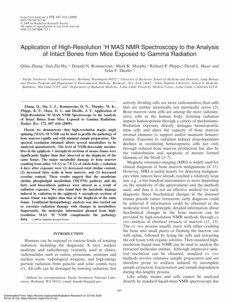
To prepare samples for high-resolution MAS 1H NMR, the leftfemur was thawed slightly, and outside surrounding tissues wereremoved under magnification. The whole femur was weighed andinserted into a Teflon tube. The Teflon tube assembly was then loadedbetween two Teflon plugs in a 7.5-mm-outside-diameter, 6-mm-internal-diameter Chemagnetics pencil rotor as shown in Fig. 1. Thetwo solid Teflon plugs can be inserted into the rotor only byprecooling the plugs in liquid nitrogen. In this way, a tight seal iscreated when the plugs warm to the targeted experimental temper-ature of 2uC. As a result, the marrow spun at the fast sample spinning
FIG. 1. Schematic of the special liquid-tight sample cell for 1HMAS NMR experiments with mouse femur. The femur was insertedinto a Teflon sleeve and closed tightly with two Teflon plugs inside thezirconium sample rotor.
608 ZHANG ET AL.
Downloaded From: https://bioone.org/journals/Radiation-Research on 23 Feb 2020Terms of Use: https://bioone.org/terms-of-use
rate remains inside the sample chamber with no fluid leakage. Nofluid leakage was found in more than 100 experiments on femursusing this setup at a sample spinning rate of 4 kHz. In otherexperiments, femurs with the surrounding tissue removed were alsodissected into two sections. One section was a combination ofepiphysis (mainly the two femoral heads) and the metaphysis (a shortportion of proximal femur beneath the head) while the other sectionwas diaphysis (most of the shaft of the femur). The sample was loadedusing the same procedures as for the whole femur.
All 1H NMR experiments were performed at 2uC on a Varian-Chemagnetics 300 MHz Infinity spectrometer operating at a protonLarmor frequency of 299.97 MHz. A standard Chemagnetics CP/MAS probe with a 7.5-mm pencil-type spinner system was used in allthe measurements. The sample spinning rate of 4.0 kHz wascontrolled automatically by a Chemagnetics MAS speed controllerat an accuracy of 1 Hz. Temperature was controlled by a Chemag-netics temperature controller in combination with an FTS heatingunit. A rotor-synchronized Carr-Purcel-Meibom-Gill pulse sequence,[90u-(t-180u-t)n-acquisition], was used for acquiring data with a totallength of 12.5 ms for the echo segment. The 90u pulse length wasadjusted for each sample individually and varied from 6.8 to 7.6 ms.The water suppression segment used in this study was a DANTE(delays alternating with nutations for tailored excitation) sequence(27) consisting of 6000 small tip angle pulses that were separated by100 ms. A total of 1920 accumulations were acquired with a totalacquisition time of ,1 h for each experiment. The strong lipid (CH2)n
signal at 1.28 ppm in each spectrum was used as the internal chemicalshift reference.
Quantification and Statistical Analysis
The spectrum was integrated with respect to different metabolitepeaks in the range of 0.5–4.5 ppm for quantitative data analysis. Forcomparison within each sample, the integrated peak intensity for eachtype of metabolite was compared to the most abundant lipid (CH2)n
peak located at 1.28 ppm in the same spectrum. For comparisonbetween treatment groups, the integrated peak intensity for each typeof metabolite in the irradiated group was compared with the same onein the age-matched control group. The reliability of this intertreat-ment group comparison was validated by performing 12 independent1H MAS experiment on a 65-mg PBS z 3 mM TSP sample during aperiod of 4 days. For each measurement, the probe was removed fromand reinserted into the magnet and the 90u pulse was calibrated.Defining the mean value of the integrated intensity for the TSP peakas 100, the mean ± SD was 100 ± 3. This small standard deviation(3%) indicated that both the spectrometer, including the MAS probe,and the pulse sequence were remarkably stable. Therefore, the
integrated metabolite peak intensities from the spectra samples thatwere acquired under similar experimental conditions could becompared directly.
The statistical significance of differences between the metabolitepeak intensities of the exposed group and those of the control groupwas determined using the Student’s t test. We used a sample size offour. Furthermore, the t test works under the assumption of aGaussian distribution of data but also works well for distributionsthat are not Gaussian (28).
RESULTS
Histopathology Findings
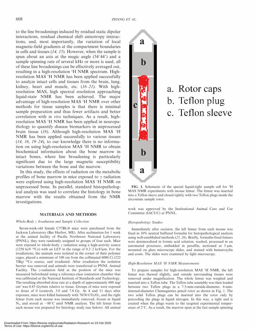
Typical examples of the cell depletion in bone marroware shown in Fig. 2. Depletion is defined as a decrease innucleated cells, which have dark blue nuclei whenstained with hematoxylin. The depleted marrow hasincreased numbers of red blood cells that are notnucleated when mature. Thus the color of the depletedmarrow cells appears less basophilic, lighter and moreeosinophilic (orange) in Fig. 2c. This indicates that thereare fewer basophilic nucleated cells, more white adiposecells, and more eosinophilic non-nucleated erythrocytes.
It is evident from Fig. 2 that there was a milddepletion of hematopoietic cells in the bone marrow ofC57BL/6 mice at 4 days after irradiation with 3 Gy(Fig. 2b). By day 11 the bone marrow had returned to anormal histology (not shown). However, the bonemarrow was severely depleted at 4 days after exposureto 7.8 Gy (Fig. 2c). The bone marrow cavity containshematopoietic cells, bone marrow stromal and adiposecells, and bone-forming cells such as osteoblasts andosteoclasts. In the studies shown in Fig. 2, the effects onhematopoietic cells were apparent, while the effects onother cellular components were not.
The Effect of High-Resolution MAS 1H NMR onWhole Femur
A typical example of the effect of MAS on the 1HNMR spectral resolution of a whole femur is shown in
FIG. 2. Histology of mouse femurs, illustrating bone marrow hematopoietic cell depletion at high radiation dose. Panel a: Nonirradiatedcontrol mouse; panel b: 4 days after whole-body exposure to 3.0 Gy c radiation; panel c: 4 days after whole-body exposure to 7.8 Gy.
1H MAS NMR OF INTACT BONE 609
Downloaded From: https://bioone.org/journals/Radiation-Research on 23 Feb 2020Terms of Use: https://bioone.org/terms-of-use
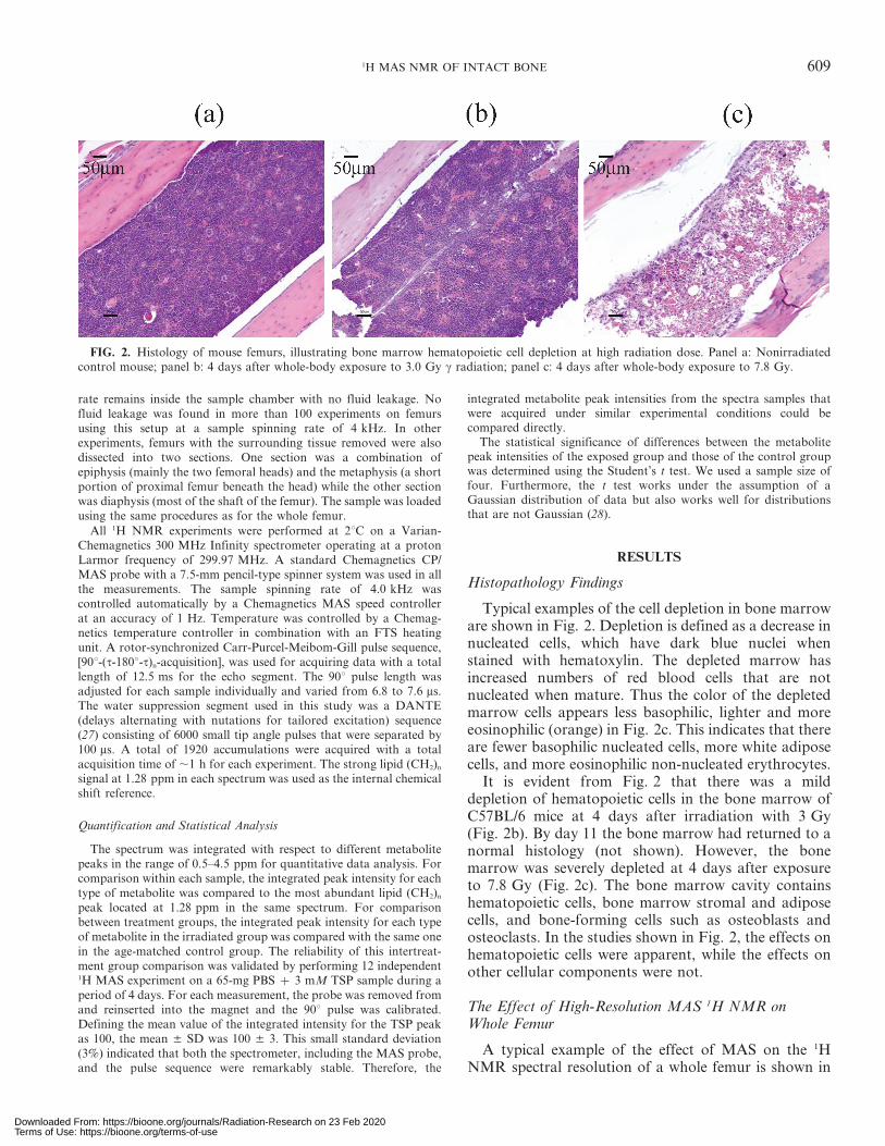
Fig. 3. Both the static (Fig. 3a) and the MAS spectra(Fig. 3b) were obtained on the same femur sample andwere acquired using the same experimental parametersexcept that a sample spinning rate of 4 kHz was used inspectrum b. The data presented in Fig. 3a were acquiredfirst, followed immediately by acquisition of thosepresented in Fig. 3b.
In the static spectrum shown in Fig. 3a, the water peakis so broad that its tail covers a chemical shift range fromabout 0 to 10 ppm. This is due to the strong magneticsusceptibility variations between the marrow tissue andthe bones. As a result, it is impossible to achieve goodwater suppression without suppressing the correspondingmetabolite signals. This prohibits the identification ofbiologically meaningful metabolites. However, by spin-ning the sample at 4 kHz (Fig. 3b), water suppressionusing the same experimental parameters as those inFig. 3a becomes efficient, and the intensity of the residualwater peak at about 4.8 ppm is less than that of the lipidpeak at 1.28 ppm. The MAS also greatly enhances thespectral resolution. As shown in Fig. 3b and its corre-sponding expanded (both horizontally and vertically)insertion spectrum between 1.5 and 4.5 ppm, a number ofdistinct metabolite peaks were obtained with a narrowpeak width. The chemical identities of these majormetabolite peaks were assigned according to the pub-
lished literature (14, 17, 22–24) and were labeled on thespectrum. The effect of spinning at 4 kHz on the samplewas also tested; there was no visible difference in thespectrum when the sample was respun at 4 kHz (data notshown). In addition, there was no observable differencewhen the measurement was repeated a few hours after thefirst measurement (data not shown), showing that there isminimal sample degradation at the experimental temper-ature of 2uC used throughout this work.
Metabolic Difference between Diaphysis and Epiphysis zMetaphysis Sections
The spectrum in Fig. 4a was acquired on the diaphysisof a femur from the control group, and the spectrum inFig. 4b was acquired on the epiphysis z metaphysissections of the same femur. Both spectra were acquiredusing the same experimental parameters except that the90u pulse width was calibrated individually.
The femurs of C57BL/6 mice exposed to 3.0 and7.8 Gy of c radiation were analyzed as outlined above;i.e., the diaphysis and the epiphysis z metaphysis of thefemur were measured separately with high-resolutionMAS 1H NMR. Typical spectra for irradiated samplesare shown in Fig. 4c–f. Quantitative results obtainedfrom statistical analysis of the high-resolution MAS 1HNMR spectra are summarized in Tables 1 and 2.
For the control groups, the relative ratio of the NMRpeaks for the diaphysis and the epiphysis z metaphysissections of the same femur was significantly different(Table 1). For example, there was a distinct difference inthe weight-averaged total peak area between 0.7–4.4 ppm on both day 4 and day 11. High-resolutionMAS 1H NMR is unique for selectively assessing themobile molecules in tissues. Large molecules such asproteins and rigid phospholipids that form the lipidbilayers of healthy cell membranes are essentiallyinvisible to high-resolution MAS 1H NMR if the samplespinning rate used is less than about 6 kHz (29). Theseresults clearly showed that the epiphysis z metaphysiscontains more mobile molecules than the diaphysis. Acareful examination revealed that the increase in thelipid (CH2)n signal at 1.28 ppm is the main cause for theincreased weight-averaged peak area of the epiphysis zmetaphysis relative to the diaphysis. This was furtherconfirmed by the data in Table 1 showing the peak areasfor the lipid (CH2)n signal at 1.28 ppm, integrated over aspectral range between 1.1 and 1.4 ppm for the controlgroup, for the diaphysis and the epiphysis z metaphysisat both day 4 and day 11. These results indicateunambiguously that the epiphysis z metaphysis containmore mobile lipids. It can also be seen in the verticallyexpanded spectral regions between 1.5 and 4.5 ppm inFig. 4 and the data in Table 2 that for the control mice,higher content of lipid moieties (peaks corresponding to0.7–1.0, 1.4–1.8 and 1.8–2.15 ppm, mainly the fatty acyl
FIG. 3. 1H NMR spectra (300 MHz) of mouse femur acquired at2uC. (a), Static spectrum; (b), MAS at 4.0 kHz. The assignments ofthe peaks are as follows: 1, terminal –CH3; 2, –(CH2)n; 3, –O–CO–CH2–CH2–; 4, –CH5CH–CH2–CH2–; 5, –O–CO–CH2–CH2–; 6, –CH5
CH–CH2–CH5CH-; 7, creatine; 8, total cholines, including choline,phosphocholine and phosphatidylcholine; 9, glycogen/glucose; 10 and11, C1, C3-glycerol ester; and 12, –CH5CH–. The peak in (b) labeledwith an asterisk is the residual water signal. The spectrum above toshows the spectral region between 1.5 and 4.5 ppm that is expandedfrom the spectrum in (b) by about 2.63 horizontally and 5.53 verti-cally.
610 ZHANG ET AL.
Downloaded From: https://bioone.org/journals/Radiation-Research on 23 Feb 2020Terms of Use: https://bioone.org/terms-of-use
groups from triglycerides) and total cholines (choline,phosphocholine and phosphatidylcholine at 3.1–3.3 ppm) were observed in the diaphysis compared withthe epiphysis z metaphysis. Similar results wereobtained from the irradiated groups. These resultsindicate that the composition of the lipids in thediaphysis is different from that in the epiphysis zmetaphysis, i.e., with increased –CH3, O–CO–CH2–, and–CH5CH–CH2– functional groups accompanied by anincreased amount of total cholines. The relative increas-es in methyl, carboxyl and unsaturated bonds presum-ably mean that the lipids in the diaphysis contain shortersaturated (CH2)n chains compared to the epiphysis zmetaphysis. Selective assessment of mobile lipids isimportant because it has been suggested that mobiliza-tion of fatty acids plays an important role in thedevelopment of cancer (30), obesity and type II diabetes(31, 32). Note that mobile lipids are observed in high-resolution MAS 1H NMR at a sample spinning ratewhile the rigid membranes are not. Therefore, high-resolution MAS 1H NMR can be considered as aselective observation for mobile lipids. The informationobtained by high-resolution MAS 1H NMR comple-ments the standard lipid analysis of cell extractions (33),
in which all of the fatty acids from the cells including cellmembranes are analyzed. The lack of similar region-specific histological changes suggests that high-resolu-tion MAS 1H NMR may represent a novel and sensitivemeans to assess subtle or early events in bone marrowpathology directly in whole bone specimens.
Impact of Radiation on the Epiphysis z Metaphysis ofthe Femur
After exposure to 3.0 Gy, the integrated peak area forthe most abundant lipid peak centered at 1.28 ppm (1.1–1.4 ppm) showed a nonsignificant increase at both 4 and11 days after irradiation. However, the peak area wasstatistically significantly increased at 4 days after exposureto 7.8 Gy radiation. The increased lipid signal at the highradiation dose indicates that radiation changed the lipidprofile in the epiphysis z metaphysis of the femur. Thisconclusion was further supported by the weight-averagedtotal peak area, which was significantly increased both at 4days after exposure to 7.8 Gy radiation and at 11 daysafter exposure to 3.0 Gy radiation.
At 4 days after exposure, the relative integrated peakarea for total choline (3.1–3.3 ppm), a combination of
FIG. 4. 1H NMR spectra (300 MHz) of C57BL/6 mouse femurs exposed to 3 and 7.8 Gy whole-body cradiation. The spectra were acquired at 2uC with MAS at 4.0 kHz. (a) and (b), Controls; (c) and (d), 4 days afterexposure to 3 Gy radiation; (e) and (f), 4 days after exposure to 7.8 Gy. (a), (c) and (e) were acquired on thediaphysis of femur while (b), (d) and (f) were acquired on the epiphysis z metaphysis. The diaphysis samplesand the epiphysis z metaphysis samples were always from the same three femurs. All the spectral traces areplotted with the lipid signal at 1.28 ppm at the same height to facilitate the comparison. The insertions aboveeach spectrum show the spectral range between 2.5 and 4.5 ppm that is expanded vertically four times.
1H MAS NMR OF INTACT BONE 611
Downloaded From: https://bioone.org/journals/Radiation-Research on 23 Feb 2020Terms of Use: https://bioone.org/terms-of-use
choline, phosphocholine and glycerol phosphocholine inthe epiphysis z metaphysis of the femur decreasedsignificantly (see the expanded insertion spectra shownin Fig. 4b–d). At 11 days after exposure to 3.0 Gyradiation, the level of total choline in the epiphysis zmetaphysis of the femur remained significantly deceasedcompared with that of the corresponding controls.
For the epiphysis z metaphysis, the relative peakarea of creatine (2.9–3.1 ppm) followed a similar trendas that of total choline (Table 2). At 4 days postirradi-ation, the level of creatine decreased significantly. At 11days after exposure to 3 Gy, creatine was decreasedcompared with the corresponding age-matched controls.Creatine is the end product associated with the cellularenergy pathway (17). The decreased level of creatinesuggests that the basic energy pathway was affected bywhole-body exposure to c radiation.
For the epiphysis z metaphysis, the relative ratios forall other peaks remained approximately the same at 4
days after exposure to 3 Gy (P . 0.05). However, at 11days after irradiation, statistically significant decreaseswere observed for many of the peaks (Table 2), indicatingthat the effect of radiation increased at longer times.
Effect of Radiation on the Diaphysis of the Femur
As in the epiphysis z metaphysis, 4 days after expo-sure to 7.8 Gy radiation, there were statistically signif-icant decreases in the relative areas of all the peaks(Table 2) for the diaphysis compared with the corre-sponding controls. However, in contrast to the resultsobtained for the epiphysis z metaphysis of the femur,there were no significant differences in total choline andcreatine 4 days after exposure to 3 Gy radiation.Furthermore, at 11 days postirradiation, except for thepeaks corresponding to the terminal methyl groups (0.7–1.0 ppm) and the –CH5CH–CH2–CH2–(1.8–2.15 ppm)groups, there were no significant differences for any of
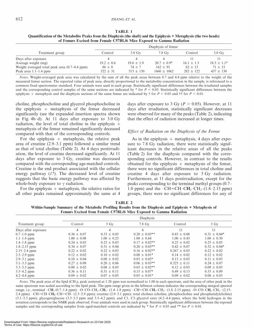
TABLE 1Quantification of the Metabolite Peaks from the Diaphysis (the shaft) and the Epiphysis + Metaphysis (the two heads)
of Femurs Excised from Female C57BL/6 Mice Exposed to Gamma Radiation
Treatment group
Diaphysis of femur
Control 3.0 Gy 7.8 Gy Control 3.0 Gy
Days after exposure 4 4 4 11 11Average weight (mg) 19.2 ± 0.6 19.6 ± 1.9 20.7 ± 0.9* 16.1 ± 1.3 18.5 ± 1.1*Weight averaged total peak area (0.7–4.4 ppm) 86 ± 8 74 ± 7 162 ± 91 62 ± 13 71 ± 15Peak area 1.1–1.4 ppm 522 ± 31 513 ± 130 1660 ± 1062 282 ± 122 437 ± 130
Notes. Weight-averaged peak area was calculated by the sum of all the peak areas between 0.7 and 4.4 ppm relative to the weight of themeasured femur section. The reported value of peak area, directly proportional to the metabolite concentration in the sample, is referenced to acommon fixed spectrometer standard. Four animals were used in each group. Statistically significant differences between the irradiated samplesand the corresponding control samples of the same sections are indicated by * for P , 0.05. Statistically significant differences between theepiphysis z metaphysis and the diaphysis sections of the same femur are indicated by { for P , 0.05 and {{ for P , 0.01.
TABLE 2Within-Sample Summary of the Metabolic Profiling Results from the Diaphysis and Epiphysis + Metaphysis of
Femurs Excised from Female C57BL/6 Mice Exposed to Gamma Radiation
Treatment group
Diaphysis
Control 3 Gy 7.8 Gy Control 3 Gy
Days after exposure 4 4 4 11 110.7–1.0 ppm 0.36 ± 0.07 0.33 ± 0.05 0.20 ± 0.03** 0.43 ± 0.08 0.31 ± 0.04*1.1–1.4 ppm 1.00 ± 0.08 1.00 ± 0.25 1.00 ± 0.64 1.00 ± 0.43 1.00 ± 0.301.4–1.8 ppm 0.24 ± 0.03 0.23 ± 0.03 0.17 ± 0.02** 0.25 ± 0.02 0.25 ± 0.031.8–2.15 ppm 0.34 ± 0.07 0.31 ± 0.04 0.20 ± 0.03** 0.42 ± 0.07 0.32 ± 0.04*2.15–2.4 ppm 0.22 ± 0.02 0.22 ± 0.03 0.16 ± 0.02** 0.267 ± 0.03 0.22 ± 0.022.5–2.9 ppm 0.12 ± 0.02 0.10 ± 0.02 0.08 ± 0.01* 0.14 ± 0.02 0.12 ± 0.022.9–3.1 ppm 0.10 ± 0.04 0.08 ± 0.02 0.03 ± 0.02* 0.13 ± 0.03 0.11 ± 0.033.1–3.3 ppm 0.27 ± 0.09 0.20 ± 0.06 0.06 ± 0.03** 0.325 ± 0.11 0.24 ± 0.073.3–3.5 ppm 0.08 ± 0.02 0.08 ± 0.05 0.03 ± 0.02** 0.12 ± 0.03 0.09 ± 0.033.5–4.2 ppm 0.36 ± 0.11 0.31 ± 0.11 0.15 ± 0.07* 0.49 ± 0.13 0.33 ± 0.094.2–4.4 ppm 0.09 ± 0.02 0.07 ± 0.05 0.05 ± 0.01* 0.09 ± 0.02 0.08 ± 0.03
Notes. The peak area of the lipid (CH2)n peak centered at 1.28 ppm was normalized to 1.00 in each spectrum, and the area of other peak in thesame spectrum was scaled according to the lipid peak. The ppm range given in the leftmost column indicates the corresponding integral spectralrange, i.e., terminal –CH3 (0.7–1.4 ppm); –O–CO–CH2–CH2– (1.4–1.8 ppm); –CH5CH–CH2–CH2– (1.8–2.15 ppm); –O–CO–CH2–CH2– (2.15–2.4 ppm); –CH5CH–CH2–CH5CH– (2.5–2.9 ppm); creatine (2.9–3.1 ppm); total cholines (choline, phosphocholine and phosphatidylcholine)(3.1–3.3 ppm); glycogen/glucose (3.3–3.5 ppm and 3.5–4.2 ppm); and C1, C3–glycerol ester (4.2–4.4 ppm), where the bold hydrogen in thenotation corresponds to the NMR peak observed. Four animals were used in each group. Statistically significant differences between the exposedsamples and the corresponding samples from aged-matched controls are indicated by * for P # 0.05 and ** for P # 0.01.
612 ZHANG ET AL.
Downloaded From: https://bioone.org/journals/Radiation-Research on 23 Feb 2020Terms of Use: https://bioone.org/terms-of-use
the other peaks. This indicated that the epiphysis zmetaphysis sections were more sensitive to radiationthan the diaphysis of the same femurs. This conclusionwas also supported by the results shown in Table 1 forthe diaphysis; although both the weight-averaged totalpeak area and the peak area of long chain lipids (CH2)n
(1.1–1.4 ppm) were increased in the irradiated animalscompared with their corresponding controls, the in-crease was not statistically significant. In contrast, asignificant increase was observed for the correspondingepiphysis z metaphysis sections of the same femur.
DISCUSSION
Our results show that the levels of NMR-detectablemetabolites in the epiphysis z metaphysis sections ofmouse femurs were significantly higher than those inthe diaphyses of the same femurs in the control and
irradiated mice. Radiation caused greater damage to themetabolic profile in the epiphysis z metaphysis sectionsthan to the diaphyses of the same femurs. This finding isnot surprising because the femoral proximal metaphysescontain more hematopoietic cells than the epiphyses inadult animals (9). It is well known that the hematopoi-etic stem cells are both metabolically active andradiosensitive (2–7).
In the present study, we showed for the first time thationizing radiation alters the lipid metabolism in bonemarrow. The significantly increased level of NMR-detectable mobile fatty acids characterized by the longchain lipid (CH2)n signal in the bone marrow ofirradiated mice indicated that the fatty acids synthesispathway is altered by radiation. Altered fatty acidbiosynthesis, i.e., increased flux from glucose into fattyacids [the Warburg effect (34, 35)], has emerged as afeature of oncogenesis (36, 37). Increased mobile lipids
TABLE 1Extended
Epiphysis z metaphysis of femur
Control 3.0 Gy 7.8 Gy Control 3.0 Gy
4 4 4 11 1123.1 ± 4.6 23 ± 6.9 21.4 ± 4.6 27.5 ± 1.8 22.0 ± 3.5*221 ± 64{{ 299 ± 95 4808 ± 188* 186 ± 30{{ 256 ± 41*
2522.3 ± 1308.3{ 3723 ± 2043 5160 ± 768* 2280 ± 513{{ 2807 ± 663
TABLE 2Extended
Epiphysis z metaphysis
Control 3 Gy 7.8 Gy Control 3 Gy
4 4 4 11 110.20 ± 0.01 0.19 ± 0.006 0.18 ± 0.007* 0.22 ± 0.02 0.19 ± 0.004*1.00 ± 0.35 1.00 ± 0.36 1.00 ± 0.40 1.00 ± 0.19 1.00 ± 0.180.16 ± 0.005 0.14 ± 0.013 0.14 ± 0.02 0.17 ± 0.01 0.15 ± 0.008**0.19 ± 0.018 0.18 ± 0.004 0.17 ± 0.006* 0.22 ± 0.01 0.18 ± 0.004**0.16 ± 0.008 0.15 ± 0.02 0.14 ± 0.004** 0.16 ± 0.01 0.15 ± 0.006**
0.067 ± 0.004 0.063 ± 0.007 0.063 ± 0.005 0.072 ± 0.006 0.067 ± 0.0020.033 ± 0.0078 0.023 ± 0.004* 0.017 ± 0.004** 0.042 ± 0.008 0.027 ± 0.003**0.097 ± 0.028 0.053 ± 0.01* 0.030 ± 0.011** 0.11 ± 0.019 0.065 ± 0.006**0.034 ± 0.008 0.03 ± 0.016 0.016 ± 0.005** 0.045 ± 0.009 0.032 ± 0.005*0.14 ± 0.04 0.11 ± 0.03 0.078 ± 0.020* 0.17 ± 0.03 0.11 ± 0.017*
0.048 ± 0.008 0.039 ± 0.008 0.037 ± 0.0046* 0.053 ± 0.0088 0.041 ± 0.0068
1H MAS NMR OF INTACT BONE 613
Downloaded From: https://bioone.org/journals/Radiation-Research on 23 Feb 2020Terms of Use: https://bioone.org/terms-of-use
are often observed in proliferating cells and tumor cellsand in the onset of apoptosis (30, 38–42). It is likely thatthe altered fatty acid synthesis after irradiation is one ofthe key biological pathways that predispose to cancer.However, more research is needed to validate thishypothesis.
The present study showed that the content of totalcholine (choline, PC and GPC) is decreased at theradiation doses studied. Phosphocholine is an importantintermediate product in membrane choline phospholip-ids metabolism, where phosphatidylcholine and glycer-ophosphocholine are synthesized and hydrolyzed (43).Phosphatidylcholine is the most abundant phospholipidin biological membranes and together with otherphospholipids, such as phosphatidylethanolamine andneutral lipids, forms the characteristic bilayer structureof cells and regulate membrane integrity (44). Priorstudies have shown that altered phosphocholine and/ortotal choline levels are associated with malignancy (43,45, 46). The significant decrease in total choline levelswith increasing radiation dose indicates an alteredmembrane choline phospholipid metabolism. This isdirect evidence that c radiation causes measurabledamage to the cell membrane.
SUMMARY
We demonstrated here that high-resolution MAS 1HNMR can be used successfully to analyze the metabo-lites in intact bones from mice without the need for bonemarrow extraction. The method is useful in profiling thepathology of bone marrow rapidly with minimal samplepreparation. The spectral resolution allows severalmetabolites, including lipids, total choline (choline,phosphocholine, glycerophosphocholine), creatine, gly-cogen/glucose and glycerol esters, to be analyzedquantitatively. We found that the amounts of NMR-detectable metabolites in the epiphysis z metaphysissections of mouse femur were significantly higher thanthose observed in the diaphysis of the same femur. Wediscovered that the major metabolite changes in thebone marrow after exposure to either 3 or 7.8 Gy of cradiation, especially 4 days after exposure, were (1)decreased total choline content, (2) increased fatty acidsin bone marrow, and (3) decreased creatine content.These results suggest that the fatty acid biosynthesispathway and the membrane choline phospholipidmetabolism (MCPM) pathway were altered in bonemarrow after irradiation. This result is similar to thosepublished recently on cell cycle arrest and death incarcinoma cells exposed to 20 Gy of c radiation (38).The metabolites associated with these two basic molec-ular pathways, which were not known from prior studies(47, 48), are possible biomarkers characterizing radia-tion damage in bone marrow. These biomarkers, ifvalidated, may serve as novel targets for medical
countermeasures against ionizing radiation. Recently, asystem-level metabolic flux profiling in cultured mam-malian cells using liquid chromatography-tandem massspectrometry has been used successfully to identify fattyacid synthesis as a target for antiviral therapy (49). Viralinfection can contribute to the death after exposure to alethal dose of ionizing radiation. We also found that themetabolic damage induced by radiation in the epiphysisz metaphysis sections of mouse femurs was higher thanthat of the diaphyses of the same femurs.
Traditional histopathology analysis was carried out inparallel to correlate histological damage with themetabolite changes. Histopathology revealed a milddepletion of hematopoietic cells in the bone marrow ofthe femurs at 4 days after exposure to 3 Gy radiation.By day 11 the bone marrow had returned to a normalhistology. However, the bone marrow was severelydepleted at 4 days after exposure to 7.8 Gy. Themolecular information from high-resolution MAS 1HNMR thus complements and expands the pathologydata in a novel way. In particular, the significantchanges in many metabolites found from high-resolutionMAS 1H NMR in the epiphysis z metaphysis sections11 days after exposure to 3 Gy whole-body radiationsuggest that metabolomics may be a more sensitivemethod for assessing radiation damage at this radiationdose. Therefore, it is reasonable to conclude that high-resolution MAS 1H NMR is a powerful method forprobing the pathology of radiation-induced damage atthe molecular level. Essentially any kind of sample froman animal can be analyzed by this method because theanalysis of intact bones represents the greatest challenge.
ACKNOWLEDGMENTS
This work was supported by the NASA Space Radiation Programunder Grant NNX07AU44G and Pacific Northwest NationalLaboratory Directed Research and Development (LDRD) fund. Allthe NMR work was performed in the Environmental MolecularSciences Laboratory (a National Scientific User Facility sponsored bythe Department of Energy’s Office of Biological and EnvironmentalResearch) located at PNNL, and operated for DOE by Battelle underContract DE-AC05-76RL01830. RPP was supported in part byNIEHS grant ES01247 and by DE011390.
Received: January 14, 2009; accepted: July 8, 2009
REFERENCES
1. J. B. Tyburski, A. D. Patterson, K. W. Krausz, J. Slavik,A. J. Fornace, Jr., F. J. Gonzalez and J. R. Idle, Radiationmetabolomics. 1. Identification of minimally invasive urinebiomarkers for gamma-radiation exposure in mice. Radiat. Res.170, 1–14 (2008).
2. D. G. C. McCann, Radiation poisoning: Current concepts in theacute radiation syndrome. Am. J. Clin. Med. 3, 13–21(2006).
3. N. Dainiak, Hematologic consequences of exposure to ionizingradiation. Exp. Hematol. 30, 513–528 (2002).
4. T. M. Fliedner, D. Graessle, C. Paulsen and K. Reimers,Structure and function of bone marrow hemopoiesis: Mecha-
614 ZHANG ET AL.
Downloaded From: https://bioone.org/journals/Radiation-Research on 23 Feb 2020Terms of Use: https://bioone.org/terms-of-use
nisms of response to ionizing radiation exposure. Cancer Biother.Radiopharm. 17, 405–426 (2002).
5. R. Coquard, Late effects of ionizing radiations on the bonemarrow. Cancer Radiother. 1, 792–800 (1997).
6. A. Banfi, G. Bianchi, M. Galotto, R. Cancedda and R. Quarto,Bone marrow stromal damage after chemo/radiotherapy:occurrence, consequences and possibilities of treatment. Leuk.Lymphoma 42, 863–870 (2001).
7. T. M. Fliedner, D. Graessle, C. Paulsen and K. Reimers,Structure and function of bone marrow hemopoiesis:mechanisms of response to ionizing radiation exposure. CancerBiother. Radiopharm. 17, 405–426 (2002).
8. S. Hwang and D. M. Panicek, Magnetic resonance imaging ofbone marrow in oncology, Part 2. Skeletal Radiol. 36, 1017–1027(2007).
9. S. Hwang and D. M. Panicek, Magnetic resonance imaging ofbone marrow in oncology, Part 1. Skeletal Radiol. 36, 913–920(2007).
10. G. P. Schmidt, S. O. Schoenberg, M. F. Reiser and A. Baur-Melnyk,Whole-body MR imaging of bone marrow. Eur. J. Radiol. 55, 33–40 (2005).
11. M. A. Tall, A. K. Thompson, T. Vertinsky and P. S. Palka, MRimaging of the spinal bone marrow. Magn. Reson. Imaging Clin.N. Am. 15, 175–198 (2007).
12. D. K. Yeung, S. L. Lam, J. F. Griffith, A. B. Chan, Z. Chen,P. H. Tsang and P. C. Leung, Analysis of bone marrow fatty acidcomposition using high-resolution proton NMR spectroscopy.Chem. Phys. Lipids 151,103–109 (2008).
13. S. Muthusami, I. Ramachandran, B. Muthusamy, G. Vasudevan,V. Prabhu, V. Subramaniam, A. Jagadeesan and S. Narasimhan,Ovariectomy induces oxidative stress and impairs boneantioxidant system in adult rats. Clin. Chim. Acta 360, 81–86(2005).
14. P. Weybright, K. Millis, N. Campbell, D. G. Cory and S. Singer,Gradient, high-resolution, magic angle spinning 1H nuclearmagnetic resonance spectroscopy of intact cells. Magn. Reson.Med. 39, 337–345 (1998).
15. R. A. Wind, J. Z. Hu and D. N. Rommereim, High-resolution 1HNMR spectroscopy in organs and tissues using slow magic anglespinning. Magn. Reson. Med. 46, 213–218 (2001).
16. L. L. Cheng, M. J. Ma, L. Becerra, T. Ptak, I. Tracey, A. Lacknerand R. G. Gonzalez, Quantitative neuropathology by highresolution magic angle spinning proton magnetic resonancespectroscopy. Proc. Natl. Acad. Sci. USA 94, 6408–6413 (1997).
17. J. Z. Hu, D. N. Rommereim, K. R. Minard, A. Woodstock,B. J. Harrer, R. A. Wind, R. P. Phillips and P. J. Sime,Metabolomics in lung inflammation: A high-resolution 1H NMRstudy of mice exposed to silica dust. Toxicol. Mech. Methods 18,385–398 (2008).
18. M. E. Bollard, A. J. Murray, K. Clarke, J. K. Nicholson andJ. L. Griffin, A study of metabolic compartmentation in the ratheart and cardiac mitochondria using high-resolution magicangle spinning 1H NMR spectroscopy. FEBS Lett. 553, 73–78(2003).
19. A. R. Tate, P. J. Foxall, E. Holmes, D. Moka, M. Spraul,J. K. Nicholson and J. C. Lindon, Distinction between normaland renal cell carcinoma kidney cortical biopsy samples usingpattern recognition of 1H magic angle spinning (MAS) NMRspectra. NMR Biomed. 13, 64–71 (2000).
20. V. Righi, A. Mucci, L. Schenetti, M. R. Tosi, W. F. Grigioni,B. Corti, A. Bertaccini, A. Franceschelli, F. Sanguedolce and V.Tugnoli, Ex vivo HR-MAS magnetic resonance spectroscopy ofnormal and malignant human renal tissues. Anticancer Res. 27,3195–3204 (2007).
21. J. H. Chen, Y. V. Wu, P. Decarolis, R. O’Connor, C. J. Sombergand S. Singer, Resolution of creatine and phosphocreatine 1Hsignals in isolated human skeletal muscle using HR-MAS 1HNMR. Magn. Reson. Med. 59, 1221–1224 (2008).
22. M. E. Bollard, S. Garrod, E. Holmes, J. C. Lindon, E. Humpfer,M. Spraul and J. K. Nicholson, High-resolution 1H and 1H-13Cmagic angle spinning NMR spectroscopy of rat liver. Magn.Reson. Med. 44, 201–207 (2000).
23. S. Garrod, E. Humpfer, M. Spraul, S. C. Connor, S. Polley,J. Connelly, J. C. Lindon, J. K. Nicholson and E. Holmes, High-resolution magic angle spinning 1H NMR spectroscopic studieson intact rat renal cortex and medulla. Magn. Reson. Med. 41,1108–1118 (1999).
24. S. Garrod, E. Humpher, S. C. Connor, J. C. Connelly, M. Spraul,J. K. Nicholson and E. Holmes, High-resolution 1H NMR andmagic angle spinning NMR spectroscopic investigation of thebiochemical effects of 2-bromoethanamine in intact renal andhepatic tissue. Magn. Reson. Med. 45, 781–790 (2001).
25. Y. F. Zhou, M. Bosch-Marce, H. Okuyama, B. Krishnamachary,H. Kimura, L. Zhang, D. L. Huso and G. L. Semenza,Spontaneous transformation of cultured mouse bone marrow-derived stromal cells. Cancer Res. 66, 10849–10854 (2006).
26. N. C. Peterson, M. D. Servinsky, A. Christian, Z. Peng, W. Qiu,J. Mann, J. Dicello and D. L. Huso, Tamoxifen resistance andHer2/neu expression in an aged, irradiated rat breast carcinomamodel. Carcinogenesis 26, 1542–1552 (2005).
27. G. Morris and R. Freeman, Selective excitation in Fouriertransform nuclear magnetic resonance. J. Magn. Reson. 29, 433–462 (1978).
28. R. J. Barlow, Statistics: A Guide to the Use of Statistical Methodsin the Physical Sciences, pp. 159–160. Wiley, Chichester, 1989.
29. J. L. Griffin, C. J. Mann, J. Scott, C. C. Shoulders andJ. K. Nicholson, Choline containing metabolites during celltransfection: an insight into magnetic resonance spectroscopydetectable changes. FEBS Lett. 509, 263–266 (2001).
30. A. Rosi, A. Luciani, P. Matarrese, G. Arancia, V. Viti andL. Guidoni, 1H-MRS lipid signal modulation and morphologicaland ultrastructural changes related to tumor cell proliferation.Magn. Reson. Med. 42, 248–257 (1999).
31. H. Bays, L. Mandarino and R. A. DeFronzo, Role of theadipocyte, free fatty acids, and ectopic fat in pathogenesis of type2 diabetes mellitus: peroxisomal proliferator-activated receptoragonists provide a rational therapeutic approach. J. Clin.Endocrinol. Metab. 89, 463–478 (2004).
32. H. J. Atherton, N. J. Bailey, W. Zhang, J. Taylor, H. Major,J. Shockcor, K. Clarke and J. L. Griffin, A combined 1H-NMRspectroscopy- and mass spectrometry-based metabolomic studyof the PPAR-alpha null mutant mouse defines profound systemicchanges in metabolism linked to the metabolic syndrome.Physiol. Genomics 27, 178–186 (2006).
33. M. Borkman, L. H. Storlien, D. A. Pan, A. B. Jenkins,D. J. Chisholm and L. V. Campbell, The relation betweeninsulin sensitivity and the fatty-acid composition of skeletal-muscle phospholipids. N. Engl. J. Med. 328, 238–244 (1993).
34. O. Warburg, The Metabolism of Tumors. Constable, London,1930.
35. O. Warburg, On the origin of cancer cells. Science 123, 309–314(1956).
36. D. E. Bauer, G. Hatzivassiliou, F. Zhao, C. Andreadis andC. B. Thompson, ATP citrate lyase is an important component ofcell growth and transformation. Oncogene 24, 6314–6322 (2005).
37. C. D. Young and S. M. Anderson, Sugar and fat—that’s whereit’s at: metabolic changes in tumors. Breast Cancer Res. 10, 202(2008).
38. A. M. Luciani, S. Grande, A. Palma, A. Rosi, C. Giovannini,O. Sapora, V. Viti and L. Guidoni, Characterization of H-1NMR detectable mobile lipids in cells from human adeno-carcinomas. FEBS J. 276, 1333–1346 (2009).
39. J. M. Hakumaki and R. A. Kauppinen, 1H NMR visible lipids inthe life and death of cells. Trends Biochem. Sci. 25, 357–362(2000).
40. E. J. Delikatny, C. M. Lander, T. M. Jeitner, R. Hancock andC. E. Mountford, Modulation of MR-visible mobile lipid levels
1H MAS NMR OF INTACT BONE 615
Downloaded From: https://bioone.org/journals/Radiation-Research on 23 Feb 2020Terms of Use: https://bioone.org/terms-of-use
by cell culture conditions and correlations with chemotacticresponse. Int. J. Cancer 65, 238–245 (1996).
41. J. M. Hakumaki and T. Liimatainen, Molecular imaging ofapoptosis in cancer. Eur. J. Radiol. 56, 143–153 (2005).
42. M. Milkevitch, H. Shim, U. Pilatus, S. Pickup, J. P. Wehrle,D. Samid, H. Poptani, J. D. Glickson and E. J. Delikatny,Increases in NMR-visible lipid and glycerophosphocholineduring phenylbutyrate-induced apoptosis in human prostatecancer cells. Biochim. Biophys. Acta 1734, 1–12 (2005).
43. E. O. Aboagye and Z. M. Bhujwalla, Malignant transformationalters membrane choline phospholipid metabolism of humanmammary epithelial cells. Cancer Res. 59, 80–84 (1999).
44. C. E. Mountford and L. C. Wright, Organization of lipids in theplasma membranes of malignant and stimulated cells: a newmodel. Trends Biochem. Sci. 13, 172–177 (1988).
45. J. Ruiz-Cabello and J. S. Cohen, Phospholipid metabolites asindicators of cancer cell function. NMR Biomed. 5, 226–233(1992).
46. W. B. Mackinnon, P. A. Barry, P. L. Malycha, D. J. Gillett,P. Russell, C. L. Lean, S. T. Doran, B. H. Barraclough, M.Bilous and C. E. Mountford, Fine-needle biopsy specimens ofbenign breast lesions distinguished from invasive cancer ex vivowith proton MR spectroscopy. Radiology 204, 661–666 (1997).
47. S. Grande, C. Giovannini, L. Guidoni, A. M. Luciani, A. Palma,A. Rosi, O. Sapora and V. Viti, 1H MRS signals from glutathionemay act as predictive markers of apoptosis in irradiated tumourcells. Radiat. Prot. Dosimetry 122, 205–206 (2006).
48. A. D. Patterson, H. Li, G. S. Eichler, K. W. Krausz,J. N. Weinstein, A. J. Fornace, Jr., F. J. Gonzalez and J. R.Idle, UPLC-ESI-TOFMS-based metabolomics and geneexpression dynamics inspector self-organizing metabolomicmaps as tools for understanding the cellular response toionizing radiation. Anal. Chem. 80, 665–674 (2008).
49. J. Munger, B. D. Bennett, A. Parikh, X. Feng, J. McArdle,H. A. Rabitz, T. Shenk and J. D. Rabinowitz, Systems-levelmetabolic flux profiling identifies fatty acid synthesis as a targetfor antiviral therapy. Nat. Biotechnol. 26, 1179–1186 (2008).
616 ZHANG ET AL.
Downloaded From: https://bioone.org/journals/Radiation-Research on 23 Feb 2020Terms of Use: https://bioone.org/terms-of-use
![[ Z b j f i ^ ` h ` g f e d c b ] k Q U R R l J J I U Q K · Y X b a ` _ ^ \ ] X \ [ Z b j f _ i ^ ` h ` g f e d c b ] k Q U R R l J J I U Q K o A q p s o t B s o @ r q p o n m B](https://img.pdfslide.us/doc/110x75/5e61126d68826e7c28591157/-z-b-j-f-i-h-g-f-e-d-c-b-k-q-u-r-r-l-j-j-i-u-q-k-y-x-b-a-x-.jpg)
![k k l m n o o p Y q r V [ s t q u...M R d Q O z LN i M P K J L P R K M a b M N R Q N i Q i M z _ z b z b J N i f M Q K i N M K M Q M K Q R M ] _ b j z I x y y y ¡ ¢](https://img.pdfslide.us/doc/110x75/5e6044c58224225c9e19f807/k-k-l-m-n-o-o-p-y-q-r-v-s-t-q-u-m-r-d-q-o-z-ln-i-m-p-k-j-l-p-r-k-m-a-b-m-n.jpg)


![M on da thru Fri da T hu rsda Fri da...d uud q j h p h q wv wr k r q r uwk h oly lqj r up h p r uld ol]h w k h g h d g $ q dfnq rz ohg j p hq wr iw k rvh e hlq j k r q r uhg r up h](https://img.pdfslide.us/doc/110x75/5e68438ed275123dd746e12c/m-on-da-thru-fri-da-t-hu-rsda-fri-da-d-uud-q-j-h-p-h-q-wv-wr-k-r-q-r-uwk-h-oly.jpg)


![C ! N P Q R R S U V X Y Z U R R Q X Z [ X X \] ^ U _ ] ` R c d d h d d d k j [ X S Z X l ] m \ o R h X q V m X X N R r Q Q R N Z [ \ X h Z X Q R X R Q Y N P] ^ R Q X m Z ` R k U X](https://img.pdfslide.us/doc/110x75/5ab3ebc17f8b9a86428b4d94/c-n-p-q-r-r-s-u-v-x-y-z-u-r-r-q-x-z-x-x-u-r-c-d-d-h-d-d-d-k-j-x.jpg)

![On q-Boson Operators and q-Analogues of the...Similarly, Corcino [11] defined the (r,β)-Stirling numbers n k r,β as coefficients in tn = Xn k=0 t−r β k βkk! ˝ n k ˛ r,β](https://img.pdfslide.us/doc/110x75/601386921126f94e192deb91/on-q-boson-operators-and-q-analogues-of-the-similarly-corcino-11-deined.jpg)
![M on da thru Fri da T hu rsda Fri da · d uud q j h p h q wv wr k r q r uwk h oly lqj r up h p r uld ol]h w k h g h d g $ q dfnq rz ohg j p hq wr iw k rvh e hlq j k r q r uhg r up](https://img.pdfslide.us/doc/110x75/5e6839ada7372a6e297cb1c6/m-on-da-thru-fri-da-t-hu-rsda-fri-da-d-uud-q-j-h-p-h-q-wv-wr-k-r-q-r-uwk-h-oly-lqj.jpg)


![m]k]q - iopb.res.insomen/RNatok/ORX/mukut_orx.pdf · m]k]q rbX¥ nAT QAk]r 3 nA](https://img.pdfslide.us/doc/110x75/5c801be609d3f2a2228c5e04/mkq-iopbresin-somenrnatokorxmukutorxpdf-mkq-rbx-nat-qakr-3-na.jpg)