Embed Size (px)
Citation preview
UvA-DARE is a service provided by the library of the University of Amsterdam (http://dare.uva.nl)
UvA-DARE (Digital Academic Repository)
Thrombopotein: its ups and downsFolman, C.C.
Link to publication
Citation for published version (APA):Folman, C. C. (2001). Thrombopotein: its ups and downs
General rightsIt is not permitted to download or to forward/distribute the text or part of it without the consent of the author(s) and/or copyright holder(s),other than for strictly personal, individual use, unless the work is under an open content license (like Creative Commons).
Disclaimer/Complaints regulationsIf you believe that digital publication of certain material infringes any of your rights or (privacy) interests, please let the Library know, statingyour reasons. In case of a legitimate complaint, the Library will make the material inaccessible and/or remove it from the website. Please Askthe Library: http://uba.uva.nl/en/contact, or a letter to: Library of the University of Amsterdam, Secretariat, Singel 425, 1012 WP Amsterdam,The Netherlands. You will be contacted as soon as possible.
Download date: 25 Feb 2019

Chapter 5
The Diagnostic Value of Thrombopoietin Measurements in Thrombocytopenia
Thrombosis and Haemostasis 1998; 79:1101-5

Diagnostic value of Tpo
The Diagnostic Value of Tpo Measurements in Thrombocytopenia
Leendert Porcelijn', Claudia C Folman', Masja de Haas', Elly Huiskes', Bernadette Bossers1
Marijke AM Overbeeke', C Ellen v.d. Schoot' and Albert EGKr von dem Borne.''2
1 Central Laboratory of the Netherlands Red Cross Blood Transfusion Service, Amsterdam 2 Department of Hematology, Academic Medical Centre, Amsterdam
ABSTRACT
It has been reported that measurement of blood thrombopoietin (Tpo) levels can be used to discriminate between thrombocytopenia due to increased platelet destruction and decreased platelet production. With our Tpo ELISA and a glycocalicin ELISA we analysed a large group of patients in detail and now confirm and amplify the above notion in detail. Tpo levels were determined in plasma from 178 clinically and serologically well-defined thrombocytopenic patients: 72 patients with idiopathic autoimmune thrombocytopenia (AITP), 29 patients with secondary AITP, 5 patients with amegakaryocytic thrombocytopenia and 72 patients who suffered from various diseases (46 in whom megakaryocyte deficiency was not and 26 in whom it was expected). In addition, we measured the level of glycocalicin as a marker of total body mass of platelets.
In all patients with primary AITP and secondary AITP, Tpo levels were within the normal range or in some (n=7) cases only slightly increased. The level of glycocalicin was not significantly different from that of the controls (n=95). The patients with amegakaryocytic thrombocytopenia had strongly elevated Tpo levels and significantly decreased glycocalicin levels. Similarly, among the 72 thrombocytopenic patients with various disorders, elevated Tpo levels were only found in patients in whom platelet production was depressed. The mean level of glycocalicin in these patients was decreased compared to that in controls and patients with AITP, but was not as low as in patients with amegakaryocytic thrombocytopenia. In conclusion, all patients with depressed platelet production had elevated levels of circulating Tpo, whereas the Tpo levels in patients with an immune-mediated thrombocytopenia were mostly within the normal range. Therefore, measurement of plasma Tpo levels provides valuable diagnostic information for the analysis of thrombocytopenia in general. Moreover, treatment with Tpo may be an option in AITP.
79
Chapter 5
INTRODUCTION
Recent studies indicate that the measurement of the serum level of thrombopoietin (Tpo) might be useful to discriminate between patients with thrombocytopenia due to increased platelet destruction and those with a deficient platelet production [1-3]. Tpo levels have been found not or only mildly increased in patients with autoimmune thrombocytopenia, drug-induced immune thrombocytopenia, post-transfusion purpura and X-linked hereditary thrombocytopenia (a variant of Wiskott-Aldrich syndrome) [1-3]. In patients with bone-marrow hypoplasia Tpo levels are significantly increased [1-3]. However, it should be emphasised that in all these studies only sera were analysed. We found serum levels in normal individuals to be 3.4 times higher than in plasma, due to the release of Tpo from platelets during coagulation [4]. Moreover, in the three published studies the number of analysed patients was small and the patient groups were not always clinically and serologically well defined. To evaluate the value of plasma Tpo levels in the differential diagnosis of thrombocytopenia, we analysed a large group of 178 patients in detail. Clinical data were obtained, Tpo serum and plasma levels determined and serological tests were performed. Furthermore, as a measure of total platelet mass, glycocalicin (GC) levels were determined [5,6]. Our results show that only in patients with a suppressed platelet production, plasma Tpo levels are significantly increased. Therefore, the measurement of Tpo levels is an important diagnostic tool for the evaluation of thrombocytopenia. Moreover, it could help in selecting those patients who might benefit from Tpo therapy.
MATERIALS AND METHODS
Patient samples EDTA-anticoagulated blood and serum samples from patients suspected of having autoimmune thrombocytopenia were sent to our laboratory for diagnostic evaluation. Informed consent and clinical data were obtained via the referring physicians by a questionnaire and/or an interview on the telephone. In this way, clinical data were collected from 217 of 377 analysed patients with various forms of thrombocytopenia. Of these 217 patients, 23 pregnant women (mostly with mild thrombocytopenia) were excluded from our study. Gestational thrombocytopenia, thrombocytopenia associated with pregnancy-induced hypertension and the HELLP syndrome account for most of these cases, and differentiation from AITP in pregnancy is difficult [7,8]. Only patients with a platelet count of less than 100 x 109/L were included. All together 178 patients were analysed. The male/female ratio was 0.76; the mean age was 55 ± 21 years (age range from 4 to 91 years). Based on the clinical data, four groups of thrombocytopenic patients were distinguished. Patients with autoimmune thrombocytopenic purpura (AITP) (n=72) were defined, in accordance with the recommendation of the American Society of Hematology (ASH) [9], by their medical history, physical examination, complete blood count and examination of the peripheral blood smear. Secondary AITP patients (n=29) were defined as patients with isolated thrombocytopenia and an autoimmune disorder frequently associated with autoimmune
Diagnostic value ofTpo
Table I: Miscellaneous group miscellaneous group A miscellaneous group B
Neoplastic diseases (n=34) CLL 4 4 (myelosuppressive drugs) AL 1 1 (myelosuppressive drugs) NHL 3 5 (myelosuppressive drugs) Myelodysplasia* 1 1 (aplasia) Breast cancer 3 (myelosuppressive drugs) Lung neoplasms 1 (myelosuppressive drugs) Intestinal tumours 2 (myelosuppressive drugs) Prostatic cancer 1 Melanoma 1 Urinary tract tumour 1 Primary tumour unknown 1 Neoplasms of the brain 1 1 (myelosuppressive drugs) Multiple myeloma 1 Waldenstrom's macroglobul. 1 (infiltration)
Infections (n=11) viral (n=9) HIV 2 2 (myelosuppressive drugs) EBV 2 HCV 1 CMV 1 unknown** 1 bacterial (n=2) Borrelia 1 Urinary tract infection 1
Drug induced (n=4) peripheral (n=2) Salazopyrin 1 Fraxiparin 1 myelotoxic (n=2) Imuran 1 (myelosuppressive drugs) Methotrexate 1 (myelosuppressive drugs)
Liver diseases 7 Cardio-vascular diseases 5 Renal disorders 2 Diabetes mellitus 2 Aplastic anemia 1 (aplasia) Bone marrow transplantation 2 (aplasia) Alpha-thalassemia 1 Pernicious anemia/hypersplenism/ portal hypertension 1 diffuse intravascular coagulation 1
* one patient with a normal number of megakaryocytes in the bone marrow ** suspected for viral infection
thrombocytopenia, such as SLE, RA or autoimmune thyroiditis. In 51 AITP and secondary AITP patients bone-marrow aspirates were taken and evaluated by the referring physicians. The bone marrow was normocellular with normal or increased numbers of megakaryocytes in all patients, which is in agreement with the diagnosis of AITP. Patients with amegakaryocytic thrombocytopenia (n=5) were AITP patients (as defined by the ASH) but with a severely decreased number of megakaryocytes in the bone marrow. All other thrombocytopenic patients, suffering from a variety of diseases (Table I), were classified as miscellaneous (n=72). Based on clinical data the patients in the miscellaneous group were divided in patients without (A) and with (B) megakaryocyte deficiency (Table I).
81
Chapter 5
Serological analysis
The direct platelet immunofluorescence test (PIFT), indirect eluate PIFT and direct mono
clonal antibody immobilisation of platelet antigens assay (MAIPA) were performed as de
scribed by von dem Borne et al. [10] and Kiefel et al. [11], respectively.
The monoclonal antibodies (MoAb) used in the MAIPA were CLBthromb/1 (CD41, anti-
GPIIb), MB45 (CD42a, anti-GPIX), SW16 (CD42d. anti-GPV), 10G11 (CD49b, anti-
GPIa/Ila) or P58 (CD36, anti-GPIV)). All MoAb were from our institute.
Not in all cases, enough platelets were isolated to perform a MAIPA with all the listed Mo-
Abs. In 90 of 178 (33 of 72 AITP, 16 of 29 SAITP and 41 of 72 miscellaneous) cases the
MAIPA was performed.
Thrombopoietin ELISA
A solid-phase sandwich ELISA for the measurement of plasma Tpo concentrations was per
formed as previously described [4]. Briefly, a mixture of two non-crossreactive MoAbs was
coated on a microtiter plate. Plates were blocked and washed, after which-samples were incu
bated together with a third biotinylated MoAb. A streptavidin-horseradish-peroxidase conju
gate and a signal amplification system were used for the final colorimetric reaction. A pool of
EDTA-anticoagulated plasma derived from thrombocytopenic patients with a high Tpo level
was used as a standard. The first dilution of this standard was arbitrarily set at 100 A.U. Nor
mal Tpo levels, as determined in a population of 193 healthy individuals, ranged from 4 to 32
A.U. (2.5lh- 97.5lh percentile). Serum Tpo levels were on average 3.4 times higher.
Glycocalicin ELISA
MoAb MB45 (CLB, Amsterdam, The Netherlands) was coated overnight on a 96-well micro
titer plate (Nunc Immunopiate Maxisorp, Rockslide Denmark) at a concentration of 2 pg/ml
in 100 pi of 0.1 M carbonate buffer pH 9.6. Plates were washed with PBS/0.02% Tween (v/v)
and the remaining binding sites were blocked for 30 minutes with 150 pi of PBS containing
2% pasteurised cows' milk. Subsequently, plates were washed 5 times and a 50-pl sample (or
standard) diluted in High Performance Elisa buffer (CLB, Amsterdam, The Netherlands) was
incubated for 2 hours together with 50 pi of biotinylated MoAb MB 15 (CLB, Amsterdam,
The Netherlands) (1 ug/ml). Again, the plates were washed and incubated for 30 minutes with
100 pi of streptavidin-polyhorseradish-peroxidase (1:10,000; CLB, Amsterdam, The Nether
lands) in PBS with 2% pasteurised cows' milk. A colorimetric reaction was obtained by addi
tion of 100 pi subtrate TMB (0.1 mg/ml) in substrate buffer (0.11 M NaAc pH 5.5 with
0.003% H202) after plates were washed. After 15 minutes the reaction was stopped with 100
pi of H,S04 . The absorbance at 450 nm was measured in a Titertek multiscan Elisa reader
(Flow laboratory, Rockville, MD). All incubations were performed at RT under shaking con
ditions. Supernatant of a platelet concentrate was used as a standard. Concentrations of glyco
calicin (GC) were expressed in Arbitrary Units. Normal plasma GC values as determined in
95 healthy individuals were between 144 and 444 A.U./ml (mean ± 2 SD).
82
Diagnostic value of Tpo
800 *
500
400
200
0 -9- J_ N = 193 72
Controls AITP
29
SAITP
5 46 26
Ameg Miscell A Miscell B
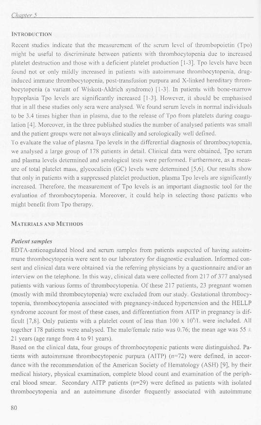
Figure 1: Plasma Tpo levels in different patient groups with thrombocytopenia. AITP; autoimmune thrombocytopenia; SAITP: secondary autoimmune thrombocytopenia; ameg.; amegakaryocytic thrombocytopenia; miscell.A; miscellaneous thrombocytopenic patients without expected megakaryocyte deficiency; miscell.B; miscellaneous thrombocytopenic patients with expected megakaryocyte deficiency. Boxes represent the interquartile range containing 50% of all values. The line across the box indicates the median whereas the whiskers extend to the highest and lowest value. The circles represent outliers, asterisks represent extremes.
Statistical analysis
Statistical analysis was performed with SPSS for Windows, release 6.1.3 (SPSS Inc.). For comparison of groups the Mann-Whitney U - Wilcoxon Rank Sum W Test was used. The correlation between two variables was calculated with Spearman correlation coefficients.
RESULTS
Tpo and GC levels
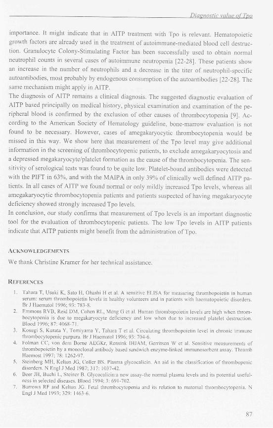
Plasma Tpo levels in AITP and SAITP patients were found to be within the normal range in most cases (89%) (13 ± 10 A.U., mean ± SD, range 2 A.U to 54 A.U.)(fig.l). Four patients in the AITP group and three patients in the SAITP group had a slightly increased Tpo level (36, 39, 42, 41 A.U./ml and 54, 53, 53 A.U./ml, respectively), whereas in four patients in the AITP group the Tpo level was lower than 4 A.U./ml (one 1 A.U./ml and three 3 A.U./ml). As shown in figure 1, the mean level of Tpo was slightly higher in the group of AITP patients as well as the SAITP group, as compared to the controls. This difference was statistically significant (p=0.03 and p=0.01, respectively). There was no correlation between the platelet count and the plasma Tpo level in either AITP or SAITP. Serum Tpo levels ranged from 6 A.U. to 81 A.U. (28 ± 14, mean±SD). The serum/plasma ratio in these two groups of patients was 2 ± 0.9 and 2 ± 0.8 (mean ± SD), respectively, which is significantly lower than the ratio found in controls (3 ± 0.6, mean ± SD)(p<0.001). There was no correlation between either the serum Tpo level or the serum/plasma ratio and the platelet number.
As shown in figure 1, all five patients, classified as amegakaryocytic thrombocytopenia had strongly elevated Tpo levels (range 258-927 A.U./ml). Furthermore, in all patients in the miscellaneous B group whose medical data indicated that a decreased hematopoiesis was the
83
Chapter 5
1000
o • OO onnn 1—I a a o
E o o E a n •— D D D G O D D 100 — • o < z • • < - O o
~ O O ö • M O O O t D c o
O O c o • • • • c o o « « » » o o • o o o o • O O » o 10 • OOO Q. O h- ~ OOOOOO ro • o £ M • re a 1 =-
B
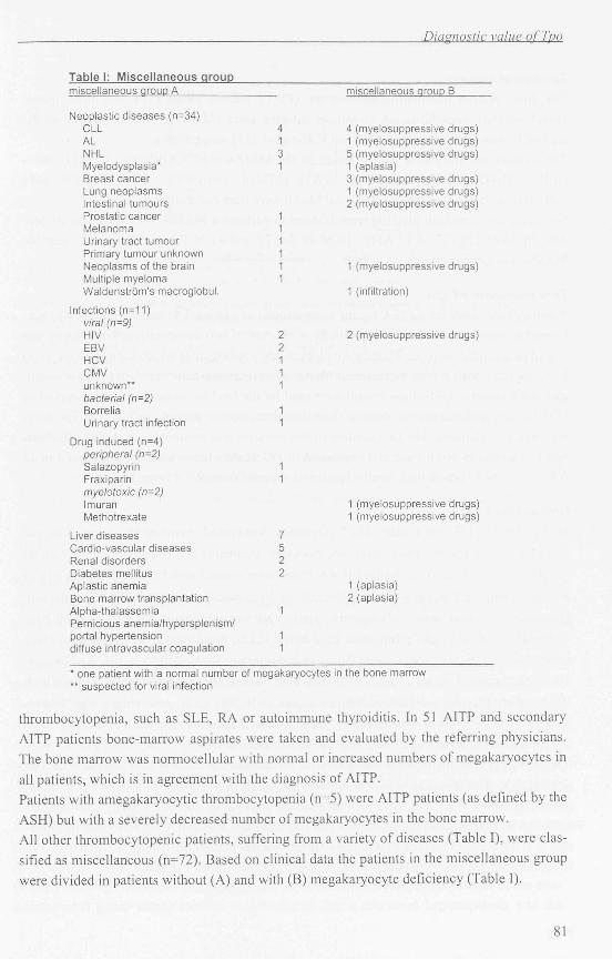
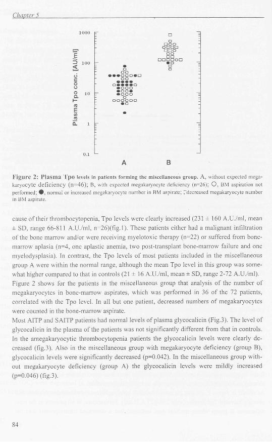
Figure 2: Plasma Tpo levels in patients forming the miscellaneous group. A, without expected megakaryocyte deficiency (n=46); B, with expected megakaryocyte deficiency (n=26); O , BM aspiration not performed; # , normal or increased megakaryocyte number in BM aspirate; fldccreased megakaryocyte number in BM aspirate.
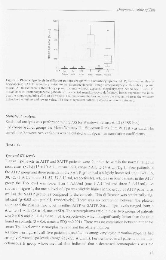
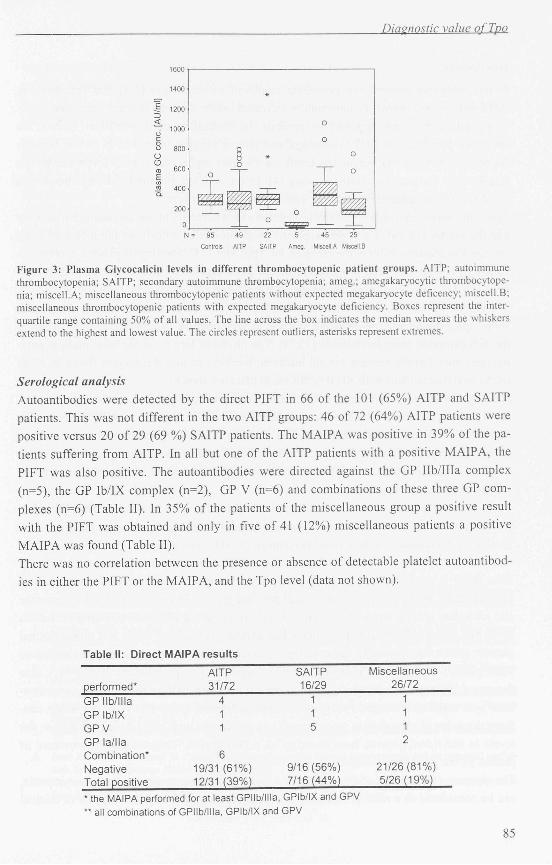
cause of their thrombocytopenia, Tpo levels were clearly increased (231 ± 160 A.U./ml, mean ± SD, range 66-811 A.U./ml, n=26)(fig.l). These patients either had a malignant infiltration of the bone marrow and/or were receiving myelotoxic therapy (n=22) or suffered from bone-marrow aplasia (n=4, one aplastic anemia, two post-transplant bone-marrow failure and one myelodysplasia). In contrast, the Tpo levels of most patients included in the miscellaneous group A were within the normal range, although the mean Tpo level in this group was somewhat higher compared to that in controls (21 ± 16 A.U./ml, mean ± SD, range 2-72 A.U./ml). Figure 2 shows for the patients in the miscellaneous group that analysis of the number of megakaryocytes in bone-marrow aspirates, which was performed in 36 of the 72 patients, correlated with the Tpo level. In all but one patient, decreased numbers of megakaryocytes were counted in the bone-marrow aspirate. Most AITP and SAITP patients had normal levels of plasma glycocalicin (Fig.3). The level of glycocalicin in the plasma of the patients was not significantly different from that in controls. In the amegakaryocytic thrombocytopenia patients the glycocalicin levels were clearly decreased (fig.3). Also in the miscellaneous group with megakaryocyte deficiency (group B), glycocalicin levels were significantly decreased (p=0.042). In the miscellaneous group without megakaryocyte deficiency (group A) the glycocalicin levels were mildly increased (p=0.046) (fig.3).
84
Diagnostic value ofTpo
N = 9 5 4 9 22 5 4 5
Controls AITP SAITP Ameg. MIScellA
2 5
Miscall E
Figure 3: Plasma Glycocalicin levels in different thrombocytopenic patient groups. AITP; autoimmune thrombocytopenia; SAITP; secondary autoimmune thrombocytopenia; ameg.; amegakaryocytic thrombocytopenia; miscell.A; miscellaneous thrombocytopenic patients without expected megakaryocyte deficency; miscell.B; miscellaneous thrombocytopenic patients with expected megakaryocyte deficiency. Boxes represent the interquartile range containing 50% of all values. The line across the box indicates the median whereas the whiskers extend to the highest and lowest value. The circles represent outliers, asterisks represent extremes.
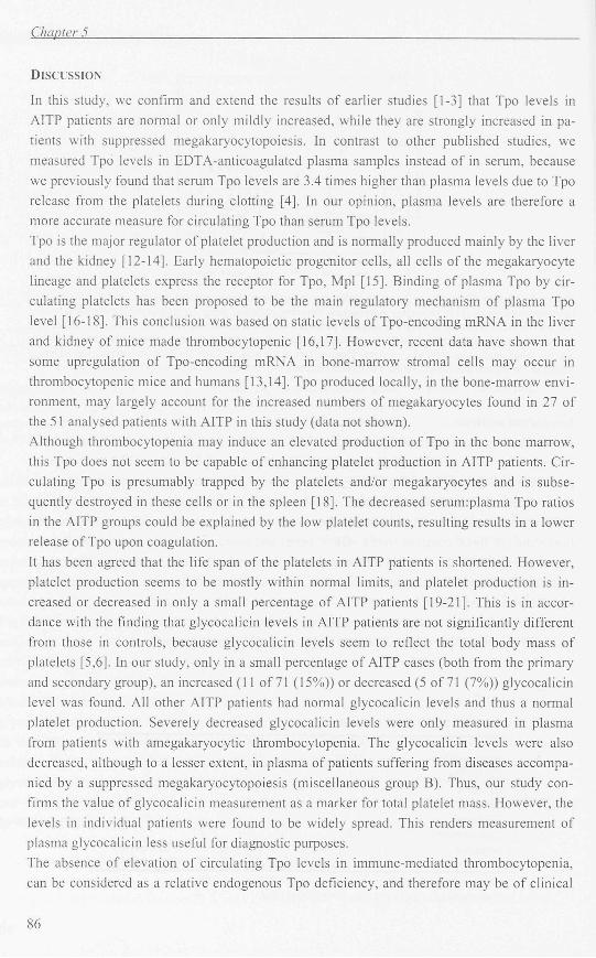
Serological analysis Autoantibodies were detected by the direct PIFT in 66 of the 101 (65%) AITP and SAITP patients. This was not different in the two AITP groups: 46 of 72 (64%) AITP patients were positive versus 20 of 29 (69 %) SAITP patients. The MAIPA was positive in 39% of the patients suffering from AITP. In all but one of the AITP patients with a positive MAIPA, the PIFT was also positive. The autoantibodies were directed against the GP Ilb/IIIa complex (n=5), the GP Ib/IX complex (n=2), GP V (n=6) and combinations of these three GP complexes (n=6) (Table II). In 35% of the patients of the miscellaneous group a positive result with the PIFT was obtained and only in five of 41 (12%) miscellaneous patients a positive MAIPA was found (Table II). There was no correlation between the presence or absence of detectable platelet autoantibodies in either the PIFT or the MAIPA, and the Tpo level (data not shown).
Table li: Direct MAIPA results
AITP SAITP Miscellaneous
performed* 31/72 16/29 26/72
GP Ilb/IIIa 4 1 1
GP Ib/IX 1 1 1 GPV 1 5 1 GP la/lla 2 Combination* 6 Negative 19/31 (61%) 9/16(56%) 21/26(81%) Total positive 12/31 (39%) 7/16(44%) 5/26(19%)
* the MAIPA performed for at least GPIIb/llla, GPIb/IX and GPV ** all combinations of GPIIb/llla, GPIb/IX and GPV
85
Chapter 5
DISCUSSION
In this study, we confirm and extend the results of earlier studies [1-3] that Tpo levels in AITP patients are normal or only mildly increased, while they are strongly increased in patients with suppressed megakaryocytopoiesis. In contrast to other published studies, we measured Tpo levels in EDTA-anticoagulated plasma samples instead of in serum, because we previously found that serum Tpo levels are 3.4 times higher than plasma levels due to Tpo release from the platelets during clotting [4]. In our opinion, plasma levels are therefore a more accurate measure for circulating Tpo than serum Tpo levels. Tpo is the major regulator of platelet production and is normally produced mainly by the liver and the kidney [12-14]. Early hematopoietic progenitor cells, all cells of the megakaryocyte lineage and platelets express the receptor for Tpo, Mpl [15]. Binding of plasma Tpo by circulating platelets has been proposed to be the main regulatory mechanism of plasma Tpo level [16-18]. This conclusion was based on static levels of Tpo-encoding mRNA in the liver and kidney of mice made thrombocytopenic [16,17]. However, recent data have shown that some upregulation of Tpo-encoding mRNA in bone-marrow stromal cells may occur in thrombocytopenic mice and humans [13,14]. Tpo produced locally, in the bone-marrow environment, may largely account for the increased numbers of megakaryocytes found in 27 of the 51 analysed patients with AITP in this study (data not shown). Although thrombocytopenia may induce an elevated production of Tpo in the bone marrow, this Tpo does not seem to be capable of enhancing platelet production in AITP patients. Circulating Tpo is presumably trapped by the platelets and/or megakaryocytes and is subsequently destroyed in these cells or in the spleen [18]. The decreased serurmplasma Tpo ratios in the AITP groups could be explained by the low platelet counts, resulting results in a lower release of Tpo upon coagulation. It has been agreed that the life span of the platelets in AITP patients is shortened. However, platelet production seems to be mostly within normal limits, and platelet production is increased or decreased in only a small percentage of AITP patients [19-21]. This is in accordance with the finding that glycocalicin levels in AITP patients are not significantly different from those in controls, because glycocalicin levels seem to reflect the total body mass of platelets [5,6]. In our study, only in a small percentage of AITP cases (both from the primary and secondary group), an increased (11 of 71 (15%)) or decreased (5 of 71 (7%)) glycocalicin level was found. All other AITP patients had normal glycocalicin levels and thus a normal platelet production. Severely decreased glycocalicin levels were only measured in plasma from patients with amegakaryocytic thrombocytopenia. The glycocalicin levels were also decreased, although to a lesser extent, in plasma of patients suffering from diseases accompanied by a suppressed megakaryocytopoiesis (miscellaneous group B). Thus, our study confirms the value of glycocalicin measurement as a marker for total platelet mass. However, the levels in individual patients were found to be widely spread. This renders measurement of plasma glycocalicin less useful for diagnostic purposes. The absence of elevation of circulating Tpo levels in immune-mediated thrombocytopenia, can be considered as a relative endogenous Tpo deficiency, and therefore may be of clinical
86
Diagnostic value ofTpo
importance. It might indicate that in AITP treatment with Tpo is relevant. Hematopoietic growth factors are already used in the treatment of autoimmune-mediated blood cell destruction. Granulocyte Colony-Stimulating Factor has been successfully used to obtain normal neutrophil counts in several cases of autoimmune neutropenia [22-28], These patients show an increase in the number of neutrophils and a decrease in the titer of neutrophil-specific autoantibodies, most probably by endogenous consumption of the autoantibodies [22-28]. The same mechanism might apply in AITP.
The diagnosis of AITP remains a clinical diagnosis. The suggested diagnostic evaluation of AITP based principally on medical history, physical examination and examination of the peripheral blood is confirmed by the exclusion of other causes of thrombocytopenia [9]. According to the American Society of Hematology guideline, bone-marrow evaluation is not found to be necessary. However, cases of amegakaryocytic thrombocytopenia would be missed in this way. We show here that measurement of the Tpo level may give additional information in the screening of thrombocytopenic patients, to exclude amegakaryocytosis and a depressed megakaryocyte/platelet formation as the cause of the thrombocytopenia. The sensitivity of serological tests was found to be quite low. Platelet-bound antibodies were detected with the PIFT in 63%, and with the MAIPA in only 39% of clinically well defined AITP patients. In all cases of AITP we found normal or only mildly increased Tpo levels, whereas all amegakaryocytic thrombocytopenia patients and patients suspected of having megakaryocyte deficiency showed strongly increased Tpo levels.
In conclusion, our study confirms that measurement of Tpo levels is an important diagnostic tool for the evaluation of thrombocytopenic patients. The low Tpo levels in AITP patients indicate that AITP patients might benefit from the administration of Tpo.
ACKNOWLEDGEMENTS
We thank Christine Kramer for her technical assistance.
REFERENCES
1. Tahara T, Usuki K, Sato H, Ohashi H et al. A sensitive ELISA for measuring thrombopoietin in human serum: serum thrombopoietin levels in healthy volunteers and in patients with haematopoietic disorders. Br J Haematol 1996; 93: 783-8.
2. Emmons RVB, Reid DM, Cohen RL, Meng G et al. Human thrombopoietin levels are high when thrombocytopenia is due to megakaryocyte deficiency and low when due to increased platelet destruction. Blood 1996;87:4068-71.
3. Kosugi S, Kurata Y, Tomiyama Y, Tahara T et al. Circulating thrombopoietin level in chronic immune thrombocytopenic purpura. Br J Haematol 1996; 93: 704-6.
4. Folman CC, von dem Bome AEGKr, Rensink IHJAM, Gerritsen W et al. Sensitive measurements of thrombopoietin by a monoclonal antibody based sandwich enzyme-linked immunosorbent assay. Thromb Haemost 1997; 78: 1262-97.
5. Steinberg MH, Kelton JG, Coller BS. Plasma glycocalicin. An aid in the classification of thrombopenic disorders. N Engl J Med 1987; 317:1037-42.
6. Beer JH, Buchi L, Steiner B. Glycocalicima new assay-the normal plasma levels and its potential usefulness in selected diseases. Blood 1994; 3: 691-702.
7. Burrows RF and Kelton JG. Fetal thrombocytopenia and its relation to maternal thrombocytopenia. N Engl J Med 1993; 329: 1463-6.
87
Chapter 5
8. Kobayashi M, Laver JH, Kato T, Miyazaki H et al. Thrombopoietin supports proliferation of human primitive hematopoietic cells in synergy with steel factor and/or interleukin-3. Blood 1996; 88: 429-36.
9. George JN. Woolf SH, Raskob GE. Idiopathic thrombocytopenic purpura: a guideline for diagnosis and management of children and adults. American Society of Hematology. Ann Med 1998; 30: 38-44.
10. von dem Borne AEGK, Verheugt FW, Oosterhof F, von Riesz E et al. A simple immunofluorescence test for the detection of platelet antibodies. Br J Haematol 1978; 39: 195-207.
11. Kiefel V, Santoso S, Weisheit M, Mueller-Eckhardt C. Monoclonal antibody-specific immobilization of platelet antigens (MAIPA): a new tool for the identification of platelet-reactive antibodies. Blood 1987; 70: 1722-6.
12. Gurney AL, Kuang WJ, Xie MH, Malloy BE et al. Genomic structure, chromosomal localization, and conserved alternative splice forms of thrombopoietin. Blood 1995; 85: 981-8.
13. Nagahisa H, Nagata Y, Ohnuki T, Osada M et al. Bone marrow stromal cells produce thrombopoietin and stimulate megakaryocyte growth and maturation but suppress proplatelet formation. Blood 1996; 87: 1309-16.
14. McCarty JM, Sprugel KH, Fox NE, Sabath DE et al. Murine thrombopoietin mRNA levels are modulated by platelet count. Blood 1995; 86: 3668-75.
15. Debili N, Wendling F, Cosman D, Titeux M et al. The Mpl receptor is expressed in the megakaryocyte lineage from late progenitors to platelets. Blood 1995; 85: 391-401.
16. Stoffel R, Wiestner A, Skoda RC. Thrombopoietin in thrombocytopenic mice: Evidence against regulation at the mRNA level and for a direct regulatory role of platelets. Blood 1996; 87: 567-73.
17. Fielder PJ, Gurney AL, Stefanich E, Marian M et al. Regulation of thrombopoietin levels by c-Mpl-mediated binding to platelets. Blood 1996; 87:2154-61.
18. Kuter DJ and Rosenberg RD. The reciprocal relationship of thrombopoietin (c-mpl ligand) to changes in the platelet mass during busulfan-induced thrombocytopenia in the rabbit. Blood 1995; 85: 2720-30.
19. Heyns Ad, Badenhorst PN, Lotter MG, Pieters H et al. Platelet turnover and kinetics in immune thrombocytopenic purpura: results with autologous 111 In-labeled platelets and homologous 51 Cr- labeled platelets differ. Blood 1986; 67: 86-92.
20. Ballem PJ, Segal GM, Stratton JR, Gemsheimer T et al. Mechanisms of thrombocytopenia in chronic autoimmune thrombocytopenic purpura. Evidence of both impaired platelet production and increased platelet clearance. J Clin Invest 1987; 80: 33-40.
21. Siegel R S, Coleman R E, Kurlander R, and Rosse W F. Platelet turnover: An important factor in predicting response to splenectomy in autoimmune thrombocytopenia. Blood (abstract) 1984; 64.
22. Ganser A, Ottmann OG, Erdmann H, Schulz G et al. The effect of recombinant human granulocyte-macrophage colony- stimulating factor on neutropenia and related morbidity in chronic severe neutropenia. Ann Intem Med 1989; 111: 887-92.
23. Klumpp TR, Herman JH, Macdonald JS, Schnell MK et al. Autoimmune neutropenia following peripheral blood stem cell transplantation. Am J Hematol 1992; 41: 215-7.
24. Stroncek DF, Shapiro RS, Filipovich AH, Plachta LB et al. Prolonged neutropenia resulting from antibodies to neutrophil-specific antigen NB1 following marrow transplantation. Transfusion 1993; 33: 158-63.
25. Takahashi K, Taniguchi S, Akashi K, Fujimoto K et al. Human recombinant granulocyte colony-stimulating factor for the treatment of autoimmune neutropenia. Acta Haematol 1991; 86: 95-8.
26. Taniguchi S, Shibuya T, Harada M, Niho Y. Decreased levels of myeloid progenitor cells associated with long-term administration of recombinant human granulocyte colony-stimulating factor in patients with autoimmune neutropenia. Br J Haematol 1993; 83: 384-7.
27. Kuijpers TW, de Haas M, de Groot CJ, von dem Borne et al. The use of rhG-CSF in chronic autoimmune neutropenia: reversal of autoimmune phenomena, a case history [see comments]. Br J Haematol 1996; 94: 464-9.
28. Vlasveld LT, de Haas M, Ermens AA, Porcelijn L et al. G-CSF-induced decrease of the anti-granulocyte autoantibody levels in a patient with autoimmune granulocytopenia. Ann Hematol 1997; 75: 59-64.
88