Embed Size (px)
Citation preview
UvA-DARE is a service provided by the library of the University of Amsterdam (http://dare.uva.nl)
UvA-DARE (Digital Academic Repository)
Biochemical and genetic aspects of mevalonate kinase and its deficiency
Houten, S.M.
Link to publication
Citation for published version (APA):Houten, S. M. (2002). Biochemical and genetic aspects of mevalonate kinase and its deficiency.
General rightsIt is not permitted to download or to forward/distribute the text or part of it without the consent of the author(s) and/or copyright holder(s),other than for strictly personal, individual use, unless the work is under an open content license (like Creative Commons).
Disclaimer/Complaints regulationsIf you believe that digital publication of certain material infringes any of your rights or (privacy) interests, please let the Library know, statingyour reasons. In case of a legitimate complaint, the Library will make the material inaccessible and/or remove it from the website. Please Askthe Library: https://uba.uva.nl/en/contact, or a letter to: Library of the University of Amsterdam, Secretariat, Singel 425, 1012 WP Amsterdam,The Netherlands. You will be contacted as soon as possible.
Download date: 22 Mar 2020
Chapterr 4
Identificationn and characterization of three novel
missensee mutations in mevalonate kinase cDNA causing
mevalonicc aciduria, a disorder of isoprene biosynthesis
S.M.. Houten, GJ. Romeijn, J. Koster, R.G.F. Gray, P. Darbyshire, G.P.A. Smit,
J.B.C.. de Klerk, M. Duran, K.M. Gibson, RJ.A. Wanders and H.R. Waterham.
Humann Molecular Genetics 8:1523-1528, 1999.

Threee novel missense mutations in MVK causing mevalonic aciduria
Mevalonicc aciduria is a rare autosomal recessive metabolic disorder, characterized by psychomotorr retardation, failure to thrive, hepatosplenomegaly, anemia and recurrent febrilee crises. The disorder is caused by a deficient activity of mevalonate kinase due to mutationss in the encoding gene. Thus far, only two disease-causing mutations have been identified.. We now report four different missense mutations including three novel ones, whichh were identified by sequence analysis of mevalonate kinase cDNA from three mevalonicc aciduria patients. All mutations affect conserved amino acids. Heterologous expressionn of the corresponding mutant mevalonate kinases as fusion proteins with glutathionee S-transferase in Escherichia coli cells showed a profound effect of each of thee mutations on enzyme activity. In addition, immunoblot analysis of fibroblast lysates fromm patients using specific antibodies against mevalonate kinase identified virtually no protein.. These results demonstrate that the mutations not only affect the activity but alsoo the stability of the mutant proteins.
Introduction n Thee isoprene pathway supplies cells with intermediates for the biosynthesis of a variety of compoundss with important functions in cellular processes. These compounds include ubiquinone-100 and heme A, both necessary for electron transport, dolichol, essential for proteinn glycosylation, isopentenyl tRNA, involved in protein translation, and farnesyl and geranylgeranyll groups used for the isoprenylation of proteins which function in cell signaling andd cell differentiation. In addition to these non-sterol isoprenoids, the pathway produces cholesterol,, a structural component of membranes and precursor for bile acids and steroid hormones. .
Att present, three different inherited disorders have been recognized, which are caused byy defects in this pathway. Two of these disorders, Smith-Lemli-Opitz (SLO) syndrome and desmosterolosis,, only affect the synthesis of cholesterol and its derivatives and are caused by aa deficient activity of 7-dehydrocholesterol reductase and desmosterol reductase, respectively Tl] ,, The biochemical defect in SLO syndrome was-confirmed recently by the identification of mutationss in the gene coding for 7-dehydrocholesterol reductase [2-4], but the gene mutated inn desmosterolosis patients remains to be identified.
Thee third disorder of the pathway is mevalonic aciduria, which is caused by a deficient activityy of mevalonate kinase (MK). MK is the first enzyme to follow 3-hydroxy-3-methylglutaryl-CoAA reductase in the isoprene pathway and converts mevalonate into 5-phosphomevalonate.. As a consequence of the MK deficiency, high levels of mevalonic acid aree present in plasma and urine from mevalonic aciduria patients [5, 6].
Soo far, ~15 patients with mevalonic aciduria have been reported. Although most patientss suffered from an often fatal, multi-sytemic disease, their clinical presentation varied considerably.. Severely affected patients had profound developmental delay, dysmorphic features,, cataracts, hepatosplenomegaly, lymphadenopathy, anemia, diarrhea and malabsorption,, and died early in infancy, while milder affected patients only showed psychomotorr retardation, hypotonia, myopathy and ataxia. All patients were characterized by recurrentt febrile attacks associated with lymphadenopathy, an increase in the size of the liver andd spleen, arthralgia, edema and morbilliform rash [6]. Since the enzyme reaction catalyzed byy MK occurs in the first, unbranched part of the pathway, an effect on the synthesis of both steroll and non-sterol isoprenoids is predicted. Accordingly, the ubiquinone-10 concentration
59 9
Chapterr 4
inn plasma from some mevalonic aciduria patients appeared to be decreased [7]. The plasma cholesteroll level, however, was usually normal or, at most, slightly decreased [6].
Sincee the cloning of human MK cDNA [8], only two disease-causing missense mutationss have been reported. The resulting two amino acid changes, N301T [8] and A334T [9],, have a profound effect on enzyme activity as shown by heterologous expression of the mutantt proteins in COS-7 cells and in Escherichia coli. Here we report four different missensee mutations causing mevalonic aciduria, three of which are novel. These were identifiedd after sequence analysis of MK cDNAs from three patients clinically diagnosed with mevalonicc aciduria. Subsequent studies demonstrated that all four mutations affect both enzymee activity and protein stability of the resulting mutant MKs.
Results s BiochemicalBiochemical characterization of three mevalonic aciduria patients Alll three patients used for this study were diagnosed with mevalonic aciduria based on clinicall signs (Table 1) and on the basis of increased concentrations of mevalonic acid in plasmaa and urine. Judged from their clinical presentation and the fact that they died at an early age,, patients 1 and 3 were severely affected. This is also reflected by the almost complete absencee of MK activity in fibroblasts of both patients (Table 2). Since patient 2 showed a milderr clinical presentation and is currently 20 years old, he was predicted to have some residuall MK activity. Measurements in fibroblasts of this patient, however, also revealed virtuallyy no MK activity (Table 2).
Tablee 1. Clinical findings in the three mevalonic aciduria patients used in this study-Clinicall findings Patient 1 (F) Patient 2 (M) Patient 3 (F) Psychomotorr retardation Recurrentt febrile crises Anemia a Failuree to thrive Hypotoniaa and myopathy Ataxiaa and cerebellar atrophy Hepatosplenomegaly y Dysmorphicc features Cataracts s Outcome2 2
+++ + +++ +
+ + +++ +
----+ + + +
--4.55 monthsf
+ + + + --
+ + + +
++/+++ +
--_ _
--200 years
++ + +++ + ++ + + + ----
++ +
----
55 monthsf aa Present age or f age at death. +++,, severe; ++, moderate; +, present; -, absent.
MutationMutation analysis ofMK cDNA Too identify the mutations that cause the mevalonic aciduria, MK mRNAs of the three patients weree amplified by RT-PCR in two overlapping fragments. Sequence analysis of the amplified cDNAA fragments showed that patient 1 was homozygous for a T>C transition at nucleotide 803,, which changes the isoleucine at position 268 into a threonine (I268T). Patient 2 was compoundd heterozygous for an A>C transversion at nucleotide 59 and a G>A transition at nucleotidee 1000, which change the histidine at position 20 into a proline (H20P) and the alaninee at position 334 into a threonine (A334T), respectively. Compound heterozygosity was
60 0
Threee novel missense mutations in MVK causing mevalonic aciduria
confirmedd by separate cloning of the two mutant cDNAs. Patient 3 was homozygous for a G>AA transition at nucleotide 928, which changes the valine at position 310 into a methionine (V310M). .
Tablee 2. MK and glutamate dehydrogenase (GDH) activities in cultured skin fibroblast lysatess from the three mevalonic aciduria patients and a control subject. Subject t
Control l Patientt 1 Patientt 2 Patientt 3
Aminoo acid change(s) ) --I268T T H20P,, A334T V310M M
(MK)a a
171 1 0.3 3 0.5 5 0.4 4
(GDH)b b
350 0 149 9 174 4 203 3
Ratioo x 1000 (MK/GDH) )
0.489 9 0.002 2 0.003 3 0.002 2
aa Expressed as pmol/min/mg protein. bb Expressed as nmol/min/mg protein.
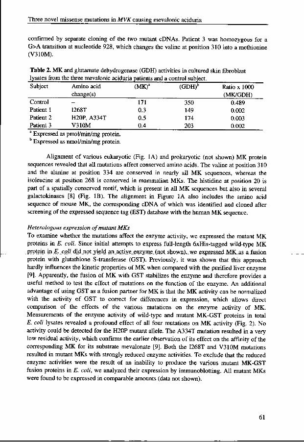
Alignmentt of various eukaryotic (Fig. 1A) and prokaryotic (not shown) MK protein sequencess revealed that all mutations affect conserved amino acids. The valine at position 310 andd the alanine at position 334 are conserved in nearly all MK sequences, whereas the isoleucinee at position 268 is conserved in mammalian MKs. The histidine at position 20 is partt of a spatially conserved motif, which is present in all MK sequences but also in several galactokinasess [8] (Fig. IB). The alignment in Figure 1A also includes the amino acid sequencee of mouse MK, the corresponding cDNA of which was identified and cloned after screeningg of the expressed sequence tag (EST) database with the human MK sequence.
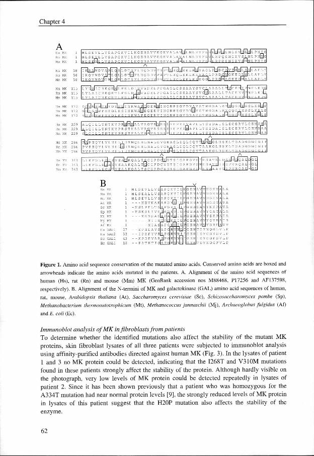
HeterologousHeterologous expression of mutant MKs Too examine whether the mutations affect the enzyme activity, we expressed the mutant MK proteinss in E. coll Since initial attempts to express full-length 6xHis-tagged wild-type MK proteinn in E. coli didjipt jield an_actiye_enzyme_(not shown),, we .expressed MKas a fusion proteinn with glutathione S-transferase (GST). Previously, it was shown that this approach hardlyy influences the kinetic properties of MK when compared with the purified liver enzyme [9].. Apparently, the fusion of MK with GST stabilizes the enzyme and therefore provides a usefull method to test the effect of mutations on the function of the enzyme. An additional advantagee of using GST as a fusion partner for MK is that the MK activity can be normalized withh the activity of GST to correct for differences in expression, which allows direct comparisonn of the effects of the various mutations on the enzyme activity of MK. Measurementss of the enzyme activity of wild-type and mutant MK-GST proteins in total E.E. coli lysates revealed a profound effect of all four mutations on MK activity (Fig. 2). No activityy could be detected for the H20P mutant allele. The A334T mutation resulted in a very loww residual activity, which confirms the earlier observation of its effect on the affinity of the correspondingg MK for its substrate mevalonate [9]. Both the I268T and V310M mutations resultedd in mutant MKs with strongly reduced enzyme activities. To exclude that the reduced enzymee activities were the result of an inability to produce the various mutant MK-GST fusionn proteins in E. coli, we analyzed their expression by immunoblotting. All mutant MKs weree found to be expressed in comparable amounts (data not shown).
61 1
Chapterr 4
Hss MK Rnn MK Mmm MK
Rn n Mm m
Hs s Rn n M~ ~
MK K MK K
MK K MK K MK K
58 8 58 8
115 5 115 5 115 5
Hss MK Rnn MK Mmm MK
Hss MK Rnn MK Mmm MK
Hss MK Rnn MK Mmm MK
M L S E V L L V S A P G K V I L H G E H A V V H G K V A LL A \ 4 S JL N L R T F L M L SS E_V_L L V S A P G K V I L H G E H A V V H G K V A L A-VA L N L R T F L
KK V AL A J A I A L N L R T F L ~-\
;_VLL L V :[AILL L v S A P G K V II ; , H G E H A V V H G >
V|LL R P Q S N G K V S L
I . R P O S N G K V S I V I N LL P N I G I
| S JLL P N I (
d N LL P N 0<
LL D T SF L E Q GD VlT T P~TISJEQIV |E K L KIE [ LL D T [ 3 F L E Q G D V P A P T L E Q L E K L KK
I K | RR AIW D V A|R L Q S
II K Q V W D V A | T L Q L II K o V W D VTGMI L O I R I L D T S F L E O G D V I S V I P T L E O L E K L K
GG L PlDjD C AlVTiElRl L A|y)L A F L Y AGG L P R D C p GN E G L | si L L A F L Y
KK G D|L P R DIRIA G N E GIMIA I, L A F L
229 9 229 9 229 9
286 6 286 6 286 6
343 3 343 3 343 3
LL Y L [ S JI C R KQ R[A|L P S L D I V V W S E L P P G A G L G S S A AY S VC LA A A L L TIYJC E Eh P N P L K D G|
L Y L A I C R K Q R T L P S LL D I[M! V W S E L P P G A G L G S S A A Y SV C 0A A A L L T A C E E v i l N P L K I H r,, Y L A I C R K O R T L P S L DIMI V V W S E L P P G A G L G S S A A Y S V C L A A A L L T A C E E VI S|N P L K D GI
^JITT KI E D L I E LI I N K w A F I O I G E RIMI I H G N P S G V D N A V S T W G G AL R Y[H|Q G KJi i s s L K R[SJP
J I W P E E D L K SS I N K W A I Y J EG E R V I H G N P S G V D N Ü JV S T W G G A L R Y Q Q GKM S SL K R L P
i s lRWW P E E D L K S I N K W A F E G E R V I H G N P S G V D N A V S T W G G AL RIFIO O GPTIM S SL KISIL P
SILL O I L L T K T .K V P R S T K A L
VA GG V R S vv I ;-, o
AA L Q I L L T N T K V P RINJTIRJA L V A G V R I N I RT L K F P E 1 ^ A P L L T S I D A I S L E C E R V L G E K ^ L Q I L L T N T K V P R S T K A L VV A^.V R S R L KK F P E I [M] A P L L T S I D A I S L E C E R V L G E M|A|A A
K F P E I V A P L L T S I D A I S L E C E R V L G EE MIVIA A
PIAJPP E Q Y L V L E E L I D M N Q H H L N A L G V G H A S L D Q L C Q VTIRJAIRJG L H S K L T G A G G G G C G I T P V P E Q Y L V L E EE L 0 D M N Q H H L N A L G V G H A S L D Q L C Q V T A A H G L H S K L T G A G G G G C G I T PP V P E O Y L V L E E L I D M N O H H L N A L G V G H[N! S L D O L C O V T A A H G L H S K L T G A G G G G C G I T
A A
LL LK P GL E LL L K P G L E|
LL L K P G L E
P E V EE ALTJK Q A L T S C G F D CILJE T S I G A P G VS
KVEAAKQALL T0C G F D C W E T S I G A P G V S
V E A A K O A L T S C G F D C W E T S I G A P G V S S
GG L GG L
B B Hss MK Mmm MK Rnn MK Att MK Scc MK Spp MK Mtt MK Mjj MK Aff MK Hss GAL1 Hss GAL2 Scc GAL1 ECC GAL1
M L S E V L L V S S MLSEALLVS S M L S E V L L V S S
M E V K A R R - M S L P F L T S S -- M S K
M M M i ll E T|P|
AA P G KV I L AA PG KV I L A P GK K AA PG - M , A.. P G K̂ V, l | T
KK V 1 K Q I I
L..,, - _ K V i L SS L I V s"Ü PG KITI I L
KK S S A s £ P[A] KIAJ I L
A PG G AA PG
MM I A S E P E L A V S
-- I P K F Y VRLA PG - K P D F V AA R^l P G -- P A T H T I Q[A P
GG K V GKK V GG K V G ST T NKK P G A T T
A V V Y S K P A | I A A AA V V Y G AA VlVjYG R H T D Y N N II D Y C II D Y C T D Y N N
L A A L A A
AJLL A AA V A AA V A A L A A
AjVV V QQ G L V L P GYY S VL P DFF S VL P DGG F V L P
Figuree 1. Amino acid sequence conservation of the mutated amino acids. Conserved amino acids are boxed and
arrowheadss indicate the amino acids mutated in the patients. A. Alignment of the amino acid sequences of
humann (Hs), rat (Rn) and mouse (Mm) MK (GenBank accession nos M88468, PI7256 and AF137598,
respectively).. B. Alignment of the N-termini of MK and galactokinase (GAL) amino acid sequences of human,
rat,, mouse, Arabidopsis thaliana (At), Saccharomyces cerevisiae (Sc), Schizosaccharomyces pombe (Sp),
MethanobacteriumMethanobacterium thermoautotrophicum (Mt), Methanococcus jannaschii (Mj), Archaeoglobus fulgidus (Af)
andd E. coli (Ec).
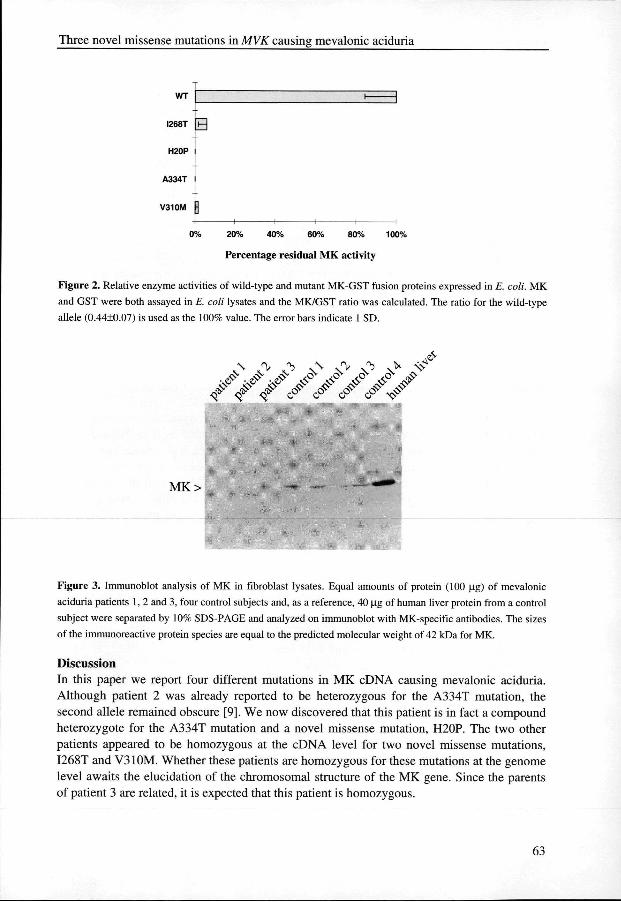
ImmunoblotImmunoblot analysis ofMK in fibroblasts from patients Too determine whether the identified mutations also affect the stability of the mutant MK proteins,, skin fibroblast lysates of all three patients were subjected to immunoblot analysis usingg affinity-purified antibodies directed against human MK (Fig. 3). In the lysates of patient 11 and 3 no MK protein could be detected, indicating that the I268T and V310M mutations foundd in these patients strongly affect the stability of the protein. Although hardly visible on thee photograph, very low levels of MK protein could be detected repeatedly in lysates of patientt 2. Since it has been shown previously that a patient who was homozygous for the A334TT mutation had near normal protein levels [9], the strongly reduced levels of MK protein inn lysates of this patient suggest that the H20P mutation also affects the stability of the enzyme. .
62 2
Threee novel missense mutations in MVK causing mevalonic aciduria
WT T
I268T T
H20P P
A334T T
V310M M
0%% 20% 40% 60% 80% 100%
Percentagee residual MK activity
Figuree 2. Relative enzyme activities of wild-type and mutant MK-GST fusion proteins expressed in E. coli. MK
andd GST were both assayed in E. coli lysates and the MK/GST ratio was calculated. The ratio for the wild-type
allelee ) is used as the 100% value. The error bars indicate 1 SD.
Figuree 3. Immunoblot analysis of MK in fibroblast lysates. Equal amounts of protein (100 ug) of mevalonic
aciduriaa patients 1, 2 and 3, four control subjects and, as a reference, 40 ug of human liver protein from a control
subjectt were separated by 10% SDS-PAGE and analyzed on immunoblot with MK-specific antibodies. The sizes
off the immunoreactive protein species are equal to the predicted molecular weight of 42 kDa for MK.
Discussion n Inn this paper we report four different mutations in MK cDNA causing mevalonic aciduria. Althoughh patient 2 was already reported to be heterozygous for the A334T mutation, the secondd allele remained obscure [9]. We now discovered that this patient is in fact a compound heterozygotee for the A334T mutation and a novel missense mutation, H20P. The two other patientss appeared to be homozygous at the cDNA level for two novel missense mutations, I268TT and V310M. Whether these patients are homozygous for these mutations at the genome levell awaits the elucidation of the chromosomal structure of the MK gene. Since the parents off patient 3 are related, it is expected that this patient is homozygous.
63 3
Chapterr 4
Thee enzyme activity measurements revealed almost no MK activity in fibroblasts from alll three patients and thus no apparent correlation between residual enzyme activity and the severityy of the disease. The identified mutations in the two severely affected patients 1 and 3, I268TT and V310M, respectively, resulted in strongly reduced activity upon expression in E. coli,coli, although some residual activity could still be measured. In immunoblots of fibroblast lysatess of these patients, however, no MK protein was identified, indicating that these mutationss also, or maybe primarily, affect the stability of the protein.
Thee A334T and H20P mutations identified in the relatively mildly affected patient 2 resultt in a strongly reduced and a complete absence of enzyme activity, respectively, when expressedd in E. coli. In addition, the levels of MK protein in fibroblast lysates from this patientt were strongly reduced as revealed by immunoblotting. Since it was shown previously thatt the A334T mutation results in a stable protein with a 30-fold elevated Km for the substrate mevalonatee [9], these results suggest that the H20P allele also codes for an unstable protein. Thee relatively mild phenotype of patients with the A334T allele might be explained by the factt that the encoded mutant protein still has substantial residual activity when plasma mevalonatee levels are elevated [9].
Thee H20P mutation occurs in a spatially conserved region found in all MKs and galactokinasess (Fig. 1). Two putative functions for this region have been proposed [10]. One functionn is that this region is involved in targeting of MK to peroxisomes. The histidine at positionn 20 is part of an N-terminal amino acid stretch that shows a perfect match with the consensuss sequence of peroxisomal targeting signal type 2 (PTS2), which is involved in targetingg of a certain class of peroxisomal matrix proteins [11]. Several observations are in favorr of a function for this domain in peroxisomal targeting. For instance, it has been reported thatt MK is localized in rat liver peroxisomes [12, 13]. Furthermore, MK deficiency has been reportedd in patients diagnosed with the peroxisomal biogenesis disorders Zellweger syndrome andd rhizomelic chondrodysplasia punctata type 1 [12, 14, 15]. In Zellweger syndrome, there is aa generalized loss of peroxisomal functions, while in rhizomelic chondrodysplasia punctata typee 1, there is only a partial loss of peroxisomal functions due to an impaired import of proteinss with a PTS2 [16-18]. However, although the H20P mutation disrupts the PTS2 consensuss sequence, the actual role of this sequence in peroxisomal targeting of MK still remainss to be demonstrated.
Thee second function of this conserved N-terminal region may be in modulating the activityy and stability of MK. It has been reported that amino acid changes introduced in the N-terminuss of MK greatly affect the enzyme activity. For instance, when the lysine at position 133 is changed into a methionine, a strong effect on the Vmax and affinity of the enzyme for ATPATP is observed [10]. In addition, changing the glutamic acid at position 19 into an alanine or aa glutamine, or the histidine at position 20 into an alanine, resulted in unstable and completely insolublee proteins upon expression in E. coli, suggesting structural importance for these aminoo acids [19]. This is in accordance with our expression studies in E. coli, which showed thatt the H20P mutation resulted in an inactive enzyme. Furthermore, the strongly reduced proteinn levels in an immunoblot of fibroblasts of patient 2 suggest instability in vivo of the MKK protein encoded by this allele. Although these data suggest that the N-terminal region is importantt for enzyme activity and/or stability, they do not necessarily exclude a second functionn in peroxisomal targeting.
64 4
Threee novel missense mutations in MVK causing mevalonic aciduria
Materialss and Methods MevalonicMevalonic aciduria patients Patientt 1 [20] was a girl born to unrelated parents who presented with minor dysmorphic featuress (micrognathia), hepatosplenomegaly, gastrointestinal problems and failure to thrive. Hematologicall parameters were suggestive of a congenital infection, but this was excluded. Thee girl had a massive mevalonic aciduria, which showed a steady increase during her life. Shee died suddenly in a state of shock at the age of 4.5 months.
Patientt 2 is a boy born to unrelated Dutch parents. At the age of 6 years, he was clinicallyy evaluated for his cerebellar ataxia, hypotonia and strongly elevated creatine kinase activity.. NMR and computed tomography of the brain revealed cerebellar hypoplasia together withh an enlarged IVth ventricle. Following the detection of mevalonic acid in the urine [21], loww concentrations of bile acids in the blood and an almost complete absence of the fat-solublee vitamins A, E and D were noticed. Serum concentration of cholesterol was normal [22].. From his 7th year, the patient has been on an oral therapy providing him with bile acid (Chenofalkk 2x100 mg daily) and vitamin A, E and D. At present, the patient is 20 years of age andd in a good clinical and mental condition (height p90 and weight p20).
Patientt 3 was a girl born to first cousin-related parents of Pakistani origin. She presentedd with recurrent fever, joint pains and swelling, hepatosplenomegaly and a moderate failuree to thrive. She died during one of the febrile episodes having made littl e developmental progress. .
Tablee 1 summarizes the clinical findings in these patients.
MutationMutation analysis Firstt strand cDNA was synthesized from RNA isolated from cultured human skin fibroblasts ass described elsewhere [23]. The first strand cDNA was used as template in a PCR with two differentt sets of primers tagged with either a -21M13 or an M13rev extension and designed to amplifyy the MK cDNA in two overlapping fragments. The first fragment was amplified using primerr set MK_2i_j(5'-tgtaaaacgacg gcc agt GCGJ3CA GGA TTC CCA GGAG-3') and MK622-6033 (5'-cag gaa aca get atg ace TGA CAG CAT TGT CCA CTC CG-3'). The second fragmentt was amplified using primer set MK521.540 (5'-tgt aaa acg acg gcc agt TCA ACA GGTT GGA CCA AGG AG-3') and MK2i238-i2i9 (5'-cag gaa aca get atg ace ATC CAG AAA GGGG GCA TCT GG-3'). The PCR fragments were sequenced in both directions by means of -21M133 and M13rev fluorescent primers, on an applied biosystems 377A automated DNA sequencer,, according to the manufacturer's protocol (Perkin-Elmer, Foster City, CA).
CloningCloning of wild-type and mutant MK cDNAs Thee open reading frame (ORF) of the wild-type MK cDNA was amplified by PCR using primerr set MK10.29 (5'-cg ata sea tec GAA GTC CTA CTG GTG TCT GC-3') and MKii 195.1177 (5'-cga taggtaCCT CTC AG A GGC CAT CCA G-3'). The primers introduce a 5'' BamHl site and a 3' Kpnl site (underlined). The PCR product was ligated into the pGEM-T vectorr (Promega Corp., Madison, WI) and sequenced to exclude PCR-introduced mutations. Thee wild-type ORF subsequently was released as a BamKl-Notl fragment from pGEM-T and ligatedd in-frame with the GST ORF into the BamHl and Notl sites of pGEX-4T-l (Pharmacia Biotech,, Uppsala, Sweden). cDNAs containing the mutations H20P, V310M and I268T were clonedd from patients into pGEM-T as described for the wild-type MK cDNA. The H20P mutationn was introduced into the pGEX-4T-l plasmid by replacing the BamiÜ-HindlJl
65 5
Chapterr 4
fragmentt of the wild-type ORF. To introduce the I268T mutation, a Smal-Sacl fragment of thee wild-type ORF in pGEX-4T-l was replaced. V310M was introduced in the wild-type ORF inn pGEX-4T-l by replacing a Sacl-Kpnl fragment.
CloningCloning of the mouse MK cDNA Inn order to identify the mouse MK cDNA, the EST database of the National Center for Biotechnologyy Information (NCBI) was screened for mouse sequences homologous to human MK.. Numerous homologous mouse EST sequences were identified which together spanned thee entire putative ORF. The ORF was subsequently amplified by PCR using primer set mmMK_2o-ii (5'-GGG CAG AAG TCT CAG AAG CC-3') and mmMK1225-i209 (5'-eg ata gaa ttcc GGA TGG TGT GTC GGG TGG T-3') and primer set mmMK4-24 (5'-eg ata gga tec TTG TCAA GAA GCT CTG CTG GTG-3') and mmMK1225-i209. Both PCR products were ligated intoo pGEM-T and sequenced. The second primer set introduced a 5' BamHl and a 3' EcoRl site,, which were used for subcloning of the ORF into the pGEX-4T-l vector to confirm its identityy by means of expression as a GST fusion protein in E. coli.
EnzymeEnzyme assays MKK was measured radiochemically making use of 14C-labeled mevalonate [24], Glutamate dehydrogenasee (GDH) activity served as a control. GST activity was assayed spectrophotometricallyy at 335 nm by measuring the formation of the conjugate of glutathione andd l-chloro-2,4-dinitrobenzene [25].
MKMK expression in E. coli pGEX-4T-ll plasmids containing the various MK ORFs were transformed into CaCl2 competentt DH5a or commercially available Inva (Invitrogen, Carlsbad, CA) E. coli cells. Cellss were grown from a 100-fold diluted fresh overnight culture for 4 hours in LB medium, inducedd with 2 mM isopropyl-p-thiogalactopyranoside (Promega) and subsequently grown for ann additional 2 hours. Cells were lysed by sonication (twice for 15 s at 8 W output, with 1 min off cooling between the pulse periods). Lysates were adjusted to equal levels of GST activity beforee assaying MK activity.
ImmunoblotImmunoblot analysis AA synthetic peptide corresponding to amino acid residues 76 to 90 of human MK (H2N-EQGDVTTPTSEQVEK-COOH)) was conjugated with keyhole limpet hemocyanin and used too produce antibodies in rabbits (Eurogentech, Seraing, Belgium). The crude antiserum was affinity-purifiedd on a column containing MK-GST fusion protein coupled to cyanogen bromide-Sepharosee as described [26]. The MK-GST fusion protein was affinity purified on gluthatione-agarosee columns according to the protocol of the supplier (Pharmacia Biotech). In immunoblots,, the affinity-purified antibodies were used at a 1:100 dilution.
Forr immunoblot analysis, fibroblast cultures were harvested and lysed by sonication in 200 mM MOPS (pH 7.4) and 0.25% Triton X-100, supplemented with 25 ug/ml phenylmethylsulfonyll fluoride and 10 ug/ml leupeptin. Equal amounts of total protein were separatedd by SDS-PAGE and transferred onto nitrocellulose by semi-dry immunoblotting [27].. Antigen-antibody complexes were visualized using anti-rabbit IgG-alkaline phosphatase conjugatee (BioRad, Hercules, CA).
66 6
Threee novel missense mutations in MVK causing mevalonic aciduria
References s [I ]] P.T. Clayton. (1998) Arch Dis Child 78,185-9. [2]] H.R. Waterham, F.A. Wijburg, R.C. Hennekam, P. Vreken, B.T. Poll-The, L. Dorland, M. Duran, P.E.
Jira,, J.A. Smeitink, R.A. Wevers and R.J. Wanders. (1998) Am J Hum Genet 63,329-38. [3]] B.U. Fitzky, M. Witsch-Baumgartner, M. Erdel, J.N. Lee, Y.K. Paik, H. Glossmann, G. Utermann and
F.F.. Moebius. (1998) Proc Natl Acad Sci U S A 95, 8181-6. [4]] C.A. Wassif, C. Maslen, S. Kachilele-Linjewile, D. Lin, L.M. Linck, W.E. Connor, R.D. Steiner and
F.D.. Porter. (1998) Am J Hum Genet 63,55-62. [5]] G. Hoffmann, K.M. Gibson, I.K. Brandt, P.I. Bader, R.S. Wappner and L. Sweetman. (1986) N Engl J
Medd 314, 1610-4. [6]] G.F. Hoffmann, C. Charpentier, E. Mayatepek, J. Mancini, M. Leichsenring, K.M. Gibson, P. Divry, M.
Hrebicek,, W. Lehnert, K. Sartor and et al. (1993) Pediatrics 91, 915-21. [7]] C. Hubner, G.F. Hoffmann, C. Charpentier, K.M. Gibson, B. Finckh, H. Puhl, H.A. Lehr and A.
Kohlschutter.. (1993) PediatrRes 34, 129-33. [8]] B.L. Schafer, R.W. Bishop, V.J. Kratunis, S.S. Kalinowski, S.T. Mosley, K.M. Gibson and R.D.
Tanaka.. (1992) J Biol Chem 267,13229-38. [9]] D.D. Hinson, K.L. Chambliss, G.F. Hoffmann, S. Krisans, R.K. Keller and K.M. Gibson. (1997) J Biol
Chemm 272, 26756-60. [10]] D. Potter, J.M. Wojnar, C. Narasimhan and H.M. Miziorko. (1997) J Biol Chem 272, 5741-6. [II ]] S. Subramani. (1996) J Biol Chem 271, 32483-32486. [12]] L. Biardi, A. Sreedhar, A. Zokaei, N.B. Vartak, R.L. Bozeat, J.E. Shackelford, G.A. Keller and S.K.
Krisans.. (1994) J Biol Chem 269, 1197-205. [13]] K.D. Stamellos, J.E. Shackelford, R.D. Tanaka and S.K. Krisans. (1992) J Biol Chem 267, 5560-8. [14]] R.J. Wanders and G.J. Romeijn. (1996) J Inherit Metab Dis 19,193-6. [15]] R.J. Wanders and G.J. Romeijn. (1998) Biochem Biophys Res Commun 247, 663-7. [16]] N. Braverman, G. Steel, C. Obie, A. Moser, H. Moser, S.J. Gould and D. Valle. (1997) Nat Genet 15,
369-76. . [17]] A.M. Motley, E.H. Hettema, E.M. Hogenhout, P. Brites, A.L. ten Asbroek, F.A. Wijburg, F. Baas, H.S.
Heijmans,, H.F. Tabak, R.J. Wanders and B. Distel. (1997) Nat Genet 15, 377-80. [18]] P.E. Purdue, J.W. Zhang, M. Skoneczny and P.B. Lazarow. (1997) Nat Genet 15, 381-4. [19]] D. Potter and H.M. Miziorko. (1997) J Biol Chem 272, 25449-54. [20]] J.B. de Klerk, M. Duran, L. Dorland, H.A. Brouwers, L. Bruinvis and D. Ketting. (1988) J Inherit
Metabb Dis 11 Suppl 2,233-6. [21]] R. Berger, G.P. Smit, H. Schierbeek, K. Bijsterveld and R. le Coultre. (1985) Clin Chim Acta 152,219-
22. . [22]] K.M. Gibson, G. Hoffmann, W.L. Nyhan, L. Sweetman, R. Berger, R. le Coultre and G.P. Smit. (1988)
Eürr J Pediaff 148, 250-2. [23]] L. Dist, R.J. Wanders, S. Ushikubo, T. Kamijo and T. Hashimoto. (1994) Biochim Biophys Acta 1215,
347-50. . [24]] G.F. Hoffmann, S.U. Brendel, S.R. Scharfschwerdt, Y.S. Shin, I.M. Speidel and K.M. Gibson. (1992) J
Inheritt Metab Dis 15, 738-46. [25]] W.H. Habig, M.J. Pabst and W.B. Jakoby. (1974) J Biol Chem 249,7130-9. [26]] C.K. Raymond, P.J. OHara, G. Eichinger, J.H. Rothman and T.H. Stevens. (1990) J Cell Biol 111, 877-
92. . [27]] J. Kyhse-Andersen. (1984) J Biochem Biophys Methods 10, 203-9.
67 7


![Autosomal recessive ichthyosis with limb reduction defect ... · including autosomal dominant, autosomal recessive and X-linked inheritance [1,2]. Associated cutaneous and extracutaneous](https://img.pdfslide.us/doc/110x75/5ec8c9b91adfdf12ab3e663c/autosomal-recessive-ichthyosis-with-limb-reduction-defect-including-autosomal.jpg)