Embed Size (px)
Citation preview

337Srp Arh Celok Lek. 2014 May-Jun;142(5-6):337-341 DOI: 10.2298/SARH1406337J
ПРИКАЗ БОЛЕСНИКА / CASE REPORT UDC: 616-056.7-07
Correspondence to:
Nebojša J. JOVIĆClinic of Neurology and Psychiatry for Children and YouthDr. Subotića 6a Str.11000 [email protected]
SUMMARYIntroduction L-2-Hydroxyglutaric aciduria (L-2-HGA) is an autosomal recessive neurometabolic disease with a slowly progressive course and characterized by increased levels of hydroxyglutaric acid in urine, cerebrospinal fluid and plasma. In this condition clinical features mainly consist of mental deterioration, ataxia and motor deficits.Case Outline The patient is a 16-year-old girl, the first and only child of healthy, non-consanguineous parents of Serbian origin. At the age of 4 years her walk became unsteady and ataxic. Other signs of cerebellar involvement were soon observed. Head circumference was above two standard deviations (55 cm). Mild mental retardation was revealed by formal intelligence testing (IQ 60). MR examination of the brain showed confluent subcortical white matter lesions spread centripetally, and atrophy of the cerebellar vermis with involvement of dentate nuclei, without deep white matter abnormalities. Laboratory investigation revealed increased amounts and a very large peak of HGA in urine and plasma. Enantiomeric analysis confirmed the L-configuration (>90%) establishing the diagnosis of L-2-HGA. The first epileptic seizure, partial with secondary generalization, occurred at age of 8 years. Favorable seizure control was achieved. A slow progression of neurological impairment was noted. Therapeutic trials with oral coenzyme Q10 and with oral riboflavin showed no biochemical and clinical effects. Recently, the diagnosis was proven by the presence of a mutation in the L-2-HGA gene.Conclusion To our knowledge, this is the first report of L-2-HGA in Serbia. L-2-HGA must be considered in the differential diagnosis based on specific findings in cranial MRI.Keywords: hydroxyglutaric aciduria; epilepsy; white matter lesions; ataxia
L-2-Hydroxyglutaric Aciduria: A Case ReportNebojša J. Jović1,2, Ana Kosać1, Katarina Koprivšek3
1Clinic of Neurology and Psychiatry for Children and Youth, Belgrade, Serbia;2School of Medicine, University of Belgrade, Belgrade, Serbia;3Imaging Diagnostic Center, Oncology Institute of Vojvodina, Sremska Kamenica, Serbia
INTRODUCTION
L-2-Hydroxyglutaric aciduria (L-2-HGA) is an autosomal recessive neurometabolic disease, belonging to the group of organic acidurias with a slowly progressive course. It is characterized by increased levels of L-2-hydroxyglutaric acid in urine, cerebrospinal fluid and plasma [1]. L-2-hydroxyglutarate accumulates as a result of deficiency in flavin–adenine dinucleotide (FAD)-linked L-2-hydroxyglutarate dehydroge-nase (L2HGDH), a mitochondrial enzyme con-verting L-2-hydroxyglutarate to a-ketoglutarate. L-2-HGA is linked to the chromosome 14q22.1 and its gene encodes a putative mitochondrial protein with homology to FAD-dependent ox-idoreductases hydroxyglutaric aciduria [2, 3]. So far, more than 30 mutations have been re-ported in the L2HGDH gene [4].
L-2-HGA was first described in 1980 and since then more than 200 cases have been re-ported worldwide. The clinical diagnosis is often delayed because of the insidious onset of symptoms [5]. Although the disease is usually of early infantile onset, it may be diagnosed in adults with a somewhat milder phenotype [4, 6]. A solitary large and persistent increase of L-2-hydroxyglutaric acid in urine was reported for the first time in 1980 in a 5-year-old boy from Morocco (Berber), who was investigated for nonspecific mental and motor delay and growth deficiency [7]. Nevertheless, a distinct clinical and neuroradiological picture has
emerged, as the disease has a relatively con-sistent pattern of presentation.
Patients usually display a delayed mental and motor development in the first years of life. About two-third of them have epilepsy and cerebellar dysfunction with progressive ataxia, dysarthria and moderate to severe mental de-terioration. Macrocephaly, pyramidal and ex-trapyramidal signs and dystonia are present in the majority of patients [8, 9].
Briefly, magnetic resonance imaging (MRI) findings, which seem to be consistently unique of L-2-HGA, are very specific and affect the central white matter, basal ganglia, and cerebel-lum [10]. Neuroimaging findings include sub-cortical white matter loss and abnormalities of the dentate nucleus, globus pallidus, putamen, and caudate nucleus [9].
It has been suggested that the deficiency of L2HGDH induces a neoplastic state [11] and that patients with L-2-HGA have a predispo-sition to cerebral neoplasms of various types, including medulloblastoma and glioblastoma [11, 12, 13]. There is no effective therapy. Iso-lated cases are reported of successful treatment with riboflavin and FAD sodium and levocar-nitine chloride supplements [11, 14].
CASE REPORT
The patient was a 16-year-old girl, the first and only child of healthy, non-consanguineous
338
doi: 10.2298/SARH1406337J
Jović N. J. et al. L-2-Hydroxyglutaric Aciduria: A Case Report
parents of Caucasian, Serbian origin. Family history was uninformative. Pregnancy was uneventful. Caesarean sec-tion was performed, as an elective procedure, because of the fetal malpresentation. Birth weight was 2800 g, birth length 50 cm and head circumference 36 cm. The neonatal period was normal. A psychomotor delay was evident in the second year of life. She was able to stand up and walk without support at about 26th months of life. Development of her speech was delayed.
At the age of 4 years her gait became worse, she was unsteady, with ataxic walk and other signs of cerebellar involvement as dysmetria, truncal ataxia, intention tremor and dysarthria. Her head circumference was above two standard deviations (55 cm). Psychomotor development, measured with the Brunet-Lezine scale, showed the de-velopment quotient of 60. Ocular fundus examination was normal, but high degree myopia had been diagnosed. Electroencephalogram (EEG) showed diffuse non-specif-ic abnormalities. Molecular genetic tests for Friedreich’s ataxia were negative. Electroneurographic examination was normal.
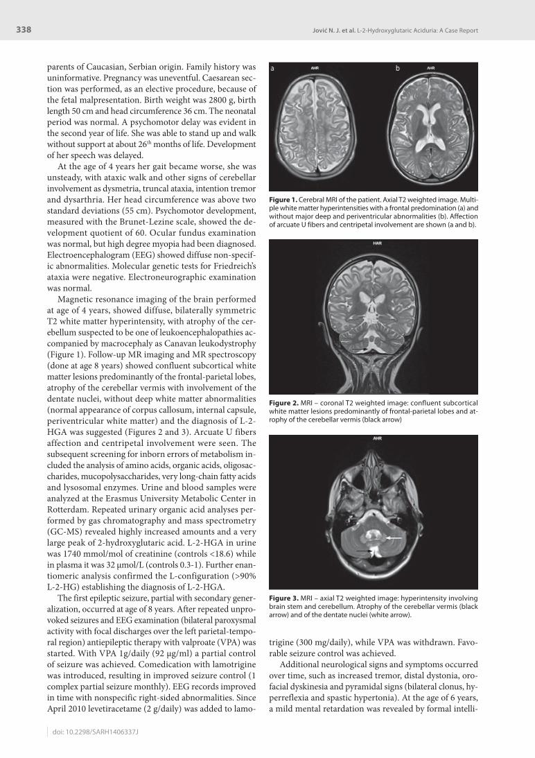
Magnetic resonance imaging of the brain performed at age of 4 years, showed diffuse, bilaterally symmetric T2 white matter hyperintensity, with atrophy of the cer-ebellum suspected to be one of leukoencephalopathies ac-companied by macrocephaly as Canavan leukodystrophy (Figure 1). Follow-up MR imaging and MR spectroscopy (done at age 8 years) showed confluent subcortical white matter lesions predominantly of the frontal-parietal lobes, atrophy of the cerebellar vermis with involvement of the dentate nuclei, without deep white matter abnormalities (normal appearance of corpus callosum, internal capsule, periventricular white matter) and the diagnosis of L-2-HGA was suggested (Figures 2 and 3). Arcuate U fibers affection and centripetal involvement were seen. The subsequent screening for inborn errors of metabolism in-cluded the analysis of amino acids, organic acids, oligosac-charides, mucopolysaccharides, very long-chain fatty acids and lysosomal enzymes. Urine and blood samples were analyzed at the Erasmus University Metabolic Center in Rotterdam. Repeated urinary organic acid analyses per-formed by gas chromatography and mass spectrometry (GC-MS) revealed highly increased amounts and a very large peak of 2-hydroxyglutaric acid. L-2-HGA in urine was 1740 mmol/mol of creatinine (controls <18.6) while in plasma it was 32 μmol/L (controls 0.3-1). Further enan-tiomeric analysis confirmed the L-configuration (>90% L-2-HG) establishing the diagnosis of L-2-HGA.
The first epileptic seizure, partial with secondary gener-alization, occurred at age of 8 years. After repeated unpro-voked seizures and EEG examination (bilateral paroxysmal activity with focal discharges over the left parietal-tempo-ral region) antiepileptic therapy with valproate (VPA) was started. With VPA 1g/daily (92 μg/ml) a partial control of seizure was achieved. Comedication with lamotrigine was introduced, resulting in improved seizure control (1 complex partial seizure monthly). EEG records improved in time with nonspecific right-sided abnormalities. Since April 2010 levetiracetame (2 g/daily) was added to lamo-
trigine (300 mg/daily), while VPA was withdrawn. Favo-rable seizure control was achieved.
Additional neurological signs and symptoms occurred over time, such as increased tremor, distal dystonia, oro-facial dyskinesia and pyramidal signs (bilateral clonus, hy-perreflexia and spastic hypertonia). At the age of 6 years, a mild mental retardation was revealed by formal intelli-
Figure 3. MRI – axial T2 weighted image: hyperintensity involving brain stem and cerebellum. Atrophy of the cerebellar vermis (black arrow) and of the dentate nuclei (white arrow).
Figure 1. Cerebral MRI of the patient. Axial T2 weighted image. Multi-ple white matter hyperintensities with a frontal predomination (a) and without major deep and periventricular abnormalities (b). Affection of arcuate U fibers and centripetal involvement are shown (a and b).
Figure 2. MRI – coronal T2 weighted image: confluent subcortical white matter lesions predominantly of frontal-parietal lobes and at-rophy of the cerebellar vermis (black arrow)
339Srp Arh Celok Lek. 2014 May-Jun;142(5-6):337-341
www.srp-arh.rs
gence testing (IQ 60) without later deterioration. Her head circumference stopped increasing, but remained signifi-cantly above the 97th percentile (60 cm).
Because of spastic pes equinovarus, her walk was pos-sible with assistance only. She developed lumbosacral scoliosis. A retention orthosis was recommended at age of 12. Last year applied therapy with botulinus toxin was beneficial and resulted in decreased spasticity. In 2011 the diagnosis was proven by the presence of a mutation in the L-2-HGA gene (missense mutation c.185C>A located in the exon 2). Therapeutic trials with oral coenzyme Q10 (400 mg/day) during 6 months and with oral riboflavin (200 mg/day) during 4 months showed neither biochemi-cal nor clinical effects.
DISCUSSION
The progression of L-2-HGA and the consequent disabil-ity are different among patients. Topçu et al. [15] report the clinical features of 29 patients from 22 families. The mean age at the time of diagnosis was 13.4 years (2.5-32 years). The main clinical findings were mental retardation and cerebellar involvement with ataxic gait and intention tremor. Additional findings were mental retardation, mac-rocephaly and seizures. During the follow-up period (1.5-16 years), all patients had a static encephalopathy course.
In a group of 7 Italian patients, three patients developed severe motor and mental impairment with epilepsy, and worsened rapidly. One patient was severely demented, with spastic tetraparesis, ataxia and frequent seizures and died at 21 years of age. Other patients had milder cerebellar symptoms and mental retardation. Their clinical follow-up showed a slow progression of the disease [12]. Our patient presented with symptoms of cerebellar involvement at age of 4 years, additionally to the developmental delay, psycho-motor regression and macrocephaly, as usually reported.
Macrocephaly is present in almost 50% of patients with L-2-HGA and it can be the first manifestation of the disease [16]. Therefore, it could be seen in other organic acidurias, such as glutaric aciduria type I, HMG-CoA lyase deficiency and 3-methylglutaconyl-CoA-hydratase deficiency [17]. Initial manifestation of severe autism and pervasive developmental disorders has been very rarely described in patients with L-2-HGA [16].
Seizures occur in more than 50% of patients with L-2-HGA, usually late in the course of the disease [8]. Epileptic seizures or even status epilepticus can be found among the presenting symptoms in organic acidurias with a slow course, such as L-2-HGA [17]. A patient with neonatal onset of L-2-HGA showed a burst suppression pattern on EEG, making this disorder another entity, which should be considered in the differential diagnosis of neonatal sei-zures [18]. The patient, a 9-month-old female infant, ho-mozygous for the p.Lys81Glu (c.241A>G) missense muta-tion in the L-2-HGA gene, was reported with acute hemi-convulsion-hemiplegia-epilepsy syndrome as a presenting feature [19]. A 5-year-old boy with eyelid myoclonia with absences, bilaterally synchronous EEG polyspike/spike and
wave discharges and L-2-OHG was described. The patient became seizure-free with a combination therapy of clon-azepam, levetiracetam, and lamotrigine [20]. Our patient presented with complex partial seizures with secondary generalization. Favorable seizure control was achieved with valproate, lamotrigine and levetiracetam in combi-nations.
Marked intra- and inter-familial variability in clinical phenotype has been reported [16, 21]. In the majority of cases, as in our patient, the severity of associated neurolog-ic and cognitive impairments appears to progress slowly, with many of them surviving adulthood. However, a sud-den deterioration may occur in occasional cases and some patients have a downhill neurologic course at a later age, sometimes as late as at 6-17 years of age [8]. There is one report of a neonate with L-2-HGA and a rapidly fatal out-come [18]. An 11-month-old girl, born to consanguineous parents of Tamil origin suddenly died at 11 months of age, during an intercurrent illness [22]. There was no correla-tion between the severity of clinical symptoms and the amount of L-2-OHG acid in urine or CSF [12]. Delay of L-2-HGA diagnosis until adulthood has been described [5, 14], because of mild clinical symptoms and lack of typical MRI abnormalities [4].
Neuroimaging findings of L-2-HGA are very specific. Cranial MRI reveals characteristic scattered or diffuse sub-cortical white-matter abnormalities fading centripetally [14]. A retrospective review of MR images in patients with L-2-HGA disclosed that initially, abnormalities of subcor-tical white matter were at least partially multifocal, later became more confluent and spread centripetally, but the periventricular rim remained relatively spared. Bilateral in-volvement of the globus pallidus, caudate nucleus, putamen and dentate nucleus was seen at all stages. The cerebellar white matter was never affected [9]. Confluent subcorti-cal white matter lesions, atrophy of the cerebellar vermis with involvement of the dentate nuclei, without deep white matter abnormalities, were shown on MRI scans in our patients, supporting the diagnosis of L-2-HGA.
In all Turkish patients MRI showed subcortical leu-koencephalopathy with bilateral high signal intensity in the dentate nuclei and putamens [15]. Atypical imaging anomalies, such as basal ganglia atrophy were described in an adult patient with L-2-OHG [5]. Moroni et al. [12] observed a good correlation between the severity of the disease and the extent of lesions on MRI. As cranial MRI findings raise suspicion of L-2-HGA, one must consider other diseases that have similar MRI findings for a dif-ferential diagnosis. These diseases include Van der Knaap disease, Canavan disease and Alexander disease. We en-countered the same initial diagnostic difficulties.
Increased incidence of brain tumors has been noted among patients with L-2-HGA [12]. Meta-analysis of pub-lished data identified 14 patients with L-2-HGA associated with cerebral neoplasms, suggesting an approximately 5% prevalence rate of CNS neoplasms in these patients [13]. A male infant with L-2-HGA and Wilms tumor, the most com-mon renal malignancy of childhood, was reported as exam-ple of an extracranial tumor associated with L-2-HGA [11].

340
doi: 10.2298/SARH1406337J
Jović N. J. et al. L-2-Hydroxyglutaric Aciduria: A Case Report
Neither cerebral nor extracranial neoplasm was found in our patient.
A possible therapeutic role of FAD, cofactor of the enzyme, and of its precursor riboflavin was investigated. Nevertheless, it appears that this approach is only effective in “mild” missense mutations of L2HGDH (supplementa-tion with FAD 30 mg/day) and levocarnitine chloride (900 mg/day)), whereas truncated enzymes (presumed null mu-
tations) are not responsive [1]. Therapeutic trials with oral coenzyme Q10 (400 mg/day) during 6 months and with oral riboflavin (200 mg/day) during 2 months showed no biochemical and clinical effects [22]. We could confirm this observation in our patient. No significant clinical ben-efit of oral coenzyme Q10 (400 mg/day) during 6 months and with oral riboflavin (200 mg/day) during 4 months was noted.
REFERENCES
1. Kranendijk M, Struys EA, Salomons GS, Van der Knaap MS, Jakobs C. Progress in understanding 2-hydroxyglutaric acidurias. J Inherit Metab Dis. 2012; 35:571-87.
2. Rzem R, Veiga-da-Cunha M, Noel G, Goffette S, Nassogne MC, Tabarki B, et al. A gene encoding a putative FAD-dependent L-2-hydroxyglutarate dehydrogenase is mutated in L-2-hydroxyglutaric aciduria. Proc Natl Acad Sci USA. 2004; 101(48):16849-54.
3. Topçu M, Jobard F, Halliez S, Coskun T, Yalcinkayal C, Gerceker FO, et al. L-2-hydroxyglutaric aciduria: identification of a mutant gene C14orf160, localized on chromosome 14q22.1. Hum Mol Genet. 2004; 13(22):2803-11.
4. Sass JO, Romrell JS, Vinson SY, Fernandez HH, Fischer J, Rodriguez RL, et al. Tracing the origin of L-2-hydroxyglutaric aciduria in a family. Int J Neurosci. 2009; 119:2118-23.
5. Mazzei R,Ungaro C, Garreffa G, Conforti FL, Mollo A, Sprovieri T, et al. Clinical, genetic and magnetic resonance findings in an Italian patient affected by L-2-hydroxyglutaric aciduria. Neurol Sci. 2011; 32:95-9.
6. Marcel C, Mallaret M, Lagha-Boukbiza O, Kremer S, Echaniz-Laguna A, Tranchant C. L-2-hydroxyglutaric aciduria diagnosed in a young adult with progressive cerebellar ataxia and facial dyskinesia. Rev Neurol (Paris). 2012; 168(2):187-91.
7. Duran M, Kamerling JP, Bakker HD, van Gennip AH, Wadman SK. L-2-hydroxy-glutaric aciduria: an inborn error of metabolism? J Inherit Metab Dis. 1980; 3:109-12.
8. De Klerk LB, Huijmans JG, Stroink H, Robben SG, Jakobs C, Duran M. L-2-Hydroxyglutaric aciduria: clinical heterogeneity versus biochemical homogeneity in a sibship. Neuropediatrics. 1997; 28:314-7.
9. Steenweg ME, Salomons GS, Yapici Z, Uziel G, Scalais E, Zafeiriou DI, et al. L-2-hydroxyglutaric aciduria: pattern of MR imaging abnormalities in 56 patients. Radiology. 2009; 251(3):856-65.
10. Seijo-Martinez M, Navarro C, Castro del Río M, Vila O, Puig M, Ribes A, et al. L-2-hydroxyglutaric aciduria. Clinical, neuroimaging and neuropathological findings. Arch Neurol. 2005; 62:666-70.
11. Rogers RE, Deberardinis RJ, Klesse LJ, Boriack RL, Margraf LR, Rakheja D. Wilms tumor in a child with L-2-hydroxyglutaric aciduria. Pediatr Dev Pathol. 2010; 13(5):408-11.
12. Moroni I, Bugiani M, D’Incerti L, Maccagnano C, Rimoldi M, Bissola L, et al. L-2-hydroxyglutaric aciduria and brain malignant tumors: a predisposing condition? Neurology. 2004; 62:1882-4.
13. Patay Z, Mills JC, Löbel U, Lambert A, Sablauer A, Ellison DW. Cerebral neoplasms in L-2 hydroxyglutaric aciduria: 3 new cases and meta-analysis of literature data. AJNR Am J Neuroradiol. 2012; 33(5):940-3.
14. Karatas H, Saygi S, Bastan B. L-2-hydroxyglutaric aciduria. Report of four Turkish adult patients. Neurologist. 2010; 16:44-6.
15. Topçu M, Aydin OF, Yalçinkaya C, Haliloğlu G, Aysun S, Anlar B, et al. L-2-hydroxyglutaric aciduria: a report of 29 patients. Turk J Pediatr. 2005; 47(1):1-7.
16. Zafeiriou DI, Ververi A, Salomonset GS, Vargiami E, Haas D, Papadopoulou V, et al. L-2 Hydroxyglutaric aciduria presenting with severe autistic features. Brain Dev. 2008; 30(4):305-7.
17. Zafeiriou DI, Sewellb A, Savvopoulou-Augoustidoua P, Gombakisa N, Katzoset G. L-2-Hydroxyglutaric aciduria presenting as status epilepticus. Brain Dev. 2001; 23(4):255-7.
18. Chen E, Nyhan WL, Jakobs C, Greco CM, Barkovich AJ, Cox VA, et al. L-2-Hydroxyglutaric aciduria: neuropathological correlations and first report of severe neurodegenerative disease and neonatal death. J Inherit Metab Dis. 1996; 19:335-43.
19. Lee C, Born M, Salomons GS, Jakobs C, Woelfle J. Hemiconvulsion-hemiplegia-epilepsy syndrome as a presenting feature of L-2-hydroxyglutaric aciduria. J Child Neurol. 2006; 21:538-40.
20. Mete A, Isikay S, Sirikci A, Ozkur A, Bayram M. Eyelid myoclonia with absence seizures in a child with L-2 hydroxyglutaric aciduria: findings of magnetic resonance imaging. Ped Neurol. 2012; 46:195-7.
21. Larnaout A, Amouri R, Kefi M, Hentati F. L-2-hydroxyglutaric aciduria: clinical and molecular study in three Tunisian families. Identification of a new mutation and inter-familial phenotype variability. J Inherit Metab Dis. 2008; 31(Suppl 2):S375-9.
22. Gygax MJ, Roulet-Perez E, Meagher-Villemure K, Jakobs C, Salomons GS, Boulat O, et al. Sudden unexpected death in an infant with L-2-hydroxyglutaric aciduria. Eur J Pediatr. 2009; 168:957-62.
341Srp Arh Celok Lek. 2014 May-Jun;142(5-6):337-341
www.srp-arh.rs
КРАТАК САДРЖАЈУвод L-2-хи дрок си глу та рич ка аци ду ри ја (L-2-ХГА) је ауто-зом но ре це сив на ме та бо лич ка бо лест са спо ро про гре сив-ним то ком, ко ја се од ли ку је по ви ше ним ни во и ма хи дрок си-глу та рич ке ки се ли не (L-2-ХГ) у мо кра ћи, це ре бро спи нал ној теч но сти и пла зми. Кли нич ка сли ка нај че шће укљу чу је мен-тал ну на ру ше ност, атак си ју и мо тор не ис па де.При каз бо ле сни ка Ше сна е сто го ди шња бо ле сни ца ро ђе на је као пр во и је ди но де те здра вих ро ди те ља срп ског по ре-кла и без крв ног срод ства. У уз ра сту од че ти ри го ди не ње но хо да ње је по стало не ста бил но и атак сич но. Дру ги зна ци за-хва ће но сти ма лог мо зга, ди сме три ја, атак си ја тру па, ин тен-ци о ни тре мор и ди зар три ја, за па же ни су вр ло бр зо. Обим гла ве је био из над две стан дард не де ви ја ци је (55 cm). Бла га ум на за о ста лост по ка за на је фор мал ном про це ном ин те ли-ген ци је (IQ је био 60). Маг нет на ре зо нан ци ја мо зга по ка за ла је кон флу ент на, суп кор ти кал на оште ће ња бе ле ма се са цен-три пе тал ним ши ре њем и атро фи ју ма ло мо жда ног вер ми са
уз за хва ће ност ден тат них је да ра, без оште ће ња ду бо ке бе ле ма се. Ла бо ра то риј ска ис пи ти ва ња по ка за ла су ве о ма ви со ке вред но сти L-2-ХГ у мо кра ћи и пла зми. Енан то ме рич ка ана-ли за по твр ди ла је Л-кон фи гу ра ци ју (>90% L-2-ХГ), чи ме је по ста вље на ди јаг но за L-2-ХГА. Пр ви епи леп тич ки на пад, жа-ри шни са се кун дар ном ге не ра ли за ци јом, ја вио се у осмој го-ди ни. По стиг ну та је по вољ на кон тро ла на па да. За па же но је спо ро на пре до ва ње не у ро ло шке на ру ше но сти. Те ра пиј ска орал на при ме на ко ен зи ма Q10 и ри бо фла ви на ни је до ве ла до би о хе миј ских и кли нич ких ефе ка та. Уско ро је ди јаг но за по твр ђе на на ла зом му та ци је L-2-ХГА ге на.За кљу чак Ко ли ко нам је по зна то, ово је пр ви при каз бо ле-сни ка са L-2-ХГА у Ср би ји. У скла ду с кли нич ком сли ком и маг нет но ре зо нант ним сним ком мо зга, L-2-ХГА тре ба да се увр сти у ди фе рен ци јал ну ди јаг но сти ку не у ро ме та бо лич ких бо ле сти.Кључ не ре чи: хи дрок си глу та рич ка аци ду ри ја; епи леп си ја; оште ће ња бе ле ма се; атак си ја
L-2-хидроксиглутаричка ацидурија: приказ болесникаНебојша Ј. Јовић1,2, Ана Косаћ1, Катарина Копрившек3
1Клиника за неурологију и психијатрију за децу и омладину, Београд, Србија;2Медицински факултет, Универзитет у Београду, Београд, Србија;3Дијагностички имиџинг центар, Институт за онкологију Војводине, Сремска Каменица, Србија
Примљен • Received: 23/05/2013 Прихваћен • Accepted: 02/08/2013


![Three Main Causes of Homocystinuria: of Metabolism ... · most frequent causes are classical homocystinuria [deficiency of cystathionine beta-synthase (CBS)], methylmalonic aciduria](https://img.pdfslide.us/doc/110x75/5e951dcb19bd325819567b57/three-main-causes-of-homocystinuria-of-metabolism-most-frequent-causes-are.jpg)