Embed Size (px)
Citation preview

UTILIZATION OF NEXT GENERATION SEQUENCING FOR IDENTIFYING STREPTOCOCCUS
SUIS CLONES RESPONSIBLE FOR RECURRENT OUTBREAKS AND IDENTIFYING
EFFECTIVE S. SUIS AUTOGENOUS VACCINE CANDIDATES
Pork CRC project 2C-126
Report prepared for the
Co-operative Research Centre for High Integrity Australian Pork
By
Dr. Sam Abraham, Dr. Mark O’Dea, Ms Tanya Laird, Mr Alec Truswell, Mrs Rebecca Abraham
Antimicrobial Resistance and Infectious Disease Laboratory
School of Veterinary and Life Sciences
Murdoch University, Perth, WA
May 2018
i
Executive Summary
Streptococcus suis is an important cause of mortality in swine production and a zoonotic
bacteria. Current management protocols consist of treatment with β-lactam antibiotics, and
administration of autogenous vaccines with variable results.
This study assessed the potential for tonsil swabs from live pigs to be a suitable method for
rapid identification of circulating S. suis clones for production of autogenous vaccines.
Tonsil swabs did not yield any isolates with genomic signatures indicative of pathogenic S.
suis circulating in Australia, however a large number of S. suis-like organisms were isolated.
Genomic comparison indicated that S. suis isolated from tonsils were genetically distinct from
pathogenic S. suis causing invasive clinical infections in Australian pigs, and likely to be a
separate subspecies. Whole genome sequencing of 148 clinical isolates from diseased pigs
demonstrated serotypes 2, 3 and 1/2 are the predominant serotypes in Australia, and a
trivalent vaccine incorporating these serotypes is indicated.

ii
Table of Contents
Executive Summary ............................................................................................................. i
1. Introduction.................................................................................................................. 1
2. Methodology ................................................................................................................ 2
3. Results ......................................................................................................................... 4
4. Discussion ................................................................................................................... 9
5. Conclusion ..................................................................................................................10
6. Recommendations ......................................................................................................10
7. References ..................................................................................................................11
8. Appendix 1 ..................................................................................................................12

1
Introduction
The increase in reports of infection of Streptococcus suis in Australian pig farms in
recent years has been associated with significant production losses. Clinical disease
caused by S. suis can result in meningitis, joint effusions, endocarditis, septicemia
and sudden death. Despite these serious consequences of infection, commensal S.
suis strains can be cultured from the tonsils, nasal cavity, genital and
gastrointestinal tracts of healthy pigs. This is a complicating factor in management
and treatment of infection and disease, with difficulty in determining the pathogenic
potential of commensal strains, and these strains potentially masking the presence
of more pathogenic strains.
Globally, S. suis infections also have human health impacts with infection causing
endocarditis, sepsis, arthritis, endopthalmitis and meningitis. The zoonotic agent has
been identified to pass to humans from the consumption of undercooked pork
products and the close exposure of slaughter workers. In Australia three cases of
zoonotic infection have been reported; one in an abattoir worker in 2007 and two
piggery workers in 2008.
Current management techniques focus on the use of antimicrobials and autogenous
vaccines, both having significant complications in controlling disease. The use of
antimicrobials to prevent S. suis infections is decreasing in value due to the increase
in resistance against antimicrobials used in veterinary medicine. Recent studies
from Korea have determined relatively high rates of resistance to β-lactams,
florfenicols and ceftiofur with 56.4%, 56.4% and 55.9% of S. suis isolated from
swine [1], although resistance to ampicillin and florfenicol in Australian isolates is
lower at 8.1% and 14.9% respectively (O’Dea, Abraham unpublished).
The lack of a highly discriminatory bacterial typing system for S. suis results in
autogenous vaccines that target specific pathogenic clones however do not provide
cross protection against other serogroups, or is some cases, isolates within the
same serogroup [2]. It is envisaged that an increased knowledge of pathogenic and
colonizing S. suis isolates will allow the production of wider, more effective
autogenous vaccines, preventing the carriage of commensal clones belonging to
serogroups with high potential of becoming pathogenic and causing systemic
infection.
This study aimed to identify the dominant clones that colonise healthy pigs and
cause clinical disease, and to determine whether tonsillar sampling of live pigs is a
feasible diagnostic method, to develop potential autogenous vaccine candidates.
Sampling was undertaken on different age groups to determine whether clonal
carriage was age-associated.

2
Methodology
Carriage study
Tonsil swabs of 30 healthy pigs from three age groups (suckers, pre-weaners and
growers) were collected from two piggeries at two sampling time points.
Round 1
- 2wk old x 30 (sucker)
- 6wk old x 30 (weaner)
- 10wk old x 30 (grower)
Round 2 - 8wk later
- 10wk old x 30 (2wk old pigs from round 1)
- 14wk old x 30 (6wk old pigs from round 1)
- 18wk old x 30 (10wk old pigs from round 1)
Immediately after collection, the swabs were sent to Murdoch University for
processing.
The swabs were cultured onto Streptococcus suis selective agar, termed NNCC
medium [3]. This was prepared from Todd Hewitt broth (OXOID), 1.5%
bacteriological agar, 50mg/L sodium azide, 2mg/L crystal violet, 5% sheep blood,
25mg/L nalidixic acid and 12.5mg/L colistin. All swabs from Farm A were vortexed
into 5mL buffered peptone water (BPW). This solution was plated onto the NNCC
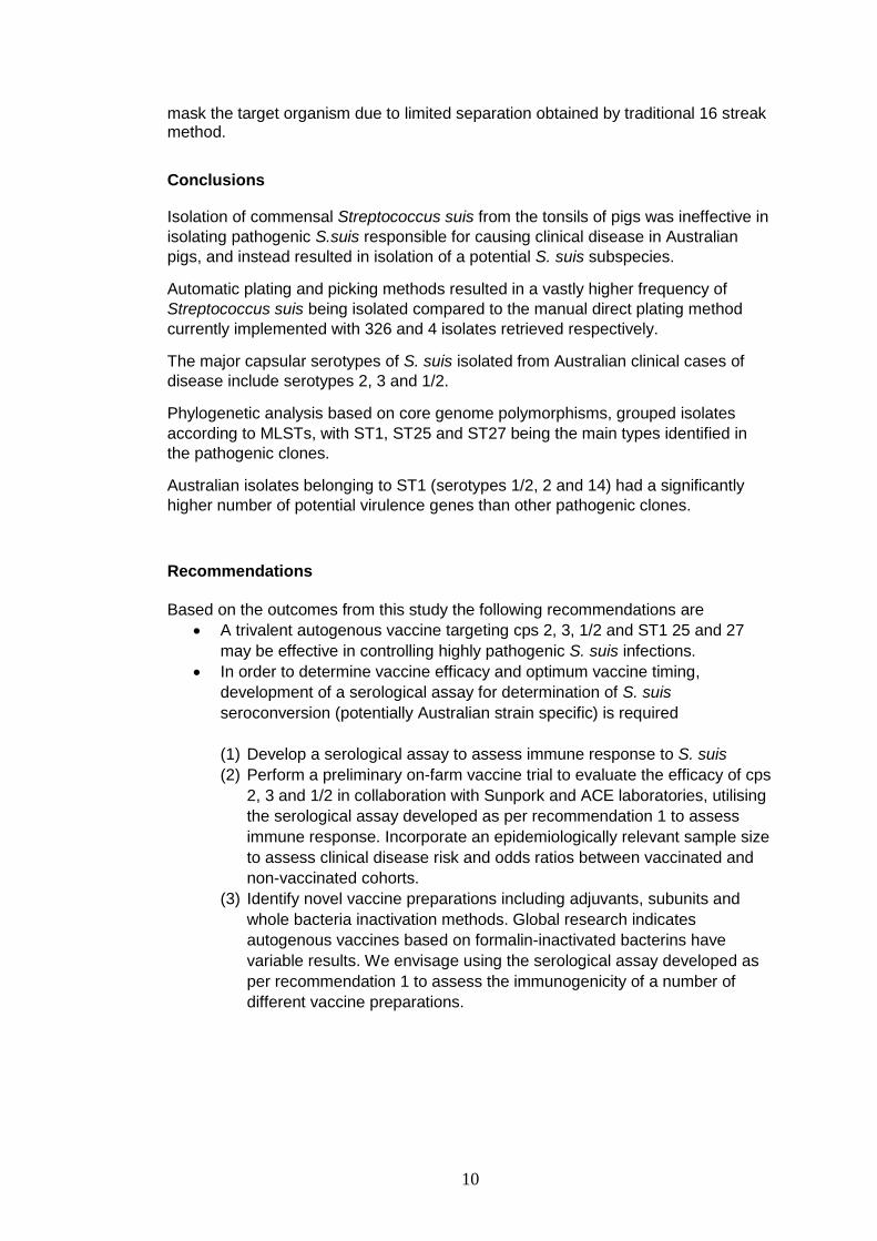
medium via an automated robotic streaking method with separation of over 100
colonies/ swab developed by the antimicrobial resistance and infectious diseases
laboratory, whereas swabs from Farm B were plated using traditional 16 streak
methods of manual direct plating. The plates were incubated throughout the project
at 37°C for 24 hours with 5% CO2. Following overnight incubation, up to 50 colonies
morphologically resembling Streptococcus suis from the Farm A plates were re-
cultured onto sheep blood agar (Edwards) and incubated. A single colony was
selected for re-culture colony from Farm B plates.
Identification of the colonies was undertaken using matrix assisted laser deionisation
time of flight (MALDI-TOF) (Bruker). All isolates confirmed to be S. suis were
transferred into microcentrifuge tubes containing BHI broth with 30% glycerol and
10% fetal calf serum. These were then stored at -80°C.
Clinical Streptococcus suis strains
A collection of 148 S. suis, isolated from clinically diseased pigs in Australian pig
herds, were sent to Murdoch University from ACE Laboratory Services. Isolates
spanned a seven year period with 2, 1, 29, 27, 9, 49 and 29 isolates from years
2010, 2011, 2013, 2014, 2015, 2016 and 2017 respectively, and two isolates for
which the year was unknown. Initial swabs from diseased pigs were obtained from
tissues outlined in Table 1. All swabs were streaked onto Columbia sheep blood

3
agar and plates incubated. A single colony was subcultured and incubated.
Confirmation of the identification of each isolate was performed using a MALDI-TOF
(Bruker) with isolates confirmed to be S. suis being harvested into microcentrifuge
tubes and stored at -80°C.
Table 1 Number of isolates and site of origin used in the study
Isolation Site Number of Isolates
Abdomen 2
Brain 12
Heart 12
Joint 6
Lung 93
Lymph node 1
Neck abscess 3
Upper respiratory tract 3
Unknown 16
MIC Testing
All isolates were subjected to antimicrobial susceptibility testing via broth
microdilution according to the Clinical Laboratory Standards Institute (CLSI)
Performance Standards.
PCR
DNA extraction for PCR reactions was performed on all the commensal isolates via
chelex method. All isolates were cultured overnight in 500mL LB broth (using a 96
deep well plate) prior to extraction. The plate was centrifuged at 4000 rpm for one
minute and supernatant was removed before 100 µL of MilliQ water was added to
each well, and all samples transferred to a PCR 96-well plate. Centrifugation of the
plate was repeated. The supernatant was removed with 100 µL of 6% chelex
(BioRad) added to all samples. The plate was placed in a thermocycler at 56°C for
20 mins followed by 100°C for 5 mins and then placed straight onto ice.
The presence of the S. suis gdh gene and the virulence genes mrp, sly and epf was
determined via PCR based on primers developed by Silva et al. (2006) and Liu et al.
(2013) (Table 2) [4, 5].
Table 2: primer sequences used in this study for detection of S. suis gdh, mrp, sly
and epf genes and capsular types 1, 2 and 3.
Gene Primer sequence
gdhF
gdhR
AAGTTCCTCGGTTTTGAGCA,
GCAGCGTATTCTGTCAAACG
mrpF
mrpR
ATTGCTCCACAAGAGGATGG,
TGAGCTTTACCTGAAGCGGT
slyF
slyR
GCTTGACTTACGAGCCACAA, CCGCGCAATACTGATAAGC

4
epfF
epfR
CGCAGACAACGAAAGATTGA, AAGAATGTCTTTGGCGATGG
cps1(14)F
cps1(14)R
TCTTATAACAGGCGTCAAAACA
ATCGGTATAAAAGCAAGACACA
cps2(1/2)F
cps2(1/2)R
TTCGTATTAACTTACTTGGCGT
TAAATCCCCATATGCCAAATCC
cps3
cps3
ACATCCATTGCAGGAGTAGT
TGCAGTTCCAAAATTCTTCGT
Whole genome sequencing
DNA extraction was performed using MagMax DNA multi sample kit (ThermoFisher
Scientific) with the only variation from the manufacturer’s instructions being the
omission of the RNAse treatment step. Whole genome sequencing library
preparation was conducted using a Nextera XT kit (Illumina). The tagmentation time
was increased to 7 minutes with all steps completed as per the manufacturer’s
instructions. Sequencing of the isolates was performed with a mid output V2 (2 x
150 cycles) kit on an Illumina Nextseq 500 platform.
Analysis of whole genome sequencing data was performed using the Nullarbor
bioinformatics pipeline (v1.20). Determination of capsular serotypes was performed
by querying contig files against a database of reference serotype genes using
Abricate (0.4) at a 90% cutoff. To distinguish serotype 2 from serotype 1/2 and
serotype 1 from serotype 14, analysis was manually performed using CLC
genomics. All contig files from the tonsillar strains were also run through the species
identification online tool JSpecies (v3.0.17).
Results
Isolation of Streptococcus suis
The total number of swabs processed was 300, inclusive of 150 (30 from suckers
and 60 from weaners and growers) from each farm. A total of 326 and 4 S. suis
strains as confirmed by MALDI-TOF were isolated from Farm A and Farm B
respectively, with the automatic robotic plating and picking method (Farm A) leading
to significantly higher numbers of S. suis isolated. From Farm A, 40 isolates were
from suckers (note only a single sample point), 209 isolates were obtained from
weaners and 77 isolates were obtained from growers. Farm B isolates were all from
weaners.
AMR Testing
A high proportion of the isolates were resistant to both tetracycline (99.3%) and
erythromycin (83.8%). Low levels of resistance were observed for florfenicol
(14.9%), penicillin G (8.1%), ampicillin (0.7%) and trimethoprim/ sulphamethoxazole
(0.7%). None of the isolates were resistant to enrofloxacin (Table 3)

5
Table 3: Distribution of minimum inhibitory concentrations for S. suis.
drug 0.13 0.25 0.5 1 2 4 8 16 32 64 128 256 ns_ci cr_ci Ampicillin 99.3 .7 0.7(0,3.7) 0.7(0,3.7) (147) (1) Ceftriaxone 88.5 7.4 2 1.4 .7 . . (131) (11) (3) (2) (1) Ciprofloxacin 37.2 37.2 22.3 3.4 . . (55) (55) (33) (5) Enrofloxacin 14.2 44.6 40.5 .7 0.7(0,3.7) 0(0,2.5) (21) (66) (60) (1) Erythromycin 11.5 4.1 .7 1.4 2 1.4 79.1 84.5(77.6,89.9) 83.8(76.8,89.3) (17) (6) (1) (2) (3) (2) (117) Florfenicol 2 4.1 79.1 11.5 .7 1.4 .7 .7 93.9(88.8,97.2) 14.9(9.6,21.6) (3) (6) (117) (17) (1) (2) (1) (1) Gentamicin 6.1 40.5 43.2 9.5 .7 . . (9) (60) (64) (14) (1) PenicillinG 81.8 4.7 4.7 3.4 3.4 .7 .7 12.8(7.9,19.3) 8.1(4.3,13.7) (121) (7) (7) (5) (5) (1) (1) Tetracycline .7 .7 1.4 2 1.4 93.9 100 (97.5,100) 99.3(96.3,100) (1) (1) (2) (3) (2) (139) TMS 91.9 5.4 2 .7 8.1(4.3,13.7) 0.7(0,3.7) (136) (8) (3) (1)
Percentage of isolates classified as non-susceptible (ns ci) and/or clinically resistant (cr ci) with corresponding 95% confidence intervals for
those where breakpoints are available. Shaded areas indicate the range of dilutions evaluated. Vertical bars indicate clinical breakpoint (where
available). TMS; trimethoprim/sulfamethoxazole

6
Determination of species and presence of virulence genes via PCR
In total, 342 isolates underwent PCR screening for gdh and virulence factors. 273
(79.8%) of isolates were positive on the gdh PCR, indicating S. suis or closely
related species. Putative virulence factor PCRs for mrp, sly and epf were positive for
146 (42.7%), 19 (5.6%) and 5 (0.5%) isolates respectively. No isolates were positive
on the cps1 PCR, and 3 (1.1%) and 22 (6.4%) were positive on cps 2 and cps 3
PCRs respectively. All other samples returned negative or indeterminate results. It
should be noted that no PCR results were confirmed by sequencing.
WGS and bioinformatics analysis A representative subsample of isolates (n=42) that was identified as S. suis by MALDI-TOFF underwent whole genome sequencing. 37 (37/42) sequenced strains identified most similarly to S. suis however the mean level of identification was low at 37% (range 5-70%),with the other strains identified as most similar to Streptococcus pneumoniae.
Genomic analysis of the 37 commensal Streptococcus suis isolates by WGS identified only 6 capsular serotypes across 10 isolates (Table 3). These 6 serotypes were 5, 9, 19, 28, 29 and 30. Twenty seven (73%) isolates had unidentifiable serotypes. None of the commensal isolates had identifiable multi locus sequence types. 30/37 (81%) were positive for the gdh, which was consistent with the proportion identified by targeted PCR. Table 4: Frequency of capsular serotypes detected from tonsillar swab isolates in this study
Capsular serotype Frequency
5 1
9 1
19 1
28 4
29 2
30 1
Three isolates were sent to DPIRD WA for Vitek biochemical analysis. Species
identifications of S. mitis/S. oralis grouping was returned for two isolates and the
third isolate could not be identified by Vitek analysis.
The dominant capsular serotypes detected amongst the clinical isolates were cps 2
(39/148), 3 (37/148) and 1/2 (11/14) (Figure 1). Other common serotypes include
cps 16 (n=8), 19 (n=8), 4 (n=7) and 8 (n=7) with a total of 20 different serotypes
detected. Genomic analysis of the 148 clinical disease isolates identified 11 different
known MLST with 39 (26.2%) isolates resulting in 26 newly identified MLSTs,
designated ST1031-ST1056 inclusive. The most common MLSTs were ST27
(n=27), ST25 (n=26), ST28 (n=11) and ST483 (n=11) (Figure 2).

7
Figure 1: S. suis serotypes from Australian clinical swine isolates (n=148)
Figure 2: S. suis MLSTs from Australian clinical swine isolates (n=148)
The carriage of antimicrobial resistance genes was similar between the commensal
and pathogenic strains with the most common resistance genes being tetO
(tetracycline) at 91.9% and 89.9% respectively, and ermB (erythromycin) at 89.2%
and 83.9% respectively. Other common genes in commensal and pathogenic strains
included lnuB (lincosamides) at 51.4% and 4.0% respectively, aadE
(aminoglycosides) at 48.6% and 2.7% respectively and fexA at 10.8% and 0.7%
(Table 4). All commensal isolates and 81.9% of pathogenic isolates carried genes
against multiple antimicrobial drug classes. The most common combination of genes
was tetO and ermB in 35.1% and 57.0% of commensal and pathogenic isolates
respectively.

8
Table 5: Proportion of S. suis isolates carrying antimicrobial resistance genes. Note
commensal strains refers to tonsillar isolates.
Gene % present in commensal
strains
% present in clinical
strains
aadD - 1.3
aadE 48.6 2.7
ant(6)-Ia 5.4 3.4
aph(3')-III 8.1 4.7
ermB 89.2 83.9
ermA - 6.0
fexA 10.8 0.7
lnuB 51.4 4.0
lnuC - 0.7
lsa 24.3 -
mefA 2.7 1.3
msrD 2.7 2.0
optrA - 2.7
Spc 2.7 5.4
tetO 91.9 89.9
tetM - 1.3
tetW - 5.4

9
Discussion
While not directly providing the outcomes anticipated when this study was designed, the findings have produced a large amount of information regarding Australian strains of S. suis, and the differences between commensal tonsillar flora and isolates from diseased pigs. Of particular interest was the degree of genetic difference between the tonsil isolates and the clinical isolates. While identified by MALDI-TOF as S. suis, many of the isolates did not display genetic signatures of S. suis, such as the presence of gdh, absence of putative virulence factors, inability to be genetically serotyped and non-typeable MLSTs. In addition to this, many of these isolates showed very low identities of 5-70% to S. suis using our in-house WGS pipeline, which returned identities of 60-95% for clinical, easily typed, isolates. Biochemical testing on a small number of isolates also did not detect S. suis. This indicates that that these commensal isolates may be normal tonsillar-pharyngeal bacteria, which are similar to S. suis, but are a separate species or subspecies which are not involved in disease, and it is our conclusion that these isolates are not representative of commensal S. suis causing clinical disease. It is likely the identity of these isolates could only be resolved through long read sequencing such as PacBio to obtain the complete genome. This was beyond the scope of this study. The isolation of these bacteria indicates that tonsil sampling in the live pig is unlikely to be suitable as a diagnostic tool, or as a tool to predict circulating clones from which to develop autogenous vaccines. While isolation of S. suis from tonsils is used in studies, it is generally from postmortem samples [6, 7], enabling deep sampling of the tonsillar crypts, potentially allowing access to S. suis which are not present on the tonsil surfaces. In this study, it is likely that swabs of the tonsil-pharyngeal region were dominated by recovery of indigenous oropharyngeal flora, which masked recovery of S. suis. In contrast to the results from tonsil swabs, WGS of clinical isolates obtained from postmortem samples demonstrated that circulating clones associated with disease in Australia are predominantly serotypes 2, 3 and 1/2, and MLSTs 1, 25, 27 and 483. The WGS pipeline used here has been demonstrated to rapidly determine clonal identity, including virulence potential and carriage of antimicrobial resistance genes, from clinical samples, and can be applied for future autogenous vaccine production. As part of this study, and incorporating results from concurrent work being undertaken, the development of a whole genome sequencing pipeline for evaluating multi-locus sequence types, virulence genes, antimicrobial resistance genes and phylogenetic relationships of S. suis, has been a particularly positive outcome. This pipeline has recently been used in field situations for selection of autogenous vaccine candidates for producers, and identification of pathogenic S. suis strains responsible for causing outbreaks on farms. This genomic pipeline will be of great benefit in the future for any further genomic studies on Australian S. suis strains, and rapid outbreak typing. We have also demonstrated that using conventional methods, the recovery of potential S. suis isolates was quite low. The robotics methods used in Farm A demonstrated that in a carriage study separating individual bacteria on selective agar is very important for identifying the carriage of non-disease causing indicative bacteria. The robotics method also allows much larger numbers of isolates to be picked and identified in a short timeframe. Standard manual streaking methods may
10
mask the target organism due to limited separation obtained by traditional 16 streak method.
Conclusions
Isolation of commensal Streptococcus suis from the tonsils of pigs was ineffective in
isolating pathogenic S.suis responsible for causing clinical disease in Australian
pigs, and instead resulted in isolation of a potential S. suis subspecies.
Automatic plating and picking methods resulted in a vastly higher frequency of
Streptococcus suis being isolated compared to the manual direct plating method
currently implemented with 326 and 4 isolates retrieved respectively.
The major capsular serotypes of S. suis isolated from Australian clinical cases of
disease include serotypes 2, 3 and 1/2.
Phylogenetic analysis based on core genome polymorphisms, grouped isolates
according to MLSTs, with ST1, ST25 and ST27 being the main types identified in
the pathogenic clones.
Australian isolates belonging to ST1 (serotypes 1/2, 2 and 14) had a significantly
higher number of potential virulence genes than other pathogenic clones.
Recommendations
Based on the outcomes from this study the following recommendations are
A trivalent autogenous vaccine targeting cps 2, 3, 1/2 and ST1 25 and 27
may be effective in controlling highly pathogenic S. suis infections.
In order to determine vaccine efficacy and optimum vaccine timing,
development of a serological assay for determination of S. suis
seroconversion (potentially Australian strain specific) is required
(1) Develop a serological assay to assess immune response to S. suis
(2) Perform a preliminary on-farm vaccine trial to evaluate the efficacy of cps
2, 3 and 1/2 in collaboration with Sunpork and ACE laboratories, utilising
the serological assay developed as per recommendation 1 to assess
immune response. Incorporate an epidemiologically relevant sample size
to assess clinical disease risk and odds ratios between vaccinated and
non-vaccinated cohorts.
(3) Identify novel vaccine preparations including adjuvants, subunits and
whole bacteria inactivation methods. Global research indicates
autogenous vaccines based on formalin-inactivated bacterins have
variable results. We envisage using the serological assay developed as
per recommendation 1 to assess the immunogenicity of a number of
different vaccine preparations.

11
References 1. Gurung, M., et al., Molecular Basis of Resistance to Selected Antimicrobial
Agents in the Emerging Zoonotic Pathogen Streptococcus suis. J Clin Microbiol, 2015. 53(7): p. 2332-6.
2. Segura, M., Streptococcus suis vaccines: candidate antigens and progress. Expert Rev Vaccines, 2015. 14(12): p. 1587-608.
3. Marois, C., et al., Detection and molecular typing of Streptococcus suis in tonsils from live pigs in France. Can J Vet Res, 2007. 71(1): p. 14-22.
4. Silva, L.M., et al., Virulence-associated gene profiling of Streptococcus suis isolates by PCR. Vet Microbiol, 2006. 115(1-3): p. 117-27.
5. Liu, Z., et al., Development of multiplex PCR assays for the identification of the 33 serotypes of Streptococcus suis. PLoS One, 2013. 8(8): p. e72070.
6. Luque, I., et al., Genetic analysis of Streptococcus suis isolates recovered from diseased and healthy carrier pigs at different stages of production on a pig farm. Vet J, 2010. 186(3): p. 396-8.
7. Ngo, T.H., et al., Slaughterhouse pigs are a major reservoir of Streptococcus suis serotype 2 capable of causing human infection in southern Vietnam. PLoS One, 2011. 6(3): p. e17943.

12
Appendix 1
Manual 16 streak plating method. Note marked overgrowth on plate and very few of the white/grey pinpoint colonies present as individual colonies suitable for subculture.
13
Robotic spiral plating method. Note much larger number of pinpoint white/grey colonies discernible as separate and suitable for subculture.