Embed Size (px)
Citation preview
Zornitsa Aleksieva1, Harpreet Auby1, Jill Prigge1, Miraj Shah1, Matt Vlasaty1, Rudolph Webster1, Wayne Oras1, Yu-Sheng Chen2, Grass Wang2, Tieyan Chang2, Ting-Zheng Xie3
1Hoffman Estates High School, Hoffman Estates, IL 60169 , 2ChemMatCARS, University of Chicago, Chicago, IL 60637, 3Polymer Science, University of Akron, Akron, OH 44325
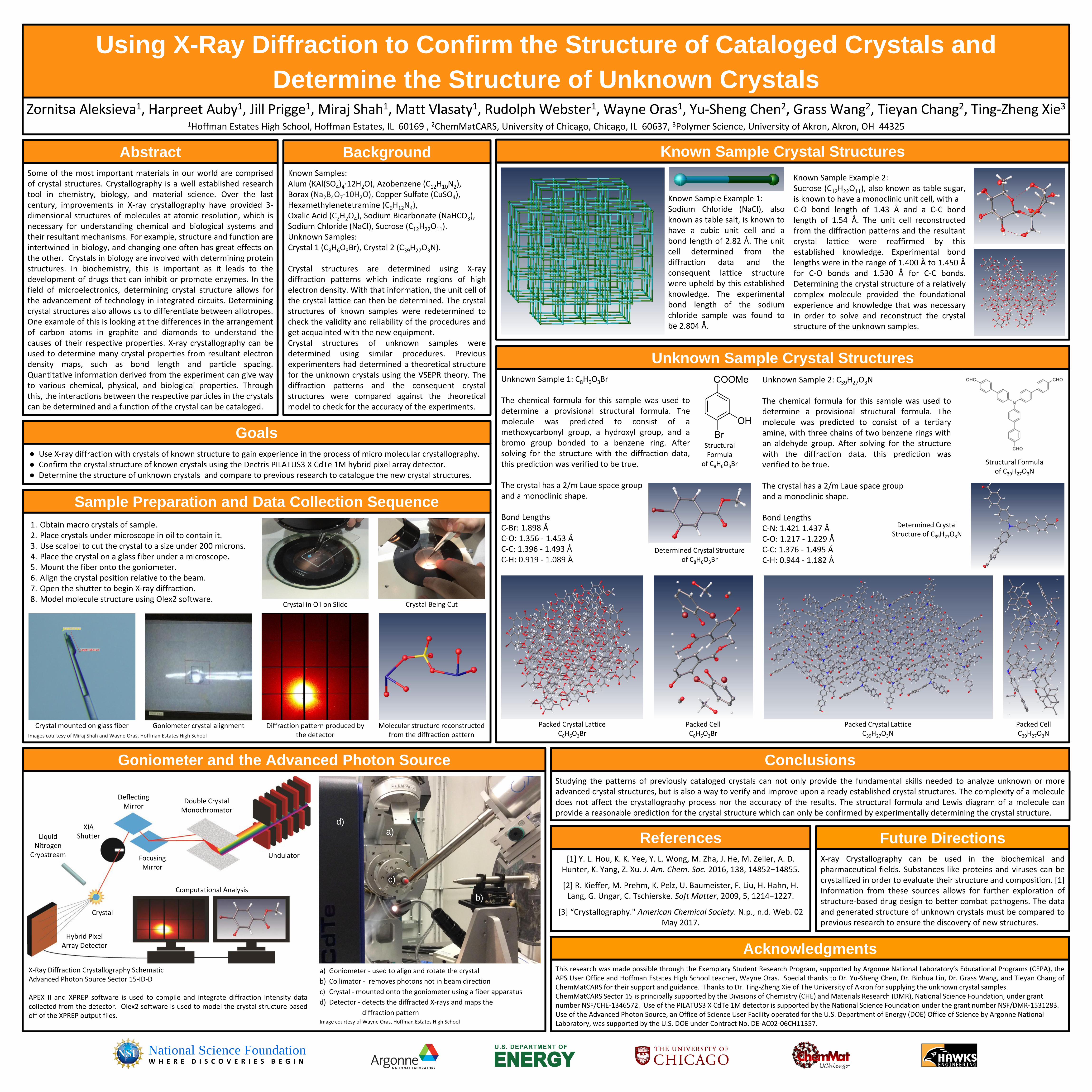
Using X-Ray Diffraction to Confirm the Structure of Cataloged Crystals and
Determine the Structure of Unknown Crystals
Some of the most important materials in our world are comprisedof crystal structures. Crystallography is a well established researchtool in chemistry, biology, and material science. Over the lastcentury, improvements in X-ray crystallography have provided 3-dimensional structures of molecules at atomic resolution, which isnecessary for understanding chemical and biological systems andtheir resultant mechanisms. For example, structure and function areintertwined in biology, and changing one often has great effects onthe other. Crystals in biology are involved with determining proteinstructures. In biochemistry, this is important as it leads to thedevelopment of drugs that can inhibit or promote enzymes. In thefield of microelectronics, determining crystal structure allows forthe advancement of technology in integrated circuits. Determiningcrystal structures also allows us to differentiate between allotropes.One example of this is looking at the differences in the arrangementof carbon atoms in graphite and diamonds to understand thecauses of their respective properties. X-ray crystallography can beused to determine many crystal properties from resultant electrondensity maps, such as bond length and particle spacing.Quantitative information derived from the experiment can give wayto various chemical, physical, and biological properties. Throughthis, the interactions between the respective particles in the crystalscan be determined and a function of the crystal can be cataloged.
Abstract
Known Samples: Alum (KAl(SO4)4·12H2O), Azobenzene (C12H10N2), Borax (Na2B4O7·10H2O), Copper Sulfate (CuSO4), Hexamethylenetetramine (C6H12N4),Oxalic Acid (C2H2O4), Sodium Bicarbonate (NaHCO3), Sodium Chloride (NaCl), Sucrose (C12H22O11).Unknown Samples:Crystal 1 (C8H6O3Br), Crystal 2 (C39H27O3N).
Crystal structures are determined using X-raydiffraction patterns which indicate regions of highelectron density. With that information, the unit cell ofthe crystal lattice can then be determined. The crystalstructures of known samples were redetermined tocheck the validity and reliability of the procedures andget acquainted with the new equipment.Crystal structures of unknown samples weredetermined using similar procedures. Previousexperimenters had determined a theoretical structurefor the unknown crystals using the VSEPR theory. Thediffraction patterns and the consequent crystalstructures were compared against the theoreticalmodel to check for the accuracy of the experiments.
Background
● Use X-ray diffraction with crystals of known structure to gain experience in the process of micro molecular crystallography.● Confirm the crystal structure of known crystals using the Dectris PILATUS3 X CdTe 1M hybrid pixel array detector.● Determine the structure of unknown crystals and compare to previous research to catalogue the new crystal structures.
Goals
X-Ray Diffraction Crystallography SchematicAdvanced Photon Source Sector 15-ID-D
APEX II and XPREP software is used to compile and integrate diffraction intensity datacollected from the detector. Olex2 software is used to model the crystal structure basedoff of the XPREP output files.
Goniometer and the Advanced Photon Source
b)
d)a)
c)
a) Goniometer - used to align and rotate the crystal
b) Collimator - removes photons not in beam direction
c) Crystal - mounted onto the goniometer using a fiber apparatus
d) Detector - detects the diffracted X-rays and maps the
diffraction patternImage courtesy of Wayne Oras, Hoffman Estates High School
Sample Preparation and Data Collection Sequence
Crystal mounted on glass fiber Goniometer crystal alignment Diffraction pattern produced by the detector
Molecular structure reconstructed from the diffraction pattern
Studying the patterns of previously cataloged crystals can not only provide the fundamental skills needed to analyze unknown or moreadvanced crystal structures, but is also a way to verify and improve upon already established crystal structures. The complexity of a moleculedoes not affect the crystallography process nor the accuracy of the results. The structural formula and Lewis diagram of a molecule canprovide a reasonable prediction for the crystal structure which can only be confirmed by experimentally determining the crystal structure.
Conclusions
This research was made possible through the Exemplary Student Research Program, supported by Argonne National Laboratory’s Educational Programs (CEPA), theAPS User Office and Hoffman Estates High School teacher, Wayne Oras. Special thanks to Dr. Yu-Sheng Chen, Dr. Binhua Lin, Dr. Grass Wang, and Tieyan Chang ofChemMatCARS for their support and guidance. Thanks to Dr. Ting-Zheng Xie of The University of Akron for supplying the unknown crystal samples.ChemMatCARS Sector 15 is principally supported by the Divisions of Chemistry (CHE) and Materials Research (DMR), National Science Foundation, under grant number NSF/CHE-1346572. Use of the PILATUS3 X CdTe 1M detector is supported by the National Science Foundation under the grant number NSF/DMR-1531283. Use of the Advanced Photon Source, an Office of Science User Facility operated for the U.S. Department of Energy (DOE) Office of Science by Argonne National Laboratory, was supported by the U.S. DOE under Contract No. DE-AC02-06CH11357.
X-ray Crystallography can be used in the biochemical andpharmaceutical fields. Substances like proteins and viruses can becrystallized in order to evaluate their structure and composition. [1]Information from these sources allows for further exploration ofstructure-based drug design to better combat pathogens. The dataand generated structure of unknown crystals must be compared toprevious research to ensure the discovery of new structures.
[1] Y. L. Hou, K. K. Yee, Y. L. Wong, M. Zha, J. He, M. Zeller, A. D. Hunter, K. Yang, Z. Xu. J. Am. Chem. Soc. 2016, 138, 14852−14855.
[2] R. Kieffer, M. Prehm, K. Pelz, U. Baumeister, F. Liu, H. Hahn, H. Lang, G. Ungar, C. Tschierske. Soft Matter, 2009, 5, 1214−1227.
[3] “Crystallography." American Chemical Society. N.p., n.d. Web. 02 May 2017.
Future DirectionsReferences
Acknowledgments
W H E R E D I S C O V E R I E S B E G I NNational Science Foundation
Images courtesy of Miraj Shah and Wayne Oras, Hoffman Estates High School
1. Obtain macro crystals of sample.2. Place crystals under microscope in oil to contain it. 3. Use scalpel to cut the crystal to a size under 200 microns.4. Place the crystal on a glass fiber under a microscope.5. Mount the fiber onto the goniometer. 6. Align the crystal position relative to the beam.7. Open the shutter to begin X-ray diffraction.8. Model molecule structure using Olex2 software.
Crystal Being CutCrystal in Oil on Slide
Unknown Sample 1: C8H6O3Br
The chemical formula for this sample was used todetermine a provisional structural formula. Themolecule was predicted to consist of amethoxycarbonyl group, a hydroxyl group, and abromo group bonded to a benzene ring. Aftersolving for the structure with the diffraction data,this prediction was verified to be true.
The crystal has a 2/m Laue space groupand a monoclinic shape.
Bond LengthsC-Br: 1.898 ÅC-O: 1.356 - 1.453 ÅC-C: 1.396 - 1.493 ÅC-H: 0.919 - 1.089 Å
Unknown Sample Crystal Structures
Unknown Sample 2: C39H27O3N
The chemical formula for this sample was used todetermine a provisional structural formula. Themolecule was predicted to consist of a tertiaryamine, with three chains of two benzene rings withan aldehyde group. After solving for the structurewith the diffraction data, this prediction wasverified to be true.
The crystal has a 2/m Laue space groupand a monoclinic shape.
Bond LengthsC-N: 1.421 1.437 ÅC-O: 1.217 - 1.229 ÅC-C: 1.376 - 1.495 ÅC-H: 0.944 - 1.182 Å
Packed Crystal LatticeC8H6O3Br
Determined Crystal Structure of C8H6O3Br
Packed CellC8H6O3Br
Structural Formulaof C39H27O3N
Packed Crystal LatticeC39H27O3N
Packed CellC39H27O3N
Known Sample Crystal Structures
Known Sample Example 1:Sodium Chloride (NaCl), alsoknown as table salt, is known tohave a cubic unit cell and abond length of 2.82 Å. The unitcell determined from thediffraction data and theconsequent lattice structurewere upheld by this establishedknowledge. The experimentalbond length of the sodiumchloride sample was found tobe 2.804 Å.
Known Sample Example 2:Sucrose (C12H22O11), also known as table sugar,is known to have a monoclinic unit cell, with aC-O bond length of 1.43 Å and a C-C bondlength of 1.54 Å. The unit cell reconstructedfrom the diffraction patterns and the resultantcrystal lattice were reaffirmed by thisestablished knowledge. Experimental bondlengths were in the range of 1.400 Å to 1.450 Åfor C-O bonds and 1.530 Å for C-C bonds.Determining the crystal structure of a relativelycomplex molecule provided the foundationalexperience and knowledge that was necessaryin order to solve and reconstruct the crystalstructure of the unknown samples.
Determined Crystal Structure of C39H27O3N
Undulator
Double CrystalMonochromator
FocusingMirror
DeflectingMirror
XIAShutter
Crystal
LiquidNitrogen
Cryostream
Hybrid PixelArray Detector
Computational Analysis
StructuralFormula
of C8H6O3Br