Embed Size (px)
Citation preview
JOURNAL OF CLINICAL MICROBIOLOGY, Nov. 1994, p. 2692-26970095-1137/94/$04.00+0Copyright © 1994, American Society for Microbiology
Epidemiologic Investigation by Macrorestriction Analysis and byUsing Monoclonal Antibodies of Nosocomial Pneumonia
Caused by Legionella pneumophila Serogroup 10tP. CHRISTIAN LUCK,`* JURGEN H. HELBIG,' ULLRICH GUNTER,2 MICHAEL ASSMANN,2
RALF BLAU,' HEIDEMARIE KOCH,3 AND MARIA KLEPP3
Institut fJr Medizinische Mikrobiologie und Hygiene, Universitatsklinikum, TU Dresden, D-01307 Dresden,,Kreiskrankenhaus Riesa, Innere Klinik, D-01589 Riesa,2 and Fachgebiet Krankenhaushygiene,
Landesuntersuchungsanstalt fuir das Gesundheits- und Veterinarwesen,Bereich Leipzig, D-04107 Leipzig,3 Germany
Received 7 February 1994/Returned for modification 11 April 1994/Accepted 8 August 1994
A 67-year-old woman was hospitalized with an acute pneumonia of the left lower lobe. Legionella pneumophilaserogroup 10 was cultured from two sputum specimens taken on days 18 and 20 and was also detected by directimmunofluorescence assay by using a commercially available species-specific monoclonal antibody as well as
serogroup 10-specific monoclonal antibodies. Antigenuria was detected in enzyme-linked immunosorbentassays by using serogroup 10-specific polyclonal and monoclonal antibodies. In the indirect immunofluores-cence test rising antibody titers against serogroups 1, 4, 5, 8, 9, 10, 14, and 15 were found in serum, with thehighest titers found against serogroups 8, 9, and 10. L. pneumophila serogroups 10 and 6 and a strain thatreacted with serogroup 4 and 14 antisera were cultured from both central and peripheral hot water systems ofthe hospital. Macrorestriction analyses of the genomic DNAs by pulsed-field gel electrophoresis showed thatthe isolate from the patient was identical to the serogroup 10 strains from the hospital hot water system. Incontrast, the genomic DNAs of 16 unrelated L. pneumophila serogroup 10 strains showed 12 different restrictionpatterns. Monoclonal antibody subtyping revealed only minor differences in L. pneumophila serogroup 10strains isolated from different sources. In conclusion, macrorestriction analysis is a valuable tool for studyingthe molecular epidemiology of L. pneumophila serogroup 10.
Legionellae can be found in both natural water systems(rivers, lakes, soils) and artificial water systems (hot watersupplies, cooling towers, dental units) (6, 8, 9, 17). In aquatichabitats they live in close association with free-living amoebaeand ciliated protozoa (17). Legionellae multiply within thevacuoles of the amoebae, especially at temperatures of be-tween 30 and 45°C. Infected amoebae or the legionellaereleased from amoebae spread from contaminated aquaticenvironments to susceptible persons and in this way may causehuman illness (17).
Legionella pneumophila is now recognized as an importantcause of nosocomial infections. Among hospitalized patients,those with impaired host defenses are at an increased risk ofacquiring legionellosis. Up to now, 15 serogroups of L. pneu-mophila have been defined by using absorbed polyclonal rabbitantisera (1, 2). Serogroup 1 is still the most common clinicaland environmental isolate; this is followed by serogroup 6 (8,17). Other serogroups account for 10 to 20% of clinical isolatesand 20 to 60% of environmental isolates. We describe here a
case of nosocomial legionellosis caused by L. pneumophilaserogroup 10. In addition, we report on the usefulness ofmacrorestriction analysis by pulsed-field gel electrophoresis(PFGE) for detecting the causative strain in the hospital watersupply.
* Corresponding author. Mailing address: Institut fur MedizinischeMikrobiologie und Hygiene, Universitatsklinikum, TU Dresden, Dur-erstrasse 24, D-01307 Dresden, Germany. Phone: 0049-351-4579 362.Fax: 0049-351-4573286.
t This paper is dedicated in memoriam to Karsten Seidel.
MATERIALS AND METHODS
Case report. A 67-year-old woman was admitted to thepsychiatric clinic (hospital A) for depression on 21 September1992. She was treated with tri- and tetracyclic thymolepticdrugs and dexamethasone. Except for a 5-day holiday overChristmas, she stayed in hospital A for 4 months before shedeveloped pneumonia on 20 January 1993. The patient wasfebrile (38.8°C) on admission to the internal medicine ward ofhospital B. Laboratory data and radiological findings wereconsistent with those reported for Legionella pneumonia (13).The patient was put on cefotiam therapy (4 g/day). On day 10the therapy was changed to roxithromycin (300 mg/day) be-cause of a clinical suspicion of Legionella pneumonia. Duringthis therapy the infiltrations regressed slowly but continuously.Bronchoscopy performed on day 22 revealed oropharyngealflora on sheep's blood and chocolate agars as well as a negativeculture for tuberculosis.
Microbiological methods for LegioneUla species. The immu-nofluorescence assay (IFA) technique with Formalin-killedbacteria was used for the Legionella species (13). Antigens forthe test were prepared from L. pneumophila serogroups 1 to14, L. pneumophila Lansing-3 (serogroup 15), L. micdadei, L.bozemanii serogroup 1, L. dumoffii, L. jordanis, and L. long-beachae serogroups 1 and 2. For the detection of antibodies tothese antigens in the patient's serum, polyvalent antibodyconjugate to human immunoglobulins (Institut fur Immun-praparate und Nahrmedien, Berlin, Germany) was used.Sputum samples were cultured on selective buffered char-
coal-yeast extract (BCYE) agar supplemented with 0.1% ot-ke-toglutarate (Sigma Corp., Munich, Germany), 0.3% glycine(Serva, Heidelberg, Germany), 1 mg of vancomycin (Lilly,
2692
Vol. 32, No. 11
Dow
nloa
ded
from
http
s://j
ourn
als.
asm
.org
/jour
nal/j
cm o
n 21
Nov
embe
r 20
21 b
y 12
2.25
4.18
1.25
0.
MOLECULAR EPIDEMIOLOGY OF L. PNEUMOPHILA SEROGROUP 10
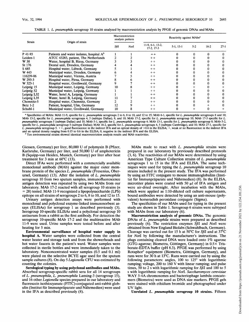
TABLE 1. L. pneumophila serogroup 10 strains analyzed by macrorestriction analysis by PFGE of genomic DNAs and MAbs
Macrorestriction Reactivity against MAbSaanalysis pattern
Strain Origin of strainSfil NotI 11-9, 6-1, 13-2, 5-1, 13-1 5-2 18-2 27-117-2, 17-3
P 41-93 Patients and water isolates, hospital A" 1 1 + + 0 0 0 0Leiden-1 ATCC 43283, patient, The Netherlands 2 2 + + 0 0 0 0W 38 Water, hospital B, Riesa, Germany 3 3 ++ 0 0 0 0St 176 Dental unit, Dresden, Germany 4 4 ++ 0 0 0 0S 685 Hospital water, Lubeck, Germany 5 1 + + 0 0 0 0W 443 Municipal water, Dresden, Germany 6 4 + + 0 0 0 0116239-86 Municipal water, Vienna, Austria 7 5 + + 0 0 0 0W 293-3 Hospital water, Pirna, Germany 8 6 + + 0 0 0 0W 323-1 Hospital water, Greifswald, Germany 9 7 + + 0 0 0 0Leipzig 15 Municipal water, Leipzig, Germany 10 8 + + 0 + 0 +Leipzig 52 Municipal water, Leipzig, Germany 1 1 +±+ 0 0 0 0Leipzig L32 Water, hotel A, Leipzig, Germany 1 1 + + 0 0 0 0Leipzig I35 Water, hotel B, Leipzig, Germany 11 7 + + 0 0 0 0Chemnitz-5 Hospital water, Chemnitz, Germany 2 2 + + 0 0 0 0Betz 1-2 Patient, hospital, Ulm, Germany 12 9 + + 0 0 + 0Schubl-1 Municipal water, Greifswald, Germany 10 8 + + 0 + 0 +
a Specificities of MAbs: MAb 11-9, specific for L. pneumophila serogroups 2 to 6, 8 to 10, and 12 to 15; MAb 6-1, specific for L. pneumophila serogroups 8 and 10;MAb 13-2, specific for L. pneumophila serogroups 4, 5 (subtype Dallas), 8, and 10; MAb 17-2, specific for L. pneunophila serogroup 10; MAb 17-3 specific for L.pneumophila serogroups 5 (subtype Dallas) and 10; MAb 5-1, specific for L. pneumophila serogroup 4; MAb 13-1, specific for L. pneumophila serogroup 8; MAb 5-2,specific for L. pneumophila serogroup 5 (subtype Dallas); MAb 18-2, specific for L. pneumophila serogroups 4, 5 (subtype Cambridge), and 10; MAb 27-1, specific forL. pneumophila serogroup 5. + +, good fluorescence in the indirect IFA and an optical density of >0.6 in the ELISA; +, weak or no fluorescence in the indirect IFAand an optical density ranging from 0.15 to 0.6 in the ELISA; 0, negative in the indirect IFA and the ELISA.
b Ten environmental strains showed identical macrorestriction analysis results and MAb reactivities.
Giessen, Germany) per liter, 80,000 U of polymyxin B (Pfizer,Karlsruhe, Germany) per liter, and 50,000 U of amphotericinB (Squipp-van Heiden, Munich, Germany) per liter after heattreatment for 3 min at 60°C (13).
Direct IFAs were performed with a commercially availablemonoclonal antibody (MAb) against the major outer mem-brane protein of the species L. pneumophila (Fresenius, Ober-ursel, Germany) (13). After the isolation of L. pneumophilaserogroup 10 from two sputum samples, immunofluorescenceantigen detection was repeated by using two MAbs from ourlaboratory. MAb 17-2 reacted with all serogroup 10 strains (n= 28) tested. MAb 11-9 recognized a lipopolysaccharide (LPS)epitope on all strains of serogroups 2 to 6, 8 to 10, and 12 to 15.
Urinary antigen detection assays were performed withmonoclonal and polyclonal enzyme-linked immunosorbent as-says (ELISAs) for serogroup 1 as described previously (3).Serogroup 10-specific ELISAs used a polyclonal serogroup 10antiserum from a rabbit as the first antibody. For detection theserogroup 10-specific MAb 17-2 and the multireactive MAb11-9 were used. Urine samples were tested before and afterheating for 5 min.
Environmental surveillance of hospital water supply inhospital A. Water samples were collected from the centralwater heater and storage tank and from the showerheads andhot water faucets in the patient's ward. Water samples werecollected in sterile bottles and were immediately taken to thelaboratory. Nonconcentrated water samples (0.5 and 0.1 ml)were plated on the selective BCYE agar used for the sputumsample cultures (8). On day 5 Legionella CFU was estimated bycounting the colonies.
Serological typing by using polyclonal antibodies and MAbs.Absorbed serogroup-specific rabbit sera for all 14 serogroupsof L. pneumophila, L. pneumophila Lansing-3 (serogroup 15),and 16 other Legionella species prepared in our laboratory andfluorescein isothiocyanate (FITC)-conjugated anti-rabbit glob-ulin (Institut fur Immunpraparate und Nahrmedien) were usedin the IFA as described previously (8).
MAbs made to react with L. pneumophila strains wereprepared in our laboratory by previously described protocols(3, 6). The reactivities of our MAbs were determined by usingAmerican Type Culture Collection strains of L. pneumophilaserogroups 1 to 15 in the IFA and ELISA. The same tech-niques were used for typing the L. pneumophila serogroup 10strains included in the present study. The IFA was performedby using an FITC conjugate to mouse immunoglobulins (Insti-tut fur Immunpraparate und Nahrmedien) (8). In the ELISA,microtiter plates were coated with 50 ,ul of IFA antigen andwere air-dried overnight. After incubation with the MAbs,which were applied as 1:10-diluted cell culture supernatants,bound antibodies were detected by using an anti-mouse (poly-valent) horseradish peroxidase conjugate (Sigma).The specificities of our MAbs used for typing in the present
study are shown in Table 1. Serogroup 6 strains were subtypedwith MAbs from our laboratory (6).
Macrorestriction analysis of genomic DNAs. The genomicDNAs of L. pneumophila strains were prepared as describedpreviously (6). The restriction enzymes Sfil and NotI wereobtained from New England Biolabs (Schwahlbach, Germany).Cleavage was carried out for 15 h at 50°C for Sfil and at 37°Cfor NotI by following the manufacturer's instructions. Theplugs containing cleaved DNA were loaded onto 1% agarose(GTG-agarose; Biometra, Gottingen, Germany) in 0.5x Tris-borate-EDTA buffer (pH 8.3). PFGE was performed by usingRotaphor° equipment (Biometra, Gottingen, Germany), andruns were for 30 h at 13°C. Runs were carried out by using thefollowing parameters: angles, 100 to 1250 with logarithmicramping; voltage, 200 to 160 V with linear ramping; and pulsetimes, 50 to 2 s with logarithmic ramping for Sfil and 100 to 5s with logarithmic ramping for NotI. Saccharomyces cerevisiaeWAY 5-4A chromosomes and bacteriophage lambda concate-mers (Biometra) were used as DNA size markers. PFGE gelswere stained with ethidium bromide and photographed underUV light.
Unrelated L. pneumophila serogroup 10 strains. Fifteen
VOL. 32, 1994 2693
Dow
nloa
ded
from
http
s://j
ourn
als.
asm
.org
/jour
nal/j
cm o
n 21
Nov
embe
r 20
21 b
y 12
2.25
4.18
1.25
0.
2694 LUCK ET AL.
unrelated L. pneumophila serogroup 10 strains were investi-gated for comparison (Table 1). These strains were eitherisolated in our laboratory or sent to us by colleagues. All butstrain Betz 1-2 were isolated from water specimens collected inGermany or Austria. Strain Betz 1-2 was cultured from asputum sample taken from a 65-year-old man suffering frompneumonia. The American Type Culture Collection strainLeiden-1 (ATCC 43283) was originally isolated during anoutbreak of nosocomial legionellosis in The Netherlands (9).
RESULTS
Microbiological results in a case of L. pneumophila sero-group 10 infection. Legionella-like colonies were isolated after4 days of incubation from two sputum samples from the casepatient obtained on days 18 and 20 of the illness. Altogether,18 colonies were picked and serotyped as serogroup 10 byusing absorbed rabbit antisera and MAbs. IFAs with thespecies (major outer membrane protein)-specific MAb, theserogroup 10 (LPS)-specific MAb 17-2, and MAb 11-9, whichrecognizes an LPS epitope present on all strains of serogroups2 to 6, 8 to 10, and 12 to 15, were positive for both sputumsamples. One sputum specimen collected on day 19 wasnegative by IFAs and culture.
Urinary antigen was detected by using serogroup 10-specificELISAs, but it was not found in our serogroup 1-specificassays. Both the serogroup 10-specific and cross-reactingMAbs used in the IFAs of sputum were able to detect urinaryantigen in two urine samples collected on days 18 and 19. Athird urine specimen (day 20) was positive in the ELISA withthe cross-reacting MAb 11-9.An acute-phase serological sample obtained on day 6 was
negative for serogroup 1 and revealed nonsignificant titers forserogroups 5, 8, 10, and 14. Serum samples collected latershowed significant rises in titer for serogroups 1, 4, 5, 8, 9, 10,12, 13, 14, and 15, with the highest titers (1,024/2,048) beingagainst serogroups 4, 5, 8, 10, and 14.Environmental studies. Legionellae were isolated from the
hot water tap in the patient's room, the hot water faucet andshowerhead in the bathroom of the patient's ward, as well asthe central heating boilers, the cold water inlet to one of them,and the main pipe connecting the central heating station to thewards.
Using absorbed rabbit antisera for serogroups 1 to 15, threedifferent strains were identified. The serogroup 10 strain wasthe prevalent one in all water samples taken from peripheralsites. Of 103 colonies tested, 70 (68%) belonged to thisserogroup. Thirteen strains were typed as serogroup 6, mono-clonal subtype Dresden. Twenty colonies did not react brightlywith our absorbed antisera for serogroups 1 to 15 but werepositive when unabsorbed antisera were used. The highesttiters were found against serogroups 4 and 14. This strain isthus recorded as either a new serogroup or a serovariant ofserogroups 4 or 14.
Legionella counts were between 10 and 300 CFU/ml. Exceptfor one cold water sample (12°C), the temperatures of all otherwater samples were between 30 and 46°C.Three weeks after the temperature in the central storage
tanks was raised to 60°C, the Legionella organism count wasfound to be reduced, but all three Legionella strains were stillpresent in the water system.
Serological subtyping of L. pneumophila serogroup 10strains. The results of subtyping of serogroup 10 strains withMAbs are given in Table 1. All serogroup 10 strains obtainedfrom the patient and related environmental sources as well as15 unrelated strains showed the same reaction patterns when
kB
1630800
610
460
245
48.5
(0
U) E 'r
CMl LO LL)Ln Xf com
L0..J.j NN
_Xe
) 7 := NU)~~ ~~~*U1.>1 Q t:
col C'M N. NLw m~~~~~~~~~~4
-j /-i c
FIG. 1. PFGE of SfiI-cleaved DNAs of the L. pneumophila sero-group 10 strains listed in Table 1. DNA sizes (in kilobases) areindicated to the left of the gel.
we used MAbs that recognized the major epitopes detectablein the IFA. Cross-reactive epitopes associated with strains ofserogroups 4, 5, 8, and 10 were found on the surfaces of a fewserogroup 10 strains. In terms of reactivity, they were attrib-uted as minor antigenic determinants, producing optical den-sities of from 0.15 to 0.6 in the ELISA. No significant reactivitywas found when these MAbs were used in the IFA. Theseresults showed that L. pneumophila serogroup 10 strains arehomogeneous in their antigenic compositions, with small dif-ferences in minor epitopes.
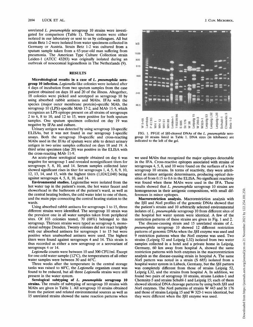
Macrorestriction analysis. Macrorestriction analysis withthe Sfil and NotI profiles of the genomic DNAs showed thatthe patient's strains and 10 arbitrarily selected environmentalisolates of L. pneumophila serogroup 10 from different sites ofthe hospital hot water system were identical. A few of therestriction patterns of these strains are given in Fig. 1 and 2.The disease-causing strain and 15 unrelated strains of L.
pneumophila serogroup 10 showed 12 different restrictionpatterns of genomic DNAs when the SfiI enzyme was used and9 restriction patterns when the NotI enzyme was used. Twostrains (Leipzig 52 and Leipzig L32) isolated from two watersamples collected in a hotel and a private home in Leipzig,Germany, 60 km away from hospital A, showed the samerestriction patterns with both enzymes in the macrorestrictionanalysis as the disease-causing strain in hospital A. The sameNotI pattern was noted in a strain (S 685) isolated from ahospital water system in Lubeck, Germany, but the Sfil patternwas completely different from those of strains Leipzig 52,Leipzig L32, and the strains from hospital A. In addition, wefound two pairs of serogroup 10 strains, strains Leiden-1 andChemnitz-5 and strains Schubl-1 and Leipzig 15; each of themshowed identical DNA cleavage patterns by using both Sfil andNotI enzymes. The NotI patterns of strains W 443 and St 176and those of strains Leipzig 15 and W 323-1 were identical, butthey were different when the Sfil enzyme was used.
J. CLIN. MICROBIOL.
Dow
nloa
ded
from
http
s://j
ourn
als.
asm
.org
/jour
nal/j
cm o
n 21
Nov
embe
r 20
21 b
y 12
2.25
4.18
1.25
0.
MOLECULAR EPIDEMIOLOGY OF L. PNEUMOPHILA SEROGROUP 10 2695
kB
160c
6CC
245
48
CI LO) LOLDCN Q n nrq~CIIC-
C1_, n
nf~ w6 N iZ OD0-cm )(D LoqtN N CL: : VC~~~~cL .Xc:Osm=mOnCN I Co
I-
gX¢ X> ¢¢ m >)-- -
a 3:
m 3i:u'r--FIG. 2. PFGE of NotI-cleaved DNAs of the L. pneumophila serogroup 10 strains listed in Table 1. DNA sizes (in kilobases) are indicated to
the left of the gel.
DISCUSSION
The clinical status of the patient described here providedunequivocal evidence of a hospital-acquired L. pneumophilaserogroup 10 infection. The primary illness in our patient wasdepression. She was treated with thymoleptic drugs and corti-costeroids, a known risk factor for acquiring legionellosis (17).The clinical course was typical for Legionnaires' disease (13).Because of a clinical suspicion of Legionella pneumonia thepatient was given roxithromycin therapy for 20 days. Duringthis therapy she slowly improved.The microbiological diagnosis was based on the isolation of
L. pneumophila serogroup 10 from two sputum samples,positive direct IFA results for sputum specimens, positiveresults in the assays for urinary antigen, and seroconversion.Only L. pneumophila serogroup 10 (18 colonies tested) wasgrown from two subsequent sputum samples. The serogroup ofthe isolated strains was confirmed in the IFA with bothabsorbed polyclonal antibodies and MAbs. It is noteworthythat the causative agent was cultured after 8 and 10 days ofroxithromycin therapy. This pointed out that attempts tocultivate legionellae from patient specimens are worthwhile,even when the patient has received Legionella-directed ther-apy. It might be speculated that the strain survived in macro-phages with impaired microbicidal function following cortico-steroid treatment and was still cultivable. Our results underlinethe fact that erythromycin, which acts bacteriostatically, shouldbe administered for at least 3 weeks to eliminate legionellae(17). For epidemiologic studies it is important to obtainLegionella strains from patients. The comparison of thesestrains with environmental isolates provides evidence of ordisproves the transmission of the strains from a suspectedsource of infection to humans.The species-specific MAb, which recognizes the major outer
membrane protein, the serogroup 10-specific MAb 17-2, andthe multireactive MAb 11-9, were successfully used for the
detection of L. pneumophila serogroup 10 in the two sputumsamples from which the strain was grown.
It was possible to detect urinary antigen by using serogroup10-specific assays with a polyclonal serogroup 10 antibody asthe first antibody. In this case both serogroup 10-specific andcross-reactive MAbs could be used to detect urinary antigen.The monoclonal and polyclonal serogroup 1-specific assaysused in our laboratory since 1987 failed to do so. With regardto the polyclonal serogroup 1-specific ELISA, this observationis in contrast to that in the report of Kohler et al. (5), whofound Legionella antigen in the urine of patients with culture-proven infections caused by serogroups 1, 4, and 10. It might bespeculated that the sensitivity of our assay was too low for ourpatient with the serogroup 10 infection. On the other hand, wereported the usefulness of the same polyclonal serogroup1-specific assay in a case of L. pneumophila serogroup 3infection verified by isolation of the causative strain (7). Onlylimited information is available concerning the cross-reactivi-ties of antigens detected in urine specimens. Our results pointto the necessity of using broad-spectrum assays for detectingurinary antigen. Tang et al. (16) reported the usefulness ofsuch an ELISA for diagnosing legionellosis caused by differentserogroups and species.An evaluation of the indirect IFA for antibody detection
with antigens other than L. pneumophila serogroup 1 has beenlimited by the rareness of culture-confirmed cases of infectioncaused by other serogroups. In our case the antibody responseagainst serogroup 1 was not significant until day 20. In contrast,in serum samples collected on days 6 and 13, presumptive titersof 128 and rising titers to 512, respectively, were found againstserogroup 8 and 10 strains. To improve the serological diag-nosis of legionellosis it is therefore recommended that multipleantigens be used in the IFA. Serogroups 4, 5, 8, and 10, whichare known to cross-react with each other (9), gave the highesttiters in our assay. Our results again confirm that serological
VOL. 32, 1994
Dow
nloa
ded
from
http
s://j
ourn
als.
asm
.org
/jour
nal/j
cm o
n 21
Nov
embe
r 20
21 b
y 12
2.25
4.18
1.25
0.
2696 LUCK ET AL.
data are of little value in epidemiologic studies because it wasimpossible to state with any certainty that the antibodies in thepatient's sera were induced by the disease-causing strain, L.pneumophila serogroup 10.
L. pneumophila serogroups 10, 6, and 4/14 were isolatedfrom central and peripheral outlets of the hospital watersystem. The disease-causing strain, L. pneumophila serogroup10, was the most prevalent, accounting for 68% of the coloniestested.
In the IFA all serogroup 10 strains used in the present studyreacted with a serogroup 10-specific MAb and with multireac-tive MAbs recognizing LPS epitopes on strains of differentserogroups. Considering these phenotypic characterizations,all serogroup 10 strains were identical. By the more sensitiveELISA technique, small differences were disclosed when cross-reactive MAbs were used. In conclusion, MAb typing is notuseful for the subtyping of L. pneumophila serogroup 10isolates. These findings are in contrast to observations con-cerning serogroup 1 strains, which could be divided into severalMAb subtypes (4).
For subtyping of L. pneumophila serogroup 10 strains weapplied macrorestriction analysis using PFGE. This techniquehas successfully been used for subtyping L. pneumophilaserogroups 1 and 6, according to recent reports (6, 12, 14).Macrorestriction analysis of the genomic DNAs of 1 arbitrarilyselected serogroup 10 strain from the patient as well as 10strains from the hot water supply system revealed identicalrestriction patterns for both Sfil and Notl enzymes (Fig. 1 and2, respectively). We are thus certain that the strain wastransferred from the hot water supply system to the patient.Our results provide further evidence that hot water systems arethe main source of Legionella infection, especially in immuno-compromised patients (9-12, 14). It could not be ascertainedwhere the patient acquired her infection, since the serogroup10 strain was isolated in the patient's room, in the bathroom(showerhead and faucet), the ward's physiotherapeutic cabi-net, as well as other wards of the hospital.
Cooling towers and air-conditioning systems, which havebeen reported previously to be sources of infection (17), werenot in operation in hospital A.Genome analysis revealed that the serogroup 10 strains
isolated from 16 different locations showed 12 different Sfi1and 9 different NotI cleavage patterns. A comparison of theSfi1 and NotI patterns indicated that both enzymes are useful insubtyping L. pneumophila serogroup 10 strains. The resultsobtained by these two techniques were in close agreement.However, the use of the restriction enzyme Sfi1 allowed thesubdivision of NotI patterns. This is not surprising since NotIproduced fewer bands than Sfi1 in the macrorestriction analy-sis. Interestingly, two strains (Leipzig 52 and Leipzig L32)isolated from the water systems of two different buildings inLeipzig, 60 km away from hospital A, were identical to thedisease-causing strain in hospital A. Two sets of L. pneumo-phila serogroup 10 strains, strains Leiden-1 and Chemnitz-5and strains Schubl-1 and Leipzig 35, isolated in locations atdistances of 1,500 and 750 km from each other, respectively,were shown to be identical to each other by using both enzymesas well as by MAb typing. These results underline the obser-vation made by Selander et al. (15), viz., that although Legio-nella pneumophila strains are genetically heterogeneous, adistribution of clones occurs throughout the world. Therefore,the investigation of Legionella outbreaks should include sub-typing data as well as epidemiologic information.A serogroup 10 strain (W 38) isolated from the hospital
water supply system in hospital B showed a completely differ-
ent restriction pattern. A nosocomial superinfection acquiredin hospital B could thus be excluded.The true incidence of serogroup 10 infections has not yet
been defined. In 1992, 33 culture-confirmed cases caused byserogroups 2 to 14 were reported by the European WorkingGroup on Legionella infections (8th Meeting, Vienna, 10 to 12May 1993). Of these, six (18%) were serogroup 10 infections.Thus, serogroup 10 is one of the prominent serogroups amongclinical isolates in Europe, bearing in mind that serogroup 1accounts for 70 to 80% of infections. This observation agreesvery well with our findings made while investigating hot watersupply systems in southeastern Germany (8).
L. pneumophila serogroup 10 strains are phenotypicallyuniform but heterogeneous in their genomic structures. Thus,macrorestriction analysis is a very powerful tool for subtypingstrains of this serogroup. To our knowledge this is the firstreport on the subtyping of serogroup 10 strains by macro-restriction analysis.
ACKNOWLEDGMENTS
We are grateful to Reinhard Marre, Ulm, Germany; GuntherWewalka, Vienna, Austria; Werner Ehret, Regensburg, Germany;Ingrid Carmienke, Leipzig, Germany; Frank Huke, Chemnitz, Ger-many; and Ingrid Schubl, Greifswald, Germany, for submission of L.pneumophila serogroup 10 strains. We thank Jutta Moller, SigridGabler, Sylvia Petsche, and Ines Wolf for technical assistance andVolker Bellmann for preparing the photographs.
This study was supported by the Deutsche Forschungsgemeinschaft(Lu 485/1-1).
REFERENCES1. Benson, R. F., W. L. Thacker, H. W. Wilkinson, R. J. Fallon, and
D. J. Brenner. 1988. Legionellapneumophila serogroup 14 isolatedfrom patients with fatal pneumonia. J. Clin. Microbiol. 26:382.
2. Brenner, D. J., A. G. Steigerwald, P. Epple, W. F. Bibb, R M.McKinney, R. W. Starnes, J. M. Colville, R K. Selander, P. H.Edelstein, and C. W. Moss. 1988. Legionella pneumophila sero-group Lansing 3 isolated from a patient with fatal pneumonia, anddescription of L. pneumophila subsp. pneumophila subsp. nov., L.pneumophila subsp. fraseri subsp. nov., and L. pneumophila subsp.pascullei subsp. nov. J. Clin. Microbiol. 26:1695-1703.
3. Helbig, J. H., P. C. Luck, C. Pilz, and W. Witzleb. 1990. Commonepitope on urinary antigen derived from different Legionellapneumophila serogroup 1 strains recognized by a monoclonalantibody. Zentralbl. Bakteriol. Parasitenkd. Infektionskr. Hyg.Abt. 1 Orig. 273:478-480.
4. Joly, J. R., R. M. McKinney, J. O'H. Tobin, W. F. Bibb, I. D.Watkins, and D. Ramsay. 1986. Development of a standardizedsubgrouping scheme for Legionella pneumophila serogroup 1 usingmonoclonal antibodies. J. Clin. Microbiol. 23:768-771.
5. Kohler, R B., L. J. Wheat, M. L. V. French, P. L. Meenhorst, W. C.Winn, and P. H. Edelstein. 1985. Cross-reactive urinary antigensamong patients with Legionella pneumophila serogroup 1 and 4and the Leiden 1 strain. J. Infect. Dis. 152:1007-1012.
6. Luck, P. C., L. Bender, M. Ott, J. H. Helbig, and J. Hacker. 1991.Analysis of Legionella pneumophila serogroup 6 strains isolatedfrom a hospital warm water supply over a three-year period usinggenomic long range mapping techniques and monoclonal antibod-ies. Appl. Environ. Microbiol. 57:3226-3231.
7. Luck, P. C., J. H. Helbig, E. Wunderlich, H. Foelske, M. Sel-bitschka, D. Wenzel, L. Patzold, and W. Witzleb. 1989. Isolation ofLegionella pneumophila serogroup 3 from pericardial fluid in acase of pericarditis. Infection 17:388-390.
8. Luck, P. C., I. Leupold, M. Hlawitschka, J. H. Helbig, I. Car-mienke, L. Jatzwauk, and T. Guderitz. 1993. Prevalence of Legio-nella species, serogroups, and monoclonal subgroups in hot watersystems in south-eastern Germany. Zentralbl. Bakteriol. Para-sitenkd. Infektionskr. Hyg. Abt. 1 Orig. 193:450-460.
9. Meenhorst, P. L., A. L. Reingold, D. Groothuis, G. W. Gorman,H. W. Wilkinson, R. M. McKinney, J. C. Feeley, D. J. Brenner, and
J. CLIN. MICROBIOL.
Dow
nloa
ded
from
http
s://j
ourn
als.
asm
.org
/jour
nal/j
cm o
n 21
Nov
embe
r 20
21 b
y 12
2.25
4.18
1.25
0.
MOLECULAR EPIDEMIOLOGY OF L. PNEUMOPHILA SEROGROUP 10 2697
van R. Furth. 1985. Water related nosocomial pneumonia causedby Legionella pneiumophila serogroups 1 and 10. J. Infect. Dis.152:356-363.
10. Muder, R. R., V. L. Yu, and J. J. Zuravleff. 1986. Mode oftransmission of Legionella pneumophila: a critical review. Arch.Intern. Med. 146:1607-1612.
11. Neill, M. A., G. W. Gorman, C. Gibert, A. Roussel, A. W.Hightower, R. M. McKinney, and C. V. Broome. 1984. Nosocomiallegionellosis, Paris, France-evidence of transmission by potablewater. Am. J. Med. 78:581-588.
12. Ott, M., L. Bender, R. Marre, and J. Hacker. 1991. Pulsed-fieldelectrophoresis of genomic restriction fragments for the detectionof nosocomial Legionella pneumophila in hospital water supplies. J.Clin. Microbiol. 29:813-815.
13. Rodgers, F. G., and A. W. Pasculle. 1991. Legionella, p. 442-453.In A. Ballows, W. J. Hausler, Jr., K. L. Herrmann, H. D. Isenberg,and H. J. Shadomy (ed.), Manual of clinical microbiology, 5th ed.American Society for Microbiology, Washington, D.C.
14. Schoonmaker, D., T. Heimberger, and G. Burkhead. 1992. Com-parison of ribotyping and restriction enzyme analysis using pulsed-field-gel-electrophoresis for distinguishing Legionella pnelumophilaisolates obtained during a nosocomial outbreak. J. Clin. Microbiol.30: 1491-1498.
15. Selander, R. K., R. M. McKinney, T. S. Whittam, W. F. Bibb, D. J.Brenner, F. S. Nolte, and P. E. Pattison. 1985. Genetic structure ofpopulations of Legionella pneumophila. J. Bacteriol. 163:1021-1037.
16. Tang, P. W., and C. Krishnan. 1993. Legionella antigenuria:six-year study of broad spectrum enzyme-linked immunosorbentassay as a routine diagnostic test, p. 12-13. In J. M. Barbaree, R. F.Breiman and A. P. Dufour (ed.), Legionella-current status andemerging perspectives. American Society for Microbiology, Wash-ington, D.C.
17. World Health Organization. 1990. Epidemiology, prevention andcontrol of legionellosis: memorandum from a WHO meeting. Bull.W. H. 0. 68:155-164.
VOL. 32, 1994
Dow
nloa
ded
from
http
s://j
ourn
als.
asm
.org
/jour
nal/j
cm o
n 21
Nov
embe
r 20
21 b
y 12
2.25
4.18
1.25
0.


![Guidelines for Prevention of Nosocomial Pneumonia · comial pneumonia. MMWR 1997;46(No. RR-1):[inclusive page numbers]. Copies can be purchased from Superintendent of Documents, U.S](https://img.pdfslide.us/doc/110x75/6052972dc7d81c05f22392ce/guidelines-for-prevention-of-nosocomial-pneumonia-comial-pneumonia-mmwr-199746no.jpg)


![Nosocomial Pneumonia: [Print] - eMedicine Infectious Diseases](https://img.pdfslide.us/doc/110x75/613cffa50c37c14a830ceb8a/nosocomial-pneumonia-print-emedicine-infectious-diseases.jpg)