Embed Size (px)
Citation preview
CLINICAL MICROBIOLOGY REVIEWS, Oct. 2006, p. 637–657 Vol. 19, No. 40893-8512/06/$08.00�0 doi:10.1128/CMR.00051-05Copyright © 2006, American Society for Microbiology. All Rights Reserved.
Ventilator-Associated Pneumonia: Diagnosis, Treatment, and PreventionSteven M. Koenig and Jonathon D. Truwit*
Pulmonary and Critical Care Medicine, University of Virginia, Charlottesville, Virginia 22908
INTRODUCTION .......................................................................................................................................................637DIAGNOSIS ................................................................................................................................................................637
Clinical Diagnosis ...................................................................................................................................................637Radiologic Diagnosis ..............................................................................................................................................638Microbiologic Diagnosis.........................................................................................................................................638
Blood and pleural fluid cultures.......................................................................................................................638Nonquantitative or semiquantitative airway sampling .............................................................................................638Quantitative cultures of airway specimens .....................................................................................................639
(i) Diagnostic accuracy ..................................................................................................................................641(ii) Bronchoscopic protected specimen brush and BAL............................................................................641(iii) Quantitative endotracheal aspirate ......................................................................................................641(iv) Blind BBS, PSB, and BAL .....................................................................................................................641(v) Comparisons among the different quantitative culturing techniques: bronchoscopic versus
nonbronchoscopic techniques................................................................................................................................642TREATMENT..............................................................................................................................................................643
Quantitative Culture Strategy...............................................................................................................................644Clinical Strategy......................................................................................................................................................646Antibiotic Management..........................................................................................................................................646
PREVENTION.............................................................................................................................................................649Preventing Multidrug Resistance .........................................................................................................................651
CONCLUSION............................................................................................................................................................652REFERENCES ............................................................................................................................................................652
INTRODUCTION
Patients in the intensive care unit (ICU) are at risk for dyingnot only from their critical illness but also from secondaryprocesses such as nosocomial infection. Pneumonia is the sec-ond most common nosocomial infection in critically ill pa-tients, affecting 27% of all critically ill patients (170). Eighty-sixpercent of nosocomial pneumonias are associated with mechan-ical ventilation and are termed ventilator-associated pneumonia(VAP). Between 250,000 and 300,000 cases per year occur in theUnited States alone, which is an incidence rate of 5 to 10 cases per1,000 hospital admissions (134, 170). The mortality attributable toVAP has been reported to range between 0 and 50% (10, 41, 43,96, 161). Studies have provided different results when deter-mining attributable mortality, in part because of very differentpopulations (less-acute trauma patients, acute respiratory dis-tress syndrome [ARDS] patients, and medical and surgicalICU patients) and in part as a result of variances in appropri-ate empirical medical therapy during the initial 2 days. Fur-thermore, the organisms recovered have an impact on out-come, with higher mortality rates seen in VAP caused byPseudomonas aeruginosa, Acinetobacter spp., and Stenotroph-omonas maltophilia (109). Beyond mortality, the economics ofVAP include increased ICU lengths of stays (LOS) (from 4 to13 days), and incremental costs associated with VAP have been
estimated at between $5,000 and $20,000 per diagnosis (20,206, 211).
Ventilator-associated pneumonia is defined as pneumoniaoccurring more than 48 h after patients have been intubatedand received mechanical ventilation. Diagnosing VAP requiresa high clinical suspicion combined with bedside examination,radiographic examination, and microbiologic analysis of respi-ratory secretions. Aggressive surveillance is vital in under-standing local factors leading to VAP and the microbiologicmilieu of a given unit. Judicious antibiotic usage is essential, asresistant organisms continue to plague intensive care units andcritically ill patients. Simple nursing and respiratory therapyinterventions for prevention should be adopted. Over the pastseveral decades our understanding of VAP has grown signifi-cantly with regard to pathogenesis, risk factors, diagnostic test-ing, therapies, and prevention by modifying risk factors. Thispaper is designed for the practicing clinician in addressingdiagnosis, treatment, and prevention of VAP.
DIAGNOSIS
Clinical Diagnosis
Ventilator-associated pneumonia is usually suspected whenthe individual develops a new or progressive infiltrate on chestradiograph, leukocytosis, and purulent tracheobronchial secre-tions. Unfortunately, and unlike for community-acquired pneu-monia, accepted clinical criteria for pneumonia are of limiteddiagnostic value in definitively establishing the presence ofVAP. In a postmortem study by Fabregas et al., when findingson histologic analysis and cultures of lung samples obtained
* Corresponding author. Mailing address: Pulmonary and CriticalCare Medicine, P.O. Box 800546, UVa HS, Charlottesville, VA 22908.Phone: (434) 924-5270. Fax: (434) 924-9682. E-mail: [email protected].
637
on March 22, 2019 by guest
http://cmr.asm
.org/D
ownloaded from
immediately after death were used as references, a new andpersistent (�48-h) infiltrate on chest radiograph plus two ormore of the three criteria (i) fever of �38.3°C, (ii) leukocytosisof �12 � 109/ml, and/or (iii) purulent tracheobronchial secre-tions had a sensitivity of 69% and a specificity of 75% forestablishing the diagnosis of VAP (60). When all three clinicalvariables were required for the diagnosis, the sensitivitydeclined further (23%); the use of a single variable resultedin a decrease in specificity (33%). The poor accuracy ofclinical criteria for diagnosing VAP should not be surprisingconsidering that purulent tracheobronchial secretions areinvariably present in patients receiving prolonged mechan-ical ventilation and are seldom caused by pneumonia. Inaddition, the systemic signs of pneumonia such as fever,tachycardia, and leukocytosis are nonspecific; they can becaused by any state that releases the cytokines interleukin-1,interleukin-6, tumor necrosis factor alpha, and gamma in-terferon (33, 34, 63, 135). Examples of such conditions in-clude trauma, surgery, the fibroproliferative phase of ARDS,deep vein thrombosis, pulmonary embolism, and pulmonaryinfarction. Reasonable clinical criteria for the suspicion ofVAP include a new and persistent (�48-h) or progressiveradiographic infiltrate plus two of the following: temperatureof �38°C or �36°C, blood leukocyte count of �10,000 cells/mlor �5,000 cells/ml, purulent tracheal secretions, and gas ex-change degradation (5, 103).
The sensitivity of the clinical criteria for VAP outlined aboveis even lower in patients with ARDS, where it may be difficultto detect new radiographic infiltrates. In the setting of ARDS,Bell et al. reported a false-negative rate of 46% for the clinicaldiagnosis of VAP (11). Consequently, suspicion for VAP in thesetting of ARDS should be high. The presence of even one ofthe clinical criteria for VAP, unexplained hemodynamic insta-bility, or an unexplained deterioration in arterial blood gasesshould prompt consideration of further diagnostic testing(129).
When purulent sputum, a positive sputum culture, fever, andleukocytosis are present without a new lung infiltrate, the di-agnosis of nosocomial tracheobronchitis should be enter-tained. In mechanically ventilated patients, nosocomial tra-cheobronchitis has been associated with a longer ICU stay andtime on the ventilator, without increased mortality (158). Inone randomized trial of intubated patients with community-acquired tracheobronchitis, antibiotic therapy resulted in a de-creased incidence of subsequent pneumonia and mortality(156). However, prospective, randomized, controlled trials arerequired before antibiotic therapy can be recommended forthe routine treatment of nosocomial tracheobronchitis. Fur-thermore, differentiation of tracheobronchitis from pneumo-nia is dependent upon the radiograph, which in the ICU isportable and often of poor quality. Hence, the clinician shouldutilize a clinical pulmonary infection score (CPIS) (see below)to direct therapy.
Radiologic Diagnosis
While the portable chest radiograph still remains a manda-tory component in the diagnosis of ventilated patients withsuspected pneumonia, as with clinical criteria for diagnosingVAP, it too has problems with both sensitivity and specificity.
Poor-quality films further compromise the accuracy of chest Xrays. Although a normal chest radiograph makes VAP unlikely,in one study of surgical patients, 26% of opacities were de-tected by computed tomography (CT) scan but not by portablechest X ray (25). In addition, asymmetric pulmonary infiltratesconsistent with VAP can be caused by numerous noninfectiousdisorders, including atelectasis, chemical pneumonitis, asym-metric cardiac pulmonary edema, pulmonary embolism, cryp-togenic organizing pneumonia, pulmonary contusion, pulmo-nary hemorrhage, drug reaction, and asymmetric ARDS. Theoverall radiographic specificity of a pulmonary opacity consis-tent with pneumonia is only 27% to 35% (116, 216).
Nonetheless, because of their high specificity, certain chestradiograph findings can be useful in establishing the diagnosisof pneumonia when present. Based on several studies, includ-ing an autopsy study by Wunderink et al., these useful findingsinclude rapid cavitation of the pulmonary infiltrate, especiallyif progressive; an air space process abutting a fissure (specific-ity, 96%); and an air bronchogram, especially if single (speci-ficity, 96%). Unfortunately, such radiographic abnormalitiesare uncommon (216).
Microbiologic Diagnosis
Blood and pleural fluid cultures. Although VAP spreads tothe blood or pleural space in �10% of cases, if an organismknown to cause pneumonia is cultured in the setting of clini-cally suspected pneumonia, treatment is warranted. Conse-quently, most experts recommend that two sets of blood cul-tures and a thoracentesis for nonloculated pleural effusions of�10 mm in diameter on a lateral decubitus chest radiographshould be part of the evaluation of suspected VAP (30). If theeffusion is loculated, ultrasound guidance may be required.However, it is important to keep in mind not only that thesensitivity of blood cultures for the diagnosis of VAP is lessthan 25% but also that when positive, the organisms mayoriginate from an extrapulmonary site of infection in as manyas 64% of cases and even when VAP is present (23, 124).
Nonquantitative or semiquantitative airway sampling. Gramstaining and nonquantitative and semiquantitative cultures oftracheal secretions have the advantages of reproducibility andof requiring little technical expertise and no specialized equip-ment or technique. However, these studies add little to thesensitivity and specificity of the clinical diagnosis of VAP, asthe upper respiratory tract is rapidly, within hours of intuba-tion, colonized by potential pulmonary pathogens, even whenpneumonia is not present (57, 91). Thus, if an organism iscultured or noted on Gram stain, one does not know if it is thecause of the pneumonia or simply colonization. In a study of 48patients with respiratory failure, concordance between trachealnonquantitative cultures and cultures of lung tissue from openlung biopsy was only 40% (82). In that study, of those patientswith pneumonia on lung histology, endotracheal aspirate(ETA) had a sensitivity of 82% but a specificity of only 27%. Inaddition, routine surveillance cultures of ETAs to anticipatethe etiology of a subsequent pneumonia can be misleading in asignificant percentage of patients, though recent data indicatethat quantitative ETAs may be helpful (see below) (78, 146).
Only 15% of ETAs are adequate specimens when strictdefinitional criteria (organisms on Gram staining and fewer
638 KOENIG AND TRUWIT CLIN. MICROBIOL. REV.
on March 22, 2019 by guest
http://cmr.asm
.org/D
ownloaded from
than 10 squamous epithelial cells per low-power field [magni-fication, �100]) are followed (153). Furthermore, the numberof polymorphonuclear leukocytes is not predictive of an inter-pretable specimen in patients with VAP (153). Nonquantita-tive and semiquantitative cultures of ETAs for the diagnosis ofVAP are most useful if the specimen is adequate and antimi-crobial therapy has not been added or changed in the prior72 h. The negative predictive value of these cultures in thissetting is high (94%) (15). Alternative causes for the patient’spresentation, including nonpulmonary sites of infection,should be investigated. In addition, the absence of growth ofmultidrug-resistant organisms in this circumstance providesstrong evidence that these bacteria are not causative. Antibi-otics should be adjusted accordingly. Overall, the presence ofprior antibiotics results in a false-negative rate of 10 to 40%(200).
Because of the poor specificity of the clinical diagnosis ofVAP and of qualitative evaluation of ETAs, Pugin et al. de-veloped a composite clinical score, called the clinical pulmo-nary infection score (CPIS), based on six variables: tempera-ture, blood leukocyte count, volume and purulence of trachealsecretions, oxygenation, pulmonary radiography, and semi-quantitative culture of tracheal aspirate. The score varied from0 to 12. A CPIS of �6 had a sensitivity of 93% and a specificityof 100% (164). However, there were several limitations of thatinvestigation, including that only 28 patients with 40 episodesof pneumonia were studied and that the diagnosis was basedupon a “bacterial index” that has not been a well-acceptedreference test for pulmonary infection. The “bacterial index” isthe sum of the logarithm of the number of bacteria culturedper milliliter of bronchoalveolar lavage (BAL) fluid. Two sub-sequent studies evaluated the accuracy of the CPIS by usingboth histology and lung tissue cultures as the reference tests(60, 162). In these investigations, the sensitivity was 72% to77%, and the specificity was 42% to 85%. However, the studywith the greater diagnostic accuracy used the tracheal aspirateculture recorded within the 48 to 72 h preceding the investi-gation, information that may not be available routinely. More-over, with a sensitivity of 72 to 77%, a CPIS of �6 is stillinsufficient to withhold antibiotic therapy safely in patientswith suspected VAP.
Using quantitative culture of BAL fluid as the diagnosticcriteria for VAP and a CPIS of �5 as the diagnostic cutoff, thesensitivity and specificity of the CPIS were 83% and 17%,respectively. The addition of Gram staining via blind or bron-choscopically directed BAL or PTC (see below) did improvethe overall sensitivity and specificity of the CPIS (65). How-ever, the false-negative rate was still 16 to 25% (65).
Due to poor specificity and poor positive predictive value,reliance on clinical parameters, chest X-ray findings, and non-and semiquantitative sputum analysis will result in overdiag-nosis and therefore overtreatment of VAP. Such an approachwill result in excess antibiotic use with its attendant cost, po-tential toxicity, and selection of drug-resistant organisms. Arecent decision analysis suggested that more deaths occurred ifpatients were treated with antibiotics on the basis of onlyclinical suspicion of VAP than if antibiotics were withheld(190).
Overreliance on the clinical diagnosis of VAP may also re-sult in undertreating alternative infectious and noninfectious
causes of fever and pulmonary infiltrates in mechanically ven-tilated patients. Meduri et al. prospectively studied 50 patientswith clinically suspected VAP (139). Twenty-two patients hadARDS. Based on quantitative cultures of bronchoscopic pro-tected specimen brush (PSB) and bronchoalveolar lavage,pneumonia was diagnosed in 42%. Of the infectious causes offever and pulmonary infiltrates on chest radiography, 84%were pneumonia, sinusitis, urinary tract infection, or catheter-related infection. Less frequent infectious causes included in-tra-abdominal abscess, peritonitis, acalculous cholecystitis,Clostridium difficile colitis, empyema, wound infection, primarybacteremia, and candidemia. Twenty-four percent of feverswere secondary to noninfectious causes, including deep venousthrombosis, pulmonary embolism, pancreatitis, chemical aspi-ration, fibroproliferative stage of ARDS, and drugs. Fifty-sixpercent of the chest X-ray abnormalities were due to nonin-fectious causes. Concomitant infections were found in 62% ofcases, with 60% of these being caused by a different pathogen.On average, there were 1.7 causes of fever per patient.
Quantitative cultures of airway specimens. To potentiallyimprove the specificity of the diagnosis of VAP and the con-sequent unnecessary antibiotic use and its associated problems,numerous studies have investigated the role of quantitativecultures of respiratory secretions. These have included non-bronchoscopic methods such as quantitative cultures of ETAs(QEAs) and sampling of secretions from distal airways“blindly” via an endobronchial catheter. Blind bronchial sam-pling (BBS), PSB, protected telescoping catheter (PTC), BAL,and protected BAL (mini-BAL) samples can be obtained viathe latter method. Bronchoscopic sampling methods permitquantitative cultures of PSB, PTC, and protected and nonpro-tected BAL specimens.
The PSB and PTC are double-sheathed catheters with abiodegradable plug occluding the distal end of the inner cath-eter to minimize bacterial contamination. The PSB and PTCprocedures involve placing the tip of the bronchoscope or“blindly placed” catheter next to an involved bronchial seg-mental orifice. With bronchoscopy, direct visualization is pos-sible. With a “blind” procedure, the catheter is advanced untilresistance is met and then retracted a few centimeters. Theinner catheter is then advanced 2 or 3 cm beyond the outercatheter, ejecting the plug. With PSB, a brush is further ad-vanced and rotated several times; with PTC, a 10-ml syringe isused to perform three brief aspirations of secretions. BALinvolves the infusion and aspiration of sterile saline through aflexible fiber-optic bronchoscope or “blindly placed” catheterwedged into a bronchial segmental orifice. Protected BALinvolves a specialized balloon-tipped catheter with a distalejectable plug. When performing a BAL to diagnose VAP,instillation of at least 140 ml of saline is required to maximizediagnostic yield (70, 138, 145).
If a bronchoscopically directed quantitative culture is cho-sen, the patient should receive adequate sedation, with consid-eration of a short-acting paralytic agent to prevent coughingduring the procedure. The endotracheal tube must be �1.5mm larger than the external diameter of the flexible broncho-scope. The patient should receive a fraction of inspired oxygen(FiO2) of 100%, and positive-end expiratory pressure shouldbe reduced as much as tolerated. To maximize ventilation andminimize air trapping, the peak inspiratory flow should be
VOL. 19, 2006 VENTILATOR-ASSOCIATED PNEUMONIA 639
on March 22, 2019 by guest
http://cmr.asm
.org/D
ownloaded from
decreased to �60 liters/min, the respiratory rate set between10 and 20 breaths/min, and the peak inspiratory pressure alarmincreased. The patient should be carefully monitored through-out the procedure, with particular attention to exhaled tidalvolume, peak inspiratory pressure, oxygen saturation, the elec-trocardiogram, and vital signs. Secondary hypotension shouldbe anticipated, and appropriate intravenous fluids and vaso-pressors should be available for immediate administration(70).
The sampling area should be chosen based on the location ofthe infiltrate on chest X ray or CT scan. This typically corre-sponds to the bronchial segment with purulent secretionsand/or where endobronchial abnormalities are maximal, whichcan be clues in the setting of diffuse pulmonary infiltrates orminimal changes in a previously abnormal chest X ray (137).When in doubt, sample the posterior right lower lobe, sinceautopsy studies have indicated that VAP frequently involvesthis area (61, 92, 130, 173). Multiple specimens are no moreaccurate than single specimens (136).
As with nonquantitative and semiquantitative cultures, onlyadequate specimens should be processed. The presence ofmore than 1% epithelial cells or 10 epithelial cells per low-power field (magnification, �100) in bronchoscopic or “blind”BAL, PSB, PTC, or bronchial sampling suggests heavy oropha-ryngeal colonization. Returns of �10% of the instilled BALfluid are probably not representative of the lower respiratorytract (70). Since interpretation of such specimens is unreliable,they should not be cultured. For QEAs, the same criteriamentioned above for nonquantitative and semiquantitative cul-tures of an ETA should be utilized.
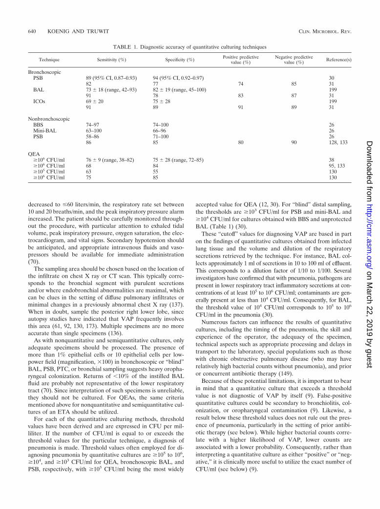
For each of the quantitative culturing methods, thresholdvalues have been derived and are expressed in CFU per mil-liliter. If the number of CFU/ml is equal to or exceeds thethreshold values for the particular technique, a diagnosis ofpneumonia is made. Threshold values often employed for di-agnosing pneumonia by quantitative cultures are �105 to 106,�104, and �103 CFU/ml for QEA, bronchoscopic BAL, andPSB, respectively, with �105 CFU/ml being the most widely
accepted value for QEA (12, 30). For “blind” distal sampling,the thresholds are �103 CFU/ml for PSB and mini-BAL and�104 CFU/ml for cultures obtained with BBS and unprotectedBAL (Table 1) (30).
These “cutoff” values for diagnosing VAP are based in parton the findings of quantitative cultures obtained from infectedlung tissue and the volume and dilution of the respiratorysecretions retrieved by the technique. For instance, BAL col-lects approximately 1 ml of secretions in 10 to 100 ml of effluent.This corresponds to a dilution factor of 1/10 to 1/100. Severalinvestigators have confirmed that with pneumonia, pathogens arepresent in lower respiratory tract inflammatory secretions at con-centrations of at least 105 to 106 CFU/ml; contaminants are gen-erally present at less than 104 CFU/ml. Consequently, for BAL,the threshold value of 104 CFU/ml corresponds to 105 to 106
CFU/ml in the pneumonia (30).Numerous factors can influence the results of quantitative
cultures, including the timing of the pneumonia, the skill andexperience of the operator, the adequacy of the specimen,technical aspects such as appropriate processing and delays intransport to the laboratory, special populations such as thosewith chronic obstructive pulmonary disease (who may haverelatively high bacterial counts without pneumonia), and prioror concurrent antibiotic therapy (149).
Because of these potential limitations, it is important to bearin mind that a quantitative culture that exceeds a thresholdvalue is not diagnostic of VAP by itself (9). False-positivequantitative cultures could be secondary to bronchiolitis, col-onization, or oropharyngeal contamination (9). Likewise, aresult below these threshold values does not rule out the pres-ence of pneumonia, particularly in the setting of prior antibi-otic therapy (see below). While higher bacterial counts corre-late with a higher likelihood of VAP, lower counts areassociated with a lower probability. Consequently, rather thaninterpreting a quantitative culture as either “positive” or “neg-ative,” it is clinically more useful to utilize the exact number ofCFU/ml (see below) (9).
TABLE 1. Diagnostic accuracy of quantitative culturing techniques
Technique Sensitivity (%) Specificity (%) Positive predictivevalue (%)
Negative predictivevalue (%) Reference(s)
BronchoscopicPSB 89 (95% CI, 0.87–0.93) 94 (95% CI, 0.92–0.97) 30
82 77 74 85 31BAL 73 � 18 (range, 42–93) 82 � 19 (range, 45–100) 199
91 78 83 87 31ICOs 69 � 20 75 � 28 199
91 89 91 89 31
NonbronchoscopicBBS 74–97 74–100 26Mini-BAL 63–100 66–96 26PSB 58–86 71–100 26
86 85 80 90 128, 133
QEA�106 CFU/ml 76 � 9 (range, 38–82) 75 � 28 (range, 72–85) 38�106 CFU/ml 68 84 95, 133�105 CFU/ml 63 55 130�106 CFU/ml 75 85 130
640 KOENIG AND TRUWIT CLIN. MICROBIOL. REV.
on March 22, 2019 by guest
http://cmr.asm
.org/D
ownloaded from
(i) Diagnostic accuracy. Before discussing and comparingthe diagnostic accuracies of the different quantitative culturingmethods and their advantages and disadvantages, it is impor-tant to understand that there exists a significant amount ofcontroversy in the medical literature regarding the use of quan-titative cultures in general as well as the use of bronchoscopic(invasive) versus nonbronchoscopic (noninvasive) methods. Atleast part of the reason for this controversy is that the studiesevaluating the accuracies of ETA and of bronchoscopic andnonbronchoscopic PSB, PTC, and BAL to diagnose VAP haveshown a significant degree of variability in sensitivity, specific-ity, and positive and negative predictive values for each of thetechniques. This variability has resulted from the use of differ-ent “gold standards” for the diagnosis of VAP, the use ofdifferent cutoff thresholds for quantitative cultures, differencesin equipment and protocols, and differences between the pop-ulations studied, in particular, the use of antibiotics. Even themost accepted “gold standard,” histopathologic examinationand culture of lung tissue obtained by biopsy or at autopsy, hasinherent problems. Among other things, patients included inautopsy studies may not be representative of most patients withVAP. Moreover, the recognition of histologic pneumonia var-ies among pathologists. In a study by Corley et al., the preva-lence of pneumonia in postmortem open lung biopsies deter-mined by each of four pathologists varied from 18% to 38%(40). Nonetheless, histopathology and lung tissue culture re-main our best “gold standard” for the diagnosis of VAP.
Numerous studies have demonstrated that prior and concur-rent antibiotic therapy decrease the accuracy, sensitivity, andnegative predictive value of Gram staining, including the per-centage of cells containing intracellular organisms (ICOs), aswell as quantitative, semiquantitative, and nonquantitative cul-tures (184). In a study of 76 patients with VAP by Montraverset al., PSB quantitative cultures obtained after the administra-tion of effective antibiotic therapy showed complete eradica-tion of the causative organisms after only 3 days of treatmentin 67% of patients (150). Even 24 h of administration of anantibiotic can affect culture results (189). This effect of priorantibiotics on the false-negative rate of microbiologic studies isof great concern, particularly since VAP is a potentially lethaldisorder. However, if antibiotics have not been changed in thelast 72 h, the diagnostic yield of any culture technique is un-affected (189, 197).
(ii) Bronchoscopic protected specimen brush and BAL.Chastre and Fagon pooled the results of 18 studies that eval-uated the bronchoscopically directed PSB technique for diag-nosing VAP (30). A total of 795 critically ill patients wereincluded in the analysis. The overall diagnostic accuracy of thismethod was very good, with a sensitivity of 89% (95% confi-dence interval [CI], 87 to 93%) and a specificity of 94% (95%CI, 92 to 97%). However, studies investigating the reproduc-ibility and variability of bronchoscopic PSB have raised con-cerns about this technique. Timsit et al. and Marquette et al.repeated PSB sampling in the same lung subsegment andnoted that results of quantitative cultures were on each side ofthe 103-CFU/ml threshold in 16.7% to 13.6% of cases, respec-tively, with 59 to 67% of samples having CFU/ml counts vary-ing by more than 10-fold (131, 196). These investigators con-cluded that, as with all quantitative culturing techniques,borderline PSB quantitative culture results should be inter-
preted with caution. In such a circumstance, one should con-sider repeating the test if suspicion of VAP persists and anti-biotics have not yet been started.
Torres and El-Ebiary reviewed 23 studies that evaluated theaccuracy of bronchoscopic BAL in diagnosing VAP. A total of957 patients were included in the analysis. Sensitivity rangedfrom 42 to 93%, with a mean � standard deviation of 73% �18%. The specificity ranged from 45 to 100%, with a meanspecificity � standard deviation of 82% � 19% (199). In 12studies, the detection of ICOs in 2 to 5% of recovered cells hada sensitivity of 69% � 20% and a specificity of 75% � 28% fordiagnosing VAP (199). Reproducibility of BAL is excellentwhen the culture is sterile. However, for positive cultures, thequantitative repeatability was only 53% in one study (73).
Chastre et al. compared PSB and BAL to the “gold stan-dard” histopathologic findings and quantitative tissue cultureresults from the same areas of lungs of patients in the terminalphase of their illness. Patients were included in the study onlyif they never had pneumonia or had acquired it during theterminal phase of their illness. Antibiotics had not beenchanged or added in the 3 days prior to the sampling. In thisinvestigation, PSB had a sensitivity of 82%, a specificity of77%, a positive predictive value of 74%, and a negative pre-dictive value of 85%; BAL had a sensitivity of 91%, a specificityof 78%, a positive predictive value of 83%, and a negativepredictive value of 87%; and the presence of �5% ICOs had asensitivity, specificity, positive predictive value, and negativepredictive value of 91%, 89%, 91%, and 89%, respectively(31).
(iii) Quantitative endotracheal aspirate. Using a thresholdvalue of �106 CFU/ml, the sensitivity and specificity of QEAhave varied widely from study to study. Sensitivity ranged from38% to 82% with a mean of 76% � 9%; specificity ranged from72% to 85% with a mean of 75% � 28%. When the diagnosisof VAP was based on postmortem lung examination, the sen-sitivity/specificity for 105-CFU/ml and 106-CFU/ml thresholdswere 63%/75% and 55%/85%, respectively (130). Patients onantibiotics were included in that study, which may have de-creased the sensitivity of the procedure.
Jourdain et al. found the QEA to have a sensitivity and aspecificity of as high as 68% and 84%, respectively, and afalse-negative rate of as high as 32% compared to broncho-scopic quantitative PSB and BAL (95, 133). The diagnosticthresholds for QEA, PSB, and BAL in that study were �106
CFU/ml, �103 CFU/ml and �5% ICOs, respectively. In addi-tion, only 40% of the organisms isolated from QEAs wereconcomitantly isolated from PSB specimens. Strengths of thisstudy included a well-defined “gold standard,” which is as closeas one can get to the “true gold standard” of histopathologyand lung tissue culture, and the absence of an addition to orchange in antibiotics in the 3 days prior to the appearance ofthe new pulmonary infiltrate.
(iv) Blind BBS, PSB, and BAL. Campbell reviewed 15 stud-ies evaluating the accuracy of blinded sampling methods (26).A total of 654 episodes of pneumonia were included in theanalysis. Sensitivities for BBS, mini-BAL, and PSB were 74 to97%, 63 to 100%, and 58 to 86%, respectively. Specificitiesranged from 74 to 100% for BBS, from 66 to 96% for mini-BAL, and from 71 to 100% for PSB. Marik and Brown com-pared blind PSB to PSB performed by bronchoscopy. In that
VOL. 19, 2006 VENTILATOR-ASSOCIATED PNEUMONIA 641
on March 22, 2019 by guest
http://cmr.asm
.org/D
ownloaded from
study both diagnostic techniques were performed in the ab-sence of antibiotic therapy and blind PSB preceded bronchos-copy, to minimize contamination of the lower respiratory tract.In addition, the study used a well-defined and reasonable “goldstandard” for the diagnosis of VAP, though not histopatho-logic and lung tissue culture. In that investigation, blind PSBhad a sensitivity of 86%, a specificity of 85%, a positive pre-dictive value of 80%, and a negative predictive value of 90%(128, 133).
(v) Comparisons among the different quantitative culturingtechniques: bronchoscopic versus nonbronchoscopic techniques.Inherent advantages of nonbronchoscopic techniques includeless invasiveness; less compromise of oxygenation, ventilation,and respiratory mechanics during the procedure; less likeli-hood of increasing intracranial pressure; less likelihood of in-ducing arrhythmias; availability where there is no bronchosco-pist; lack of contamination presented by the bronchoscopicchannel; availability to patients with small endotracheal tubes;and lower cost. Of the quantitative techniques, QEA is leastinvasive, most readily available, and least expensive, and itrequires the least experience and is easily repeatable.
Where comparisons have been made, the authors of moststudies have concluded that the diagnostic accuracies of non-bronchoscopic and bronchoscopic techniques are similar.Nonetheless, and although not noted by all studies, certaingeneralizations regarding the overall medical literature can bemade.
(i) In some studies, the concordance between the sensitivityof bronchoscopic versus nonbronchoscopic quantitative cul-tures has been only approximately 80% (30, 94, 140). Conse-quently in some patients, particularly if the pneumonia is notdiffuse and involves the left lung or upper lobes, the diagnosisof VAP could be missed by blind sampling.
(ii) Compared to nonbronchoscopic sampling methods,bronchoscopic quantitative cultures have greater specificity(104, 162).
(iii) Because BAL samples larger areas of lung, it as at leastas sensitive as PSB and PTC (135). The sensitivity and negativepredictive value of a culture for pneumonia are affected by thesize of the sampling area and the amount of retrieved secre-tions. Bronchoalveolar lavage, which samples approximately 1million alveoli, is estimated to recover 5 to 10 times the num-ber of organisms obtained by PSB. Quantitative endotrachealaspirates would likewise be expected to provide more repre-sentative samples than PSB and PTC. Combining the results ofPSB and BAL may increase sensitivity (187).
(iv) Other “technical” advantages of BAL over PSB are thatthe technique of smear preparation for direct microscopic ex-amination of BAL is better established and that BAL is lesslikely to cause bleeding (28).
(v) Protected sampling methods such as PSB, PTC, andprotected BAL, because they “bypass” the oropharyngeal andupper airway bacterial contamination/colonization, have supe-rior specificity and positive predictive values (135).
(vi) Protected specimen brush is more specific than sensitivefor the diagnosis of VAP. Consequently, a positive result in-creases the likelihood of pneumonia being present (199).
(vii) Direct visualization of the airways by bronchoscopypermits sampling from the airway that corresponds to the ab-normal region on chest X ray or CT scan, to purulent secre-
tions, and/or to maximal endobronchial abnormalities. Conse-quently, bronchoscopy should theoretically improve the sensitivityof the procedure, particularly for pneumonias involving the upperlobes and the left lung (94, 115).
(a) Timsit et al. reported that the presence of two or more ofthe following had a sensitivity of 78% and a specificity of 89%for diagnosing VAP: a decrease in the partial pressure ofarterial oxygen (PaO2)/FiO2 ratio of �50 mmHg, distal puru-lent secretions, or persistence of distal secretions surging fromdistal bronchi during exhalation (195).
(b) In a study with autopsy serving as the gold standard todiagnose VAP, a bronchoscopic BAL with �50% neutrophildifferential had a 100% negative predictive value for the diag-nosis of VAP (98).
(viii) Compared to QEA and BBS, blind and bronchoscopicPSB, PTC, and BAL provide additional information that maybe clinically useful.
(a) In one study of PSB, having fewer than 10% neutrophilson direct examination was uniformly associated with negativecultures, a finding that would contribute significantly to thespecificity and positive predictive value of the procedure (142).
(b) Immediate performance of a direct microscopic exami-nation, BAL, PBC, and PTC also enable a search for ICOs. If�2 to 5% of recovered cells contain ICOs, this result canpotentially serve as a guide for the initial selection of empiricaltherapy. Unfortunately, as indicated above, the accuracy of thisprocedure is too low to be clinically useful in most circum-stances. Not surprisingly, concomitant antibiotic administra-tion increases the likelihood of false-negative results (53, 205).Moreover in one study, one-third of episodes of VAP causedby Pseudomonas aeruginosa were associated with negative di-rect stainings (205). Consequently, in the majority of cases, anegative direct staining still requires initial broad-spectrumantibiotics until culture results are returned.
(c) In a recent study by Michel et al., QEAs performed twicea week anticipated the etiology of a subsequent pneumonia in83% of cases (146). This contrasts with the results of similarstudies utilizing nonquantitative cultures of ETAs (78).
The key question, however, is not which quantitative cultur-ing technique is more accurate but whether these techniquesaffect outcomes from VAP. Four studies have prospectivelyevaluated the impact of an invasive diagnostic strategy on themorbidity of, the use of antimicrobial drugs in, and the mor-tality of VAP. Unfortunately, the four studies had differentdesigns as well as methodological flaws, including an inappro-priate selection of diagnostic techniques for comparison, in-sufficient control for previous antimicrobial treatment, incon-sistent ways of managing antibiotic treatment in patients whohad negative microbiologic assays, and insufficient power todetect clinically important differences among alternativestrategies.
In the three, randomized, controlled Spanish studies, nodifferences in mortality and morbidity were found when eitherinvasive (PSB and/or BAL) or QEA techniques were used todiagnose VAP (175, 176, 186). However, these studies con-tained relatively few patients (51, 76, and 88 patients) andtherefore were not powered sufficiently to demonstrate a dif-ference in mortality. Moreover, antibiotics were continued inall patients, thereby negating one of the major potential ad-vantages of any diagnostic test in patients clinically suspected
642 KOENIG AND TRUWIT CLIN. MICROBIOL. REV.
on March 22, 2019 by guest
http://cmr.asm
.org/D
ownloaded from
of having VAP. In other studies, it has been shown that anti-biotics can be safely stopped in patients with negative quanti-tative cultures (30). Although these studies did not demon-strate any difference in clinical outcomes, they did confirm thatinvasive tools are associated with a greater ability to narrow ordiscontinue antibiotics.
In a large, multicenter, randomized, unblinded French studyof 413 critically ill patients with a clinical suspicion of pneu-monia, bronchoscopy with quantitative cultures of PSB or BALwas compared to nonquantitative endotracheal aspirates (64).Patients with recent changes in antibiotic therapy were ex-cluded, limiting the ability to generalize the results. Patients inthe invasive diagnostic group had more antibiotic-free days ina 28-day period (11.4 versus 7.5 days), fewer antibiotics per day(1.0 versus 1.3), and less organ dysfunction at day 3 and 7. Themortality rate at 14 days was significantly lower in the invasivediagnostic group (16.2% versus 25.8%; P � 0.022); there wasno difference at 28 days. However, when a multivariate analysiswas performed, there was an improvement in mortality at 28days (hazard ratio, 1.54; 95% confidence interval, 1.10 to 2.16;P � 0.01).
One problem with this study that could have confoundedresults was that the invasive diagnostic group had a much lowerrate of inappropriate initial antibiotics (1 patient [0.5%] versus24 patients [13%]; P � 0.001). Of the cohort that receivedinappropriate antibiotics, 33% died, including all in the non-invasive diagnostic group before day 14. Numerous studieshave documented the importance of appropriate and earlyantimicrobial therapy for VAP (2, 90, 125). Therefore, theimproved outcome at 14 days in the invasive diagnostic groupmay have been secondary to higher use of inappropriate anti-biotics in the noninvasive diagnostic group and not to theactual invasive procedures.
A recent meta-analysis of the impact of invasive approacheson the diagnosis of VAP concluded that invasive lower airwaysampling does not alter hospital mortality but consistently re-sults in changes to the antibiotic regimen (182). In that study,the odds ratio for change in antibiotic management after in-vasive testing was 2.85 (95% CI, 1.45 to 5.59). In anotheroutcomes study, the invasive bronchoscopic evaluation of VAPwas also shown to allow de-escalation or narrowing of antibi-otics to occur once organisms and their susceptibilities were
identified (169). In contrast, in a decision analysis of antibioticand diagnostic strategies for VAP, Ost et al. concluded thatfrom the perspective of minimizing cost, minimizing antibioticuse, and maximizing survival, the best strategy was employingmini-BAL and treating with three antibiotics (160). Whilemini-BAL did not improve survival, it did decrease cost andantibiotic use (160).
At the present time, based on the available data, theoptimal strategy for diagnosing VAP remains to be defined.The American Thoracic Society (ATS)/Infectious DiseaseSociety of America guidelines do provide expert opinionsupporting quantitative or semiquantitative cultures of respi-ratory specimens, although the panel favors invasive quantita-tive techniques (5). However, a large, better matched, multi-center, randomized study comparing quantitative cultures ofendotracheal aspirates to quantitative cultures of broncho-scopic specimens to a clinical strategy using scoring systemsand nonquantitative and semiquantitative cultures is stillneeded. Potential confounding variables such as antibiotic reg-imens and antibiotic discontinuation protocols must be con-trolled. Until such evidence exists, the use of invasive broncho-scopic techniques cannot be required for the routine diagnosisof VAP. Therefore, which diagnostic approach for VAP shouldbe undertaken is up to the discretion of the clinician. Factorsto consider include local experience, expertise, availability, andcost.
TREATMENT
Principles to apply when choosing appropriate therapy forVAP include knowledge of organisms likely to be present, localresistance patterns within the ICU, a rational antibiotic regi-men, and a rationale for antibiotic de-escalation or stoppage.Although the clinician could know the organisms and sensitiv-ities prior to the development of VAP (see “Antibiotic Man-agement” below), this is often not the case. In the latter situ-ation, empirical choices that provide adequate coverage arecritical. Early effective therapy for VAP is associated withreduced mortality. Luna et al. demonstrated that inadequatetherapy during the initial 48 h, despite provision of adequatetherapy after BAL results, was associated with a mortality rateof 91% (125). When empirical therapy was appropriate, mor-
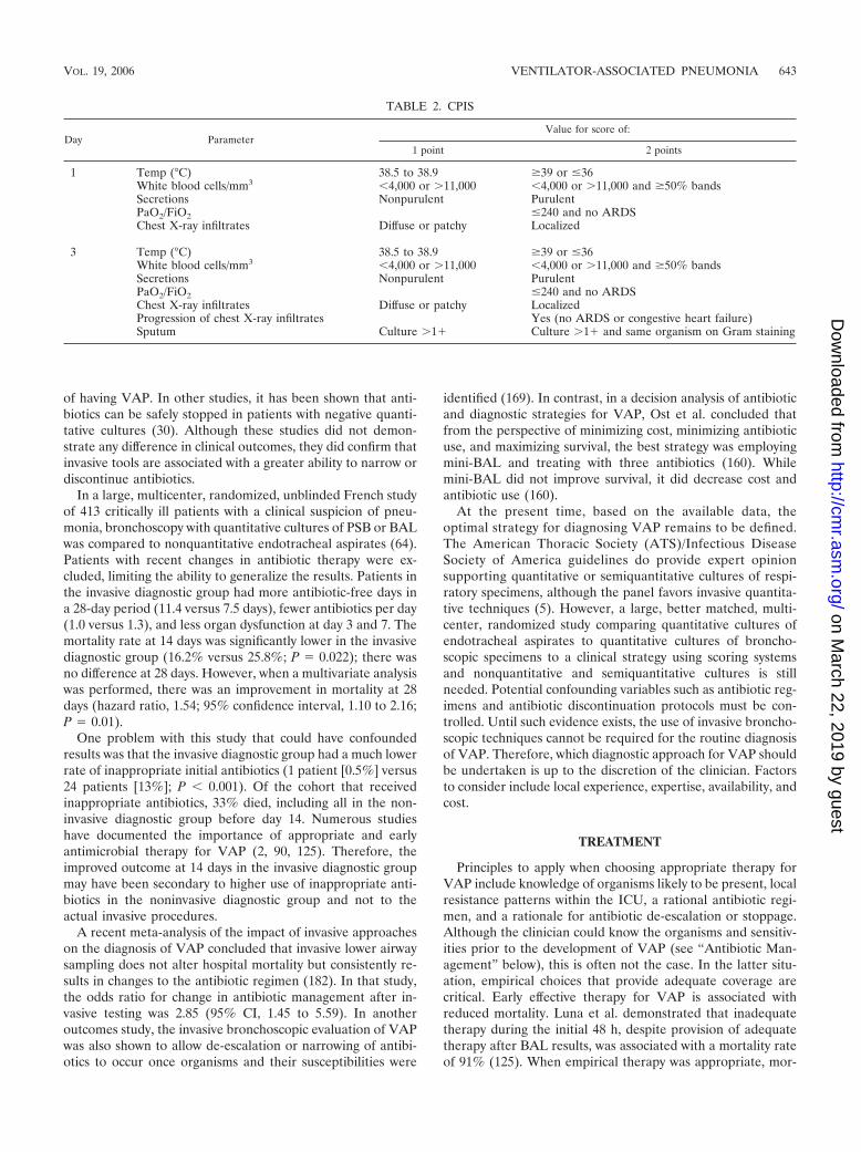
TABLE 2. CPIS
Day ParameterValue for score of:
1 point 2 points
1 Temp (°C) 38.5 to 38.9 �39 or �36White blood cells/mm3 �4,000 or �11,000 �4,000 or �11,000 and �50% bandsSecretions Nonpurulent PurulentPaO2/FiO2 �240 and no ARDSChest X-ray infiltrates Diffuse or patchy Localized
3 Temp (°C) 38.5 to 38.9 �39 or �36White blood cells/mm3 �4,000 or �11,000 �4,000 or �11,000 and �50% bandsSecretions Nonpurulent PurulentPaO2/FiO2 �240 and no ARDSChest X-ray infiltrates Diffuse or patchy LocalizedProgression of chest X-ray infiltrates Yes (no ARDS or congestive heart failure)Sputum Culture �1� Culture �1� and same organism on Gram staining
VOL. 19, 2006 VENTILATOR-ASSOCIATED PNEUMONIA 643
on March 22, 2019 by guest
http://cmr.asm
.org/D
ownloaded from
tality rates were much lower (38%). Delays in the administra-tion of appropriate antibiotic therapy for VAP have been as-sociated with excess mortality (2, 90, 125). In one study, a delayin appropriate therapy for 24 h or more was associated with a69.7% mortality, compared to 28.4% in patients treated with-out the delay (P � 0.001) (90). Consequently, once VAP isconsidered, cultures must be obtained quickly and treatmentinitiated without delay. VAP should be considered with a CPISscore of �6, as illustrated in Table 2, or alternatively, with anew pulmonary infiltrate and at least two of the following:fever, leukocytosis, and purulent secretions.
As multiple etiologies may explain why patients develop afever and pulmonary infiltrates while receiving mechanicalventilation, we often search for other infectious and noninfec-tious etiologies concurrently with evaluation for VAP. Theextent of this investigation is dictated by the clinical circum-stances, including physical examination, laboratory findings,and the severity of illness (Tables 3 and 4). In patients withsepsis, a definite site of infection cannot be found in 20 to 30%(212). As delays in treating severe sepsis significantly increasemortality, we are very hesitant to discontinue antibiotics inpatients with severe sepsis, even if initial respiratory and othercultures are negative. In such critically ill individuals, we usu-ally continue broad-spectrum antibiotics as we continue toaggressively pursue other infectious and noninfectious causesof the patient’s presentation (Tables 3 and 4). However, be-cause VAP is rarely occult, we direct our antibiotic coverageand diagnostic efforts at non-VAP causes of sepsis.
There is a general consensus that VAP is very likely incertain situations. These circumstances are outlined in Table 5(198, 215). However, either such scenarios are uncommon orthe procedures required are undesirable or contraindicated in
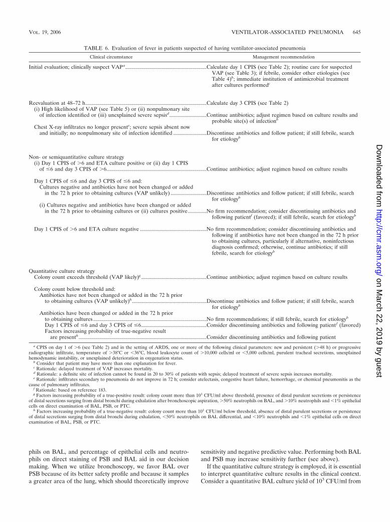
critically ill patients on mechanical ventilators. Therefore, inmost cases the clinician has a choice of two strategies formanaging suspected VAP (Table 6). One strategy is based onclinical criteria and nonquantitative or semiquantitative cul-tures of tracheal aspirates. The other strategy utilizes quanti-tative cultures of respiratory specimens. The quantitative cul-ture approach can be further divided into bronchoscopic(invasive) and nonbronchoscopic (noninvasive) strategies. Asoutlined above, each strategy has its own advantages and dis-advantages.
Bronchoscopy allows the direct examination of respiratorysecretions from BAL, PSB, and PTC to determine the percent-age of cells containing ICOs. Some experts cite this potentialfor early guidance of antibiotic management as a factor favor-ing the bronchoscopic approach to the management of VAPover other strategies (29, 30). However, and as outlined above,the false-negative rate of direct staining is alarmingly high,particularly with concomitant antibiotic use and with VAPcaused by Pseudomonas. Consequently, we contend that a neg-ative direct staining still requires initial broad-spectrum anti-biotics until culture results are returned, particularly if antibi-otics have been added or changed in the previous 72 h.
Quantitative Culture Strategy
Although there is no definitive evidence that quantitativecultures clearly improve patient outcomes, we favor a quan-titative culture strategy for the management of suspectedVAP. The superior specificity of quantitative compared tononquantitative and semiquantitative culture techniquespermits us to more confidently discontinue antibiotics andthereby avoid the attendant complications, including thepotential for increased bacterial resistance. In addition, anegative quantitative culture compels us to more aggres-sively search for other noninfectious and nonpulmonary in-fectious causes of the patient’s presentation.
Our ICU practice is to rely on QEAs, as most studies haveconcluded that the sensitivities of nonbronchoscopic and bron-choscopic quantitative techniques are comparable. However,the overall concordance in some studies has been only approx-imately 80% (30, 94, 140). That is, in some patients, the diag-nosis of VAP could be missed by blind, nonbronchoscopicsampling, particularly if the pneumonia involves the left lungor upper lobes. Moreover, the additional information obtainedfrom direct visualization of the airways, percentage of neutro-
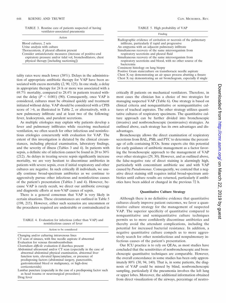
TABLE 3. Routine care of patients suspected of havingventilator-associated pneumonia
Action
Blood cultures, 2 setsUrine analysis with cultureThoracentesis, if pleural effusion presentConsider antiatelectatic measures (increase of positive-end
expiratory pressure and/or tidal vol, bronchodilators, chestphysical therapy �including suctioning�)
TABLE 4. Evaluation for infectious (other than VAP) andnoninfectious causes of fever
Action to be considered
Changing and/or culturing intravenous linesCT scan of sinuses, with fine needle aspirate if abnormalEvaluation for venous thromboembolismClostridium difficile evaluation if diarrhea presentAbdominal ultrasound and/or CT scan (especially in the case of
abnormal abdominal physical examination, abnormal liverfunction tests, elevated lipase/amylase, or presence ofpredisposing factors (abdominal surgery, pancreatitis,gastrointestinal bleed or malignancy, or high-dosecorticosteroids)
Lumbar puncture (especially in the case of a predisposing factor suchas head trauma or neurosurgical procedure)
Drug fever
TABLE 5. High probability of VAP
Finding
Radiographic evidence of cavitation or necrosis of the pulmonaryinfiltrate, particularly if rapid and progressive
An empyema with an adjacent pulmonary infiltrateSimultaneous recovery of the same microorganism from
respiratory secretions and pleural fluidSimultaneous recovery of the same microorganism from
respiratory secretions and blood, with no other source of thebacteremia
Consistent histology on lung biopsyPositive Gram stain/culture on transthoracic needle aspirateChest X ray demonstrating an air space process abutting a fissureChest X ray demonstrating an air bronchogram, especially if single
644 KOENIG AND TRUWIT CLIN. MICROBIOL. REV.
on March 22, 2019 by guest
http://cmr.asm
.org/D
ownloaded from
phils on BAL, and percentage of epithelial cells and neutro-phils on direct staining of PSB and BAL aid in our decisionmaking. When we utilize bronchoscopy, we favor BAL overPSB because of its better safety profile and because it samplesa greater area of the lung, which should theoretically improve
sensitivity and negative predictive value. Performing both BALand PSB may increase sensitivity further (see above).
If the quantitative culture strategy is employed, it is essentialto interpret quantitative culture results in the clinical context.Consider a quantitative BAL culture yield of 103 CFU/ml from
TABLE 6. Evaluation of fever in patients suspected of having ventilator-associated pneumonia
Clinical circumstance Management recommendation
Initial evaluation; clinically suspect VAPa.............................................................Calculate day 1 CPIS (see Table 2); routine care for suspectedVAP (see Table 3); if febrile, consider other etiologies (seeTable 4)b; immediate institution of antimicrobial treatmentafter cultures performedc
Reevaluation at 48–72 h...........................................................................................Calculate day 3 CPIS (see Table 2)(i) High likelihood of VAP (see Table 5) or (ii) nonpulmonary site
of infection identified or (iii) unexplained severe sepsisd............................Continue antibiotics; adjust regimen based on culture results andprobable site(s) of infectionb
Chest X-ray infiltrates no longer presente; severe sepsis absent nowand initially; no nonpulmonary site of infection identified .........................Discontinue antibiotics and follow patient; if still febrile, search
for etiologyb
Non- or semiquantitative culture strategy(i) Day 1 CPIS of �6 and ETA culture positive or (ii) day 1 CPIS
of �6 and day 3 CPIS of �6...........................................................................Continue antibiotics; adjust regimen based on culture results
Day 1 CPIS of �6 and day 3 CPIS of �6 and:Cultures negative and antibiotics have not been changed or added
in the 72 h prior to obtaining cultures (VAP unlikely) ...........................Discontinue antibiotics and follow patient; if still febrile, searchfor etiologyb
(i) Cultures negative and antibiotics have been changed or addedin the 72 h prior to obtaining cultures or (ii) cultures positive..............No firm recommendation; consider discontinuing antibiotics and
following patientf (favored); if still febrile, search for etiologyb
Day 1 CPIS of �6 and ETA culture negative ..................................................No firm recommendation; consider discontinuing antibiotics andfollowing if antibiotics have not been changed in the 72 h priorto obtaining cultures, particularly if alternative, noninfectiousdiagnosis confirmed; otherwise, continue antibiotics; if stillfebrile, search for etiologyb
Quantitative culture strategyColony count exceeds threshold (VAP likely)g .................................................Continue antibiotics; adjust regimen based on culture results
Colony count below threshold and:Antibiotics have not been changed or added in the 72 h prior
to obtaining cultures (VAP unlikely)h........................................................Discontinue antibiotics and follow patient; if still febrile, searchfor etiologyb
Antibiotics have been changed or added in the 72 h priorto obtaining cultures .....................................................................................No firm recommendations; if still febrile, search for etiologyb
Day 1 CPIS of �6 and day 3 CPIS of �6.................................................Consider discontinuing antibiotics and following patient f (favored)Factors increasing probability of true-negative result
are presenth................................................................................................Consider discontinuing antibiotics and following patient
a CPIS on day 1 of �6 (see Table 2) and in the setting of ARDS, one or more of the following clinical parameters: new and persistent (�48 h) or progressiveradiographic infiltrate, temperature of �38°C or �36°C, blood leukocyte count of �10,000 cells/ml or �5,000 cells/ml, purulent tracheal secretions, unexplainedhemodynamic instability, or unexplained deterioration in oxygenation status.
b Consider that patient may have more than one explanation for fever.c Rationale: delayed treatment of VAP increases mortality.d Rationale: a definite site of infection cannot be found in 20 to 30% of patients with sepsis; delayed treatment of severe sepsis increases mortality.e Rationale: infiltrates secondary to pneumonia do not improve in 72 h; consider atelectasis, congestive heart failure, hemorrhage, or chemical pneumonitis as the
cause of pulmonary infiltrates.f Rationale: based on reference 183.g Factors increasing probability of a true-positive result: colony count more than 101 CFU/ml above threshold, presence of distal purulent secretions or persistence
of distal secretions surging from distal bronchi during exhalation after bronchoscopic aspiration, �50% neutrophils on BAL, and �10% neutrophils and �1% epithelialcells on direct examination of BAL, PSB, or PTC.
h Factors increasing probability of a true-negative result: colony count more than 101 CFU/ml below threshold, absence of distal purulent secretions or persistenceof distal secretions surging from distal bronchi during exhalation, �50% neutrophils on BAL differential, and �10% neutrophils and �1% epithelial cells on directexamination of BAL, PSB, or PTC.
VOL. 19, 2006 VENTILATOR-ASSOCIATED PNEUMONIA 645
on March 22, 2019 by guest
http://cmr.asm
.org/D
ownloaded from
a mechanically ventilated patient obtained 48 h after adminis-tration of broad-spectrum antibiotics. This is below the 104-CFU/ml threshold, but antibiotics given or changed within the72 h prior to obtaining a quantitative culture can decrease thebacterial burden and result in a false-negative quantitativeculture. In the appropriate clinical context, such a result can beinterpreted as consistent with the presence of VAP. In con-trast, the same culture result obtained for an individual on noantibiotics or without a change in the previous 72 h would beless indicative of VAP. Moreover, if available, the percentageof neutrophils on BAL differential, the percentage of neutro-phils and epithelial cells on direct staining of BAL and PSB,the percentage of organisms containing ICOs, and visual in-spection of the airways can also be useful in individual cases(see above).
Clinical Strategy
The primary advantage of a clinical strategy for diagnosingVAP is that it does not require specific expertise or specializedequipment or techniques and is noninvasive. Therefore, suchan approach can be utilized anywhere. However, because ofthe poor specificity of clinical signs and symptoms of VAP andof nonquantitative or semiquantitative cultures of tracheal se-cretions, relying on the clinical approach would be expected toresult in treating noninfectious processes with broad-spectrumantibiotics as well as potentially failing to recognize and pursuenoninfectious mimics of VAP and nonpulmonary infections.
Antibiotic Management
The ATS has recently published guidelines to guide empir-ical antibiotic choices (5). These guidelines are divided intothose for patients at risk for VAP caused by multidrug-resis-tant organisms and those for patients without such risk. Riskfactors for multidrug-resistant organisms include prior antimi-crobial therapy in the preceding 90 days, current hospitaliza-tion exceeding 5 days (not necessarily ICU days), high fre-quency of resistance in the community or local hospital unit,and immunosuppressive disease and/or therapy. In addition,the clinician must consider risk factors for health care-associ-ated pneumonia, as such a pneumonia may present with multi-drug-resistant organisms even upon hospital admission (5).Such risk factors for the intubated patient include a hospital-ization for �2 days within the preceding 90 days, residence ina long-term care facility, chronic dialysis within 30 days, homewound care, home infusion therapy (inclusive of antibiotics),and a family member with a multidrug-resistant pathogen.
In the absence of risk factors for multidrug-resistant bacteria,the clinician should choose empirical therapy for Streptococcuspneumoniae, Haemophilus influenzae, methicillin-sensitive Staph-ylococcus aureus, and antibiotic-sensitive gram-negative entericorganisms. Antibiotic choices include ceftriaxone, quinolones(levofloxacin, moxifloxacin, or ciprofloxacin), ampicillin/sulbac-tam, or ertapenem (Fig. 1). When risk factors for multidrug-resistant organisms are present, the clinician must consider notonly the organisms listed above but also Pseudomonas aeruginosa,Klebsiella, Enterobacter, Serratia, Acinetobacter, Stenotrophomonasmaltophilia, Burkholderia cepacia, and methicillin-resistant S. au-reus. Empirical therapy is broadened to include (i) either an
antipseudomonal cephalosporin (cefepime or ceftazadime), anantipseudomonal carbepenem (imipenem or meropenem), or a-lactam/-lactamase inhibitor (pipercacillin-tazobactam) plus(ii) an antipseudomonal fluoroquinolone (ciprofloxacin or levo-floxacin) or an aminoglycoside (amikacin, gentamicin, or tobra-mycin) plus linezolid or vancomycin.
While the complex regimen outlined above is appropriate,creating a milieu of further resistant organism must be a con-cern, as it will lead to fewer opportunities to choose effectiveempirical therapy. As noted in “DIAGNOSIS” above, there isconsiderable controversy over the use of quantitative culturesand which quantitative culturing technique to use. Michel et al.applied QEA as a surveillance tool and routinely obtainedsamples twice weekly (146). Sensitivities were determined formicroorganisms present at a concentration of �103 CFU/ml.When VAP occurred, the most recent QEA preceding VAPwas used to direct antibiotic therapy, and a BAL was obtainedto assess the appropriateness of the antibiotic regimen. Thoseauthors also compared results of BAL to empirical regimensthat would have been chosen by the classification of Trouilletet al. and the 1996 ATS consensus guidelines (6, 203). Theantibiotic regimen as guided by QEA was appropriate in 95%of cases. This was not statistically different from the appropri-ateness of the empirical regimens chosen by the strategy ofTrouillet et al. (83% appropriate) but was superior to that ofthe empirical choices suggested by the 1996 ATS guidelines(68% appropriate). This approach is very new, and the cost isthat of culturing and determining sensitivities (if the thresholdis exceeded). The benefit is that it appears to provide a highlikelihood for appropriate initial therapy. Furthermore, it willlikely reduce the application of overly broad antibiotic regi-mens, hence reducing the likelihood of inducing more multi-drug-resistant organisms.
Considerable controversy surrounds monotherapy versuscombination therapy for patients with VAP. The primary rea-sons for combination therapy are to prevent the developmentof resistance, improve outcomes, provide synergy, and providesufficient antibiotic coverage should the pathogen be resistantto the agent that would have been chosen as single therapy.The former two arguments, while logical, have yet to be proven(36, 209). In fact, a meta-analysis suggested that clinical failurewas more common with combination therapy, as was nephro-toxicity; aminoglycosides were the second agent, and combina-tion therapy did not prevent new resistance patterns (209).However, given that mortality is higher when therapy is inap-propriate during the first 48 h, we favor initiating combinationtherapy for patients at risk for multidrug-resistant organismsuntil sensitivities are known. This is consistent with an ap-proach suggested by Gruson et al. (75).
Commonly employed methods to reduce the development ofresistance include de-escalation therapy, truncated courses ofantibiotics, dosing regimens that account for patient-antibioticpharmacokinetics and pharmacodynamics (PK/PD), antibioticcycling, and surveillance cultures. Most intensivists have em-braced the former two; however, the latter two remain contro-versial. The ATS has put forth a management strategy to ad-dress de-escalation and early stoppage of antibiotics (5). Uponsuspicion of VAP, empirical antibiotics are initiated and lowerrespiratory tract cultures obtained. At 48 to 72 h, if the patientis improving and cultures are negative, strong consideration
646 KOENIG AND TRUWIT CLIN. MICROBIOL. REV.
on March 22, 2019 by guest
http://cmr.asm
.org/D
ownloaded from
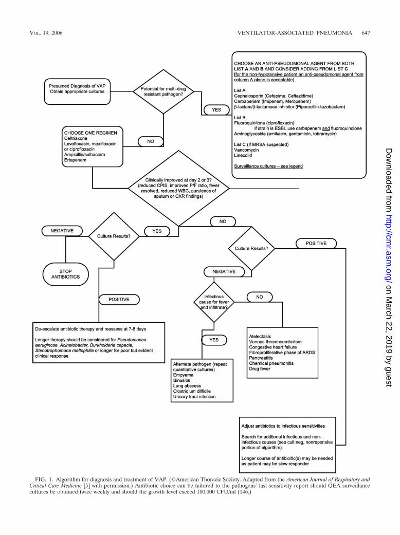
FIG. 1. Algorithm for diagnosis and treatment of VAP. (©American Thoracic Society. Adapted from the American Journal of Respiratory andCritical Care Medicine [5] with permission.) Antibiotic choice can be tailored to the pathogens’ last sensitivity report should QEA surveillancecultures be obtained twice weekly and should the growth level exceed 100,000 CFU/ml (146.)
VOL. 19, 2006 VENTILATOR-ASSOCIATED PNEUMONIA 647
on March 22, 2019 by guest
http://cmr.asm
.org/D
ownloaded from
should be given to stopping antibiotics. Rello et al. have sug-gested truncating the course at �5 days provided the patienthas been afebrile for �48 h (169). Should the culture results bepositive and the patient has improved at 48 to 72 h, then theATS guidelines suggest de-escalation (reduction in antibioticsto be administered, including potential for monotherapy) andtreating patients without P. aeruginosa, Acinetobacter, or Stenotro-phomonas maltophilia for 7 to 8 days. A longer course is indicatedfor P. aeruginosa, Acinetobacter, and Stenotrophomonas malto-philia.
The antibiotic regimen (choice and dosing) should be re-evaluated for change or prolongation in patients with poorclinical responses, which may be assessed by a rising CPIS. Arising CPIS has been associated with higher mortality (183).These recommendations are based on the results from studiesby Dennesen et al., Luna et al., Singh et al., and Ibrahim et al.(49, 88, 122, 183). Such strategies are dependent upon clearevidence of patient improvement as defined by reduction inserial CPISs or improvement of the PaO2/FiO2 ratio at days 3to 5 (122). The technique chosen in obtaining microbiologicdata may indeed affect the clinical decision to de-escalate ther-apy. Heyland et al. reported that the choice of bronchoscopicBAL and PSB resulted in increased physician confidence in thediagnosis and management of VAP; this resulted in a greatertendency to limit or discontinue antibiotics, an outcome thatwas echoed in a recent meta-analysis (79, 182).
Singh et al. proposed another potential clinical strategy tominimize unnecessary antibiotic use for VAP and the potentialconsequences (183). In this study, patients with a modifiedCPIS of �6 on day 1 (Table 2) were randomized to receiveeither standard antimicrobial therapy or ciprofloxacin mono-therapy, with reevaluation at 3 days. In the ciprofloxacin mono-therapy group, if the CPIS remained at �6 at day 3, antibioticswere discontinued. Continuation of antibiotics in the standardtherapy group was left up to the discretion of the attending
physician but occurred in 96% of patients. Despite mono-therapy with ciprofloxacin, a shorter duration of treatment(P � 0.0001) and lower cost (P � 0.003), mortality, and lengthof ICU stay did not differ. In addition, antimicrobial resistance,superinfections, or both were less in the experimental groupthan in the standard therapy group (15% versus 36%; P �0.017). Such an approach recognizes that a gold standard fordiagnosing VAP does not exist, and consequently the approachdoes not attempt to discern whether the patient did or did nothave pneumonia. Rather, the goal was to identify patients forwhom a shorter course of antibiotic therapy would suffice. In asubsequent study that incorporated the modified CPIS, 41% ofpatients with a score of �6 did not have pneumonia by quan-titative BAL culture (65). Therefore, one potential explanationfor the good outcome in the study by Singh and colleagues isthat many of the patients did not have pneumonia.
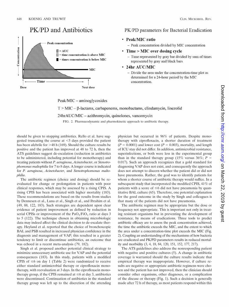
The antibiotic regimen may be appropriate but the dose orfrequency not appropriate. This is important not only in treat-ing resistant organisms but in preventing the development ofresistance, by means of eradications. Three tools to predictantibiotic efficacy are to assess the peak concentration achieved,the time the antibiotic exceeds the MIC, and the extent to whichthe area under a concentration-time plot exceeds the MIC (Fig.2). Coupling an understanding of the mechanisms of how bacteriaare eradicated and PK/PD parameters results in reduced mortal-ity and morbidity (3, 4, 18, 84, 120, 151, 152, 177, 217).
The ATS guidelines also address the nonresponding patientwith negative and positive cultures (5). A change in antibioticcoverage is warranted should the culture results indicate thatempirical therapy was inappropriate. However, if culture re-sults are negative or appropriate antibiotic regimens were cho-sen and the patient has not improved, then the clinician shouldconsider other organisms, other diagnoses, or a complicationof the disease or therapy (Fig. 1). Such a decision is generallymade after 72 h of therapy, as most patients respond within this
FIG. 2. Pharmacodynamic and pharmokinetic approach to antibiotic therapy.
648 KOENIG AND TRUWIT CLIN. MICROBIOL. REV.
on March 22, 2019 by guest
http://cmr.asm
.org/D
ownloaded from
time frame (122, 123). At this juncture, quantitative culturesare warranted. Montravers et al. have demonstrated that clin-ical failure rarely occurred when protected brush samples re-covered organisms at �103 CFU/ml (7% failure rate). Con-versely, higher failure rates were seen when culture resultsexceeded 103 CFU/ml (55.8% failure rate) (150). Persistentfever or failure to improve with antibiotic therapy may indicatethat the inciting process is noninfectious. Other diagnoses in-clude atelectasis, congestive heart failure, venous thromboem-bolic disease, pancreatitis, chemical pneumonitis from aspira-tion, proliferative phase of acute respiratory distress syndrome,drug fever, or pulmonary hemorrhage (5). Alternatively, theprocess may be infectious but not VAP. The clinician shouldconsider empyema, lung abscess, Clostridium difficile colitis,urinary tract infection, and sinusitis (139, 172) (Table 4).
Candida species, while commonly cultured from patients,rarely cause invasive pulmonary disease, even when quantita-tive thresholds are exceeded (58, 166, 210). However, Candidamay be a marker that the patient is more likely to develop VAPwith P. aeruginosa (a causal relationship has not been demon-strated) (8). Azoulay et al., in a surveillance study, noted thatpatients colonized with Candida species were 1.58 times morelikely to develop VAP and 2.22 times more likely to develop P.aeruginosa (8). The three most common Candida species re-
covered were C. albicans (2/3), C. glabrata (1/5), and C. tropi-calis (1/8). Those colonized were older and were more likely tohave respiratory failure as a reason for ICU admission. Thesepatients had longer courses of mechanical ventilation, receivedmore antibiotics, and experienced higher hospital mortality.However, there are no data to support the routine administra-tion of antifungal therapy when Candida species are found inpulmonary secretions of mechanically ventilated patients.
After considering VAP, the clinician should promptly insti-tute therapy. Choosing an appropriate antibiotic regimen, de-fined by sensitivities of the organism cultured and by dosingregimen ordered, is paramount, as the first 48 h is critical topatient survival. Surveillance cultures may provide better guid-ance than empirical strategies. Truncated courses of antibioticsare indicated for culture-negative improving patients and forVAP not caused by P. aeruginosa, Acinetobacter, and Stenotro-phomonas maltophilia. Such a practice may reduce the likeli-hood of colonization with multidrug-resistant organisms orcreating a local environment of resistant organisms.
PREVENTION
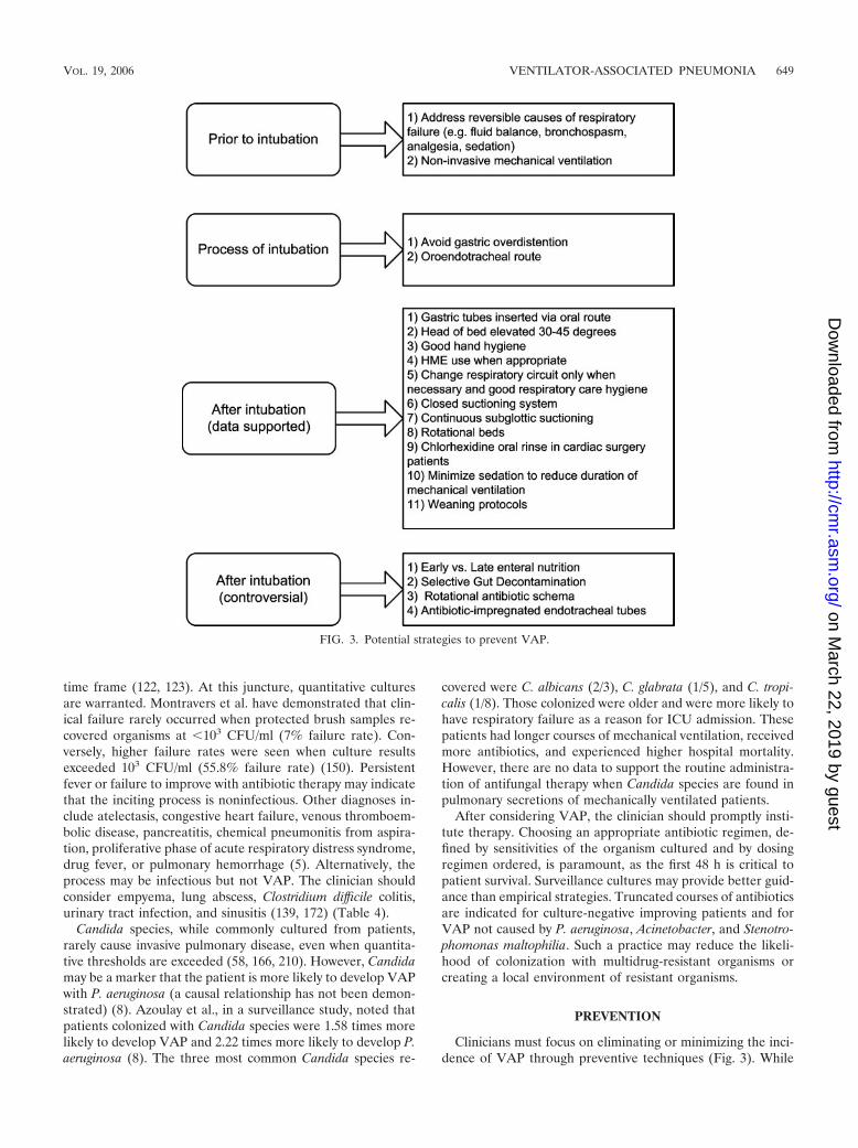
Clinicians must focus on eliminating or minimizing the inci-dence of VAP through preventive techniques (Fig. 3). While
FIG. 3. Potential strategies to prevent VAP.
VOL. 19, 2006 VENTILATOR-ASSOCIATED PNEUMONIA 649
on March 22, 2019 by guest
http://cmr.asm
.org/D
ownloaded from
little has affected the incidence of late-onset VAP, the occur-rence of early-onset VAP can be reduced by simple measures.Data have accumulated to support interventions and establishguidelines, yet translation into practice is lacking. Health careteam compliance rates vary between 30 and 64% (39). Thefocus should be addressing modifiable risk factors such asendotracheal and nasogastric tubes, tracheotomy, reintuba-tion, enteral nutrition, corticosteroid administration, gastricpH-modifying agents, supine positioning, prior antibiotic us-age, poor infection control practice, and contaminated respi-ratory equipment, medications, or water (24, 42, 100, 102, 155,191).
Noninvasive mechanical ventilation (NIV) has been associ-ated with more favorable outcomes (mortality and morbidity)in comparison to endotracheal tube placement in patients withacute exacerbations of chronic obstructive pulmonary diseaseor acute pulmonary edema (7, 21, 117, 118). The incidence ofnosocomial pneumonia was reduced in the group randomizedto NIV (7, 27, 74, 76, 157). Furthermore, immunocompro-mised patients with bilateral infiltrates also benefited fromNIV over invasive mechanical ventilation (IMV) with regard toboth mortality and morbidity (81). Yet clinicians have signifi-cant reluctance to initiate NIV, perhaps because of patientintolerance or increased resource consumption (nursing andrespiratory therapy).
Once the decision to intubate is made, the practice of VAPprevention should be directed at reducing colonization andaspiration (volume of organisms presented to the lungs). Thisbegins with choosing the oral route of intubation and focusingon minimizing the duration of mechanical ventilation (DOMV).Oral intubation is preferred over nasal intubation, as the latterhas been associated with both VAP and sinusitis, with the samebacteria identified in both. Rouby et al., demonstrated a sig-nificant reduction in nosocomial sinusitis when patients areorally cannulated with endotracheal and gastric tubes (172).Holzapfel et al. have linked the reduction in nosocomial sinus-itis to a reduction in VAP (83). Furthermore, the clinicianmust give careful attention to the mundane and seeminglysmall interventions, such as regularly assessing endotrachealcuff pressure, performing endotracheal suctioning, drainingventilator tube condensate, avoiding gastric overdistention,avoiding the supine position, avoiding unnecessary ventilatorcircuit changes, application of heat and moisture exchangers(HMEs) when appropriate, minimizing out-of-ICU transports,and regular hand cleaning with soap or alcohol disinfectant.Maintaining cuff pressure of endotracheal tubes at �20 mmHg reduces nosocomial pneumonia, presumably by minimizingthe passage of oropharyngeal contents into the trachea (192).
The duration of intubation directly affects the likelihood ofVAP, which is more evident in patients with ICU LOS exceed-ing 5 days. Fagon et al. suggested that the incidence of VAPincreases by 1% per day of IMV (62). However, Cook et al.found that the incidence per day varies over time, with 3% perday during first 5 days of IMV, 2% for the second 5 days, and1% for the subsequent 5-day period (39). This observation issupported by Ibrahim et al., who identified an incidence rate ofVAP of 11.5%, 56% of which were early onset (�5 day) (87).Hence, the greatest attack rates appear to be during the initialdays of mechanical ventilation. Additionally, significant risk
factors for early-onset VAP include cardiopulmonary resusci-tation and continuous sedation (167).
Continuous sedation is more often administered in the acutephase of an illness. In addition to treating the primary cause ofrespiratory failure, the DOMV can be reduced through judi-cious use of sedatives and analgesics. Studies by Brook et al.and Kress et al. have demonstrated that protocols for sedativeand analgesic administration with the goal of minimizing con-stant infusions led to reduced DOMV (22, 113). Furthermore,daily interruption of sedation results in a reduced incidence ofintensive care unit complications, in which VAP was included(127, 180). Weaning protocols have also resulted in reducedDOMV, whether respiratory therapist initiated or not (59, 67).
Patients should be cared for in the semirecumbent positionto reduce the extent of aspiration, especially when receivingenteral feeds. Radionuclide studies reveal increased trachealpenetration of gastric contents when intubated patients aresupine (85, 159, 201). Drakulovic et al. found that the simpleelevation of the head of bed to 45° results in dramatic reduc-tions in VAP incidence and a trend toward reduced mortality(54). Nonetheless, a recent survey by the University HospitalConsortium revealed that compliance with the simple andno-cost intervention of elevating the head is woefully low,and a study by Heyland et al. revealed that the head of bedis on average elevated to 29° and not 45° (80). Kinetic bedtherapy has also led to a reduction in the incidence of VAP(46, 48, 69, 72, 97, 202, 213). However, this is costly and hasnot been directly compared to head-of-bed elevation, a no-cost option.
Some VAP is contracted from inhalation of bacteriathrough the ventilator circuit and may be a result of con-taminated aerosols, condensate, or suction catheters. Tra-ditionally, ventilator circuit changes have been on a regularschedule and often daily. However, the data examining thispractice reveal that there is no benefit to changing the cir-cuit on a regular basis, and the present recommendationsare to change the circuit when soiled (56, 108, 121). Such apractice would likely reduce the rate of accidental spillage ofcondensate into the airway. As heated humidifiers enhancethe amount of condensate, attention has been focused onHMEs. These devices have led to a reduction in VAP, albeitsmall, and should be used in patients without significantsecretions or concern over the risk of obstruction (17, 55, 99,107, 132, 141, 174). While changing the HME less frequentlythan every 48 h may lead to further reductions in VAP, caremust be taken to carefully monitor for trapped secretionsand subsequent airway obstruction or increments in thework of breathing (45, 193).
Endotracheal suctioning of intubated patients can be per-formed through an open or closed system. In theory the closedsystem could reduce the incidence of VAP, but in practice thishas not been demonstrated (35, 50, 93, 106, 219). Cost analysisfavors the closed system, as the enveloped catheter can bereused for suctioning and needs to be changed only whendysfunctional (52). However, respiratory therapists have voicedconcerns over residue buildup within the lumen of the endotra-cheal tube.
As most VAP follows from aspiration of oropharyngeal se-cretions, attention to proper cuff inflation pressures and endo-tracheal suctioning can affect the volume presented to the
650 KOENIG AND TRUWIT CLIN. MICROBIOL. REV.
on March 22, 2019 by guest
http://cmr.asm
.org/D
ownloaded from
trachea. The application of continuous suction of subglotticsecretions through specialized endotracheal tubes will reducethe incidence of VAP (110, 126, 181, 185, 204). Surprisingly,this was not associated a reduction in mortality, ICU LOS, orduration of mechanical ventilation. While studying the appli-cation of continuous subglottic suctioning, Rello et al. noted atrend of increased VAP in patients with endotracheal cuffpressures of �20 cm H2O (168). Hence, it is recommended notonly to assess cuff pressure for tracheal ischemia (which occurswhen pressure exceeds 30 cm H2O) but also to ensure thatadequate cuff pressure (�20 mm Hg) is present.
The endotracheal tube itself is a reservoir for gram-negativebacteria. The buildup of a biofilm within endotracheal tubesoccurs frequently. One study demonstrated that 84% of endo-tracheal tubes examined had a biofilm (188). As documentedby Inglis et al., this biofilm is heavily laden with bacteria,usually gram-negative organisms (66, 89). At present, ongoingstudies are directed at either eliminating this biofilm or reduc-ing the bacterial load associated with it.
Oral decontamination with chlorhexidine has been shown toreduce the incidence of VAP in patients undergoing cardiacsurgery, presumably by reducing oropharyngeal colonization(51). Furthermore, numerous studies with oral decontamina-tion antibiotic pastes alone or coadministered with systemicantibiotics have shown a reduction in early VAP (1, 13, 47, 163,171). Two meta-analyses have suggested better results withoral decontamination alone than with the combination of oraland systemic prophylaxis (44, 154). With either approach, how-ever, concern over the emergence of antibiotic-resistant organ-isms has tempered use, as has the labor intensity required toapply these regimens at the bedside. This is particularly true inICUs housing organisms with high antibiotic resistance rates(68, 71, 77, 119, 147, 148, 208, 214). While often recommended,it appears not to be routinely practiced. Two recent studies willfurther the debate, as they demonstrated significant reductionsin VAP and mortality with selective decontamination of thedigestive tract (47, 114). These two studies were performedunder conditions where selective decontamination of the di-gestive tract is most effective, i.e., surgical intensive care unitshousing patients less likely to be colonized with resistant bac-teria.
Gastric volumes and acidity affect the incidence of VAP.Reducing the acidity of gastric secretions and feeding willreduce bacterial overgrowth. However, in high-risk patients(ventilated for �48 h and coagulopathic), the risk of bleedingoutweighs the risk of VAP from pH-modifying agents (52).Hence, it is difficult to recommend against H2 blockers orproton pump inhibitors. Sucralafate may indeed be superiorfrom the viewpoint of VAP, but it is less effective with regardto prophylaxis of gastrointestinal bleeding, and thus it use isnot warranted over H2 blockers or proton pump inhibitors (16,37, 143).
Multiple studies have examined postpyloric versus gastricfeedings with regard to incidence of aspiration and develop-ment of VAP. These studies were small and inconclusive. In ameta-analysis, postpyloric feedings reduce the incidence ofVAP and increased the nutrition delivered (80). However, nosingle trial demonstrated that postpyloric tube feedings pre-vent VAP. The improved delivery of nutrition was likely theresult of decreased gastric residual assessments and conse-
quently fewer stoppages in continuous tube feedings. A recentpublication favored a delay of greater than 5 days before ini-tiating tube feedings, as the incidence of VAP was reduced(86). Further data are needed to unconditionally embrace thispractice.
Preventing Multidrug Resistance
Antibiotic cycling remains controversial. Employing a rota-tional schedule for empirical antibiotic administration for sus-pected VAP may indeed lead to a reduced incidence of resis-tant organisms (75, 111, 165). While such a strategy may notreduce the incidence of VAP, reductions in mortality may beseen (165). This is likely a result of changes in resistancepatterns resulting in a higher likelihood of choosing appropri-ate antibiotic regimens (112). Because rotational scheduleshave primarily targeted reducing the resistance of gram-nega-tive organisms, we do not know the impact of rotating antibi-otics against gram-positive organisms, such as methicillin-re-sistant S. aureus. Furthermore, the frequency with which torotate antibiotics remains unclear, as monthly and quarterlyregimens have been assessed with documented successes (75,165). Furthermore, the probability of antibiotic cycling leadingto a reduction in antimicrobial resistance is low as determinedthrough mathematical modeling (14). At this juncture, it ispremature to recommend rotating antibiotics or a rotationalschema.
Multidrug resistance can also be reduced when patient-antibi-otic PK/PD characteristics are accounted for. Early eradicationminimizes the opportunity for a population of organisms to de-velop resistance. Peak concentrations for aminoglycosides 10-foldgreater than MIC appear to inhibit the emergence of resistantorganisms (178, 207). When choosing fluoroquinolones, resis-tant organisms are less likely to be seen when the 24-h area-under-the-curve/MIC levels are �100 for gram-negative bac-teria and �40 for gram-positive bacteria (177, 179, 194).Changes in medication frequency or infusion rates can increasethe time that the antibiotic concentration exceeds the MIC.For -lactams, monobactams, glycopeptides, and cabapenemsthis can be important in enhancing bactericidal activity, againreducing opportunities for resistant organisms to emerge (18,19, 105, 120, 217).
In summary, several opportunities to reduce the incidence ofVAP are available to the clinician. Many are no-cost or mini-mal-cost interventions and should be implemented as part ofroutine care protocols. Care of the critically ill should be di-rected at applying interventions that reduce mortality, mini-mize morbidity, shorten the length of stay, and reduce cost.Reducing VAP through the simple measures outlined doesexactly that. We recommend that the clinician’s practice in-clude noninvasive mechanical ventilation over intubation whenappropriate, oral intubation when an endotracheal tube is nec-essary, orogastric over nasogastric tubes, elevation of the headto at least 30°, minimization of sedation, administration of aproton pump inhibitor when prophylaxis is indicated, a fre-quency of ventilator tubing changes at 7 days or when soiled,avoidance or elimination of endotracheal tube leak, good tech-nique in removal of condensate, and of course excellent handhygiene. At this time we do not support the routine use ofendotracheal tubes with subglottic suction capabilities, rota-
VOL. 19, 2006 VENTILATOR-ASSOCIATED PNEUMONIA 651
on March 22, 2019 by guest
http://cmr.asm
.org/D
ownloaded from
tional beds, in-line suction systems, rotational antibioticschemes, or selective gut decontamination.
Strategies and a more thorough discussion on prevention arewithin the ATS/Infectious Disease Society of America state-ment and papers by Kollef and by Dodek et al. (5, 52, 101).Zack et al. have demonstrated that a multifaceted and multi-disciplinary approach to VAP prevention can indeed reducethe incidence (218). Success is dependent upon persistent at-tention to detail, high compliance rates, and a champion.
CONCLUSION
A low threshold for suspicion of VAP is needed when apatient’s clinical course deteriorates. The day 1 CPIS can beuseful, especially when combined with quantitative cultures.The choice of which quantitative culture methodology to use isan open debate. However, diagnostic cost favors QEA, whichcan also be implemented as a surveillance technique. However,the clinician is more likely to stop antibiotics with a moreinvasive quantitative culture, resulting in increased savings.
Antibiotic administration should be promptly initiated whenVAP is suspected and quantitative cultures obtained andshould be broad in coverage. Knowledge of local antibiogramsshould guide the choice of antibiotics, in addition to likelihoodof organisms (early- or late-onset VAP). For patients alreadyon antibiotics at the time of suspected VAP, the clinicianshould choose antibiotics from different classes, as it is likelythat resistance to “in-use” antibiotics has developed.
Assessment of the likelihood of VAP by day 3 is needed todecide whether antibiotics should be continued. The assess-ment should include a repeat CPIS, as the change in CPIS canguide clinical decisions, even stoppage of antibiotics. Assess-ment of quantitative culture results and sensitivities at thisjuncture is prudent, as it may permit early antibiotic de-esca-lation by choosing a more narrowly focused agent(s). Mono-therapy may be appropriate in many instances of VAP andshould reduce the incidence of drug resistance. A change tomonotherapy may be possible in a responding patient whereorganism sensitivity results permit. A short course (6 to 8 days)can be administered to patients with VAP but is dependent onthe patient physiologic response to treatment along with whichorganisms have been recovered (see above) (32, 144).
Simple and effective preventive measures can be institutedeasily and at minimal costs. Such measures might include NIV,diligent respiratory care, hand hygiene, elevation of head, oraland not nasal cannulation, minimization of sedation, institu-tion of weaning protocols, judicious use of antibiotics, de-escalation, and leveraging PK/PD characteristics for antibioticsadministered. More costly interventions should be reserved forappropriate situations.
Utilizing the preventive, diagnostic, and treatment recom-mendations outlined in this paper should allow for improvedoutcomes for a common and serious medical complicationseen in ICU mechanically ventilated patients.
REFERENCES
1. Abele-Horn, M., A. Dauber, A. Bauernfeind, W. Russwurm, I. Seyfarth-Metzger, P. Gleich, and G. Ruckdeschel. 1997. Decrease in nosocomialpneumonia in ventilated patients by selective oropharyngeal decontamina-tion (SOD). Intensive Care Med. 23:187–195.
2. Alvarez-Lerma, F., et al. 1996. Modification of empiric antibiotic treatment
in patients with pneumonia acquired in the intensive care unit. IntensiveCare Med. 22:387–394.
3. Ambrose, P. G., D. M. Grasela, T. H. Grasela, J. Passarell, H. B. Mayer,and P. F. Pierce. 2001. Pharmacodynamics of fluoroquinolones againstStreptococcus pneumoniae in patients with community-acquired respiratorytract infections. Antimicrob. Agents Chemother. 45:2793–2797.
4. Ambrose, P. G., R. C. Owens, Jr., M. J. Garvey, and R. N. Jones. 2002.Pharmacodynamic considerations in the treatment of moderate to severepseudomonal infections with cefepime. J. Antimicrob. Chemother. 49:445–453.
5. American Thoracic Society. 2005. Guidelines for the management of adultswith hospital-acquired, ventilator-associated, and healthcare-associatedpneumonia. Am. J. Respir. Crit. Care Med. 171:388–416.
6. American Thoracic Society. 1996. Hospital-acquired pneumonia in adults:diagnosis, assessment of severity, initial antimicrobial therapy, and preven-tive strategies. A consensus statement, American Thoracic Society, Novem-ber 1995. Am. J. Respir. Crit. Care Med. 153:1711–1725.
7. Antonelli, M., G. Conti, M. Rocco, M. Bufi, R. A. De Blasi, G. Vivino, A.Gasparetto, and G. U. Meduri. 1998. A comparison of noninvasive positive-pressure ventilation and conventional mechanical ventilation in patientswith acute respiratory failure. N. Engl. J. Med. 339:429–435.
8. Azoulay, E., J. F. Timsit, M. Tafflet, A. de Lassence, M. Darmon, J. R.Zahar, C. Adrie, M. Garrouste-Orgeas, Y. Cohen, B. Mourvillier, and B.Schlemmer. 2006. Candida colonization of the respiratory tract and subse-quent Pseudomonas ventilator-associated pneumonia. Chest 129:110–117.
9. Baker, A. M., D. L. Bowton, and E. F. Haponik. 1995. Decision making innosocomial pneumonia. An analytic approach to the interpretation of quan-titative bronchoscopic cultures. Chest 107:85–95.
10. Baker, A. M., J. W. Meredith, and E. F. Haponik. 1996. Pneumonia inintubated trauma patients. Microbiology and outcomes. Am. J. Respir. Crit.Care Med. 153:343–349.
11. Bell, R. C., J. J. Coalson, J. D. Smith, and W. G. Johanson, Jr. 1983.Multiple organ system failure and infection in adult respiratory distresssyndrome. Ann. Intern. Med. 99:293–298.
12. Bergmans, D. C., M. J. Bonten, P. W. De Leeuw, and E. E. Stobberingh.1997. Reproducibility of quantitative cultures of endotracheal aspiratesfrom mechanically ventilated patients. J. Clin. Microbiol. 35:796–798.
13. Bergmans, D. C., M. J. Bonten, C. A. Gaillard, J. C. Paling, S. van derGeest, F. H. van Tiel, A. J. Beysens, P. W. de Leeuw, and E. E. Stobberingh.2001. Prevention of ventilator-associated pneumonia by oral decontamina-tion: a prospective, randomized, double-blind, placebo-controlled study.Am. J. Respir. Crit. Care Med. 164:382–388.
14. Bergstrom, C. T., M. Lo, and M. Lipsitch. 2004. Ecological theory suggeststhat antimicrobial cycling will not reduce antimicrobial resistance in hospi-tals. Proc. Natl. Acad. Sci. USA 101:13285–13290.
15. Blot, F., B. Raynard, E. Chachaty, C. Tancrede, S. Antoun, and G. Niten-berg. 2000. Value of gram stain examination of lower respiratory tractsecretions for early diagnosis of nosocomial pneumonia. Am. J. Respir.Crit. Care Med. 162:1731–1737.
16. Bonten, M. J., C. A. Gaillard, S. van der Geest, F. H. van Tiel, A. J. Beysens,H. G. Smeets, and E. E. Stobberingh. 1995. The role of intragastric acidityand stress ulcus prophylaxis on colonization and infection in mechanicallyventilated ICU patients. A stratified, randomized, double-blind study ofsucralfate versus antacids. Am. J. Respir. Crit. Care Med. 152:1825–1834.
17. Boots, R. J., S. Howe, N. George, F. M. Harris, and J. Faoagali. 1997.Clinical utility of hygroscopic heat and moisture exchangers in intensivecare patients. Crit. Care Med. 25:1707–1712.
18. Boselli, E., D. Breilh, M. Cannesson, F. Xuereb, T. Rimmele, D. Chassard,M. C. Saux, and B. Allaouchiche. 2004. Steady-state plasma and intrapul-monary concentrations of piperacillin/tazobactam 4 g/0.5 g administered tocritically ill patients with severe nosocomial pneumonia. Intensive CareMed. 30:976–979.
19. Boselli, E., D. Breilh, T. Rimmele, J. C. Poupelin, M. C. Saux, D. Chassard,and B. Allaouchiche. 2004. Plasma and lung concentrations of ceftazidimeadministered in continuous infusion to critically ill patients with severenosocomial pneumonia. Intensive Care Med. 30:989–991.
20. Boyce, J. M., G. Potter-Bynoe, L. Dziobek, and S. L. Solomon. 1991. Noso-comial pneumonia in Medicare patients. Hospital costs and reimbursementpatterns under the prospective payment system. Arch. Intern. Med. 151:1109–1114.
21. Brochard, L., J. Mancebo, M. Wysocki, F. Lofaso, G. Conti, A. Rauss, G.Simonneau, S. Benito, A. Gasparetto, F. Lemaire, et al. 1995. Noninvasiveventilation for acute exacerbations of chronic obstructive pulmonary dis-ease. N. Engl. J. Med. 333:817–822.
22. Brook, A. D., T. S. Ahrens, R. Schaiff, D. Prentice, G. Sherman, W. Shannon,and M. H. Kollef. 1999. Effect of a nursing-implemented sedation protocol onthe duration of mechanical ventilation. Crit. Care Med. 27:2609–2615.
23. Bryan, C. S. 1999. Nosocomial pneumonia: blood cultures remain useful.Chest 116:859–860.
24. Bryan, C. S., and K. L. Reynolds. 1984. Bacteremic nosocomial pneumonia.Analysis of 172 episodes from a single metropolitan area. Am. Rev. Respir.Dis. 129:668–671.
652 KOENIG AND TRUWIT CLIN. MICROBIOL. REV.
on March 22, 2019 by guest
http://cmr.asm
.org/D
ownloaded from
25. Butler, K. L., K. E. Sinclair, V. J. Henderson, G. McKinney, D. A. Mesidor,I. Katon-Benitez, and W. L. Weaver. 1999. The chest radiograph in criticallyill surgical patients is inaccurate in predicting ventilator-associated pneu-monia. Am. Surg. 65:805–810.
26. Campbell, G. D., Jr. 2000. Blinded invasive diagnostic procedures in ven-tilator-associated pneumonia. Chest 117:207S–211S.
27. Carlucci, A., J. C. Richard, M. Wysocki, E. Lepage, and L. Brochard. 2001.Noninvasive versus conventional mechanical ventilation. An epidemiologicsurvey. Am. J. Respir. Crit. Care Med. 163:874–880.
28. Chastre, J. 2005. Conference summary: ventilator-associated pneumonia.Respir. Care 50:975–983.
29. Chastre, J., A. Combes, and C. E. Luyt. 2005. The invasive (quantitative)diagnosis of ventilator-associated pneumonia. Respir. Care 50:797–812.
30. Chastre, J., and J. Y. Fagon. 2002. Ventilator-associated pneumonia. Am. J.Respir. Crit. Care Med. 165:867–903.
31. Chastre, J., J. Y. Fagon, M. Bornet-Lecso, S. Calvat, M. C. Dombret, R.al Khani, F. Basset, and C. Gibert. 1995. Evaluation of bronchoscopictechniques for the diagnosis of nosocomial pneumonia. Am. J. Respir. Crit.Care Med. 152:231–240.
32. Chastre, J., M. Wolff, J. Y. Fagon, S. Chevret, F. Thomas, D. Wermert, E.Clementi, J. Gonzalez, D. Jusserand, P. Asfar, D. Perrin, F. Fieux, and S.Aubas. 2003. Comparison of 8 vs 15 days of antibiotic therapy for ventilator-associated pneumonia in adults: a randomized trial. JAMA 290:2588–2598.
33. Chollet-Martin, S., P. Montravers, C. Gibert, C. Elbim, J. M. Desmonts,J. Y. Fagon, and M. A. Gougerot-Pocidalo. 1993. High levels of interleu-kin-8 in the blood and alveolar spaces of patients with pneumonia and adultrespiratory distress syndrome. Infect. Immun. 61:4553–4559.
34. Chollet-Martin, S., P. Montravers, C. Gibert, C. Elbim, J. M. Desmonts,J. Y. Fagon, and M. A. Gougerot-Pocidalo. 1992. Subpopulation of hyper-responsive polymorphonuclear neutrophils in patients with adult respira-tory distress syndrome. Role of cytokine production. Am. Rev. Respir. Dis.146:990–996.
35. Combes, P., B. Fauvage, and C. Oleyer. 2000. Nosocomial pneumonia inmechanically ventilated patients, a prospective randomised evaluation ofthe Stericath closed suctioning system. Intensive Care Med. 26:878–882.
36. Cometta, A., J. D. Baumgartner, D. Lew, W. Zimmerli, D. Pittet, P.Chopart, U. Schaad, C. Herter, P. Eggimann, O. Huber, et al. 1994. Pro-spective randomized comparison of imipenem monotherapy with imipenemplus netilmicin for treatment of severe infections in nonneutropenic pa-tients. Antimicrob. Agents Chemother. 38:1309–1313.
37. Cook, D., G. Guyatt, J. Marshall, D. Leasa, H. Fuller, R. Hall, S. Peters, F.Rutledge, L. Griffith, A. McLellan, G. Wood, A. Kirby, et al. 1998. Acomparison of sucralfate and ranitidine for the prevention of upper gastro-intestinal bleeding in patients requiring mechanical ventilation. N. Engl.J. Med. 338:791–797.
38. Cook, D., and L. Mandell. 2000. Endotracheal aspiration in the diagnosis ofventilator-associated pneumonia. Chest 117:195S–197S.
39. Cook, D. J., S. D. Walter, R. J. Cook, L. E. Griffith, G. H. Guyatt, D. Leasa,R. Z. Jaeschke, and C. Brun-Buisson. 1998. Incidence of and risk factors forventilator-associated pneumonia in critically ill patients. Ann. Intern. Med.129:433–440.
40. Corley, D. E., S. H. Kirtland, R. H. Winterbauer, S. P. Hammar, D. H. Dail,D. E. Bauermeister, and J. W. Bolen. 1997. Reproducibility of the histologicdiagnosis of pneumonia among a panel of four pathologists: analysis of agold standard. Chest 112:458–465.
41. Craig, C. P., and S. Connelly. 1984. Effect of intensive care unit nosocomialpneumonia on duration of stay and mortality. Am. J. Infect Control 12:233–238.
42. Craven, D. E., L. M. Kunches, V. Kilinsky, D. A. Lichtenberg, B. J. Make,and W. R. McCabe. 1986. Risk factors for pneumonia and fatality in pa-tients receiving continuous mechanical ventilation. Am. Rev. Respir. Dis.133:792–796.
43. Cunnion, K. M., D. J. Weber, W. E. Broadhead, L. C. Hanson, C. F. Pieper,and W. A. Rutala. 1996. Risk factors for nosocomial pneumonia: comparingadult critical-care populations. Am. J. Respir. Crit. Care Med. 153:158–162.
44. D’Amico, R., S. Pifferi, C. Leonetti, V. Torri, A. Tinazzi, and A. Liberati.1998. Effectiveness of antibiotic prophylaxis in critically ill adult patients:systematic review of randomised controlled trials. Br. Med J. 316:1275–1285.
45. Davis, K., Jr., S. L. Evans, R. S. Campbell, J. A. Johannigman, F. A.Luchette, D. T. Porembka, and R. D. Branson. 2000. Prolonged use of heatand moisture exchangers does not affect device efficiency or frequency rateof nosocomial pneumonia. Crit. Care Med. 28:1412–1418.
46. deBoisblanc, B. P., M. Castro, B. Everret, J. Grender, C. D. Walker, andW. R. Summer. 1993. Effect of air-supported, continuous, postural oscilla-tion on the risk of early ICU pneumonia in nontraumatic critical illness.Chest 103:1543–1547.
47. de Jonge, E., M. J. Schultz, L. Spanjaard, P. M. Bossuyt, M. B. Vroom, J.Dankert, and J. Kesecioglu. 2003. Effects of selective decontamination ofdigestive tract on mortality and acquisition of resistant bacteria in intensivecare: a randomised controlled trial. Lancet 362:1011–1016.
48. Demarest, G. B., W. W. Schmidt-Nowara, L. W. Vance, and A. R. Altman.
1989. Use of the kinetic treatment table to prevent the pulmonary compli-cations of multiple trauma. West. J. Med. 150:35–38.
49. Dennesen, P. J., A. J. van der Ven, A. G. Kessels, G. Ramsay, and M. J.Bonten. 2001. Resolution of infectious parameters after antimicrobial ther-apy in patients with ventilator-associated pneumonia. Am. J. Respir. Crit.Care Med. 163:1371–1375.
50. Deppe, S. A., J. W. Kelly, L. L. Thoi, J. H. Chudy, R. N. Longfield, J. P.Ducey, C. L. Truwit, and M. R. Antopol. 1990. Incidence of colonization,nosocomial pneumonia, and mortality in critically ill patients using a TrachCare closed-suction system versus an open-suction system: prospective,randomized study. Crit. Care Med. 18:1389–1393.
51. DeRiso, A. J., II, J. S. Ladowski, T. A. Dillon, J. W. Justice, and A. C.Peterson. 1996. Chlorhexidine gluconate 0.12% oral rinse reduces theincidence of total nosocomial respiratory infection and nonprophylacticsystemic antibiotic use in patients undergoing heart surgery. Chest 109:1556–1561.
52. Dodek, P., S. Keenan, D. Cook, D. Heyland, M. Jacka, L. Hand, J. Mus-cedere, D. Foster, N. Mehta, R. Hall, and C. Brun-Buisson. 2004. Evidence-based clinical practice guideline for the prevention of ventilator-associatedpneumonia. Ann. Intern. Med. 141:305–313.
53. Dotson, R. G., and S. K. Pingleton. 1993. The effect of antibiotic therapy onrecovery of intracellular bacteria from bronchoalveolar lavage in suspectedventilator-associated nosocomial pneumonia. Chest 103:541–546.
54. Drakulovic, M. B., A. Torres, T. T. Bauer, J. M. Nicolas, S. Nogue, and M.Ferrer. 1999. Supine body position as a risk factor for nosocomial pneu-monia in mechanically ventilated patients: a randomised trial. Lancet 354:1851–1858.
55. Dreyfuss, D., K. Djedaini, I. Gros, L. Mier, G. Le Bourdelles, Y. Cohen, P.Estagnasie, F. Coste, and Y. Boussougant. 1995. Mechanical ventilationwith heated humidifiers or heat and moisture exchangers: effects on patientcolonization and incidence of nosocomial pneumonia. Am. J. Respir. Crit.Care Med. 151:986–992.
56. Dreyfuss, D., K. Djedaini, P. Weber, P. Brun, J. J. Lanore, J. Rahmani, Y.Boussougant, and F. Coste. 1991. Prospective study of nosocomial pneu-monia and of patient and circuit colonization during mechanical ventilationwith circuit changes every 48 hours versus no change. Am. Rev. Respir. Dis.143:738–743.
57. du Moulin, G. C., D. G. Paterson, J. Hedley-Whyte, and A. Lisbon. 1982.Aspiration of gastric bacteria in antacid-treated patients: a frequent causeof postoperative colonisation of the airway. Lancet i:242–245.
58. el-Ebiary, M., A. Torres, N. Fabregas, J. P. de la Bellacasa, J. Gonzalez, J.Ramirez, D. del Bano, C. Hernandez, and M. T. Jimenez de Anta. 1997.Significance of the isolation of Candida species from respiratory samples incritically ill, non-neutropenic patients. An immediate postmortem histo-logic study. Am. J. Respir. Crit. Care Med. 156:583–590.
59. Ely, E. W., M. O. Meade, E. F. Haponik, M. H. Kollef, D. J. Cook, G. H.Guyatt, and J. K. Stoller. 2001. Mechanical ventilator weaning protocolsdriven by nonphysician health-care professionals: evidence-based clinicalpractice guidelines. Chest 120:454S–463S.
60. Fabregas, N., S. Ewig, A. Torres, M. El-Ebiary, J. Ramirez, J. P. de LaBellacasaqq, T. Bauer, and H. Cabello. 1999. Clinical diagnosis of ventilatorassociated pneumonia revisited: comparative validation using immediatepost-mortem lung biopsies. Thorax 54:867–873.
61. Fabregas, N., A. Torres, M. El-Ebiary, J. Ramirez, C. Hernandez, J. Gonzalez,J. P. de la Bellacasa, J. de Anta, and R. Rodriguez-Roisin. 1996. Histopatho-logic and microbiologic aspects of ventilator-associated pneumonia. Anesthe-siology 84:760–771.
62. Fagon, J. Y., J. Chastre, Y. Domart, J. L. Trouillet, J. Pierre, C. Darne, andC. Gibert. 1989. Nosocomial pneumonia in patients receiving continuousmechanical ventilation. Prospective analysis of 52 episodes with use of aprotected specimen brush and quantitative culture techniques. Am. Rev.Respir. Dis. 139:877–884.
63. Fagon, J. Y., J. Chastre, A. J. Hance, M. Guiguet, J. L. Trouillet, Y. Domart,J. Pierre, and C. Gibert. 1988. Detection of nosocomial lung infection inventilated patients. Use of a protected specimen brush and quantitativeculture techniques in 147 patients. Am. Rev. Respir. Dis. 138:110–116.
64. Fagon, J. Y., J. Chastre, M. Wolff, C. Gervais, S. Parer-Aubas, F. Stephan,T. Similowski, A. Mercat, J. L. Diehl, J. P. Sollet, and A. Tenaillon. 2000.Invasive and noninvasive strategies for management of suspected ventila-tor-associated pneumonia. A randomized trial. Ann. Intern. Med. 132:621–630.
65. Fartoukh, M., B. Maitre, S. Honore, C. Cerf, J. R. Zahar, and C. Brun-Buisson. 2003. Diagnosing pneumonia during mechanical ventilation: theclinical pulmonary infection score revisited. Am. J. Respir. Crit. Care Med.168:173–179.
66. Feldman, C., M. Kassel, J. Cantrell, S. Kaka, R. Morar, A. GoolamMahomed, and J. I. Philips. 1999. The presence and sequence of endotra-cheal tube colonization in patients undergoing mechanical ventilation. Eur.Respir. J. 13:546–551.
67. Ferrer, M., A. Esquinas, F. Arancibia, T. T. Bauer, G. Gonzalez, A. Carrillo,R. Rodriguez-Roisin, and A. Torres. 2003. Noninvasive ventilation during
VOL. 19, 2006 VENTILATOR-ASSOCIATED PNEUMONIA 653
on March 22, 2019 by guest
http://cmr.asm
.org/D
ownloaded from
persistent weaning failure: a randomized controlled trial. Am. J. Respir.Crit. Care Med. 168:70–76.
68. Ferrer, M., A. Torres, J. Gonzalez, J. Puig de la Bellacasa, M. el-Ebiary, M.Roca, J. M. Gatell, and R. Rodriguez-Roisin. 1994. Utility of selectivedigestive decontamination in mechanically ventilated patients. Ann. Intern.Med. 120:389–395.
69. Fink, M. P., C. M. Helsmoortel, K. L. Stein, P. C. Lee, and S. M. Cohn.1990. The efficacy of an oscillating bed in the prevention of lower respira-tory tract infection in critically ill victims of blunt trauma. A prospectivestudy. Chest 97:132–137.
70. Gallego, M., and J. Rello. 1999. Diagnostic testing for ventilator-associatedpneumonia. Clin. Chest Med. 20:671–679.
71. Gastinne, H., M. Wolff, F. Delatour, F. Faurisson, S. Chevret, et al. 1992. Acontrolled trial in intensive care units of selective decontamination of thedigestive tract with nonabsorbable antibiotics. N. Engl. J. Med. 326:594–599.
72. Gentilello, L., D. A. Thompson, A. S. Tonnesen, D. Hernandez, A. S.Kapadia, S. J. Allen, B. A. Houtchens, and M. E. Miner. 1988. Effect of arotating bed on the incidence of pulmonary complications in critically illpatients. Crit. Care Med. 16:783–786.
73. Gerbeaux, P., V. Ledoray, A. Boussuges, F. Molenat, P. Jean, and J. M.Sainty. 1998. Diagnosis of nosocomial pneumonia in mechanically venti-lated patients: repeatability of the bronchoalveolar lavage. Am. J. Respir.Crit. Care Med. 157:76–80.
74. Girou, E., F. Schortgen, C. Delclaux, C. Brun-Buisson, F. Blot, Y. Lefort, F.Lemaire, and L. Brochard. 2000. Association of noninvasive ventilationwith nosocomial infections and survival in critically ill patients. JAMA284:2361–2367.
75. Gruson, D., G. Hilbert, F. Vargas, R. Valentino, C. Bebear, A. Allery, C.Bebear, G. Gbikpi-Benissan, and J. P. Cardinaud. 2000. Rotation andrestricted use of antibiotics in a medical intensive care unit. Impact on theincidence of ventilator-associated pneumonia caused by antibiotic-resistantgram-negative bacteria. Am. J. Respir. Crit. Care Med. 162:837–843.
76. Guerin, C., R. Girard, C. Chemorin, R. De Varax, and G. Fournier. 1997.Facial mask noninvasive mechanical ventilation reduces the incidence ofnosocomial pneumonia. A prospective epidemiological survey from a singleICU. Intensive Care Med. 23:1024–1032.
77. Hammond, J. M., P. D. Potgieter, G. L. Saunders, and A. A. Forder. 1992.Double-blind study of selective decontamination of the digestive tract inintensive care. Lancet 340:5–9.
78. Hayon, J., C. Figliolini, A. Combes, J. L. Trouillet, N. Kassis, M. C.Dombret, C. Gibert, and J. Chastre. 2002. Role of serial routine microbi-ologic culture results in the initial management of ventilator-associatedpneumonia. Am. J. Respir. Crit. Care Med. 165:41–46.
79. Heyland, D. K., D. J. Cook, J. Marshall, M. Heule, B. Guslits, J. Lang, R.Jaeschke, et al. 1999. The clinical utility of invasive diagnostic techniques inthe setting of ventilator-associated pneumonia. Chest 115:1076–1084.
80. Heyland, D. K., J. W. Drover, R. Dhaliwal, and J. Greenwood. 2002. Opti-mizing the benefits and minimizing the risks of enteral nutrition in thecritically ill: role of small bowel feeding. J Parenter. Enteral. Nutr. 26:S51–S57.
81. Hilbert, G., D. Gruson, F. Vargas, R. Valentino, G. Gbikpi-Benissan, M.Dupon, J. Reiffers, and J. P. Cardinaud. 2001. Noninvasive ventilation inimmunosuppressed patients with pulmonary infiltrates, fever, and acuterespiratory failure. N. Engl. J. Med. 344:481–487.
82. Hill, J. D., J. L. Ratliff, J. C. Parrott, M. Lamy, R. J. Fallat, E. Koeniger,E. M. Yaeger, and G. Whitmer. 1976. Pulmonary pathology in acute respi-ratory insufficiency: lung biopsy as a diagnostic tool. J. Thoracic Cardiovasc.Surg. 71:64–71.
83. Holzapfel, L., S. Chevret, G. Madinier, F. Ohen, G. Demingeon, A. Coupry,and M. Chaudet. 1993. Influence of long-term oro- or nasotracheal intu-bation on nosocomial maxillary sinusitis and pneumonia: results of a pro-spective, randomized, clinical trial. Crit. Care Med. 21:1132–1138.
84. Hyatt, J. M., P. S. McKinnon, G. S. Zimmer, and J. J. Schentag. 1995. Theimportance of pharmacokinetic/pharmacodynamic surrogate markers tooutcome. Focus on antibacterial agents. Clin. Pharmacokinet. 28:143–160.
85. Ibanez, J., A. Penafiel, J. M. Raurich, P. Marse, R. Jorda, and F. Mata.1992. Gastroesophageal reflux in intubated patients receiving enteral nu-trition: effect of supine and semirecumbent positions. J. Parenter. Enteral.Nutr. 16:419–422.
86. Ibrahim, E. H., L. Mehringer, D. Prentice, G. Sherman, R. Schaiff, V.Fraser, and M. H. Kollef. 2002. Early versus late enteral feeding of me-chanically ventilated patients: results of a clinical trial. J. Parenter. Enteral.Nutr. 26:174–181.
87. Ibrahim, E. H., S. Ward, G. Sherman, and M. H. Kollef. 2000. A compar-ative analysis of patients with early-onset vs late-onset nosocomial pneu-monia in the ICU setting. Chest 117:1434–1442.
88. Ibrahim, E. H., S. Ward, G. Sherman, R. Schaiff, V. J. Fraser, and M. H.Kollef. 2001. Experience with a clinical guideline for the treatment ofventilator-associated pneumonia. Crit. Care Med. 29:1109–1115.
89. Inglis, T. J., M. R. Millar, J. G. Jones, and D. A. Robinson. 1989. Tracheal
tube biofilm as a source of bacterial colonization of the lung. J. Clin.Microbiol. 27:2014–2018.
90. Iregui, M., S. Ward, G. Sherman, V. J. Fraser, and M. H. Kollef. 2002.Clinical importance of delays in the initiation of appropriate antibiotictreatment for ventilator-associated pneumonia. Chest 122:262–268.
91. Johanson, W. G., Jr., A. K. Pierce, J. P. Sanford, and G. D. Thomas. 1972.Nosocomial respiratory infections with gram-negative bacilli. The signifi-cance of colonization of the respiratory tract. Ann. Intern. Med. 77:701–706.
92. Johanson, W. G., Jr., J. J. Seidenfeld, R. de los Santos, J. J. Coalson, andP. Gomez. 1988. Prevention of nosocomial pneumonia using topical andparenteral antimicrobial agents. Am. Rev. Respir. Dis. 137:265–272.
93. Johnson, K. L., P. A. Kearney, S. B. Johnson, J. B. Niblett, N. L. MacMillan,and R. E. McClain. 1994. Closed versus open endotracheal suctioning: costsand physiologic consequences. Crit. Care Med. 22:658–666.
94. Jorda, R., F. Parras, J. Ibanez, J. Reina, J. Bergada, and J. M. Raurich.1993. Diagnosis of nosocomial pneumonia in mechanically ventilated pa-tients by the blind protected telescoping catheter. Intensive Care Med.19:377–382.
95. Jourdain, B., A. Novara, M. L. Joly-Guillou, M. C. Dombret, S. Calvat, J. L.Trouillet, C. Gibert, and J. Chastre. 1995. Role of quantitative cultures ofendotracheal aspirates in the diagnosis of nosocomial pneumonia. Am. J.Respir. Crit. Care Med. 152:241–246.
96. Kappstein, I., G. Schulgen, U. Beyer, K. Geiger, M. Schumacher, and F. D.Daschner. 1992. Prolongation of hospital stay and extra costs due to ven-tilator-associated pneumonia in an intensive care unit. Eur. J. Clin. Micro-biol. Infect. Dis. 11:504–508.
97. Kirschenbaum, L., E. Azzi, T. Sfeir, P. Tietjen, and M. Astiz. 2002. Effectof continuous lateral rotational therapy on the prevalence of ventilator-associated pneumonia in patients requiring long-term ventilatory care. Crit.Care Med. 30:1983–1986.
98. Kirtland, S. H., D. E. Corley, R. H. Winterbauer, S. C. Springmeyer, K. R.Casey, N. B. Hampson, and D. F. Dreis. 1997. The diagnosis of ventilator-associated pneumonia: a comparison of histologic, microbiologic, and clin-ical criteria. Chest 112:445–457.
99. Kirton, O. C., B. DeHaven, J. Morgan, O. Morejon, and J. Civetta. 1997. Aprospective, randomized comparison of an in-line heat moisture exchangefilter and heated wire humidifiers: rates of ventilator-associated early-onset(community-acquired) or late-onset (hospital-acquired) pneumonia and in-cidence of endotracheal tube occlusion. Chest 112:1055–1059.
100. Koerner, R. J. 1997. Contribution of endotracheal tubes to the pathogenesisof ventilator-associated pneumonia. J. Hosp. Infect 35:83–89.
101. Kollef, M. H. 2004. Prevention of hospital-associated pneumonia and ven-tilator-associated pneumonia. Crit. Care Med. 32:1396–1405.
102. Kollef, M. H. 1993. Ventilator-associated pneumonia. A multivariate anal-ysis. JAMA 270:1965–1970.
103. Kollef, M. H. 2005. What is ventilator-associated pneumonia and why is itimportant? Respir. Care 50:714–724.
104. Kollef, M. H., K. R. Bock, R. D. Richards, and M. L. Hearns. 1995. Thesafety and diagnostic accuracy of minibronchoalveolar lavage in patientswith suspected ventilator-associated pneumonia. Ann. Intern. Med. 122:743–748.
105. Kollef, M. H., and S. T. Micek. 2005. Strategies to prevent antimicrobialresistance in the intensive care unit. Crit. Care Med. 33:1845–1853.
106. Kollef, M. H., D. Prentice, S. D. Shapiro, V. J. Fraser, P. Silver, E. Trovil-lion, P. Weilitz, B. von Harz, and R. St. John. 1997. Mechanical ventilationwith or without daily changes of in-line suction catheters. Am. J. Respir.Crit. Care Med. 156:466–472.
107. Kollef, M. H., S. D. Shapiro, V. Boyd, P. Silver, B. Von Harz, E. Trovillion,and D. Prentice. 1998. A randomized clinical trial comparing an extended-use hygroscopic condenser humidifier with heated-water humidification inmechanically ventilated patients. Chest 113:759–767.
108. Kollef, M. H., S. D. Shapiro, V. J. Fraser, P. Silver, D. M. Murphy, E.Trovillion, M. L. Hearns, R. D. Richards, L. Cracchilo, and L. Hossin.1995. Mechanical ventilation with or without 7-day circuit changes. A ran-domized controlled trial. Ann. Intern. Med. 123:168–174.
109. Kollef, M. H., P. Silver, D. M. Murphy, and E. Trovillion. 1995. The effectof late-onset ventilator-associated pneumonia in determining patient mor-tality. Chest 108:1655–1662.
110. Kollef, M. H., N. J. Skubas, and T. M. Sundt. 1999. A randomized clinicaltrial of continuous aspiration of subglottic secretions in cardiac surgerypatients. Chest 116:1339–1346.
111. Kollef, M. H., J. Vlasnik, L. Sharpless, C. Pasque, D. Murphy, and V.Fraser. 1997. Scheduled change of antibiotic classes: a strategy to decreasethe incidence of ventilator-associated pneumonia. Am. J. Respir. Crit. CareMed. 156:1040–1048.
112. Kollef, M. H., S. Ward, G. Sherman, D. Prentice, R. Schaiff, W. Huey, andV. J. Fraser. 2000. Inadequate treatment of nosocomial infections is asso-ciated with certain empiric antibiotic choices. Crit. Care Med. 28:3456–3464.
113. Kress, J. P., A. S. Pohlman, M. F. O’Connor, and J. B. Hall. 2000. Daily
654 KOENIG AND TRUWIT CLIN. MICROBIOL. REV.
on March 22, 2019 by guest
http://cmr.asm
.org/D
ownloaded from
interruption of sedative infusions in critically ill patients undergoing me-chanical ventilation. N. Engl. J. Med. 342:1471–1477.
114. Krueger, W. A., F. P. Lenhart, G. Neeser, G. Ruckdeschel, H. Schreckhase,H. J. Eissner, H. Forst, J. Eckart, K. Peter, and K. E. Unertl. 2002.Influence of combined intravenous and topical antibiotic prophylaxis on theincidence of infections, organ dysfunctions, and mortality in critically illsurgical patients: a prospective, stratified, randomized, double-blind, pla-cebo-controlled clinical trial. Am. J. Respir. Crit. Care Med. 166:1029–1037.
115. Leal-Noval, S. R., E. Alfaro-Rodriguez, F. Murillo-Cabeza, J. Garnacho-Montero, J. Rey-Perez, and M. A. Munoz-Sanchez. 1992. Diagnostic valueof the blind brush in mechanically ventilated patients with nosocomialpneumonia. Intensive Care Med. 18:410–414.
116. Lefcoe, M. S., G. A. Fox, D. J. Leasa, R. K. Sparrow, and D. G. McCormack.1994. Accuracy of portable chest radiography in the critical care setting.Diagnosis of pneumonia based on quantitative cultures obtained from pro-tected brush catheter. Chest 105:885–887.
117. Liesching, T., H. Kwok, and N. S. Hill. 2003. Acute applications of nonin-vasive positive pressure ventilation. Chest 124:699–713.
118. Lightowler, J. V., J. A. Wedzicha, M. W. Elliott, and F. S. Ram. 2003.Non-invasive positive pressure ventilation to treat respiratory failure result-ing from exacerbations of chronic obstructive pulmonary disease: Cochranesystematic review and meta-analysis. Br. Med. J. 326:185.
119. Lingnau, W., J. Berger, F. Javorsky, M. Fille, F. Allerberger, and H. Benzer.1998. Changing bacterial ecology during a five-year period of selectiveintestinal decontamination. J. Hosp. Infect. 39:195–206.
120. Lipman, J., S. C. Wallis, and C. Rickard. 1999. Low plasma cefepime levelsin critically ill septic patients: pharmacokinetic modeling indicates im-proved troughs with revised dosing. Antimicrob. Agents Chemother. 43:2559–2561.
121. Long, M. N., G. Wickstrom, A. Grimes, C. F. Benton, B. Belcher, and A. M.Stamm. 1996. Prospective, randomized study of ventilator-associated pneu-monia in patients with one versus three ventilator circuit changes per week.Infect. Control Hosp. Epidemiol. 17:14–19.
122. Luna, C. M., D. Blanzaco, M. S. Niederman, W. Matarucco, N. C. Baredes,P. Desmery, F. Palizas, G. Menga, F. Rios, and C. Apezteguia. 2003. Res-olution of ventilator-associated pneumonia: prospective evaluation of theclinical pulmonary infection score as an early clinical predictor of outcome.Crit. Care Med. 31:676–682.
123. Luna, C. M., and M. S. Niederman. 2002. What is the natural history ofresolution of nosocomial pneumonia? Semin. Respir. Crit. Care Med. 23:471–480.
124. Luna, C. M., A. Videla, J. Mattera, C. Vay, A. Famiglietti, P. Vujacich, andM. S. Niederman. 1999. Blood cultures have limited value in predictingseverity of illness and as a diagnostic tool in ventilator-associated pneumo-nia. Chest 116:1075–1084.
125. Luna, C. M., P. Vujacich, M. S. Niederman, C. Vay, C. Gherardi, J. Matera,and E. C. Jolly. 1997. Impact of BAL data on the therapy and outcome ofventilator-associated pneumonia. Chest 111:676–685.
126. Mahul, P., C. Auboyer, R. Jospe, A. Ros, C. Guerin, Z. el Khouri, M.Galliez, A. Dumont, and O. Gaudin. 1992. Prevention of nosocomial pneu-monia in intubated patients: respective role of mechanical subglottic secre-tions drainage and stress ulcer prophylaxis. Intensive Care Med. 18:20–25.
127. Marelich, G. P., S. Murin, F. Battistella, J. Inciardi, T. Vierra, and M.Roby. 2000. Protocol weaning of mechanical ventilation in medical andsurgical patients by respiratory care practitioners and nurses: effect onweaning time and incidence of ventilator-associated pneumonia. Chest 118:459–467.
128. Marik, P. E., and W. J. Brown. 1995. A comparison of bronchoscopic vsblind protected specimen brush sampling in patients with suspected venti-lator-associated pneumonia. Chest 108:203–207.
129. Markowicz, P., M. Wolff, K. Djedaini, Y. Cohen, J. Chastre, C. Delclaux, J.Merrer, B. Herman, B. Veber, A. Fontaine, D. Dreyfuss, et al. 2000. Multi-center prospective study of ventilator-associated pneumonia during acuterespiratory distress syndrome. Incidence, prognosis, and risk factors. Am. J.Respir. Crit. Care Med. 161:1942–1948.
130. Marquette, C. H., M. C. Copin, F. Wallet, R. Neviere, F. Saulnier, D.Mathieu, A. Durocher, P. Ramon, and A. B. Tonnel. 1995. Diagnostic testsfor pneumonia in ventilated patients: prospective evaluation of diagnosticaccuracy using histology as a diagnostic gold standard. Am. J. Respir. Crit.Care Med. 151:1878–1888.
131. Marquette, C. H., F. Herengt, D. Mathieu, F. Saulnier, R. Courcol, and P.Ramon. 1993. Diagnosis of pneumonia in mechanically ventilated patients.Repeatability of the protected specimen brush. Am. Rev. Respir. Dis.147:211–214.
132. Martin, C., G. Perrin, M. J. Gevaudan, P. Saux, and F. Gouin. 1990. Heatand moisture exchangers and vaporizing humidifiers in the intensive careunit. Chest 97:144–149.
133. Mayhall, C. G. 1997. Nosocomial pneumonia. Diagnosis and prevention.Infect. Dis. Clin. N. Am. 11:427–457.
134. McEachern, R., and G. D. Campbell, Jr. 1998. Hospital-acquired pneumo-
nia: epidemiology, etiology, and treatment. Infect. Dis. Clin. N. Am. 12:761–779.
135. Meduri, G. U. 1995. Diagnosis and differential diagnosis of ventilator-associated pneumonia. Clin. Chest Med. 16:61–93.
136. Meduri, G. U., J. M. Belenchia, R. J. Estes, R. G. Wunderink, M. el Torky,and K. V. Leeper, Jr. 1991. Fibroproliferative phase of ARDS. Clinicalfindings and effects of corticosteroids. Chest 100:943–952.
137. Meduri, G. U., and J. Chastre. 1992. The standardization of bronchoscopictechniques for ventilator-associated pneumonia. Chest 102:557S–564S.
138. Meduri, G. U., and W. G. Johanson, Jr. 1992. International ConsensusConference: clinical investigation of ventilator-associated pneumonia. In-troduction. Chest 102:551S–552S.
139. Meduri, G. U., G. L. Mauldin, R. G. Wunderink, K. V. Leeper, Jr., C. B.Jones, E. Tolley, and G. Mayhall. 1994. Causes of fever and pulmonarydensities in patients with clinical manifestations of ventilator-associatedpneumonia. Chest 106:221–235.
140. Meduri, G. U., R. C. Reddy, T. Stanley, and F. El-Zeky. 1998. Pneumoniain acute respiratory distress syndrome. A prospective evaluation of bilateralbronchoscopic sampling. Am. J. Respir. Crit. Care Med. 158:870–875.
141. Memish, Z. A., G. A. Oni, W. Djazmati, G. Cunningham, and M. W. Mah.2001. A randomized clinical trial to compare the effects of a heat andmoisture exchanger with a heated humidifying system on the occurrencerate of ventilator-associated pneumonia. Am. J. Infect. Control 29:301–305.
142. Mertens, A. H., J. M. Nagler, D. I. Galdermans, H. R. Slabbynck, B. Weise,and D. Coolen. 1998. Quality assessment of protected specimen brushsamples by microscopic cell count. Am. J. Respir. Crit. Care Med. 157:1240–1243.
143. Messori, A., S. Trippoli, M. Vaiani, M. Gorini, and A. Corrado. 2000.Bleeding and pneumonia in intensive care patients given ranitidine andsucralfate for prevention of stress ulcer: meta-analysis of randomised con-trolled trials. Br. Med. J. 321:1103–1106.
144. Micek, S. T., S. Ward, V. J. Fraser, and M. H. Kollef. 2004. A randomizedcontrolled trial of an antibiotic discontinuation policy for clinically sus-pected ventilator-associated pneumonia. Chest 125:1791–1799.
145. Michaud, S., S. Suzuki, and S. Harbarth. 2002. Effect of design-related biasin studies of diagnostic tests for ventilator-associated pneumonia. Am. J.Respir. Crit. Care Med. 166:1320–1325.
146. Michel, F., B. Franceschini, P. Berger, J. M. Arnal, M. Gainnier, J. M.Sainty, and L. Papazian. 2005. Early antibiotic treatment for BAL-con-firmed ventilator-associated pneumonia: a role for routine endotrachealaspirate cultures. Chest 127:589–597.
147. Misset, B., M. D. Kitzis, G. Conscience, F. Goldstein, A. Fourrier, andJ. Carlet. 1994. Mechanisms of failure to decontaminate the gut withpolymixin E, gentamicin and amphotericin B in patients in intensive care.Eur. J. Clin. Microbiol. Infect. Dis. 13:165–170.
148. Misset, B., M. D. Kitzis, P. Mahe, G. Conscience, F. W. Goldstein, A.Fourrier, and J. Carlet. 1993. Bacteriological side effects of gut decontam-ination with polymyxin E, gentamicin, and amphotericin B. Infect. ControlHosp. Epidemiol. 14:62–64.
149. Monso, E., J. Ruiz, A. Rosell, J. Manterola, J. Fiz, J. Morera, and V.Ausina. 1995. Bacterial infection in chronic obstructive pulmonary disease.A study of stable and exacerbated outpatients using the protected specimenbrush. Am. J. Respir. Crit. Care Med. 152:1316–1320.
150. Montravers, P., J. Y. Fagon, J. Chastre, M. Lecso, M. C. Dombret, J. L.Trouillet, and C. Gibert. 1993. Follow-up protected specimen brushes toassess treatment in nosocomial pneumonia. Am. Rev. Respir. Dis. 147:38–44.
151. Moore, R. D., P. S. Lietman, and C. R. Smith. 1987. Clinical response toaminoglycoside therapy: importance of the ratio of peak concentration tominimal inhibitory concentration. J. Infect. Dis. 155:93–99.
152. Moore, R. D., C. R. Smith, and P. S. Lietman. 1984. The association ofaminoglycoside plasma levels with mortality in patients with gram-negativebacteremia. J. Infect. Dis. 149:443–448.
153. Morris, A. J., D. C. Tanner, and L. B. Reller. 1993. Rejection criteria forendotracheal aspirates from adults. J. Clin. Microbiol. 31:1027–1029.
154. Nathens, A. B., and J. C. Marshall. 1999. Selective decontamination of thedigestive tract in surgical patients: a systematic review of the evidence.Arch. Surg. 134:170–176.
155. Niederman, M. S., D. E. Craven, A. M. Fein, and D. E. Schultz. 1990.Pneumonia in the critically ill hospitalized patient. Chest 97:170–181.
156. Nouira, S., S. Marghli, M. Belghith, L. Besbes, S. Elatrous, and F. Abroug.2001. Once daily oral ofloxacin in chronic obstructive pulmonary diseaseexacerbation requiring mechanical ventilation: a randomised placebo-con-trolled trial. Lancet 358:2020–2025.
157. Nourdine, K., P. Combes, M. J. Carton, P. Beuret, A. Cannamela, and J. C.Ducreux. 1999. Does noninvasive ventilation reduce the ICU nosocomialinfection risk? A prospective clinical survey. Intensive Care Med. 25:567–573.
158. Nseir, S., C. Di Pompeo, P. Pronnier, S. Beague, T. Onimus, F. Saulnier, B.Grandbastien, D. Mathieu, M. Delvallez-Roussel, and A. Durocher. 2002.Nosocomial tracheobronchitis in mechanically ventilated patients: inci-dence, aetiology and outcome. Eur. Respir. J. 20:1483–1489.
VOL. 19, 2006 VENTILATOR-ASSOCIATED PNEUMONIA 655
on March 22, 2019 by guest
http://cmr.asm
.org/D
ownloaded from
159. Orozco-Levi, M., A. Torres, M. Ferrer, C. Piera, M. el-Ebiary, J. P. de laBellacasa, and R. Rodriguez-Roisin. 1995. Semirecumbent position pro-tects from pulmonary aspiration but not completely from gastroesophagealreflux in mechanically ventilated patients. Am. J. Respir. Crit. Care Med.152:1387–1390.
160. Ost, D. E., C. S. Hall, G. Joseph, C. Ginocchio, S. Condon, E. Kao, M.LaRusso, R. Itzla, and A. M. Fein. 2003. Decision analysis of antibiotic anddiagnostic strategies in ventilator-associated pneumonia. Am. J. Respir.Crit. Care Med. 168:1060–1067.
161. Papazian, L., F. Bregeon, X. Thirion, R. Gregoire, P. Saux, J. P. Denis, G.Perin, J. Charrel, J. F. Dumon, J. P. Affray, and F. Gouin. 1996. Effect ofventilator-associated pneumonia on mortality and morbidity. Am. J. Respir.Crit. Care Med. 154:91–97.
162. Papazian, L., P. Thomas, L. Garbe, I. Guignon, X. Thirion, J. Charrel, C.Bollet, P. Fuentes, and F. Gouin. 1995. Bronchoscopic or blind samplingtechniques for the diagnosis of ventilator-associated pneumonia. Am. J.Respir. Crit. Care Med. 152:1982–1991.
163. Pugin, J., R. Auckenthaler, D. P. Lew, and P. M. Suter. 1991. Oropharyn-geal decontamination decreases incidence of ventilator-associated pneumo-nia. A randomized, placebo-controlled, double-blind clinical trial. JAMA265:2704–2710.
164. Pugin, J., R. Auckenthaler, N. Mili, J. P. Janssens, P. D. Lew, and P. M.Suter. 1991. Diagnosis of ventilator-associated pneumonia by bacteriologicanalysis of bronchoscopic and nonbronchoscopic “blind” bronchoalveolarlavage fluid. Am. Rev. Respir. Dis. 143:1121–1129.
165. Raymond, D. P., S. J. Pelletier, T. D. Crabtree, T. G. Gleason, L. L. Hamm,T. L. Pruett, and R. G. Sawyer. 2001. Impact of a rotating empiric antibioticschedule on infectious mortality in an intensive care unit. Crit. Care Med.29:1101–1108.
166. Rello, J., M. E. Esandi, E. Diaz, D. Mariscal, M. Gallego, and J. Valles.1998. The role of Candida sp isolated from bronchoscopic samples innonneutropenic patients. Chest 114:146–149.
167. Rello, J., J. A. Paiva, J. Baraibar, F. Barcenilla, M. Bodi, D. Castander, H.Correa, E. Diaz, J. Garnacho, M. Llorio, M. Rios, A. Rodriguez, and J.Sole-Violan. 2001. International Conference for the Development of Con-sensus on the Diagnosis and Treatment of Ventilator-Associated Pneumo-nia. Chest 120:955–970.
168. Rello, J., R. Sonora, P. Jubert, A. Artigas, M. Rue, and J. Valles. 1996.Pneumonia in intubated patients: role of respiratory airway care. Am. J.Respir. Crit. Care Med. 154:111–115.
169. Rello, J., L. Vidaur, A. Sandiumenge, A. Rodriguez, B. Gualis, C. Boque,and E. Diaz. 2004. De-escalation therapy in ventilator-associated pneumo-nia. Crit. Care Med. 32:2183–2190.
170. Richards, M. J., J. R. Edwards, D. H. Culver, R. P. Gaynes, et al. 1999.Nosocomial infections in medical intensive care units in the United States.Crit. Care Med. 27:887–892.
171. Rodriguez-Roldan, J. M., A. Altuna-Cuesta, A. Lopez, A. Carrillo, J. Gar-cia, J. Leon, and A. J. Martinez-Pellus. 1990. Prevention of nosocomial lunginfection in ventilated patients: use of an antimicrobial pharyngeal nonab-sorbable paste. Crit. Care Med. 18:1239–1242.
172. Rouby, J. J., P. Laurent, M. Gosnach, E. Cambau, G. Lamas, A. Zouaoui,J. L. Leguillou, L. Bodin, T. D. Khac, C. Marsault, et al. 1994. Risk factorsand clinical relevance of nosocomial maxillary sinusitis in the critically ill.Am. J. Respir. Crit. Care Med. 150:776–783.
173. Rouby, J. J., E. Martin De Lassale, P. Poete, M. H. Nicolas, L. Bodin, V.Jarlier, Y. Le Charpentier, J. Grosset, and P. Viars. 1992. Nosocomialbronchopneumonia in the critically ill. Histologic and bacteriologic aspects.Am. Rev. Respir. Dis. 146:1059–1066.
174. Roustan, J. P., J. Kienlen, P. Aubas, S. Aubas, and J. du Cailar. 1992.Comparison of hydrophobic heat and moisture exchangers with heatedhumidifier during prolonged mechanical ventilation. Intensive Care Med.18:97–100.
175. Ruiz, M., A. Torres, S. Ewig, M. A. Marcos, A. Alcon, R. Lledo, M. A.Asenjo, and A. Maldonaldo. 2000. Noninvasive versus invasive microbialinvestigation in ventilator-associated pneumonia: evaluation of outcome.Am. J. Respir. Crit. Care Med. 162:119–125.
176. Sanchez-Nieto, J. M., A. Torres, F. Garcia-Cordoba, M. El-Ebiary, A.Carrillo, J. Ruiz, M. L. Nunez, and M. Niederman. 1998. Impact of invasiveand noninvasive quantitative culture sampling on outcome of ventilator-associated pneumonia: a pilot study. Am. J. Respir. Crit. Care Med. 157:371–376.
177. Schentag, J. J. 1999. Antimicrobial action and pharmacokinetics/pharma-codynamics: the use of AUIC to improve efficacy and avoid resistance.J. Chemother. 11:426–439.
178. Schentag, J. J., M. C. Birmingham, J. A. Paladino, J. R. Carr, J. M. Hyatt,A. Forrest, G. S. Zimmer, M. H. Adelman, and T. J. Cumbo. 1997. Innosocomial pneumonia, optimizing antibiotics other than aminoglycosidesis a more important determinant of successful clinical outcome, and a bettermeans of avoiding resistance. Semin. Respir. Infect. 12:278–293.
179. Schentag, J. J., K. K. Gilliland, and J. A. Paladino. 2001. What have welearned from pharmacokinetic and pharmacodynamic theories? Clin. In-fect. Dis. 32(Suppl. 1):S39–S46.
180. Schweickert, W. D., B. K. Gehlbach, A. S. Pohlman, J. B. Hall, and J. P.Kress. 2004. Daily interruption of sedative infusions and complications ofcritical illness in mechanically ventilated patients. Crit. Care Med. 32:1272–1276.
181. Shorr, A. F., and P. G. O’Malley. 2001. Continuous subglottic suctioning forthe prevention of ventilator-associated pneumonia: potential economic im-plications. Chest 119:228–235.
182. Shorr, A. F., J. H. Sherner, W. L. Jackson, and M. H. Kollef. 2005. Invasiveapproaches to the diagnosis of ventilator-associated pneumonia: a meta-analysis. Crit. Care Med. 33:46–53.
183. Singh, N., P. Rogers, C. W. Atwood, M. M. Wagener, and V. L. Yu. 2000.Short-course empiric antibiotic therapy for patients with pulmonary infil-trates in the intensive care unit. A proposed solution for indiscriminateantibiotic prescription. Am. J. Respir. Crit. Care Med. 162:505–511.
184. Sirvent, J. M., L. Vidaur, S. Gonzalez, P. Castro, J. de Batlle, A. Castro, andA. Bonet. 2003. Microscopic examination of intracellular organisms in pro-tected bronchoalveolar mini-lavage fluid for the diagnosis of ventilator-associated pneumonia. Chest 123:518–523.
185. Smulders, K., H. van der Hoeven, I. Weers-Pothoff, and C. Vandenbroucke-Grauls. 2002. A randomized clinical trial of intermittent subglottic se-cretion drainage in patients receiving mechanical ventilation. Chest 121:858–862.
186. Sole Violan, J., J. A. Fernandez, A. B. Benitez, J. A. Cardenosa Cendrero,and F. Rodriguez de Castro. 2000. Impact of quantitative invasive diagnos-tic techniques in the management and outcome of mechanically ventilatedpatients with suspected pneumonia. Crit. Care Med. 28:2737–2741.
187. Sole Violan, J., F. Rodriguez de Castro, J. Caminero Luna, A. BordesBenitez, and J. L. Manzano Alonso. 1993. Comparative efficacy of bron-choalveolar lavage and telescoping plugged catheter in the diagnosis ofpneumonia in mechanically ventilated patients. Chest 103:386–390.
188. Sottile, F. D., T. J. Marrie, D. S. Prough, C. D. Hobgood, D. J. Gower, L. X.Webb, J. W. Costerton, and A. G. Gristina. 1986. Nosocomial pulmonaryinfection: possible etiologic significance of bacterial adhesion to endotra-cheal tubes. Crit. Care Med. 14:265–270.
189. Souweine, B., B. Veber, J. P. Bedos, B. Gachot, M. C. Dombret, B. Regnier,and M. Wolff. 1998. Diagnostic accuracy of protected specimen brush andbronchoalveolar lavage in nosocomial pneumonia: impact of previous an-timicrobial treatments. Crit. Care Med. 26:236–244.
190. Sterling, T. R., E. J. Ho, W. T. Brehm, and M. B. Kirkpatrick. 1996.Diagnosis and treatment of ventilator-associated pneumonia—impact onsurvival. A decision analysis. Chest 110:1025–1034.
191. Sutherland, K. R., K. P. Steinberg, R. J. Maunder, J. A. Milberg, D. L.Allen, and L. D. Hudson. 1995. Pulmonary infection during the acuterespiratory distress syndrome. Am. J. Respir. Crit. Care Med. 152:550–556.
192. Tablan, O. C., L. J. Anderson, N. H. Arden, R. F. Breiman, J. C. Butler,M. M. McNeil, et al. 1994. Guideline for prevention of nosocomial pneu-monia. Am. J. Infect. Control 22:247–292.
193. Thomachot, L., M. Leone, K. Razzouk, F. Antonini, R. Vialet, and C.Martin. 2002. Randomized clinical trial of extended use of a hydrophobiccondenser humidifier: 1 vs. 7 days. Crit. Care Med. 30:232–237.
194. Thomas, J. K., A. Forrest, S. M. Bhavnani, J. M. Hyatt, A. Cheng, C. H.Ballow, and J. J. Schentag. 1998. Pharmacodynamic evaluation of factorsassociated with the development of bacterial resistance in acutely ill pa-tients during therapy. Antimicrob Agents Chemother. 42:521–527.
195. Timsit, J. F., B. Misset, E. Azoulay, B. Renaud, M. Garrouste-Orgeas, andJ. Carlet. 1996. Usefulness of airway visualization in the diagnosis of noso-comial pneumonia in ventilated patients. Chest 110:172–179.
196. Timsit, J. F., B. Misset, S. Francoual, F. W. Goldstein, P. Vaury, andJ. Carlet. 1993. Is protected specimen brush a reproducible method todiagnose ICU-acquired pneumonia? Chest 104:104–108.
197. Timsit, J. F., B. Misset, B. Renaud, F. W. Goldstein, and J. Carlet. 1995.Effect of previous antimicrobial therapy on the accuracy of the main pro-cedures used to diagnose nosocomial pneumonia in patients who are usingventilation. Chest 108:1036–1040.
198. Torres, A., R. Aznar, J. M. Gatell, P. Jimenez, J. Gonzalez, A. Ferrer, R.Celis, and R. Rodriguez-Roisin. 1990. Incidence, risk, and prognosis factorsof nosocomial pneumonia in mechanically ventilated patients. Am. Rev.Respir. Dis. 142:523–528.
199. Torres, A., and M. El-Ebiary. 2000. Bronchoscopic BAL in the diagnosis ofventilator-associated pneumonia. Chest 117:198S–202S.
200. Torres, A., and S. Ewig. 2004. Diagnosing ventilator-associated pneumonia.N. Engl. J. Med. 350:433–435.
201. Torres, A., J. Serra-Batlles, E. Ros, C. Piera, J. Puig de la Bellacasa, A.Cobos, F. Lomena, and R. Rodriguez-Roisin. 1992. Pulmonary aspiration ofgastric contents in patients receiving mechanical ventilation: the effect ofbody position. Ann. Intern. Med. 116:540–543.
202. Traver, G. A., M. L. Tyler, L. D. Hudson, D. L. Sherrill, and S. F. Quan.1995. Continuous oscillation: outcome in critically ill patients. J. Crit. Care10:97–103.
203. Trouillet, J. L., J. Chastre, A. Vuagnat, M. L. Joly-Guillou, D. Combaux,M. C. Dombret, and C. Gibert. 1998. Ventilator-associated pneumonia
656 KOENIG AND TRUWIT CLIN. MICROBIOL. REV.
on March 22, 2019 by guest
http://cmr.asm
.org/D
ownloaded from
caused by potentially drug-resistant bacteria. Am. J. Respir. Crit. CareMed. 157:531–539.
204. Valles, J., A. Artigas, J. Rello, N. Bonsoms, D. Fontanals, L. Blanch, R.Fernandez, F. Baigorri, and J. Mestre. 1995. Continuous aspiration ofsubglottic secretions in preventing ventilator-associated pneumonia. Ann.Intern. Med. 122:179–186.
205. Valles, J., J. Rello, R. Fernandez, L. Blanch, F. Baigorri, J. Mestre, L.Matas, A. Marin, and A. Artigas. 1994. Role of bronchoalveolar lavage inmechanically ventilated patients with suspected pneumonia. Eur. J. Clin.Microbiol. Infect. Dis. 13:549–558.
206. van Nieuwenhoven, C. A., E. Buskens, D. C. Bergmans, F. H. van Tiel, G.Ramsay, and M. J. Bonten. 2004. Oral decontamination is cost-saving in theprevention of ventilator-associated pneumonia in intensive care units. Crit.Care Med. 32:126–130.
207. Verpooten, G. A., R. A. Giuliano, L. Verbist, G. Eestermans, and M. E. DeBroe. 1989. Once-daily dosing decreases renal accumulation of gentamicinand netilmicin. Clin. Pharmacol. Ther. 45:22–27.
208. Verwaest, C., J. Verhaegen, P. Ferdinande, M. Schetz, G. Van den Berghe,L. Verbist, and P. Lauwers. 1997. Randomized, controlled trial of selectivedigestive decontamination in 600 mechanically ventilated patients in a mul-tidisciplinary intensive care unit. Crit. Care Med. 25:63–71.
209. Vidal, L., M. Paul, I. Ben-Dor, E. Pokroy, K. Soares-Weiser, and L. Leibovici.2004. Oral versus intravenous antibiotic treatment for febrile neutropenia incancer patients. Cochrane Database Syst. Rev. CD003992.
210. Vincent, J. L., D. J. Bihari, P. M. Suter, H. A. Bruining, J. White, M. H.Nicolas-Chanoin, M. Wolff, R. C. Spencer, M. Hemmer, et al. 1995. Theprevalence of nosocomial infection in intensive care units in Europe. Re-sults of the European Prevalence of Infection in Intensive Care (EPIC)Study. JAMA 274:639–644.
211. Warren, D. K., S. J. Shukla, M. A. Olsen, M. H. Kollef, C. S. Hollenbeak,M. J. Cox, M. M. Cohen, and V. J. Fraser. 2003. Outcome and attributable
cost of ventilator-associated pneumonia among intensive care unit patientsin a suburban medical center. Crit. Care Med. 31:1312–1317.
212. Wheeler, A. P., and G. R. Bernard. 1999. Treating patients with severesepsis. N. Engl. J. Med. 340:207–214.
213. Whiteman, K., L. Nachtmann, D. Kramer, S. Sereika, and M. Bierman.1995. Effects of continuous lateral rotation therapy on pulmonary compli-cations in liver transplant patients. Am. J. Crit. Care. 4:133–139.
214. Wiener, J., G. Itokazu, C. Nathan, S. A. Kabins, and R. A. Weinstein. 1995.A randomized, double-blind, placebo-controlled trial of selective digestivedecontamination in a medical-surgical intensive care unit. Clin. Infect. Dis.20:861–867.
215. Wunderink, R. G., C. G. Mayhall, and C. Gibert. 1992. Methodology forclinical investigation of ventilator-associated pneumonia. Epidemiology andtherapeutic intervention. Chest 102:580S–588S.
216. Wunderink, R. G., L. S. Woldenberg, J. Zeiss, C. M. Day, J. Ciemins, andD. A. Lacher. 1992. The radiologic diagnosis of autopsy-proven ventilator-associated pneumonia. Chest 101:458–463.
217. Wysocki, M., F. Delatour, F. Faurisson, A. Rauss, Y. Pean, B. Misset, F.Thomas, J. F. Timsit, T. Similowski, H. Mentec, L. Mier, and D. Dreyfuss.2001. Continuous versus intermittent infusion of vancomycin in severestaphylococcal infections: prospective multicenter randomized study. Anti-microb. Agents Chemother. 45:2460–2467.
218. Zack, J. E., T. Garrison, E. Trovillion, D. Clinkscale, C. M. Coopersmith,V. J. Fraser, and M. H. Kollef. 2002. Effect of an education program aimedat reducing the occurrence of ventilator-associated pneumonia. Crit. CareMed. 30:2407–2412.
219. Zeitoun, S. S., A. L. de Barros, S. Diccini, and Y. Juliano. 2001. Incidenceof ventilator-associated pneumonia in patients using open-suction systemsand closed-suction systems: a prospective study—preliminary data. Rev.Lat. Am. Enfermagem. 9:46–52.
VOL. 19, 2006 VENTILATOR-ASSOCIATED PNEUMONIA 657
on March 22, 2019 by guest
http://cmr.asm
.org/D
ownloaded from