Embed Size (px)
Citation preview
USING CARBOHYDRATE-BINDING MODULES AS TOOLS FOR IN VIVO
VISUALIZATION/MODULATION OF PLANT CELL WALL POLYSACCHARIDES
by
TIANTIAN ZHANG
(Under the direction of Michael G Hahn)
ABSTRACT
Plant cell walls are highly complex and dynamic cell compartments. Until now, most
knowledge of cell wall is derived from the biochemical analysis of fractionated walls and
immunolabeling of fixed tissues. Consequently, the majority of temporal and developmental
information is lost. Recently, Carbohydrate-Binding Modules (CBMs) from microbial cell wall
polysaccharides hydrolases have been developed as probes for cell wall analysis in vitro. CBMs
are particularly attractive as in vivo cell wall probes because of their intrinsic specificities toward
polysaccharides, ease of heterologous expression, and convenience of modification with
fluorescent protein markers.
Preliminary research found xylan-binding CBM is more suitable to be utilized as in vivo
tagging xylan tool since expression of fluorescent protein mCherry tagged CBM2b-1-2 has no
deleterious effect on plant morphology or development. The heterologously expressed
fluorescent protein-tagged CBM2b-1-2 selectively labels cell walls that contain xylan, including
those of xylem vessels and interfascicular fibers associated with phloem in wild-type
Arabidopsis plants. As a functional in vivo visualization tool, we were interested in testing
whether fluorescent protein-tagged CBM2b-1-2 would allow us observe effects that arise from
mutations in genes that affect the synthesis and deposition of xylan in vivo. Mutation of a
WRKY transcription factor causes ectopic xylan deposition in pith cells of Arabidopsis. The
plant line carrying a mutation in this WRKY gene was crossed with the stable CBM2b-1-
2:mCherry expression line. Examination of F2 plants resulting from this cross demonstrated that
the ectopic deposition of xylan can be observed simply and conveniently via hand sectioning.
These results suggest that the fluorescent protein-tagged xylan-binding CBM2b-1-2 is a
functional in vivo visualization tool to follow xylan dynamics during plant development.
In contrast, heterologous expression of cellulose-binding CBM3a affected plant cell wall
polysaccharides network and modulated plant growth and developmental processes. Transgenic
CBM3a:mCherry plants displayed a dwarfed phenotype, had reduced cellulose and xylan content,
and showed changes in glycan epitope extractability patterns. These results indicate that
expression of a specific CBM in Arabidopsis may lead to changes in the composition and/or
structure of the cell wall, which in turn, may have an impact on the development of the plant.
INDEX WORDS: Carbohydrate-Binding Module (CBM), Plant Cell Wall, in vivo Visualization
Tool, CBM2b-1-2, modulation, CBM3a

USING CARBOHYDRATE-BINDING MODULES AS TOOLS FOR IN VIVO
VISUALIZATION/MODULATION OF PLANT CELL WALL POLYSACCHARIDES
by
TIANTIAN ZHANG
(Under the direction of Michael G Hahn)
B.S. Nanjing Normal University, China, 2004
M.S. Texas Tech University, Texas, United States, 2008
A Dissertation Submitted to the Graduate Faculty of The University of Georgia in Partial
Fulfillment of the Requirements for the Degree
DOCTOR OF PHILOSOPHY
ATHENS, GEORGIA
2014

© 2014
TIANTIAN ZHANG
All Rights Reserved
USING CARBOHYDRATE-BINDING MODULES AS TOOLS FOR IN VIVO
VISUALIZATION/MODULATION OF PLANT CELL WALL POLYSACCHARIDES
by
TIANTIAN ZHANG
Major Professor: Michael G Hahn
Committee: Debra Mohnen
Kelly Dawe
William S York
Wolfgang Lukowitz
Electronic Version Approved:
Julie Coffield
Interim Dean of the Graduate School
The University of Georgia
December 2014

DEDICATION
This dissertation is dedicated to my parents and my husband for their forever love and
unconditional support.
v
ACKNOWLEDGEMENTS
First and most importantly, I would like to express my gratitude and appreciation to my
advisor, Dr. Michael G Hahn, for giving me the opportunity to study and work in his group with
an extraordinary environment. I am really grateful to his constant guidance, support and
encouragement throughout all the years of my graduate study.
I would like to thank my committee, Dr. Debra Mohnen, Dr. Kelly Dawe, Dr. William
York and Dr. Wolfgang Lukowitz for their invaluable time and expertise. I would also like to
thank Dr. Harry Gilbert, for his countless help.
Many thanks go to the past and present members of the Complex Carbohydrate Research
Center: Yingzhen Kong, Sivakumar Pattathil, Utku Avci, Fangfang Fu, Claudia L. Cardenas, Ron
Clay, Christina Hopper, Sivasankari Venketachalam, Sindhu Kandemkavil, Lei Zhao, Supriya
Ratnaparkhe, Xiaoyang Zhang, Ann (Zhangyin) Hao, Li Tan, Melani Atmodjo. Best wishes in
their future endeavors.
Last but not least, I would like to thank my family and all my friends for their love and
support.
vi
TABLE OF CONTENTS
Page
ACKNOWLEDGEMENTS………………………………………………………………………v
LIST OF TABLES……………………………………………………………………………….vii
LIST OF FIGURES……………………………………………………………………………..viii
CHAPTER
1 INTRODUCTION AND LITERATURE REVIEW…………………………………1
2 CHARACTERIZATION OF THE BINDING SPECIFICITIES OF FLUORESCENT
PROTEIN TAGGED CBMS………………………………………………………...32
3 IN VIVO VISUALIZATION OF XYLAN IN WILD-TYPE AND MUTANT
PLANTS USING CBM2B-1-2………………………………………………………64
4 HETEROLOGOUS EXPRESSION OF CRYSTALLINE CELLULOSE DIRECTED-
CARBOHYDRATE BINDING MODULE 3A IN ARABIDOPSIS MODULATES
PLANT CELL WALLS AND DEVELOPMENT…………………………………...95
5 CONCLUSIONS…………………………………………………………………...130
vii
LIST OF TABLES
Page
Table 1.1: List of CBMs…………………………………………………………………………15
Table 2.1: List of CBMs in this project………………………………………………………….34
Table 2.2: Size of the fluorescent protein mCherry tagged CBMs in this study………………...38
viii
LIST OF FIGURES
Page
Figure 1.1: Schematic representation of the polysaccharides in plant cell walls………………….3
Figure 1.2: Cartoon of network models of the primary cell wall………………………………….8
Figure 1.3: The genes encoding polysaccharide synthases involved in xyloglucan biosynthesis in
Arabidopsis………………………………………………………………………………………..9
Figure 2.1: Expression of mCherry fluorescent protein tagged CBMs………………………….38
Figure 2.2: Binding specificity of CBM2a:mCherry towards 55 plant polysaccharide antigens..40
Figure 2.3: Binding specificity of CBM3a:mCherry towards 55 plant polysaccharide antigens..41
Figure 2.4: Binding specificity of CBM2b-1-2:mCherry towards 55 plant polysaccharide
antigens…………………………………………………………………………………………..43
Figure 2.5: Binding specificity of CBM35:mCherry towards 55 plant polysaccharide antigens..44
Figure 2.6: Binding specificity of CBMXG34/1-X:mCherry towards 55 plant polysaccharide
antigens…………………………………………………………………………………………..45
Figure 2.7: Binding specificity of CBMXG34/1-X:mCherry towards 55 plant polysaccharide
antigens…………………………………………………………………………………………..46
Figure 2.8: Cellulose Pull-Down Assay of mCherry tagged CBMs………………………..........48
Figure 2.9: Labeling of Arabidopsis stem sections in vitro with tagged CBM fusion
proteins……………………………………………….…………………………………………..49
Figure 2.10: Binding studies of fluorescent protein tagged CBMs on Arabidopsis cell wall
extracts…………………………………………………………………………………………...51
ix
Figure 3.1: Outline of heterologous expression of CBM2b-1-2:mCherry in transgenic plants….72
Figure 3.2: Observation of WT plants in vivo and in vitro………………………………………75
Figure 3.3: Observation of transgenic SP:mCherry plants in vivo and in vitro………………….76
Figure 3.4: Observation of transgenic CBM2b-1-2:mCherry plants in vivo and in vitro………..77
Figure 3.5: Identification and phenotypic characterization of CBM2b-1-2:mCherry X wrky12-1,
CBM2b-1-2:mCherry and SP:mCherry plants…………………………………………………..79
Figure 3.6: Observation of CBM2b-1-2:mCherry X wrky12-1 plants in vivo and in vitro……...82
Figure 3.7: Observation of wrky12-1 plants in vivo and in vitro………………………………...83
Figure 3.8: In vivo localization of CBM2b-1-2:mCherry/SP:mCherry proteins in maturation zone
of 3-day-old CBM2b-1-2:mCherry x wrky12-1, CBM2b-1-2:mCherry, wrky12-1, SP:mCherry
and WT plants……………………………………………………………………………………85
Figure 4.1: (A) Outline of heterologous expression of CBM3a:mCherry in transgenic plants; (B)
Agarose gels showing the presence of transgene CBM3a by RT-PCR in T1 plants; (C) RT-PCR
analysis confirming equal expression of CBM3a gene in the T3 plants used in this
study…………………………………………………………………………………………….105
Figure 4.2: Morphological phenotypes of transgenic CBM3a:mCherry plants………………...107
Figure 4.3: Fluorescent images of root and stem of transgenic Arabidopsis plants expressing
CBM3a:mCherry and of wild type plants………………………………………………………108
Figure 4.4: Compositional analysis of plant cell walls from wild type and transgenic
CBM3a:mCherry plants………………………………………………………………………...111
Figure 4.5: Detection of cellulose in stem sections of 2-month-old WT plant (Upper) and
transgenic CBM3a:mCherry plant (Lower)…………………………………………………….112
x
Figure 4.6: Glycome Profiling of sequential cell wall extracts from 2-month-old Wild-Type and
transgenic CBM3a:mCherry plants…………………………………………………………….114

1
CHAPTER 1
INTRODUCTION AND LITERATURE REVIEW
Plant cells are surrounded by a highly complex and dynamic cell compartment known as
the cell wall. This cell wall plays important roles in the physiology, growth and development of
plants, and provides structural integrity and mechanical protection for plant cells (Carpita and
McCann, 2000). Plant cell walls are also important sources for human nutrition, animal
feedstock, natural textile fibers, paper and wood products, and raw materials for biofuel
production (Somerville, 2007). The major polysaccharide components of plant cell walls are
currently classed as cellulose, hemicelluloses (e.g., xyloglucan, xylan, mannan, and mixed-
linkage glucan), and pectic polysaccharides (e.g., homogalacturonan, rhamnogalacturonan I and
rhamnogalacturonan II). In the primary cell walls, which surround growing cells, cellulose is
thought to form load-bearing microfibrils that are cross-linked by, or are co-extensive with
xyloglucan and pectin (Carpita and Gibeaut, 1993). Secondary cell wall polysaccharides
surround cells that have stopped growing and are differentiated; these walls often contain
cellulose, xylan and/or mannan and lignin. Increasing knowledge of the structure and function of
the polysaccharide components of plant cell walls, and how their synthesis is coordinated and
regulated, is required to understand how plant cell wall polysaccharide components form a
functional wall.
2
The structure and biosynthesis of the plant cell wall
Combinations of genetic, biochemical and functional genomic approaches have led to the
identification of hundreds of genes that are involved in the biosynthesis and modification of cell
wall components. However, a detailed understanding about how these genes impact the
construction and maintenance of cell walls during plant development and how the corresponding
proteins function in the cellular context is still limited. Here, we will briefly overview the
current state of knowledge in relation to the structure and biosynthesis of cellulose, xyloglucan
and xylan, which are the main focus of the studies reported in this thesis.
Cellulose structure and function
On average, primary cell walls contain between 20 and 30% cellulose, while secondary
cell walls can contain up to 50% cellulose (Albersheim et al., 2010). Cellulose is the most stable
wall polysaccharide and is thought to be the major load-bearing constituent of plant cell walls.
Cellulose is a relatively simple polysaccharide in that it is composed of unsubstituted β-1-4-
glucan chains (Figure 1.1). Each cellulose polymer is arranged in 3-nm thick microfibrils, which
are currently thought to contain roughly 36 crystalline, parallel β-1-4-glucan chains (Carpita and
McCann, 2000). Cellulose provides the major mechanical resistance to external stresses and
internal osmotic pressure (Brown, 2004) and also serves as scaffold for other cell wall
polysaccharides such as hemicelluloses and pectins (Carpita and McCann, 2000).
3
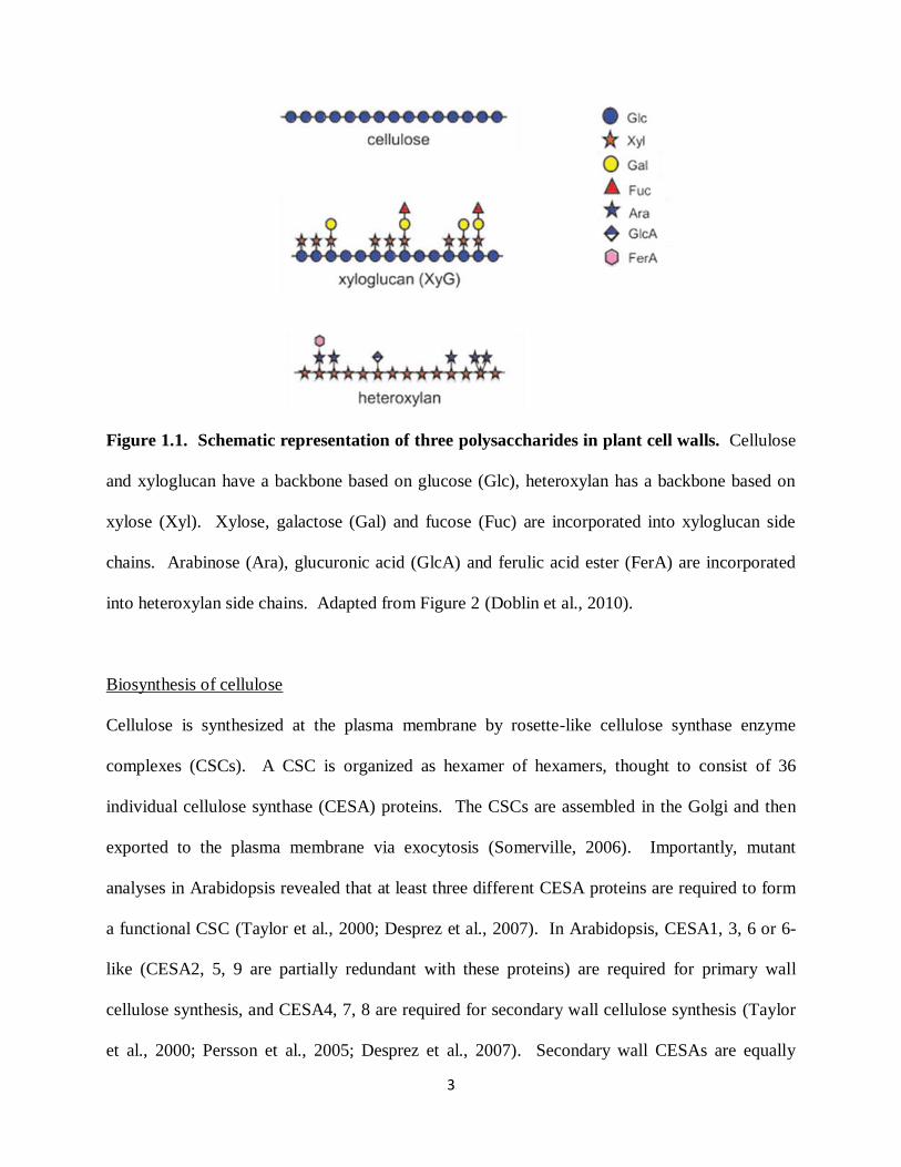
Figure 1.1. Schematic representation of three polysaccharides in plant cell walls. Cellulose
and xyloglucan have a backbone based on glucose (Glc), heteroxylan has a backbone based on
xylose (Xyl). Xylose, galactose (Gal) and fucose (Fuc) are incorporated into xyloglucan side
chains. Arabinose (Ara), glucuronic acid (GlcA) and ferulic acid ester (FerA) are incorporated
into heteroxylan side chains. Adapted from Figure 2 (Doblin et al., 2010).
Biosynthesis of cellulose
Cellulose is synthesized at the plasma membrane by rosette-like cellulose synthase enzyme
complexes (CSCs). A CSC is organized as hexamer of hexamers, thought to consist of 36
individual cellulose synthase (CESA) proteins. The CSCs are assembled in the Golgi and then
exported to the plasma membrane via exocytosis (Somerville, 2006). Importantly, mutant
analyses in Arabidopsis revealed that at least three different CESA proteins are required to form
a functional CSC (Taylor et al., 2000; Desprez et al., 2007). In Arabidopsis, CESA1, 3, 6 or 6-
like (CESA2, 5, 9 are partially redundant with these proteins) are required for primary wall
cellulose synthesis, and CESA4, 7, 8 are required for secondary wall cellulose synthesis (Taylor
et al., 2000; Persson et al., 2005; Desprez et al., 2007). Secondary wall CESAs are equally
4
important for cellulose biosynthesis in secondary walls, as CESA4, CESA7, CESA8 have the
same expression pattern in xylem and display interactions in the BiFc and co-
immunoprecipitation assays (Atanassov et al., 2009; Timmers et al., 2009). The cesa4, cesa7
and cesa8 mutants were initially identified by collapsed xylem phenotypes and the xylem defect
was associated with an up to eightfold reduction in the total amount of cellulose in stems (Turner
and Somerville, 1997). On the other hand, the three primary wall CESAs have unequal
contributions to cellulose biosynthesis. No null mutants have been reported for either CESA1 or
CESA3 (Persson et al., 2007). Missense mutations in CESA1 or CESA3 result in severely
retarded growth phenotypes and missense mutations in the catalytic domain of CESA1 cause
embryo-lethal phenotypes (Beeckman et al., 2002; Gillmor et al., 2002). However, null
mutations for CESA6 exhibit relatively mild phenotypes such as anisotropic cell swelling (Fagard
et al., 2000).
Live-cell imaging of a yellow fluorescent protein (YFP) fusion to CESA6 in Arabidopsis
has greatly advanced understanding of how cellulose is synthesized (Paredez et al., 2006). Such
YFP-tagged CESA6 shows that CESA rosettes move with an average velocity of 300 nm min -1
in vivo (Paredez et al., 2006). To further observe the spatial relationship between microtubules
and membrane-localized cellulose synthase complexes, the YFP tagged CESA6 line was crossed
with a CFP-tagged tubulin (TUA1) marker line. Co-localization of YFP:CESA and CFP:TUA1
showed that CESA localization and guidance are spatially and temporally coupled to
microtubules (Paredez et al., 2006). In the absence of cortical microtubules by treatment with
MT-destabilizing drug Oryzalin, the same velocities of CESA complexes were maintained as in
untreated samples (Paredez et al., 2006). This observation demonstrated that MTs might not be
required for cellulose synthase motility and the motive force for CSC motility is likely provided
5
by cellulose polymerization (Paredez et al., 2006). Interestingly, in the disorganized MT
resulting from treatment with Morlin, the velocities of CESA complexes were significantly
reduced (DeBolt et al., 2007). Therefore, Morlin affects an unknown agent that is possibly
involved in mediating interactions between the CESA complex and MTs, or a signaling mediator
that coordinates the CSC and MT activities (Hematy et al., 2007).
Apart from the CESA proteins, other proteins in the CSC also participate in cellulose
biosynthesis. These include the KORRIGAN (KOR1) glucanase, the KOBITO1 (KOB1) protein,
the COBRA (COB) glycosylphosphatidylinositol-anchored protein, and the Tracheary Element
Differentiation-Related (TED)-6 protein. Several mutations in the KOR1 gene lead to a marked
reduction in cellulose content, lateral organ swelling, and altered pectin composition, which may
arise as a compensation for the loss of cellulose (Nicol et al., 1998; Zuo et al., 2000; Sato et al.,
2001). There are three hypotheses about KOR1: first, KOR1 functions in removing the growing
cellulose chain from some type of primer or initiator (Peng et al., 2002); second, KOR1 alters the
crystallization properties within microfibrils (Szyjanowicz et al., 2004; Takahashi et al., 2009);
or third, KOR1 participates in the release of completed cellulose microfibrils from the CSC
(Szyjanowicz et al., 2004). The KOB1 protein appears to be involved directly in cellulose
biosynthesis. kob1 deletion mutants show a dwarf phenotype with swollen roots, and random
deposition of cellulose microfibrils (Pagant et al., 2002). COB is anchored to the extracellular
face of the plasma membrane and contains a motif with some similarities to a cellulose-binding
domain (Roudier et al., 2002). cob mutants exhibit defects in anisotropic expansion due to
altered cellulose microfibril orientations (Roudier et al., 2002; Roudier et al., 2005). TED6
interacts with the CESA7 subunit of the secondary wall CSC in vivo (Endo et al., 2009).
Suppression of TED6 results in defects of secondary cell-wall formation in vessels (Endo et

6
al.2009). However, the specific functions of these proteins involved in cellulose synthesis are
still unclear.
Xyloglucan structure and function
Of the hemicelluloses in dicot and non-grass monocot primary cell walls, xyloglucan is
the most abundant hemicellulosic polysaccharide, representing approximately 20% of leaf cell
walls in dicots (Zablackis et al., 1995). Xyloglucan consists of a β-1,4-linked glucan backbone
that is highly branched, with substitutions of α-1,6-linked xylosyl residue or side chains
composed of xylosyl, galactosyl and fucosyl residues (Figure 1.1). Some acetylated xyloglucans
have also been found in plant cell walls (Jia et al., 2005). A new discovery reports that
Arabidopsis root hair walls contain a previously unidentified xyloglucan that is composed of
both neutral and galacturonic acid-containing side chains (Peña et al., 2012). Xyloglucan was
believed to work as a tether between cellulose microfibrils. Therefore, xyloglucan was
hypothesized to contribute to the rigidity of cell walls when it associates with and non-covalently
links adjacent microfibrils, and to the loosening of the cell wall when it degrades (Hayashi et al.,
1988) (Figure 1.2A). Xyloglucan has been proposed to regulate cell expansion, to control cell
growth, and to prevent self-association of cellulose microfibrils (Liepman et al., 2010).
However, recent studies argue against a major mechanical role for xyloglucan tethers
spanning the space between adjacent microfibrils, since xxt1 xxt2 double mutant and xxt1 xxt2
xxt5 triple mutant plants, which lack detectable xyloglucan in their cell walls, can survive with
minor defects in root hairs (Cavalier et al., 2008; Park and Cosgrove, 2012; Zabotina et al., 2012).
The study of the structure and interactions of plant cell wall polysaccharides by two-and three-
dimensional magic-angle-spinning Solid-State NMR revealed that load bearing in plant cell
7
walls is accomplished by a single network of all three types of polysaccharides instead of a
cellulose-xyloglucan network (Dick-Perez et al., 2011). Upon treating cucumber hypocotyl
walls with a set of homologous endoglucanases with varying substrate specificities: xyloglucan-
specific endoglucanase (XEG from Aspergillus aculeatus) and cellulose-specific endoglucanase
(CEG from Aspergillus niger) failed to induce cell wall creep, either by themselves or in
combination, whereas an endoglucanase that hydrolyzes both xyloglucan and cellulose (Cel12A
from Hypocrea jecorina) induced high cell wall creep (Park and Cosgrove, 2012).
Measurements of elastic and plastic compliance revealed that both XEG and Cel12A hydrolyzed
xyloglucan in intact walls, and Cel12A could hydrolyze a minor xyloglucan compartment
recalcitrant to XEG digestion (Park and Cosgrove, 2012). Therefore, these results point to a
revised model for primary cell wall networks (Figure 1.2B) in which only a minor xyloglucan
component is located in the limited regions of tight contact between cellulose fibers to form
load-bearing connections (Park and Cosgrove, 2012).
8
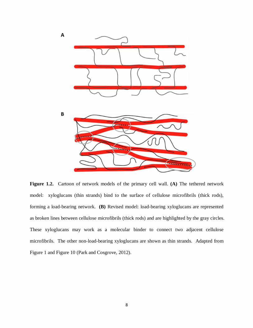
Figure 1.2. Cartoon of network models of the primary cell wall. (A) The tethered network
model: xyloglucans (thin strands) bind to the surface of cellulose microfibrils (thick rods),
forming a load-bearing network. (B) Revised model: load-bearing xyloglucans are represented
as broken lines between cellulose microfibrils (thick rods) and are highlighted by the gray circles.
These xyloglucans may work as a molecular binder to connect two adjacent cellulose
microfibrils. The other non-load-bearing xyloglucans are shown as thin strands. Adapted from
Figure 1 and Figure 10 (Park and Cosgrove, 2012).
9
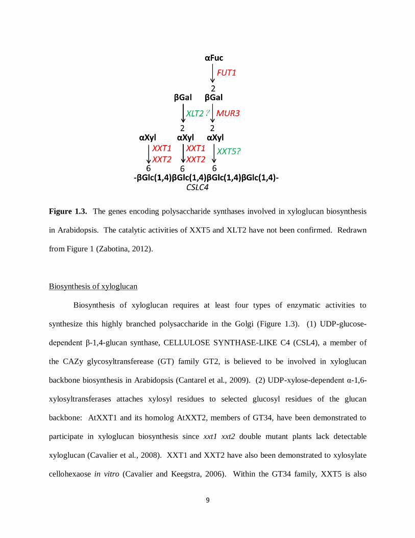
Figure 1.3. The genes encoding polysaccharide synthases involved in xyloglucan biosynthesis
in Arabidopsis. The catalytic activities of XXT5 and XLT2 have not been confirmed. Redrawn
from Figure 1 (Zabotina, 2012).
Biosynthesis of xyloglucan
Biosynthesis of xyloglucan requires at least four types of enzymatic activities to
synthesize this highly branched polysaccharide in the Golgi (Figure 1.3). (1) UDP-glucose-
dependent β-1,4-glucan synthase, CELLULOSE SYNTHASE-LIKE C4 (CSL4), a member of
the CAZy glycosyltransferease (GT) family GT2, is believed to be involved in xyloglucan
backbone biosynthesis in Arabidopsis (Cantarel et al., 2009). (2) UDP-xylose-dependent α-1,6-
xylosyltransferases attaches xylosyl residues to selected glucosyl residues of the glucan
backbone: AtXXT1 and its homolog AtXXT2, members of GT34, have been demonstrated to
participate in xyloglucan biosynthesis since xxt1 xxt2 double mutant plants lack detectable
xyloglucan (Cavalier et al., 2008). XXT1 and XXT2 have also been demonstrated to xylosylate
cellohexaose in vitro (Cavalier and Keegstra, 2006). Within the GT34 family, XXT5 is also
10
involved in xyloglucan biosynthesis, but its activity has not been demonstrated in vitro (Zabotina
et al., 2008). (3) UDP-galactose-dependent β-1,2-galactosyltransferase attaches galactosyl
residues to selected xylosyl residues: a screen for mutants with significant reduction in the fucose
content of cell walls identified AtMUR3, which encodes a GT47 galactosyltransferase (Reiter et
al., 1997). Recently, a second xyloglucan galactosyltransferase named Xyloglucan L-side Chain
Galactosyltransferase (XLT2) was found to be required for galactosylation of the second xylose
in the xyloglucan subunit, although the catalytic activity of XLT2 has not been confirmed
(Jensen et al., 2012). (4) GDP-fucose-dependent α-1,2-fucosyltransferase attaches fucosyl
residues to selected galactosyl residues: the AtFUT1 gene from the GT37 family encodes a
xylolucan α-1,2-fucosyltransferase that terminally fucosylates xyloglucan (Perrin et al., 1999).
Arabidopsis fut1 T-DNA knock-out mutants exhibit complete elimination of fucosylated
xyloglucan subunits (Perrin et al., 1999).
Xylan structure and function
Xylan consists of a linear backbone of β-1,4-linked xylose residues substituted with
acetyl, glucuronic acid (GlcA), 4-O-methylglucuronic acid (Me-GlcA), and arabinose residues
(Figure 1.1). Depending on the plant species and even tissues in the same species, there is
variation in xylan structures (Rennie and Scheller, 2014). In dicots, xylan is the second most
abundant polysaccharide after cellulose in plant secondary cell walls, and it is also found at much
lower level in primary walls. Dicot xylans contain the tetrasaccharide reducing-end sequence,
known as β-D-Xyl-(1,3)-α-L-Rhap-(1,2)-α-D-GalA-(1,4)-D-Xyl, which has been proposed to
serve either as an initiator or terminator of xylan backbone biosynthesis (York and O'Neill, 2008).
In Arabidopsis, the ratio of Me-GlcA to GlcA is 2:1 and Me-GlcA substitution occurs on one out
of eight xylose residues on average (Bromley et al., 2013). No or very few arabinosyl
11
substitutions have been reported in Arabidopsis and Poplar; hence these xylans are referred to as
Glucuronoxylans (GX) (Rennie and Scheller, 2014). In contrast, grass xylans are typically
substituted with arabinosyl residues, and are referred to as Glucuronoarabinoxylans (GAX).
Xylans have been proposed to coat cellulose microfibrils and crosslink with other
polysaccharides via hydrogen bonds. Xylans are believed to link to lignin via ester bonds to
GlcA and/or ether bonds to Xyl or Ara (Imamura et al., 1994; Balakshin et al., 2011). A recent
study discovered that within Arabidopsis, a large proteoglycan complex, Arabinoxylan Pectin
Arabinogalactan Protein 1 (APAP1) has short stretches of arabinoxylan/xylan that appear to be
covalently linked to both pectin and arabinogalactans (Tan et al., 2013).
Xylan provides shape, structural strength and protection for plant cells, and is required
for normal plant growth and development. Xylan makes up a large fraction of plant biomass.
However, xylan is composed almost entirely of pentose sugars, which cannot be efficiently
fermented. Therefore, plants that have reduced amounts of xylan, but still maintain normal
growth and development, are desired for converting biomass to biofuel (Petersen et al., 2012).
Biosynthesis of xylan
GX biosynthesis involves at least three types of glycosyltransferase activities: (1)
formation of the β-1,4-linked xylose backbone; (2) formation of the sequence β-D-Xyl-(1,3)-α-
L-Rhap-(1,2)-α-D-GalA-(1,4)-D-Xyl at the reducing end (at least in those plants that make this
sequence); (3) addition and modification of GlcA and Me-GlcA side chains.
Two members of GT43, Irregular Xylem (IRX) 9 and IRX14, and one member of GT47,
IRX10, which encode putative xylosyltransferases, are required to synthesize the xylan backbone
(Brown et al., 2005; Brown et al., 2007; Lee et al., 2007). Mutations in IRX9 and IRX14 result in
12
decreases in xylan contents and length of the xylan chains. IRX9 and IRX14 are not functionally
redundant in backbone synthesis and are suggested to work cooperatively, since IRX9 did not
rescue the irx14 phenotype (Lee et al., 2010). However, the catalytic activities of IRX9 and
IRX14 have not been demonstrated in vitro. Mutations in IRX10/GUT2 and its homolog IRX10-
L/GUT1 lead to a reduction in xylan content and the proteins encoded by these genes are thought
to function redundantly in synthesizing the xylan backbone, as the reduction in xylan content is
much greater in irx10 irx10-L double mutants (Brown et al., 2009; Wu et al., 2009). IRX10-L
has recently been demonstrated to have UDP-Xyl:β-(1,4)-xylosyl transferase activity and has
been renamed XYLAN SYNTHASE-1 (XYS1) (Urbanowicz et al., 2014). Interestingly, the
authors also showed that TBL29/ESK1 catalyzes the subsequent addition of O-acetyl groups
from acetyl-CoA to the 2-position of xylosyl backbone residues (Urbanowicz et al., 2014). A
recent study found that an IRX10 homolog from the moss, Physcomitrella patens, and an IRX10
homolog from the dicot plant, Plantago ovate, are conserved in having xylan β-1,4-
xylosyltransferase activities (Jensen et al., 2014).
The Arabidopsis genes IRX7/FRA8 (GT47), a homolog of IRX7/FRA8 termed F8H,
GAUT12/IRX8 (GT8), and GATL1/PARVUS (GT8) have been implicated in the synthesis of the
xylan reducing end sequencing (Zhong et al., 2005; Brown et al., 2007; Peña et al., 2007;
Persson et al., 2007; Kong et al., 2009; Lee et al., 2009). Mutations in these genes cause reduced
amounts of the reducing-end tetrasaccharide, more heterogeneous distribution of xylan chain
length, and yet microsomal extracts exhibit no change in xylan synthase activity. There are two
proposed functions of this reducing end tetrasaccharide in xylan biosynthesis, either acting as a
GX chain terminator or acting as a GX primer, assuming that GX chain elongation occurs by
sequential addition of xylosyl residues to this sequence (York and O'Neill, 2008).
13
The GlcA side chains are added by Glucuronic Acid Substitution of Xylan (GUX) 1 and
GUX2, two members of GT8 that catalyze the transfer of α-GlcA onto the O-2 position of xylose
in xylan (Mortimer et al., 2010). The gux1 gux2 double mutant has almost no detectable GlcA
substitution on xylan, yet shows a visibly normal phenotype (Mortimer et al., 2010). A recent
detailed study further points out that GUX1 is responsible for adding GlcA to evenly spaced
xylose residues, while GUX2 is responsible for adding GlcA to both evenly and oddly spaced
xylose residues of the xylan backbone (Bromley et al., 2013). 4-O-Methyl groups are transferred
from S-adenosylmethionine (SAM) to GlcA by Glucuronoxylan Methyltransferase 1 (GXMT1),
a protein containing a Domain of Unknown Fuction 579 (DUF579) (Urbanowicz et al., 2012).
The gxmt1 mutants have a 75% reduction in Me-GlcA residues in their secondary cell wall xylan
(Urbanowicz et al., 2012).
Using Carbohydrate Binding Modules (CBMs) as tools for analysis of cell wall
polysaccharides
To degrade the highly complex and dynamic cell wall structure, microorganisms produce
an extensive repertoire of polysaccharide-degrading enzymes, including glycoside hydrolases,
polysaccharide lyases, carbohydrate esterases and polysaccharide oxidases (Gilbert, 2010).
These enzymes often contain protein domains called Carbohydrate-Binding Modules (CBMs).
CBMs display no hydrolytic function and it has been suggested that CBMs enhance the
efficiency of hydrolytic enzymes by mediating prolonged and intimate contact between their
catalytic modules and their target substrates (Boraston et al., 2004; Herve et al., 2010). Based on
sequence comparisons, CBMs have been grouped into 69 families
(http://www.cazy.org/Carbohydrate-Binding-Modules.html). Recently, CBMs have been used as

14
probes to assess and document the cell biological contexts of polysaccharides and cell wall
diversity in vitro (Knox, 2008). CBMs that have been used to study polysaccharides include
those that bind to cellulose, xylan, mannan, xyloglucan and pectin according to their binding
specificities, as listed in Table 1.1. CBM binding specificity for pectin was underrepresented
until recently, when a new family of CBMs, CBM61, was identified (Cid et al., 2010).
The capacity of these CBMs to recognize polysaccharides in cell walls was usually
assessed using an indirect triple labeling immuofluorescence procedure (His-tagged CBM, anti-
His mouse-Ig, anti-mouse Ig fluorescein isothiocyanate) on sections of plant tissues (Blake et al.,
2006; McCartney et al., 2006). Within the same polysaccharide-directed CBM group, diverse
origins of CBMs displayed differential binding capacities to cell walls depending on cell type,
tissue, and taxon of origin (Blake et al., 2006; McCartney et al., 2006). For instance, the xylan-
directed CBM2b-1-2 targets all of the secondary cell walls of specific cell types such as xylem
vessels and sclerechyma fibers associated with phloem in sections of a range of dicotyledonous
plant materials (tobacco, pea and flax) (McCartney et al., 2006). However, xylan-directed
CBM35 did not bind to all secondary cell walls of xylem vessels, exhibiting a preference for an
unmethylated form of xylan (GlcA not Me-GlcA) in tobacco sections (McCartney et al., 2006).
CBM35 bound specifically to the secondary cell walls of pea sections, but to both primary and
secondary cell walls of flax (McCartney et al., 2006). This diversity reveals that a variety of
polysaccharide microstructures exists in plants, and points out a biological rationale for the large
number of CBMs: different CBMs have the capacity to target appended catalytic modules to
specific cell wall structures in diverse species (McCartney et al., 2006).
Compared to another set of cell wall probes, antibodies, CBMs have the advantages that
gene sequences and protein structures are often known (Knox, 2008). This information

15
generates great opportunities for the engineering of CBM specificities and the generation of
fluorescent protein-tagged CBMs. To date, the only existing xyloglucan CBMs were created by
protein engineering of the xylan-binding CBM4-2, by modification of the CBM protein through
random mutagenesis of the corresponding gene in combination with phage display technology
(Gunnarsson et al., 2006; von Schantz et al., 2009). By fusing the xylan-binding CBM2b-1-2
and/or cellulose-directed CBM3a to the xylanases Xyl11A/Xyl10B (either by appending single
CBM or tandem CBMs), degradation of xylan polysaccharides in secondary cell walls was
potentiated (Herve et al., 2010). A recent study also found that cellulose-binding CBM3a and
mannan-binding CBM27 have a great impact on the removal of mannan from tobacco and
Physcomitrella cell walls, respectively, by fusion to GH5 and GH26 mannanases and CE2
esterases (Zhang et al., 2014). Although the functions of CBMs in cell wall degrading processes
are still not fully understood, these studies proposed a mechanism that polysaccharide degrading
enzymes are bound to the cell wall through their appended CBM(s), and that the CBM(s) greatly
increase the concentration of the enzymes in the vicinity of the substrate by targeting
polysaccharides that are in close proximity to the substrate of the catalytic module, leading to the
observed increase in polysaccharide hydrolysis (Herve et al., 2010).
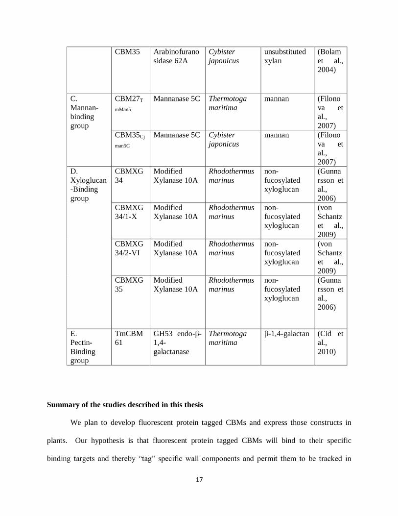
Table1.1. List of CBMs
Group Protein Source
enzymes
Organism Type Refere
nce
A.
Cellulose-
Binding
group
CBM1 Cellulase Trichoderma
reesei
crystalline
cellulose
(Reinik
ainen et
al.,
1992)
CBM2a Xylanase 10A Cybister
japonicus
crystalline
cellulose
(Bolam
et al.,
1998)
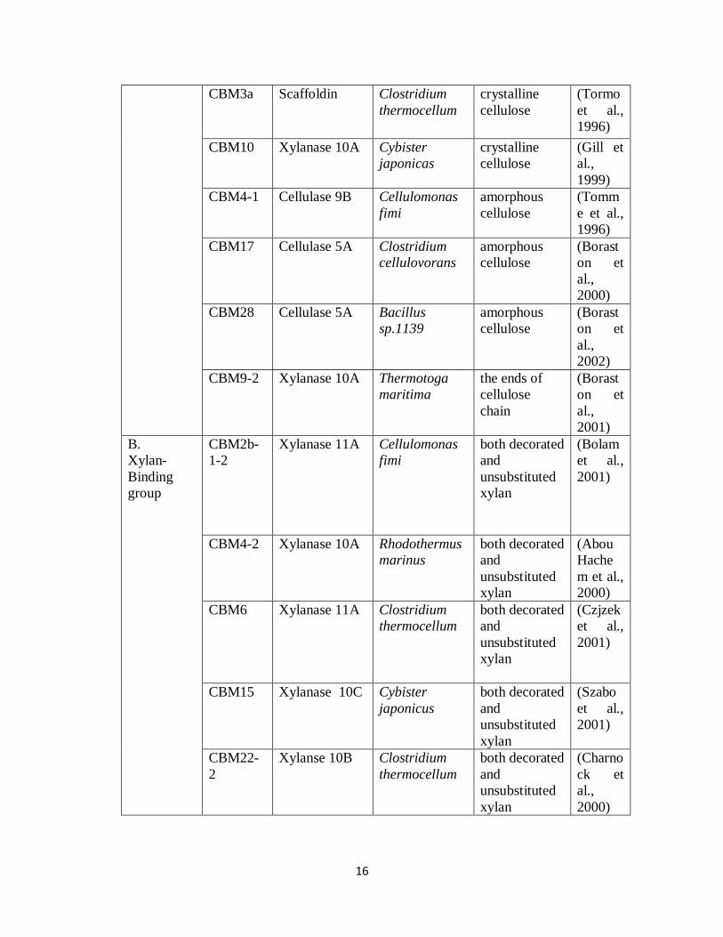
16
CBM3a Scaffoldin Clostridium
thermocellum
crystalline
cellulose
(Tormo
et al.,
1996)
CBM10 Xylanase 10A Cybister
japonicas
crystalline
cellulose
(Gill et
al.,
1999)
CBM4-1 Cellulase 9B Cellulomonas
fimi
amorphous
cellulose
(Tomm
e et al.,
1996)
CBM17 Cellulase 5A Clostridium
cellulovorans
amorphous
cellulose
(Borast
on et
al.,
2000)
CBM28 Cellulase 5A Bacillus
sp.1139
amorphous
cellulose
(Borast
on et
al.,
2002)
CBM9-2 Xylanase 10A Thermotoga
maritima
the ends of
cellulose
chain
(Borast
on et
al.,
2001)
B.
Xylan-
Binding
group
CBM2b-
1-2
Xylanase 11A Cellulomonas
fimi
both decorated
and
unsubstituted
xylan
(Bolam
et al.,
2001)
CBM4-2 Xylanase 10A Rhodothermus
marinus
both decorated
and
unsubstituted
xylan
(Abou
Hache
m et al.,
2000)
CBM6 Xylanase 11A Clostridium
thermocellum
both decorated
and
unsubstituted
xylan
(Czjzek
et al.,
2001)
CBM15 Xylanase 10C Cybister
japonicus
both decorated
and
unsubstituted
xylan
(Szabo
et al.,
2001)
CBM22-
2
Xylanse 10B Clostridium
thermocellum
both decorated
and
unsubstituted
xylan
(Charno
ck et
al.,
2000)
17
CBM35 Arabinofurano
sidase 62A
Cybister
japonicus
unsubstituted
xylan
(Bolam
et al.,
2004)
C.
Mannan-
binding
group
CBM27T
mMan5
Mannanase 5C Thermotoga
maritima
mannan (Filono
va et
al.,
2007)
CBM35Cj
man5C
Mannanase 5C Cybister
japonicus
mannan (Filono
va et
al.,
2007)
D.
Xyloglucan
-Binding
group
CBMXG
34
Modified
Xylanase 10A
Rhodothermus
marinus
non-
fucosylated
xyloglucan
(Gunna
rsson et
al.,
2006)
CBMXG
34/1-X
Modified
Xylanase 10A
Rhodothermus
marinus
non-
fucosylated
xyloglucan
(von
Schantz
et al.,
2009)
CBMXG
34/2-VI
Modified
Xylanase 10A
Rhodothermus
marinus
non-
fucosylated
xyloglucan
(von
Schantz
et al.,
2009)
CBMXG
35
Modified
Xylanase 10A
Rhodothermus
marinus
non-
fucosylated
xyloglucan
(Gunna
rsson et
al.,
2006)
E.
Pectin-
Binding
group
TmCBM
61
GH53 endo-β-
1,4-
galactanase
Thermotoga
maritima
β-1,4-galactan (Cid et
al.,
2010)
Summary of the studies described in this thesis
We plan to develop fluorescent protein tagged CBMs and express those constructs in
plants. Our hypothesis is that fluorescent protein tagged CBMs will bind to their specific
binding targets and thereby “tag” specific wall components and permit them to be tracked in

18
living plants. There are two potential outcomes: A) The binding of the fluorescent tagged CBM
to the polysaccharide in vivo is benign, that is, has no effect on polysaccharide function and
therefore the incorporation of the tag will allow us track the dynamics of plant cell wall
polysaccharides during plant growth and development; or B) The binding of the fluorescent
tagged CBMs to the polysaccharide in vivo is NOT benign, that is, has an effect on the bound
polysaccharide and leads to changes in the function or structure of the wall, which in turn, may
have an impact on the development of the plant. Before setting up the specific objectives, we
verified the binding specificities of fluorescent protein tagged CBMs in vitro and examined the
morphological and developmental effects of the expression of these CBMs in Arabidopsis. Our
preliminary data suggest that BOTH outcomes A AND B mentioned above occur, depending on
the CBM and the polysaccharide that the CBM binds to. Even within the same group, expression
of different CBMs can lead to different results. It appears that the xylan-binding CBM group is
more suitable to be utilized as an in vivo tagging tool for xylans, since expression of either
CBM2b-1-2 (see Chapter 3) or CBM35 (data not shown) has no deleterious effect on plant
morphology and development. On the other hand, cellulose binding CBMs appear to have
deleterious effects when expressed in plants, making them tools for selectively modulating cell
wall function(s) (see Chapter 4).
19
References
Abou Hachem M, Nordberg Karlsson E, Bartonek-Roxa E, Raghothama S, Simpson PJ,
Gilbert HJ, Williamson MP, Holst O (2000) Carbohydrate-binding modules from a
thermostable Rhodothermus marinus xylanase: cloning, expression and binding studies.
Biochem J 345 Pt 1: 53-60
Albersheim P, Darvill A, Roberts K, Sederoff R, Staehelin A (2010) Plant Cell Walls.
Garland Science, New York
Atanassov, II, Pittman JK, Turner SR (2009) Elucidating the mechanisms of assembly and
subunit interaction of the cellulose synthase complex of Arabidopsis secondary cell walls.
J Biol Chem 284: 3833-3841
Balakshin M, Capanema E, Gracz H, Chang HM, Jameel H (2011) Quantification of lignin-
carbohydrate linkages with high-resolution NMR spectroscopy. Planta 233: 1097-1110
Beeckman T, Przemeck GK, Stamatiou G, Lau R, Terryn N, De Rycke R, Inze D, Berleth T
(2002) Genetic complexity of cellulose synthase a gene function in Arabidopsis
embryogenesis. Plant Physiol 130: 1883-1893
Blake AW, McCartney L, Flint JE, Bolam DN, Boraston AB, Gilbert HJ, Knox JP (2006)
Understanding the biological rationale for the diversity of cellulose-directed
carbohydrate-binding modules in prokaryotic enzymes. J Biol Chem 281: 29321-29329
Bolam DN, Ciruela A, McQueen-Mason S, Simpson P, Williamson MP, Rixon JE, Boraston
A, Hazlewood GP, Gilbert HJ (1998) Pseudomonas cellulose-binding domains mediate
their effects by increasing enzyme substrate proximity. Biochem J 331 ( Pt 3): 775-781

20
Bolam DN, Xie H, Pell G, Hogg D, Galbraith G, Henrissat B, Gilbert HJ (2004) X4 modules
represent a new family of carbohydrate-binding modules that display novel properties. J
Biol Chem 279: 22953-22963
Bolam DN, Xie H, White P, Simpson PJ, Hancock SM, Williamson MP, Gilbert HJ (2001)
Evidence for synergy between family 2b carbohydrate binding modules in Cellulomonas
fimi xylanase 11A. Biochemistry 40: 2468-2477
Boraston AB, Bolam DN, Gilbert HJ, Davies GJ (2004) Carbohydrate-binding modules: fine-
tuning polysaccharide recognition. Biochem J 382: 769-781
Boraston AB, Chiu P, Warren RA, Kilburn DG (2000) Specificity and affinity of substrate
binding by a family 17 carbohydrate-binding module from Clostridium cellulovorans
cellulase 5A. Biochemistry 39: 11129-11136
Boraston AB, Creagh AL, Alam MM, Kormos JM, Tomme P, Haynes CA, Warren RA,
Kilburn DG (2001) Binding specificity and thermodynamics of a family 9 carbohydrate-
binding module from Thermotoga maritima xylanase 10A. Biochemistry 40: 6240-6247
Boraston AB, Ghaffari M, Warren RA, Kilburn DG (2002) Identification and glucan-binding
properties of a new carbohydrate-binding module family. Biochem J 361: 35-40
Bromley JR, Busse-Wicher M, Tryfona T, Mortimer JC, Zhang Z, Brown DM, Dupree P
(2013) GUX1 and GUX2 glucuronyltransferases decorate distinct domains of
glucuronoxylan with different substitution patterns. Plant J 74: 423-434
Brown DM, Goubet F, Wong VW, Goodacre R, Stephens E, Dupree P, Turner SR (2007)
Comparison of five xylan synthesis mutants reveals new insight into the mechanisms of
xylan synthesis. Plant J 52: 1154-1168

21
Brown DM, Zeef LA, Ellis J, Goodacre R, Turner SR (2005) Identification of novel genes in
Arabidopsis involved in secondary cell wall formation using expression profiling and
reverse genetics. Plant Cell 17: 2281-2295
Brown DM, Zhang Z, Stephens E, Dupree P, Turner SR (2009) Characterization of IRX10
and IRX10-like reveals an essential role in glucuronoxylan biosynthesis in Arabidopsis.
Plant J 57: 732-746
Brown RM (2004) Cellulose structure and biosynthesis: What is in store for the 21st century?
Journal of Polymer Science Part A: Polymer Chemistry 42: 487-495
Carpita NC, Gibeaut DM (1993) Structural models of primary cell walls in flowering plants:
consistency of molecular structure with the physical properties of the walls during growth.
Plant J 3: 1-30
Cavalier DM, Keegstra K (2006) Two xyloglucan xylosyltransferases catalyze the addition of
multiple xylosyl residues to cellohexaose. J Biol Chem 281: 34197-34207
Cavalier DM, Lerouxel O, Neumetzler L, Yamauchi K, Reinecke A, Freshour G, Zabotina
OA, Hahn MG, Burgert I, Pauly M, Raikhel NV, Keegstra K (2008) Disrupting two
Arabidopsis thaliana xylosyltransferase genes results in plants deficient in xyloglucan, a
major primary cell wall component. Plant Cell 20: 1519-1537
Charnock SJ, Bolam DN, Turkenburg JP, Gilbert HJ, Ferreira LM, Davies GJ, Fontes CM
(2000) The X6 "thermostabilizing" domains of xylanases are carbohydrate-binding
modules: structure and biochemistry of the Clostridium thermocellum X6b domain.
Biochemistry 39: 5013-5021
22
Cid M, Pedersen HL, Kaneko S, Coutinho PM, Henrissat B, Willats WG, Boraston AB
(2010) Recognition of the helical structure of beta-1,4-galactan by a new family of
carbohydrate-binding modules. J Biol Chem 285: 35999-36009
Czjzek M, Bolam DN, Mosbah A, Allouch J, Fontes CM, Ferreira LM, Bornet O, Zamboni
V, Darbon H, Smith NL, Black GW, Henrissat B, Gilbert HJ (2001) The location of
the ligand-binding site of carbohydrate-binding modules that have evolved from a
common sequence is not conserved. J Biol Chem 276: 48580-48587
DeBolt S, Gutierrez R, Ehrhardt DW, Melo CV, Ross L, Cutler SR, Somerville C, Bonetta
D (2007) Morlin, an inhibitor of cortical microtubule dynamics and cellulose synthase
movement. Proc Natl Acad Sci U S A 104: 5854-5859
Desprez T, Juraniec M, Crowell EF, Jouy H, Pochylova Z, Parcy F, Hofte H, Gonneau M,
Vernhettes S (2007) Organization of cellulose synthase complexes involved in primary
cell wall synthesis in Arabidopsis thaliana. Proc Natl Acad Sci U S A 104: 15572-15577
Dick-Perez M, Zhang Y, Hayes J, Salazar A, Zabotina OA, Hong M (2011) Structure and
interactions of plant cell-wall polysaccharides by two- and three-dimensional magic-
angle-spinning solid-state NMR. Biochemistry 50: 989-1000
Doblin MS, Pettolino F, Bacic A (2010) Plant cell walls: the skeleton of the plant world.
Functional Plant Biology 37: 357-381
Endo S, Pesquet E, Yamaguchi M, Tashiro G, Sato M, Toyooka K, Nishikubo N, Udagawa-
Motose M, Kubo M, Fukuda H, Demura T (2009) Identifying new components
participating in the secondary cell wall formation of vessel elements in zinnia and
Arabidopsis. Plant Cell 21: 1155-1165
23
Fagard M, Desnos T, Desprez T, Goubet F, Refregier G, Mouille G, McCann M, Rayon C,
Vernhettes S, Hofte H (2000) PROCUSTE1 encodes a cellulose synthase required for
normal cell elongation specifically in roots and dark-grown hypocotyls of Arabidopsis.
Plant Cell 12: 2409-2424
Filonova L, Kallas AM, Greffe L, Johansson G, Teeri TT, Daniel G (2007) Analysis of the
surfaces of wood tissues and pulp fibers using carbohydrate-binding modules specific for
crystalline cellulose and mannan. Biomacromolecules 8: 91-97
Gilbert HJ (2010) The biochemistry and structural biology of plant cell wall deconstruction.
Plant Physiol 153: 444-455
Gill J, Rixon JE, Bolam DN, McQueen-Mason S, Simpson PJ, Williamson MP, Hazlewood
GP, Gilbert HJ (1999) The type II and X cellulose-binding domains of Pseudomonas
xylanase A potentiate catalytic activity against complex substrates by a common
mechanism. Biochem J 342 ( Pt 2): 473-480
Gillmor CS, Poindexter P, Lorieau J, Palcic MM, Somerville C (2002) Alpha-glucosidase I is
required for cellulose biosynthesis and morphogenesis in Arabidopsis. J Cell Biol 156:
1003-1013
Gunnarsson LC, Zhou Q, Montanier C, Karlsson EN, Brumer H, 3rd, Ohlin M (2006)
Engineered xyloglucan specificity in a carbohydrate-binding module. Glycobiology 16:
1171-1180
Hayashi T, Koyama T, Matsuda K (1988) Formation of UDP-Xylose and Xyloglucan in
Soybean Golgi Membranes. Plant Physiol 87: 341-345
24
Hematy K, Sado PE, Van Tuinen A, Rochange S, Desnos T, Balzergue S, Pelletier S, Renou
JP, Hofte H (2007) A receptor-like kinase mediates the response of Arabidopsis cells to
the inhibition of cellulose synthesis. Curr Biol 17: 922-931
Herve C, Rogowski A, Blake AW, Marcus SE, Gilbert HJ, Knox JP (2010) Carbohydrate-
binding modules promote the enzymatic deconstruction of intact plant cell walls by
targeting and proximity effects. Proc Natl Acad Sci U S A 107: 15293-15298
Imamura T, Watanabe T, Kuwahara M, Koshijima T (1994) Ester linkages between lignin
and glucuronic acid in lignin-carbohydrate complexes from Fagus crenata.
Phytochemistry 37: 1165-1173
Jensen JK, Johnson NR, Wilkerson CG (2014) Arabidopsis thaliana IRX10 and two related
proteins from psyllium and Physcomitrella patens are xylan xylosyltransferases. The
Plant Journal 80: 207-215
Jensen JK, Schultink A, Keegstra K, Wilkerson CG, Pauly M (2012) RNA-Seq analysis of
developing nasturtium seeds (Tropaeolum majus): identification and characterization of
an additional galactosyltransferase involved in xyloglucan biosynthesis. Mol Plant 5:
984-992
Jia Z, Cash M, Darvill AG, York WS (2005) NMR characterization of endogenously O-
acetylated oligosaccharides isolated from tomato (Lycopersicon esculentum) xyloglucan.
Carbohydr Res 340: 1818-1825
Knox JP (2008) Revealing the structural and functional diversity of plant cell walls. Current
Opinion in Plant Biology 11: 308-313
25
Kong Y, Zhou G, Avci U, Gu X, Jones C, Yin Y, Xu Y, Hahn MG (2009) Two poplar
glycosyltransferase genes, PdGATL1.1 and PdGATL1.2, are functional orthologs to
PARVUS/AtGATL1 in Arabidopsis. Mol Plant 2: 1040-1050
Lee C, O'Neill MA, Tsumuraya Y, Darvill AG, Ye ZH (2007) The irregular xylem9 mutant is
deficient in xylan xylosyltransferase activity. Plant Cell Physiol 48: 1624-1634
Lee C, Teng Q, Huang W, Zhong R, Ye ZH (2009) The F8H glycosyltransferase is a
functional paralog of FRA8 involved in glucuronoxylan biosynthesis in Arabidopsis.
Plant Cell Physiol 50: 812-827
Lee C, Teng Q, Huang W, Zhong R, Ye ZH (2010) The Arabidopsis family GT43
glycosyltransferases form two functionally nonredundant groups essential for the
elongation of glucuronoxylan backbone. Plant Physiol 153: 526-541
Liepman AH, Wightman R, Geshi N, Turner SR, Scheller HV (2010) Arabidopsis - a
powerful model system for plant cell wall research. Plant J 61: 1107-1121
McCartney L, Blake AW, Flint J, Bolam DN, Boraston AB, Gilbert HJ, Knox JP (2006)
Differential recognition of plant cell walls by microbial xylan-specific carbohydrate-
binding modules. Proceedings of the National Academy of Sciences of the United States
of America 103: 4765-4770
Mortimer JC, Miles GP, Brown DM, Zhang Z, Segura MP, Weimar T, Yu X, Seffen KA,
Stephens E, Turner SR, Dupree P (2010) Absence of branches from xylan in
Arabidopsis gux mutants reveals potential for simplification of lignocellulosic biomass.
Proceedings of the National Academy of Sciences 107: 17409-17414
26
Nicol F, His I, Jauneau A, Vernhettes S, Canut H, Höfte H (1998) A plasma membrane‐
bound putative endo‐1,4‐β‐d‐glucanase is required for normal wall assembly and cell
elongation in Arabidopsis, Vol 17
Pagant S, Bichet A, Sugimoto K, Lerouxel O, Desprez T, McCann M, Lerouge P,
Vernhettes S, Hofte H (2002) KOBITO1 encodes a novel plasma membrane protein
necessary for normal synthesis of cellulose during cell expansion in Arabidopsis. Plant
Cell 14: 2001-2013
Paredez AR, Somerville CR, Ehrhardt DW (2006) Visualization of Cellulose Synthase
Demonstrates Functional Association with Microtubules. Science 312: 1491-1495
Park YB, Cosgrove DJ (2012) A Revised Architecture of Primary Cell Walls Based on
Biomechanical Changes Induced by Substrate-Specific Endoglucanases. Plant Physiology
158: 1933-1943
Peña MJ, Kong Y, York WS, O’Neill MA (2012) A Galacturonic Acid–Containing
Xyloglucan Is Involved in Arabidopsis Root Hair Tip Growth. The Plant Cell Online 24:
4511-4524
Peña MJ, Zhong R, Zhou G-K, Richardson EA, O'Neill MA, Darvill AG, York WS, Ye Z-
H (2007) Arabidopsis irregular xylem8 and irregular xylem9: Implications for the
Complexity of Glucuronoxylan Biosynthesis. The Plant Cell Online 19: 549-563
Peng L, Kawagoe Y, Hogan P, Delmer D (2002) Sitosterol-beta-glucoside as primer for
cellulose synthesis in plants. Science 295: 147-150
Perrin RM, DeRocher AE, Bar-Peled M, Zeng W, Norambuena L, Orellana A, Raikhel NV,
Keegstra K (1999) Xyloglucan fucosyltransferase, an enzyme involved in plant cell wall
biosynthesis. Science 284: 1976-1979
27
Persson S, Caffall KH, Freshour G, Hilley MT, Bauer S, Poindexter P, Hahn MG, Mohnen
D, Somerville C (2007) The Arabidopsis irregular xylem8 mutant is deficient in
glucuronoxylan and homogalacturonan, which are essential for secondary cell wall
integrity. Plant Cell 19: 237-255
Persson S, Paredez A, Carroll A, Palsdottir H, Doblin M, Poindexter P, Khitrov N, Auer M,
Somerville CR (2007) Genetic evidence for three unique components in primary cell-
wall cellulose synthase complexes in Arabidopsis. Proceedings of the National Academy
of Sciences 104: 15566-15571
Persson S, Wei H, Milne J, Page GP, Somerville CR (2005) Identification of genes required
for cellulose synthesis by regression analysis of public microarray data sets. Proc Natl
Acad Sci U S A 102: 8633-8638
Petersen PD, Lau J, Ebert B, Yang F, Verhertbruggen Y, Kim JS, Varanasi P,
Suttangkakul A, Auer M, Loque D, Scheller HV (2012) Engineering of plants with
improved properties as biofuels feedstocks by vessel-specific complementation of xylan
biosynthesis mutants. Biotechnol Biofuels 5: 84
Reinikainen T, Ruohonen L, Nevanen T, Laaksonen L, Kraulis P, Jones TA, Knowles JK,
Teeri TT (1992) Investigation of the function of mutated cellulose-binding domains of
Trichoderma reesei cellobiohydrolase I. Proteins 14: 475-482
Reiter WD, Chapple C, Somerville CR (1997) Mutants of Arabidopsis thaliana with altered
cell wall polysaccharide composition. Plant J 12: 335-345
Rennie EA, Scheller HV (2014) Xylan biosynthesis. Current Opinion in Biotechnology 26:
100-107
28
Roudier F, Fernandez AG, Fujita M, Himmelspach R, Borner GH, Schindelman G, Song S,
Baskin TI, Dupree P, Wasteneys GO, Benfey PN (2005) COBRA, an Arabidopsis
extracellular glycosyl-phosphatidyl inositol-anchored protein, specifically controls highly
anisotropic expansion through its involvement in cellulose microfibril orientation. Plant
Cell 17: 1749-1763
Roudier F, Schindelman G, DeSalle R, Benfey PN (2002) The COBRA family of putative
GPI-anchored proteins in Arabidopsis. A new fellowship in expansion. Plant Physiol 130:
538-548
Sato S, Kato T, Kakegawa K, Ishii T, Liu YG, Awano T, Takabe K, Nishiyama Y, Kuga S,
Sato S, Nakamura Y, Tabata S, Shibata D (2001) Role of the putative membrane-
bound endo-1,4-beta-glucanase KORRIGAN in cell elongation and cellulose synthesis in
Arabidopsis thaliana. Plant Cell Physiol 42: 251-263
Somerville C (2006) Cellulose synthesis in higher plants. Annu Rev Cell Dev Biol 22: 53-78
Somerville C (2007) Biofuels. Curr Biol 17: R115-119
Szabo L, Jamal S, Xie H, Charnock SJ, Bolam DN, Gilbert HJ, Davies GJ (2001) Structure
of a family 15 carbohydrate-binding module in complex with xylopentaose. Evidence that
xylan binds in an approximate 3-fold helical conformation. J Biol Chem 276: 49061-
49065
Szyjanowicz PM, McKinnon I, Taylor NG, Gardiner J, Jarvis MC, Turner SR (2004) The
irregular xylem 2 mutant is an allele of korrigan that affects the secondary cell wall of
Arabidopsis thaliana. Plant J 37: 730-740
Takahashi J, Rudsander UJ, Hedenstrom M, Banasiak A, Harholt J, Amelot N, Immerzeel
P, Ryden P, Endo S, Ibatullin FM, Brumer H, del Campillo E, Master ER, Scheller
29
HV, Sundberg B, Teeri TT, Mellerowicz EJ (2009) KORRIGAN1 and its aspen
homolog PttCel9A1 decrease cellulose crystallinity in Arabidopsis stems. Plant Cell
Physiol 50: 1099-1115
Tan L, Eberhard S, Pattathil S, Warder C, Glushka J, Yuan C, Hao Z, Zhu X, Avci U,
Miller JS, Baldwin D, Pham C, Orlando R, Darvill A, Hahn MG, Kieliszewski MJ,
Mohnen D (2013) An Arabidopsis Cell Wall Proteoglycan Consists of Pectin and
Arabinoxylan Covalently Linked to an Arabinogalactan Protein. The Plant Cell Online 25:
270-287
Taylor NG, Laurie S, Turner SR (2000) Multiple Cellulose Synthase Catalytic Subunits Are
Required for Cellulose Synthesis in Arabidopsis. The Plant Cell Online 12: 2529-2539
Timmers J, Vernhettes S, Desprez T, Vincken JP, Visser RG, Trindade LM (2009)
Interactions between membrane-bound cellulose synthases involved in the synthesis of
the secondary cell wall. FEBS Lett 583: 978-982
Tomme P, Creagh AL, Kilburn DG, Haynes CA (1996) Interaction of polysaccharides with
the N-terminal cellulose-binding domain of Cellulomonas fimi CenC. 1. Binding
specificity and calorimetric analysis. Biochemistry 35: 13885-13894
Tormo J, Lamed R, Chirino AJ, Morag E, Bayer EA, Shoham Y, Steitz TA (1996) Crystal
structure of a bacterial family-III cellulose-binding domain: a general mechanism for
attachment to cellulose. Embo j 15: 5739-5751
Turner SR, Somerville CR (1997) Collapsed xylem phenotype of Arabidopsis identifies
mutants deficient in cellulose deposition in the secondary cell wall. Plant Cell 9: 689-701
Urbanowicz BR, Peña MJ, Moniz HA, Moremen KW, York WS (2014) Two Arabidopsis
proteins synthesize acetylated xylan in vitro. The Plant Journal 80: 197-206
30
Urbanowicz BR, Pena MJ, Ratnaparkhe S, Avci U, Backe J, Steet HF, Foston M, Li H,
O'Neill MA, Ragauskas AJ, Darvill AG, Wyman C, Gilbert HJ, York WS (2012) 4-
O-methylation of glucuronic acid in Arabidopsis glucuronoxylan is catalyzed by a
domain of unknown function family 579 protein. Proc Natl Acad Sci U S A 109: 14253-
14258
von Schantz L, Gullfot F, Scheer S, Filonova L, Cicortas Gunnarsson L, Flint JE, Daniel G,
Nordberg-Karlsson E, Brumer H, Ohlin M (2009) Affinity maturation generates
greatly improved xyloglucan-specific carbohydrate binding modules. BMC Biotechnol 9:
92
Wu AM, Rihouey C, Seveno M, Hornblad E, Singh SK, Matsunaga T, Ishii T, Lerouge P,
Marchant A (2009) The Arabidopsis IRX10 and IRX10-LIKE glycosyltransferases are
critical for glucuronoxylan biosynthesis during secondary cell wall formation. Plant J 57:
718-731
York WS, O'Neill MA (2008) Biochemical control of xylan biosynthesis - which end is up?
Curr Opin Plant Biol 11: 258-265
Zablackis E, Huang J, Muller B, Darvill AG, Albersheim P (1995) Characterization of the
cell-wall polysaccharides of Arabidopsis thaliana leaves. Plant Physiol 107: 1129-1138
Zabotina OA (2012) Xyloglucan and its biosynthesis. Front Plant Sci 3: 134
Zabotina OA, Avci U, Cavalier D, Pattathil S, Chou YH, Eberhard S, Danhof L, Keegstra
K, Hahn MG (2012) Mutations in multiple XXT genes of Arabidopsis reveal the
complexity of xyloglucan biosynthesis. Plant Physiol 159: 1367-1384

31
Zabotina OA, van de Ven WT, Freshour G, Drakakaki G, Cavalier D, Mouille G, Hahn
MG, Keegstra K, Raikhel NV (2008) Arabidopsis XXT5 gene encodes a putative alpha-
1,6-xylosyltransferase that is involved in xyloglucan biosynthesis. Plant J 56: 101-115
Zhang X, Rogowski A, Zhao L, Hahn MG, Avci U, Knox JP, Gilbert HJ (2014)
Understanding How the Complex Molecular Architecture of Mannan-degrading
Hydrolases Contributes to Plant Cell Wall Degradation. Journal of Biological Chemistry
289: 2002-2012
Zhong R, Pena MJ, Zhou GK, Nairn CJ, Wood-Jones A, Richardson EA, Morrison WH,
3rd, Darvill AG, York WS, Ye ZH (2005) Arabidopsis fragile fiber8, which encodes a
putative glucuronyltransferase, is essential for normal secondary wall synthesis. Plant
Cell 17: 3390-3408
Zuo J, Niu QW, Nishizawa N, Wu Y, Kost B, Chua NH (2000) KORRIGAN, an Arabidopsis
endo-1,4-beta-glucanase, localizes to the cell plate by polarized targeting and is essential
for cytokinesis. Plant Cell 12: 1137-1152
32
CHAPTER 2
CHARACTERIZATION OF THE BINDING SPECIFICITIES OF FLUORESCENT
PROTEIN TAGGED CBMS
Abstract
Carbohydrate-Binding Modules (CBMs) are a diverse set of non-catalytic protein
domains that are present in diverse microbial glycoside hydrolases. CBMs are particularly
attractive as cell wall probes because of their individual, intrinsic specificities toward
polysaccharides, ease of heterologous expression, and convenience of modification with peptide
and small molecule detection tags. We have developed fluorescent protein tagged CBMs and
expressed those constructs in Arabidopsis. Before transferring the fluorescent protein tagged
CBMs to Arabidopsis, the binding specificities of the fluorescent CBM fusion proteins were
examined by ELISA against cell wall glycans and labeling of fixed Arabidopsis stem sections.
The variations in CBM specificity depend on the target configuration of cell wall polymers, and
points to the utility of these modules in further studies.
Introduction
Glycoside hydrolases deployed by microbes that degrade plant cell walls have multi-
modular structures in which one or more catalytic modules are appended to non-catalytic
carbohydrate-binding modules (CBMs). CBMs promote binding to polysaccharides and recently
have been developed as probes for in vitro cell wall analysis. Compared to antibodies, CBMs
have advantages in that the gene/protein sequences and protein structures are often known,

33
reducing the difficulties for the generation of probes (Bertani, 1951; McCann and Knox, 2010).
These features of CBMs generate opportunities for engineering of fluorescent protein-tagged
CBMs and further expression of these fusion proteins in Arabidopsis to allow the tagged CBMs
to interact with their glycan ligands in vivo.
Three groups of CBMs were selected for our study, based on their binding specificities,
listed in Table 2.1, that display specificity for cellulose, xylan or xyloglucan. The capacities of
these CBMs to bind to plant cell walls had been assessed by using an indirect triple labeling
immuofluorescence procedure (His-tagged CBM, anti-His mouse-Ig, anti-mouse Ig fluorescein
isothiocyanate) in sections of tobacco stems and tamarind seeds (Blake et al., 2006; McCartney
et al., 2006; von Schantz et al., 2009). CBM2a and CBM3a, which bind to cellulose, are
effective binders to both primary and secondary cell walls (Blake et al., 2006). CBM2b-1-2,
which binds to xylans, targets the secondary cell walls of specific cell types such as xylem
vessels and interfascicular fibres (McCartney et al., 2006). CBM35 displays no binding to
arabinoxylan or methylglucuronoxylan, exhibiting a preference for an unsubstituted form of
xylan (McCartney et al., 2006). In tobacco, CBM35 does not bind to all secondary cell walls of
xylem vessels (McCartney et al., 2006). Xyloglucan-binding CBMXG34/1-X and CBM34/2-VI
were created by CBM engineering, which modified xylan-directed CBM proteins through
random mutagenesis in combination with phage display technology (Gunnarsson et al., 2006;
von Schantz et al., 2009). CBMXG34/1-X and CBMXG34/2-VI bind specifically to the
endosperm of tamarind seed sections, which is rich in non-fucosylated xyloglucan (von Schantz
et al., 2009). Before undertaking experiments to transfer the fluorescent protein-tagged CBMs to
Arabidopsis, the binding specificities of the tagged fluorescent CBM fusion proteins were

34
analyzed by binding of fluorescent protein-tagged CBMs to a diverse panel of 55 plant
polysaccharides.
Table 2.1. List of CBMs in this project
Group Protein Source
enzymes
Organism Binding
Ligand
Refere
nce
A. Cellulose-
Binding group
CBM2a Xylanase 10A C. japonicas Glucose
resides of
celluolse
chains
(Blake
et al.,
2006)
CBM3a Scaffoldin C.
thermocellum
Glucose
resides of
celluolse
chains
(Blake
et al.,
2006)
B.
Xylan-Binding
group
CBM2b-
1-2
Xylanase 11A C. fimi Xylose
residues in the
xylan
backbone
(McCar
tney et
al.,
2006)
CBM35 Arabino-
furanosidase
62A
C. japonicas Undecorated
xylose
residues in the
xylan
backbone
(McCar
tney et
al.,
2006)
C.
Non-
fucosylated
Xyloglucan-
Binding group
CBMXG3
4/1-X
Modified
Xylanase 10A
R. marinus Xylose
residues in
xyloglucan
(von
Schantz
et al.,
2009)
CBMXG3
4/2-VI
Modified
Xylanase 10A
R. marinus Xylose
residues in
xyloglucan
(von
Schantz
et al.,
2009)
Materials and Methods
Preparation of Fluorescent Protein-tagged CBMs
The fluorescent protein tagged CBM:mCherry probes were produced as His-tagged
recombinant proteins using the T7 expression-system consisting of the pET22b vector (Novagen,
Madison, WI) harbored in Escherichia coli BL21(DE3). The CBM genes were inserted into the

35
vector using PCR. Digestion of purified PCR products and pET22b with NotI and XhoI
(Thermo Scientific Inc) enabled cloning of the genes between NdeI/XhoI sites in the vector
resulting in constructs encoding recombinant proteins containing a hexahistidine tag. The
bacteria were routinely cultured at 37°C in Luria broth (Bertani, 1951; Sambrook et al., 1989),
and expression of the recombinant proteins was induced with 100 µM isopropyl-β-D-thio-
galactopyranoside at 16°C overnight. The fluorescent protein/His tagged CBM:mCherry fusion
proteins, which were produced in soluble form in the cytoplasm of E. coli, were purified by
immobilized metal ion affinity chromatography (Sigma) using TalonTM
Buffer (10 mM Tris/HCl
pH 8.0 containing 300 mM NaCl) as the column matrix. The concentrations of purified
CBM:mCherry fusion proteins were determined spectrophotometrically from absorbance
measurements at 280nm.
Preparation of Polysaccharides
Polysaccharides from various plant sources were obtained from commercial sources
(Megazyme, Sigma, and Sunkist) and our lab stocks. Detailed information about these
polysaccharides is given in Supplemental Table S2.1. Stock solutions were prepared by
dissolving the polysaccharides at 1mg mL-1
in deionized water and solutions were stored
at -20°C.
Enzyme-Linked Immunosorbent Assay (ELISA)
Polysaccharides were applied (50 µl of 10 µg ml-1
in deionized water per well) to 96-well
plates and were dried to the well surfaces by evaporation overnight at 37°C. Control wells were
coated with deionized water. The plates were blocked with 200 µl of 1% (w/v) instant nonfat
dry milk in Tris-buffered saline (50 mM Tris-HCl, pH 7.6, containing 100 mM sodium chloride)
for 1 h. All subsequent aspiration and wash steps were performed using an ELx405 microplate
36
washer (Bio-Tek Instruments). Blocking agent was removed by aspiration. First, 50 µl of
fluorescent protein tagged CBM proteins (100 µM) were added to each well and incubated for 1
h. Then wells were washed three times with 300 µl of 0.1% (w/v) instant nonfat dry milk in
Tris-buffered saline (wash buffer) and peroxidase-conjugated mouse anti-his antibodies (Sigma-
Aldrich) were diluted 1:5000 in wash buffer and 50 µl were added to each well and incubated for
another 1 h. Finally, wells were washed five times with 300 µl of wash buffer. Substrate
solution (3,3’,5,5’-Tetramethylbenzidine) (Vector Laboratories) was freshly prepared according
to the manufacturer’s instructions, and 50 µl were added to each well. After 20 min, the reaction
was stopped by adding 50 µl of 0.5 N sulfuric acid to each well. The OD of each well was read
as the difference in A450 and A655 using a model 680 microplate reader (Bio-Rad). The
reading from each test well was subtracted from that of a control well on the same plate that
contained the fluorescent protein tagged CBM and antibodies but no immobilized polysaccharide.
Cellulose Pull-Down Assay
Avicel® microcrystalline cellulose (10 mg) that was purified from fibrous plants (Sigma-
Aldrich, Ireland) was mixed with 50 µg fluorescent protein tagged CBM fusion proteins and
incubated at 30°C for 1 hour with 80 rpm rotation speed (about 50-200 µl). Loosely bound
proteins were removed by washing with 5 volumes of Talon Buffer (about 1 ml) (Clontech). The
cellulose-binding proteins were eluted with 50 µl 10% (w/v) SDS Talon Buffer and boiled. The
eluate was collected as the cellulose-binding fraction and separated by 12% SDS-PAGE and
stained with Coomassie blue G-250 (Bio-Rad Laboratories, Hercules, CA).
Tissue Fixation
Two-month-old Arabidopsis inflorescence stems were fixed for 2.5 h in 1.6% (w/v)
paraformaldehyde and 0.2% (w/v) glutaraldehyde in 25 mM sodium phosphate buffer, pH 7.1.
37
Tissue was washed with buffer twice for 15 min, and dehydrated through a graded ethanol series
[35%, 50%, 75%, 95% (v/v), 100%, 100%, and 100% ethanol] for 30 min for each step. The
dehydrated tissue was moved to 4°C and gradually infiltrated with cold LR White embedding
resin (Ted Pella) using 33% (v/v) and 66% (v/v) resin in 100% ethanol for 24 h each, followed
by 100% resin for three times of 24 h infiltration. The infiltrated tissue was transferred to gelatin
capsules containing 100% resin for embedding, and resin was polymerized by exposing the
capsules to 365-nm UV light at 4°C for 48 h.
Direct fluorescence labeling by fluorescent protein tagged CBMs
Semi-thin sections (250 nm) were cut using a Leica EM UC6 ultramicrotome (Leica
Microsystems) and mounted on glass slides (colorfrost/plus; Fisher Scientific). Labeling was
performed at room temperature as follows. Sections were blocked with 3% (w/v) nonfat dry
milk in KPBS (0.01 M potassium phosphate, pH 7.1, containing 0.5 M NaCl) for 20 min and
then washed with KPBS for 5 min. 15 µl fluorescent protein-tagged CBM proteins (1-2 mg/ml)
were applied and incubated for 1 h. Sections were washed with KPBS three times for 5 min and
distilled water for 5 min. Prior to applying a coverslip, Citifluor antifade mounting medium AF1
(Electron Microscopy Sciences) was applied to the sections.
Results
Fluorescent protein tagged CBM construction and production
Fluorescent protein-tagged CBMs were constructed by joining fluorescent protein
(mCherry or YFP or GFP) to the C-terminus of CBMs via a 15-residue proline-threonine (PT)
linker. Fluorescent protein mCherry tagged CBMs were produced as His-tagged recombinant
proteins expressed in E. coli BL21(DE3) and purified by immobilized metal ion affinity
chromatography. The molecular weight of these six mCherry tagged CBMs is between 40 KDa

38
and 55 KDa (Table 2.2) based on their amino acid sequences. The purified fluorescent protein
tagged CBMs were examined to confirm whether they were homogenous by SDS-PAGE
(Fig.2.1). The purified proteins electrophoresed as single bands with the expected sizes under
typical SDS-PAGE conditions. The binding of these six CBM fusion proteins to cell wall
polysaccharides was explored in more detail as follows.
Table 2.2. Size of the fluorescent protein mCherry tagged CBMs used in this study
Protein Size (KDa)
_____________________________________________________________
CBM2a:mCherry 41.8
CBM3a:mCherry 46.3
CBM2b-1-2:mCherry 50.4
CBM35:mCherry 47.5
CBMXG34/1-X:mCherry 48.6
CBMXG34/2-VI:mCherry 48.6
Figure 2.1. Expression of mCherry fluorescent protein tagged CBMsSDS-PAGE of
immobilized metal ion affinity chromatography purified total soluble protein isolated from E.
coli cell expressing mCherry fluorescent protein tagged CBMs.
39
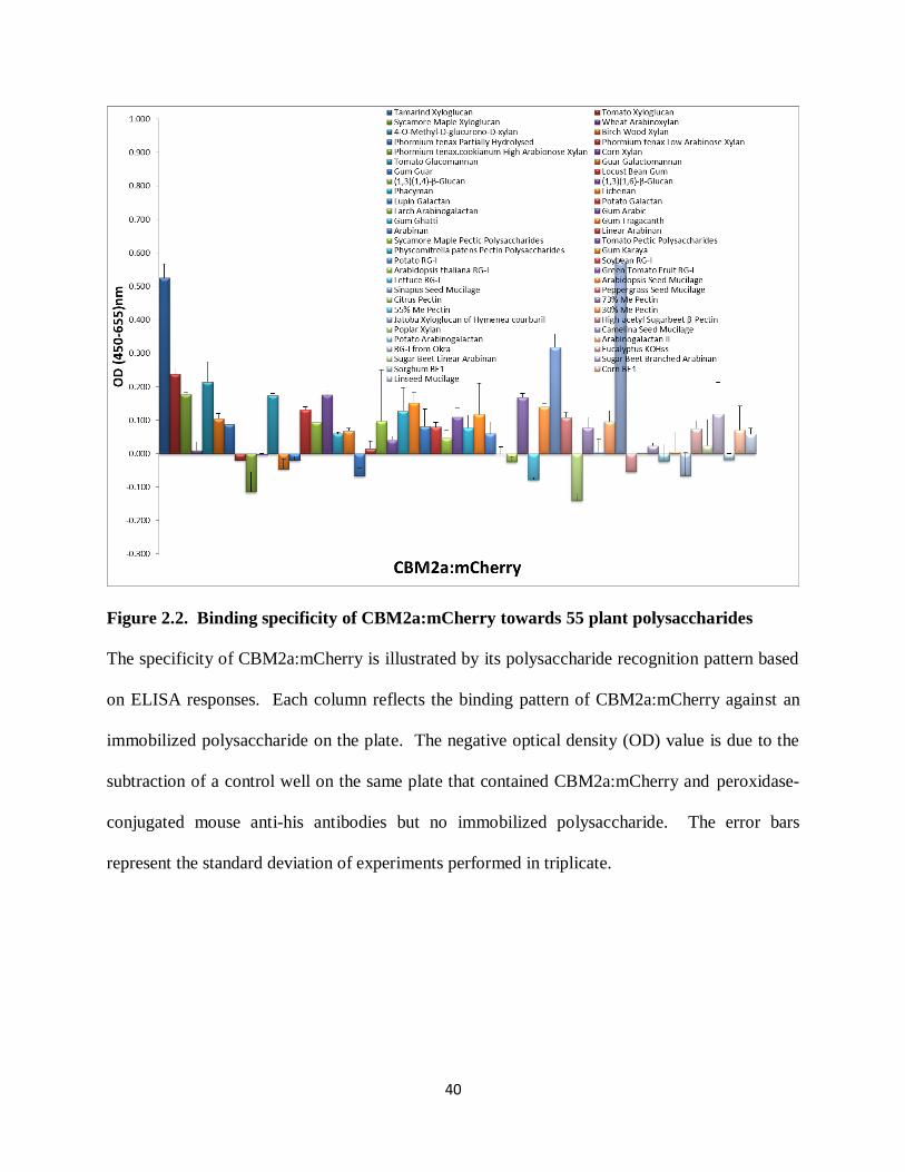
Binding of fluorescent protein tagged CBMs to a diverse panel of 55 plant polysaccharides
In vitro binding characteristics of these six fluorescent protein mCherry tagged CBMs
towards 55 diverse plant polysaccharide preparations, whose detailed chemical compositions
were previously known (Pattathil et al., 2010), were screened by ELISA. The 55 plant
polysaccharide preparations were divided into groups of Xyloglucans, Xylans, Mannans,
β-Glucans, Galactans, Arabinogalactans, Rhamnogalacturonans, Mucilages, Homogalacturonans
(See detail information in Supplemental Table S2.1). Due to the high insolubility, cellulose
cannot be dissolved in deionized water and in turn applied to 96-well plate for ELISA screens.
Thus, cellulose is not included in this screening.
Cellulose-binding Group
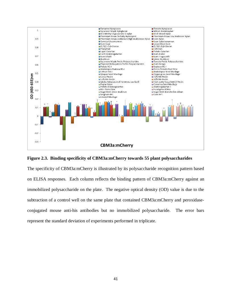
The ELISA results with the fluorescent protein mCherry tagged CBM2a and CBM3a
were in agreement with known specificities of CBM2a and CBM3a for crystalline cellulose.
That is, the ELISA screen revealed that neither CBM2a:mCherry nor CBM3a:mCherry
effectively recognized most samples of hemicellulose and pectin tested in this screen. However,
CBM2a:mCherry displayed some binding capacity to tamarind seed xyloglucan, Sinapus seed
mucilage and seed xyloglucan of Hymenea courbaril (Figure 2.2). Mucilage is extruded from
the seed coat of epidermal cells and is typically composed primarily of pectin and cellulose. It
has been suggested that within the cellulose-binding CBM group, CBMs exhibit both similarities
and differences in cell wall specificity that are plant- and tissue-specific (Blake et al., 2006). In
our studies, CBM2a:mCherry showed some binding to xyloglucan from tamarind seed, jatoba
seed and mucilage from sinapus seed, while CBM3a:mCherry did not bind to these
hemicellulose and pectin samples (Figure 2.3), implicating variations between these two CBMs
towards plant-specific cell wall components.
40
Figure 2.2. Binding specificity of CBM2a:mCherry towards 55 plant polysaccharides
The specificity of CBM2a:mCherry is illustrated by its polysaccharide recognition pattern based
on ELISA responses. Each column reflects the binding pattern of CBM2a:mCherry against an
immobilized polysaccharide on the plate. The negative optical density (OD) value is due to the
subtraction of a control well on the same plate that contained CBM2a:mCherry and peroxidase-
conjugated mouse anti-his antibodies but no immobilized polysaccharide. The error bars
represent the standard deviation of experiments performed in triplicate.
41
Figure 2.3. Binding specificity of CBM3a:mCherry towards 55 plant polysaccharides
The specificity of CBM3a:mCherry is illustrated by its polysaccharide recognition pattern based
on ELISA responses. Each column reflects the binding pattern of CBM3a:mCherry against an
immobilized polysaccharide on the plate. The negative optical density (OD) value is due to the
subtraction of a control well on the same plate that contained CBM3a:mCherry and peroxidase-
conjugated mouse anti-his antibodies but no immobilized polysaccharide. The error bars
represent the standard deviation of experiments performed in triplicate.
42
Xylan-binding group
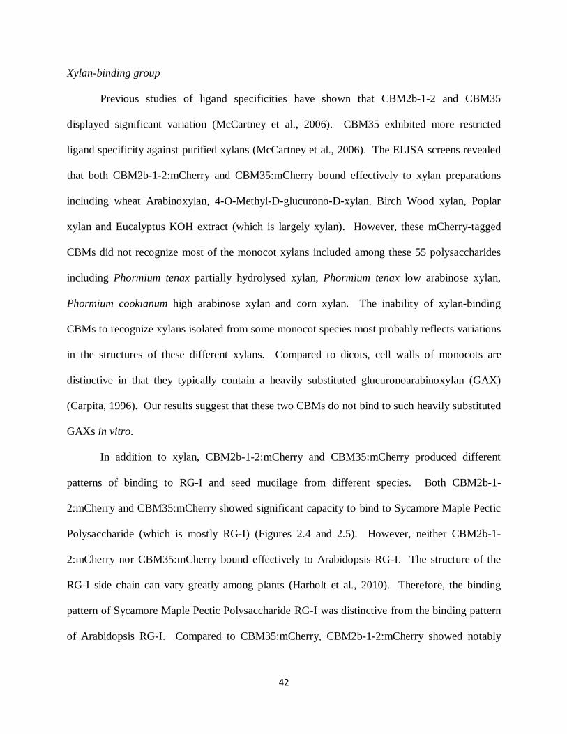
Previous studies of ligand specificities have shown that CBM2b-1-2 and CBM35
displayed significant variation (McCartney et al., 2006). CBM35 exhibited more restricted
ligand specificity against purified xylans (McCartney et al., 2006). The ELISA screens revealed
that both CBM2b-1-2:mCherry and CBM35:mCherry bound effectively to xylan preparations
including wheat Arabinoxylan, 4-O-Methyl-D-glucurono-D-xylan, Birch Wood xylan, Poplar
xylan and Eucalyptus KOH extract (which is largely xylan). However, these mCherry-tagged
CBMs did not recognize most of the monocot xylans included among these 55 polysaccharides
including Phormium tenax partially hydrolysed xylan, Phormium tenax low arabinose xylan,
Phormium cookianum high arabinose xylan and corn xylan. The inability of xylan-binding
CBMs to recognize xylans isolated from some monocot species most probably reflects variations
in the structures of these different xylans. Compared to dicots, cell walls of monocots are
distinctive in that they typically contain a heavily substituted glucuronoarabinoxylan (GAX)
(Carpita, 1996). Our results suggest that these two CBMs do not bind to such heavily substituted
GAXs in vitro.
In addition to xylan, CBM2b-1-2:mCherry and CBM35:mCherry produced different
patterns of binding to RG-I and seed mucilage from different species. Both CBM2b-1-
2:mCherry and CBM35:mCherry showed significant capacity to bind to Sycamore Maple Pectic
Polysaccharide (which is mostly RG-I) (Figures 2.4 and 2.5). However, neither CBM2b-1-
2:mCherry nor CBM35:mCherry bound effectively to Arabidopsis RG-I. The structure of the
RG-I side chain can vary greatly among plants (Harholt et al., 2010). Therefore, the binding
pattern of Sycamore Maple Pectic Polysaccharide RG-I was distinctive from the binding pattern
of Arabidopsis RG-I. Compared to CBM35:mCherry, CBM2b-1-2:mCherry showed notably

43
stronger binding to Green Tomato Fruit RG-I, Camelina Seed mucilage (Xylose sugar
composition 2.2 Mol %), Peppergrass seed mucilage (Xylose sugar composition 9.8 Mol %) and
Arabidopsis Seed mucilage (Xylose sugar composition 1.5 Mol %) (Pattathil et al., 2010) (Figure
2.4).
Figure 2.4. Binding specificity of CBM2b-1-2:mCherry towards 55 plant polysaccharides
The specificity of CBM2b-1-2:mCherry is illustrated by its polysaccharide recognition pattern
based on ELISA responses. Each column reflects the binding pattern of CBM2b-1-2:mCherry
against an immobilized polysaccharide on the plate. The negative optical density (OD) value are
due to the subtraction of a control well on the same plate that contained CBM2b-1-2:mCherry
and peroxidase-conjugated mouse anti-his antibodies but no immobilized polysaccharide. The
error bars represent the standard deviation of experiments performed in triplicate.
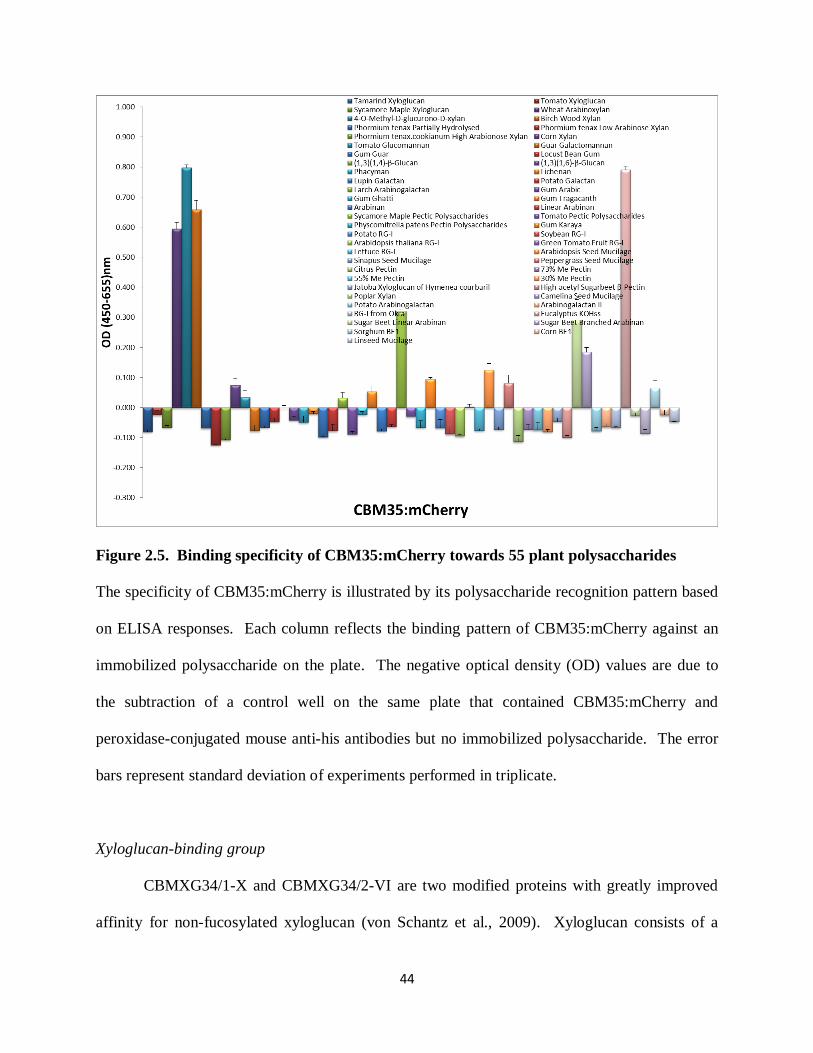
44
Figure 2.5. Binding specificity of CBM35:mCherry towards 55 plant polysaccharides
The specificity of CBM35:mCherry is illustrated by its polysaccharide recognition pattern based
on ELISA responses. Each column reflects the binding pattern of CBM35:mCherry against an
immobilized polysaccharide on the plate. The negative optical density (OD) values are due to
the subtraction of a control well on the same plate that contained CBM35:mCherry and
peroxidase-conjugated mouse anti-his antibodies but no immobilized polysaccharide. The error
bars represent standard deviation of experiments performed in triplicate.
Xyloglucan-binding group
CBMXG34/1-X and CBMXG34/2-VI are two modified proteins with greatly improved
affinity for non-fucosylated xyloglucan (von Schantz et al., 2009). Xyloglucan consists of a

45
cellulose-like β-1-4 glucan backbone that may be substituted up to 75% with xylose units. The
xyloses in turn can be decorated with galactose and fucose units. Some xyloglucans occur
without any fucose on side chains and are collectively referred as non-fucosylated xyloglucan.
CBMXG34/1-X:mCherry and CBMXG34/2-VI:mCherry exhibited strong specificity and
binding capacity to xyloglucans, especially to Tamarind seed xyloglucan and seed xyloglucan of
Hymenea courbail, which are rich in non-fucosylated xyloglucans (Buckeridge, 2010).
CBMXG34/1-X:mCherry and CBMXG34/2-VI:mCherry discriminated non-fucosylated
xyloglucan from fucosylated xyloglucan as shown by their notably weaker ability to bind to
fucosylated Sycamore Maple xyloglucan.
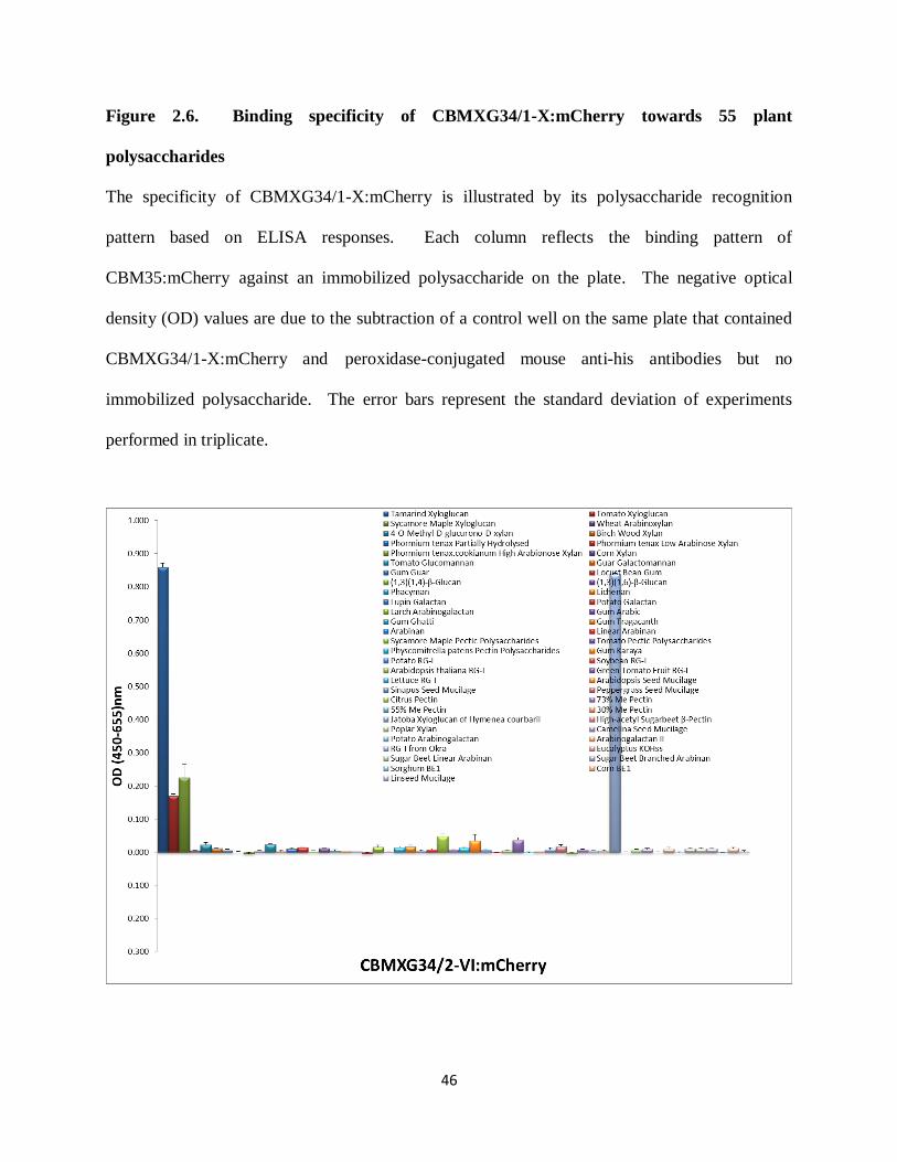
46
Figure 2.6. Binding specificity of CBMXG34/1-X:mCherry towards 55 plant
polysaccharides
The specificity of CBMXG34/1-X:mCherry is illustrated by its polysaccharide recognition
pattern based on ELISA responses. Each column reflects the binding pattern of
CBM35:mCherry against an immobilized polysaccharide on the plate. The negative optical
density (OD) values are due to the subtraction of a control well on the same plate that contained
CBMXG34/1-X:mCherry and peroxidase-conjugated mouse anti-his antibodies but no
immobilized polysaccharide. The error bars represent the standard deviation of experiments
performed in triplicate.
47
Figure 2.7. Binding specificity of CBMXG34/1-X:mCherry towards 55 plant
polysaccharides
The specificity of CBMXG34/2-VI:mCherry is illustrated by its polysaccharide recognition
pattern based on ELISA responses. Each column reflects the binding pattern of
CBM35:mCherry against an immobilized polysaccharide on the plate. The negative optical
density (OD) values are due to the subtraction of a control well on the same plate that contained
CBMXG34/2-VI:mCherry and peroxidase-conjugated mouse anti-his antibodies but no
immobilized polysaccharide. The error bars represent the standard deviation of experiments
performed in triplicate.
Binding capacity of mCherry tagged cellulose-binding CBMs towards cellulose in vitro
The in vitro binding capacity of the CBM2a:mCherry and CBM3a:mCherry fusion
proteins towards cellulose was confirmed by Cellulose Pull-Down assays. In this experiment,
Avicel (insoluble substrate) were used as bait and CBM fusion proteins were tested as prey.
Xylan-binding CBM2b-1-2:mCherry and CBM35:mCherry, as expected, did not bind to Avicel
(Figure 2.8). The xyloglucan-binding CBMXG34/1-X:mCherry and CBMXG34/2-VI:mCherry
did not bind to Avicel also (data not shown). In contrast, CBM2a:mCherry and
CBM3a:mCherry were pulled down by Avicel. Thus, the CBM2a:mCherry and
CBM3a:mCherry fusion proteins functioned as cellulose-binding modules.

48
Figure 2.8. Cellulose Pull-Down Assay of mCherry tagged CBMs
Left: xylan-binding CBM2b-1-2:mCherry and CBM35:mCherry were not pulled down by Avicel;
Right: cellulose-binding CBM2a:mCherry and CBM3a:mCherry were pulled down by Avicel.
Direct and indirect labeling of fixed Arabidopsis stem sections using tagged CBMs
The binding capacity of these six fluorescent protein-tagged CBMs to Arabidopsis cell
walls was initially assessed by direct labeling of stem sections with His-tagged CBM:mCherry
fusion proteins, and also by indirect labeling of stem sections with His-tagged CBM:mCherry
fusion proteins and Alexa Fluor® 488 attached to an anti-His monoclonal antibody. As shown in
Figure 2.8, the two cellulose-binding CBMs, CBM2a:mCherry and CBM3a:mCherry, displayed
different patterns on Arabidospsis stems (Figure 2.8 A1, B1). CBM3a:mCherry recognized the
walls of every tissue in the Arabidopsis stem including epidermis, cortex, phloem, interfascicular
fibres, vascular xylem and pith. CBM2a:mCherry displayed binding mainly to cellulose within
cell walls of xylem vessels and interfascicular fibres. Both xylan-binding CBM2b-1-2:mCherry
and CBM35:mCherry showed labeling to secondary cell walls of xylem vessels and
interfascicular fibres. However, CBM35:mCherry weakly labeled secondary walls of some
vascular xylem cells (indicated by stars in Figure 2.8 D1). The xyloglucan-binding
CBMXG34/1-X:mCherry and CBM34/2-VI:mCherry did not bind to Arabidopsis stem sections
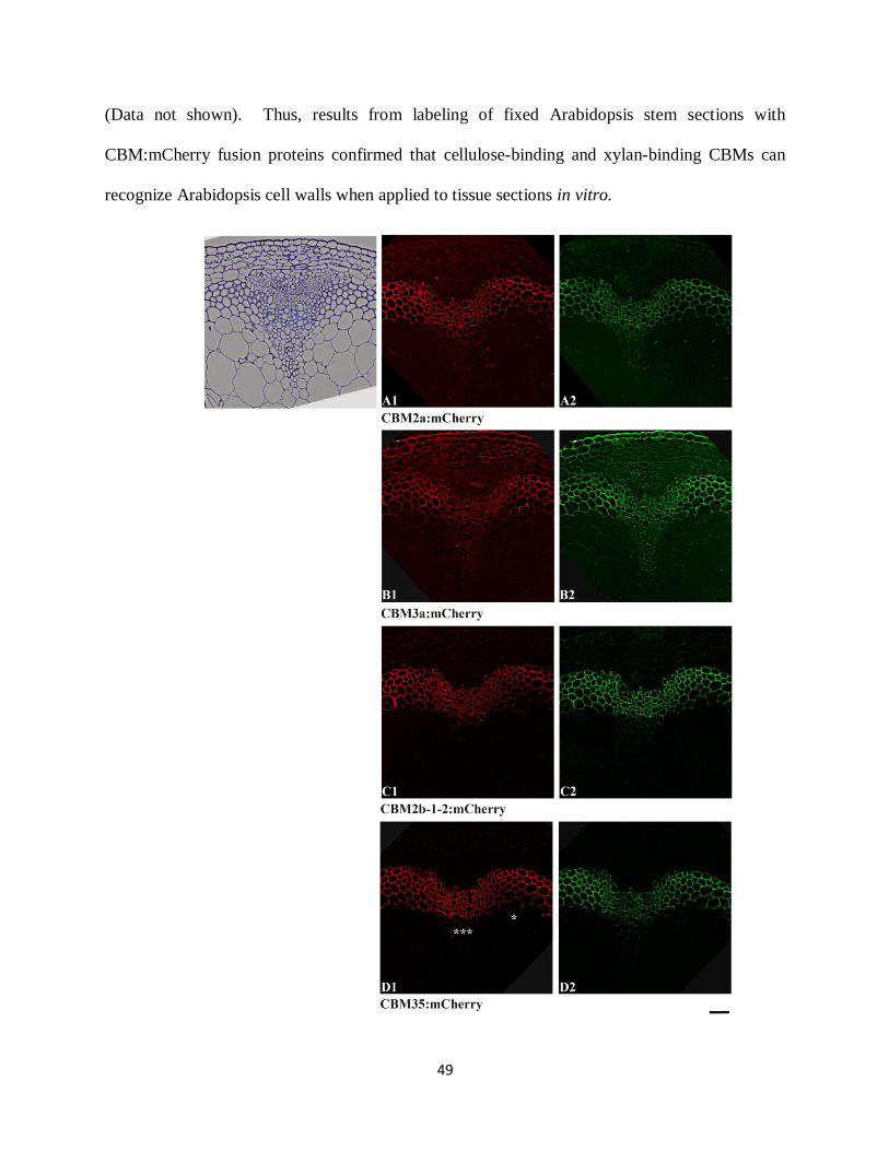
49
(Data not shown). Thus, results from labeling of fixed Arabidopsis stem sections with
CBM:mCherry fusion proteins confirmed that cellulose-binding and xylan-binding CBMs can
recognize Arabidopsis cell walls when applied to tissue sections in vitro.
50
Figure 2.9. Labeling of Arabidopsis stem sections in vitro with tagged CBM fusion proteins.
Left (A1, B1, C1, D1): Direct labeling of transverse section of WT Arabidopsis basal stem by
His-tagged fluorescent protein CBM fusion proteins. Right (A2, B2, C2, D2): Immunolabeling
of transverse sections of WT Arabidopsis basal stem with His-tagged fluorescent protein CBM
fusion proteins and Alexa Fluor® 488 attached to an anti-His monoclonal antibody. Tissues
were taken from the base of 6-week-old Col-0 (WT) stems, fixed and embedded in LR White
resin, and 250 nm thick transverse sections were cut by microtome. Scale bar = 50 µm.
Binding of Xyloglucan-Binding CBMs to Arabidopsis cell wall KOH extracts
Since xyloglucan-binding CBMXG34/1-X:mCherry and CBMXG34/2-VI:mCherry did
not exhibit labeling capacity on fixed Arabidopsis stem sections, the binding capacities of these
xyloglucan-binding CBMs was explored in more detail by ELISA with Arabidopsis cell wall
extracts from both leaves and stems. Arabidopsis cell walls were sequentially extracted with
increasingly harsh solvents to release cell wall components. Cell walls were treated with 1M
KOH and 4M KOH to solubilize primarily hemicelluloses, mainly xylan and xyloglucan
(Zablackis et al., 1995). In this experiment, CCRC-M1, which recognizes terminal α-1,2-linked
fucosyl residues in fucosylated xyloglucans, and CCRC-M87, which recognizes non-fucosylated
xyloglucan epitope, were used as controls. Both CCRC-M1 and CCRC-M87 exhibited
significant binding capacity to 4M KOH Arabidopsis cell wall extracts and moderate binding
capacity to 1M KOH extracts. These results are consistent with results obtained by others in the
Hahn lab. In contrast, CBMXG34/1-X:mCherry and CBMXG34/2-VI:mCherry bound poorly to
Arabidopsis cell wall KOH extracts, though they bound strongly to tamarind xyloglucan, which
is a non-fucosylated xyloglucan. Therefore, the xyloglucan structure(s) recognized by
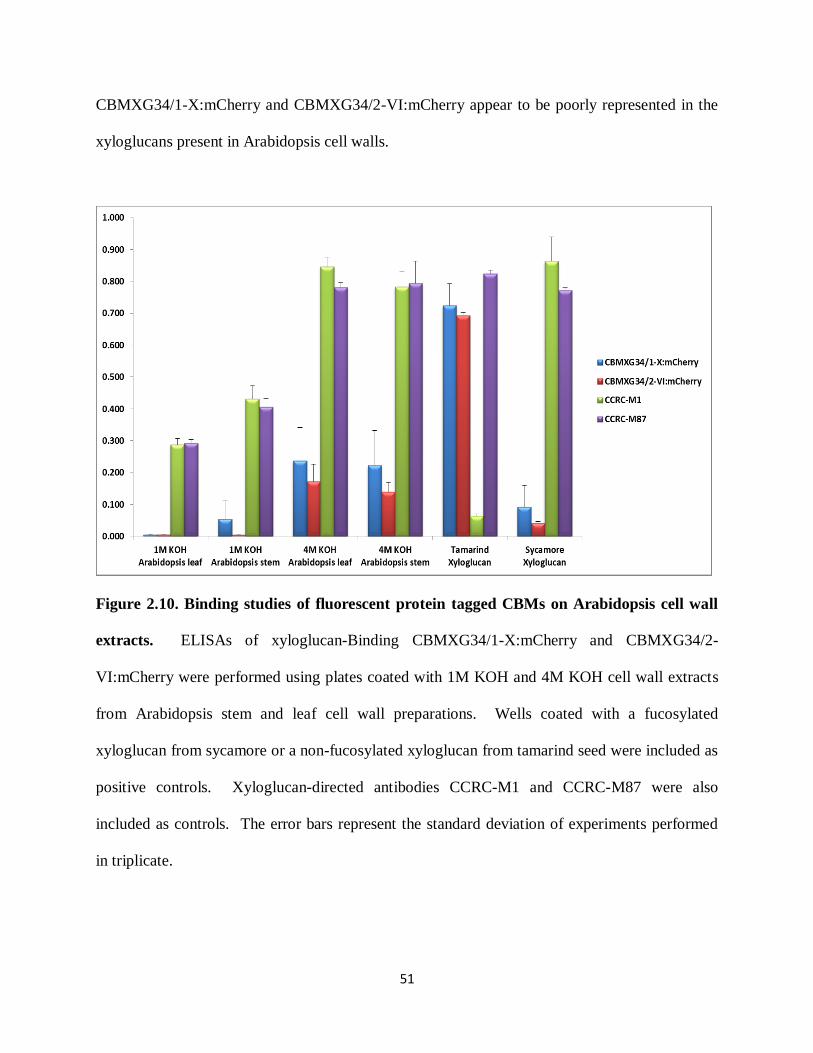
51
CBMXG34/1-X:mCherry and CBMXG34/2-VI:mCherry appear to be poorly represented in the
xyloglucans present in Arabidopsis cell walls.
Figure 2.10. Binding studies of fluorescent protein tagged CBMs on Arabidopsis cell wall
extracts. ELISAs of xyloglucan-Binding CBMXG34/1-X:mCherry and CBMXG34/2-
VI:mCherry were performed using plates coated with 1M KOH and 4M KOH cell wall extracts
from Arabidopsis stem and leaf cell wall preparations. Wells coated with a fucosylated
xyloglucan from sycamore or a non-fucosylated xyloglucan from tamarind seed were included as
positive controls. Xyloglucan-directed antibodies CCRC-M1 and CCRC-M87 were also
included as controls. The error bars represent the standard deviation of experiments performed
in triplicate.
52
Discussion
We have investigated the binding specificities of several tagged CBMs that bind to
different plant cell wall polysaccharides. Within the cellulose-binding group, CBM3a:mCherry
bound evenly to primary and secondary cell walls, whereas CBM2a:mCherry displayed a
preference for secondary cell walls of interfascicular fibres and vascular xylem in labeling of
fixed Arabidopsis stem sections. CBM3a is a module in the cellulosome, a multi-enzyme plant
cell wall-degrading complex produced by the anaerobic bacterium Clostridium thermocellum
(Bayer et al., 2004). Thus, there will be a strong selective pressure for CBM3a to display broad
specificity for cellulose microfibrils present in a range of cell walls in order for CBM3a to target
the cellulosome to the array of crystalline cellulosic material that is likely to exist in the C.
thermocellum environment (Blake et al., 2006). CBM2a is the carbohydrate-binding module in
Xylanase 10A. It is possible that CBM2a is used to target the enzyme preferentially to cellulose
within secondary cell walls, where cellulose is in intimate contact with xylan. In ELISAs,
CBM2a:mCherry showed binding capacity to non-fucosylated xyloglucan, whereas
CBM3a:mCherry did not exhibit any cross-reactivity with these xyloglucan samples. Cellulose
and xyloglucan are not covalently linked to each other as far as is known. These two
polysaccharides share common structural features (xyloglucan has a cellulose-like β1-4 glucan
backbone which may be substituted up to 75% with xylose units). Cellulose-binding CBMs
appear to make few significant hydrogen bonds with the microfibril cellulose structures (Blake et
al., 2006). It has been suggested that the binding face of the cellulose-binding CBMs may
interact with other polysaccharides that are in intimate contact with the cellulose (Blake et al.,
2006). Therefore, it is possible for CBM2a:mCherry to also have binding capacity to
xyloglucans, though this still requires additional experimental verification.
53
In dicots, glucurononxylan (GX) and methylglucurononxylan (MGX) are abundant in the
secondary cell walls of specific cell types such as xylem vessels and interfascicular fibers;
primary cell walls have low levels of xylans that are of the GX type (Darvill et al., 1980; O’Neill
and York, 2003). Within the xylan-binding group, CBM2b-1-2 binds to the backbone of linear
and substituted xylans, whereas CBM35 binds to GX, but not to MGX (McCartney et al., 2006).
Consistent with a recent study (Urbanowicz et al., 2012), CBM2b-1-2:mCherry extensively
labeled the secondary walls of interfascicular fibers and vascular bundles in Arabidopsis stem
section. CBM35:mCherry weakly labeled secondary cell walls of some vascular xylem cells in
Arabidopsis stem section, indicating that the xylans in the cell walls of these cells are highly
methylated. In ELISAs, CBM2b-1-2:mCherry and CBM35:mCherry exhibited notable
recognition of some RG-I preparations. Xylan and pectin structure were not thought to be
interdependent wall polysaccharides until the recent identification of ARABINOXYLAN
PECTIN ARABINOGALACTAN PROTEIN1 (APAP1), which was found to carry both pectin
(RGI/homogalacturonan) and xylan structures linked to the same arabinogalactan (Tan et al.,
2013). Thus, these observed cross-reactivities of the xylan-directed CBMs might be explained
by the presence of APAP1 or APAP1-like molecules in those RG-I preparations.
CBMs exhibit plant- and tissue-specific differences in cell wall recognition, providing
insights into the complex molecular architecture of plant cell wall (Knox, 2008). Previous
studies revealed that xyloglucan-binding CBMXG34/1-X and CBMXG34/2-VI bound
selectively to non-fucosylated xyloglucan of tamarind seed (von Schantz et al., 2009). ELISA
screens of mCherry fluorescent protein CBMXG34/1-X:mCherry and CBMXG34/2-VI:mCherry
fusion proteins demonstrated their ability to bind to tamarind seed xyloglucan. However, these
two xyloglucan-binding CBMs did not exhibit strong binding capacity to Arabidopsis cell walls,
54
even though Arabidopsis stem cell walls are known to contain non-fucosylated xyloglucan
structural features as shown by binding of monoclonal antibodies directed against these epitopes
(Cavalier et al., 2008; Zabotina et al., 2008; Zabotina et al., 2012). Therefore, CBMXG34/1-X
and CBMXG34/2-VI appear to bind to non-fucosylated xyloglucan structural features that are
not abundant in Arabidopsis cell walls. Thus, these two CBMs are not suitable for our further
studies which focus on Arabidopsis cell walls.
55
References
Bayer EA, Belaich JP, Shoham Y, Lamed R (2004) The cellulosomes: multienzyme machines
for degradation of plant cell wall polysaccharides. Annu Rev Microbiol 58: 521-554
Bertani G (1951) STUDIES ON LYSOGENESIS I.: The Mode of Phage Liberation by
Lysogenic Escherichia coli. Journal of Bacteriology 62: 293-300
Blake AW, McCartney L, Flint JE, Bolam DN, Boraston AB, Gilbert HJ, Knox JP (2006)
Understanding the biological rationale for the diversity of cellulose-directed
carbohydrate-binding modules in prokaryotic enzymes. J Biol Chem 281: 29321-29329
Buckeridge MS (2010) Seed Cell Wall Storage Polysaccharides: Models to Understand Cell
Wall Biosynthesis and Degradation. Plant Physiology 154: 1017-1023
Carpita NC (1996) STRUCTURE AND BIOGENESIS OF THE CELL WALLS OF GRASSES.
Annu Rev Plant Physiol Plant Mol Biol 47: 445-476
Cavalier DM, Lerouxel O, Neumetzler L, Yamauchi K, Reinecke A, Freshour G, Zabotina
OA, Hahn MG, Burgert I, Pauly M, Raikhel NV, Keegstra K (2008) Disrupting Two
Arabidopsis thaliana Xylosyltransferase Genes Results in Plants Deficient in Xyloglucan,
a Major Primary Cell Wall Component. The Plant Cell Online 20: 1519-1537
Darvill JE, McNeil M, Darvill AG, Albersheim P (1980) Structure of Plant Cell Walls: XI.
GLUCURONOARABINOXYLAN, A SECOND HEMICELLULOSE IN THE
PRIMARY CELL WALLS OF SUSPENSION-CULTURED SYCAMORE CELLS.
Plant Physiology 66: 1135-1139
Gunnarsson LC, Zhou Q, Montanier C, Karlsson EN, Brumer H, 3rd, Ohlin M (2006)
Engineered xyloglucan specificity in a carbohydrate-binding module. Glycobiology 16:
1171-1180

56
Harholt J, Suttangkakul A, Vibe Scheller H (2010) Biosynthesis of Pectin. Plant Physiology
153: 384-395
Knox JP (2008) Revealing the structural and functional diversity of plant cell walls. Current
Opinion in Plant Biology 11: 308-313
McCann MC, Knox JP (2010) Plant Cell Wall Biology: Polysaccharides in Architectural and
Developmental Contexts. In Annual Plant Reviews. Wiley-Blackwell, pp 343-366
McCartney L, Blake AW, Flint J, Bolam DN, Boraston AB, Gilbert HJ, Knox JP (2006)
Differential recognition of plant cell walls by microbial xylan-specific carbohydrate-
binding modules. Proceedings of the National Academy of Sciences of the United States
of America 103: 4765-4770
O’Neill MA, York WS (2003) The composition and structure of plant primary cell walls. The
plant cell wall: 1-54
Pattathil S, Avci U, Baldwin D, Swennes AG, McGill JA, Popper Z, Bootten T, Albert A,
Davis RH, Chennareddy C, Dong R, O'Shea B, Rossi R, Leoff C, Freshour G, Narra
R, O'Neil M, York WS, Hahn MG (2010) A Comprehensive Toolkit of Plant Cell Wall
Glycan-Directed Monoclonal Antibodies. Plant Physiology 153: 514-525
Sambrook J, Fritsch EF, Maniatis T (1989) Molecular Cloning: A Laboratory Manual, Ed 2nd.
Cold Spring Harbor Laboratory Press, Plainview, N.Y
Tan L, Eberhard S, Pattathil S, Warder C, Glushka J, Yuan C, Hao Z, Zhu X, Avci U,
Miller JS, Baldwin D, Pham C, Orlando R, Darvill A, Hahn MG, Kieliszewski MJ,
Mohnen D (2013) An Arabidopsis Cell Wall Proteoglycan Consists of Pectin and
Arabinoxylan Covalently Linked to an Arabinogalactan Protein. The Plant Cell Online 25:
270-287
57
Urbanowicz BR, Pena MJ, Ratnaparkhe S, Avci U, Backe J, Steet HF, Foston M, Li H,
O'Neill MA, Ragauskas AJ, Darvill AG, Wyman C, Gilbert HJ, York WS (2012) 4-
O-methylation of glucuronic acid in Arabidopsis glucuronoxylan is catalyzed by a
domain of unknown function family 579 protein. Proc Natl Acad Sci U S A 109: 14253-
14258
von Schantz L, Gullfot F, Scheer S, Filonova L, Cicortas Gunnarsson L, Flint JE, Daniel G,
Nordberg-Karlsson E, Brumer H, Ohlin M (2009) Affinity maturation generates
greatly improved xyloglucan-specific carbohydrate binding modules. BMC Biotechnol 9:
92
Zablackis E, Huang J, Muller B, Darvill AG, Albersheim P (1995) Characterization of the
Cell-Wall Polysaccharides of Arabidopsis thaliana Leaves. Plant Physiology 107: 1129-
1138
Zabotina OA, Avci U, Cavalier D, Pattathil S, Chou YH, Eberhard S, Danhof L, Keegstra
K, Hahn MG (2012) Mutations in multiple XXT genes of Arabidopsis reveal the
complexity of xyloglucan biosynthesis. Plant Physiol 159: 1367-1384
Zabotina OA, van de Ven WT, Freshour G, Drakakaki G, Cavalier D, Mouille G, Hahn
MG, Keegstra K, Raikhel NV (2008) Arabidopsis XXT5 gene encodes a putative alpha-
1,6-xylosyltransferase that is involved in xyloglucan biosynthesis. Plant J 56: 101-115
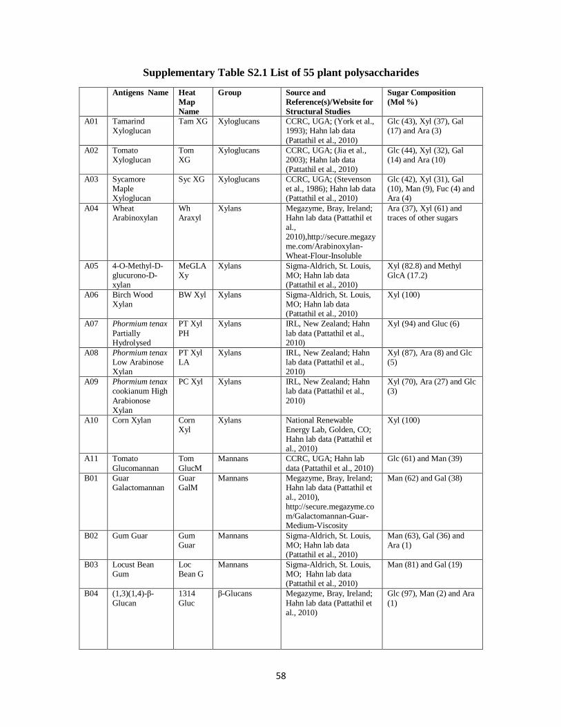
58
Supplementary Table S2.1 List of 55 plant polysaccharides
Antigens Name Heat
Map
Name
Group Source and
Reference(s)/Website for
Structural Studies
Sugar Composition
(Mol %)
A01 Tamarind Xyloglucan
Tam XG Xyloglucans CCRC, UGA; (York et al., 1993); Hahn lab data
(Pattathil et al., 2010)
Glc (43), Xyl (37), Gal (17) and Ara (3)
A02 Tomato Xyloglucan
Tom XG
Xyloglucans CCRC, UGA; (Jia et al., 2003); Hahn lab data (Pattathil et al., 2010)
Glc (44), Xyl (32), Gal (14) and Ara (10)
A03 Sycamore Maple Xyloglucan
Syc XG Xyloglucans CCRC, UGA; (Stevenson et al., 1986); Hahn lab data (Pattathil et al., 2010)
Glc (42), Xyl (31), Gal (10), Man (9), Fuc (4) and Ara (4)
A04 Wheat Arabinoxylan
Wh Araxyl
Xylans Megazyme, Bray, Ireland; Hahn lab data (Pattathil et al., 2010),http://secure.megazyme.com/Arabinoxylan-Wheat-Flour-Insoluble
Ara (37), Xyl (61) and traces of other sugars
A05 4-O-Methyl-D-glucurono-D-xylan
MeGLA Xy
Xylans Sigma-Aldrich, St. Louis, MO; Hahn lab data (Pattathil et al., 2010)
Xyl (82.8) and Methyl GlcA (17.2)
A06 Birch Wood Xylan
BW Xyl Xylans Sigma-Aldrich, St. Louis, MO; Hahn lab data (Pattathil et al., 2010)
Xyl (100)
A07 Phormium tenax
Partially Hydrolysed
PT Xyl
PH
Xylans IRL, New Zealand; Hahn
lab data (Pattathil et al., 2010)
Xyl (94) and Gluc (6)
A08 Phormium tenax Low Arabinose Xylan
PT Xyl LA
Xylans IRL, New Zealand; Hahn lab data (Pattathil et al., 2010)
Xyl (87), Ara (8) and Glc (5)
A09 Phormium tenax cookianum High
Arabionose Xylan
PC Xyl Xylans IRL, New Zealand; Hahn lab data (Pattathil et al.,
2010)
Xyl (70), Ara (27) and Glc (3)
A10 Corn Xylan Corn Xyl
Xylans National Renewable Energy Lab, Golden, CO; Hahn lab data (Pattathil et al., 2010)
Xyl (100)
A11 Tomato
Glucomannan
Tom
GlucM
Mannans CCRC, UGA; Hahn lab
data (Pattathil et al., 2010)
Glc (61) and Man (39)
B01 Guar Galactomannan
Guar GalM
Mannans Megazyme, Bray, Ireland; Hahn lab data (Pattathil et al., 2010), http://secure.megazyme.com/Galactomannan-Guar-Medium-Viscosity
Man (62) and Gal (38)
B02 Gum Guar Gum Guar
Mannans Sigma-Aldrich, St. Louis, MO; Hahn lab data (Pattathil et al., 2010)
Man (63), Gal (36) and Ara (1)
B03 Locust Bean Gum
Loc Bean G
Mannans Sigma-Aldrich, St. Louis, MO; Hahn lab data (Pattathil et al., 2010)
Man (81) and Gal (19)
B04 (1,3)(1,4)-β-
Glucan
1314
Gluc
β-Glucans Megazyme, Bray, Ireland;
Hahn lab data (Pattathil et al., 2010)
Glc (97), Man (2) and Ara
(1)

59
B05 (1,3)(1,6)-β-Glucan
1315 Gluc
β-Glucans CCRC, UGA; (Hahn et al., 1992); Hahn lab data (Pattathil et al., 2010)
Glc (91), Man (7) and Ara (2)
B06 Pachyman Pachyman
β-Glucans Megazyme, Bray, Ireland; Hahn lab data (Pattathil et al., 2010), http://secure.megazyme.com/Pachyman
D-glucose essentially all of which is 1,3-ß-linked (>98)
B07 Lichenan Lichenan
β-Glucans Megazyme, Bray, Ireland; Hahn lab data (Pattathil et al., 2010),
http://secure.megazyme.com/Lichenan-Icelandic-Moss
Glc (98) and Ara (2)
B08 Lupin Galactan Lup Gal Galactans Megazyme, Bray, Ireland; Hahn lab data (Pattathil et al., 2010), http://secure.megazyme.co
m/Galactan-Lupin
Gal (83), GalA (5), Rha (5), Ara (3), Xyl (2) and traces of Glc
B09 Potato Galactan Pot Gal Galactans Megazyme, Bray, Ireland; Hahn lab data (Pattathil et al., 2010), http://secure.megazyme.com/Galactan-Potato
Gal (88), GalA (6), Ara (3) and Rha (3)
B10 Larch
Arabinogalactan
Lar Ara
Gal
Arabinogalactans Megazyme, Bray, Ireland;
Hahn lab data (Pattathil et al., 2010), http://secure.megazyme.com/Arabinogalactan-Larch-Wood
Gal (81), Ara (14) and
traces of other sugars
B11 Gum Arabic Gum Arabic
Arabinogalactans Sigma-Aldrich, St. Louis, MO; (Stephen, Phillips, and Williams, 2006); Hahn
lab data (Pattathil et al., 2010)
Ara (37), Gal (40), Rha (20) and traces of Glc
C01 Gum Ghatti Gum Ghatti
Arabinogalactans Sigma-Aldrich, St. Louis, MO; (Stephen, Phillips, and Williams, 2006); Hahn lab data (Pattathil et al., 2010)
Ara (49), Gal (31), Man (10), GlcA (8) and Xyl (2)
C02 Gum Tragacanth Gum Trag
Arabinogalactans Sigma-Aldrich, St. Louis, MO; (Stephen, Phillips, and Williams, 2006); Hahn lab data (Pattathil et al., 2010)
Gal (32.3), Ara (31.7), Xyl (12), Glc (12.3), Fuc (8.4) and Rha (3.3)
C03 Arabinan Arabinan
Arabinogalactans Megazyme, Bray, Ireland; Hahn lab data (Pattathil et
al., 2010), http://secure.megazyme.com/Arabinan-Sugar-Beet
Ara (88), Gal (3), Rha (2) and GalA (7)
C04 Linear Arabinan Linear Arabinan
Arabinogalactans Megazyme, Bray, Ireland; Hahn lab data (Pattathil et al., 2010), http://secure.megazyme.com/Linear-1-5-alpha-L-
Arabinan-Sugar-Beet
Ara (97.5), GalA (2), Gal (0.4) and Rha (0.1)
C05 Sycamore Maple Pectic Polysaccharides
Syc PecP
RG-I CCRC, UGA; (Stevenson et al., 1986); Hahn lab data (Pattathil et al., 2010)
Gal (52.4), GlcA (15), GalA (11), Xyl (7.9), Ara (7.5), Glc (2.6), Rha (2), Man (1.3) and traces of Fuc

60
C06 Tomato Pectic Polysaccharides
Tom PecP
RG-I CCRC, UGA; (Jia et al., 2003); Hahn lab data (Pattathil et al., 2010)
Gal (57.4), Ara (25.2), GalA (6.5), Rha (4.4), Xyl (4.3) and Fuc (2.2)
C07 Physcomitrella patens Pectin Polysaccharides
PhyscPecP
RG-I CCRC, UGA; (Peña et al., 2008); Hahn lab data (Pattathil et al., 2010)
GalA (46.7), Rha (24), Gal(17.4), Ara (8.1), Glc (2.3) and Xyl (1.5)
C08 Gum Karaya Gum Karaya
RG-I Sigma-Aldrich, St. Louis, MO; Hahn lab data (Pattathil et al., 2010)
GalA (57), GlcA (8), Gal (17), Rha (16) and traces of Glc
C09 Potato RG-I Pot RG-
I
RG-I Megazyme, Bray, Ireland;
Hahn lab data (Pattathil et al., 2010), http://secure.megazyme.com/Rhamnogalacturonan-I-Potato
GalA (51), Rha (28.6), Gal
(14), Ara (5.4) and Xyl (1)
C10 Soybean RG-I Soy RG-I
RG-I Megazyme, Bray, Ireland; Hahn lab data (Pattathil et al., 2010),
http://secure.megazyme.com/Rhamnogalacturonan-Soy-Bean
GalA (51), Xyl (13), Gal (11), Rha (10), Fuc (9) and Ara (6)
C11 Arabidopsis thaliana RG-I
At RG-I RG-I CCRC, UGA; (Zablackis et al., 1995); Hahn lab data (Pattathil et al., 2010)
GalA (34), Rha (27), Gal (17), Ara (14), Xyl (5), Fuc (1), GlcA (1) and GalA (1)
D01 Green Tomato Fruit RG-I
GrTomFrR
RG-I CCRC, UGA; Hahn lab data (Pattathil et al., 2010)
GalA (49.5), Gal (43.8), Rha (4.7), GlcA (1), Ara (0.5) and traces of Xyl and Glc
D02 Lettuce RG-I Let RG-I
RG-I CCRC, UGA; Hahn lab data (Pattathil et al., 2010)
Gal (54.9), GalA (23.4), Rha (6.9), GlcA (5.7), Ara (4.9) and Xyl (4.2)
D03 Arabidopsis Seed Mucilage
At Seed Mu
Mucilages CCRC, UGA; Hahn lab data (Pattathil et al., 2010)
Rha (69), GalA (27.7), Gal (1.8) and Xyl (1.5)
D04 Sinapus Seed Mucilage
Sin Seed Mu
Mucilages CCRC, UGA; Hahn lab data (Pattathil et al., 2010)
GalA (57), Gal (19), Rha (12), GlcA (10) and traces of Man and Glc
D05 Peppergrass
Seed Mucilage
Pep Gr
S Mu
Mucilages CCRC, UGA; (Deng et al.,
2009); Hahn lab data (Pattathil et al., 2010)
GlcA (22.2), GalA (22.1),
Gal (18.9), Xyl (9.8), Rha (8), Glc (7), Fuc (5.2), Ara (5) and Man (1.8)
D06 Citrus Pectin Citrus Pectin
HG Hercules, Wilmington, DE; Hahn lab data (Pattathil et al., 2010)
GalA (85), Rha (13) and Gal (2)
D07 73% Me Pectin 73%
MePec
HG Hercules, Wilmington, DE;
Hahn lab data (Pattathil et al., 2010)
GalA (81), Gal (7), Rha
(6.5), Ara (4.6) and Glc (0.9)
D08 55% Me Pectin 55% MePec
HG Hercules, Wilmington, DE; Hahn lab data (Pattathil et al., 2010)
GalA (81), Gal (10), Rha (9)
D09 33% Me Pectin 33% Me Pec
HG Hercules, Wilmington, DE; Hahn lab data (Pattathil et
al., 2010)
GalA (89), Gal (7), Rha (4)
D10 Jatoba Xyloglucan of Hymenaea courbaril
Znone Xyloglucans CCRC, UGA; Hahn lab data (unpublished)

61
D11 High-acetyl Sugarbeet β-Pectin
HAcSBPec
HG Hercules, Wilmington, DE; Hahn lab data (Pattathil et al., 2010)
GalA (56.2), Rha (21.3), Gal (15) and Ara (7.5)
E01 Poplar Xylan Pop Xyl Xylans National Renewable Energy Lab, Golden, CO; Hahn lab data (Pattathil et al., 2010)
Xyl (94), GalA (4) and Rha (2)
E02 Camelina Seed Mucilage
Cam Seed
Mucilages CCRC, UGA; Hahn lab data (Pattathil et al., 2010)
Rha (57.1), GalA (24.4), Gal (13.3), Xyl (2.2), Ara (2.3) and Man (0.8)
E03 Potato Arabinogalactan
Pot Ara Gal
Arabinogalactans Dr. Henk Schols, Laboratory of Food Chemistry, Wageningen,
The Netherlands; personal communication
Gal (63), Ara (20) and UA (17)
E04 Arabinogalactan II
Ara Gal II
Arabinogalactans Dr. Henk Schols, Laboratory of Food Chemistry, Wageningen, The Netherlands; personal communication
Gal (84) and Gal (16)
E05 RG-I from Okra Okra RG-I
RG-I Dr. Henk Schols, Laboratory of Food Chemistry, Wageningen, The Netherlands; personal communication
Gal (53), GalA (26), Rha (14), GlcA (4), Glc (2.5) Ara (1) and Man (1)
E06 Eucalyptus KOHss
Eucal KOH
Xylans Dr. Henk Schols, Laboratory of Food
Chemistry, Wageningen, The Netherlands; personal communication
Xyl (81), UA (13), Gal (3), Rha (2), Ara (1), Glc (1)
and Man (1)
E07 Sugar Beet Linear Arabinan
SB Lin Ara
Arabinogalactans Dr. Henk Schols, Laboratory of Food Chemistry, Wageningen, The Netherlands; personal communication
Ara (74), UA (14) and Gal (12)
E08 Sugar Beet Branched Arabinan
SB Branch
Arabinogalactans Dr. Henk Schols, Laboratory of Food Chemistry, Wageningen, The Netherlands; personal communication
Ara (>80)
E09 Sorghum BE1 Sorg BE1
Xylans Dr. Henk Schols, Laboratory of Food
Chemistry, Wageningen, The Netherlands; personal communication
Ara (45.8), Xyl (40.9), UA (9.8), Gal (1.8), Glc (1.7)
and Man (0.2)
E10 Corn BE1 Corn BE1
Xylans Dr. Henk Schols, Laboratory of Food Chemistry, Wageningen, The Netherlands; personal
communication
Ara (38.4), Xyl (48.3), UA (8.3), Gal (4.3), Glc (0.7) and Man (0.1)
E11 Linseed Mucilage
Lin Seed Mu
Mucilages CCRC, UGA; Hahn lab data (Pattathil et al., 2010)
Rha (44.2), GalA (18.5), Xyl (15.8), Gal (11.1), Fuc (3), Ara (4.6) and Glc (2.9)
62
Supplemental References
Deng C, O’Neill MA, Hahn MG, York WS (2009) Improved procedures for the selective
chemical fragmentation of rhamnogalacturonans. Carbohydr Res 344: 1852-1857
Hahn MG, Darvill A, Albersheim P, Bergmann C, Cheong J-J, Koller A, Lò V-M (1992)
Preparation and characterization of oligosaccharide elicitors of phytoalexin accumulation.
In S Gurr, M McPherson, DJ Bowles, eds, Molecular Plant Pathology (Volume II): A
Practical Approach, Oxford University Press, Oxford, UK, pp 103-147
Jia ZH, Qin Q, Darvill AG, York WS (2003) Structure of the xyloglucan produced by
suspension-cultured tomato cells. Carbohydr Res 338: 1197-1208
Pattathil S, Avci U, Baldwin D, Swennes AG, McGill JA, Popper Z, Bootten T, Albert A,
Davis RH, Chennareddy C, Dong R, O'Shea B, Rossi R, Leoff C, Freshour G, Narra
R, O'Neil M, York WS, Hahn MG (2010) A Comprehensive Toolkit of Plant Cell Wall
Glycan-Directed Monoclonal Antibodies. Plant Physiology 153: 514-525
Peña MJ, Darvill AG, Eberhard S, York WS, O'Neill MA (2008) Moss and liverwort
xyloglucans contain galacturonic acid and are structurally distinct from the xyloglucans
synthesized by hornworts and vascular plants. Glycobiology 18: 891-904
Stevenson TT, McNeil M, Darvill AG, Albersheim P (1986) Structure of plant cell walls
XVIII. An analysis of the extracellular polysaccharides of suspension-cultured sycamore
cells. Plant Physiol 80: 1012-1019
Stephen AM, Phillips GO, Williams PA eds, (2006) Food Polysaccharides and Their
Applications. CRC Press, Boca Raton, FL, pp 1-733
63
York WS, Harvey LK, Guillen R, Albersheim P, Darvill AG (1993) Structural analysis of
tamarind seed xyloglucan oligosaccharides using β-galactosidase digestion and
spectroscopic methods. Carbohydr Res 248: 285-301
Zablackis E, Huang J, Müller B, Darvill AG, Albersheim P (1995) Characterization of the
cell wall polysaccharides of Arabidopsis thaliana leaves. Plant Physiol 107: 1129-1138
64
CHAPTER 3
IN VIVO VISUALIZATION OF XYLAN IN WILD-TYPE AND MUTANT PLANTS
USING CBM2B-1-2
Abstract
Carbohydrate Binding Modules (CBMs) are carbohydrate-binding protein domains
commonly found attached to glycosyl hydrolases produced by diverse bacteria and fungi,
especially those that attack plant biomass. The binding specificities of a number of CBMs have
been fully elucidated biochemically and, in some cases, using crystalographic data. Knowledge
of the binding specificities of CBMs has led to their increased use as probes to localize specific
glycan structures in plant cell walls. CBM2b-1-2 from Cellulomonas fimi xylanase 11A is a
xylan-binding CBM that binds specifically to secondary plant cell walls in Arabidopsis thaliana.
Heterologous expression of fluorescent protein (mCherry/GFP)-tagged CBM2b-1-2 has no
noticeable deleterious effect on Arabidopsis morphology or development. The heterologously
expressed fluorescent protein-tagged CBM2b-1-2 selectively labels cell walls that contain xylan,
including those of xylem vessels and interfascicular fibers associated with phloem in wild-type
plants. As a functional in vivo visualization tool, we were interested in testing whether
fluorescent protein-tagged CBM2b-1-2 would allow us to observe effects that arise from
mutations in genes that affect the synthesis and deposition of xylan in vivo. Toward this end, the
stable CBM2b-1-2:mCherry expression line was crossed with a plant line carrying a mutation in
a WRKY transcription factor gene that causes ectopic xylan deposition in pith cells of
Arabidopsis. Examination of F2 plants resulting from this cross demonstrated that the ectopic
deposition of xylan can be observed simply and conveniently via hand sectioning. These results
65
suggest that the fluorescent protein-tagged xylan-binding CBM2b-1-2 is a functional in vivo
visualization tool to follow xylan dynamics during plant development.
Introduction
Xylan is a polymer of β-(1,4)-linked xylose with various additional substitutions
commonly found in monocot grasses and in dicot secondary cell walls. As a major
hemicellulose in the dicot plant cell wall, particularly in vascular tissues, xylan provides
structure and protection for plant cells and is essential for plant growth and development. Xylan
is also as a major constituent of lignocellulosic biomass for biofuel production (Carroll and
Somerville, 2009) and animal feed (Bedford and Partridge, 2010). Synthesis of xylan requires
the coordinated action and regulation of a number of proteins including biosynthetic enzymes,
transporters, and transcription factors (Rennie and Scheller, 2014). Until recently, the genes that
encode these proteins have been identified using genetic and biochemical techniques. However,
the biochemical activities of the majority of these proteins have been difficult to demonstrate
(Hao and Mohnen, 2014) and several enzymes that have been shown to affect xylan synthesis
have not yet been assigned biosynthetic roles (Rennie and Scheller, 2014), indicating that there is
still much to be elucidated. At the same time, relating to the identification and annotation of
genes involved in xylan biosynthesis, a better understanding of the occurrence of xylan during
development demands novel tools that would facilitate the genetic, biochemical, and cellular
analyses of xylan synthesis and function in the cell wall.
The traditional way to visualize cell wall polysaccharides is by immunolabeling of fixed,
sectioned tissues. The activity and distribution of cell wall polysaccharides during the dynamics
of plant development as cells grow, divide and differentiate cannot be observed in these “dead”
tissues. Live cell imaging of plant cells was ushered in just over a decade ago (Haseloff et al.,

66
1997) and has become a powerful approach towards yielding new information about enzymes
involved in cell wall biosynthesis (Paredez et al., 2006). In vivo imaging of polysaccharides is
severely limited since, unlike protein or nucleic acid synthesis where sequences are directly
encoded by or related to gene sequences, the biosynthesis of plant polysaccharides occurs
indirectly from the combined action of specific enzymes that include not only those involved in
the biosynthesis of the polymer, but also enzymes that function in polymer degradation and
modification (Fry, 2010). The biosynthesis of plant polysaccharides also depends on the
availability of the required precursor substrates and whether the availability of these precursors
may confine the biosynthetic reaction in specific cells, cellular locations, compartments or
tissues (Stone et al., 2010). More recently, localization of a plant polysaccharide has been
achieved by incorporating fluorescent dye/fluorescence-tagged probes metabolically into living
plant cells (Anderson et al., 2010; Tobimatsu et al., 2013). However, these methods suffer from
several disadvantages such as dye effects on plant growth, difficulties in achieving dye uptake by
plant cells, sample preparation times, and artifacts for probe incorporation.
Non-catalytic carbohydrate-binding modules (CBMs) from microbial cell wall
polysaccharide hydrolases play critical roles in the recognition of plant cell wall polymers and
potentiating the activity of these degradative enzymes (Gilbert et al., 2013). CBMs have been
developed as probes for in vitro cell wall analysis and have advantages over antibodies in that the
gene/protein sequences are readily obtained and at least some protein structural information is
already known (Knox, 2008). These features of CBMs generate opportunities for creating
fluorescent protein-tagged CBMs, which can be utilized as live imaging tools to study the
complex temporal and developmental relationships among cell wall polysaccharides. Analysis
of xylan-binding CBM2b-1-2 modules indicated that CBM2b-1-2 has a very shallow xylan
67
binding site, which allows this CBM to bind effectively to all dicot secondary cell walls
(McCartney et al., 2006). In this study, we exploited the ability to generate fluorescent protein-
tagged CBM2b-1-2 and evaluated the tagged CBM for its potential to allow in vivo imaging of
xylan distribution patterns in the walls of Arabidopsis cells by fluorescent microscopy.
Materials and Methods
Plant Material and Growth Conditions
Both Arabidopsis wild type and transgenic plant lines generated were in the ecotype
Columbia (Col-0) background. A homozygous plant line of atwrky12-1 (At2g44745) was
obtained from Dr. Richard A. Dixon (Noble Research Foundation, Ardmore, OK). Seeds of
Arabidopsis were sterilized, cold treated at 4°C for 48 h, and germinated on one-half-strength
Murashige and Skoog medium (Sigma) in an environmental chamber under a 16-h-light/8-h-dark
cycle at 19°C during the light period and 15°C during the dark period. The light intensity was
120 µmol m–2
s–1
, and the relative humidity was maintained at 70%. One-week-old seedlings
were transferred to soil and grown in growth chambers under the same conditions.
Gene Constructs and Plant Transformation
The coding sequence of the AtExpansin10 signal peptide (At1g26770) (Cho and
Cosgrove, 2000) was amplified by AtSP-F and AtSP-R primers (containing KpnI and XbaI
restriction enzyme sites) with Platinum Tag High Fidelity DNA Polymerase (Invitrogen 12574-
030) (all the primers designed for gene constructs are listed in Supplemental Table S3.1). A
proline-threonine (PT) linker coding sequence was amplified from a modified pET22b vector
(obtained from Dr. Gilbert, Newcastle University, UK) using PT linker Forward Primer
(containing the XmaI restriction enzyme site) and PT linker Reverse Primer (containing the AgeI
68
restriction enzyme site). The mCherry gene was amplified from the pmCherry vector (Clontech
PT3973-5) using mCherry Forward Primer (containing the AgeI restriction enzyme site) and
mCherry Reverse Primer (containing the AgeI restriction enzyme site). The CBM2b-1-2 gene
was amplified from a Cellulomonas fimi strain harboring the CBM2b-1-2 gene (obtained from Dr.
Gilbert, Newcastle University, UK) using CBM2b-1-2 Forward primer (containing the XmaI
restriction enzyme site) and CBM2b-1-2 Reverse primer (containing the XhoI restriction enzyme
site). PCR products were cleaved by the appropriate restriction enzymes (Thermo) and ligated
into the pBI121 vector (Clontech). All constructs were confirmed by sequencing (Macrogen,
USA) and individually transformed into Agrobacterium tumefaciens GV3101 competent cells by
electroporation. Transformation of Arabidopsis was conducted by floral dipping (Clough and
Bent, 1998).
Screening of Homozygous Plants with T-DNA Insertion
To identify homozygous plants with T-DNA insertions in the WRKY12 gene among the
F1 generation of crosses, genomic DNA was extracted by GeneJET Genomic DNA Purification
Kit (Thermo). T-DNA insertions were identified using the flanking primers (LP and RP)
generated by the T-DNA verification primer design website
(http://signal.salk.edu/tdnaprimers.html) and primers are listed in Supplemental Table S3.2.
RNA Isolation and Reverse Transcript (RT)-PCR Analysis
Plant stems to be used for RNA isolation were frozen immediately in liquid nitrogen and
stored at –80°C until use. RNA was isolated using RNeasy plant mini kit (Qiagen) and treated
with RNase-free DNase (Qiagen) to remove contaminating genomic DNA. Total RNA (500 ng)
was reverse-transcribed using Superscript® III Reverse Transcriptase (Invitrogen). RT-PCR was
performed in 20 µl of reaction mixture, composed of 2 µl of a given cDNA, 0.4 µl dNTP
69
(10mM), 0.2 µl DreamTaq DNA Polymerase (5 U/µl) (Thermo), 2 µl 10X DreamTaq DNA
Polymerase Buffer, and 14.6 µl Nuclease-free water. Amplifications were performed using a
Tetrad 2 DNA Engine PCR machine (Bio-Rad) under the following conditions: initial
polymerase activation: 95°C, 3 min; then 35 cycles of 30 s at 95°C and 30 s at 55°C. Actin2
(At3g18780) was used as reference gene. Gene-specific primers are listed in Supplemental
Table S3.3.
Fluorescent Microscopy
Live plants expressing CBM2b-1-2:mCherry fusion proteins (hand-sectioned 5-week-old
and 9-week-old stems, 3-day-old roots) were examined by fluorescence microscopy on an
Eclipse 80i microscope (Nikon) equipped with epifluorescence optics. Images were captured
with a Nikon DS-Ri1 camera head using NIS-Element Basic Research software, and images
were assembled using Adobe Photoshop (Adobe Systems).
Tissue Fixation
Five-week-old and 9-week-old Arabidopsis inflorescence stem pieces were fixed for 2.5
h in 1.6% (w/v) paraformaldehyde and 0.2% (w/v) glutaraldehyde in 25 mM sodium phosphate
buffer, pH 7.1. Tissue was washed with buffer twice for 15 min, and dehydrated through a
graded ethanol series [35%, 50%, 75%, 95% (v/v), 100%, 100%, and 100% ethanol] for 30 min
for each step. The dehydrated tissue was moved to 4°C and then gradually infiltrated with cold
LR White embedding resin (Ted Pella) using 33% (v/v) and 66% (v/v) resin in 100% ethanol for
24 h each, followed by 100% resin for 24 h three times. The infiltrated tissue was transferred to
gelatin capsules containing 100% resin for embedding, and resin was polymerized by exposing
the capsules to 365-nm UV light at 4°C for 48 h.
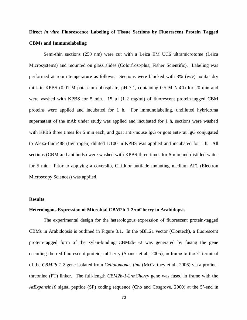
70
Direct in vitro Fluorescence Labeling of Tissue Sections by Fluorescent Protein Tagged
CBMs and Immunolabeling
Semi-thin sections (250 nm) were cut with a Leica EM UC6 ultramicrotome (Leica
Microsystems) and mounted on glass slides (Colorfrost/plus; Fisher Scientific). Labeling was
performed at room temperature as follows. Sections were blocked with 3% (w/v) nonfat dry
milk in KPBS (0.01 M potassium phosphate, pH 7.1, containing 0.5 M NaCl) for 20 min and
were washed with KPBS for 5 min. 15 µl (1-2 mg/ml) of fluorescent protein-tagged CBM
proteins were applied and incubated for 1 h. For immunolabeling, undiluted hybridoma
supernatant of the mAb under study was applied and incubated for 1 h, sections were washed
with KPBS three times for 5 min each, and goat anti-mouse IgG or goat anti-rat IgG conjugated
to Alexa-fluor488 (Invitrogen) diluted 1:100 in KPBS was applied and incubated for 1 h. All
sections (CBM and antibody) were washed with KPBS three times for 5 min and distilled water
for 5 min. Prior to applying a coverslip, Citifluor antifade mounting medium AF1 (Electron
Microscopy Sciences) was applied.
Results
Heterologous Expression of Microbial CBM2b-1-2:mCherry in Arabidopsis
The experimental design for the heterologous expression of fluorescent protein-tagged
CBMs in Arabidopsis is outlined in Figure 3.1. In the pBI121 vector (Clontech), a fluorescent
protein-tagged form of the xylan-binding CBM2b-1-2 was generated by fusing the gene
encoding the red fluorescent protein, mCherry (Shaner et al., 2005), in frame to the 3’-terminal
of the CBM2b-1-2 gene isolated from Cellulomonas fimi (McCartney et al., 2006) via a proline-
threonine (PT) linker. The full-length CBM2b-1-2:mCherry gene was fused in frame with the
AtExpansin10 signal peptide (SP) coding sequence (Cho and Cosgrove, 2000) at the 5’-end in
71
order to target the translated fusion protein to the cell wall through the secretory pathway in
Arabidopsis. The empty vector containing the AtExpansin10 signal peptide coding sequence
directly fused with mCherry via the same PT linker was produced as a negative control to verify
that expression of the mCherry protein itself does not bind to or affect the plant cell wall. After
transformation, kanamycin antibiotic-resistant transgenic plants were selected and the expression
of introduced genes was confirmed by Reverse Transcript (RT)-PCR analysis using total RNA
harvested from stems. Fluorescent localization of CBM2b-1-2:mCherry/SP:mCherry was
carefully examined in these transgenic plants in the T1 generation. For each construct, three
independent lines were isolated and selfed. T2 generations of these transgenic plants were
screened for kanamycin resistance and examined by RT-PCR and fluorescence microscopy.
Among the T2 plants, the introduced CBM2b-1-2:mCherry/SP:mCherry genes were stably
inherited and CBM2b-1-2:mCherry/SP:mCherry proteins were expressed in an active
(fluorescent) form. In our imaging analysis, transgenic plants were grown from seeds of one line
of each transgenic plant expressing CBM2b-1-2:mCherry/SP:mCherry.
Xylan is abundant in the secondary cell walls of specific cell types such as xylem vessels
and interfascicular fibers (McCartney et al., 2006). The plant cell starts producing the secondary
cell wall after the primary cell wall is complete and the cell has stopped expanding (Carpita,
2000). Thus, the secondary cell wall is located between the primary cell wall and the plasma
membrane. In relation to plant development, xylan synthesis and deposition increases with
increasing stem maturity. To observe the in vivo xylan deposition during plant development,
transgenic CBM2b-1-2:mCherry plants were examined by hand-sectioning the basal, middle, and
top parts of 5-week-old and 9-week-old stems in comparison with WT and SP:mCherry controls.
In addition, direct labeling and immunolabeling studies were carried out on the basal parts of the
72
same stems used for in vivo observation to obtain independent in vitro verification of xylan
distribution patterns to compare with results from in vivo observation of CBM2b-1-2:mCherry
localization. These in vitro studies were done using CBM2b-1-2:mCherry protein expressed in
Escherichia coli and xylan-directed mAbs (CCRC-M138, CCRC-M147, CCRC-M149, CCRC-
M153 and LM11) on these fixed stem sections.
Figure 3.1. Outline of heterologous expression of CBM2b-1-2:mCherry in transgenic plants.
[Figure is modified from Figure 1 of (Abramson et al., 2010)]
(A) Gene constructs consisting of a constitutive promoter (Cauliflower Mosaic Virus 35S
promoter (red)), a cell wall targeting signal peptide (yellow), CBM2b-1-2 gene (green),
proline and threonine (PT) linker (blue) and mCherry fluorescent protein (cherry-pink).
(B) Transformation of CBM2b-1-2:mCherry gene into the plant genome.
73
(C) CBM2b-1-2:mCherry protein is then expressed and secreted into the cell wall: (green
strands - crystalline cellulose; yellow strands - amorphous cellulose; brown strands -
hemicellulose).
(D) Transgenic CBM2b-1-2:mCherry plant is generated and expression of CBM2b-1-
2:mCherry protein is examined.
Expression of SP:mCherry in Arabidopsis
In this study, mCherry was chosen to avoid issues arising from autofluorescence of lignin
in secondary cell walls, which occurs in the green part of the spectrum where GFP fluorescence
is observed. This autofluorescence is not typically observed in fixed and embedded material
(Utku Avci, personal communication), but was observed in fresh hand-cut sections
(Supplemental Figure S3.1). Observation of WT stem (Figure 3.2A-B) revealed that
autofluorescence under microscopy conditions used for mCherry observation was almost absent
in secondary cell walls of xylem vessels and interfascicular fibers in both 5-week-old and 9-
week-old stages. This result confirmed the advantage of red fluorescent mCherry for in vivo
plant cell wall visualization by circumventing possible interference from autofluorescence of
secondary cell walls.
In 5-week-old basal stems of transgenic SP:mCherry plants, strong mCherry fluorescent
signal was detected in epidermis, cortex, xylem vessels and interfascicular fibers and weak
fluorescent signal was detected in pith cells (Figure 3.3A). Higher magnification of fiber cells
from the middle section of SP:mCherry stems revealed that the SP:mCherry protein was
localized in the intercellular spaces (Supplemental Figure S3.2). In particular, the empty
triangle-shaped region at tri-cellular junctions of fiber cells was occupied by SP:mCherry
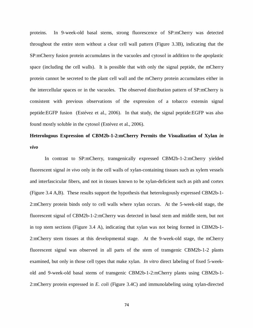
74
proteins. In 9-week-old basal stems, strong fluorescence of SP:mCherry was detected
throughout the entire stem without a clear cell wall pattern (Figure 3.3B), indicating that the
SP:mCherry fusion protein accumulates in the vacuoles and cytosol in addition to the apoplastic
space (including the cell walls). It is possible that with only the signal peptide, the mCherry
protein cannot be secreted to the plant cell wall and the mCherry protein accumulates either in
the intercellular spaces or in the vacuoles. The observed distribution pattern of SP:mCherry is
consistent with previous observations of the expression of a tobacco extensin signal
peptide:EGFP fusion (Estévez et al., 2006). In that study, the signal peptide:EGFP was also
found mostly soluble in the cytosol (Estévez et al., 2006).
Heterologous Expression of CBM2b-1-2:mCherry Permits the Visualization of Xylan in
vivo
In contrast to SP:mCherry, transgenically expressed CBM2b-1-2:mCherry yielded
fluorescent signal in vivo only in the cell walls of xylan-containing tissues such as xylem vessels
and interfascicular fibers, and not in tissues known to be xylan-deficient such as pith and cortex
(Figure 3.4 A,B). These results support the hypothesis that heterologously expressed CBM2b-1-
2:mCherry protein binds only to cell walls where xylan occurs. At the 5-week-old stage, the
fluorescent signal of CBM2b-1-2:mCherry was detected in basal stem and middle stem, but not
in top stem sections (Figure 3.4 A), indicating that xylan was not being formed in CBM2b-1-
2:mCherry stem tissues at this developmental stage. At the 9-week-old stage, the mCherry
fluorescent signal was observed in all parts of the stem of transgenic CBM2b-1-2 plants
examined, but only in those cell types that make xylan. In vitro direct labeling of fixed 5-week-
old and 9-week-old basal stems of transgenic CBM2b-1-2:mCherry plants using CBM2b-1-
2:mCherry protein expressed in E. coli (Figure 3.4C) and immunolabeling using xylan-directed

75
antibodies (Figure 3.4D) independently verified the presence of xylan in the cell walls of cells
labeled by in vivo expression of CBM2b-1-2:mCherry fluorescent protein.
Figure 3.2. Observation of WT plants in vivo and in vitro. Hand-sectioned basal, middle and
top parts of stems of (A) 5-week-old WT and (B) 9-week-old WT plants were visualized under
identical imaging conditions (left panels, excitation = 528-553 nm, emission = 590-650 nm) and
white light (right panels). (C) Direct labeling in fixed stem sections using CBM2b-1-2:mCherry
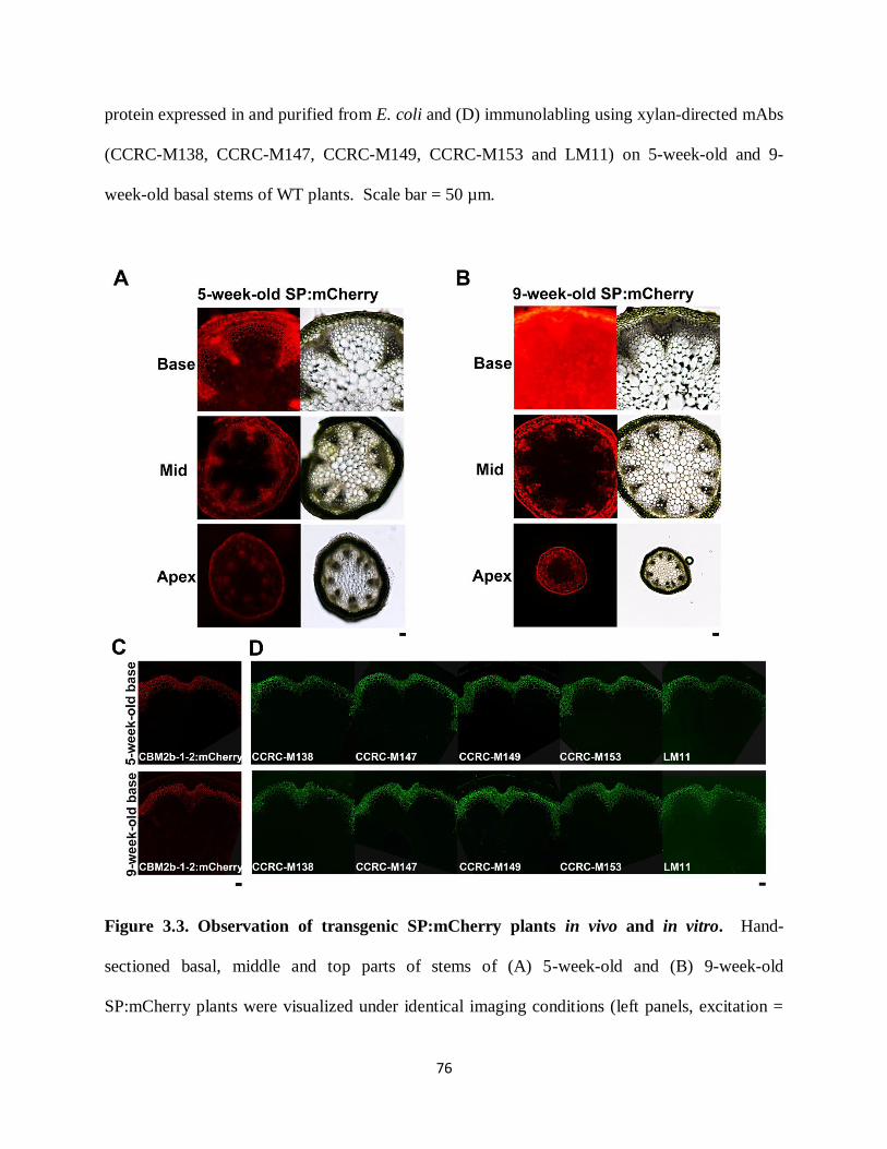
76
protein expressed in and purified from E. coli and (D) immunolabling using xylan-directed mAbs
(CCRC-M138, CCRC-M147, CCRC-M149, CCRC-M153 and LM11) on 5-week-old and 9-
week-old basal stems of WT plants. Scale bar = 50 µm.
Figure 3.3. Observation of transgenic SP:mCherry plants in vivo and in vitro. Hand-
sectioned basal, middle and top parts of stems of (A) 5-week-old and (B) 9-week-old
SP:mCherry plants were visualized under identical imaging conditions (left panels, excitation =
77
528-553 nm, emission = 590-650 nm) and white light (right panels). (C) Direct labeling in fixed
stem sections using CBM2b-1-2:mCherry protein expressed in and purified from E. coli and (D)
immunolabling using xylan-directed mAbs (CCRC-M138, CCRC-M147, CCRC-M149, CCRC-
M153 and LM11) on 5-week-old and 9-week-old basal stems of transgenic SP:mCherry plants.
Scale bar = 50 µm.
78
Figure 3.4. Observation of transgenic CBM2b-1-2:mCherry plants in vivo and in vitro.
Hand-sectioned basal, middle and top parts of stems of (A) 5-week-old and (B) 9-week-old
CBM2b-1-2:mCherry plants were visualized under identical imaging conditions (left panels,
excitation = 528-553 nm, emission = 590-650 nm) and white light (right panels). (C) Direct
labeling in fixed stem sections using CBM2b-1-2:mCherry protein expressed in and purified
from E. coli and (D) immunolabling using xylan-directed mAbs (CCRC-M138, CCRC-M147,
CCRC-M149, CCRC-M153 and LM11) on 5-week-old and 9-week-old basal stems of transgenic
CBM2b-1-2:mCherry plants. Scale bar = 50 µm.
In vivo Localization of CBM2b-1-2:mCherry Reveals Altered Patterns of Xylan Deposition
in the wrky Cell Wall Mutant
We tested the effectiveness of the CBM2b-1-2:mCherry protein as a functional in vivo
visualization tool by examining what happens to the fluorescence labeling pattern when the
distribution of xylan is altered by mutagenesis. Toward this end, we chose to work with the
wrky12 mutant in Arabidopsis (Wang et al., 2010). Pith cells of Arabidopsis normally do not
undergo secondary wall thickening, nor do they deposit xylans in their walls. However, a mutant
plant line carrying a transfer (T)-DNA insertion in the AtWRKY12 transcription factor gene
exhibits secondary thickening in pith cell walls together with ectopic xylan deposition (Wang et
al., 2010). Mutations in the AtWRKY12 gene have little impact on overall plant growth and
development (Wang et al., 2010). Thus, we crossed the stable transgenic CBM2b-1-2:mCherry
plant with the wrky12-1 plant and observed the labeling pattern in progeny carrying a
homozygous T-DNA insertion at the AtWRKY12 locus. The outline of this experiment was: F1
progeny of the cross were selfed to generate homozygous plants for the wrky12 mutation. The
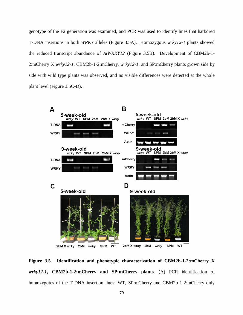
79
genotype of the F2 generation was examined, and PCR was used to identify lines that harbored
T-DNA insertions in both WRKY alleles (Figure 3.5A). Homozygous wrky12-1 plants showed
the reduced transcript abundance of AtWRKY12 (Figure 3.5B). Development of CBM2b-1-
2:mCherry X wrky12-1, CBM2b-1-2:mCherry, wrky12-1, and SP:mCherry plants grown side by
side with wild type plants was observed, and no visible differences were detected at the whole
plant level (Figure 3.5C-D).
Figure 3.5. Identification and phenotypic characterization of CBM2b-1-2:mCherry X
wrky12-1, CBM2b-1-2:mCherry and SP:mCherry plants. (A) PCR identification of
homozygotes of the T-DNA insertion lines: WT, SP:mCherry and CBM2b-1-2:mCherry only
80
have a wrky gene-specific band, whereas the insertion lines wrky12-1 and CBM2b-1-2:mCherry
X wrky12-1 have only a T-DNA-specific band. (B) RT-PCR analyses. mCherry and WRKY
expression was examined using primers covering the full length of the cDNAs. ACTIN
expression was used as the control. (C-D) From left to right: growth of CBM2b-1-2:mCherry X
wrky12-1, CBM2b-1-2:mCherry, wrky12-1, SP:mCherry and WT plants at 5-weeks (C) and 9-
weeks (D). Note that expression of CBM2b-1-2:mCherry and SP:mCherry have no visible
impact on overall plant growth and development. Scale bar = 5cm.
At the 5-week-old developmental stage, the in vivo localization pattern of CBM2b-1-
2:mCherry protein in the CBM2b-1-2 X wrky12-1 plant was similar to that observed in the
CBM2b-1-2:mCherry plant. The CBM2b-1-2:mCherry protein was localized in the xylem
vessels and interfascicular fibers of basal and middle stems, but not in the top stems (Figure
3.6A). At the 9-week-old developmental stage, fluorescent signal from CBM2b-1-2:mCherry
was detected in some pith cell walls and the intensity appeared to be indistinguishable from the
signal detected in the secondary cell walls of adjacent xylem cells in basal stem (Figure 3.4A).
This result suggested that these pith cells in CBM2b-1-2:mCherry X wrky12-1 contained ectopic
deposition of xylan in the 9-week-old basal stem. In contrast, the pattern of CBM2b-1-
2:mCherry fluorescence at the middle and top of CBM2b-1-2:mCherry X wrky12 stems (Figure
3.6B) was essentially identical to that observed at the middle and top of CBM2b-1-2:mCherry
stems (Figure 3.4B).
In vitro direct labeling with exogenous CBM2b-1-2:mCherry and immunolabeling
studies with xylan-directed mAbs confirmed there was no ectopic xylan in pith cells in basal
stems of CBM2b-1-2:mCherry X wrky12-1 and wrky12-1 at the 5-week-old stage (Figure 3.6C-
81
D; Figure 3.7C-D). These results demonstrate that pith cells in the wrky background did not
initiate secondary wall thickening at the 5-week-old stage. At the 9-week-old stage, xylan-
binding CBM2b-1-2:mCherry protein and xylan-directed mAbs labeled walls of xylem vessels,
interfascicular fibers and some pith cells in basal stems of both CBM2b-1-2:mCherry X wrky12
and wrky12, displaying the same xylan distribution pattern as in vivo localization of CBM2b-1-
2:mCherry fluorescent protein. Taken together, the in vivo and in vitro localization data indicate
that in the wrky mutant, xylan is first deposited into the xylem vessels and interfascicular fibers,
and is then ectopically deposited into pith cells with increasing stem maturity.
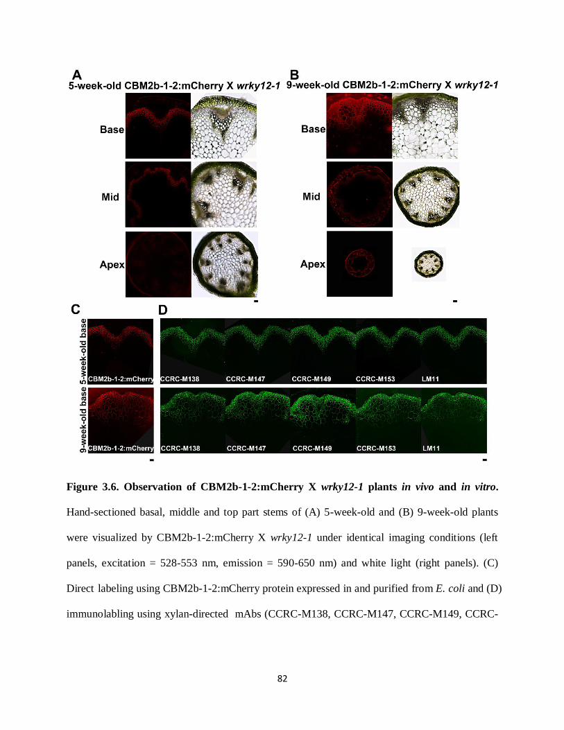
82
Figure 3.6. Observation of CBM2b-1-2:mCherry X wrky12-1 plants in vivo and in vitro.
Hand-sectioned basal, middle and top part stems of (A) 5-week-old and (B) 9-week-old plants
were visualized by CBM2b-1-2:mCherry X wrky12-1 under identical imaging conditions (left
panels, excitation = 528-553 nm, emission = 590-650 nm) and white light (right panels). (C)
Direct labeling using CBM2b-1-2:mCherry protein expressed in and purified from E. coli and (D)
immunolabling using xylan-directed mAbs (CCRC-M138, CCRC-M147, CCRC-M149, CCRC-
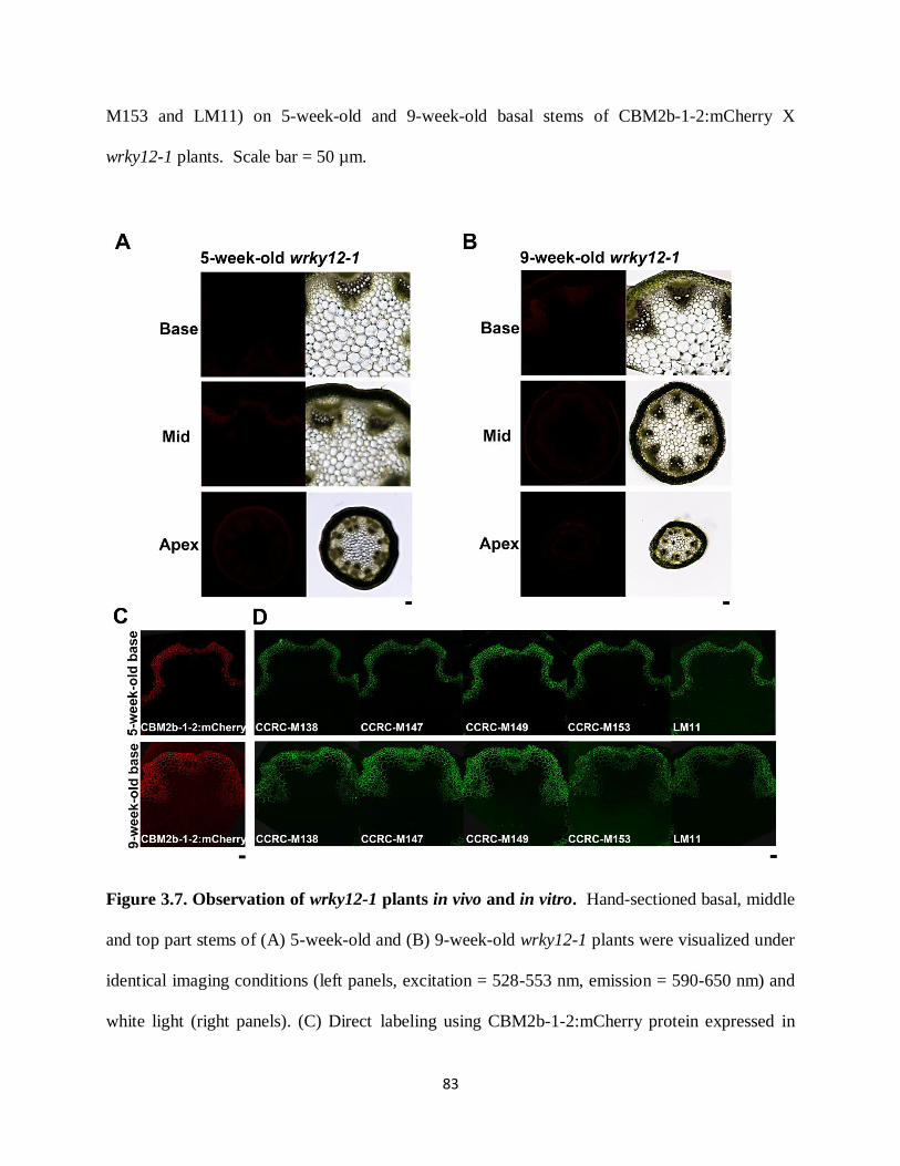
83
M153 and LM11) on 5-week-old and 9-week-old basal stems of CBM2b-1-2:mCherry X
wrky12-1 plants. Scale bar = 50 µm.
Figure 3.7. Observation of wrky12-1 plants in vivo and in vitro. Hand-sectioned basal, middle
and top part stems of (A) 5-week-old and (B) 9-week-old wrky12-1 plants were visualized under
identical imaging conditions (left panels, excitation = 528-553 nm, emission = 590-650 nm) and
white light (right panels). (C) Direct labeling using CBM2b-1-2:mCherry protein expressed in

84
and purified from E. coli and (D) immunolabling using xylan-directed mAbs (CCRC-M138,
CCRC-M147, CCRC-M149, CCRC-M153 and LM11) on 5-week-old and 9-week-old basal
stems of CBM2b-1-2:mCherry X wrky12-1 plants. Scale bar = 50 µm.
In vivo Localization of CBM2b-1-2:mCherry Reveals the Architecture of Xylan Deposition
in Roots
We also examined the maturation zone of roots of 3-day-old CBM2b-1-2:mCherry X
wrky, CBM2b-1-2:mCherry, wrky, SP:mCherry and WT seedlings by fluorescent microscopy to
test if the transgenically expressed CBM2b-1-2:mCherry would label xylan-containing walls in
this organ. The intact (unsectioned) roots of SP:mCherry seedlings exhibited robust mCherry
fluorescence throughout the root tissues, clearly showing SP:mCherry accumulation in all cells
of the root. In contrast, no fluorescence was observed in intact WT and wrky seedlings (Figure
3.8). There was not much difference in the distribution patterns of CBM2b-1-2:mCherry
between the intact CBM2b-1-2:mCherry root and the intact CBM2b-1-2:mCherry X wrky12-1
root. In both CBM2b-1-2:mCherry X wrky12-1 and CBM2b-1-2:mCherry roots, mCherry
fluorescence was primarily observed in the cell walls of the central cylinder of the root,
suggesting that xylan is already deposited into the cell walls of particular cells at the 3-day-old
stage. The specific cell types whose cell walls are labeled by CBM2b-1-2:mCherry could not be
delimited under the microscopic conditions used.
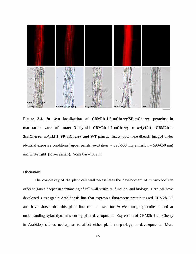
85
Figure 3.8. In vivo localization of CBM2b-1-2:mCherry/SP:mCherry proteins in
maturation zone of intact 3-day-old CBM2b-1-2:mCherry x wrky12-1, CBM2b-1-
2:mCherry, wrky12-1, SP:mCherry and WT plants. Intact roots were directly imaged under
identical exposure conditions (upper panels, excitation = 528-553 nm, emission = 590-650 nm)
and white light (lower panels). Scale bar = 50 µm.
Discussion
The complexity of the plant cell wall necessitates the development of in vivo tools in
order to gain a deeper understanding of cell wall structure, function, and biology. Here, we have
developed a transgenic Arabidopsis line that expresses fluorescent protein-tagged CBM2b-1-2
and have shown that this plant line can be used for in vivo imaging studies aimed at
understanding xylan dynamics during plant development. Expression of CBM2b-1-2:mCherry
in Arabidopsis does not appear to affect either plant morphology or development. More
86
importantly, expression of CBM2b-1-2:mCherry and targeting this probe to the secretory
pathway in Arabidopsis using the AtExpansin10 signal peptide leads to the selective labeling of
xylans in vivo in both stems and roots. Incorporation of the CBM2b-1-2:mCherry specifically
occurs in the secondary cell walls of stems and binds tightly to xylem vessels and interfascicular
fibers where xylans are being produced and deposited. The resultant fluorescent protein-labeled
xylan can be directly visualized by fluorescent microscopy. No labeling of cells that produce
only primary cell walls, such as pith cells, was observed. Labeling of fixed embedded sections
of Arabidopsis stems with diverse xylan-directed mAbs showed that the same cell walls were
labeled in vitro as had been tagged in vivo with transgenically expressed CBM2b-1-2:mCherry.
Thus, our results provide a proof of concept of the utility of transgenically expressed CBM2b-1-
2:mCherry for in vivo imaging studies of xylans in plant cell walls without apparently disturbing
plant physiology. Indeed, examination of the transgenic CBM2b-1-2:mCherry line using hand-
cut sections documented the staged appearance of xylan in relation to plant development,
showing that xylan deposition increases with increasing stem maturity without the need for
extensive tissue preparation and manipulation. In fact, in root tissue it was possible to observe
labeling of xylan in the intact organ, although confocal microscopy will be needed to provide
cellular resolution of the xylan deposition patterns.
The utility of the transgenic CBM2b-1-2:mCherry for monitoring xylan deposition and
dynamics was further illustrated by crossing this xylan tag into the wrky mutant of Arabidopsis.
Mutation of the WRKY12 gene had previously been shown to result in ectopic secondary wall
formation and the ectopic deposition of xylan in pith cells, which normally do not express either
character (Wang et al., 2010). The CBM2b-1-2:mCherry X wrky plant exhibited ectopic xylan
labeling in vivo in pith cells adjacent to xylem vessels in basal stems, a pattern that was
87
confirmed in vitro using xylan-directed mAbs to label sections taken from fixed embedded stems
of these plants. This observation demonstrates that fluorescently tagged-CBMs allow one to
monitor the effects of mutations on cell wall polysaccharide deposition in vivo. Indeed, our
results document that the ectopic deposition of xylan in pith cell walls occurs after xylan
deposition in the developing primary and secondary xylem, an observation that had not been
made in the earlier study of the wrky mutant (Wang et al., 2010).
We are still at the stage of tool development in terms of documenting cell wall structure
in relation to plant development, and to integrate this knowledge with an understanding of
polysaccharide functions. Currently, immunohistochemical techniques carried out in vitro on
fixed tissues are the main method to discern cell wall structures and to locate polysaccharides in
plant cells, tissues and organs. However, substantial effort is required to demonstrate that in
vitro measurements of polysaccharides precisely reflect the in vivo biochemical and structural
complexity of polysaccharides. The methods and results described here create new opportunities
to assay the effects of genetic, developmental, and environmental variation on xylan biosynthesis.
To our knowledge, no other tool that can detect xylan in vivo is available now. Thus,
heterologously expressed CBM2b-1-2:mCherry provides the plant research community the first
in vivo xylan tracking tool. In the future, the use of this and similar tools is likely to expand our
understanding of xylan synthesis and deposition, and bolster efforts to engineer xylan with
improved properties for applications such as biofuel production.

88
Supplemental Figure S3.1. Observation of transgenic CBM2b-1-2:GFP and WT plants in
vivo. Hand-sectioned basal part stems of 9-week-old transgenic CBM2b-1-2:GFP (upper) and
WT (lower) plants were visualized by fluorescent microscopy (left panels) and white light (right
panels). Scale bar = 50 µm.

89
Supplemental Figure S3.2. 10X magnification of interfascicular fiber cells from middle
stems of 5-week-old and 9-week-old transgenic SP:mCherry and CBM2b-1-2:mCherry
plants. Squares in the left panel indicate the areas of high magnification views. Scale bar = 50
µm.
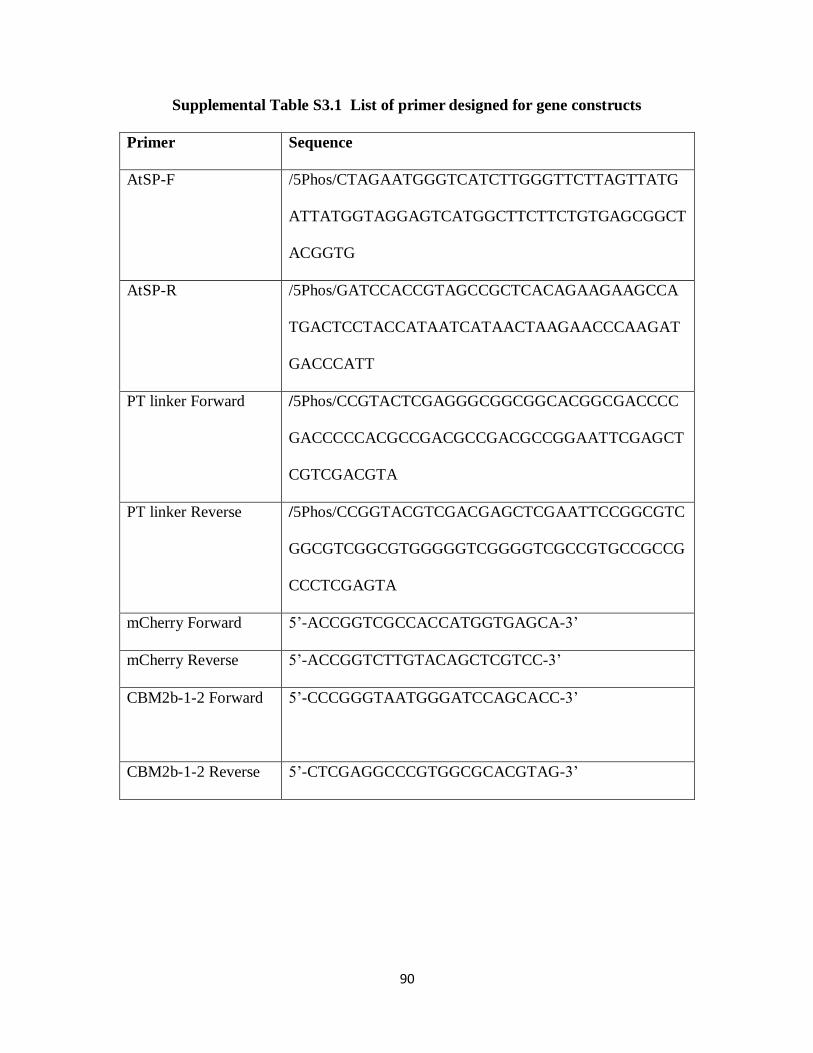
90
Supplemental Table S3.1 List of primer designed for gene constructs
Primer Sequence
AtSP-F /5Phos/CTAGAATGGGTCATCTTGGGTTCTTAGTTATG
ATTATGGTAGGAGTCATGGCTTCTTCTGTGAGCGGCT
ACGGTG
AtSP-R /5Phos/GATCCACCGTAGCCGCTCACAGAAGAAGCCA
TGACTCCTACCATAATCATAACTAAGAACCCAAGAT
GACCCATT
PT linker Forward /5Phos/CCGTACTCGAGGGCGGCGGCACGGCGACCCC
GACCCCCACGCCGACGCCGACGCCGGAATTCGAGCT
CGTCGACGTA
PT linker Reverse /5Phos/CCGGTACGTCGACGAGCTCGAATTCCGGCGTC
GGCGTCGGCGTGGGGGTCGGGGTCGCCGTGCCGCCG
CCCTCGAGTA
mCherry Forward 5’-ACCGGTCGCCACCATGGTGAGCA-3’
mCherry Reverse 5’-ACCGGTCTTGTACAGCTCGTCC-3’
CBM2b-1-2 Forward 5’-CCCGGGTAATGGGATCCAGCACC-3’
CBM2b-1-2 Reverse 5’-CTCGAGGCCCGTGGCGCACGTAG-3’
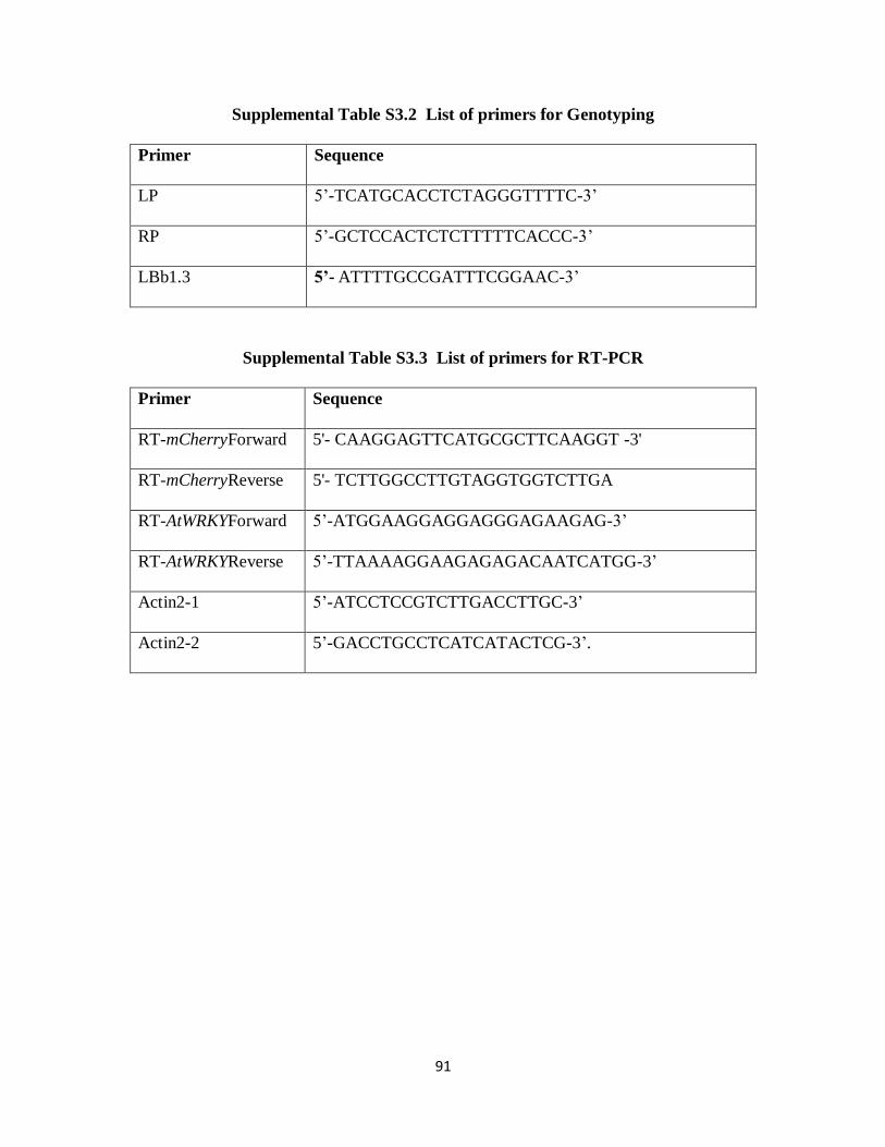
91
Supplemental Table S3.2 List of primers for Genotyping
Primer Sequence
LP 5’-TCATGCACCTCTAGGGTTTTC-3’
RP 5’-GCTCCACTCTCTTTTTCACCC-3’
LBb1.3 5’- ATTTTGCCGATTTCGGAAC-3’
Supplemental Table S3.3 List of primers for RT-PCR
Primer Sequence
RT-mCherryForward 5'- CAAGGAGTTCATGCGCTTCAAGGT -3'
RT-mCherryReverse 5'- TCTTGGCCTTGTAGGTGGTCTTGA
RT-AtWRKYForward 5’-ATGGAAGGAGGAGGGAGAAGAG-3’
RT-AtWRKYReverse 5’-TTAAAAGGAAGAGAGACAATCATGG-3’
Actin2-1 5’-ATCCTCCGTCTTGACCTTGC-3’
Actin2-2 5’-GACCTGCCTCATCATACTCG-3’.
92
References
Abramson M, Shoseyov O, Shani Z (2010) Plant cell wall reconstruction toward improved
lignocellulosic production and processability. Plant Science 178: 61-72
Anderson CT, Carroll A, Akhmetova L, Somerville C (2010) Real-time imaging of cellulose
reorientation during cell wall expansion in Arabidopsis roots. Plant Physiol 152: 787-796
Bedford MR, Partridge GG (2010) Enzymes in Farm Animal Nutrition. CABI
Carpita NM, M. (2000) The cell wall. In WG B. Buchanan, and R. Jones, ed, Biochemistry and
Molecular Biology of Plants. American Society of Plant Physiologists, Rockville, MD,
pp 52-108
Carroll A, Somerville C (2009) Cellulosic biofuels. Annu Rev Plant Biol 60: 165-182
Cho H-T, Cosgrove DJ (2000) Altered expression of expansin modulates leaf growth and
pedicel abscission in Arabidopsis thaliana. Proceedings of the National Academy of
Sciences 97: 9783-9788
Clough SJ, Bent AF (1998) Floral dip: a simplified method for Agrobacterium-mediated
transformation of Arabidopsis thaliana. Plant J 16: 735-743
Estévez JM, Kieliszewski MJ, Khitrov N, Somerville C (2006) Characterization of Synthetic
Hydroxyproline-Rich Proteoglycans with Arabinogalactan Protein and Extensin Motifs in
Arabidopsis. Plant Physiology 142: 458-470
Fry SC (2010) Cell Wall Polysaccharide Composition and Covalent Crosslinking. In Annual
Plant Reviews. Wiley-Blackwell, pp 1-42
Gilbert HJ, Knox JP, Boraston AB (2013) Advances in understanding the molecular basis of
plant cell wall polysaccharide recognition by carbohydrate-binding modules. Current
Opinion in Structural Biology 23: 669-677

93
Hao Z, Mohnen D (2014) A review of xylan and lignin biosynthesis: foundation for studying
Arabidopsis irregular xylem mutants with pleiotropic phenotypes. Crit Rev Biochem Mol
Biol 49: 212-241
Haseloff J, Siemering KR, Prasher DC, Hodge S (1997) Removal of a cryptic intron and
subcellular localization of green fluorescent protein are required to mark transgenic
Arabidopsis plants brightly. Proc Natl Acad Sci U S A 94: 2122-2127
Knox JP (2008) Revealing the structural and functional diversity of plant cell walls. Current
Opinion in Plant Biology 11: 308-313
McCartney L, Blake AW, Flint J, Bolam DN, Boraston AB, Gilbert HJ, Knox JP (2006)
Differential recognition of plant cell walls by microbial xylan-specific carbohydrate-
binding modules. Proceedings of the National Academy of Sciences of the United States
of America 103: 4765-4770
Paredez AR, Somerville CR, Ehrhardt DW (2006) Visualization of Cellulose Synthase
Demonstrates Functional Association with Microtubules. Science 312: 1491-1495
Rennie EA, Scheller HV (2014) Xylan biosynthesis. Current Opinion in Biotechnology 26:
100-107
Shaner NC, Steinbach PA, Tsien RY (2005) A guide to choosing fluorescent proteins. Nat
Methods 2: 905-909
Stone BA, Jacobs AK, Hrmova M, Burton RA, Fincher GB (2010) Biosynthesis of Plant Cell
Wall and Related Polysaccharides by Enzymes of the GT2 and GT48 Families. In Annual
Plant Reviews. Wiley-Blackwell, pp 109-165
94
Tobimatsu Y, Wagner A, Donaldson L, Mitra P, Niculaes C, Dima O, Kim JI, Anderson N,
Loque D, Boerjan W, Chapple C, Ralph J (2013) Visualization of plant cell wall
lignification using fluorescence-tagged monolignols. Plant J 76: 357-366
Wang H, Avci U, Nakashima J, Hahn MG, Chen F, Dixon RA (2010) Mutation of WRKY
transcription factors initiates pith secondary wall formation and increases stem biomass
in dicotyledonous plants. Proc Natl Acad Sci U S A 107: 22338-22343
95
CHAPTER 4
HETEROLOGOUS EXPRESSION OF CRYSTALLINE CELLULOSE DIRECTED-
CARBOHYDRATE BINDING MODULE 3A IN ARABIDOPSIS MODULATES PLANT
CELL WALLS AND DEVELOPMENT
Abstract
The non-catalytic polysaccharide-recognizing modules within diverse microbial
carbohydrate-active enzymes are defined as Carbohydrate-Binding Modules (CBMs). CBMs
can bind to specific plant cell wall polysaccharides, such as cellulose, mannan or xylan. In this
study, we have examined the effects of heterologous expression of crystalline cellulose binding-
CBM3a fused with the mCherry fluorescent protein in Arabidopsis. Compared to wild type
plants, transgenic CBM3a:mCherry plants displayed a dwarfed phenotype, had reduced cellulose
and xylan content, and showed changes in glycan epitope extractability patterns. These results
indicate that expression of a specific CBM in Arabidopsis may lead to changes in the
composition and/or structure of the cell wall, which in turn, may have an impact on the
development of the plant. Our results further suggest that creation of a set of Arabidopsis plants
expressing diverse microbial CBMs and single-chain antibody fragments (ScFvs) may provide a
toolset for manipulating plant cell wall structure to change or improve the properties of lingo-
cellulosic biomass.

96
Introduction
Growing plant cells are surrounded by a primary cell wall consisting predominantly of
carbohydrate polymers. Cellulose is the most abundant biological macromolecule and is an
extracellular, liner polymer of glucose molecules (Nishiyama et al., 2002). Cellulose exists as
microfibrils composed of parallel β-1,4-linked glucan chains that are held together laterally by
hydrogen bonds and vertically by hydrophobic stacking of the sugar rings. Cellulose is much
more ordered than other components of the primary cell wall (Bootten et al., 2004), in keeping
with its key role of providing strength and controlling growth. Xyloglucan was widely believed
to act as a tether between cellulose fibers, limiting cell enlargement and regulating cell wall
mechanical properties. However, recent studies suggests that only a minor xyloglucan
component may be located in the limited regions of tight contact between cellulose fibers,
playing an important role in wall mechanics (Cavalier et al., 2008; Zabotina et al., 2008; Dick-
Perez et al., 2011; Park and Cosgrove, 2012; Zabotina et al., 2012). Pectin can form hydrated
gels to facilitate slippage of cellulose microfibrils during cell growth. In relation to cell wall
biology, the architecture of heterogeneous functional wall polysaccharides accommodates the
dynamic processes of cell growth and differentiation.
Plant cell growth is precisely controlled by oriented expansion of the cell walls. The
driving force for cell expansion is osmotic, but the rate and direction of expansion are controlled
by the mechanical properties of the cell wall (Cho and Cosgrove, 2000). The stiffness of cell
walls is greatest in the direction of cellulose microfibrils, expansion of the cell walls needs either
widening the space (Marga et al., 2005) or facilitating slippage (Cosgrove, 2005) between
cellulose microfibrils or both. In growing cell walls, when cellulose microfibrils are first laid
down at the inner face of the primary cell wall, their orientation is normally transverse to the

97
direction of growth (Cosgrove, 2005). As the cell wall expands, cellulose microfibrils reorient
toward the direction of growth (Anderson et al., 2010). Hemicellulose and pectin co-extensive
with cellulose microfibrils resist these deformation (Zykwinska et al., 2007), and therefore help
to control the rate of growth.
There is evidence that proteins, for example expansins, are involved in modifying
cellulose-hemicellulose interactions and enhancing growth by mediating cell wall loosening
(Choi et al., 2003). Carbohydrate-binding modules (CBMs) could also affect these interactions
(Levy et al., 2002), leading to altered morphology or plant growth in transgenic plants (Safra-
Dassa et al., 2006; Obembe et al., 2007). A CBM is defined as a contiguous amino acid
sequence within a carbohydrate-active enzyme with a discreet fold having carbohydrate-binding
activity (Boraston et al., 2004). CBMs are suggested to enhance the efficiency of enzymes by
mediating prolonged and intimate contact between their cognate catalytic modules and their
target substrates (Herve et al., 2010). CBMs have been grouped into 69 sequence-based families
(http://www.cazy.org/Carbohydrate-Binding-Modules.html) and around half of these CBM
families contain members that bind to an extensive range of plant cell wall polysaccharides
(Knox, 2008), including cellulose (Blake et al., 2006), mannans (Filonova et al., 2007),
xyloglucan (von Schantz et al., 2009) and xylan (McCartney et al., 2006). Recently, CBMs have
been used as probes to assess and document the cell biological contexts of polysaccharides and
cell wall diversity in vitro (Knox, 2008). Although CBMs do occur in plant enzymes, most
CBMs being used as probes for cell wall polysaccharides are of microbial origin (McCann and
Knox, 2010).
Cellulose microfibrils can exist in highly ordered crystalline, semi-ordered para-
crystalline, and disordered noncrystalline (amorphous) states (Tomme et al., 1995). CBMs
98
located in family 3a bind to crystalline cellulose (Blake et al., 2006). A previous study has
shown that recombinant His-tagged CBM3a from Clostridium spp. effectively binds to both
primary and secondary cell walls in tobacco stem sections using an indirect triple labeling
immunofluorescence procedure (His-tagged CBM, anti-His mouse-Ig, anti-mouse Ig fluorescein
isothiocyanate) (Blake et al., 2006). We have shown previously that expression of a xylan-
binding CBM can be used effectively as a tool for in vivo monitoring of xylan deposition (see
Chapter 3 of this thesis). Since CBMs are the only cellulose-directed probes available, it was of
interest to investigate the heterologous expression of CBM3a in the plant to determine if this
CBM could be used as an in vivo probe for monitoring cellulose deposition and dynamics.
Toward that end, we have expressed fluorescent protein mCherry-tagged CBM3a in Arabidopsis
under the control of the 35S cauliflower mosaic virus (CaMV) promoter. We show here that
transgenic expression of CBM3a:mCherry modulates plant cell wall structure and affects plant
development.
Materials and Methods
Plant Material and Growth Conditions
Both Arabidopsis wild type and the transgenic plant CBM3a:mCherry lines are in the
ecotype Columbia (Col-0) background. Seeds of Arabidopsis were sterilized, cold treated at 4°C
for 48 h, and germinated on one-half-strength Murashige and Skoog medium (Sigma) in a
growth chamber under a 16-h-light/8-h-dark cycle at 19°C during the light period and 15°C
during the dark period. The light intensity was 120 µmol m–2
s–1
, and the relative humidity was
maintained at 70%. One-week-old seedlings were transferred to soil and grown in growth
chambers under the same conditions.

99
Gene Constructs and Plant Transformation
The coding sequence of the AtExpansin10 signal peptide (At1g26770) (Cho and
Cosgrove, 2000) was amplified by AtSP-F and AtSP-R primers (containing KpnI and XbaI
restriction enzyme sites) with Platinum Tag High Fidelity DNA Polymerase (Invitrogen 12574-
030) (all the primers designed for gene constructs are listed in Supplemental Table S4.1). A
proline-threonine (PT) linker coding sequence was amplified from a modified pET22b vector
(obtained from Dr. Gilbert, Newcastle University, UK) using PT linker Forward Primer
(containing the XmaI restriction enzyme site) and PT linker Reverse Primer (containing the AgeI
restriction enzyme site). Using mCherry Forward Primer (containing an AgeI restriction enzyme
site) and mCherry Reverse Primer (containing an AgeI restriction enzyme site), the mCherry
gene was amplified from the pmCherry vector (Clontech PT3973-5). Using CBM3a Forward
primer (containing the BamHI restriction enzyme site) and CBM3a Reverse primer (containing
the XhoI restriction enzyme site), the CBM3a gene was amplified from the scaffoldin of
Clostridium spp. (obtained from Dr. Gilbert, New Castle University, UK). PCR products were
cleaved by the appropriate restriction enzymes (Thermo Scientific Inc) and ligated into the
pBI121 vector (Clontech). All constructs were confirmed by sequencing (Macrogen, USA) and
individually transformed into Agrobacterium tumefaciens GV3101 competent cells by
electroporation. Transformation of Arabidopsis was conducted by floral dipping (Clough and
Bent, 1998).
RNA Isolation and Reverse Transcript (RT)-PCR Analysis
Plant stems for RNA isolation were frozen in liquid nitrogen immediately after harvesting
and stored at -80°C until use. RNA was isolated using the RNeasy plant mini kit (Qiagen) and
treated with RNase-free DNase (Qiagen) to remove contaminating genomic DNA. Total RNA
100
(500 ng) was reverse-transcribed using Superscript® III Reverse Transcriptase (Invitrogen). RT-
PCR was performed in 20 µl of reaction mixture, composed of 2 µl of a given cDNA, 0.4 µl of
each primer (10 μM), 0.4 µl dNTP (10mM), 0.2 µl DreamTaq DNA Polymerase (5 U/µl)
(Thermo), 2 µl 10X DreamTaq DNA Polymerase Buffer, and 14.6 µl Nuclease-free water.
Amplifications were performed using a Tetrad 2 DNA Engine PCR machine (Bio-Rad) under the
following conditions: initial polymerase activation: 95°C, 3 min; 35 cycles of 30 s at 95°C, and
30 s at 55°C. Actin2 (At3g18780) was used as reference gene. Gene-specific primers are listed
in Supplemental Table S4.1.
Fluorescent Microscopy
Live plants expressing CBM3a:mCherry fusion proteins (3-day-old roots and hand-
sectioned 2-month-old stems) were examined by fluorescence microscopy on an Eclipse 80i
(Nikon) microscope equipped with epifluorescence optics. Images were captured with a Nikon
DS-Ri1 camera head using NIS-Element Basic Research software, and images were assembled
using Adobe Photoshop (Adobe Systems).
Cellulose Content Analysis
Fresh samples (stems) from each plant were collected and ground immediately under
liquid nitrogen to a fine powder. Alcohol-insoluble residue (AIR) of each sample was prepared
and destarched enzymatically as described (Foster et al., 2010). Roughly 60-70 mg AIR samples
were subsequently hydrolyzed with 2 M triflouroacetic acid (TFA) for 1 h at 120°C. The pellet
material remaining after TFA hydrolysis was used for cellulose content analysis as described
(Foster et al., 2010).

101
TMS Methyl Glycoside Glycosyl Residue Composition Analysis by Gas Chromatography
The AIR samples were aliquoted (1–3 mg) as acetone suspensions to individual tubes and
allowed to air dry. Inositol (20 µg) was added to each tube and the samples were lyophilized and
analyzed for glycosyl residue composition by combined gas chromatography–mass spectrometry
(GC–MS) of the per-O-trimethylsilyl (TMS) derivatives of the monosaccharide methyl
glycosides produced from the sample by acid methanolysis basically as described (York, 1985).
The dry samples were hydrolyzed for 18 h at 80°C in 1 M methanolic-HCl. The samples were
cooled and evaporated under a stream of dry air and further dried two additional times with
anhydrous methanol. The samples were derivatized with 200 µl of TriSil Reagent (Pierce-
Endogen, Rockford, IL, USA) and heated to 80°C for 20 min. The cooled samples were
evaporated under a stream of dry air, re-suspended in 3 ml of hexane, and filtered through
packed glass wool. The dried samples were re-suspended in 150 µl of hexane and 1 µl of sample
was injected onto a fused silica capillary column in an Agilent gas chromatograph G1701EA.
Monosaccharides were quantified based on standard curves obtained with monosaccharide
standards.
Tissue Fixation
Two-month-old Arabidopsis inflorescence stems were fixed for 2.5 h in 1.6% (w/v)
paraformaldehyde and 0.2% (w/v) glutaraldehyde in 25 mM sodium phosphate buffer, pH 7.1.
Tissue was then washed with buffer twice for 15 min, and dehydrated through a graded ethanol
series [35%, 50%, 75%, 95% (v/v), 100%, 100%, and 100% ethanol] for 30 min for each step.
The dehydrated tissue was moved to 4°C and then gradually infiltrated with cold LR White
embedding resin (Ted Pella) using 33% (v/v) and 66% (v/v) resin in 100% ethanol for 24 h each,
followed by 100% resin for 24 h three times. The infiltrated tissue was transferred to gelatin
102
capsules containing 100% resin for embedding, and resin was polymerized by exposing the
capsules to 365-nm UV light at 4°C for 48 h.
Generation of Fluorescent CBM3a:mCherry protein in Escherichia coli
The fluorescent protein tagged CBM3a:mCherry was produced as a His-tagged
recombinant protein using the T7 expression-system consisting of the pET22b vector (Novagen,
Madison, WI) harbored in Escherichia coli BL21(DE3). The CBM3a:mCherry gene was
inserted into the vector using PCR. Digestion of purified PCR products and pET22b with NotI
and XhoI (Thermo Scientific Inc) enabled cloning of the gene between the NdeI/XhoI sites in the
vector resulting in a construct encoding the recombinant protein containing a hexahistidine tag.
The bacterium was routinely cultured at 37°C in Luria broth (Bertani, 1951; Sambrook et al.,
1989) and expression of the recombinant proteins was induced with 100 µM isopropyl-β-D-
thiogalactopyranoside at 16°C overnight. The fluorescent protein/His-tagged CBM3a:mCherry,
which was produced in soluble form in the cytoplasm of E. coli, was purified by immobilized
metal ion affinity chromatography (Sigma) using TalonTM
Buffer (10 mM Tris/HCl pH 8.0
containing 300 mM NaCl) as the column matrix. The concentration of purified CBM3a:mCherry
was determined spectrophotometrically from absorbance measurements at 280nm.
Direct in vitro Fluorescence Labeling of Tissue Sections by Fluorescent Protein-Tagged
CBMs and Immunolabeling
Semi-thin sections (250 nm) were cut from fixed, embedded plant tissues with a Leica
EM UC6 ultramicrotome (Leica Microsystems) and mounted on glass slides (Colorfrost/plus;
Fisher Scientific). Labeling was performed at room temperature as follows. Sections were
blocked with 3% (w/v) nonfat dry milk in KPBS (0.01 M potassium phosphate, pH 7.1,
containing 0.5 M NaCl) for 20 min and were washed with KPBS for 5 min. 15 µl (1-2 mg/ml)
103
fluorescent protein-tagged CBM3a:mCherry was applied and incubated for 1 h. For
immunolabeling, undiluted hybridoma supernatant of the mAb under study was applied and
incubated for 1 h, sections were washed with KPBS three times for 5 min each, and goat anti-
mouse IgG conjugated to Alexa-fluor488 (Invitrogen) diluted 1:100 in KPBS was applied and
incubated for 1 h. All sections were washed with KPBS three times for 5 min and distilled water
for 5 min. Prior to applying a coverslip, Citifluor antifade mounting medium AF1 (Electron
Microscopy Sciences) was applied.
Cell Wall Extraction for Glycome Profiling
The AIR samples (200 mg) of cell walls was washed with increasingly harsh reagents in
the following order: 50 mM ammonium oxalate (oxalate), 50 mM sodium carbonate with 0.5%
(w/v) of sodium borohydride (carbonate), 1 M KOH with 1% (w/v) of sodium borohydride, 4 M
KOH with 1% (w/v) of sodium borohydride, 100 mM sodium chlorite (Chlorite), and post-
chlorite 4 M KOH with 1% (w/v) of sodium borohydride (4 M KOH PC) to isolate fractions
enriched in cell wall components as previously described (Zhu et al., 2010). The 1 M KOH, 4 M
KOH, and 4 M KOH PC fractions were neutralized using glacial acetic acid. All extracts were
dialyzed against four changes of de-ionized water (sample:water~1:60) at room temperature for
a total of 48 h and then lyophilized. Total sugars in the cell wall extracts were quantified using
the phenol sulphuric acid method (Masuko et al., 2005), and ELISA analyses were done as
previously described (Pattathil et al., 2012).

104
Results
Generation and Molecular Characterization of Transgenic CBM3a:mCherry Plants
A construct (Figure 4.1A) was created to heterologously express CBM3a:mCherry
transgenically in Arabidopsis: the CBM3a:mCherry gene was inserted into a PBI121-derived
vector containing a constitutive CaMV 35S promoter (red) and a cell wall secretory signal
peptide (yellow) from AtExpansin10 (Cho and Cosgrove, 2000). After Agrobacterium-mediated
transformation, expression of the introduced CBM3a:mCherry gene in kanamycin antibiotic-
resistant Arabidopsis was confirmed by Reverse-Transcript (RT) PCR analysis with total RNA.
RT-PCR result revealed variations in the yield of CBM3a expression among individual T1 lines
(Figure 4.1B). In total, 56 transgenic plants were obtained and all transgenic plants were grown
side by side with wild-type Col-0 plants. Among these transgenic plants, 48% of the T1
transgenic CBM3a plants were not able to survive in the soil, 22% of the T1 transgenic CBM3a
plants appeared normal as wild-type plants and 30% of the T1 transgenic CBM3a plants
eventually developed into plants with reduced stem elongation. However, the expression level of
CBM3a:mCherry does not correlate entirely positively with the severity of the phenotype. The
seeds of dwarfed plants were collected, germinated and grown. This T2 generation inherited the
stunted growth phenotype. One line of the T2 generation was chosen based on its dwarf
phenotype and the relatively strong fluorescent signal of the CBM3a:mCherry fusion protein,
and a further generation of this line (T3) was used for all subsequent analyses. RT-PCR showed
that all T3 individuals had equivalent expression levels of CBM3a:mCherry compared to the
actin controls (Figure 4.1C)

105
A
B CBM3a:mCherry T1 plants WT
CBM3a Actin
C
Figure 4.1. (A) Outline of the heterologous expression of CBM3a:mCherry in transgenic plants.
[Figure is modified from Figure 1 of (Abramson et al., 2010)]: 1) Gene Construct consisting of a
constitutive promoter, CaMV 35S (red), a cell wall targeting signal peptide (yellow), a CBM
gene (green), proline and threonine (PT) linker (blue) and fluorescent marker (mCherry) (cherry-
pink). 2) Transformation of the CBM gene into plant genome. 3) CBM protein is then

106
expressed and secreted into the cell wall (green strands - crystalline cellulose; yellow strands -
amorphous cellulose; brown strands - hemicellulose). 4) Transgenic CBM-expressing plant is
generated and expression level of CBM is examined.
(B) Agarose gels showing the presence and various expression levels of the CBM3a transgene by
RT-PCR in T1 plants. Stems from 4-week-old plants were used to extract RNA. Actin2 was
amplified as the control.
(C) RT-PCR analysis confirming equal expression level of the CBM3a transgene in the T3 plants
used in this study. Stems from 4-week-old plants were used to extract RNA. Actin2 was
amplified as the control.
Developmental Effects of Expressed CBM3a:mCherry in Transgenic Plants
To observe the develpmental effects of the expressed CBM3a:mCherry, weekly
measurements of the growth of rosettes and inflorescence stems were conducted for transgenic
CBM3a:mCherry plants, along with wild type plants. Heterologous expression of
CBM3a:mCherry yielded phenotypes different than wild type in plant rosette size, plant height
and plant developmental timing. Development in the transgenic plants was delayed by 2 to 3
weeks compared with development in wild type plants (Figure 4.2). At the rosette age (4-week-
old), transgenic CBM3a:mCherry plants exhibited smaller leaves compared to wild type (Figure
4.2C). The rosette leaf eventually developed to a normal size at weeks 7 and 8 (Figure 4.2A).
CBM3a:mCherry plants started bolting two weeks later than did wild type plants, and when they
reached their final height at week 8, they averaged 29 cm tall, which was approximately 67% of
the average height of wild type plants (Figures 4.2B and 4.2C).
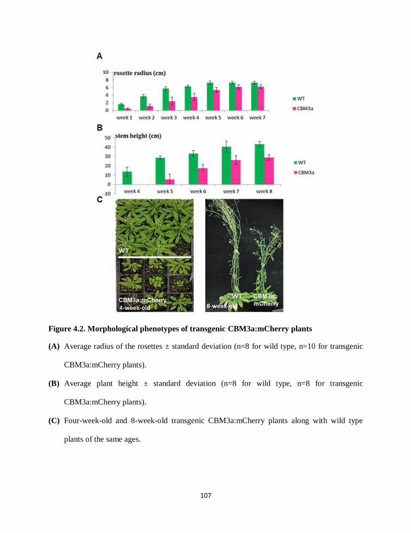
107
Figure 4.2. Morphological phenotypes of transgenic CBM3a:mCherry plants
(A) Average radius of the rosettes ± standard deviation (n=8 for wild type, n=10 for transgenic
CBM3a:mCherry plants).
(B) Average plant height ± standard deviation (n=8 for wild type, n=8 for transgenic
CBM3a:mCherry plants).
(C) Four-week-old and 8-week-old transgenic CBM3a:mCherry plants along with wild type
plants of the same ages.
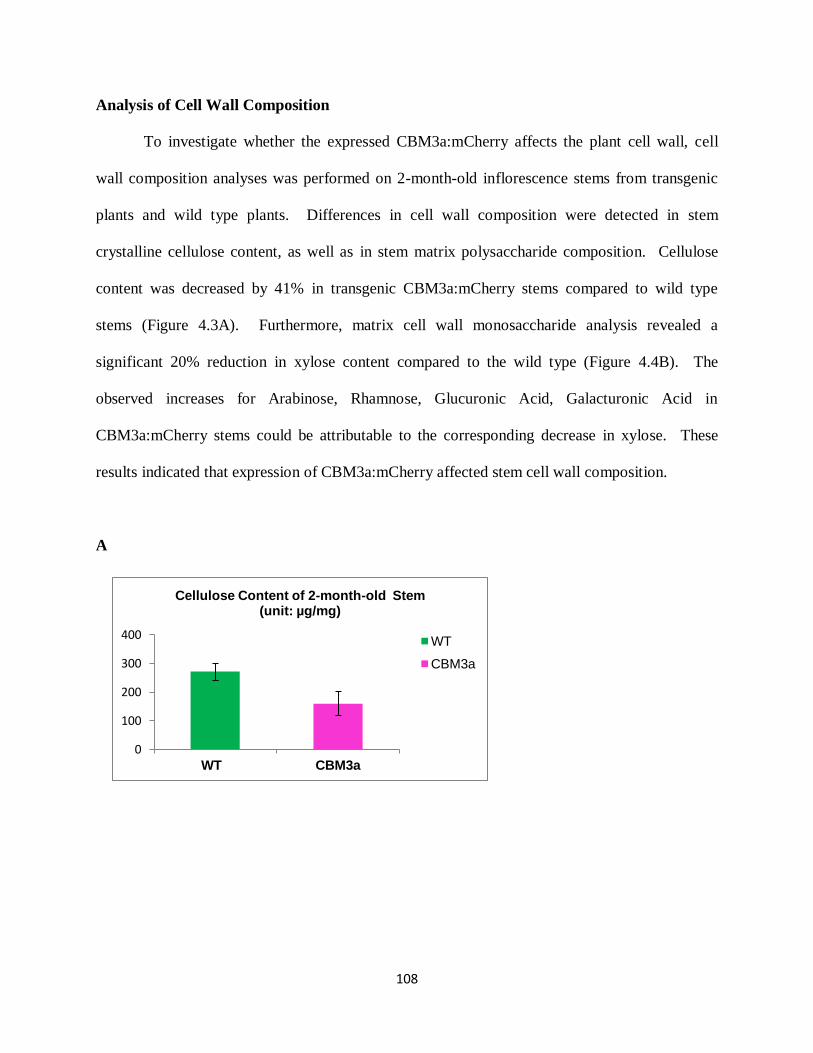
108
0
100
200
300
400
WT CBM3a
Cellulose Content of 2-month-old Stem (unit: µg/mg)
WT
CBM3a
Analysis of Cell Wall Composition
To investigate whether the expressed CBM3a:mCherry affects the plant cell wall, cell
wall composition analyses was performed on 2-month-old inflorescence stems from transgenic
plants and wild type plants. Differences in cell wall composition were detected in stem
crystalline cellulose content, as well as in stem matrix polysaccharide composition. Cellulose
content was decreased by 41% in transgenic CBM3a:mCherry stems compared to wild type
stems (Figure 4.3A). Furthermore, matrix cell wall monosaccharide analysis revealed a
significant 20% reduction in xylose content compared to the wild type (Figure 4.4B). The
observed increases for Arabinose, Rhamnose, Glucuronic Acid, Galacturonic Acid in
CBM3a:mCherry stems could be attributable to the corresponding decrease in xylose. These
results indicated that expression of CBM3a:mCherry affected stem cell wall composition.
A
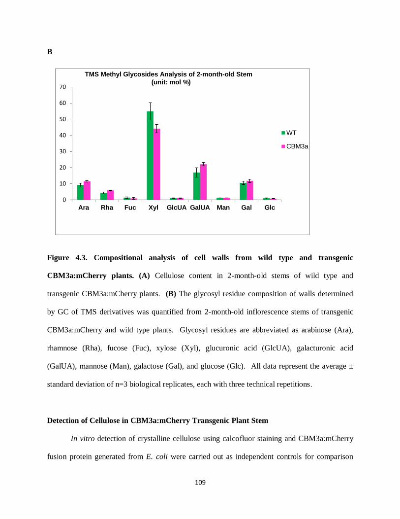
109
0
10
20
30
40
50
60
70
Ara Rha Fuc Xyl GlcUA GalUA Man Gal Glc
WT
CBM3a
B
Figure 4.3. Compositional analysis of cell walls from wild type and transgenic
CBM3a:mCherry plants. (A) Cellulose content in 2-month-old stems of wild type and
transgenic CBM3a:mCherry plants. (B) The glycosyl residue composition of walls determined
by GC of TMS derivatives was quantified from 2-month-old inflorescence stems of transgenic
CBM3a:mCherry and wild type plants. Glycosyl residues are abbreviated as arabinose (Ara),
rhamnose (Rha), fucose (Fuc), xylose (Xyl), glucuronic acid (GlcUA), galacturonic acid
(GalUA), mannose (Man), galactose (Gal), and glucose (Glc). All data represent the average ±
standard deviation of n=3 biological replicates, each with three technical repetitions.
Detection of Cellulose in CBM3a:mCherry Transgenic Plant Stem
In vitro detection of crystalline cellulose using calcofluor staining and CBM3a:mCherry
fusion protein generated from E. coli were carried out as independent controls for comparison
TMS Methyl Glycosides Analysis of 2-month-old Stem
(unit: mol %)

110
with the in vivo labeling patterns resulting from the heterologous expression of CBM3a:mCherry
protein in transgenic plant stems. Calcofluor staining in transgenic CBM3a:mCherry plant stems
showed less staining intensity in the walls of pith cells compared to wild type stem (Figure 4.4A
& 4.4B). Direct labeling of stem sections with CBM3a:mCherry fusion protein revealed that
CBM3a:mCherry binding was restricted to the epidermis and xylem cells in the
CBM3a:mCherry plant stem (Figure 4.4C), whereas CBM3a:mCherry bound indiscriminately to
the epidermis through the cortex and phloem to the vascular tissue in WT stem (Figure 4.5D).
Since mCherry red fluorescent protein is not as bright as green fluorescent protein (GFP) (Shaner
et al., 2005), indirect immunofluorescent localization using a His-tagged CBM3a:mCherry
protein and Alexa Fluor® 488 attached anti-His antibody was used in an effort to get more
information than could be obtained using direct labeling with CBM3a:mCherry. Using this in
vitro method, it was shown that CBM3a bound to every tissue of wide type stem including pith
parenchyma (Figure 4.4E). However, labeling was still weak in epidermis, phloem and pith of
the transgenic CBM3a:mCherry plant stem (Figure 4.4F), suggesting decreased cellulose content
in these tissues of the transgenic plant stem.
Expressed CBM3a:mCherry is Localized in Cell Walls
The in vivo expression pattern of the CBM3a:mCherry fusion protein was determined in
roots and stems of transgenic CBM3a:mCherry plants by fluorescent microscopy. The resulting
images showed that fluorescence of CBM3a:mCherry fusion proteins was located in the cell
walls of epidermis, cortex and endodermis in transgenic plant roots (Figure 4.5A2). In the stem,
distribution of CBM3a:mCherry protein showed a cell-type specific pattern; the fluorescent
signal of CBM3a:mCherry was specifically localized in xylem vessels and interfascicular fibres,
whereas no mCherry fluorescence was observed in pith parenchyma cells (Figure 4.5C2). The in

111
vivo distribution pattern of CBM3a:mCherry fusion protein in stem is consistent with in vitro
detection of cellulose in fixed stem sections of 2-month-old CBM3a:mCherry using exogenous
CBM3a:mCherry protein (Figure 4.4D) and CBM3a protein (Figure 4.4F), reflecting reduced
cellulose content mainly in primary walls of epidermis, cortex, phloem and pith.
Figure 4.4. In vitro detection of cellulose in stem sections of 2-month-old WT plant (Upper)
and transgenic CBM3a:mCherry plant (Lower). (A) (B) Calcofluor staining; (C) (D) Direct
labeling with CBM3a:mCherry protein; (E) (F) Immunohistochemistry using CBM3a:mCherry
protein and Alexa Fluor® 488 attached anti-his monoclonal antibody. Scale bar = 50 µm.
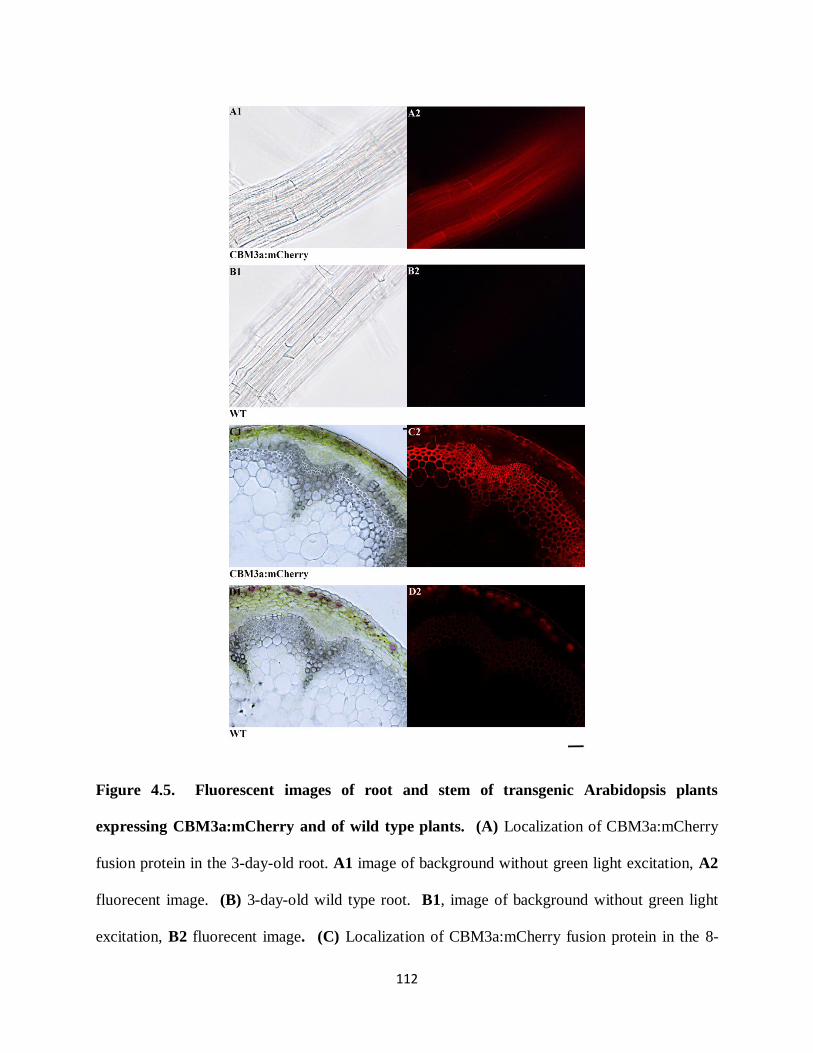
112
Figure 4.5. Fluorescent images of root and stem of transgenic Arabidopsis plants
expressing CBM3a:mCherry and of wild type plants. (A) Localization of CBM3a:mCherry
fusion protein in the 3-day-old root. A1 image of background without green light excitation, A2
fluorecent image. (B) 3-day-old wild type root. B1, image of background without green light
excitation, B2 fluorecent image. (C) Localization of CBM3a:mCherry fusion protein in the 8-
113
week-old stem. C1 image of background without green light excitation, C2 fluorecent image. (D)
8-week-old wild type stem. D1 image of background without green light excitation, D2
fluorecent image. (green light excitation = 528-553 nm, emission = 590-650 nm) Scale bar =
50 µm.
Glycome Profiling Analysis
To determine if expression of CBM3a:mCherry affected cell wall structure on a larger
scale, glycome profiling (Zhu et al., 2010) was used to compare transgenic CBM3a:mCherry cell
walls versus wild type cell walls. Stem cell walls from 2-month-old, 3-month-old and 4-month-
old transgenic plants were analyzed to obtain a more comprehensive picture of possible changes
in the cell wall composition (hemicellulose and pectin) and structure (Figure 4.6 and
Supplementary Figure S4.2 and S4.3). Glycome profiling indicated subtle differences, primarily
in epitope extractability patterns, in transgenic CBM3a:mCherry stem cell walls compared to
wild type walls. For example, the 1M KOH extracts of transgenic CBM3a:mCherry stem walls
contained less detectable levels of xylan epitopes, HG backbone epitopes and RG-I epitopes.
There were also subtle changes in the levels of xyloglucan epitopes in the Carbonate extracts of
transgenic CBM3a:mCherry stems compared with wild type stems.

114
Figure 4.6. Glycome Profiling of sequential cell wall extracts from 2-month-old Wild-Type
and transgenic CBM3a:mCherry stems.
Glycome profiling of sequential extracts prepared from total plant stem cell walls of three plants
each of 2-month-old transgenic CBM3a:mCherry plants and wild-type plants. The presence of
cell wall glycan epitopes in each extract was determined by ELISA using 150 glycan-directed
monoclonal antibodies and the data were prepared as heat maps. The panel on the right lists the

115
array of antibodies used, grouped according to the principal cell wall glycan (right-hand side)
recognized by antibodies.
Discussion
We have investigated whether or not CBM3a:mCherry could be used as a tool to localize
cellulose in vivo in the same way that we were able to localize xylans in vivo using CBM2b-1-2
(see Chapter 3). Previous literature on the effects of heterologous expression of cellulose-
directed CBMs in plants yielded plants with altered growth phenotypes. In one case, transgenic
expression of CBM29-1-2 was deleterious to plant growth in tobacco (Obembe et al., 2007). In
the other case, transgenic expression of CBM4 resulted in enhanced growth in potatoes (Safra-
Dassa et al., 2006). Thus we were interested in testing whether expression of fluorescent protein
mCherry-tagged cellulose-binding CBM3a in Arabidopsis has any effect on the growth and
development of the plant, and if so, what changes in composition or structure of plant cell wall
networks might have occurred. In this study, we generated transgenic Arabidopsis plant
expressing CBM3a:mCherry and demonstrated that expression of CBM3a:mCherry affected
plant development and cell wall composition in Arabidopsis, as reflected by a dwarfed growth
habit of the transgenic plants, decreased cellulose content, decreased xylose content, and changes
in glycan epitope extractability patterns compared to wild type plants. Thus, CBM3a cannot be
used as a benign tool for tagging cellulose in vivo. Instead, it appears that CBM3a can be used as
a tool to selectively target cellulose, thereby modulating wall structure and consequently plant
growth and development.
Unlike non-cellulosic polysaccharides, which are assembled within the Golgi and then
transported to the apoplast via secretory vesicles, cellulose is synthesized at the plasma
116
membrane by plasma membrane resident cellulose synthase (CESA) complexes (Mutwil et al.,
2008). In our study, CBM3a was fused with a signal peptide to direct the newly translated
protein into the endoplasmic reticulum (ER) and hence into the secretory pathway. Newly
translated CBM3a proteins are transferred from the ER to the Golgi, and then secreted into the
apoplast by fusion of Golgi-derived vesicles with the plasma membrane. Thus, the
heterologously expressed CBM3a can interact with cellulose only after the protein has reached
the cell wall, where it could bind to cellulose microfibrils as they are newly synthesized by the
CESA complexes or to already existing cellulose microfibrils in the wall.
Previous studies using the bacterium, Acetobacter xylinum, indicated that the fluorescent
dye Calcofluor alters the crystallization of cellulose by hydrogen bonding with individual glucan
chains, thereby preventing their assembly into a microfibril (Haigler et al., 1980). The alteration
of microfibril assembly led to an increase in the rate of glucan chain polymerization, suggesting
polymerization and crystallization are coupled processes, and that the rate of crystallization
determines the rate of polymerization (Benziman et al., 1980). Changes in the assembly of the
cellulose microfibrils might further affect composition and mechanical properties of cell walls.
Arabidopsis mutants in CESA1A903V
and CESA3T942I
, have reduced width and lower microfibril
crystallinity of cellulose, suggesting different chain assembly during microfibril formation
(Harris et al., 2012). These mutants also display decreased cellulose content (Harris et al., 2012).
Cellulose-binding CBM3a presents a planar hydrophobic face that likely binds to the surface of
the cellulose crystal (Murashima et al., 2005) and CBM3a is also suggested to make few
significant hydrogen bonds with cellulose structures (Blake et al., 2006). The polar side chains
in the binding face of CBM3a are suggested to interact with matrix polysaccharides that are in
intimate contact with the cellulose microfibirls (Blake et al., 2006). Thus, one could speculate
117
that the presence of the CBM3a in the Arabidopsis cell wall has an influence on the assembly of
cellulose microfibrils and/or on the interactions of cellulose with other wall polymers, resulting
in decreased cellulose content, decreased xylose content, and changes in glycan epitope
extractability patterns. Further measurement of the cellulose crystallinity index and analysis of
glycosyl linkage composition are planned to interpret the effects of the heterologously expressed
CBM3a on the Arabidopsis cell wall.
The continuous presence of CBM3a protein could also result in cell wall structures being
held more loosely, thereby affecting the rate of wall extension and possibly leading to a
remodeling of cell wall structure. During cell expansion, a delicate balance between cell wall
biosynthesis and cell wall remodeling needs to be maintained. That is, cell walls are loosened
enough to allow extension, but the integrity of the cell is not jeopardized (Bashline et al., 2014).
Once the balance is disturbed via the presence of CBM3a protein, cell wall biosynthesis could be
affected with resulting functional and structural alterations in the cell wall. In some cases, the
modulated wall itself could act as feedback signal to the cell. Although compensatory
mechanisms of cell wall integrity sensing are not well understood, it is noteworthy that the cell
wall responds to either chemically or genetically reduced cellulose by increasing the amount of
pectin (Albersheim et al., 2011). Thus, cell wall modification caused by the transgenically
expressed CBM3a may activate signaling pathways to affect cell wall synthesis by up- or down-
regulating polysaccharide biosynthesis genes. Detailed transcriptional analysis of transgenic
CBM3a:mCherry plant by next generation high-throughput sequencing methodology (RNAseq)
is planned to define whether the transcription levels of genes involved in cell wall
biosynthesis/modification are affected by the heterologously expressed CBM3a.
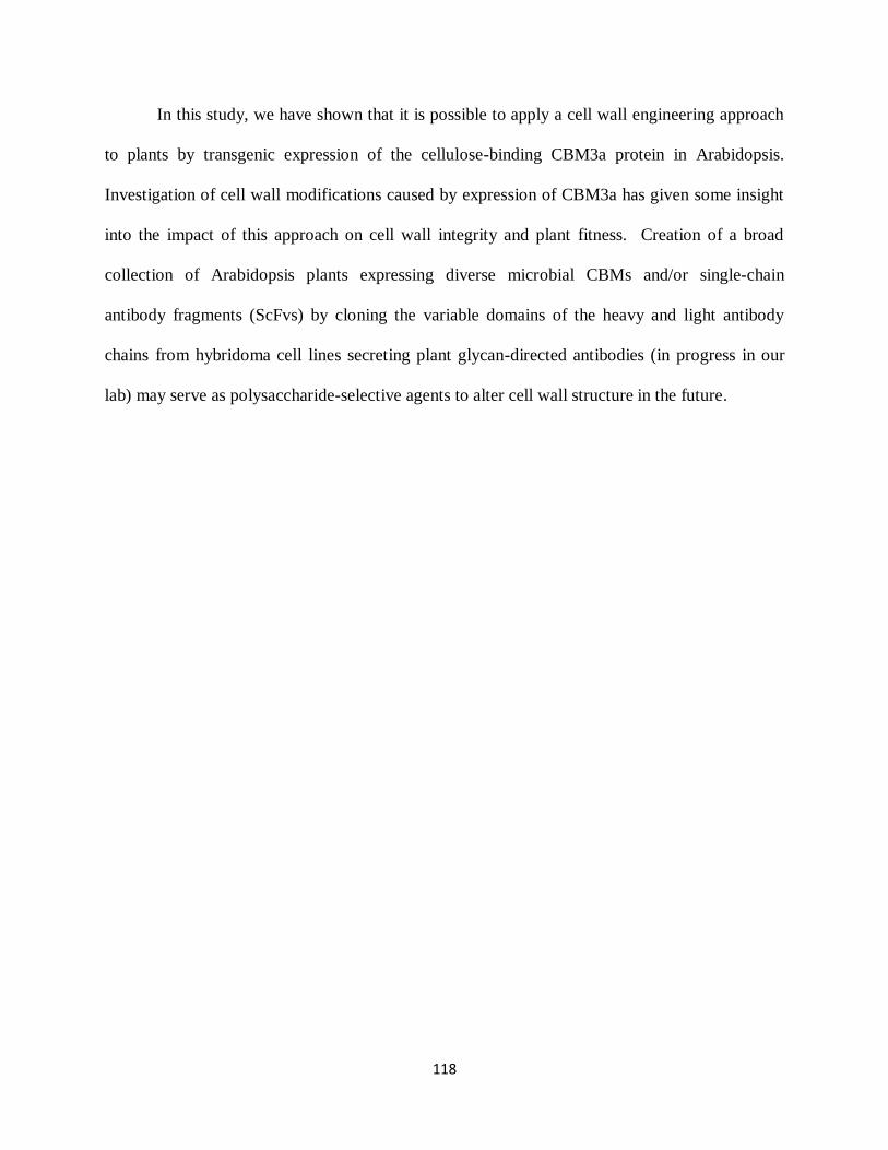
118
In this study, we have shown that it is possible to apply a cell wall engineering approach
to plants by transgenic expression of the cellulose-binding CBM3a protein in Arabidopsis.
Investigation of cell wall modifications caused by expression of CBM3a has given some insight
into the impact of this approach on cell wall integrity and plant fitness. Creation of a broad
collection of Arabidopsis plants expressing diverse microbial CBMs and/or single-chain
antibody fragments (ScFvs) by cloning the variable domains of the heavy and light antibody
chains from hybridoma cell lines secreting plant glycan-directed antibodies (in progress in our
lab) may serve as polysaccharide-selective agents to alter cell wall structure in the future.
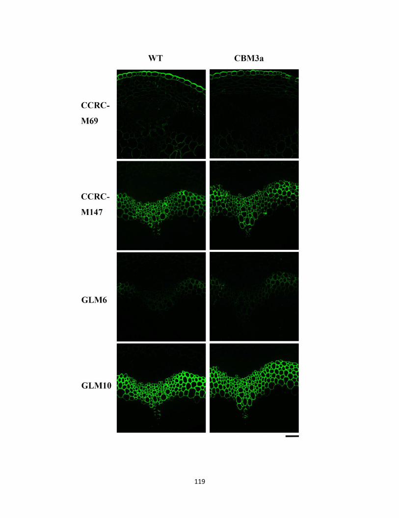
119
120
Supplemental Figure S4.1. Pectin, xylan and lignin deposition in transgenic
CBM3a:mCherry stems. Transverse sections of the basal part of 2-month-old transgenic
CBM3a:mCherry plants labeled with the pectin RG-I backbone-directed monoclonal antibody
CCRC-M69, the xylan-directed monoclonal antibody CCRC-M147, and the lignin-directed
monoclonal antibodies GLM6 and GLM10.
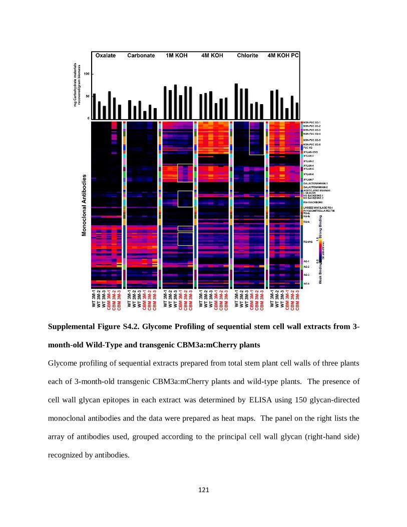
121
Supplemental Figure S4.2. Glycome Profiling of sequential stem cell wall extracts from 3-
month-old Wild-Type and transgenic CBM3a:mCherry plants
Glycome profiling of sequential extracts prepared from total stem plant cell walls of three plants
each of 3-month-old transgenic CBM3a:mCherry plants and wild-type plants. The presence of
cell wall glycan epitopes in each extract was determined by ELISA using 150 glycan-directed
monoclonal antibodies and the data were prepared as heat maps. The panel on the right lists the
array of antibodies used, grouped according to the principal cell wall glycan (right-hand side)
recognized by antibodies.
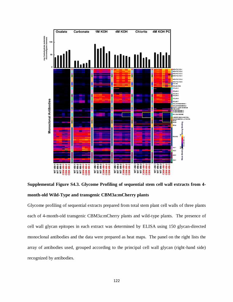
122
Supplemental Figure S4.3. Glycome Profiling of sequential stem cell wall extracts from 4-
month-old Wild-Type and transgenic CBM3a:mCherry plants
Glycome profiling of sequential extracts prepared from total stem plant cell walls of three plants
each of 4-month-old transgenic CBM3a:mCherry plants and wild-type plants. The presence of
cell wall glycan epitopes in each extract was determined by ELISA using 150 glycan-directed
monoclonal antibodies and the data were prepared as heat maps. The panel on the right lists the
array of antibodies used, grouped according to the principal cell wall glycan (right-hand side)
recognized by antibodies.
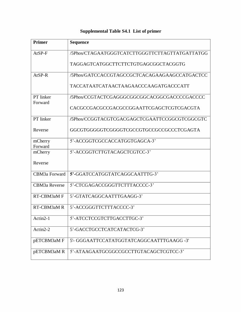
123
Supplemental Table S4.1 List of primer
Primer Sequence
AtSP-F /5Phos/CTAGAATGGGTCATCTTGGGTTCTTAGTTATGATTATGG
TAGGAGTCATGGCTTCTTCTGTGAGCGGCTACGGTG
AtSP-R /5Phos/GATCCACCGTAGCCGCTCACAGAAGAAGCCATGACTCC
TACCATAATCATAACTAAGAACCCAAGATGACCCATT
PT linker
Forward
/5Phos/CCGTACTCGAGGGCGGCGGCACGGCGACCCCGACCCC
CACGCCGACGCCGACGCCGGAATTCGAGCTCGTCGACGTA
PT linker
Reverse
/5Phos/CCGGTACGTCGACGAGCTCGAATTCCGGCGTCGGCGTC
GGCGTGGGGGTCGGGGTCGCCGTGCCGCCGCCCTCGAGTA
mCherry
Forward
5’-ACCGGTCGCCACCATGGTGAGCA-3’
mCherry
Reverse
5’-ACCGGTCTTGTACAGCTCGTCC-3’
CBM3a Forward 5’-GGATCCATGGTATCAGGCAATTTG-3’
CBM3a Reverse 5’-CTCGAGACCGGGTTCTTTACCCC-3’
RT-CBM3aM F 5’-GTATCAGGCAATTTGAAGG-3’
RT-CBM3aM R 5’-ACCGGGTTCTTTACCCC-3’
Actin2-1 5’-ATCCTCCGTCTTGACCTTGC-3’
Actin2-2 5’-GACCTGCCTCATCATACTCG-3’
pETCBM3aM F 5'- GGGAATTCCATATGGTATCAGGCAATTTGAAGG -3'
pETCBM3aM R 5’-ATAAGAATGCGGCCGCCTTGTACAGCTCGTCC-3’
124
References
Abramson M, Shoseyov O, Shani Z (2010) Plant cell wall reconstruction toward improved
lignocellulosic production and processability. Plant Science 178: 61-72
Albersheim P, Darvill A, Roberts K, Sederoff R, Staehelin A (2011) Plant Cell Walls.
Garland Science, New York
Anderson CT, Carroll A, Akhmetova L, Somerville C (2010) Real-time imaging of cellulose
reorientation during cell wall expansion in Arabidopsis roots. Plant Physiol 152: 787-796
Bashline L, Lei L, Li S, Gu Y (2014) Cell Wall, Cytoskeleton, and Cell Expansion in Higher
Plants. Molecular Plant 7: 586-600
Benziman M, Haigler CH, Brown RM, White AR, Cooper KM (1980) Cellulose biogenesis:
Polymerization and crystallization are coupled processes in Acetobacter xylinum. Proc
Natl Acad Sci U S A 77: 6678-6682
Bertani G (1951) STUDIES ON LYSOGENESIS I.: The Mode of Phage Liberation by
Lysogenic Escherichia coli. Journal of Bacteriology 62: 293-300
Blake AW, McCartney L, Flint JE, Bolam DN, Boraston AB, Gilbert HJ, Knox JP (2006)
Understanding the Biological Rationale for the Diversity of Cellulose-directed
Carbohydrate-binding Modules in Prokaryotic Enzymes. Journal of Biological Chemistry
281: 29321-29329
Blake AW, McCartney L, Flint JE, Bolam DN, Boraston AB, Gilbert HJ, Knox JP (2006)
Understanding the biological rationale for the diversity of cellulose-directed
carbohydrate-binding modules in prokaryotic enzymes. J Biol Chem 281: 29321-29329
Bootten TJ, Harris PJ, Melton LD, Newman RH (2004) Solid-state 13C-NMR spectroscopy
shows that the xyloglucans in the primary cell walls of mung bean (Vigna radiata L.)
125
occur in different domains: a new model for xyloglucan-cellulose interactions in the cell
wall. J Exp Bot 55: 571-583
Boraston AB, Bolam DN, Gilbert HJ, Davies GJ (2004) Carbohydrate-binding modules: fine-
tuning polysaccharide recognition. Biochem J 382: 769-781
Cavalier DM, Lerouxel O, Neumetzler L, Yamauchi K, Reinecke A, Freshour G, Zabotina
OA, Hahn MG, Burgert I, Pauly M, Raikhel NV, Keegstra K (2008) Disrupting Two
Arabidopsis thaliana Xylosyltransferase Genes Results in Plants Deficient in Xyloglucan,
a Major Primary Cell Wall Component. The Plant Cell Online 20: 1519-1537
Cho H-T, Cosgrove DJ (2000) Altered expression of expansin modulates leaf growth and
pedicel abscission in Arabidopsis thaliana. Proceedings of the National Academy of
Sciences 97: 9783-9788
Choi D, Lee Y, Cho H-T, Kende H (2003) Regulation of Expansin Gene Expression Affects
Growth and Development in Transgenic Rice Plants. The Plant Cell Online 15: 1386-
1398
Clough SJ, Bent AF (1998) Floral dip: a simplified method for Agrobacterium-mediated
transformation of Arabidopsis thaliana. Plant J 16: 735-743
Cosgrove DJ (2005) Growth of the plant cell wall. Nat Rev Mol Cell Biol 6: 850-861
Dick-Perez M, Zhang Y, Hayes J, Salazar A, Zabotina OA, Hong M (2011) Structure and
interactions of plant cell-wall polysaccharides by two- and three-dimensional magic-
angle-spinning solid-state NMR. Biochemistry 50: 989-1000
Filonova L, Kallas AM, Greffe L, Johansson G, Teeri TT, Daniel G (2007) Analysis of the
surfaces of wood tissues and pulp fibers using carbohydrate-binding modules specific for
crystalline cellulose and mannan. Biomacromolecules 8: 91-97
126
Foster CE, Martin TM, Pauly M (2010) Comprehensive compositional analysis of plant cell
walls (lignocellulosic biomass) part II: carbohydrates. J Vis Exp
Haigler CH, Brown RM, Jr., Benziman M (1980) Calcofluor white ST Alters the in vivo
assembly of cellulose microfibrils. Science 210: 903-906
Harris DM, Corbin K, Wang T, Gutierrez R, Bertolo AL, Petti C, Smilgies DM, Estevez
JM, Bonetta D, Urbanowicz BR, Ehrhardt DW, Somerville CR, Rose JK, Hong M,
Debolt S (2012) Cellulose microfibril crystallinity is reduced by mutating C-terminal
transmembrane region residues CESA1A903V and CESA3T942I of cellulose synthase.
Proc Natl Acad Sci U S A 109: 4098-4103
Herve C, Rogowski A, Blake AW, Marcus SE, Gilbert HJ, Knox JP (2010) Carbohydrate-
binding modules promote the enzymatic deconstruction of intact plant cell walls by
targeting and proximity effects. Proc Natl Acad Sci U S A 107: 15293-15298
Knox JP (2008) Revealing the structural and functional diversity of plant cell walls. Current
Opinion in Plant Biology 11: 308-313
Levy I, Shani Z, Shoseyov O (2002) Modification of polysaccharides and plant cell wall by
endo-1,4-beta-glucanase and cellulose-binding domains. Biomol Eng 19: 17-30
Marga F, Grandbois M, Cosgrove DJ, Baskin TI (2005) Cell wall extension results in the
coordinate separation of parallel microfibrils: evidence from scanning electron
microscopy and atomic force microscopy. Plant J 43: 181-190
Masuko T, Minami A, Iwasaki N, Majima T, Nishimura S, Lee YC (2005) Carbohydrate
analysis by a phenol-sulfuric acid method in microplate format. Anal Biochem 339: 69-
72
127
McCann MC, Knox JP (2010) Plant Cell Wall Biology: Polysaccharides in Architectural and
Developmental Contexts. In Annual Plant Reviews. Wiley-Blackwell, pp 343-366
McCartney L, Blake AW, Flint J, Bolam DN, Boraston AB, Gilbert HJ, Knox JP (2006)
Differential recognition of plant cell walls by microbial xylan-specific carbohydrate-
binding modules. Proceedings of the National Academy of Sciences of the United States
of America 103: 4765-4770
Murashima K, Kosugi A, Doi RH (2005) Site-directed mutagenesis and expression of the
soluble form of the family IIIa cellulose binding domain from the cellulosomal
scaffolding protein of Clostridium cellulovorans. J Bacteriol 187: 7146-7149
Mutwil M, Debolt S, Persson S (2008) Cellulose synthesis: a complex complex. Curr Opin
Plant Biol 11: 252-257
Nishiyama Y, Langan P, Chanzy H (2002) Crystal Structure and Hydrogen-Bonding System in
Cellulose Iβ from Synchrotron X-ray and Neutron Fiber Diffraction. Journal of the
American Chemical Society 124: 9074-9082
Obembe OO, Jacobsen E, Timmers J, Gilbert H, Blake AW, Knox JP, Visser RG, Vincken
JP (2007) Promiscuous, non-catalytic, tandem carbohydrate-binding modules modulate
the cell-wall structure and development of transgenic tobacco (Nicotiana tabacum) plants.
J Plant Res 120: 605-617
Park YB, Cosgrove DJ (2012) A Revised Architecture of Primary Cell Walls Based on
Biomechanical Changes Induced by Substrate-Specific Endoglucanases. Plant Physiology
158: 1933-1943

128
Pattathil S, Avci U, Miller J, Hahn M (2012) Immunological Approaches to Plant Cell Wall
and Biomass Characterization: Glycome Profiling. In ME Himmel, ed, Biomass
Conversion, Vol 908. Humana Press, pp 61-72
Safra-Dassa L, Shani Z, Danin A, Roiz L, Shoseyov O, Wolf S (2006) Growth modulation of
transgenic potato plants by heterologous expression of bacterial carbohydrate-binding
module. Molecular Breeding 17: 355-364
Sambrook J, Fritsch EF, Maniatis T (1989) Molecular Cloning: A Laboratory Manual, Ed 2nd.
Cold Spring Harbor Laboratory Press, Plainview, N.Y
Shaner NC, Steinbach PA, Tsien RY (2005) A guide to choosing fluorescent proteins. Nat
Methods 2: 905-909
Tomme P, Warren RA, Gilkes NR (1995) Cellulose hydrolysis by bacteria and fungi. Adv
Microb Physiol 37: 1-81
von Schantz L, Gullfot F, Scheer S, Filonova L, Cicortas Gunnarsson L, Flint JE, Daniel G,
Nordberg-Karlsson E, Brumer H, Ohlin M (2009) Affinity maturation generates
greatly improved xyloglucan-specific carbohydrate binding modules. BMC Biotechnol 9:
92
York WS, Darvill, A.G., McNeil, M., Stevenson, T.T., Albersheim, P. (1985) Isolation and
characterization of plant cell
walls and cell wall components. Methods Enzymol 118: 3-40
Zabotina OA, Avci U, Cavalier D, Pattathil S, Chou YH, Eberhard S, Danhof L, Keegstra
K, Hahn MG (2012) Mutations in multiple XXT genes of Arabidopsis reveal the
complexity of xyloglucan biosynthesis. Plant Physiol 159: 1367-1384

129
Zabotina OA, van de Ven WT, Freshour G, Drakakaki G, Cavalier D, Mouille G, Hahn
MG, Keegstra K, Raikhel NV (2008) Arabidopsis XXT5 gene encodes a putative alpha-
1,6-xylosyltransferase that is involved in xyloglucan biosynthesis. Plant J 56: 101-115
Zhu X, Pattathil S, Mazumder K, Brehm A, Hahn MG, Dinesh-Kumar SP, Joshi CP (2010)
Virus-induced gene silencing offers a functional genomics platform for studying plant
cell wall formation. Mol Plant 3: 818-833
Zykwinska A, Thibault JF, Ralet MC (2007) Organization of pectic arabinan and galactan side
chains in association with cellulose microfibrils in primary cell walls and related models
envisaged. J Exp Bot 58: 1795-1802
130
CONCLUSIONS
The complex and dynamic nature of the plant cell wall has led to the need for novel tools
and approaches to characterize wall structure, and the genes responsible for its synthesis and
modification. As imaging tools develop in scope and sophistication, efforts are now focusing on
observation of structurally and functionally diverse cell walls during plant development. In vivo
imaging of polysaccharides is severely limited, since unlike protein or nucleic acid synthesis,
where sequences are directly encoded by gene sequences, the biosynthesis of plant
polysaccharides occurs from the combined action of specific enzymes including not only the
ones involved in the biosynthesis, but also ones that function in degradation and modification
(Fry, 2010). Also, the biosynthesis of plant polysaccharides depends on the availability of the
necessary precursors and whether these precursors will confine the reactions in specific cells,
cellular locations, compartments or tissues (Stone et al., 2010). We have demonstrated that
heterologous expression of CBMs can provide a platform for tracking cell wall polysaccharides
in vivo. Our transgenic CBM2b-1-2:mCherry plant is enabling a new approach to monitor active
xylan deposition in vivo without unnecessarily disturbing plant physiology and development. In
this respect, CBM2b-1-2:mCherry constitutes a novel imaging tool for xylan detection in living
plants, and is in contrast to traditional immunolabeling of fixed plant sections, where the activity
and distribution of cell wall polysaccharides during the dynamics of plant development as cells
grow, divide, and differentiate cannot be observed because these tissues are “dead”. The in vivo
feature of transgenic CBM expression provides scientists with a substantial advantage in studies
of the detailed alterations of plant cell walls during development and during cell expansion and
cell separation processes. This year, the development of super-resolved fluorescence microscopy

131
was awarded the Nobel Prize in chemistry. Super-resolved fluorescence microscopy enables
scientists to visualize the pathways of individual molecules inside living cells. We expect our in
vivo polysaccharide tracking tool, in combination with recent microscopic advances, will provide
important new insights into the biosynthesis of cell walls, and the contribution of
polysaccharides in cellular processes.
On the other hand, we also demonstrated that heterologous expression of cellulose-
binding CBM3a modifies the cell wall in transgenic plants. Future investigations of cell wall
modifications caused by heterologously expressed specific CBMs and their impact on cell wall
structure and plant development will help us to understand the contribution of different
polysaccharides to the biology of plant cells. Currently, steady progress is being made in
characterizing polysaccharide biosynthesis genes in order to enable the design of plants biomass
with higher yield and altered cell wall composition suited for various biotechnological
applications, including biofuel production. However, there are still technical difficulties in the
genetic engineering of plant cell walls due to the complexity of polysaccharide metabolism,
which is regulated through the balance between biosynthesis, modification, and degradation.
More than 2000 genes are involved in these processes. Heterologous expression of specific
CBMs may provide an effective alternative strategy to modify biomass quality and quantity by
use of CBMs to alter cell wall structure and content in the future. Whether the heterologous
expression of fluorescent protein tagged CBMs is suitable for utilization as an in vivo tracking
tool or as a cell wall modification tool depends on the CBM itself and the polysaccharide that the
CBM binds to. Indeed, even within the same polysaccharide binding group, different CBMs
could lead to different results, since these modules can display very different specificities in cell
walls due to their different topologies (Gilbert et al., 2013).
132
References
Fry SC (2010) Cell Wall Polysaccharide Composition and Covalent Crosslinking. In Annual
Plant Reviews. Wiley-Blackwell, pp 1-42
Gilbert HJ, Knox JP, Boraston AB (2013) Advances in understanding the molecular basis of
plant cell wall polysaccharide recognition by carbohydrate-binding modules. Current
Opinion in Structural Biology 23: 669-677
Stone BA, Jacobs AK, Hrmova M, Burton RA, Fincher GB (2010) Biosynthesis of Plant Cell
Wall and Related Polysaccharides by Enzymes of the GT2 and GT48 Families. In Annual
Plant Reviews. Wiley-Blackwell, pp 109-165