Embed Size (px)
Citation preview
Rev Bras Ter Intensiva. 2010; 22(3):291-299
Usefulness of Extended-FAST (EFAST-Extended Focused Assessment with Sonography for Trauma) in critical care setting
Utilização do FAST-Estendido (EFAST-Extended Focused Assessment with Sonography for Trauma) em terapia intensiva
REVIEW ARTICLE
INTRODUCTION
The use of ultrasonography (US) in polytrauma patients, special-ly according to the FAST (Focused Assessment with Sonography for Trauma) protocol,(1-3) is not restricted to the initially stable or unstable patient’ evaluation, but is additionally a valuable tool for the follow-up. Considering that being “stable” not necessarily means being free of threats to life, but, depending on the trauma mechanics may be a potentially dangerous condition, portable devices providing early iden-tification of life threatening injuries (consequently changing its natural course), may become life-and-death deciders. A practical example could include a closed abdominal trauma patient without cardiac tamponade clinical signs, in who early US identifies signs of pericardial effusion
Uri Adrian Prync Flato1, Hélio Penna Guimarães2-4, Renato Delascio Lopes3-4, Jorge Luís Valiatti5, Elias Marcos Silva Flato6, Ricardo Gonçalves Lorenzo7
1. Physician of the Intensive Care Unit of Instituto Dante Pazzanese de Cardiologia – São Paulo (SP), Brazil. 2. Coordinator for the Training Center of the Hospital do Coração - CETES-HCor – São Paulo (SP), Brazil. 3. Adjunct Professor for the Division of Cardiology of the Department of Internal Medicine of the Duke Clinical Research Institute, Duke Medical Center, Duke University, Durham - USA.4. Physician of the Discipline of Internal Medicine of the Universidade Federal de São Paulo – UNIFESP – São Paulo (SP), Brazil.5. Titular Professor for the Discipline of Intensive Care Medicine of the Faculdade de Medicina de Catanduva – Catanduva (SP), Brazil.6. Internal Medicine Resident Physician of the Santa Casa de São Paulo – SCMSP - São Paulo (SP), Brazil. 7. Physician of the Center Diagnostic Imaging of Hospital Alemão Oswaldo Cruz – São Paulo (SP), Brazil.
ABSTRACT
Trauma is the leading cause of death in people below 45 years-old in Brazil, and responsible for one third of all intensive care unit admis-sions. The increasing knowledge on ultrasound diagnosis methods and its availability for life-threatening inju-ries (such as cardiac tamponade and abdominal cavity solid organs rup-ture leading to hemorrhagic shock) diagnosis and monitoring, lead to the development o the FAST (Focused Assessment with Sonography for Trauma) protocol, aimed to be used both in the emergency and intensive care unit settings. Due to its repro-ducibility, lack of radiation exposure, and bedside feasibility, this technol-ogy is being increasingly accepted. A new protocol extension, the Extend-
ed-FAST, provides valuable informa-tion for improved patients’ manage-ment, extending its availability from the abdominal conditions to other diagnosis such as hemothorax, pleu-ral effusion and pneumothorax. We must underline that this technique is able to replace computed tomography and diagnostic peritoneal wash, and do not delay surgical procedure in-stead of perform this exam . Thus, its careful appraisal in connection with the clinical information should guide the therapeutic approaches, specially in inhospitable sites such as intensive care units in war zones, rural or dis-tant places, were other imagery meth-ods are not available.
Keywords: Ultrasonography/utilization; Trauma; Intensive care/trends; Point-of-care systems
Received from the Intensive Care Unit of the Instituto Dante Pazzanese de Cardiologia - São Paulo (SP), Brazil.
Submitted on March 20, 2010Accepted on August 13, 2010
Author for correspondence:Uri Adrian Prync FlatoInstituto Dante Pazzanese de Cardiologia Av. Dr. Dante Pazzanese, 500- 3rd floorZip Code: 04012-180- São Paulo (SP), Brazil.Phone/Fax: +55 (11) 5081-4531Email: [email protected]
292 Flato UAP, Guimarães HP, Lopes RD, Valiatti JL, Flato EMS, Lorenzo RG
Rev Bras Ter Intensiva. 2010; 22(3):291-299
with myocardial restriction – which would radically change the approach to an immediate intervention. A FAST protocol extension, the Extended-FAST, was developed aimed to extend the evaluation, previously restricted to heart and abdominal wall evaluations, to the chest cavity, allowing pneumothorax, hemo-thorax and diaphragm rupture diagnosis. The Chart 1 describes the main EFAST(4) indications.
Chart 1- EFAST IndicationsEFAST IndicationsPenetrating heart trauma Closed heart traumaClosed abdominal traumaChest traumaPneumothoraxHemothorax Undefined cause hypotension
“Stable” should mean “stay alert and keep mon-itoring the patient”; on the other hand, instabil-ity should be understood as a clinical condition where immediate measures are required to prevent a catastrophic outcomes. Peitzman et al. evaluated the closed trauma shock etiology, and described as primary cause (decreasing frequency) hypovolemia (59%), head trauma (16%), obstruction (pneu-mothorax, cardiac tamponade) (13%), neurogenic shock (7%) and others (7%).(5) The bleeding site identification and its control is vital, as resuscita-tion measures, such as crystalloid infusions, may have either transitory or no response at all if the bleeding site is left untreated. In case of a vascu-lar injury, the definitive treatment is vital, as is the Damage Control Therapy in selected cases.
During the last decade, several international so-cieties have strongly recommended the FAST proto-col (Grade I).(6-10) It is suitable for closed abdominal trauma, closed or penetrating chest trauma, either in stable or unstable patients. As it is an operator-dependent test, the apprenticeship curve, which is correlated with the results, is required. The litera-ture describes variable outcomes according to dif-ferent operators (radiologist versus emergencist), associated parenchyma injuries (which the method was not planned for) and hemodynamic status (ar-terial hypotension versus normotension).(11-14)
A positive FAST test points to in-cavity bleed-ing and likely requires exploratory laparotomy if the patient is hemodynamically unstable, or con-
tinued diagnosis with computed tomography (CT), if available, in clinically stable patients (Figure 1).
Scalea TM, Rodriguez A, Chiu WC, et al. Focused assessment with so-nography for trauma (FAST): results from an international consensus conference. J Trauma 1999;46(3):466–72.CT – computed tomography; FAST - Focused Assessment with Sonography for Trauma.
Figure 1 - FAST consensus.
A FAST peculiarity is its feasibility even in restrict-ed complementary methods sites, as seen in natural catastrophes (e.g. earthquakes), war conflicts, spatial stations, where additional data may be decisive and no other complementary methods are available.(15-17)
Isolated free fluid in the cavity itself does not mean immediate surgery requirement, as it should be associated with other aspects such as the amount of cavity free fluid, number of sites (recesses, gut-ters) with fluid and the patient’s clinical status. Some authors have proposed FAST positive patients scoring protocols, aiming risk stratification (either low or high) and surgical indication, e.g. free flu-id collection observed in more than three recesses was correlated with a more than 1,000 mL intra-peritonial volume.(18,19) Identification of high risk patients (closed abdominal trauma and arterial hy-pertension) evidenced the test accuracy to be about 95%, the sensitivity 85% and specificity 96%, ac-cording to Lee et al., for exploratory laparotomy and consequent therapeutic intervention.(20)
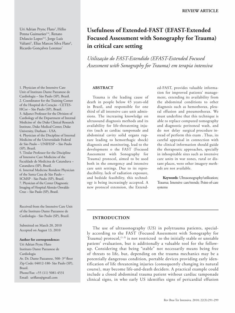
With the EFAST(21) development and use (Figure 2) the chest cavity may be evaluated just sliding the transducer cranially after the Morrison (hepatore-nal) and splenorenal spaces evaluations. The EFAST sequence indicates initially the abdomen evalu-ation, and then the chest evaluation as described in the secondary Advanced Trauma Life Support evaluation (feet to head evaluation), i.e., following the primary evaluation. However, the WINFOCUS
Usefulness of Extended-FAST (EFAST-Extended Focused Assessment with Sonography for Trauma) in critical care setting 293
Rev Bras Ter Intensiva. 2010; 22(3):291-299
(World Interactive Network Focused on Critical Ultrasound) is currently developing the use of ul-trasound for initial polytrauma patient’s evaluation, starting from the primary evaluation until resolu-tion or surgical procedure. This protocol will be included in the Ultrasound Trauma Life Support (US-ATLS) algorithm.(22) Based on this sequence, the ABC approach to trauma is suggested, starting by the airways (confirmation of patent airways and support to surgical airway), breathing (pneumotho-rax and hemothorax) and circulation, by bleedings as hemoperitoneum investigation.
FAST - Focused Assessment with Sonography for Trauma; EFAST – Extended Focused Assessment with Sonography for Trauma.
Figure 2- FAST (A) and EFAST (B) anatomical refe-rences.
Pulmonary ultrasound The correct chest anatomical references as dia-
phragm, pulmonary parenchyma, costal arcs and US artifacts identification is vital for appropriate reading. Changes as pleural effusion, hemothorax, lack of pleural sliding (pneumothorax, selective in-tubation), pulmonary complications related to the intubated patient transportation (as tube displace-ment, among others) may be diagnosed. The rele-vance of this method parallels with the frequency of chest changes found in major trauma patients. One pneumothorax was evidenced out of every 5 major traumas, which, if not identified, could lead to se-rious hemodynamic changes and death. Blaivas et
al.(23) evaluated the chest X-ray (anterior-posterior incidence) versus pulmonary US accuracy for oc-cult pneumothorax identification (normal chest X-ray reading and pneumothorax evidenced on CT or US) in polytrauma patients, and found this last to have an approximately 94% accuracy versus X-ray. Occult pneumothorax alone may not directly deter-mine the patient’s deterioration, however when as-sociated with secondary injuries such as pulmonary contusion, hypothermia, hypoxia, and positive pres-sure ventilation, may lead to serious consequences. In observational studies occult pneumothorax in trauma patients was described by chest CT in about 55%.(24,25) Its quick bedside identification possibili-ties to be done in hospital settings /or in battle-field scenarios, not implying risks for the patient or other devices unavailability can optimize the patient care, and thus reduce mortality. This tool is aimed to support the treating physician’s deci-sion making process, and to support the monitoring of pharmacological and surgical interventions. The medical experience with EFAST US is related its correct use as well as technical limitations and read-ing mistakes.(26-28)
Appropriate awareness of human anatomy and its correlation with this method’s bidimensional images are required, and two different orthogo-nal approaches are recommended. As this proto-col is aimed to identify free fluids within cavities and pneumothorax, rather than complex organs evaluations, its apprenticeship curve is short and relatively easy. Free abdominal fluid detection is made, according to the magnitude, first in the up-per right quadrant(29) (hepatorenal recess), which when systematically conducted takes just 19 sec-onds. A full negative FAST examination takes about 3 minutes. Again, it should be emphasized that a negative result does not preclude life threat-ening injuries such as retroperitonial bleedings and hollow viscerae injuries, which are not included in the method.
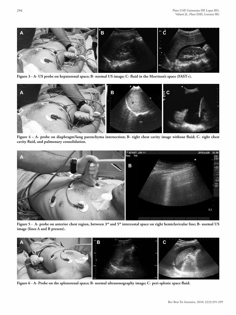
EFAST ultrasound references are:- Hepatorenal recess (Figure 3)- Right anterior axillary line (liver-lung (dia-
phragm) transition) Figure 4)- Right anterior hemiclavicular line, between
the 3rd and 5th intercostal space (Figure 5)- Splenorenal recess (Figure 6)- Left anterior axillary line, spleen-lung transi-
tion (Figure 7)
294 Flato UAP, Guimarães HP, Lopes RD, Valiatti JL, Flato EMS, Lorenzo RG
Rev Bras Ter Intensiva. 2010; 22(3):291-299
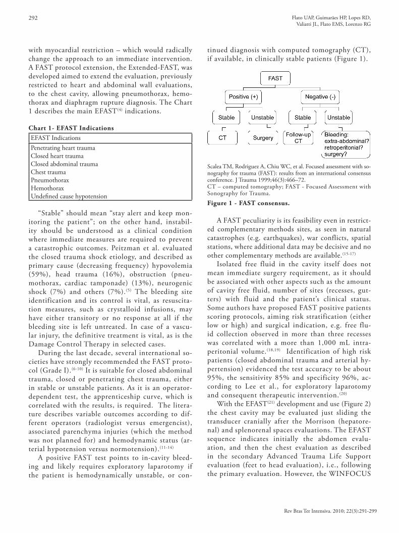
Figure 6 - A- Probe on the splenorenal space; B- normal ultrasonography image; C- peri-splenic space fluid.
Figure 5 - A- probe on anterior chest region, between 3rd and 5th intercostal space on right hemiclavicular line; B- normal US image (lines A and B present).
Figure 4 – A- probe on diaphragm/lung parenchyma intersection; B- right chest cavity image without fluid; C- right chest cavity fluid, and pulmonary consolidation.
Figure 3 - A- US probe on hepatorenal space; B- normal US image; C- fluid in the Morrison’s space (FAST+).
A
A
A
A
B
B
B
B
C
C
C
Usefulness of Extended-FAST (EFAST-Extended Focused Assessment with Sonography for Trauma) in critical care setting 295
Rev Bras Ter Intensiva. 2010; 22(3):291-299
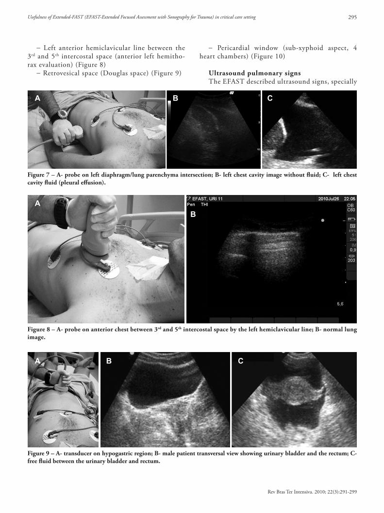
- Left anterior hemiclavicular line between the 3rd and 5th intercostal space (anterior left hemitho-rax evaluation) (Figure 8)
- Retrovesical space (Douglas space) (Figure 9)
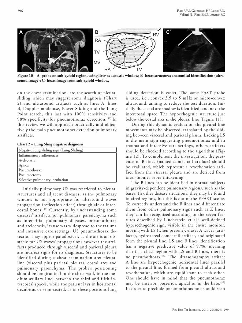
- Pericardial window (sub-xyphoid aspect, 4 heart chambers) (Figure 10)
Ultrasound pulmonary signsThe EFAST described ultrasound signs, specially
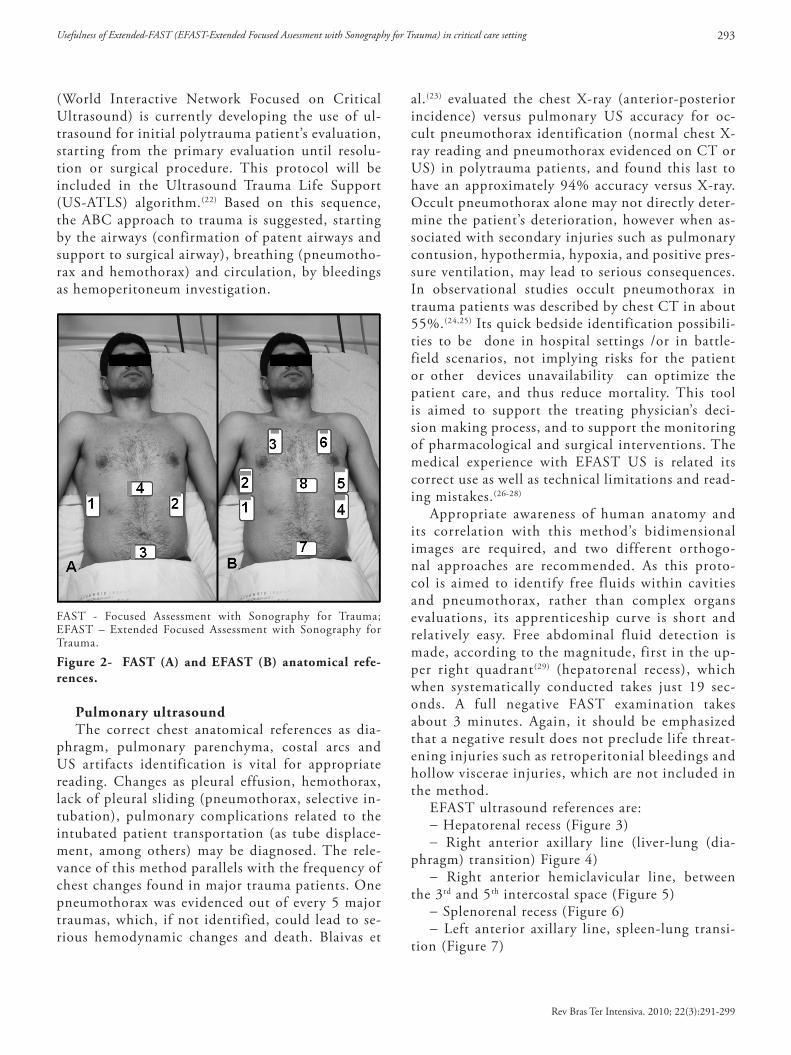
Figure 9 – A- transducer on hypogastric region; B- male patient transversal view showing urinary bladder and the rectum; C- free fluid between the urinary bladder and rectum.
Figure 8 – A- probe on anterior chest between 3rd and 5th intercostal space by the left hemiclavicular line; B- normal lung image.
Figure 7 – A- probe on left diaphragm/lung parenchyma intersection; B- left chest cavity image without fluid; C- left chest cavity fluid (pleural effusion).
A
A
A
B
B
B
C
C
296 Flato UAP, Guimarães HP, Lopes RD, Valiatti JL, Flato EMS, Lorenzo RG
Rev Bras Ter Intensiva. 2010; 22(3):291-299
on the chest examination, are the search of pleural sliding which may suggest some diagnosis (Chart 2) and ultrasound artifacts such as lines A, lines B, Doppler mode use, Power Sliding and the Lung Point search, this last with 100% sensitivity and 98% specificity for pneumothorax detection.(30) In this review we will approach practically and objec-tively the main pneumothorax detection pulmonary artifacts.
Chart 2 – Lung Sling negative diagnosis Negative lung sliding sign (Lung Sliding)Inflammatory adherences AtelectasisApneaPneumothoraxPneumectomySelective pulmonary intubation
Initially pulmonary US was restricted to pleural structures and adjacent diseases, as the pulmonary window is not appropriate for ultrasound waves propagation (reflection effect) through air or inter-costal bones.(31) Currently, by understanding some diseases’ artifacts on pulmonary parenchyma such as interstitial pulmonary diseases, pneumothorax and atelectasis, its use was widespread to the trauma and intensive care settings. US pneumothorax de-tection may appear paradoxical, as the air is an ob-stacle for US waves’ propagation; however the arti-facts produced through visceral and parietal pleura are indirect signs for its diagnosis. Structures to be identified during a chest examination are: pleural line (visceral plus parietal pleura), costal arcs and pulmonary parenchyma. The probe’s positioning should be longitudinal to the chest wall, in the me-dium axillary line, between the third and fifth in-tercostal spaces, while the patient lays in horizontal decubitus or semi-seated, as in these positions lung
Figure 10 – A- probe on sub-xyfoid region, using liver as acoustic window; B- heart structures anatomical identification (ultra-sound image); C- heart image from sub-xyfoid window.
sliding detection is easier. The same FAST probe is used, i.e., convex 3.5 to 5 mHz or micro-convex ultrasound, aiming to reduce the test duration. Ini-tially the costal arc shadow is identified, and next the intercostal space. The hypoechogenic structure just below the costal arcs is the pleural line (Figure 11).
During this dynamic evaluation the pleural line movements may be observed, translated by the slid-ing between visceral and parietal pleura. Lacking LS is the main sign suggesting pneumothorax and in trauma and intensive care settings, others artifacts should be checked according to the algorithm (Fig-ure 12). To complement the investigation, the pres-ence of B lines (named comet tail artifact) should be evaluated, which represent a reverberation arti-fact from the visceral pleura and are derived from inter-lobules septa thickening.
The B lines can be identified in normal subjects in gravity-dependent pulmonary regions, such as the bases. In other disease situations, they may be found in aired regions, but this is out of the EFAST scope. To correctly understand the B lines and differentiate them from other pulmonary signs such as Z lines, they can be recognized according to the seven fea-tures described by Linchestein et al.: well-defined hyperechogenic sign, visible in the entire monitor, moving with LS (when present), erases A waves (arti-facts), hydroaereal comet tail artifact, and originated form the pleural line. LS and B lines identification has a negative predictive value of 97%, meaning that in a chest region with LS and B lines, there is no pneumothorax.(32) The ultrasonography artifact A line are hypoechogenic horizontal lines parallel to the pleural line, formed from pleural ultrasound reverberation, which are equidistant to each other. One should have in mind that the pneumothorax may be anterior, posterior, apical or in the base.(32) In order to preclude pneumothorax one should scan
A B CRA
TV
MVRA LV
RV

Usefulness of Extended-FAST (EFAST-Extended Focused Assessment with Sonography for Trauma) in critical care setting 297
Rev Bras Ter Intensiva. 2010; 22(3):291-299
both lungs in the order anterior-lateral-posterior. In the EFAST sequence, the so called lung point (LP) ultrasound sign (Figure 12) can be searched, with a 98% sensitivity and 100% specificity for pneumo-thorax identification, as well to determine its volume and extension. LP is the dynamic identification of a chest cavity point, generally during the inspiration, where the LS transition is observed, along with B lines, A lines, identifying the probable pneumotho-rax. A practical way to perform this maneuver is to start from the posterior region, followed by anterior and lateral. The more lateral the LP is observed, the larger is the pneumothorax, as well as pneumothorax extending to the posterior region may show no LP. It should be underlined that in some situations LS may be absent, with present A lines and no LP, as in atelectasis and close to liver or spleen areas simulat-ing LP in the absence of pneumothorax. These eight
segments ultrasound evaluation may bring relevant data for our patients’ management. With the above described method, it is possible to extrapolate the EFAST for pulmonary evaluation,(33-37) rendering possible the evaluation of central venous lines inser-tion-related complications, such as pneumothorax.
CONCLUSION
The ultrasonography use in the emergency and in-tensive care settings provides very relevant information which should be interpreted along with the patient’s clinical data. Currently full-body CT (foot to head scan) is the preferential method in trauma centers and inten-sive care units, associated to its indiscriminate and liberal use. CT-related complications are increased risk of can-cer and neprotoxicity, widely described in the literature, and in case of extreme age patients, these frets should be kept in mind. New portable and self-sustaining technol-ogies, such as US devices, are able to use solar energy and to be transmitted in real time to experts (telemedicine). Another advantage would involve its use in inhospitable places and even in intensive care units, where patients transportation is usually not advisable and the diagnosis agility is a plus. Its cost-effectiveness is very relevant, as some high cost tests used for injuries diagnosis add over-head costs to very limited resources regions. The training and certification of professionals involved in critically ill patients’ care in Brazil is likely to be seen in a near future, as this tool is currently fully consolidated among Euro-pean and North American emergency doctors for some years. Thus, the EFAST protocol may be an alternative
Lung sliding - Lung sliding +
Figure 12 - Lung point (LP) ultrasound sign.
PNTX = pneumothorax.Figure 11 - A- structures identification; B- pulmonary artifacts algorithm for pneumothorax detection.
A B
298 Flato UAP, Guimarães HP, Lopes RD, Valiatti JL, Flato EMS, Lorenzo RG
Rev Bras Ter Intensiva. 2010; 22(3):291-299
for these critical patients’ bedside management, keeping in mind that the physical examination and medical rea-soning should be connected with complementary meth-ods, but never replaced.
RESUMO
A principal causa de morte no Brasil, em pacientes com idade inferior a 45 anos, está relacionada ao trauma, sendo responsável por um terço das internações em unidades de terapia intensiva. Em virtude do crescente conhecimento e disponibilidade da ultrassonografia para o diagnóstico e mo-nitoramento de lesões ameaçadoras à vida, como tampona-mento cardíaco e ruptura de órgão sólido na cavidade abdo-minal com choque hemorrágico, foi desenvolvido um proto-colo denominado FAST (Focused Assesment with Sonography for Trauma) no ambiente de emergência e terapia intensiva. Esta tecnologia está ganhando adeptos por sua reprodutibili-dade, ausência de exposição à radiação ao paciente e facilida-
de beira leito. Uma nova complementação a este protocolo, denominada FAST-Estendido, proporciona informações va-liosas na condução desses pacientes, ampliando o diagnóstico de doenças antes reservadas à cavidade abdominal e pericár-dica, conjuntamente com doenças localizadas na cavidade torácica, em busca de hemotórax, derrame pleural e pneu-motórax. Devemos salientar que esta modalidade de exame complementar substitui a tomografia computadorizada e o lavado peritoneal diagnóstico, mas não o retardo de inter-venções cirúrgicas. Sua avaliação criteriosa, conjuntamente com dados clínicos, deve nortear as condutas terapêuticas, principalmente em locais inóspitos e/ou com limitações de recursos, como pré-hospitalar, unidades de terapia intensiva em zonas de conflito armado, áreas rurais e/ou geografica-mente distantes, nas quais não há disponibilidade de outros métodos de imagem.
Descritores: Ultrassonografia/utilização; Trauma; Cuidados intensivos/tendências; Sistemas automatizados de assistência junto ao leito
REFERENCES
1. Centers for Disease Control and Prevention - CDC. National estimates of the ten leading causes of nonfatal injuries. Atlanta (GA): Centers for Disease Control and Prevention; 2004.
2. Ten leading causes of death. Atlanta (GA): Centers for Disease Control and Prevention; 2003.
3. Kirkpatrick AW, Sirois M, Laupland KB, Liu D, Rowan K, Ball CG, et al. Hand-held thoracic sonography for detecting post-traumatic pneumothoraces: The Extended Focused Assessment with Sonography for Trauma (EFAST). J Trauma. 2004;57(2):288-95.
4. Lichtenstein DA. Pneumothorax and introduction to ultrasound signs in the lung. In: Lichtenstein DA, Pinsky MR, Jardin F. . General ultrasound in the critically ill. Berlin: Springer; 2002. p. 105-15.
5. Peitzman AB, Billiar TR, Harbrecht BG, Kelly E, Udekwu AO, Simmons RL. Hemorrhagic shock. Curr Probl Surg. 1995;32(11):925-1002. Review.
6. Scalea TM, Rodriguez A, Chiu WC, Brenneman FD, Fallon WF Jr, Kato K, et al. Focused Assessment with Sonography for Trauma (FAST): results from an international consensus conference. J Trauma. 1999;46(3):466-72.
7. Kirkpatrick AW, Sustic A, Blaivas M. Introduction to the use of ultrasound in critical care medicine. Crit Care Med. 2007;35:S123-5.
8. Wherrett LJ, Boulanger BR, McLellan BA, Brenneman FD, Rizoli SB, Culhane J, Hamilton P. Hypotension after blunt abdominal trauma: the role of emergent abdominal
sonography in surgical triage. J Trauma.1996;41(5):815-20. 9. Rozycki GS, Ochsner MG, Schmidt JA, Frankel HL,
Davis TP, Wang D, Champion HR. A prospective study of surgeon-performed ultrasound as the primary adjuvant modality for injured patient assessment. J Trauma. 1995;39(3):492-8; discussion 498-500.
10. Hoff WS, Holevar M, Nagy KK, Patterson L, Young JS, Arrillaga A, Najarian MP, Valenziano CP; Eastern Asociation for the Surgery of Trauma. Practice management guidelines for the evaluation of blunt abdominal trauma: the EAST practice management guidelines work group. J Trauma. 2002;53(3):602-15.
11. McElveen TS, Collin GR. The role of ultrasonography in blunt abdominal trauma: a prospective study. Am Surg. 1997;63(2):184-8.
12. Bode PJ, Edwards MJ, Kruit MC, van Vugt AB. Sonography in a clinical algorithm for early evaluation of 1671 patients with blunt abdominal trauma. AJR Am J Roentgenol. 1999;172(4):905-11.
13. Thomas B, Falcone RE, Vasquez D, Santanello S, Townsend M, Hockenberry S, et al. Ultrasound evaluation of blunt abdominal trauma: program implementation, initial experience, and learning curve. J Trauma. 1997;42(3):380-8; discussion 388-90.
14. Gracias VH, Frankel HL, Gupta R,Malcynski J, Gandhi R, Collazzo L, et al. Defining the learning curve for the Focused Abdominal Sonogram for Trauma (FAST) examination: implications for credentialing. Am Surg. 2001;67(4):364-8.
15. Sarkisian AE, Khondkarian RA, Amirbekian NM,
Usefulness of Extended-FAST (EFAST-Extended Focused Assessment with Sonography for Trauma) in critical care setting 299
Rev Bras Ter Intensiva. 2010; 22(3):291-299
Bagdasarian NB, Khojayan RL, Oganesian YT. Sonographic screening of mass casualties for abdominal and renal injuries following the 1988 Armenian earthquake. J Trauma. 1991;31(2):247-50.
16. Beck-Razi N, Fischer D, Michaelson M, Engel A, Gaitini D. The utility of focused assessment with sonography for trauma as a triage tool in multiple-casualty incidents during the second Lebanon war. J Ultrasound Med. 2007;26(9):1149-56.
17. Sargsyan EA, Hamilton DR, Jones JA, Melton S, Whitson PA, Kirkpatrick AW, et al. FAST at MACH 20: clinical ultrasound aboard the International Space Station. J Trauma. 2005;58(1):35-9.
18. Farahmand N, Sirlin CB, Brown MA, Shragg GP, Fortlage D, Hoyt DB, Casola G. Hypotensive patients with blunt abdominal trauma: performance of screening US. Radiology. 2005;235(2):436-43.
19. McKenney KL, McKenney MG, Cohn SM, Compton R, Nunez DB, Dolich M, Namias N. Hemoperitoneum score helps determine need for therapeutic laparotomy. J Trauma. 2001;50(4):650-4; discussion 654-6.
20. Lee BC, Ormsby EL, McGahan JP, Melendres GM, Richards JR. The utility of sonography for the triage of blunt abdominal trauma patients to exploratory laparotomy. AJR Am J Roentgenol. 2007;188(2):415-21.
21. Kirkpatrick AW. Clinician-performed focused sonography for the resuscitation of trauma. Crit Care Med. 2007;35(5 Suppl):S162-72. Review.
22. World Interactive Network Focused on Critical Ultrasound. (WINFOCUS). Ultrasound in critical care medicine - Continuing medical education. [cited 2010 Jul 1]. Available from: http://www.winfocus.org/usccm .
23. Blaivas M, Lyon M, Duggal S. A prospective comparison of supine chest radiography and bedside ultrasound for the diagnosis of traumatic pneumothorax. Acad Emerg Med. 2005;12(9):844-9.
24. Ball CG, Kirkpatrick AW, Laupland KB, Fox DI, Nicolaou S, Anderson IB, et al. Incidence, risk factors, and outcomes for occult pneumothoraces in victims of major trauma. J Trauma. 2005;59(4):917-24; discussion 924-5.
25. Ball CG, Kirkpatrick AW, Laupland KB, Fox DL, Litvinchuk S, Dyer DM, et al. Factors related to the failure of radiographic recognition of occult posttraumatic pneumothoraces. Am J Surg. 2005;189(5):541-6; discussion 546.
26. Rozycki GS, Shackford SR. Ultrasound, what every trauma
surgeon should know. J Trauma. 1996;40(1):1-4. 27. Reardon R, Moscati R Beyond the FAST Exam: additional
applications of sonography in trauma. In: Jehle D, Heller M, editors. Ultrasonography in trauma: the FAST exam. Dallas,TX: American College of Emergency Physicians; 2003. p. 107-26.
28. Moscati R, Reardon R. Clinical application of the FAST exam. In: Jehle D, Heller M, editors. Ultrasonography in trauma: the FAST exam. Dallas,TX: American College of Emergency Physicians; 2003. p. 39-60.
29. Rozycki GS, Ochsner MG, Feliciano DV, Thomas B, Boulanger BR, Davis FE, et al. Early detection of hemoperitoneum by ultrasound examination of the right upper quadrant: a multicenter study. J Trauma. 1998;45(5):878-83.
30. Lichtenstein DA, Mezière G, Lascols N, Biderman P, Courret JP, Gepner A, et al. Ultrasound diagnosis of occult pneumothorax. Crit Care Med. 2005;33(6):1231-8.
31. Weinberger SE, Drazen JM. Diagnostic procedures in respiratory diseases. In: Kasper D, editor. Harrison’s principles of internal medicine. 16th ed. New York: McGraw-Hill; 2005. p. 1505-8.
32. Melniker LA, Leibner E, McKenney MG, Lopez P, Briggs WM, Mancuso CA. Randomized controlled trial of point-of-care, limited ultrasonography for trauma in the emergency department: the first sonography outcomes assessment program trial. Ann Emerg Med. 2006;48(3):227-35.
33. Noble VE, Nelson B, Sutingco AN. Manual of emergency and critical care ultrasound. New York: Cambridge University Press; 2007.
34. Jones R. Recognition of pneumoperitoneum using bedside ultrasound in ctitically ill patients presenting with acute abdominal pain. Am J Emerg Med. 2007;25(7):838-41.
35. Lichtenstein DA, Mezière GA. Revelance of lung ultrasound in the diagnosis of acute respiratory failure: the BLUE protocol. Chest. 2008;134(1):117-25. 2010.
36. Nicolaou S, Talsky A, Khashoggi K, Venu V. Ultrasound-guided intervencional radiology in critical care. Crit Care Med. 2007;35(5 Suppl): S186-97. Review.
37. Dyer D, Cusden J, Turner C, Boyd J, Hall R, Lautner D, et al. The clinical and technical evaluation of a remote telementored telesonography system during the acute resuscitation and transfer of the injured patient. J Trauma. 2008;65(6):1209-16.