Embed Size (px)
Citation preview

8/10/2019 Use of ultrasound Doppler to determine tooth vitality in a discolored tooth
http://slidepdf.com/reader/full/use-of-ultrasound-doppler-to-determine-tooth-vitality-in-a-discolored-tooth 1/32
Use of ultrasound Doppler to determine tooth
vitality in a discolored tooth after traumatic
injury: its prospects and limitationsYong-Wook Cho and Sung-Ho Park Author information ► Article notes ► Copyright and License information ► Go to:
Abstract
When a tooth shows discoloration and does not respond to the cold test or electric pulp test(EPT) after a traumatic injury, its diagnosis can be even more difficult due to the lack of properdiagnostic methods to evaluate its vitality. In these case reports, we hope to demonstrate that
ultrasound Doppler might be successfully used to evaluate the vitality of the tooth after trauma,and help reduce unnecessary endodontic treatments. In all three of the present cases, the teethwere discolored after traumatic injuries and showed negative responses to the cold test and EPT.However, they showed distinctive vital reactions in the ultrasound Doppler test during the wholeobservation period. In the first case, the tooth color returned to normal, and the tooth showed a positive response to the cold test and EPT at 10 wk after the injury. In the second case, the toothcolor had returned to its normal shade at 10 wk after the traumatic injury but remainedinsensitive to the cold test and EPT. In the third case, the discoloration was successfully treatedwith vital tooth bleaching.
Keywords: Tooth discoloration, Tooth vitality, Traumatic injury, Ultrasound Doppler
Go to:
Introduction
Tooth vitality is determined using the cold test, electric pulp test (EPT), radiographicexamination, or clinical signs such as tooth discoloration. However, tooth vitality could be more properly evaluated by the blood supply in the pulp rather than these other tests, such as the coldtest and EPT, which actually evaluate the sensitivity of the nerves.1 When the tooth experience atraumatic injury, the evaluation of tooth vitality is difficult because they occasionally do notrespond to the cold test or EPT due to the reduced conduction ability of the sensory nerves ornerve endings.2 This lack of response seems to be caused by the damage, inflammation,compression or tension state of the apical nerve fibers, which require approximately eight weeksor more to return to normal functioning.3
Tooth discoloration may follow a traumatic injury.4,5 When the tooth shows discoloration andalso does not respond to the cold test or EPT after a traumatic injury, its diagnosis can be evenmore difficult due to the lack of proper diagnostic methods to evaluate its vitality. The discoloredtooth may return to its original shade and translucency completely or incompletely when the

8/10/2019 Use of ultrasound Doppler to determine tooth vitality in a discolored tooth
http://slidepdf.com/reader/full/use-of-ultrasound-doppler-to-determine-tooth-vitality-in-a-discolored-tooth 2/32
tooth vitality is preserved.4,5 Malgren and Hubel reported that the discoloration disappearedwithin 4 weeks to 6 months in eight out of nine permanent teeth that had been root fractured andshowed tooth discoloration after the trauma.6 They reported that all of the teeth had regainedtheir normal sensibility when the discoloration disappeared. Transient color changes were alsodescribed in connection with transient apical breakdown (TAB) after luxation injuries in
permanent teeth.7,8 The discoloration and loss of electrometric sensibility returned to normalwhen there was radiographic evidence of the resolution of the TAB. However, this resolutionusually takes a long time to be confirmed.
Ultrasound Doppler imaging has been used in many medical fields as a non-invasive andradiation-free technique to assess the blood flow in micro-vascular systems. Ultrasound has alsorecently been applied to dentistry. Some studies have shown that ultrasound Doppler imaging provides sufficient information on micro-vascularity for dental treatment.9-11
Recently, Yoon et al. reported that ultrasound Doppler could be effectively used to evaluate the pulp blood flow in the pulp spaces.1,12 They reported that it can measure the reduced blood
stream speed after a local anesthetic injection containing 1 : 80,000 epinephrine. They alsoindicated the possibility that this Doppler system could be used effectively in the diagnosis oftraumatic injury.12
In this paper, three cases are presented that were seen in the Department of ConservativeDentistry, Yonsei University Dental Hospital, Seoul, Korea, during the past two years. In the beginning, all three teeth were discolored after a traumatic injury and showed negative responsesto the thermal test and EPT but also showed a distinctive vital reaction in the ultrasound Dopplertest unit (MM-D-K, Minimax, Moscow, Russia). In the first and second cases, the toothdiscolorations returned to normal at 10 weeks after the injuries. In the third case, the toothdiscoloration was successfully treated by vital bleaching. In this case series, we hope to
demonstrate that ultrasound Doppler might be successfully used to evaluate the vitality of teethafter trauma and help reduce unnecessary endodontic treatments.
Go to:
Case reports
Case 1
A 47-year-old female patient visited our department due to traumatic injury to her upper rightlateral incisor (tooth #12). She sustained the injury 3 days before she visited our clinic by a fist
blow injury to her face. Tooth #12 was subluxated, and showed a positive response to a percussion test. It did not show any response to a cold test or EPT. The tooth was diagnosed withsubluxation, and we decided to wait and observe its course. There was no discomfort during the2 weeks after the injury, but there was no response to the thermal test or EPT, and a reddishdiscoloration was observed (Figure 1a).
8/10/2019 Use of ultrasound Doppler to determine tooth vitality in a discolored tooth
http://slidepdf.com/reader/full/use-of-ultrasound-doppler-to-determine-tooth-vitality-in-a-discolored-tooth 3/32
Figure 1 (a) In case 1, discoloration of tooth #12 was observed at 2 weeks after the injury; (b) The resultof an ultrasound Doppler test at 6 weeks after the injury. It shows a typical pulsated image,
which represents normal vital pulp; (c) At 10 weeks after ...
At 6 weeks after the injury, the patient did not show any discomfort, but the discoloration lasted,and the tooth did not respond to cold or EPT. We decided to use the ultrasound Doppler unit toevaluate the vitality of the pulp, and the result was shown in Figure 1b. Tooth #12 produced atypical pulsated image, which represents normal vital pulp (Figure 1b). We explained the resultsand implications of the test to the patient. We decided to continue to wait and observe the tooth because the patient had no discomfort, did not mind the discoloration at that time, and waswilling to wait to determine whether the tooth could recover to normal without any treatment. At10 weeks after the injury, the tooth had returned to a normal shade and regained its normalresponses to the cold test and EPT (Figure 1c).
Case 2
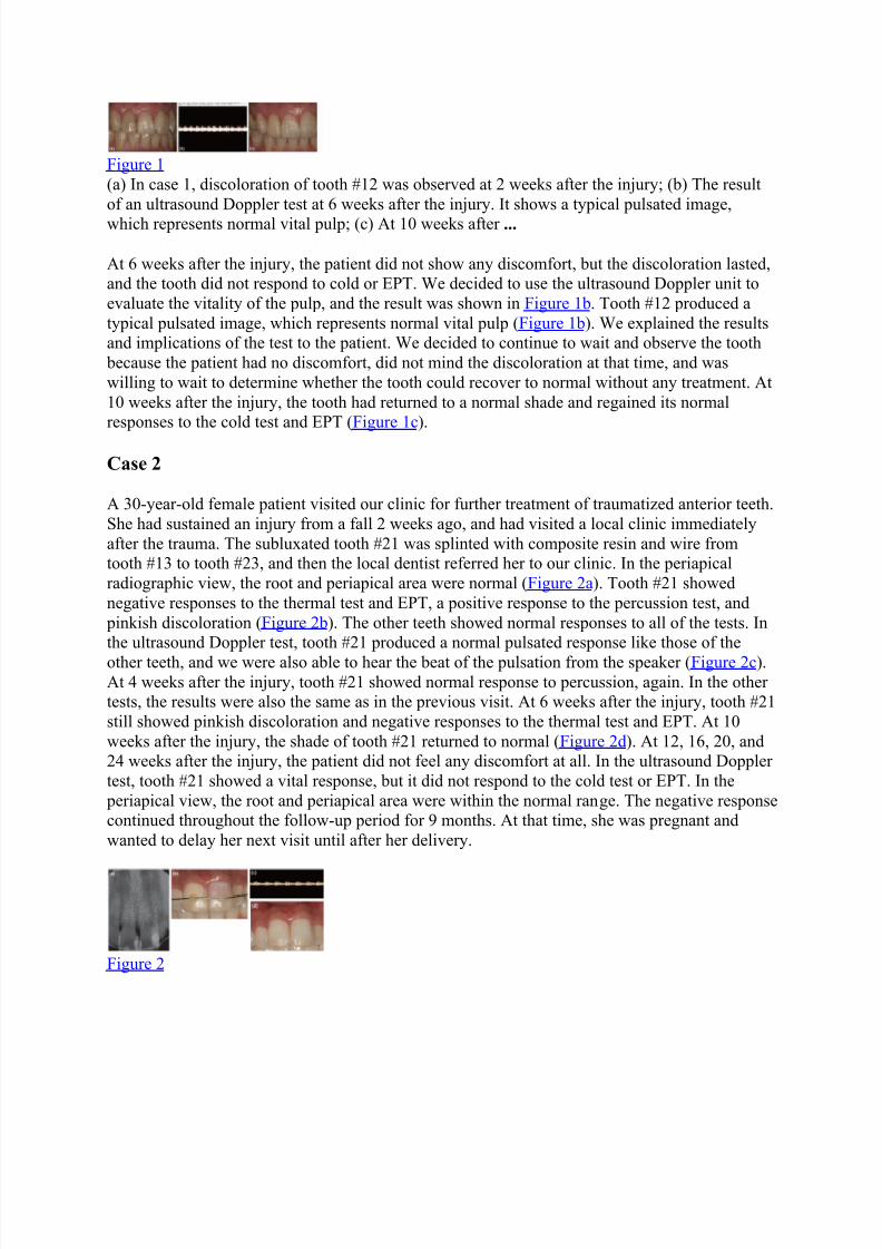
A 30-year-old female patient visited our clinic for further treatment of traumatized anterior teeth.She had sustained an injury from a fall 2 weeks ago, and had visited a local clinic immediatelyafter the trauma. The subluxated tooth #21 was splinted with composite resin and wire fromtooth #13 to tooth #23, and then the local dentist referred her to our clinic. In the periapicalradiographic view, the root and periapical area were normal (Figure 2a). Tooth #21 showednegative responses to the thermal test and EPT, a positive response to the percussion test, and pinkish discoloration (Figure 2b). The other teeth showed normal responses to all of the tests. Inthe ultrasound Doppler test, tooth #21 produced a normal pulsated response like those of the
other teeth, and we were also able to hear the beat of the pulsation from the speaker (Figure 2c).At 4 weeks after the injury, tooth #21 showed normal response to percussion, again. In the othertests, the results were also the same as in the previous visit. At 6 weeks after the injury, tooth #21still showed pinkish discoloration and negative responses to the thermal test and EPT. At 10weeks after the injury, the shade of tooth #21 returned to normal (Figure 2d). At 12, 16, 20, and24 weeks after the injury, the patient did not feel any discomfort at all. In the ultrasound Dopplertest, tooth #21 showed a vital response, but it did not respond to the cold test or EPT. In the periapical view, the root and periapical area were within the normal range. The negative responsecontinued throughout the follow-up period for 9 months. At that time, she was pregnant andwanted to delay her next visit until after her delivery.
Figure 2
8/10/2019 Use of ultrasound Doppler to determine tooth vitality in a discolored tooth
http://slidepdf.com/reader/full/use-of-ultrasound-doppler-to-determine-tooth-vitality-in-a-discolored-tooth 4/32
(a) In case 2, tooth #21 was splinted at a local clinic after a subluxation injury that had occurred2 weeks before the patient visited our clinic. It showed a negative response to the thermal testand EPT, and a positive response to the percussion test; ...
Case 3
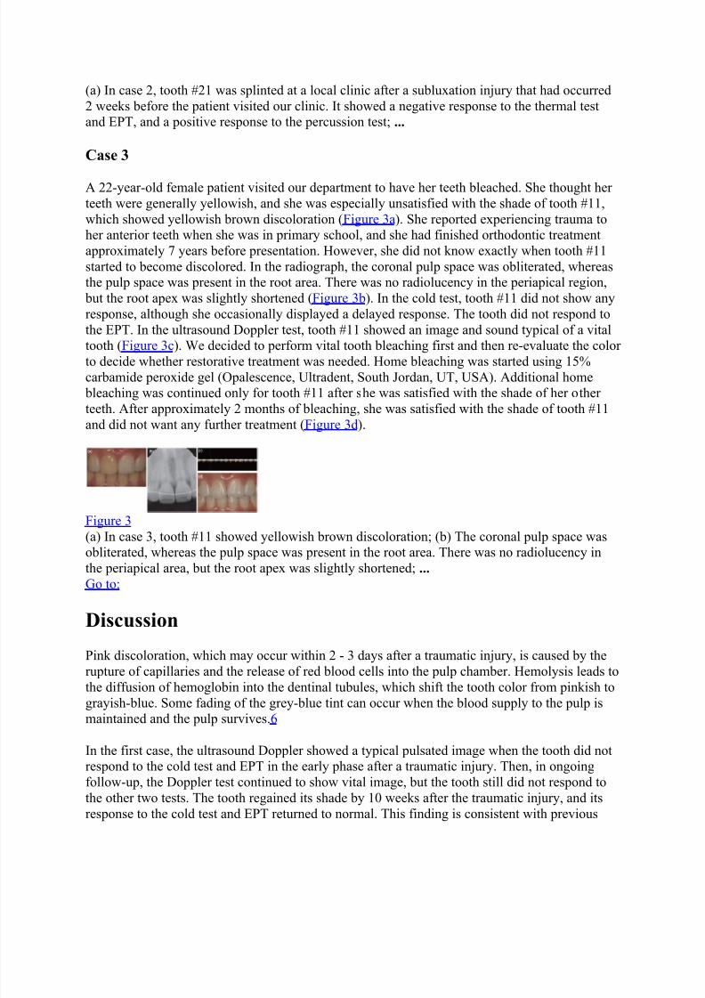
A 22-year-old female patient visited our department to have her teeth bleached. She thought herteeth were generally yellowish, and she was especially unsatisfied with the shade of tooth #11,which showed yellowish brown discoloration (Figure 3a). She reported experiencing trauma toher anterior teeth when she was in primary school, and she had finished orthodontic treatmentapproximately 7 years before presentation. However, she did not know exactly when tooth #11started to become discolored. In the radiograph, the coronal pulp space was obliterated, whereasthe pulp space was present in the root area. There was no radiolucency in the periapical region, but the root apex was slightly shortened (Figure 3b). In the cold test, tooth #11 did not show anyresponse, although she occasionally displayed a delayed response. The tooth did not respond tothe EPT. In the ultrasound Doppler test, tooth #11 showed an image and sound typical of a vital
tooth (Figure 3c). We decided to perform vital tooth bleaching first and then re-evaluate the colorto decide whether restorative treatment was needed. Home bleaching was started using 15%carbamide peroxide gel (Opalescence, Ultradent, South Jordan, UT, USA). Additional home bleaching was continued only for tooth #11 after she was satisfied with the shade of her otherteeth. After approximately 2 months of bleaching, she was satisfied with the shade of tooth #11and did not want any further treatment (Figure 3d).
Figure 3 (a) In case 3, tooth #11 showed yellowish brown discoloration; (b) The coronal pulp space wasobliterated, whereas the pulp space was present in the root area. There was no radiolucency inthe periapical area, but the root apex was slightly shortened; ... Go to:
Discussion
Pink discoloration, which may occur within 2 - 3 days after a traumatic injury, is caused by therupture of capillaries and the release of red blood cells into the pulp chamber. Hemolysis leads tothe diffusion of hemoglobin into the dentinal tubules, which shift the tooth color from pinkish to
grayish-blue. Some fading of the grey-blue tint can occur when the blood supply to the pulp ismaintained and the pulp survives.6
In the first case, the ultrasound Doppler showed a typical pulsated image when the tooth did notrespond to the cold test and EPT in the early phase after a traumatic injury. Then, in ongoingfollow-up, the Doppler test continued to show vital image, but the tooth still did not respond tothe other two tests. The tooth regained its shade by 10 weeks after the traumatic injury, and itsresponse to the cold test and EPT returned to normal. This finding is consistent with previous
8/10/2019 Use of ultrasound Doppler to determine tooth vitality in a discolored tooth
http://slidepdf.com/reader/full/use-of-ultrasound-doppler-to-determine-tooth-vitality-in-a-discolored-tooth 5/32
reports that indicated that the discoloration returned to normal when the teeth regained theirvitality and demonstrating that ultrasound Doppler can be successfully used to determine thevitality of teeth during the period when they do not respond to the cold test and EPT after atraumatic injury.4-8 Ultrasound Doppler may help decrease unnecessary endodontic treatments,which could be performed due to a lack of the proper diagnostic methods after a traumatic injury.
In the first and second cases, 10 weeks were needed to regain the tooth's color and responses tothe cold test and EPT. This result is consistent with a previous study in which the discolorationdisappeared within 4 weeks to 6 months after root fracture resulting in tooth discoloration aftertrauma.6 The second case was interesting in that the discoloration returned to normal by 10weeks after injury, but the tooth did not respond to the cold test and EPT even at 9 months afterthe traumatic injury, although it showed a consistent vital image in the Doppler test from the beginning. False positive responses in the ultrasound Doppler test have not yet been studied. Inthe present study, a 20-MHz ultrasound Doppler probe was used. The frequency of ultrasound isvery important because it determines the penetration depth of the ultrasound wave. Although a20-MHz frequency was reported to efficiently penetrate the enamel and dentin, and detect the
blood flow in the pulp spaces, it might be possible to detect the blood flow outside of the pulpspaces if the thickness of the hard tissue is very thin.1,12 The potential for false positiveresponses with the ultrasound Doppler probe requires further investigation. In the second case,long-term follow-up is necessary to verify whether the vitality was actually maintained, whichcould be confirmed by a positive response to the cold test and EPT. However, in this case, thetooth returned to its normal shade by 10 weeks after the traumatic injury, which suggests that the blood supply to the pulp was maintained and the pulp survived.6 More time might be needed forthe nerve fiber to heal. Further follow-up is required to determine whether the test results are trueor false positive.
In the third case, the patient did not respond to the cold test and EPT, although she occasionallyshowed an obscure positive delayed response to the cold test. The cold test depends on thehydrodynamic movement of fluid within the dentinal tubules, which excites the A-fibers.13 Teeth with calcified pulp spaces might have normal and healthy pulps, but cold stimuli might not be able to excite the nerve endings due to the insulating effect of the thicker layer of dentin,which is the result of secondary and reactionary dentin formation.14 Ehrmann reported that EPTis particularly effective in older patients and in teeth that have limited fluid movement throughthe dentinal tubules as a result of dentine sclerosis and calcification of the pulp space becausethermal pulp tests are usually inadequate in these situations.14 Klein reported that a patient wasunlikely to respond to a cold test but may respond to an EPT if the pulp space had beensignificantly calcified.15 In their case, more electric pulp current was often needed to elicit aresponse because there was an increased dentin layer and a diminished pulp cavity or a fibrotic pulp. In the third case, tooth #11 was diagnosed as a vital tooth based on the results of theultrasound Doppler test because it displayed a consistent positive sign throughout the observation period. In this case, the coronal pulp space was obliterated, whereas the pulp space was presentin the root area. Because the ultrasound Doppler probe tip was positioned apically, there was a possibility of detecting the blood flow of the root canal. Furthermore, the patient showed aresponse to the cold test, although the response was delayed and inconsistent. For furtherresearch, we need more cases and studies related to ultrasound Doppler.
8/10/2019 Use of ultrasound Doppler to determine tooth vitality in a discolored tooth
http://slidepdf.com/reader/full/use-of-ultrasound-doppler-to-determine-tooth-vitality-in-a-discolored-tooth 6/32
Other methods for evaluating the vascularity of pulp are laser Doppler and pulse oximetry.16-20 Laser Doppler applies a laser to transmit light into the pulp blood vessels through the toothstructure, and a red and infrared LED light beam is used in pulse oximetry for the same purpose.However, the discoloration of the tooth caused by the deposition of blood pigments in thetraumatized tooth may hinder the penetration of light in both laser Doppler and pulse
oximetry.18,20,21 The ultrasound wave used in the ultrasound Doppler unit can detect bloodflow regardless of coronal discoloration, so it can be more useful for discolored teeth.
Go to:
Conclusions
Tooth discoloration after a traumatic injury was corrected when the ultrasound Doppler produceda typical pulsated image, which represents normal vital pulp. Ultrasound Doppler might be aneffective tool to evaluate tooth vitality when the cold test and EPT do not give properinformation, especially after a traumatic injury. However, the use of ultrasound Doppler requires
further research on the potential for false positive and negative responses to increase its clinicalreliability.
Go to:
Acknowledgement
This case report is a part of the research that was supported by the Basic Science ResearchProgram through the National Research Foundation of Korea (NRF) funded by the Ministry ofEducation, Science and Technology (2011-0021235).
Go to:
Footnotes
No potential conflict of interest relevant to this article was reported.
Go to:
References
1. Yoon MJ, Kim E, Lee SJ, Bae YM, Kim S, Park SH. Pulpal blood flow measurement withultrasound Doppler imaging. J Endod. 2010;36:419 – 422. [PubMed] 2. Abd-Elmeguid A, Yu DC. Dental pulp neurophysiology: part 2. Current diagnostic tests toassess pulp vitality. J Can Dent Assoc. 2009;75:139 – 143. [PubMed] 3. Ozçelik B, Kuraner T, Kendir B, Aşan E. Histopathological evaluation of the dental pulps incrown-fractured teeth. J Endod. 2000;26:271 – 273. [PubMed]

8/10/2019 Use of ultrasound Doppler to determine tooth vitality in a discolored tooth
http://slidepdf.com/reader/full/use-of-ultrasound-doppler-to-determine-tooth-vitality-in-a-discolored-tooth 7/32
4. Aguiló L, Gandía JL. Transient red discoloration: report of case. ASDC J Dent Child.1998;65:346 – 348. 356. [PubMed] 5. Andreasen FM. Pulpal healing after luxation injuries and root fracture in the permanentdentition. Endod Dent Traumatol. 1989;5:111 – 131. [PubMed] 6. Malmgren B, Hübel S. Transient discoloration of the coronal fragment in intra-alveolar root
fractures. Dent Traumatol. 2012;28:200 – 204. [PubMed] 7. Andreasen FM. Transient apical breakdown and its relation to color and sensibility changesafter luxation injuries to teeth. Endod Dent Traumatol. 1986;2:9 – 19. [PubMed] 8. Cohenca N, Karni S, Rotstein I. Transient apical breakdown following tooth luxation. DentTraumatol. 2003;19:289 – 291. [PubMed] 9. Cotti E, Campisi G, Ambu R, Dettori C. Ultrasound real-time imaging in the differentialdiagnosis of periapical lesions. Int Endod J. 2003;36:556 – 563. [PubMed] 10. Rajendran N, Sundaresan B. Efficacy of ultrasound and color power Doppler as a monitoringtool in the healing of endodontic periapical lesions. J Endod. 2007;33:181 – 186. [PubMed] 11. Lustig JP, London D, Dor BL, Yanko R. Ultrasound identification and quantitativemeasurement of blood supply to the anterior part of the mandible. Oral Surg Oral Med Oral
Pathol Oral Radiol Endod. 2003;96:625 – 629. [PubMed] 12. Yoon MJ, Lee SJ, Kim E, Park SH. Doppler ultrasound to detect pulpal blood flow changesduring local anaesthesia. Int Endod J. 2012;45:83 – 87. [PubMed] 13. Cohen S, Hargreaves KM. Pathways of the pulp. 9th ed. Louis: Mosby; 2006. pp. 504 – 508.14. Ehrmann EH. Pulp testers and pulp testing with particular reference to the use of dry ice.Aust Dent J. 1977;22:272 – 279. [PubMed] 15. Klein H. Pulp responses to an electric pulp stimulator in the developing permanent anteriordentition. ASDC J Dent Child. 1978;45:199 – 202. [PubMed] 16. Olgart L, Gazelius B, Lindh-Strömberg U. Laser Doppler flowmetry in assessing vitality inluxated permanent teeth. Int Endod J. 1988;21:300 – 306. [PubMed] 17. Sasano T, Onodera D, Hashimoto K, Iikubo M, Satoh-Kuriwada S, Shoji N, Miyahara T.Possible application of transmitted laser light for the assessment of human pulp vitality. Part 2.Increased laser power for enhanced detection of pulpal blood flow. Dent Traumatol. 2005;21:37 – 41. [PubMed] 18. Gopikrishna V, Tinagupta K, Kandaswamy D. Comparison of electrical, thermal, and pulseoximetry methods for assessing pulp vitality in recently traumatized teeth. J Endod.2007;33:531 – 535. [PubMed] 19. Gopikrishna V, Tinagupta K, Kandaswamy D. Evaluation of efficacy of a new custom-made pulse oximeter dental probe in comparison with the electrical and thermal tests for assessing pulpvitality. J Endod. 2007;33:411 – 414. [PubMed] 20. Jafarzadeh H, Rosenberg PA. Pulse oximetry: review of a potential aid in endodonticdiagnosis. J Endod. 2009;35:329 – 333. [PubMed] 21. Heithersay GS, Hirsch RS. Tooth discoloration and resolution following a luxation injury:significance of blood pigment in dentin to laser Doppler flowmetry readings. Quintessence Int.1993;24:669 – 676. [PubMed]

8/10/2019 Use of ultrasound Doppler to determine tooth vitality in a discolored tooth
http://slidepdf.com/reader/full/use-of-ultrasound-doppler-to-determine-tooth-vitality-in-a-discolored-tooth 8/32
Abstract
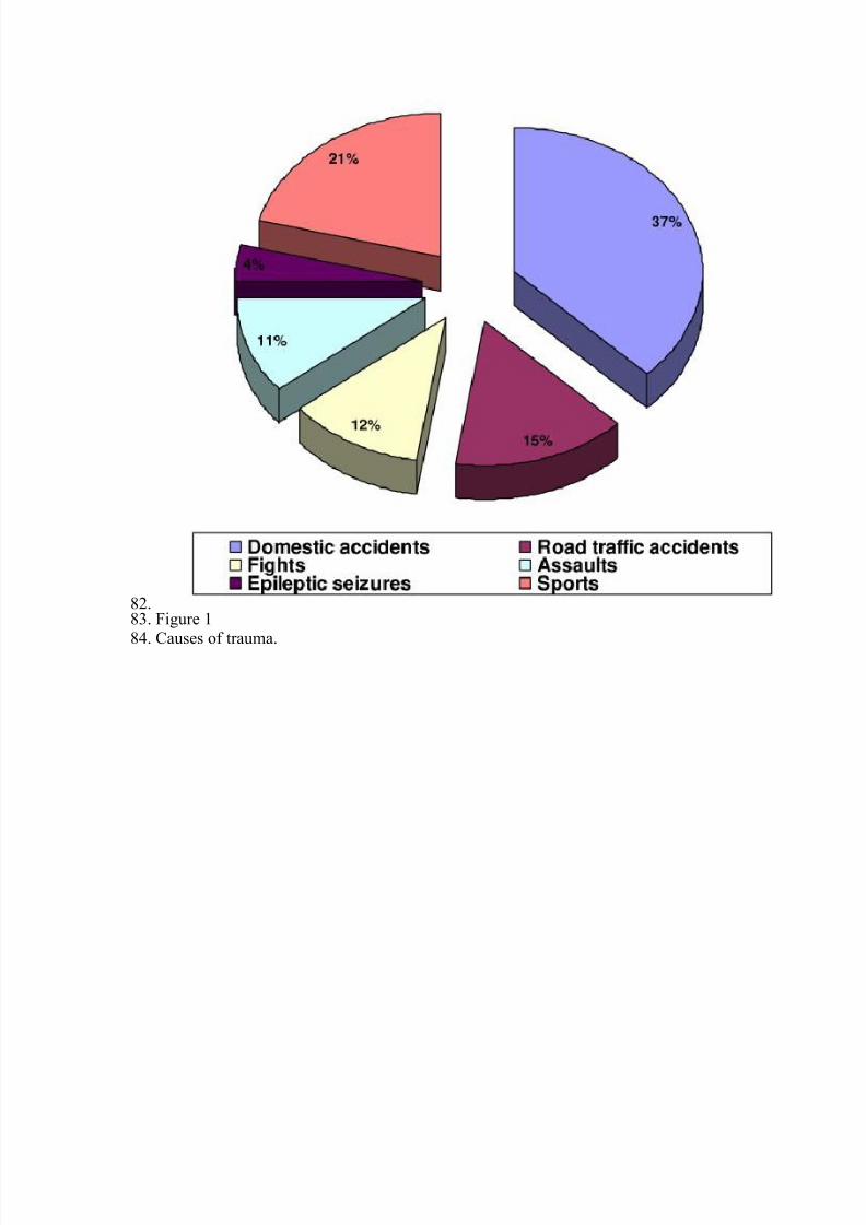
Objectives
Vertical root fractures (VRFs) are a common cause of tooth loss. Little evidence exists though,
relating the incidence of VRFs to the type of endodontic retreatment. This retrospective studyaimed at evaluating the impact of conventional versus surgical endodontics on root canal-filledteeth with VRFs.
Materials and methods
Over a period of 13 years, 200 endodontically retreated teeth from 192 patients with VRFs wereextracted and further examined. VRFs were assessed in relation to age, gender, tooth group,clinical signs, extension on the root surface, patency, as well as type of endodontic retreatmentand restoration. Statistical analysis was conducted using a Cox PH Model, Chi-squared,Wilcoxon rank-sum, and Log rank tests at a significance level of 5 %.
Results
The majority of teeth with VRFs (62.31 %) had undergone the combination of conventional rootcanal retreatment and apical surgery. Women (64.06 %) presented VRFs more frequently thanmen (35.94 %) at the mean age of 51.1 and 55.1 years, respectively. Maxillary first (17.5 %) andsecond (16.5 %) premolars, restored by a resin-based material without a post (56.28 %) weremore susceptible to VRFs. Apically initiated (84.1 %) VRFs could be diagnosed more easily onradiographs.
Conclusions
The type of endodontic treatment strongly correlated with VRFs. The prevalence of VRFs inteeth having undergone both conventional and surgical endodontic retreatment could beattributed, among others, to additive dentin damage related to the aforementioned endodontic procedures.
Clinical relevance
The possible involvement of endodontic retreatment in the multifactorial etiology of VRFs needsto be taken into consideration in clinical practice.
Original Article
8/10/2019 Use of ultrasound Doppler to determine tooth vitality in a discolored tooth
http://slidepdf.com/reader/full/use-of-ultrasound-doppler-to-determine-tooth-vitality-in-a-discolored-tooth 9/32
Comparative assessment of the incidence of
vertical root fractures between conventional
versus surgical endodontic retreatmentL. Karygianni1, M. Krengel2, M. Winter 3, S. Stampf 4 and K. T. Wrbas1, 5
(1)Department of Operative Dentistry and Periodontology, Center for Dental Medicine, MedicalCenter, University of Freiburg, Hugstetterstrasse 55, Freiburg, 79106, Germany(2)Bergisch Gladbach, Germany(3)Rheinbach, Germany(4)
Institute of Medical Biometry and Medical Informatics, Albert-Ludwigs-University, Freiburg,Germany(5)Department of Endodontics, Centre for Operative Dentistry and Periodontology, University ofDental Medicine and Oral Health, Danube Private University (DPU), Krems, Austria
K. T. WrbasEmail: [email protected] Received: 8 August 2013Accepted: 26 December 2013Published online: 10 January 2014AbstractObjectivesVertical root fractures (VRFs) are a common cause of tooth loss. Little evidence exists though,relating the incidence of VRFs to the type of endodontic retreatment. This retrospective studyaimed at evaluating the impact of conventional versus surgical endodontics on root canal-filledteeth with VRFs.Materials and methodsOver a period of 13 years, 200 endodontically retreated teeth from 192 patients with VRFs wereextracted and further examined. VRFs were assessed in relation to age, gender, tooth group,clinical signs, extension on the root surface, patency, as well as type of endodontic retreatmentand restoration. Statistical analysis was conducted using a Cox PH Model, Chi-squared,Wilcoxon rank-sum, and Log rank tests at a significance level of 5 %.ResultsThe majority of teeth with VRFs (62.31 %) had undergone the combination of conventional rootcanal retreatment and apical surgery. Women (64.06 %) presented VRFs more frequently thanmen (35.94 %) at the mean age of 51.1 and 55.1 years, respectively. Maxillary first (17.5 %) andsecond (16.5 %) premolars, restored by a resin-based material without a post (56.28 %) weremore susceptible to VRFs. Apically initiated (84.1 %) VRFs could be diagnosed more easily onradiographs.Conclusions

8/10/2019 Use of ultrasound Doppler to determine tooth vitality in a discolored tooth
http://slidepdf.com/reader/full/use-of-ultrasound-doppler-to-determine-tooth-vitality-in-a-discolored-tooth 10/32
The type of endodontic treatment strongly correlated with VRFs. The prevalence of VRFs inteeth having undergone both conventional and surgical endodontic retreatment could beattributed, among others, to additive dentin damage related to the aforementioned endodontic procedures.Clinical relevance
The possible involvement of endodontic retreatment in the multifactorial etiology of VRFs needsto be taken into consideration in clinical practice.KeywordsApical surgery Clinical signs Endodontic retreatment Vertical root fractures
Introduction
Novel NiTi retreatment systems and root canal obturation techniques allow for minimized dentinloss of root canals during endodontic procedures and thus, for fracture resistance of root canal-treated teeth in the long term [1]. However, these vast improvements in modern endodontics arestill accompanied by the unexpected occurrence of vertical root fractures (VRFs) in root canal-
treated teeth. According to the American Association of Endodontists, vertically fractured teethare characterized by a crack that begins in the root at any level and extends toward the occlusalsurface, usually in the bucco-lingual direction [2].The diagnosis of VRFs, usually years after the final crown restoration of the root canal-treatedteeth, may be confusing because of the existence of nonspecific radiographic and clinical signsthat imitate endodontic treatment failure or periodontal disease [3, 4]. Representative clinicalfeatures of VRFs usually include a deep, thread-thin, isolated periodontal pocket, and multiplesinus tracts, sometimes situated coronally on both the buccal and lingual gingiva [5].Furthermore, an angular resorption pattern (―halo‖ lesion) which incorporates a periapical alongwith a lateral radiolucency extending apically has been shown as a typical radiographic feature ofVRFs on conventional X-rays [6 ]. High-resolution visualization techniques with image accuracy
and low radiation doses such as local CT, tuned-aperture computed tomography, and opticalcoherence tomography have been also successfully employed for monitoring VRFs [7 , 8]. Nevertheless, when the diagnosis of a VRF is still inconclusive, exploratory surgical proceduresare usually applied to verify its occurrence [6 ].Clinical management has to be undertaken as soon as possible to prevent additional bone loss,which might pose difficulties for the further reconstruction of the region later on. Verticallyfractured teeth inevitably lead to extraction in most of the cases. Alternative treatment procedures involve extraction only of the fractured root in multirooted teeth [9, 10]. Someauthors suggest also removing the fractured tooth and rebonding the fractured parts extraorallyfollowed by the reimplantation of the tooth [11, 12]. The introduction of cone-beam computedtomography in dentistry facilitated the three-dimensional high-resolution visualization of VRFs
[13, 14].The higher incidence of VRFs in root canal-treated teeth is mainly attributed to factors relating toconventional root-canal treatment such as excessive biomechanical preparation and extremelateral-vertical forces during compaction of root canal filling materials [15 – 17 ]. The use ofirrigants (NaOCl, EDTA) and intracanal medicaments (Ca(OH)2) for more than 30 days can alsoinduce VRFs [18]. Additionally, tooth structural loss is mainly due to dehydration of dentine andmicrobe-induced degradation or modification of collagen constitutes risk factors for fracture predisposition in root canal-treated teeth [19]. Various restorative parameters such as insufficient
8/10/2019 Use of ultrasound Doppler to determine tooth vitality in a discolored tooth
http://slidepdf.com/reader/full/use-of-ultrasound-doppler-to-determine-tooth-vitality-in-a-discolored-tooth 11/32
ferrule effect, extreme widening of the root canal for posttreatment, and inappropriate postdesignmay all contribute to VRF formation [20, 21]. Nowadays, the widespread conduction of endodontic surgery is considered an alternativeapproach with good prognosis in cases where an orthograde attempt at retreatment is notindicated [22]. Despite that modern microtechniques coupled with the appropriate surgical
magnification have been introduced, VRFs relating to apical surgery may still occur. Theremoval of the apical part of the root, the use of specially designed ultrasonic tips as well as theretrograde MTA filling are treatment parameters that may be associated with VRFs in theframework of their multifactorial etiology.The purpose of the present retrospective study was to evaluate potential etiological, clinical, andradiographic features of 200 root canal-treated teeth referred for extraction after a clinicaldiagnosis of VRF. All teeth had received an endodontic retreatment previously, either by aconventional root-canal retreatment, root-end resection or the combination of both techniques.
Materials and methods
Inclusion and exclusion criteria
The cross-sectional study was conducted over a period of 13 years in a total of 192 patients with200 root canal-retreated teeth with a diagnosis of VRF. The vertically fractured teeth that wereselected for this study had received endodontic treatment by general practitioners. It could beassumed that the prevailing quality guidelines for endodontic treatment at that time werefollowed to eliminate procedural complications [23]. However, if the quality of the root canalretreatment controlled by another endodontist prior to the study was questionable, the teeth wereexcluded from the study. Based on the type of endodontic retreatment they had received, theteeth were divided into three groups. In the first group, a conventional retreatment had beenconducted; the second group had undergone apical surgery and the third group had been treated
by both methods. Endodontic retreatment of all teeth had been completed at least 2 years earlier.In all cases, retreatment was conducted after the initial root canal treatment had been considereda failure.Teeth with VRF were finally excluded from the amount of teeth studied if patient records lackedsufficient information about dental history of the fractured tooth or if the extraction wasassociated with dental trauma as well as other types of tooth fractures. Teeth with insufficientcoronal restorations, with direct exposure of the root canal filling material to the oral cavity orwith obturation material that did not reach within 2 mm of the radiographic apex were alsoexcluded from the study.
Clinical procedure
When the inclusion criteria were fulfilled, the following clinical parameters were furtherevaluated by an endodontist: gender and age of the patient, endodontic history, tooth type(incisor, premolar, or molar in upper/lower jaw), clinical signs (depth and extent of periodontal pockets and presence of sinus tract and pus), type of endodontic retreatment (conventionalretreatment, root-end resection, and combined use of both methods), and type of definiterestoration (composite, post, and crown). Radiographic alterations were detected with the aid of aview-box with background illumination and magnification; loss of attachment was demonstrated
8/10/2019 Use of ultrasound Doppler to determine tooth vitality in a discolored tooth
http://slidepdf.com/reader/full/use-of-ultrasound-doppler-to-determine-tooth-vitality-in-a-discolored-tooth 12/32
as a deep, narrow, isolated periodontal pocket; and distinct separation of the cracked toothfragments was diagnosed on X-rays. Exploratory surgery was additionally conducted to confirman uncertain diagnosis. The use of magnifying loops (Carl Zeiss, Oberkochen, Germany) andstaining with methylene blue solution along fractures enabled the operator not only to visualizethe fracture lines but also to identify the extension of the fracture on the root surface (apical,
central) and patency of fractures depending on separation of root fragments (complete,incomplete). All vertically fractured teeth were finally extracted by two oral surgeons, collectedand stored in a 0.9 % saline solution at 4 °C until use.
Statistical analysis
Frequency tables and cross tables were used for the statistical evaluation of the data. To evaluateassociations between categorical variables, the Chi-squared test was used. The Wilcoxon ranksum test was applied to detect significant differences in age between gender groups.The analysis for evaluating differences between treatment groups was performed with a Cox PHmodel and corresponding Kaplan – Meier curves were presented. The log rank test was used to
examine the difference between patients with different endodontic treatment. All statistical testswere done at the significance level of 5 % and were performed using the statistical software SAS9.1.2.
Results
Type of endodontic treatment
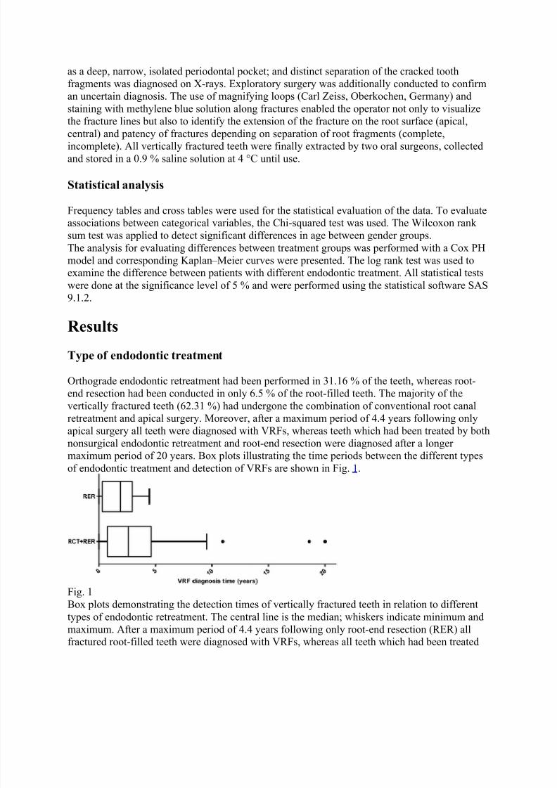
Orthograde endodontic retreatment had been performed in 31.16 % of the teeth, whereas root-end resection had been conducted in only 6.5 % of the root-filled teeth. The majority of thevertically fractured teeth (62.31 %) had undergone the combination of conventional root canalretreatment and apical surgery. Moreover, after a maximum period of 4.4 years following onlyapical surgery all teeth were diagnosed with VRFs, whereas teeth which had been treated by bothnonsurgical endodontic retreatment and root-end resection were diagnosed after a longermaximum period of 20 years. Box plots illustrating the time periods between the different typesof endodontic treatment and detection of VRFs are shown in Fig. 1.
Fig. 1Box plots demonstrating the detection times of vertically fractured teeth in relation to differenttypes of endodontic retreatment. The central line is the median; whiskers indicate minimum andmaximum. After a maximum period of 4.4 years following only root-end resection (RER) allfractured root-filled teeth were diagnosed with VRFs, whereas all teeth which had been treated

8/10/2019 Use of ultrasound Doppler to determine tooth vitality in a discolored tooth
http://slidepdf.com/reader/full/use-of-ultrasound-doppler-to-determine-tooth-vitality-in-a-discolored-tooth 13/32
by both root canal retreatment and root-end resection (RCT + RER) were diagnosed after alonger maximum period of 20 years
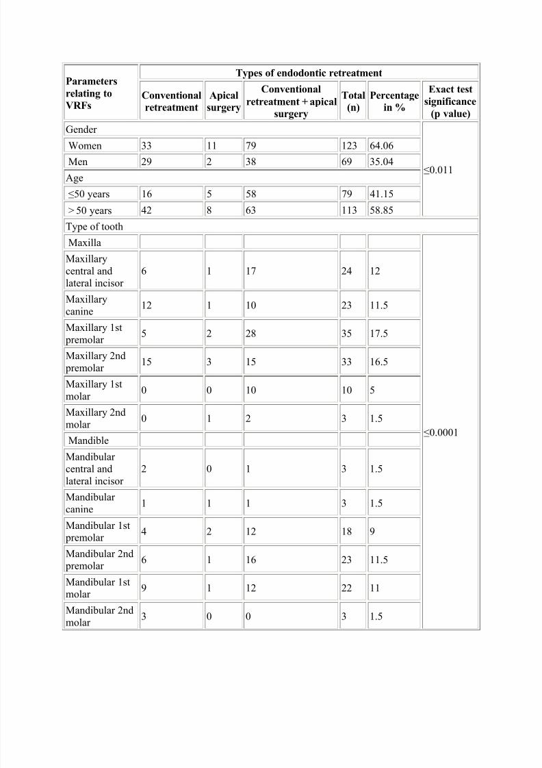
Gender and age
The percentages of female and male patients demonstrating VRFs were 64.06 and 35.94 %,respectively. The mean age of patients presenting VRFs was 52.6 (±13.5; range, 22 to 79) years.The mean age of female and male patients with VRFs was 55.1 (±13.1; range, 22 to 78) yearsand 51.1 (±13.5; range, 22 to 79) years, respectively. This difference in age between gender wassignificant (p value = 0.02, Wilcoxon rank-sum test).
Tooth group
The most commonly extracted teeth were the maxillary first premolars (n
=
35, 17.5 %),maxillary second premolars (n = 33, 16.5 %), maxillary central and lateral incisors (n = 24,12 %), mandibular first molars (n
=
23, 11.5 %), mandibular second premolars (n
=
23, 11.5 %),
and maxillary canines (n
=
23, 11.5 %). There were very few extracted mandibular and maxillarysecond molars as well as mandibular incisors (n = 3, 1.5 %).
Clinical signs
The presence of deep periodontal pockets (23.9 %) was revealed along with the presence of asinus tract without pus (28.93 %), sinus tract with pus (3.31 %) and combination of periodontal pocket and fistula (43.8 %).
Extension of VRF on the root surface
After the extraction of the vertically fractured teeth the extension of VRFs on the root surfacewas assessed macroscopically using magnifying loops (Carl Zeiss, Oberkochen, Germany). If thelocalization of the fracture line was difficult (especially by incomplete VRFs) staining withmethylene blue solution along fractures enabled the inspection and categorization of VRFs(coronal, central, and apical) according to their extension on the root surface (Fig. 2). Theextension point of VRFs was located apically in 56.77 % of the teeth which weremacroscopically examined, whereas 43.23 % of the fractures reached the middle of the rootsurface. The mean distance measured between the extension point of VRFs and the cement – enamel junction was 5.8 (±3.5; range, 1 to 15) mm.

8/10/2019 Use of ultrasound Doppler to determine tooth vitality in a discolored tooth
http://slidepdf.com/reader/full/use-of-ultrasound-doppler-to-determine-tooth-vitality-in-a-discolored-tooth 14/32
Fig. 2A vertically fractured mandibular first molar (46) with a fracture line reaching the middle thirdof the root surface. According to their extension point on the root surface (coronal third, middle
third, and apical third), vertical root fractures (VRFs) were characterized as coronal, central, andapical, respectively. CEJ cement – enamel junction
Partial/complete VRFs
Based on separation of root fragments, 57.39 % of the VRFs were incomplete, whereas 42.61 %were complete. The inspection of the VRFs was conducted both radiographically (mainlycomplete VRFs) and visually (partial VRFs) with magnifying loops after the extraction of thevertically fractured teeth. Teeth with VRFs that could not be extracted under atraumaticconditions were excluded from the study in order to avoid the risk of turning an incomplete VRFinto a complete one.
Radiographic diagnosis
Almost only half of the total amount of VRFs (56.5 %) was radiographically recognizable. Therewas a statistically significant association between radiographically recognizable VRFs and theirlocation on the root. It was found that apically initiated VRFs were more easily observed on theradiographs (84.1 %) compared with the centrally located fractures (p < 0.001, Chi-square test).
Type of restoration
One hundred twelve teeth (56.28 %) had been previously coronally restored by resin-based
composite without a post, 25 teeth (12.56 %) had been restored by a composite material with a post, 15 teeth (7.54 %) with posts were crowned, and 47 teeth (23.62 %) were only crowned.An overview of the major findings of this study relating different clinical and radiographic parameters to VRFs after conventional root-canal retreatment, root-end resection or thecombination of both techniques is presented in Table 1. Table 1Overview of the correlation between various clinical and radiographic parameters and verticalroot fractures (VRF) according to the type of endodontic retreatment
8/10/2019 Use of ultrasound Doppler to determine tooth vitality in a discolored tooth
http://slidepdf.com/reader/full/use-of-ultrasound-doppler-to-determine-tooth-vitality-in-a-discolored-tooth 15/32
Parameters
relating to
VRFs
Types of endodontic retreatment
Conventional
retreatment
Apical
surgery
Conventional
retreatment + apical
surgery
Total
(n)
Percentage
in %
Exact test
significance
(p value)
Gender
≤0.011
Women 33 11 79 123 64.06
Men 29 2 38 69 35.04
Age
≤50 years 16 5 58 79 41.15
> 50 years 42 8 63 113 58.85
Type of tooth
Maxilla
≤0.0001
Maxillary
central andlateral incisor
6 1 17 24 12
Maxillarycanine
12 1 10 23 11.5
Maxillary 1st premolar
5 2 28 35 17.5
Maxillary 2nd premolar
15 3 15 33 16.5
Maxillary 1stmolar
0 0 10 10 5
Maxillary 2ndmolar
0 1 2 3 1.5
Mandible
Mandibularcentral andlateral incisor
2 0 1 3 1.5
Mandibularcanine
1 1 1 3 1.5
Mandibular 1st
premolar
4 2 12 18 9
Mandibular 2nd premolar
6 1 16 23 11.5
Mandibular 1stmolar
9 1 12 22 11
Mandibular 2ndmolar
3 0 0 3 1.5

8/10/2019 Use of ultrasound Doppler to determine tooth vitality in a discolored tooth
http://slidepdf.com/reader/full/use-of-ultrasound-doppler-to-determine-tooth-vitality-in-a-discolored-tooth 16/32
Parameters
relating to
VRFs
Types of endodontic retreatment
Conventional
retreatment
Apical
surgery
Conventional
retreatment + apical
surgery
Total
(n)
Percentage
in %
Exact test
significance
(p value)
Clinical signsPeriodontal
pocket11 1 17 29 23.97
≥0.95
Sinus tractwithout pus
13 1 21 35 28.93
Sinus tract with pus
1 0 3 4 3.31
Periodontal pocket
+
sinustract
21 3 29 53 43.8
Initiation of VRFCentral 20 2 45 67 56.49
≥0.65 Apical 25 5 57 87 43.51
Patency of VRF
Complete 18 3 28 49 42.98≥0.65
Incomplete 21 2 42 65 57.02
Radiographic diagnosis
Yes 41 3 69 113 56.5≥0.6
No 22 10 55 87 43.5Type of restoration
Resin-basedcomposite
32 4 76 112 56.28
≤0.01 Resin-basedcomposite + post
20 0 5 25 12.56
Crown 4 8 35 47 7.54
Crown + post 8 1 6 15 23.62
Discussion
The susceptibility of root canal-filled teeth to VRFs is highlighted in some recent reports [24 – 26 ]. The innovation of the present study is that it relates the type of endodontic retreatment withthe incidence of VRFs. Specifically, orthograde endodontic retreatment had been previously performed in 31.16 % of the teeth, whereas 62.31 % of the vertically fractured teeth hadundergone the combination of root canal retreatment and root-end resection. 6.5 % of the teethhad been retreated only with root-end resection. Procedures associated to the initial endodontic

8/10/2019 Use of ultrasound Doppler to determine tooth vitality in a discolored tooth
http://slidepdf.com/reader/full/use-of-ultrasound-doppler-to-determine-tooth-vitality-in-a-discolored-tooth 17/32
treatment such as the use of irrigants, medicaments, and root canal fillings can pave the way forthe occurrence of VRFs [17 , 18, 27 ]. The additional influence of the low moisture content andthe reduced structural tooth integrity after access cavity preparation are also common VRF- predisposing side effects of the conventional root canal therapy [19]. However, a more profounddamage is made to dentin during retreatment procedures. The additional mechanical widening of
the canal system for the efficient removal of the old root canal filling, the use of variousdissolving agents to soften gutta-percha as well as the removal of separated instruments and posts can cumulatively promote the defect progression in dentin [28 – 30].Topical anomalies or more extensive pre-existing flaws located in the canal wall can inducesubcritical cracks resulting in catastrophic root fractures after cyclic loading or immense occlusalstress [31]. Given also the fact that circumferential and radial stresses on root dentin are doubled by the presence of a root canal itself, the excessive removal of the apical part of roots, theapplication of ultrasonic instruments and retrograde filling materials as well as the mechanicalstress during surgical endodontic procedures could further trigger comprehensive stress at theroot surface and hence crack initiation [32]. The anisotropic mechanical properties of dentin because of the orientation of the tubules still challenges further research in this field [33].
Nevertheless, the effects of additional potentially harmful procedures during apical surgery arereflected in the higher incidence of VRFs (62.31 %) among teeth having undergone thecombination of root canal retreatment and root-end resection. Excluding the cases where theconventional root canal therapy or retreatment failed because of pre-existing VRFs, it seems thatthe cumulative dentine damage associated with the initial endodontic treatment, retreatment andapical surgery could be possibly responsible for the outstanding presence of VRFs in teethhaving undergone both conventional and surgical retreatment.The multifactorial origin of VRFs in root canal-treated teeth has been well studied, so far. Theinfluence of chemical agents (irrigants, intracanal medicaments), obturation biomaterials (gutta- percha, sealer), instrumentation and compaction techniques as well as restorative parameters(posts, crowns) in VRF predilection in teeth after initial endodontic treatment has beenhighlighted in previous studies [17 – 19, 27 – 29]. Nonetheless, endodontic retreatment proceduresare not similar and thus, not comparable to the initial endodontic therapy. Root canalsundergoing endodontic retreatment are subjected to additional preparation for the removal of theold obturation material, the use of different irrigants, ultrasonication, and the retrieval of postsand separated files. Given the different treatment protocols followed during retreatment casesresulting in additional stress of root canal walls, the comparison to primary cases was notconsidered meaningful in this report.The fact that the root-canal treatments were conducted by general practicioners was one of thelimitations of this study. Nevertheless, teeth were excluded from the study if the quality of theroot canal retreatment was questionable. Under-/overinstrumented and insufficiently filled rootcanals were therefore rejected as they would compromise the results of this study.As far as the detection time of VRF in the vertically fractured teeth is concerned, all surgicallyretreated teeth without conventional retreatment were diagnosed after 4.4 years. Allnonsurgically retreated teeth following apical surgery were diagnosed with VRF after a longer period of 20 years. The delayed VRF diagnosis and thus, tooth removal in these cases can beattributed to the time-consuming treatment sessions as well as long inter-appointment,observation and recall periods in the framework of orthograde endodontic retreatment. Secondly,given their great desire to retain the tooth, patients having received nonsurgical retreatment probably seeked for dental procedures as conservative as possible in order to postpone or even
8/10/2019 Use of ultrasound Doppler to determine tooth vitality in a discolored tooth
http://slidepdf.com/reader/full/use-of-ultrasound-doppler-to-determine-tooth-vitality-in-a-discolored-tooth 18/32
avoid the extraction of the fractured teeth. Finally, the role of the dentist should also be takeninto account. Specialized endodontists conducting conventional retreatment usually possessdental operating microscopes allowing for better discrimination of anatomic variations, controlof instruments and prevention of intraoperative complications prior to surgery [34].The gender distribution manifests a higher incidence of VRFs in women, a finding that
contradicts with the results of other studies, where they are equally divided [35
, 36
]. Theassumption that bleaching usually preferred by female patients attributes to dentin dehydrationand thus to crack initiation seems a plausible explanation for this phenomenon [37 ]. Although itis difficult to ascertain why women have a higher degree of VRFs, it is much easier to surmisewhy older patients are prone to VRFs. Teeth of older patients that are longer in everyday use aremore likely to receive root canal therapy over the years. The propensity of VRFs in older population is also related to low moisture content in dentine, closure of dentin tubules followed by an increased mineral concentration in dentin, as well as decrease in fracture toughness andfatigue crack growth resistance with advancing patient age [32, 38 – 40].Maxillary first premolars (n = 35, 17.5 %) and maxillary second premolars (n = 33, 16.5 %) werefound to have more VRFs than any other tooth, a fact which is consistent with other studies [41, 42
]. The combination of all the maxillary and mandibular premolars constituted about 56 % ofall the teeth seen with VRFs.Despite that deep, narrow, osseous periodontal defects were present in many cases of VRFs(23.9 %), the simultaneous occurrence of sinus tracts showed significantly higher rates (43.8 %)invertically fractured teeth. However, the absence of the aforementioned clinical signs wasverified in more than half of the VRFs examined, a fact that highlights the lack of specificclinical features for the diagnosis of VRFs. A recent report demonstrated a strong correlation between periodontal pockets and VRFs [43].The radiographic diagnosis of VRFs can be a very challenging task. Although the detection ofVRFs on radiographs is theoretically possible, the X-ray beam must be aligned with the fractureto enable its observation. Considering this technical restriction, radiographs have been proved anunreliable method for the diagnosis of VRFs [44]. Their low sensitivity to detect longitudinalfractures can be further attributed to the superimposition of other structures [13]. Thedevelopment of three-dimensional intraoral radiography systems such as cone-beam computedtomography or digital volume tomography has facilitated a more accurate visualization ofvertically fractured teeth and their adjacent structures. [14]. The present study confirmed thatonly about half of the VRFs (56.5 %) examined were radiographically recognizable. Nonetheless, the detection of apically extended VRFs on X-rays is probably more feasible because of their greater extension on the root surface compared with the centrally extendedfractures.The type of definite restoration plays an essential role in the process of fracturing [26 ]. Withinthe limitations of the present cross-sectional study, the use of resin composite as filling materialappears to increase the susceptibility of root canal-treated teeth to VRFs. However, the presenceof posts combined with composite restorations or with crowns seemed to prevent the appearanceof VRFs despite that in some studies they increased fracture risk because of the stress theycaused to dentin [40, 45]. It is generally believed that the least intraradicular stress is produced by fiber reinforced composite posts [46 ]. Nevertheless, posts should be utilized only in caseswhere there is little remaining tooth structure and hard tissue supports the apical portion of the post [21, 22].Acknowledgments

8/10/2019 Use of ultrasound Doppler to determine tooth vitality in a discolored tooth
http://slidepdf.com/reader/full/use-of-ultrasound-doppler-to-determine-tooth-vitality-in-a-discolored-tooth 19/32
The authors express their gratitude to Dr. Fadil Elamin, Jonathan Bass, and Dr. Dougal Laird fortheir valuable scientific and linguistic contribution to this report.Conflict of interestWe declare that this manuscript is original, has not been published before and is not currently being considered for publication elsewhere. We wish to confirm that there are no known
conflicts of interest associated with this publication and there has been no significant financialsupport for this work that could have influenced its outcome. The manuscript has been read andapproved by all named authors.
1.
Tang W, Wu Y, Smales RJ (2010) Identifying and reducing risks for potential fractures inendodontically treated teeth. J Endod 36:609 – 617 CrossRef
2. Fuss Z, Lustig J, Tamse A (1999) Prevalence of vertical root fractures in extractedendodontically treated teeth. Int Endod J 32:283 – 286 CrossRef
3.
Fuss Z, Lustig J, Katz A, Tamse A (2001) An evaluation of endodontically treated
vertical root fractured teeth: impact of operative procedures. J Endod 27:46 – 48 CrossRef 4. Tamse A, Kaffe I, Lustig J, Ganor Y, Fuss Z (2006) Radiographic features of verticallyfractured endodontically treated mesial roots of mandibular molars. Oral Surg Oral MedOral Pathol Oral Radiol Endod 101:797 – 802 CrossRef
5. Tamse A, Fuss Z, Lustig J, Kaplavi J (1999) An evaluation of endodontically treatedvertically fractured teeth. J Endod 25:506 – 508 CrossRef
6.
Tamse A, Fuss Z, Lustig J, Ganor Y, Kaffe I (1999) Radiographic features of verticallyfractured, endodontically treated maxillary premolars. Oral Surg Oral Med Oral PatholOral Radiol Endod 88:348 – 352 CrossRef
7.
Shemesh H, van Soest G, Wu MK, Wesselink PR (2008) Diagnosis of vertical rootfractures with optical coherence tomography. J Endod 34:739 – 742 CrossRef
8. Ozer SY (2010) Detection of vertical root fractures of different thicknesses inendodontically enlarged teeth by cone beam computed tomography versus digitalradiography. J Endod 36:1245 – 1249 CrossRef
9. Lin CC, Tsai YL, Li UM, Chang YC, Lin CP, Jeng JH (2008) Horizontal/oblique rootfractures in the palatal root of maxillary molars with associated periodontal destruction:case reports. Int Endod J 41:442 – 447 CrossRef
10. Floratos SG, Kratchman SI (2012) Surgical management of vertical root fractures for posterior teeth: report of four cases. J Endod 38:550 – 555 CrossRef
11.
Hayashi M, Kinomoto Y, Miura M, Sato I, Takeshige F, Ebisu S (2002) Short-termevaluation of intentional reimplantation of vertically fractured roots reconstructed withdentin-bonded resin. J Endod 28:120 – 124 CrossRef
12. Kawai K, Masaka N (2002) Vertical root fracture treated by bonding fragments androtational replantation. Dent Traumatol 18:42 – 45 CrossRef
13. Khedmat S, Rouhi N, Drage N, Shokouhinejad N, Nekoofar MH (2012) Evaluation ofthree imaging techniques for the detection of vertical root fractures in the absence and presence of gutta-percha root fillings. Int Endod J 45:1004 – 1009 CrossRef
14.
Metska ME, Aartman IH, Wesselink PR, Özok AR (2012) Detection of vertical rootfractures in vivo in endodontically treated teeth by cone-beam computed tomographyscans. J Endod 38:1344 – 1347 CrossRef
8/10/2019 Use of ultrasound Doppler to determine tooth vitality in a discolored tooth
http://slidepdf.com/reader/full/use-of-ultrasound-doppler-to-determine-tooth-vitality-in-a-discolored-tooth 20/32
15. Joyce AP, Loushine RJ, West LA, Runyan DA, Cameron SM (1998) Photoelasticcomparison of stress induced by using stainless-steel versus nickel-titanium spreaders invitro. J Endod 24:714 – 715 CrossRef
16. Gharai SR, Thorpe JR, Strother JM, McClanahan SB (2005) Comparison of generatedforces and apical microleakage using nickel – titanium and stainless steel finger spreaders
in curved canals. J Endod 31:198 – 200 CrossRef 17.
Hammad M, Qualtrough A, Silikas N (2007) Effect of new obturating materials onvertical root fracture resistance of endodontically treated teeth. J Endod 33:732 – 736CrossRef
18.
Doyon GE, Dumsha T, von Fraunhofer JA (2005) Fracture resistance of human rootdentin exposed to intracanal calcium hydroxide. J Endod 31:895 – 897 CrossRef
19. Kishen A (2006) Mechanisms and risk factors for fracture predilection in endodonticallytreated teeth. Endod Topics 13:57 – 83 CrossRef
20.
Schmitter M, Huy C, Ohlmann B, Gabbert O, Gilde H, Rammelsberg P (2006) Fractureresistance of upper and lower incisors restored with glass fiber reinforced posts. J Endod32:328 – 330 CrossRef
21.
Naumann M, Preuss A, Frankenberger R (2007) Reinforcement effect of adhesively lutedfiber reinforced composite versus titanium posts. Dent Mater 23:138 – 144 CrossRef 22. Pop I (2013) Oral surgery: part 2. Endodontic surgery. Br Dent J 215:279 – 286 CrossRef 23.
European Society of Endodontology (1994) Consensus report of the European Society ofEndodontology on quality guidelines for endodontic treatment. Int Endod J 27:115 – 124CrossRef
24. Santos AF, Tanaka CB, Lima RG, Espósito CO, Ballester RY, Braga RR, Meira JB(2009) Vertical root fracture in upper premolars with endodontic posts: finiteelementanalysis. J Endod 35:117 – 120 CrossRef
25. Touré B, Faye B, Kane AW, Lo CM, Niang B, Boucher Y (2011) Analysis of reasons forextraction of endodontically treated teeth: a prospective study. J Endod 37:1512 – 1515CrossRef
26.
Seo DG, Yi YA, Shin SJ, Park JW (2012) Analysis of factors associated with crackedteeth. J Endod 38:288 – 292 CrossRef
27.
Grigoratos D, Knowles J, Ng YL, Gulabivala K (2001) Effect of exposing dentinetosodium hypochlorite and calcium hydroxide on its flexural strength and elasticmodulus. Int Endod J 34:113 – 119 CrossRef
28.
Erdemir A, Eldeniz AU, Belli S (2004) Effect of the gutta-percha solvents on themicrohardness and the roughness of human root dentine. J Oral Rehabil 31:1145 – 1148CrossRef
29.
Shemesh H, Bier CA, Wu MK, Tanomaru-Filho M, Wesselink PR (2009) The effects ofcanal preparation and filling on the incidence of dentinal defects. Int Endod J 42:208 – 213CrossRef
30.
Shemesh H, Roeleveld AC, Wesselink PR, Wu MK (2011) Damage to root dentin duringretreatment procedures. J Endod 37:63 – 66 CrossRef
31.
Lertchirakarn V, Palamara JE, Messer HH (2003) Patterns of vertical root fracture:factors affecting stress distribution in the root canal. J Endod 29:523 – 528 CrossRef
32. Winter W, Karl M (2012) Dehydration-induced shrinkage of dentin as a potential causeof vertical root fractures. J Mech Behav Biomed Mater 14:1 – 6 CrossRef

8/10/2019 Use of ultrasound Doppler to determine tooth vitality in a discolored tooth
http://slidepdf.com/reader/full/use-of-ultrasound-doppler-to-determine-tooth-vitality-in-a-discolored-tooth 21/32
33. Lertchirakarn V, Palamara JE, Messer HH (2001) Anisotropy of tensile strength of rootdentin. J Dent Res 80:453 – 456 CrossRef
34.
Torabinejad M, Corr R, Handysides R, Shabahang S (2009) Outcomes of nonsurgicalretreatment and endodontic surgery: a systematic review. J Endod 35:930 – 937 CrossRef
35. Chan CP, Lin CP, Tseng SC, Jeng JH (1999) Vertical root fracture in endodontically
versus nonendodontically treated teeth: a survey of 315 cases in Chinese patients. OralSurg Oral Med Oral Pathol Oral Radiol Endod 87:504 – 507 CrossRef 36. Cohen S, Berman LH, Blanco L, Bakland L, Kim JS (2006) A demographic analysis of
vertical root fractures. J Endod 32:1160 – 1163 CrossRef 37.
Betke H, Kahler E, Reitz A, Hartmann G, Lennon A, Attin T (2006) Influence of bleaching agents and desensitizing varnishes on the water content of dentin. Oper Dent31:536 – 542 CrossRef
38.
Bajaj D, Sundaram N, Nazari A, Arola D (2006) Age, dehydration and fatigue crackgrowth in dentin. Biomaterials 27:2507 – 2517 CrossRef
39. Soares CJ, Santana FR, Silva NR, Preira JC, Pereira CA (2007) Influence of theendodontic treatment on mechanical properties of root dentin. J Endod 33:603 – 606
CrossRef 40.
Mireku AS, Romberg E, Fouad AF, Arola D (2010) Vertical fracture of root filled teethrestored with posts: the effects of patient age and dentine thickness. Int Endod J 43:218 – 225 CrossRef
41.
Llena-Puy MC, Forner-Navarro L, Barbero-Navarro I (2001) Vertical root fracture inendodontically treated teeth: a review of 25 cases. Oral Surg Oral Med Oral Pathol OralRadiol Endod 92:553 – 555 CrossRef
42.
Kahler B, Heithersay GS (2008) An evidence-based appraisal of splinting luxated,avulsed and root-fractured teeth. Dent Traumatol 24:2 – 10 CrossRef
43. Takeuchi N, Yamamoto T, Tomofuji T, Murakami C (2009) A retrospective study on the prognosis of teeth with root fracture in patients during the maintenance phase of periodontal therapy. Dent Traumatol 25:332 – 337 CrossRef
44.
Özer SY, Ünlü G, Değer Y (2011) Diagnosis and treatment of endodontically treatedteeth with vertical root fracture: three case reports with 2-year follow-up. J Endod 37:97 – 102 CrossRef
45. Meira JB, Quitero MF, Braga RR, Placido E, Rodrigues FP, Lima RG, Ballester RY(2008) The suitability of different FEA models for studying root fractures caused bywedge effect. J Biomed Mater Res A 84:442 – 446 CrossRef
46. Hayashi M, Sugeta A, Takahashi Y, Imazato S, Ebisu S (2008) Static and fatigue fractureresistances of pulpless teeth restored with post-cores. Dent Mater 24:1178 – 1186CrossRef
47. Pulpal sequelae after trauma to
anterior teeth among adult Nigerian
dental patients48. Adeleke O Oginni1 and Comfort A Adekoya-Sofowora2
49.
(1)

8/10/2019 Use of ultrasound Doppler to determine tooth vitality in a discolored tooth
http://slidepdf.com/reader/full/use-of-ultrasound-doppler-to-determine-tooth-vitality-in-a-discolored-tooth 22/32
50. Department of Restorative Dentistry, Faculty of Dentistry, College of Health Sciences,Obafemi Awolowo University, Ile-Ife, Nigeria
51.
(2)52. Department of Child Dental Health, Faculty of Dentistry, College of Health Sciences,
Obafemi Awolowo University, Ile-Ife, Nigeria
53.
54.
55. Adeleke O Oginni (Corresponding author)56. Email: [email protected] 57.
58. Comfort A Adekoya-Sofowora (Corresponding author)59. Email: [email protected] 60.
Received: 20 December 2006Accepted: 31 August 2007Published online:31 August 2007
61. Abstract62. Background
63.
Epidemiological studies show that about 11.6% to 33.0% of all boys and about 3.6% to19.3% of all girls suffer dental trauma of varying severity before the age of 12 years.Moderate injuries to the periodontium such as concussion and subluxation are usuallyassociated with relatively minor symptoms and hence may go unnoticed by the patient orthe dentist, if consulted. Patients with these kinds of injuries present years after atraumatic accident most of the time with a single discoloured tooth. This study sets out todocument the incidence of various posttraumatic sequelae of discoloured anterior teethamong adult Nigerian dental patients.
64. Methods65. One hundred and sixty eight (168) traumatized discoloured anterior teeth in 165 patients
were studied. Teeth with root canal treatment were excluded from the study. Partialobliteration was recorded when the pulp chamber or root canal was not discernible orreduced in size on radiographs, total obliteration was recorded when pulp chamber androot canal were not discernible. A retrospective diagnosis of concussion was made from patient's history of trauma to the tooth without abnormal loosening, while subluxationwas made from patient's history of trauma to the tooth with abnormal loosening.
66. Results67.
Of the 168 traumatized discoloured anterior teeth, 47.6% and 31.6% had partial and totalobliteration of the pulp canal spaces respectively, 20.8% had pulpal necrosis. Concussionand subluxation injuries resulted more in obliteration of the pulp canal space, whilefracture of the teeth resulted in more pulpal necrosis (p < 0.001). Injuries sustained duringthe 1st and 2nd decade of life resulted more in obliteration of the pulp canal space, whileinjuries sustained in the 3rd decade resulted in more pulpal necrosis.
68.
Conclusion69.
Calcific metamorphosis developed more in teeth with concussion and subluxationinjuries. Pulpal necrosis occurred more often in traumatized teeth including fractures.
70. Background71. Epidemiological studies show that about 11.6% to 33.0% of all boys and about 3.6% to
19.3% of all girls suffer dental trauma of varying severity before the age of 12 years [1 – 3]. The male: female ratio ranged from 1.3 – 2.3:1 [1 – 3]. In Nigeria, the prevalence of
8/10/2019 Use of ultrasound Doppler to determine tooth vitality in a discolored tooth
http://slidepdf.com/reader/full/use-of-ultrasound-doppler-to-determine-tooth-vitality-in-a-discolored-tooth 23/32
traumatized anterior teeth in rural population has been reported to be 6.5% [4] while inthe metropolitan population; it is much higher, 14.5% [5]. The number, type and severityof dental injuries differ according to the age of the patient and the cause of the accident.Most of the time, these results in coronal fractures that are easily recognizable by both the patients and their parents, and are also easy to diagnose by the dental practitioner [6 ].
Moderate injuries to the periodontium such as concussion and subluxation are usuallyassociated with relatively minor symptoms and hence may go unnoticed by the patient orthe dentist, if consulted [7 ]. The maxillary central incisors were the most frequentlyinjured teeth in all studies. While many studies reported the maxillary lateral incisors asthe second most frequently injured teeth that of Forsberg and Tedestam [8] reported themandibular central incisors as the second most frequently injured teeth.
72. Concussion may be defined as an injury to the tooth supporting structures withoutabnormal loosening or displacement of the tooth but with marked reaction to percussion.Subluxation is an injury to the tooth supporting structures with abnormal loosening, butwithout displacement of the tooth. Patients with these kinds of injuries present years aftera traumatic accident most of the time with a single discoloured tooth. This discolouration
may be the result of obliteration of the pulp canal space, the pulp cavity being filled withdark tertiary dentine resulting in a tooth with less translucent appearance. Analysis bymeans of scanning and transmission electron microscopy shows that the tissues occludingthe pulpal lumen are either dentine like (49%), bone like (19%), or fibrotic (9%) whichcould not be correlated with explicit clinical diagnoses [9]. This calcific metamorphosismay be recognized clinically as early as 3 months after injury [10]. The pulp calcificationand subsequent discolouration increases with time.
73.
Approximately 3.8% to 24% of traumatized teeth develop varying degrees of obliteration.Studies indicate that pulpal necrosis will develop in about 1% – 16% of these [10]. While pulpal necrosis only occurs in 3% of teeth subjected to concussion [11]. Following asevere traumatic injury to permanent immature teeth, the growth of calcified tissue in pulp canal space may occasionally occur [12]. Also the pulp may become necroticleading to the formation of a periapical lesion around a wide-open apex. All these presents various endodontic challenges to the dentist, in cases of symptomatic teeth with partial or complete obliteration of the pulp canal space, root canal treatment may becomea difficult or an impossible task respectively [13]. In traumatic teeth with periapicallesion and open apexes, it will be difficult to get a hermetic apical seal with conventionalroot canal treatment.
74. The present study sets out to document the incidence of various post traumatic sequelaein discoloured anterior teeth among adult Nigerian patients attending the Dental Hospitalof the Obafemi Awolowo University, Ile-Ife, Nigeria.
75. Methods
76.
One hundred and sixty eight (168) traumatized discoloured anterior teeth in 165 patients(95 males and 70 females) were studied. Their ages ranged from 20 – 56 years (mean age± SD 31.3 ± 8.6 years). These included all patients presenting with traumatizeddiscoloured anterior teeth between August 2003 and July 2005 at the Oral Diagnosis Unitand the Conservative Clinic of the Dental Hospital, Obafemi Awolowo University Ile-Ife, Nigeria. The traumatized discoloured teeth may or may not be the cause of presentingcomplaint. Discoloured teeth with root canal treatment were excluded from the study, soalso were discoloured teeth with no history of reported injury/trauma.

8/10/2019 Use of ultrasound Doppler to determine tooth vitality in a discolored tooth
http://slidepdf.com/reader/full/use-of-ultrasound-doppler-to-determine-tooth-vitality-in-a-discolored-tooth 24/32
77. Information extracted from the patients include the history of the discoloured tooth, wasthere any previous injury/trauma to the tooth? If yes, how long ago was it? How longafter the injury/trauma was the discolouration first noticed? Is the discolourationincreasing? Has there been any other associated symptom such as pain, swelling, anddischarge from the gum around the tooth (sinus tract)? On examination, any fracture or
loss of tooth structure, intrusion or extrusion was recorded. Results of sensibility test andradiographic examinations were also recorded. Was there obliteration of the pulp canalspace, and/or apical radiolucency? Was the root formation complete or incomplete?Partial obliteration was recorded when the pulp chamber or root canal was not discernibleor reduced in size on radiographs, total obliteration was recorded when pulp chamber androot canal were not discernible. A retrospective diagnosis of concussion was made from patient's history of trauma to the tooth without abnormal loosening, while subluxationwas made from patient's history of trauma to the tooth with abnormal loosening. Thediagnosis of pulpal status was based on a combination of coronal discolouration,sensibility test, clinical symptoms, and radiographic evaluation [6 ].
78. Data were subjected to descriptive and statistical analyses using SPSS for windows
statistical software package Version 11.0. A significance level p < 0.05 was defined asstatistically significant.
79. Results80. A total of 165 patients (95 male, 70 female) presented with 168 traumatized discoloured
anterior teeth, with a male: female ratio of 1.36:1. All the discoloured teeth included inthis study had histories of some form of traumatic injury leading to fracture of the dentalhard tissues in 38(22.6%) of cases, concussion in 53(31.6%) of cases and subluxation in77(45.8%) of cases. Causes of injuries were domestic accidents (Impact with person,impact with objects, fell or pushed), sports, road traffic accidents (RTA), fights (Physicalcombat), assault (Abuse), and epileptic seizures (Figure 1). The discolouration resultingfrom the traumatic injuries were first noticed 4 – 24 months (mean = 13.2 months and
median = 11.0 months) after injury and the discolourations increased with time. The ageof the patients at the time of injury ranged from 7 to 30 years (mean age ± SD 14.2 ± 6.1years). About 60.1% of injuries had occurred by age 12. Figure 2 shows the time lapse between trauma and presentation of discoloured teeth, majority of patients presented 6 – 10 years after trauma.
81.
8/10/2019 Use of ultrasound Doppler to determine tooth vitality in a discolored tooth
http://slidepdf.com/reader/full/use-of-ultrasound-doppler-to-determine-tooth-vitality-in-a-discolored-tooth 25/32
82.
83. Figure 1
84.
Causes of trauma.

8/10/2019 Use of ultrasound Doppler to determine tooth vitality in a discolored tooth
http://slidepdf.com/reader/full/use-of-ultrasound-doppler-to-determine-tooth-vitality-in-a-discolored-tooth 26/32
85.
86. Figure 287.
Time lapse between trauma and presentation of discoloured teeth.
88.
Of the 168 traumatized discoloured anterior teeth (167 maxillary incisors; 150 centrals,17 laterals and 1 mandibular central incisor), 133(79.2%) had obliteration of the pulpcanal spaces; partial obliteration in 80(47.6%) of cases, and total obliteration in53(31.6%) of cases. Thirty-five (20.8%) had necrosis of the pulp out of which 29 hadclosed apexes and 6 had open apexes (Table 1). Fifty-six, (70.0%) and 26.4% of teeththat had partial and total obliteration of the pulp canal space respectively presented with pain and also showed pathological periapical changes. Teeth with pulp necrosis presentedwith pain in 51.4%, swelling in 34.4%, and sinus tract in 14.3% of cases. Table 2 showsthat concussion and subluxation injuries resulted more in obliteration of the pulp canalspace, while fracture of the teeth resulted in more pulpal necrosis. The differences werestatistically significant (p < 0.001). Partial obliteration of the pulp canal space occurred
more frequently from all the injury types than total obliteration, the differences were notstatistically significant (p > 0.05), Table 2. In 72(42.9%) of cases, the injury to the teethwas sustained during the first decade of life, while in 32.7% and 24.4% of cases, theinjury occurred during the 2nd and 3rd decade of life respectively. Obliteration of the pulpcanal space was more frequent in teeth that were traumatized during the 1st and 2nd decade of life, while pulpal necrosis was more frequent in teeth traumatized during the 3 rd decade of life. The differences were statistically significant (p < 0.001), Table 3. Pulpalnecrosis occurred more frequently in fractured teeth. Fracture, secondary to road traffic
8/10/2019 Use of ultrasound Doppler to determine tooth vitality in a discolored tooth
http://slidepdf.com/reader/full/use-of-ultrasound-doppler-to-determine-tooth-vitality-in-a-discolored-tooth 27/32
accident (RTA) resulted to pulpal necrosis more in teeth traumatized during the 3 rd decade of life.
89.
Table 190. Incidence of post traumatic sequelae
Post traumatic sequelae No (%)
Partial obliteration 80 (47.6)Total obliteration 53 (31.6)
Pulp necrosis 35 (20.8)
Total 168 (100.0)
91. Table 292.
Injury type and post traumatic sequelae
Injury typePartial obliteration No
(%)
Total obliteration No
(%)
Pulpal necrosis No
(%)
Fracture (n = 38) 8 (21.0) 6 (15.8) 24 (63.2)
Concussion (n =53)
28 (52.8) 20 (37.7) 5 (9.4)
Subluxation (n =77)
44 (57.1) 27 (35.1) 6 (7.8)
93. (A+B)vsC: χ 2 = 53.4, df = 2, p < 0.001; AvsB: χ 2 = 0.22, df = 2, p = 0.994. Table 395.
Age at time of injury and post traumatic sequelae
Age group
(yrs)
Partial obliteration
No (%)
Total obliteration No
(%)
Pulp necrosis No
(%)
Total No
(%)
1 – 10 38 (52.8) 29 (40.3) 5 (6.9) 72 (100)
11 – 20 29 (52.8) 17 (30.9) 9 (16.3) 55 (100)
21 – 30 13 (31.7) 7 (17.1) 21 (51.2) 41 (100)
96.
(A+B)vsC: χ 2 = 31.57, df = 2, p < 0.001; AvsB: χ 2 = 1.07, df = 2, p = 0.59
97.
Discussion98.
To determine the frequency of calcific metamorphosis in traumatized teeth, it would have been better to follow-up traumatized teeth for a long period of time. However, from ourexperience, response to recall and follow-up visit is very poor. Therefore, it was decidedto look into the incidence of calcific metamorphosis and pulpal necrosis in patients presenting with discoloured anterior teeth secondary to traumatic injuries. The study was
carried out in Southwestern Nigeria; hence the population studied may not berepresentative of the total Nigerian population.99.
Most international surveys reported that males experienced significantly more dentaltrauma to the permanent dentition than females [14, 15]. In this study, we got a male:female ratio of 1.36:1, this falls within the usually quoted range of 1.3 – 2.3:1 [1 – 3].However, a lower ratio of 0.9:1.0 has been reported for children less than seven years old[16 ]. Domestic accidents accounted for most of the injuries in the present study (37.0%),
8/10/2019 Use of ultrasound Doppler to determine tooth vitality in a discolored tooth
http://slidepdf.com/reader/full/use-of-ultrasound-doppler-to-determine-tooth-vitality-in-a-discolored-tooth 28/32
this is in agreement with earlier studies [16 , 17 ] that reported accidents at home andschool to account for most injuries to the permanent dentition.
100.
In the discoloured traumatized anterior teeth presented in this study, subluxationswere the most frequent type of injury (45.8%), followed by concussions (31.6%) andfractures (22.6%). These were contrary to the findings of Petti et. al. [18] in which
fractures (enamel, 67%; enamel-dentine, 19.3%) were the most frequent type of injuryfollowed by concussions (8.3%). Also Rocha and Cardoso [19] reported fractures(51.4%) to be more frequent than luxation (48.6%). The differences are to be expectedsince the present study dealt with discoloured teeth secondary to trauma and not a surveyof all the traumatized anterior teeth. It may be that patients who sustained severe injury totheir teeth resulting in serious fractures had earlier sought treatment, hence the lowfrequency of fractures in this study. Because of the difficulty in determining the pulpalsequelae in traumatized teeth that have already been treated, they were excluded from thestudy. Also it is widely accepted that moderate injuries such as concussions andsubluxations most of the time go unnoticed. Patients with such injuries usually presentslater with discoloured teeth.
101.
The reactions of the dental pulp to traumatic injuries can be extremely varied.They ranged from almost immediate pulp death to long-term slow pulp canalobliterations [20]. In the sequelae of calcific degeneration, the clinical crown frequently becomes discoloured. In this study, obliteration of the pulp canal space was morefrequent in concussion and subluxation injuries, while pulpal necrosis was more frequentin fractures. The differences were statistically significant p < 0.001. However, thedifferences in the frequency of partial or total obliteration of the pulp canal space werenot statistically significant (p > 0.05) in relation to the injury type. In the present study, pulpal necrosis occurred in 9.4% of teeth subjected to concussions. This is much higherthan the 3.0% reported by Andreasen and Vestergaard Pedersen [11]. The authors couldnot proffer any reason for this. Injuries sustained during the 1 st and 2nd decade of liferesulted more in obliteration of the pulp canal space, while injuries sustained in the 3rd decade resulted in more pulpal necrosis. The differences were statistically significant (p <0.001). It was observed that road traffic accident (RTA) was the major cause of injuriesin the 3rd decade of life leading to enamel-dentine fractures.
102. Although prophylactic endodontic treatment in teeth displaying pulp canalobliteration on a routine basis does not seem justified, it has been reported that theincidence of pulpal necrosis increases over the course of time [21]. In this study, themajority (70.0%) of teeth with partial obliteration of the pulp canal space presented with pain and showed pathologic periapical changes, which may have resulted from pulpalnecrosis. However, this runs contrary to the findings of Jacobsen and Kerekes [22] whoreported normal periapical conditions in all teeth with partial obliteration. Only 26.4% ofteeth with total obliteration presented with pain and showed pathologic periapicalchanges. This is in partial agreement with the findings of Jacobsen and Kerekes [22] inwhich 21.0% of teeth with total obliteration developed pathologic periapical changes.From these, teeth with partial obliteration of the pulp canal space are more likely to besymptomatic than those with total obliteration. Although, an earlier study had suggestedthat increase in the amount of calcification might lead to partial or complete radiographic but not microscopic obliteration of the pulp chamber and root canals [23].
103. Conclusion

8/10/2019 Use of ultrasound Doppler to determine tooth vitality in a discolored tooth
http://slidepdf.com/reader/full/use-of-ultrasound-doppler-to-determine-tooth-vitality-in-a-discolored-tooth 29/32
104. Calcific metamorphosis developed more in teeth with concussion and subluxationinjuries. Pulpal necrosis occurred more often in traumatized teeth including fractures.
105.
Acknowledgements106. The authors wish to thank all the members of staff working in the Dental
Hospital, Obafemi Awolowo University Ile-Ife Nigeria, for their support during the
collection of data for this study.107.
References108. 1.109. Clarkson BH, Longhurst P, Sheiham A: The prevalence of injured anterior teeth
in English school children and adults. J Dent Child 1973, 4:21 – 24.110. 2.111. Jarvinen S: Fractured and avulsed permanent incisors in Finnish children. A
retrospective study. Acta Odontol Scand 1979, 37:47 – 50.CrossRefPubMed 112.
3.113. Baghdady VS, Ghose LJ, Enke H: Traumatized anterior teeth in Iraqi and
Sudanese children-A comparative study. J Dent Res 1981, 60:677 – 680.PubMed
114.
4.115.
Otuyemi OD, Sofowora CA: Traumatic anterior dental injuries in selected ruralPrimary school children in Ile-Ife, Nigeria. Nig Dent J 1991, 10:20 – 25.
116.
5.117.
Akpata ES: Traumatised anterior teeth in Lagos school children. Nig Med J 1969,6:40 – 45.
118. 6.119.
Andreasen JO, Andreasen FM: Textbook and Color Atlas of Traumatic Injuries tothe teeth. 3 Edition Copenhagen: Munksgaard 1994.
120. 7.121.
Ebeleseder KA, Glockner K: Diagnostik des dentalen Traumas-Erstuntersuchungund Verletzungsarten. Endodontie 1999, 8:101 – 111.
122.
8.123. Forsberg CM, Tedestam G: Traumatic injuries to teeth in Swedish children living
in an urban area. Swed Dent J 1990, 14:115 – 122.PubMed 124. 9.125. Robertson A, Lundgren T, Andreasen JO, Dietz W, Hoyer I, Noren JG: Pulp
calcifications in traumatized primary incisors. A morphological and inductive analysisstudy. Eur J Oral Sci 1997, 105:196 – 206.CrossRefPubMed
126. 10.127.
Amir FA, Gutmann JL, Witherspoon DE: Calcific metamorphosis: a challenge inendodontic diagnosis and treatment. Quintessence int 2001, 32:447 – 455.PubMed
128. 11.129.
Andreasen FM, Vestergaard Pedersen B: Prognosis of luxated permanent teeth-Development of pulp necrosis. Endod Dent Traumatol 1985, 1:207 – 220.CrossRefPubMed
130. 12.131. Heling I, Slutzky-Goldberg I, Lustmann J, Ehrlich Y, Becker A: Bone-like tissue
growth in the root canal of immature permanent teeth after traumatic injuries. EndodDent Traumatol 2000, 16:298 – 303.CrossRefPubMed

8/10/2019 Use of ultrasound Doppler to determine tooth vitality in a discolored tooth
http://slidepdf.com/reader/full/use-of-ultrasound-doppler-to-determine-tooth-vitality-in-a-discolored-tooth 30/32
132. 13.133. Ngeow WC, Thong YL: Gaining access through a calcified pulp chamber: a
clinical challenge. Int Endod J 1998, 31:367 – 371.CrossRefPubMed 134. 14.135. Zerman N, Cavalleri G: Traumatic injuries to permanent incisors. Endod Dent
Traumatol 1993, 9:61 – 66.CrossRefPubMed 136.
15.137. Kaba AS, Marechaux SC: A fourteen-year follow-up study of traumatic injuries to
the permanent dentition. J Dent Child 1989, 56:417 – 425.138.
16.139. Onetto JE, Flores MT, Garbarino ML: Dental trauma in children and adolescents
in Valparaiso, Chile. Endod Dent Traumatol 1994, 10:223 – 227.CrossRefPubMed 140.
17.141.
Caliskan MK, Turkun M: Clinical investigation of traumatic injuries of permanentincisors in Izmir, Turkey. Endod Dent Traumatol 1995, 11:210 – 213.CrossRefPubMed
142. 18.
143.
Petti S, Tarsitani G, Arcadi P, Tomassini E, Romagnoli L: The prevalence ofanterior tooth trauma in children 6 to 11 years old. Minerva Stomatol 1996, 45:213 – 218.PubMed
144.
19.145.
Rocha MJ, Cardoso M: Traumatized permanent teeth in Brazilian children at theFederal University of Santa Catarina, Brazil. Dent Traumatol 2001, 17:245 – 249.CrossRefPubMed
146.
20.147. Feiglin B: Dental pulp response to traumatic injuries – a retrospective analysis
with case reports. Endod Dent Traumatol 1996, 12:1 – 8.CrossRefPubMed 148.
21.149.
Robertson A, Andreasen FM, Bergenholtz G, Andreasen JO, Noren JG: Incidenceof pulp necrosis subsequent to pulp canal obliteration from trauma of permanent incisors.J Endod 1996, 22:557 – 560.CrossRefPubMed
150.
22.151. Jacobsen I, Kerekes K: Long-term prognosis of traumatized permanent anterior
teeth showing calcifying processes in the pulp cavity. Scand J Dent Res 1977, 85:588 – 598.PubMed
152. 23.153. Piatteli A: Generalized "complete" calcific degeneration or pulp obliteration.
Endod Dent Traumatol 1992, 8:259 – 263.CrossRef
Abstract
Dental trauma is the largest single reason for successful malpractice claims against anaesthetists.The purpose of this article is to familiarize the anaesthetist with basic tooth anatomy and pathology and to provide an update on the different types of dental treatment and appliances

8/10/2019 Use of ultrasound Doppler to determine tooth vitality in a discolored tooth
http://slidepdf.com/reader/full/use-of-ultrasound-doppler-to-determine-tooth-vitality-in-a-discolored-tooth 31/32
which one may encounter in anaesthetic practice. Traumatic fractures to teeth are classified intosix categories; Class I — fracture into the enamel layer, Class II — fracture into the dentinallayer, Class III — fracture into the pulp of a tooth, Class IV — fracture of the root of a tooth,Class V — subluxation of a tooth, and Class VI — avulsion of a tooth. Treatment for each classof fracture is described as well as certain preventative ideas. Some of the more recent
developments in dental therapy such as the butterfly bridge, titanium implants and porcelainlaminate veneers are described. Such developments in their turn have led to new and different problems. Care must be taken when using the laryngoscope as these teeth may be more easilyfractured or dislodged. Several investigations into malpractice claims found that the oral airwaywas responsible for up to 55 per cent of dental complications. Prevention of dental trauma beginswith an understanding of basic tooth anatomy and pathology and a recognition of the differentdental treatments and appliances at the preoperative visit. The value of an appropriate preoperative dental consultation must not be underestimated.
The most frequent results of trauma to tooth germs are enamel hypoplasia and enamel
hypocalcification. These differing results may be due to the stage of amelogenesis at whichtrauma occurs. The cellular and biomolecullar events involved in the genesis of these defects are poorly understood. We hypothesized that one factor involved is the possibility that relativelyhigh levels of serum albumin enter the enamel matrix through the damaged enamel organ, andimpair mineralization of the matrix. The present study was undertaken toimmunohistochemically and autoradiographically localize serum albumin in the enamel organsof rat incisors after trauma was inflicted to the mandibular incisor region of 4-day-old rats.Hemorrhage was seen surrounding the enamel organ and between the detached secretory-stageameloblasts. One day after trauma, the most intense immunohistochemical (IHC) staining foralbumin was localized in the outer layer of the enamel matrix adjacent to the detached secretory-stage ameloblasts. Albumin was also detected autoradiographically in the secretory-stageameloblasts layer and enamel matrix. These findings indicate that serum albumin can leak between the detached ameloblasts and penetrate the enamel matrix after trauma. Leaked albuminwas still present in the matrix during the maturation stage. Leaked albumin in the developingenamel could inhibit crystal growth and result in hypocalcification

8/10/2019 Use of ultrasound Doppler to determine tooth vitality in a discolored tooth
http://slidepdf.com/reader/full/use-of-ultrasound-doppler-to-determine-tooth-vitality-in-a-discolored-tooth 32/32