Embed Size (px)
Citation preview
PRECLINICAL STUDY
Upregulation of mucin4 in ER-positive/HER2-overexpressingbreast cancer xenografts with acquired resistance to endocrineand HER2-targeted therapies
Albert C. Chen • Ilenia Migliaccio • Mothaffar Rimawi • Sara Lopez-Tarruella •
Chad J. Creighton • Suleiman Massarweh • Catherine Huang • Yen-Chao Wang •
Surinder K. Batra • M. Carolina Gutierrez • C. Kent Osborne • Rachel Schiff
Received: 23 April 2012 / Accepted: 25 April 2012 / Published online: 29 May 2012
� Springer Science+Business Media, LLC. 2012
Abstract We studied resistance to endocrine and HER2-
targeted therapies using a xenograft model of estrogen
receptor positive (ER)/HER2-overexpressing breast cancer.
Here, we report a novel phenotype of drug resistance in this
model. MCF7/HER2-18 xenografts were treated with endo-
crine therapy alone or in combination with lapatinib and
trastuzumab (LT) to inhibit HER2. Archival tumor tissues
were stained with hematoxylin and eosin and with mucicar-
mine. RNA extracted from tumors at early time points and
late after acquired resistance were analyzed for mucin4
(MUC4) expression by microarray and quantitative reverse
transcriptase-PCR. Protein expression of the MUC4, ER, and
HER2 signaling pathways was measured by immunohisto-
chemistry and western blotting. The combination of the
potent anti-HER2 regimen LT with either tamoxifen
(Tam ? LT) or estrogen deprivation (ED ? LT) can cause
complete eradication of ER-positive/HER2-overexpressing
tumors in mice. Tumors developing resistance to this com-
bination, as well as those acquiring resistance to endocrine
therapy alone, exhibited a distinct histological and molecular
phenotype—a striking increase in mucin-filled vacuoles and
upregulation of several mucins including MUC4. At the onset
of resistance, MUC4 mRNA and protein were increased.
These tumors also showed upregulation and reactivation of
HER2 signaling, while losing ER protein and the estrogen-
regulated gene progesterone receptor. Mucins are upregulated
in a preclinical model of ER-positive/HER2-overexpressing
breast cancer as resistance develops to the combination of
endocrine and anti-HER2 therapy. These mucin-rich tumors
reactivate the HER2 pathway and shift their molecular phe-
notype to become more ER-negative/HER2-positive.
Keywords Breast cancer � Mucin4 � Mucinated
phenotype � Mucins � Endocrine therapy � HER2 therapy �Drug resistance
Background
Found in *70 % of breast cancers, estrogen receptor-alpha
(ER) generally identifies a more indolent tumor phenotype
that can be targeted with endocrine therapy [either
A. C. Chen � M. Rimawi � C. Huang � Y.-C. Wang �M. C. Gutierrez � C. K. Osborne � R. Schiff
Lester & Sue Smith Breast Center, Baylor College of Medicine,
Houston, TX, USA
A. C. Chen � M. Rimawi � C. J. Creighton � C. Huang �Y.-C. Wang � M. C. Gutierrez � C. K. Osborne � R. Schiff (&)
Dan L. Duncan Cancer Center, Baylor College of Medicine, 1
Baylor Plaza, MS 600, Houston, TX 77030, USA
e-mail: [email protected]
A. C. Chen � C. K. Osborne � R. Schiff
Department of Molecular and Cellular Biology, Baylor College
of Medicine, Houston, TX, USA
I. Migliaccio
Translational Research Unit, Tuscany Tumor Institute,
Prato, Italy
S. Lopez-Tarruella
Instituto de Investigacion Sanataria del Hospital Gregorio
Maranon, Madrid, Spain
S. Massarweh
University of Kentucky and Markey Cancer Center, Lexington,
KY, USA
S. K. Batra
University of Nebraska Medical Center, Omaha, NE, USA
M. C. Gutierrez
Department of Pathology, Baylor College of Medicine, Houston,
TX, USA
123
Breast Cancer Res Treat (2012) 134:583–593
DOI 10.1007/s10549-012-2082-9
tamoxifen (Tam) or estrogen deprivation (ED) via aroma-
tase inhibitors]. Crosstalk between ER and human epidermal
growth factor receptor-2 (HER2/neu/c-ErbB2) contributes
to endocrine resistance in preclinical models [1, 2] and
combining endocrine therapy (Tam or ED) with drugs tar-
geting the HER pathway can significantly delay resistance
by inhibiting crosstalk in preclinical models [3–6]. This has
been shown to be an effective treatment strategy in clinical
trials [7–10]. We have modeled this phenomenon by grow-
ing MCF7 cells that stably overexpress HER2 (MCF7/
HER2) as xenograft tumors. These tumors (MCF7/HER2-
18) have been shown previously to be more endocrine
resistant than MCF7 wild-type xenografts. Giving tamoxi-
fen stimulates growth, exhibiting de novo resistance in these
tumors. ED therapy results in transient response as tumors
rapidly acquire resistance [11]. ED-resistant tumors alter
their molecular phenotype by losing ER expression and
upregulating HER2 signaling. These data suggest that both
ER and HER2 need to be targeted therapeutically in ER-
positive tumors with HER2 amplification or overexpression.
Currently, two FDA-approved medications specifically
target HER2: the monoclonal antibody trastuzumab and the
dual kinase inhibitor lapatinib. Adding a single anti-HER2
agent to endocrine therapy can temporarily restore growth
inhibition, but is inadequate to block HER2 signaling to
fully eradicate tumors [1, 3]. Combining both the anti-
HER2 drugs with endocrine therapy not only completely
shuts off ER-HER2 signaling and crosstalk but also results
in complete regression of most of these xenograft tumors in
mice [12]. However, a few tumors eventually acquire
resistance, indicating that these tumors have reactivation of
HER2 or bypass sustained HER2 inhibition with escape
mechanisms to drive tumor growth. In this report, we
present a novel and striking phenotypic shift with the
model system of MCF7/HER2 tumors resistant to a com-
bination of endocrine and anti-HER2 therapy.
Materials and methods
Cell lines and antibodies
Generation and growth conditions of MUC4-positive
CD18/HPAF, MCF7/HER2-18, and MCF7/HER2-11 cell
lines were previously described [13]. Anti-MUC4 (8G7)
was generated as previously described [14] with additional
aliquots purchased along with anti-ERa (H-184) from
Santa Cruz Biotechnologies (Santa Cruz, CA). Anti-
phospho-HER2 (Y1248) was purchased from Millipore
(Billerica, MA). Polyclonal antibodies against HER2, phos-
pho-Akt (pThr308), total Akt, phospho-MAPK (pThr202/
pTyr204), total MAPK, and b-actin were purchased from Cell
Signaling Technology (Danvers, MA).
Human breast cancer xenograft tumors
Xenograft tumors of MCF7/HER2-18, MCF7/HER2-11,
and MCF7 wild-type with cell lines previously generated
in nu/nu athymic nude mice treated with continued E2-
supplementation (control), endocrine treatment alone
(Tamoxifen or ED), or in combination with the anti-HER2
regimen (E2 ? LT, Tam ? LT, ED ? LT) for MCF7/
HER2-18 only, as previously reported [12]. Only two
tumors developed resistance to the ED ? LT regimen after
[200 days. One of these tumors demonstrated a more
stable phenotype and was transplantable. 2-mm3 fragments
of this tumor were transplanted into an additional set of
nude mice and harvested. For this study, transplants of the
more stable and transplantable tumor were collected and
combined with the original tumor and considered the
ED ? LT-resistant group. These archival tumors were
stored in liquid nitrogen or were formalin-fixed and par-
affin-embedded (FFPE).
Histological staining, immunohistochemistry (IHC),
and immunofluorescence (IF)
5-lm sections were stained with hematoxylin and eosin
(H&E) or mucicarmine, or were used for IHC. Cell pellets
were made by growing cells in 10-cm culture dishes at
*95 % confluency, washing them with pH 7.4 phosphate
buffered saline (PBS), and detaching them with Versene
(Lonza, Basel, Switzerland). Cells were washed two addi-
tional times with PBS, fixed for 2 h in 10 % neutral buf-
fered formalin, and then resuspended in PBS. Cells were
pelleted to remove PBS and resuspended into 4 % agar.
Cells were refrigerated for 30 min and then embedded into
paraffin. MUC4 IHC was performed following a protocol
previously described [14], with the modification of using a
Mouse-on-Mouse kit (Vector Labs, Burlingame, CA) to
reduce background staining. Tumors were scored using
intensity scores (IS: 1–3) and percentage scores (PS:
0–100). A histoscore for each tumor was calculated by
multiplying IS by PS. Slides were also dual stained by
combining the IHC protocol for HER2 (rabbit antibody) as
before [1] and the MUC4 (mouse antibody) IHC protocol.
Furthermore, slides were stained for IF by using anti-
mouse-AlexaFluor568 (Invitrogen, Carlsbad, CA) and anti-
rabbit-Fluorescein-Isothiocyanate (Vector Labs). Repre-
sentative IF images were obtained with an SP5 confocal
microscope (Leica, Bannockburn, IL) using a 639 oil
immersion objective with LAS Software (Leica).
Microarray analysis
Mucin family gene expression in endocrine-resistant
MCF7/HER2-18 and MCF-7 wild-type tumors was
584 Breast Cancer Res Treat (2012) 134:583–593
123
extracted from a previous study [15]. Arrays were nor-
malized and compared using DNA Chip Analyzer software
(dChip, www.dChip.org). Two-sample t tests of log-trans-
formed data were performed as criteria for determining
significant differences in mean gene mRNA levels between
groups. Expression values were visualized as heatmaps
using Java TreeView [16]. An additional MCF7/HER2-18
expression microarray was generated using similar meth-
ods to look specifically at MUC4 levels.
RNA isolation and quantitative real-time PCR assay
Tumor RNA was isolated with the EZ-1 kit (Qiagen, Ger-
mantown, MD) and the Biorobot EZ1 (Qiagen) according to
manufacturer’s specifications. CD18/HPAF RNA was iso-
lated using the RNeasy Kit (Qiagen) following manufac-
turer’s protocol. cDNA was made from RNA using
SuperScript III Reverse Transcriptase (Invitrogen) according
to manufacturer’s directions. MUC4-specific primers were
designed using Entrez ID NM018406 by Primer Express
(Applied Biosystems, Carlsbad, CA). The MUC4 forward
primer was 50 TCACTCTGGAGATTCTAGCAAGAAGT 30
and the reverse primer was 50 ACTGTGGTCTGCCAT
TGCAAT 30. b-Actin-specific primers were described pre-
viously [17]. All the primers were synthesized by Eurogentec
(San Diego, CA). The qPCR was performed on Applied
Biosystems 7700 using SYBR Green PCR Master Mix
(Applied Biosystems) following manufacturer’s instruc-
tions. MUC4 mRNA expression was normalized to b-actin
using the comparative Ct method [18], calculated as a fold
change relative to a representative E2-stimulated MCF7/
HER2-18 tumor, and log2-transformed.
Immunoblotting
Tumor extracts were generated by homogenizing frozen
tissue in Reverse Phase Protein Array buffer [1, 19]. Fif-
teen micrograms of protein extracts were loaded and sep-
arated on precast 8 % Tris–glycine sodium dodecyl sulfate
(SDS)-polyacrylamide gels (Invitrogen), before transfer-
ring to nitrocellulose membrane via iBlot (Invitrogen) as
per manufacturer’s directions. Membranes were blocked in
Fig. 1 MCF7/HER2-18 xenograft tumor growth curves and histo-
logical staining. a Growth curves of representative tumors show de
novo resistance to tamoxifen (Tam, green) and temporary response to
estrogen deprivation (ED, dark blue) and estrogen ? lapati-
nib ? trastuzumab (E2 ? LT, light blue). Tam ? LT (yellow) and
ED ? LT (red) are successful in completely decreasing tumor size,
but residual disease eventually leads to the onset of resistant tumor
growth. b Tissue sections of MCF7/HER2-18 xenograft tumors
resistant to endocrine therapy alone or with anti-HER2 therapy (LT)
exhibits a vacuolar phenotype with H&E stain (top panel), that stain
positive (pink) for mucin with mucicarmine (MC) stain (bottompanel). This phenotype is not seen in E2-stimulated or E2 ? LT-
resistant tumors. Arrowheads Signet ring cells. Bar 50 lm
Breast Cancer Res Treat (2012) 134:583–593 585
123
5 % milk solution and then incubated with primary anti-
bodies overnight at 4 �C. After washing, membranes were
incubated for an hour in appropriate horseradish peroxi-
dase-labeled secondary antibodies at room temperature.
After additional washes, chemiluminescence solution (GE
Healthcare, Pittsburgh, PA) was used as per manufacturer’s
specifications. Membranes were exposed to film and
quantified on a gel imager (Alpha Innotech, Santa Clara,
CA). Protein expression was normalized to b-actin and
log2-transformed.
Statistical analysis
The statistical significance of the difference between two
means of data was analyzed using two-sided Student’s
t test for normally distributed samples. For the IHC data, as
some data sets failed the Shapiro–Wilk normality test [20],
the two-sided generalized Wilcoxon rank sum test [21] was
used. All statistical analyses were performed using R [22].
Charts were plotted with mean ± SE. A p value of \0.05
was considered significant.
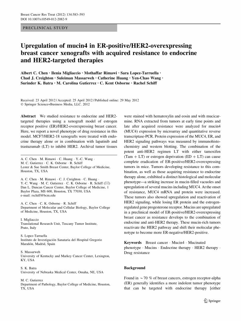
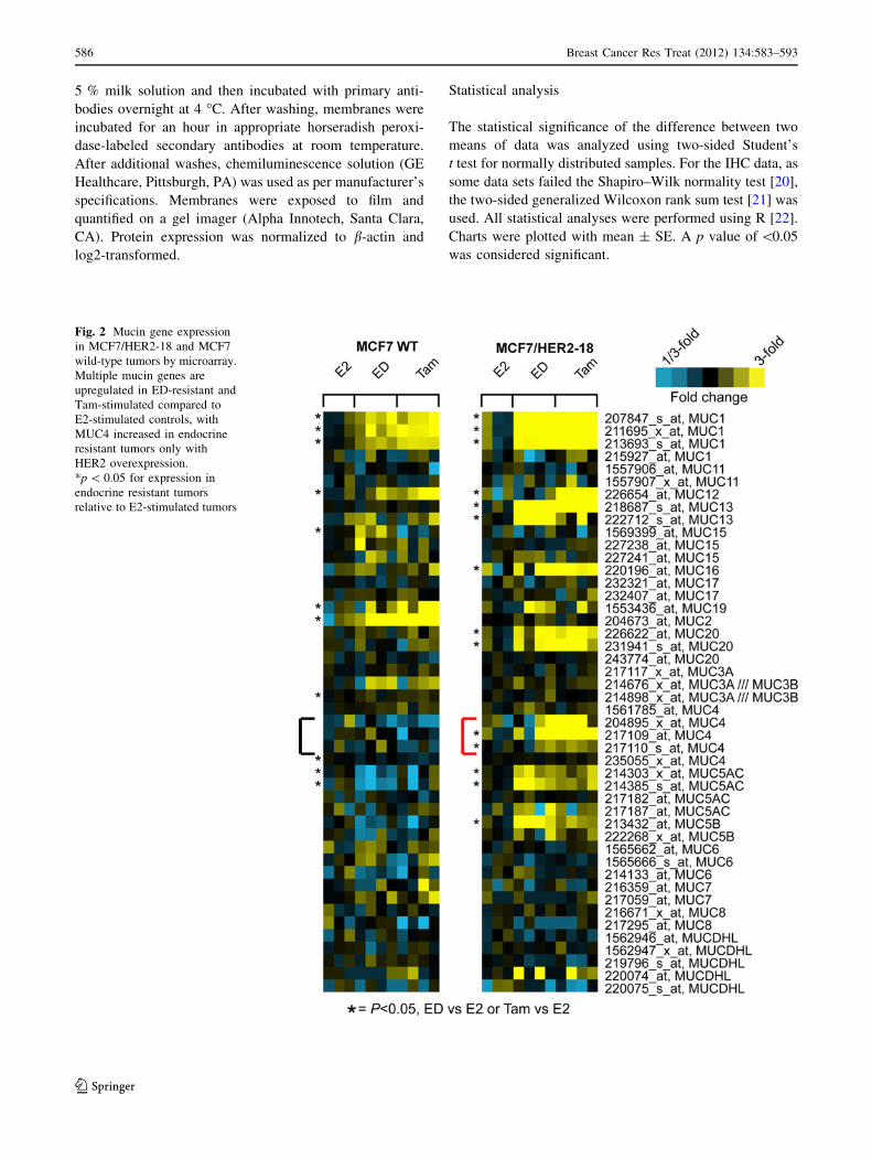
Fig. 2 Mucin gene expression
in MCF7/HER2-18 and MCF7
wild-type tumors by microarray.
Multiple mucin genes are
upregulated in ED-resistant and
Tam-stimulated compared to
E2-stimulated controls, with
MUC4 increased in endocrine
resistant tumors only with
HER2 overexpression.
*p \ 0.05 for expression in
endocrine resistant tumors
relative to E2-stimulated tumors
586 Breast Cancer Res Treat (2012) 134:583–593
123
Results
Mucinated phenotype in drug-resistant xenograft
tumors
We have previously shown [11, 12] that MCF7/HER2-18
xenografts are de novo resistant to Tam treatment and
rapidly acquire resistance to ED, and adding potent dual-
agent anti-HER2 treatment (LT) can delay this effect and
even completely eradicate some tumors (Fig. 1a). Impor-
tantly, we observed striking histological changes in these
tumors resistant to Tam and ED, alone or in combination
with LT (Fig. 1b). H&E staining of tumor sections in these
groups detected the presence of multiple occupied vacu-
oles. This phenotype was not observed in E2-stimulated
tumors alone or with LT. Mucicarmine staining confirmed
the presence of mucin in these vacuoles, which were pri-
marily intracellular, exhibiting cellular morphology similar
to signet ring cells (see arrowheads) [23].
Expression of MUC4 mRNA in resistant tumors
We analyzed previously published expression microarrays
of endocrine resistant MCF7/HER2-18 and wild-type
MCF7 tumors for mucin gene expression [15]. mRNA of
multiple mucin genes were upregulated in endocrine-
resistant tumors when compared to E2-stimulated controls
(Fig. 2). MUC4 and MUC5AC mRNA levels were
increased in Tam- and ED-resistant MCF7/HER2-18
tumors but not in Tam- and ED-resistant wild-type MCF7
tumors. Due to its reported role in HER2 stability and
signaling [24, 25], we next focused on MUC4 and tested
for its expression in tumors treated with endocrine therapy
with LT. MUC4 expression was dramatically increased in
tumors resistant to endocrine therapy alone (Tam or ED)
and in combination with LT when compared to E2-stimu-
lated control tumors alone or with LT (Fig. 3a). The MUC4
levels were similar in fold change to those we found in
previous microarray data [15].
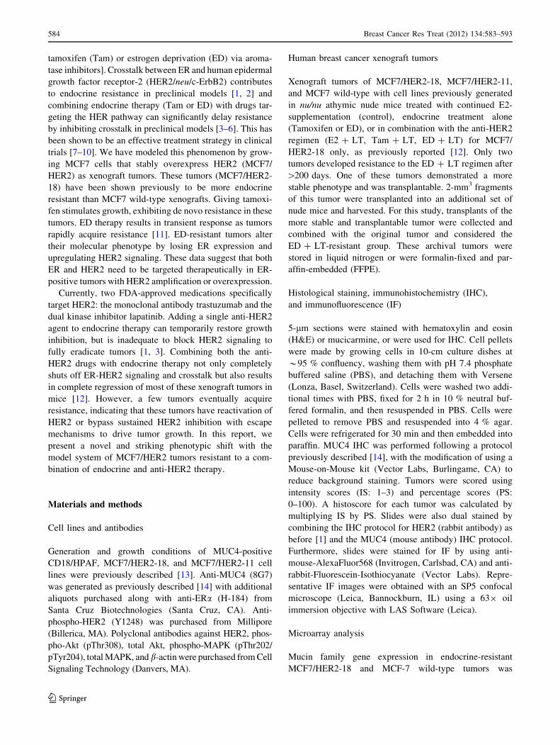
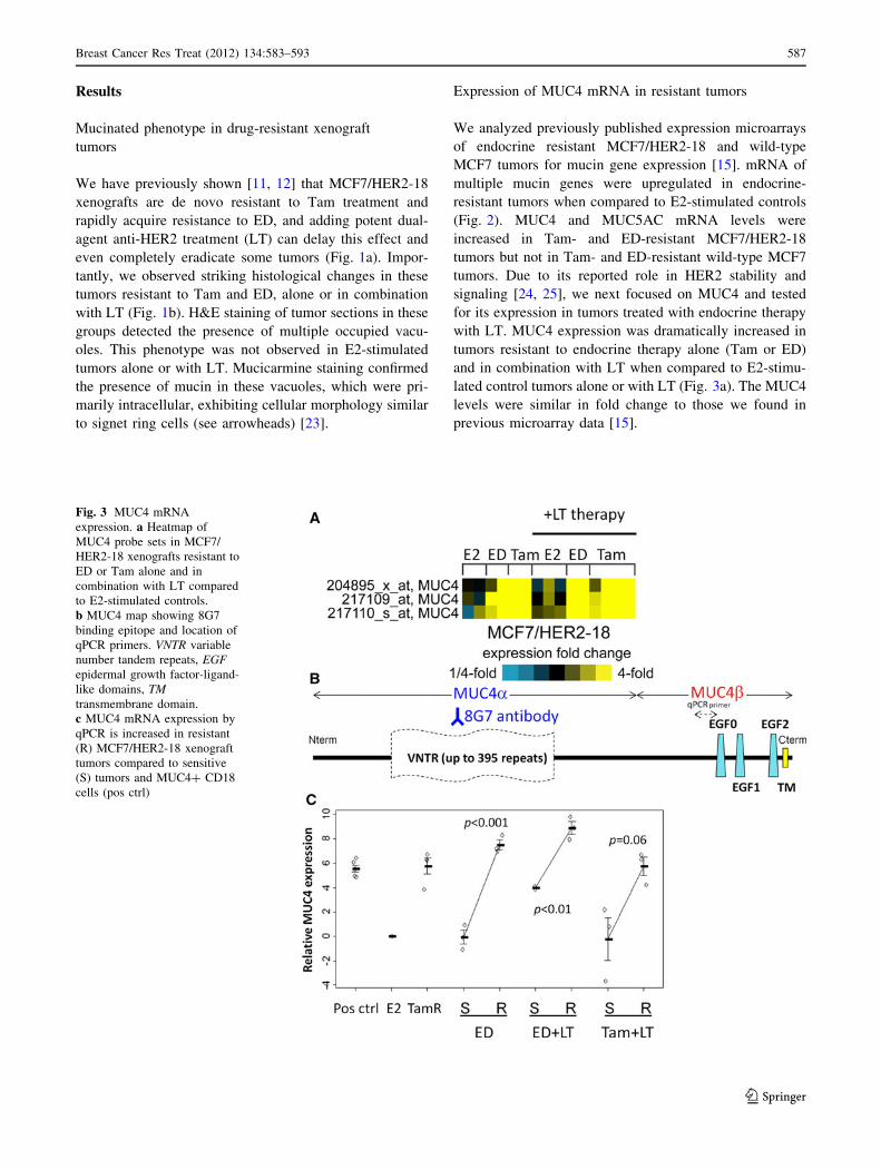
Fig. 3 MUC4 mRNA
expression. a Heatmap of
MUC4 probe sets in MCF7/
HER2-18 xenografts resistant to
ED or Tam alone and in
combination with LT compared
to E2-stimulated controls.
b MUC4 map showing 8G7
binding epitope and location of
qPCR primers. VNTR variable
number tandem repeats, EGFepidermal growth factor-ligand-
like domains, TMtransmembrane domain.
c MUC4 mRNA expression by
qPCR is increased in resistant
(R) MCF7/HER2-18 xenograft
tumors compared to sensitive
(S) tumors and MUC4? CD18
cells (pos ctrl)
Breast Cancer Res Treat (2012) 134:583–593 587
123
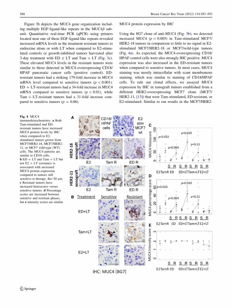
Figure 3b depicts the MUC4 gene organization includ-
ing multiple EGF-ligand-like repeats in the MUC4b sub-
unit. Quantitative real-time PCR (qPCR) using primers
located near one of these EGF-ligand-like repeats revealed
increased mRNA levels in the treatment resistant tumors to
endocrine alone or with LT when compared to E2-stimu-
lated controls or growth-inhibited tumors harvested after
3-day treatment with ED ± LT and Tam ? LT (Fig. 3c).
These elevated MUC4 levels in the resistant tumors were
similar to those detected in MUC4-overexpressing CD18/
HPAF pancreatic cancer cells (positive control). ED-
resistant tumors had a striking 179-fold increase in MUC4
mRNA level compared to sensitive tumors (p \ 0.001).
ED ? LT-resistant tumors had a 34-fold increase in MUC4
mRNA compared to sensitive tumors (p \ 0.01), while
Tam ? LT-resistant tumors had a 31-fold increase com-
pared to sensitive tumors (p = 0.06).
MUC4 protein expression by IHC
Using the 8G7 clone of anti-MUC4 (Fig. 3b), we detected
increased MUC4 (p \ 0.005) in Tam-stimulated MCF7/
HER2-18 tumors in comparison to little to no signal in E2-
stimulated MCF7/HER2-18 or MCF7/wild-type tumors
(Fig. 4a). As expected, the MUC4-overexpressing CD18/
HPAF control cells were also strongly IHC positive. MUC4
expression was also increased in the ED-resistant tumors
when compared to sensitive tumors. In most cases, MUC4
staining was mostly intracellular with scant membranous
staining, which was similar to staining of CD18/HPAF
cells. To rule out clonal effects, we assayed MUC4
expression by IHC in xenograft tumors established from a
different HER2-overexpressing MCF7 clone (MCF7/
HER2-11, [13]) that were Tam-stimulated, ED-resistant, or
E2-stimulated. Similar to our results in the MCF7/HER2-
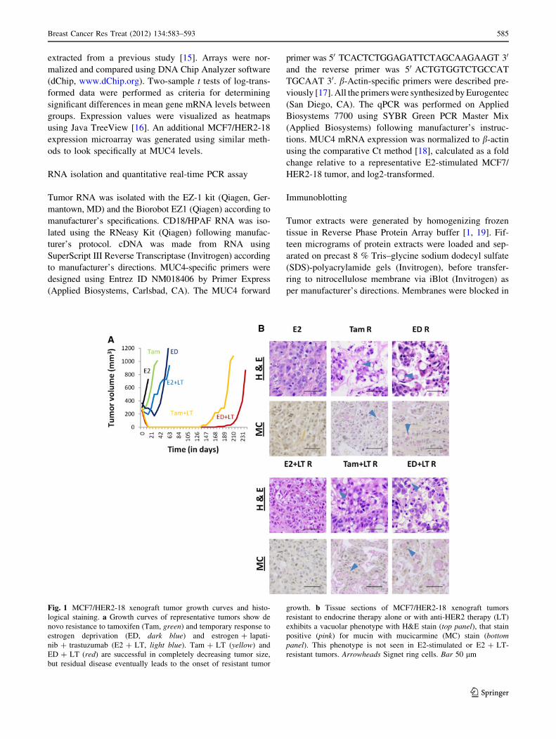
Fig. 4 MUC4
immunohistochemistry. a Both
Tam-stimulated and ED-
resistant tumors have increased
MUC4 protein levels by IHC
when compared to E2-
stimulated tumors grown from
MCF7/HER2-18, MCF7/HER2-
11, or MCF7 wild-type (WT)
cells. The MUC4 patterns are
similar to CD18 cells.
b ED ? LT and Tam ? LT but
not E2 ? LT resistance is
associated with increased
MUC4 protein expression
compared to tumors still
sensitive to therapy. Bar 50 lm.
c Resistant tumors have
increased histoscores versus
sensitive tumors. d Percentage
scores are increased between
sensitive and resistant phases,
but e intensity scores are similar
588 Breast Cancer Res Treat (2012) 134:583–593
123
18 clone, we detected upregulation of MUC4 protein in
Tam-stimulated and ED-resistant tumors but not E2-stim-
ulated tumors (Fig. 4b). Mimicking the mRNA results,
higher levels of MUC4 protein were also found in
ED ? LT- and Tam ? LT-resistant MCF7/HER2-18
tumors but not in tumors in the therapy-sensitive stage. In
contrast, in E2 ? LT-treated tumors, both the sensitive and
the resistant tumors had little or no MUC4 expression.
Overall, the histoscores of ED and ED ? LT-resistant
tumors were higher than sensitive tumors (p \ 0.01,\0.04,
respectively), but not in E2 ? LT-treated tumors (Fig. 4c).
Sensitive tumors tended to have low percentages (\10 %)
of MUC4 staining that were observed as occasional clusters
of MUC4-positive cells (Fig. 4d). Interestingly, these focal
regions of positivity had moderate to very intense staining
similar to those found in resistant tumors (Fig. 4d, e).
Molecular signaling of MCF7/HER2-18 resistant
tumors
To further understand the signaling pathways activated in
these resistant mucinated tumors, we used western blotting
analysis of protein extracts from the same Tam ? LT and
ED ? LT tumors used for qPCR analysis (Fig. 5a).
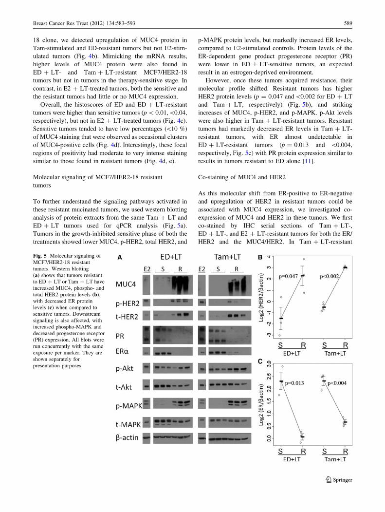
Tumors in the growth-inhibited sensitive phase of both the
treatments showed lower MUC4, p-HER2, total HER2, and
p-MAPK protein levels, but markedly increased ER levels,
compared to E2-stimulated controls. Protein levels of the
ER-dependent gene product progesterone receptor (PR)
were lower in ED ± LT-sensitive tumors, an expected
result in an estrogen-deprived environment.
However, once these tumors acquired resistance, their
molecular profile shifted. Resistant tumors has higher
HER2 protein levels (p = 0.047 and\0.002 for ED ? LT
and Tam ? LT, respectively) (Fig. 5b), and striking
increases of MUC4, p-HER2, and p-MAPK. p-Akt levels
were also higher in Tam ? LT-resistant tumors. Resistant
tumors had markedly decreased ER levels in Tam ? LT-
resistant tumors, with ER almost undetectable in
ED ? LT-resistant tumors (p = 0.013 and \0.004,
respectively, Fig. 5c) with PR protein expression similar to
results in tumors resistant to ED alone [11].
Co-staining of MUC4 and HER2
As this molecular shift from ER-positive to ER-negative
and upregulation of HER2 in resistant tumors could be
associated with MUC4 expression, we investigated co-
expression of MUC4 and HER2 in these tumors. We first
co-stained by IHC serial sections of Tam ? LT-,
ED ? LT-, and E2 ? LT-resistant tumors for both the ER/
HER2 and the MUC4/HER2. In Tam ? LT-resistant
Fig. 5 Molecular signaling of
MCF7/HER2-18 resistant
tumors. Western blotting
(a) shows that tumors resistant
to ED ? LT or Tam ? LT have
increased MUC4, phospho- and
total HER2 protein levels (b),
with decreased ER protein
levels (c) when compared to
sensitive tumors. Downstream
signaling is also affected, with
increased phospho-MAPK and
decreased progesterone receptor
(PR) expression. All blots were
run concurrently with the same
exposure per marker. They are
shown separately for
presentation purposes
Breast Cancer Res Treat (2012) 134:583–593 589
123
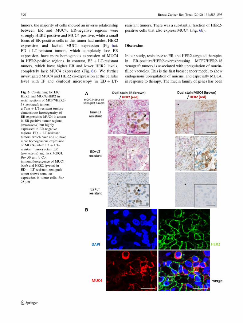
tumors, the majority of cells showed an inverse relationship
between ER and MUC4. ER-negative regions were
strongly HER2-positive and MUC4-positive, while a small
focus of ER-positive cells in this tumor had modest HER2
expression and lacked MUC4 expression (Fig. 6a).
ED ? LT-resistant tumors, which completely lose ER
expression, have more homogenous expression of MUC4
in HER2-positive regions. In contrast, E2 ? LT-resistant
tumors, which have higher ER and lower HER2 levels,
completely lack MUC4 expression (Fig. 6a). We further
investigated MUC4 and HER2 co-expression at the cellular
level with IF and confocal microscopy in ED ? LT-
resistant tumors. There was a substantial fraction of HER2-
positive cells that also express MUC4 (Fig. 6b).
Discussion
In our study, resistance to ER and HER2-targeted therapies
in ER-positive/HER2-overexpressing MCF7/HER2-18
xenograft tumors is associated with upregulation of mucin-
filled vacuoles. This is the first breast cancer model to show
endogenous upregulation of mucins, and especially MUC4,
in response to therapy. The mucin family of genes has been
Fig. 6 Co-staining for ER/
HER2 and MUC4/HER2 in
serial sections of MCF7/HER2-
18 xenograft tumors.
a Tam ? LT-resistant tumors
demonstrate heterogeneity of
ER expression; MUC4 is absent
in ER-positive tumor regions
(arrowhead) but highly
expressed in ER-negative
regions. ED ? LT-resistant
tumors, which have no ER, have
more homogeneous expression
of MUC4, while E2 ? LT-
resistant tumors retain ER
(arrowhead) and lack MUC4.
Bar 50 lm. b Co-
immunofluorescence of MUC4
(red) and HER2 (green) in
ED ? LT-resistant xenograft
tumor shows some co-
expression in tumor cells. Bar25 lm
590 Breast Cancer Res Treat (2012) 134:583–593
123
hypothesized to be associated with drug resistance in
cancer. While normally found in the epithelium of the GI
tract and respiratory tree, mucin expression is common in
multiple cancers [26]. One mucin in particular, mucin4
(MUC4) is overexpressed in various cancers [27–35].
There are few studies of MUC4 in breast cancer, although
recent preclinical data suggest that MUC4 regulates tumor
cell survival and metastasis [36, 37]. The rat form of
MUC4 can form a potent signaling complex with HER2
through EGF-ligand-like domains [38, 39]. MUC4 is
thought to have two potential mechanisms of HER2-
resistance: enhancement of HER2–HER3 signaling [40,
41] or interference with trastuzumab binding [42]. Studies
in pancreatic, gall bladder, and melanoma cancer cell lines
suggest that HER2 and MUC4 form a complex in HER2
non-overexpressing cell lines [24, 39, 43].
Most of these previous studies have focused on MUC4
expression in vitro. While we found MUC4 and HER2 co-
expression in our xenograft tumors, MUC4 expression was
mostly cytoplasmic and did not stain most mucin vacuoles.
While we focused this study on MUC4, other mucins were
also observed to be upregulated, including secreted mucins
that may stain the mucin vacuoles. A common trigger, such as
stress response or cytokine signaling, which have been pre-
viously reported to upregulate MUC4 expression in cell lines
[44–48], may be responsible for the mucinated phenotype.
MUC4 regulation in breast cancer is not well under-
stood. The MUC4-expressing cell line JIMT-1 has low
levels of HER2 expression with low p-HER2. Another
study suggests that MUC4 is downstream of MAPK sig-
naling, functioning through the Ets transcription factor
PEA3 [49]. Interestingly, while we did not detect an
increase in PEA3 by microarray, two Ets-related factors
(ELF1 and ETV6) were upregulated in resistant tumors
[15]. High HER2 activity activates MAPK; Tam ? LT and
ED ? LT-treated tumors had activation of HER2 and
MAPK in resistant tumors compared to sensitive tumors.
Tumors treated with E2 ? LT have levels of MUC4 sim-
ilar to E2-stimulated controls, showing that LT alone is
insufficient to induce MUC4 expression and suggesting
that the mucinated phenotype is not found when estrogen
signaling remains active.
Our data suggests that ER may instead have a repressive
role, as loss of ER coincides with increased MUC4.
Alternatively, increased growth factor signaling decreases
ER expression and activity [50, 51], and mucin upregula-
tion could be related to HER2 reactivation and MAPK
signaling. Although MCF7 wild-type tumors acquire
endocrine resistance by upregulating HER1/HER2 signal-
ing [3, 11], they fail to upregulate MUC4. This implies that
a threshold HER2 level may be needed to upregulate
MUC4, as MCF7/HER2-18 stably overexpresses HER2 48
times higher than MCF7 parental cells [13].
This preclinical model has shown an interesting phe-
notype associated with anti-HER2 treatment and endocrine
resistance. In the ED ? LT and Tam ? LT therapy-sen-
sitive phase of growth, HER2 is inhibited but ER signaling
is still partially active. Conversely, resistance to ED ? LT
and Tam ? LT, similar to ED- and Tam-resistant tumors,
is associated with a molecular shift away from ER sig-
naling but with reactivation of HER2. Whether MUC4
overexpression is the cause or a contributor to reactivation
of the HER2 pathway and drug resistance is unknown.
Nonetheless, based on the known cellular biology of
MUC4 there is a conceptual context to investigate MUC4
as a mechanism of resistance. MUC4 has been shown in
pancreatic and ovarian cancer cells to increase tumor cell
proliferation, motility, and tumorigenicity [52–54]. We
have been unable to examine the role of MUC4 in acquired
resistance in clinical samples, as resistant tumors from
human trials of L ? T or ED in the presence of HER2-
positive disease are currently unavailable; however, these
studies are warranted as samples will become available. In
summary, this study describes a novel mucinated pheno-
type seen in conjunction with a shift in the intimate
crosstalk between ER and HER2 resulting in an ER-
negative/HER2-positive tumor with reactivation of HER
signaling and treatment resistance [3, 11, 55]. Upon
acquiring resistance to therapy, the molecular profile of
these tumors exhibits ER plasticity as it changes its phe-
notype to ER-negative/PR-negative/HER2-positive; the
HER2 pathway is reactivated and there is marked upreg-
ulation of several mucins, including MUC4.
Acknowledgments The authors thank Rena Mao and the Baylor
Breast Center Pathology Core, Robin Ward and Maria Fernanda Prigge
for technical assistance, Dr. Susan Hilsenbeck for advice on statistical
analysis, Dr. Gary Chamness for help with manuscript writing, and
Dr. Kermit Carraway for scientific discussion. This study was sup-
ported by DOD Grant W81XWH-08-1-0264 (to A.C.C.) and NIH
SPORE Grant P50CA058183 and Cancer Center Grant P30CA125123,
the EIF/Lee Jeans Breast Cancer Research Program, Breast Cancer
Research Foundation and Stand Up 2 Cancer (to C.K.O. and R.S.).
Disclosures The experiments described in this study comply with
the current laws of the country in which they were performed. The
authors declare that they have no relevant conflict of interest.
References
1. Arpino G, Gutierrez C, Weiss H, Rimawi M, Massarweh S,
Bharwani L, De Placido S, Osborne CK, Schiff R (2007) Treat-
ment of human epidermal growth factor receptor 2-overexpress-
ing breast cancer xenografts with multiagent HER-targeted
therapy. J Natl Cancer Inst 99(9):694–705. doi:10.1093/jnci/
djk151
2. Arpino G, Wiechmann L, Osborne CK, Schiff R (2008) Crosstalk
between the estrogen receptor and the HER tyrosine kinase
receptor family: molecular mechanism and clinical implications
Breast Cancer Res Treat (2012) 134:583–593 591
123
for endocrine therapy resistance. Endocr Rev 29(2):217–233. doi:
10.1210/er.2006-0045
3. Massarweh S, Osborne CK, Creighton CJ, Qin L, Tsimelzon A,
Huang S, Weiss H, Rimawi M, Schiff R (2008) Tamoxifen
resistance in breast tumors is driven by growth factor receptor
signaling with repression of classic estrogen receptor genomic
function. Cancer Res 68(3):826–833. doi:10.1158/0008-5472.
CAN-07-2707
4. Britton DJ, Hutcheson IR, Knowlden JM, Barrow D, Giles M,
McClelland RA, Gee JM, Nicholson RI (2006) Bidirectional
cross talk between ERalpha and EGFR signalling pathways reg-
ulates tamoxifen-resistant growth. Breast Cancer Res Treat
96(2):131–146. doi:10.1007/s10549-005-9070-2
5. Knowlden JM, Hutcheson IR, Jones HE, Madden T, Gee JM,
Harper ME, Barrow D, Wakeling AE, Nicholson RI (2003)
Elevated levels of epidermal growth factor receptor/c-erbB2
heterodimers mediate an autocrine growth regulatory pathway in
tamoxifen-resistant MCF-7 cells. Endocrinology 144(3):1032–
1044
6. Leary AF, Drury S, Detre S, Pancholi S, Lykkesfeldt AE, Martin
LA, Dowsett M, Johnston SR (2010) Lapatinib restores hormone
sensitivity with differential effects on estrogen receptor signaling
in cell models of human epidermal growth factor receptor
2-negative breast cancer with acquired endocrine resistance. Clin
Cancer Res 16(5):1486–1497. doi:10.1158/1078-0432.CCR-09-
1764
7. Ellis MJ, Coop A, Singh B, Mauriac L, Llombert-Cussac A,
Janicke F, Miller WR, Evans DB, Dugan M, Brady C, Quebe-
Fehling E, Borgs M (2001) Letrozole is more effective neoad-
juvant endocrine therapy than tamoxifen for ErbB-1- and/or
ErbB-2-positive, estrogen receptor-positive primary breast can-
cer: evidence from a phase III randomized trial. J Clin Oncol
19(18):3808–3816
8. Johnston S, Pippen J Jr, Pivot X, Lichinitser M, Sadeghi S, Dieras
V, Gomez HL, Romieu G, Manikhas A, Kennedy MJ, Press MF,
Maltzman J, Florance A, O’Rourke L, Oliva C, Stein S, Pegram
M (2009) Lapatinib combined with letrozole versus letrozole and
placebo as first-line therapy for postmenopausal hormone
receptor-positive metastatic breast cancer. J Clin Oncol 27(33):
5538–5546. doi:10.1200/JCO.2009.23.3734
9. Kaufman B, Mackey JR, Clemens MR, Bapsy PP, Vaid A,
Wardley A, Tjulandin S, Jahn M, Lehle M, Feyereislova A, Revil
C, Jones A (2009) Trastuzumab plus anastrozole versus anas-
trozole alone for the treatment of postmenopausal women with
human epidermal growth factor receptor 2-positive, hormone
receptor-positive metastatic breast cancer: results from the ran-
domized phase III TAnDEM study. J Clin Oncol
27(33):5529–5537. doi:10.1200/JCO.2008.20.6847
10. Osborne CK, Neven P, Dirix LY, Mackey JR, Robert J, Underhill
C, Schiff R, Gutierrez C, Migliaccio I, Anagnostou VK, Rimm DL,
Magill P, Sellers M (2011) Gefitinib or placebo in combination
with tamoxifen in patients with hormone receptor-positive meta-
static breast cancer: a randomized phase II study. Clin Cancer Res
17(5):1147–1159. doi:10.1158/1078-0432.CCR-10-1869
11. Massarweh S, Osborne CK, Jiang S, Wakeling AE, Rimawi M,
Mohsin SK, Hilsenbeck S, Schiff R (2006) Mechanisms of tumor
regression and resistance to estrogen deprivation and fulvestrant
in a model of estrogen receptor-positive, HER-2/neu-positive
breast cancer. Cancer Res 66(16):8266–8273. doi:10.1158/0008-
5472.CAN-05-4045
12. Rimawi MF, Wiechmann LS, Wang YC, Huang C, Migliaccio I,
Wu MF, Gutierrez C, Hilsenbeck SG, Arpino G, Massarweh S,
Ward R, Soliz R, Osborne CK, Schiff R (2011) Reduced dose and
intermittent treatment with lapatinib and trastuzumab for potent
blockade of the HER pathway in HER-2/neu-overexpressing
breast tumor xenografts. Clin Cancer Res 17:1351–1361
13. Benz CC, Scott GK, Sarup JC, Johnson RM, Tripathy D, Coro-
nado E, Shepard HM, Osborne CK (1992) Estrogen-dependent,
tamoxifen-resistant tumorigenic growth of MCF-7 cells trans-
fected with HER2/neu. Breast Cancer Res Treat 24(2):85–95
14. Moniaux N, Varshney GC, Chauhan SC, Copin MC, Jain M,
Wittel UA, Andrianifahanana M, Aubert JP, Batra SK (2004)
Generation and characterization of anti-MUC4 monoclonal anti-
bodies reactive with normal and cancer cells in humans. J Histo-
chem Cytochem 52(2):253–261
15. Creighton CJ, Massarweh S, Huang S, Tsimelzon A, Hilsenbeck
SG, Osborne CK, Shou J, Malorni L, Schiff R (2008) Develop-
ment of resistance to targeted therapies transforms the clinically
associated molecular profile subtype of breast tumor xenografts.
Cancer Res 68(18):7493–7501. doi:10.1158/0008-5472.CAN-08-
1404
16. Saldanha AJ (2004) Java Treeview—extensible visualization of
microarray data. Bioinformatics 20(17):3246–3248. doi:10.1093/
bioinformatics/bth349bth349
17. Hammerich-Hille S, Kaipparettu BA, Tsimelzon A, Creighton
CJ, Jiang S, Polo JM, Melnick A, Meyer R, Oesterreich S (2010)
SAFB1 mediates repression of immune regulators and apoptotic
genes in breast cancer cells. J Biol Chem 285(6):3608–3616. doi:
10.1074/jbc.M109.066431
18. Livak KJ, Schmittgen TD (2001) Analysis of relative gene
expression data using real-time quantitative PCR and the 2(-Delta
Delta C(T)) method. Methods 25(4):402–408. doi:10.1006/meth.
2001.1262
19. Tibes R, Qiu Y, Lu Y, Hennessy B, Andreeff M, Mills GB,
Kornblau SM (2006) Reverse phase protein array: validation of a
novel proteomic technology and utility for analysis of primary
leukemia specimens and hematopoietic stem cells. Mol Cancer
Ther 5(10):2512–2521. doi:10.1158/1535-7163.MCT-06-0334
20. Shapiro SS, Wilk MB (1965) An analysis of variance test for
normality (complete samples). Biometrika 52(3–4):591–611
21. Mann HB, Whitney DR (1947) On a test of whether one of two
random variables is stochastically larger than the other. Ann Math
Stat 18(1):50–60
22. Team RDC (2010) R: a language and environment for statistical
computing. R Foundation for Statistical Computing, Vienna
23. Steinbrecher JS, Silverberg SG (1976) Signet-ring cell carcinoma
of the breast. The mucinous variant of infiltrating lobular carci-
noma? Cancer 37(2):828–840
24. Chaturvedi P, Singh AP, Chakraborty S, Chauhan SC, Bafna S,
Meza JL, Singh PK, Hollingsworth MA, Mehta PP, Batra SK
(2008) MUC4 mucin interacts with and stabilizes the HER2
oncoprotein in human pancreatic cancer cells. Cancer Res
68(7):2065–2070. doi:10.1158/0008-5472.CAN-07-6041
25. Carraway KL, Perez A, Idris N, Jepson S, Arango M, Komatsu
M, Haq B, Price-Schiavi SA, Zhang J, Carraway CA (2002)
Muc4/sialomucin complex, the intramembrane ErbB2 ligand, in
cancer and epithelia: to protect and to survive. Prog Nucleic Acid
Res Mol Biol 71:149–185
26. Hollingsworth MA, Swanson BJ (2004) Mucins in cancer: pro-tection and control of the cell surface. Nat Rev Cancer
4(1):45–60. doi:10.1038/nrc1251nrc1251
27. Karg A, Dinc ZA, Basok O, Ucvet A (2006) MUC4 expression
and its relation to ErbB2 expression, apoptosis, proliferation,
differentiation, and tumor stage in non-small cell lung cancer
(NSCLC). Pathol Res Pract 202(8):577–583. doi:10.1016/j.prp.
2006.04.002
28. Kwon KY, Ro JY, Singhal N, Killen DE, Sienko A, Allen TC,
Zander DS, Barrios R, Haque A, Cagle PT (2007) MUC4 expres-
sion in non-small cell lung carcinomas: relationship to tumor his-
tology and patient survival. Arch Pathol Lab Med 131(4):593–598
29. Tamada S, Shibahara H, Higashi M, Goto M, Batra SK, Imai K,
Yonezawa S (2006) MUC4 is a novel prognostic factor of
592 Breast Cancer Res Treat (2012) 134:583–593
123
extrahepatic bile duct carcinoma. Clin Cancer Res 12(14 Pt 1):
4257–4264. doi:10.1158/1078-0432.CCR-05-2814
30. Tsutsumida H, Goto M, Kitajima S, Kubota I, Hirotsu Y,
Wakimoto J, Batra SK, Imai K, Yonezawa S (2007) MUC4
expression correlates with poor prognosis in small-sized lung
adenocarcinoma. Lung Cancer 55(2):195–203. doi:10.1016/j.
lungcan.2006.10.013
31. Chauhan SC, Singh AP, Ruiz F, Johansson SL, Jain M, Smith
LM, Moniaux N, Batra SK (2006) Aberrant expression of MUC4
in ovarian carcinoma: diagnostic significance alone and in com-
bination with MUC1 and MUC16 (CA125). Mod Pathol
19(10):1386–1394. doi:10.1038/modpathol.3800646
32. Munro EG, Jain M, Oliva E, Kamal N, Lele SM, Lynch MP, Guo
L, Fu K, Sharma P, Remmenga S, Growdon WB, Davis JS,
Rueda BR, Batra SK (2009) Upregulation of MUC4 in cervical
squamous cell carcinoma: pathologic significance. Int J Gynecol
Pathol 28(2):127–133. doi:10.1097/PGP.0b013e318184f3e0
33. Singh AP, Chauhan SC, Bafna S, Johansson SL, Smith LM,
Moniaux N, Lin MF, Batra SK (2006) Aberrant expression of
transmembrane mucins, MUC1 and MUC4, in human prostate
carcinomas. Prostate 66(4):421–429. doi:10.1002/pros.20372
34. Miyahara N, Shoda J, Ishige K, Kawamoto T, Ueda T, Taki R,
Ohkohchi N, Hyodo I, Thomas MB, Krishnamurthy S, Carraway
KL, Irimura T (2008) MUC4 interacts with ErbB2 in human
gallbladder carcinoma: potential pathobiological implications.
Eur J Cancer 44(7):1048–1056. doi:10.1016/j.ejca.2008.03.007
35. Andrianifahanana M, Moniaux N, Schmied BM, Ringel J, Friess
H, Hollingsworth MA, Buchler MW, Aubert JP, Batra SK (2001)
Mucin (MUC) gene expression in human pancreatic adenocar-
cinoma and chronic pancreatitis: a potential role of MUC4 as a
tumor marker of diagnostic significance. Clin Cancer Res 7(12):
4033–4040
36. Workman HC, Miller JK, Ingalla EQ, Kaur RP, Yamamoto DI,
Beckett LA, Young LJ, Cardiff RD, Borowsky AD, Carraway
KL, Sweeney C, Carraway KL 3rd (2009) The membrane mucin
MUC4 is elevated in breast tumor lymph node metastases relative
to matched primary tumors and confers aggressive properties to
breast cancer cells. Breast Cancer Res 11(5):R70. doi:10.1186/
bcr2364
37. Workman HC, Sweeney C, Carraway KL 3rd (2009) The mem-
brane mucin Muc4 inhibits apoptosis induced by multiple insults
via ErbB2-dependent and ErbB2-independent mechanisms.
Cancer Res 69(7):2845–2852. doi:10.1158/0008-5472.CAN-08-
2089
38. Ramsauer VP, Pino V, Farooq A, Carothers Carraway CA, Salas
PJ, Carraway KL (2006) Muc4–ErbB2 complex formation and
signaling in polarized CACO-2 epithelial cells indicate that Muc4
acts as an unorthodox ligand for ErbB2. Mol Biol Cell
17(7):2931–2941. doi:10.1091/mbc.E05-09-0895
39. Yokoyama A, Shi BH, Kawai T, Konishi H, Andoh R, Tachikawa
H, Ihara S, Fukui Y (2007) Muc4 is required for activation of
ErbB2 in signet ring carcinoma cell lines. Biochem Biophys Res
Commun 355(1):200–203. doi:10.1016/j.bbrc.2007.01.133
40. Carraway KL 3rd, Rossi EA, Komatsu M, Price-Schiavi SA,
Huang D, Guy PM, Carvajal ME, Fregien N, Carraway CA,
Carraway KL (1999) An intramembrane modulator of the ErbB2
receptor tyrosine kinase that potentiates neuregulin signaling.
J Biol Chem 274(9):5263–5266
41. Funes M, Miller JK, Lai C, Carraway KL 3rd, Sweeney C (2006)
The mucin Muc4 potentiates neuregulin signaling by increasing
the cell-surface populations of ErbB2 and ErbB3. J Biol Chem
281(28):19310–19319. doi:10.1074/jbc.M603225200
42. Nagy P, Friedlander E, Tanner M, Kapanen AI, Carraway KL,
Isola J, Jovin TM (2005) Decreased accessibility and lack of
activation of ErbB2 in JIMT-1, a herceptin-resistant, MUC4-
expressing breast cancer cell line. Cancer Res 65(2):473–482
43. Price-Schiavi SA, Jepson S, Li P, Arango M, Rudland PS, Yee L,
Carraway KL (2002) Rat Muc4 (sialomucin complex) reduces
binding of anti-ErbB2 antibodies to tumor cell surfaces, a
potential mechanism for herceptin resistance. Int J Cancer 99(6):
783–791. doi:10.1002/ijc.10410
44. Mejias-Luque R, Peiro S, Vincent A, Van Seuningen I, de Bolos
C (2008) IL-6 induces MUC4 expression through gp130/STAT3
pathway in gastric cancer cell lines. Biochim Biophys Acta
1783(10):1728–1736. doi:10.1016/j.bbamcr.2008.05.020
45. Andrianifahanana M, Singh AP, Nemos C, Ponnusamy MP,
Moniaux N, Mehta PP, Varshney GC, Batra SK (2007) IFN-
gamma-induced expression of MUC4 in pancreatic cancer cells is
mediated by STAT-1 upregulation: a novel mechanism for IFN-
gamma response. Oncogene 26(51):7251–7261. doi:10.1038/sj.
onc.1210532
46. Andrianifahanana M, Agrawal A, Singh AP, Moniaux N, van
Seuningen I, Aubert JP, Meza J, Batra SK (2005) Synergistic
induction of the MUC4 mucin gene by interferon-gamma and
retinoic acid in human pancreatic tumour cells involves a
reprogramming of signalling pathways. Oncogene 24(40):6143–
6154. doi:10.1038/sj.onc.1208756
47. Damera G, Xia B, Ancha HR, Sachdev GP (2006) IL-9 modu-
lated MUC4 gene and glycoprotein expression in airway epi-
thelial cells. Biosci Rep 26(1):55–67. doi:10.1007/s10540-006-
9000-5
48. Damera G, Xia B, Sachdev GP (2006) IL-4 induced MUC4
enhancement in respiratory epithelial cells in vitro is mediated
through JAK-3 selective signaling. Respir Res 7:39. doi:10.1186/
1465-9921-7-39
49. Perez A, Barco R, Fernandez I, Price-Schiavi SA, Carraway KL
(2003) PEA3 transactivates the Muc4/sialomucin complex pro-
moter in mammary epithelial and tumor cells. J Biol Chem 278(38):
36942–36952. doi:10.1074/jbc.M300264200M300264200
50. Guo S, Sonenshein GE (2004) Forkhead box transcription factor
FOXO3a regulates estrogen receptor alpha expression and is
repressed by the Her-2/neu/phosphatidylinositol 3-kinase/Akt
signaling pathway. Mol Cell Biol 24(19):8681–8690. doi:
10.1128/MCB.24.19.8681-8690.200424/19/8681
51. Creighton CJ, Fu X, Hennessy BT, Casa AJ, Zhang Y, Gonzalez-
Angulo AM, Lluch A, Gray JW, Brown PH, Hilsenbeck SG,
Osborne CK, Mills GB, Lee AV, Schiff R (2010) Proteomic and
transcriptomic profiling reveals a link between the PI3K pathway
and lower estrogen-receptor (ER) levels and activity in ER?
breast cancer. Breast Cancer Res 12(3):R40. doi:10.1186/bcr2594
52. Moniaux N, Chaturvedi P, Varshney GC, Meza JL, Rodriguez-
Sierra JF, Aubert JP, Batra SK (2007) Human MUC4 mucin
induces ultra-structural changes and tumorigenicity in pancreatic
cancer cells. Br J Cancer 97(3):345–357. doi:10.1038/sj.bjc.66
03868
53. Ponnusamy MP, Singh AP, Jain M, Chakraborty S, Moniaux N,
Batra SK (2008) MUC4 activates HER2 signalling and enhances
the motility of human ovarian cancer cells. Br J Cancer
99(3):520–526. doi:10.1038/sj.bjc.6604517
54. Ponnusamy MP, Lakshmanan I, Jain M, Das S, Chakraborty S, Dey
P, Batra SK (2010) MUC4 mucin-induced epithelial to mesen-
chymal transition: a novel mechanism for metastasis of human
ovarian cancer cells. Oncogene. doi:10.1038/onc.2010.309
55. Shou J, Massarweh S, Osborne CK, Wakeling AE, Ali S, Weiss
H, Schiff R (2004) Mechanisms of tamoxifen resistance:
increased estrogen receptor-HER2/neu cross-talk in ER/HER2-
positive breast cancer. J Natl Cancer Inst 96(12):926–935
Breast Cancer Res Treat (2012) 134:583–593 593
123