Embed Size (px)
DESCRIPTION
Upper Extremity Injuries in the Pediatric Population. By Matthew Bloom, OMS IV. Overview. Birth traumas Clavicular Fracture Klumpke’s Palsy Erb’s Palsy Fractures Not related to abuse Torus, Greenstick, and Supracondylar types Salter-Harris Classification Sprains and Strains - PowerPoint PPT Presentation
Citation preview
Upper Extremity Injuries in the Pediatric Population
By Matthew Bloom, OMS IV
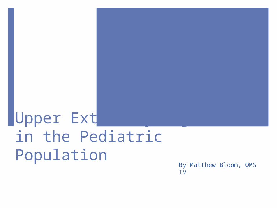
OverviewBirth traumas
Clavicular Fracture Klumpke’s Palsy Erb’s Palsy
Fractures Not related to abuse Torus, Greenstick, and Supracondylar types Salter-Harris Classification
Sprains and StrainsNursemaid’s Elbow
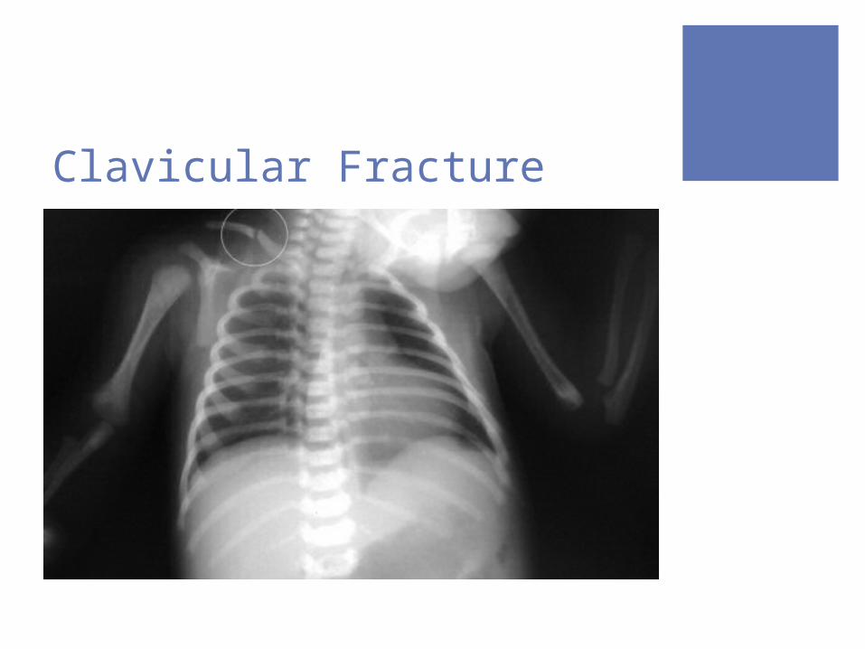
Clavicular FractureMost common bone fractured during deliveryComplete fracture symptoms include:
Decreased or absent movement Gross deformity of the clavicle Tenderness on palpation Localized crepitus Absence of Moro’s reflex
Greenstick (partial) fractures have no symptoms initially and the diagnosis is made at 7 to 10 days postpartum because of callus formation
Clavicular Fracture
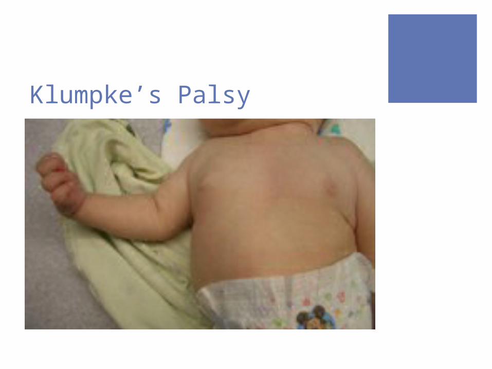
Klumpke’s Palsy Involves the lower arm Affects the C7, C8, and T1 nerve roots Hand is paralyzed and has an absent grasp reflex Causes a “claw hand” deformity Rare to have an isolated Klumpke’s palsy Often accompanied by Horner’s syndrome
P = Ptosis A = Anhidrosis M = Miosis
Klumpke’s Palsy
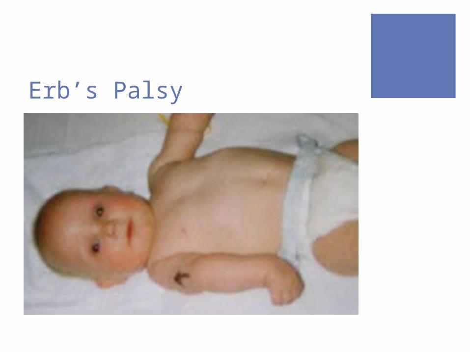
Erb’s Palsyaka Erb-Duchenne Involves the upper armMost common type of palsy during birth Involves C5 and C6 nerve rootsArm is adducted and internally rotated, but the
grasp reflex is intact
Erb’s Palsy
Treatment for PalsySymptoms resolve by two years in most casesTreatment involves early immobilization with
passive movement in order to prevent contractures, followed by physical therapy with active range of motion exercises
In severe cases, surgery may be required to replace nerves that are refractory to healing
Torus FractureAKA buckle fracture Impact injury in which the bone cortex is
buckled but not disruptedAcute angulation of the cortex is noted, as
opposed to the usual curved surfaceStableOften best visualized on lateral viewSoft tissue changes may be the only indication
of fracture (ie pronator fat pad)
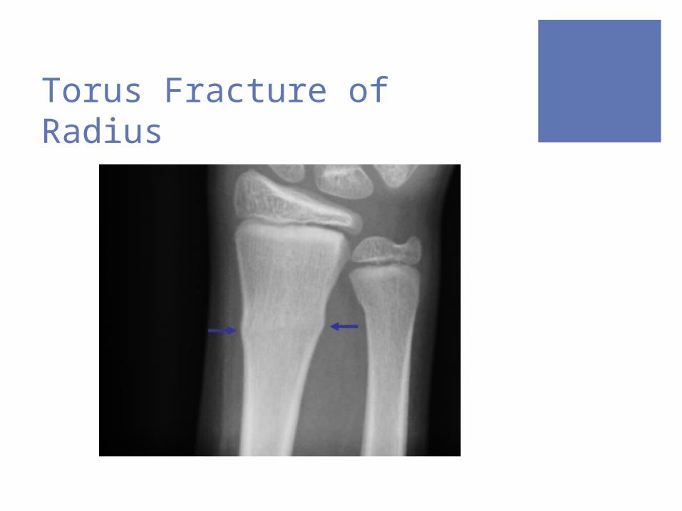
Torus Fracture of Radius
Greenstick FractureAngulation beyond the limits of plastic
deformation Incomplete fracture in which the cortex is
disrupted only on one sideRepresents bone failure (fracture) on the
tension side and a plastic (or bend) deformity on the compression side
Like breaking a green stick, hence the name
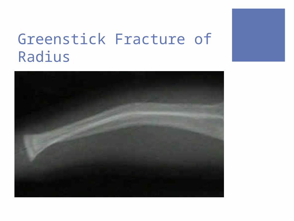
Greenstick Fracture of Radius
Supracondylar Fracture Distal humerus just above the epicondyles Most common fracture in children Associated with ligamentous laxity Extension or Flexion
Extension (80% of cases) – distal fragment is displaced posteriorly
Flexion (20%) – distal fragment is displaced anteriorly
Degree of separation Type I – undisplaced or minimally displaced Type II – partially displaced Type III – fully displaced
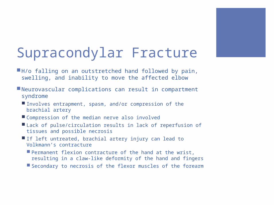
Supracondylar Fracture H/o falling on an outstretched hand followed by pain,
swelling, and inability to move the affected elbow Neurovascular complications can result in compartment
syndrome Involves entrapment, spasm, and/or compression of the
brachial artery Compression of the median nerve also involved Lack of pulse/circulation results in lack of reperfusion of
tissues and possible necrosis If left untreated, brachial artery injury can lead to Volkmann’s
contracture Permanent flexion contracture of the hand at the wrist,
resulting in a claw-like deformity of the hand and fingers Secondary to necrosis of the flexor muscles of the forearm
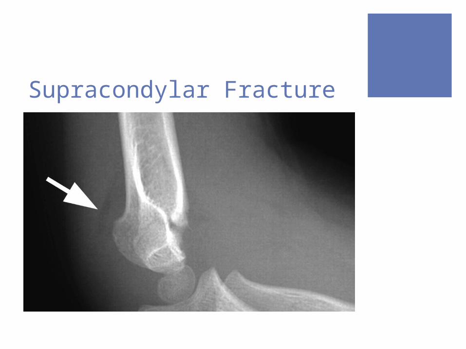
Supracondylar Fracture
Salter-Harris Fracture Classification of Growth Plate Injuries
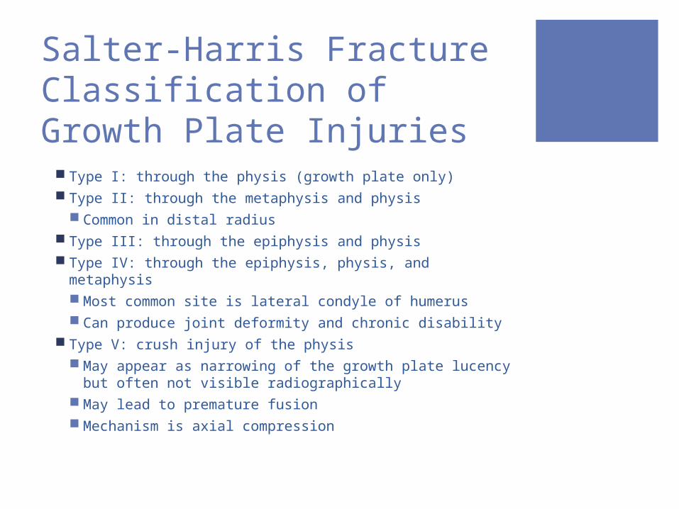
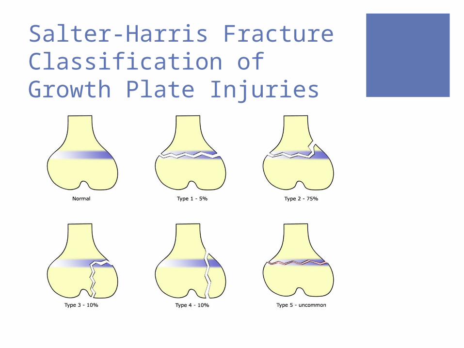
Type I: through the physis (growth plate only) Type II: through the metaphysis and physis
Common in distal radius Type III: through the epiphysis and physis Type IV: through the epiphysis, physis, and metaphysis
Most common site is lateral condyle of humerus Can produce joint deformity and chronic disability
Type V: crush injury of the physis May appear as narrowing of the growth plate
lucency but often not visible radiographically May lead to premature fusion Mechanism is axial compression
Salter-Harris Fracture Classification of Growth Plate Injuries
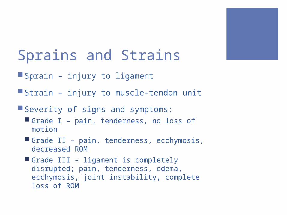
Sprains and StrainsSprain – injury to ligamentStrain – injury to muscle-tendon unitSeverity of signs and symptoms:
Grade I – pain, tenderness, no loss of motion Grade II – pain, tenderness, ecchymosis,
decreased ROM Grade III – ligament is completely disrupted; pain,
tenderness, edema, ecchymosis, joint instability, complete loss of ROM
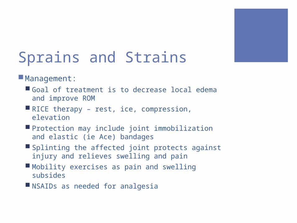
Sprains and StrainsManagement:
Goal of treatment is to decrease local edema and improve ROM
RICE therapy – rest, ice, compression, elevation Protection may include joint immobilization and
elastic (ie Ace) bandages Splinting the affected joint protects against injury
and relieves swelling and pain Mobility exercises as pain and swelling subsides NSAIDs as needed for analgesia
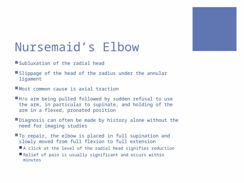
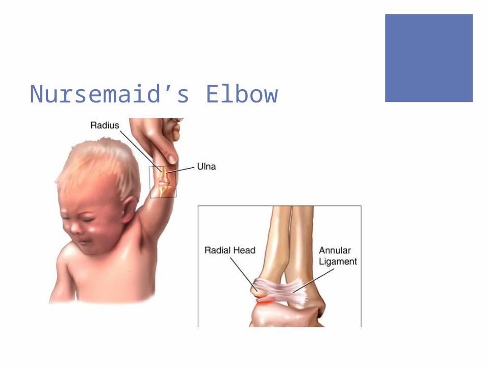
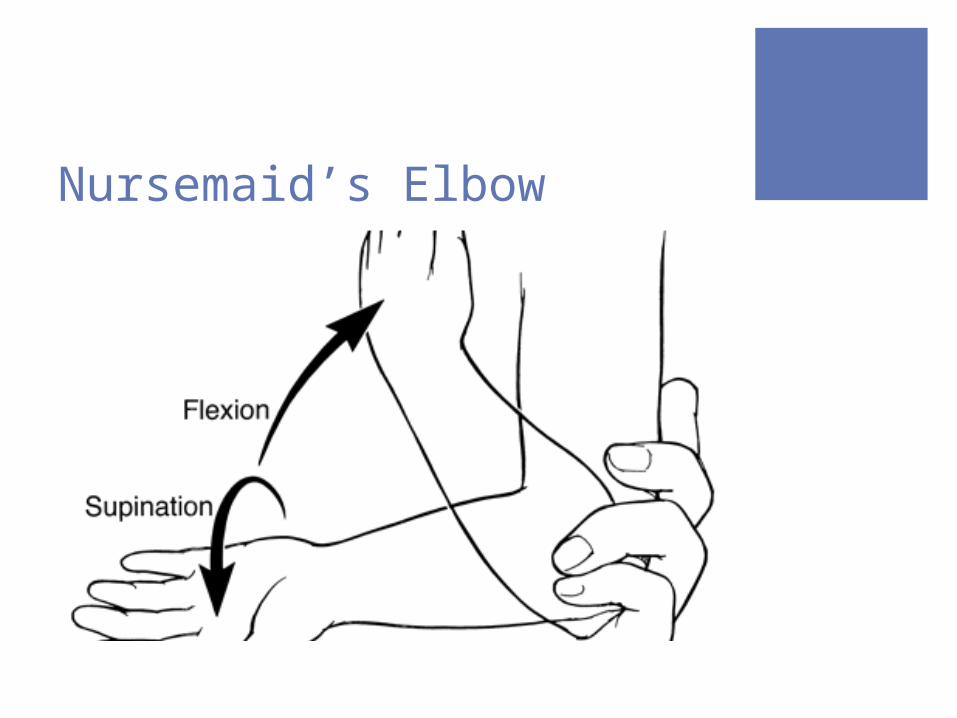
Nursemaid’s Elbow Subluxation of the radial head Slippage of the head of the radius under the annular ligament Most common cause is axial traction H/o arm being pulled followed by sudden refusal to use the
arm, in particular to supinate, and holding of the arm in a flexed, pronated position
Diagnosis can often be made by history alone without the need for imaging studies
To repair, the elbow is placed in full supination and slowly moved from full flexion to full extension A click at the level of the radial head signifies reduction Relief of pain is usually significant and occurs within minutes
Nursemaid’s Elbow
Nursemaid’s Elbow
Role of OMT Main objective of OMT is to encourage the body’s
natural ability to heal itself Restoring structure improves function In the acute setting, direct and active techniques are
normally contraindicated (ie HVLA and ME) ST, MFR, CS, and lymphatic techniques can be utilized:
to reduce tension created by compensatory mechanisms the body employs to prevent further injury, thus reducing pain and swelling at both direct and referred sites
to increase circulation, which allows for better blood flow carrying oxygen and nutrients that can help increase the rate of healing

References First-Aid for the Pediatric Clerkship OMM Manual http://niyaf.com/post/31434342/birth-trauma-fractured-clavicle-collar-bone http://pediatricneuro.com/alfonso/pg220.htm http://www.birthtraumaassociation.org.uk/articles/erbs.htm http://www.feinberg.northwestern.edu/emergencymed/residency/ortho-teaching/ped
iatrics/case13/case13background.html
http://www.medscape.com/viewarticle/446548_2 http://imaging.birjournals.org/cgi/content-nw/full/16/2/140/F15 http://en.wikipedia.org/wiki/File:SalterHarris.svg http://www.pedsinbrevard.com/wp-content/html/pa/pa_nursmaid_art.htm http://academiclifeinem.blogspot.com/2011/01/tricks-of-trade-nursemaid-elbow.html http://www.wikiradiography.com/page/Soft+Tissue+Signs-+The+Wrist