Embed Size (px)
Citation preview

Management of Distal Extremity Injuries in College Health John A. Vaughn, MD The Ohio State University

Girl trips over her flip-flops and puts her hand out to catch herself

Distal Radius (Colles’) Fracture
• Most common upper extremity fracture
• FOOSH!
• Extremity often looks normal
– Swelling may not develop immediately
– Often no deformity

Distal Radius Fx - Evaluation
• Assess neuro-vascular status
– Motor, sensory exam
– Pulses, cap refill
– Range of motion
• Obtain x-ray
– What if you don’t have x-ray?

Distal Radius Fx – Management
• Refer to Ortho
– within 3-5 days
• Reverse sugar tong splint is ideal
– 2 planes of motion
• Volar splint and a sling in a pinch

Her flip-flop fanatic friend fell the same way
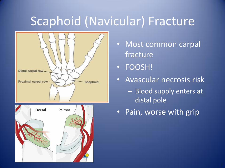
Scaphoid (Navicular) Fracture
• Most common carpal fracture
• FOOSH!
• Avascular necrosis risk
– Blood supply enters at distal pole
• Pain, worse with grip

Scaphoid Fracture - Evaluation
• Compare wrists
– May look normal
• Snuff box tenderness!
– 90% sensitive for fx
– Less specific (40%)
• Obtain x-ray…
But don’t believe them!

Scaphoid Fracture - Management
• Thumb spica splint
• Reassess in 7-10 days
– Re-check snuff box
– Repeat x-rays
• Alternatively, MRI or CT
• Refer to Ortho if fracture is present or symptoms persist

Sophomore’s girlfriend broke up with him last night and he punched a wall

Boxer’s Fracture
• 5th Metacarpal neck fx
• Direct trauma to a clenched fist
– I fought the wall and the wall won.

Boxer’s fracture - Evaluation
• Swelling of dorsum of hand
• Ecchymosis and Tenderness over 5th metacarpal head
• Assess rotational alignment
– On exam
– On x-ray

Boxer’s fracture - Management
• Ulnar gutter splint
• Refer to Ortho in 1 week
• Urgent referral
– Open fracture
– Neurovascular compromise
– “pseudo-clawing”
– Significant angulation

Freshman just got back from ski club trip over break and her thumb hurts

Skier’s Thumb
• Ulnar Collateral Ligament Tear
• Forced abduction and hyperextension of 1st MCP joint
• Ski pole injuries most common cause

Skier’s Thumb - Evaluation

Skier’s Thumb - Evaluation
• Classic history
• Swelling of entire joint
• Tender at ulnar aspect
• Pain with extension or abduction
• Laxity of MCP joint
– Compare to other thumb

Skier’s Thumb - Management
• Thumb spica splint
– At least 6 weeks
• R.I.C.E.
• Ortho referral ASAP especially if…
– Presence of fracture
– Significant laxity of joint
• Early surgical repair has better outcomes

Wrestling with fraternity brother

PIP Volar Plate Injuries
• Mechanism: hyper-extension of joint
• “Swan Neck” deformity
– PIP joint hyperextended by extensor tendons
Volar Plate Injuries
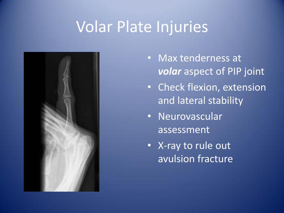
• Max tenderness at volar aspect of PIP joint
• Check flexion, extension and lateral stability
• Neurovascular assessment
• X-ray to rule out avulsion fracture

Volar Plate Injuries
• Block Splint at 30 degrees of flexion
– Progressively extend over 2-4 weeks
– Buddy taping for less severe strains
• Ortho referral
– Presence of fracture
– Unstable joint

6 weeks later his buddy comes in

Central Slip Extensor Injury
• Boutonniere Deformity
• PIP most commonly affected
• Mechanism = forced flexion of an extended PIP joint
• *Usually don’t present for 4-6 weeks

Central Extensor Slip Injury
• Max tenderness at dorsal aspect of PIP joint
• Can’t actively extend PIP joint
• X-ray to rule out dorsal avulsion fracture

Central Extensor Slip Injury
• Continuous splinting in full extension
– 6-8 weeks
• Ortho Referral
– Presence of fracture
– Inability to passively extend PIP joint
– Non-urgent
• What if you can’t tell?

Freshman tossing a baseball on the quad and “jams” his finger

Mallet Finger
• Avulsion of DIP extensor tendon
• Most common tendon injury in finger
• Pain, swelling, bruising
• *Inability to extend DIP
– Flexed DIP at rest

Mallet Finger - Evaluation
• Isolate extensor tendon
– Stabilize PIP joint
• No active extension
– Passive extension intact
• Obtain x-ray to r/o avulsion fracture

Mallet Finger - Management
• Continuous splinting in full extension
– Slight hyper-extension
– 6-8 weeks
• Refer
– Failed splinting
– Presence of fracture
– Distal phalanx subluxation

Junior playing flag football on the quad

Jersey Finger
• Flexor Digitorum Profundus (FDP) Tendon rupture
• Forced hyperextension of flexed DIP joint
• Ring finger = 75% of cases

Jersey Finger - Evaluation
• *Inability to flex DIP
• Again, isolate DIP
• X-ray to rule out fractures

Jersey Finger - Management
• Refer ALL cases to hand surgery ASAP*
– Call Ortho/Hand that day
– Requires surgical repair
• Acute care
– Splint with DIP and PIP joint in slight flexion.
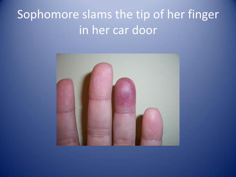
Sophomore slams the tip of her finger in her car door

Distal phalanx (tuft) fracture
• Half of all hand fractures
• Middle finger most commonly involved
• Mechanism = direct blow

Distal Phalanx fracture - evaluation
• Pain, swelling, ecchymosis
• Tuft vs. Distal Phalanx
• Neuro-vascular status
– Capillary refill
– 2-point discrimination
• X-ray to evaluate intra-articular fracture and displacement

Distal phalanx (tuft) fracture
• Splint with DIP in extension for 3-4 weeks
• Referral to ortho/hand
– Immediately • open fx, severe crush
inury, neuro compromise
– Within 3-4 days • Tendon dysfunction
• Nerve dysfunction
• Intraarticular (> 30%) or displaced

Nail bed Injury - Subungual Hematoma
• Distal phalanx fracture more likely if hematoma involves > 50% of nail bed
• Evaluate eponychial fold for disruption or deformity

Nail bed injuries - Trephination
• Indications
– Acute ( < 48 hours)
– Painful
• Electrocautery
– 18-gauge needle
– Heated paperclip
• No antibiotics
• Soapy soaks for 2 days

What if her finger looked like this?

Proximal/Middle phalanx fracture
• Pain, swelling, ecchymosis
• Neuro-vascular status
– Capillary refill
– 2-point discrimination
• X-ray to evaluate intra-articular fracture and displacement

Proximal/Middle phalanx fracture - Management
• Stable, non-displaced
– Buddy taping 4-6 weeks
– Dorsal or volar splint for added protection and pain control
• Referral to Ortho/Hand
– Comminuted , rotational, intraarticular, displaced, angulated or unstable

Guy walks in from soccer practice with his finger looking like this!

PIP dislocation - evaluation
• Most common = dorsal
– Lateral fairly common
– Volar rarely
• Pain, swelling, impaired range of motion and deformity
• X-ray to assess for associated fracture

PIP dislocation - Management
• Reduction – Pre/post x-rays*
– Gentle traction, then flexion
– Dorsal splint in flexion • Buddy tape after 3-5 days
• Prompt Referral – Irreducible
– unstable
– Tendon rupture
– Volar dislocation

Junior playing basketball twists his ankle coming down from a rebound

Ankle Sprains
• Lateral sprains most common
• Medial injuries usually result in fractures
• Syndesmotic (“high”) sprains predict poor outcomes

Ankle Sprains
• Anterior Drawer Test
– Assess ATF ligament
• Talar Tilt Test
– Assess CF ligament
• Squeeze Test
– Assess syndesmotic structures

To X-Ray or Not: Ottowa Rules
• Pain over malleolus and/or midfoot
AND
• Tenderness over malleolus and/or midfoot
OR
• Inability to bear weight immediately and at visit

Ankle Sprain – Management
• Functional Treatment*
I. Start PRICE protocol within 24 hours
II. Strength and ROM exercises in 48-72 hours
III. Endurance and balance training

Ankle fractures
• 60-70% malleolar
– Lateral most common and most stable
• 15-20% bi-malleolar
• 7-12% tri-malleolar
• Isolated medial malleolar fx are rare and unstable
– Treat like bi- or tri-malleolar

Ankle fracture - Management
• Small, non-displaced avulsion fractures
– Like ankle sprain
• Isolated Malleolar fx
– Stirrup splint • At 90 degrees (neutral
position)
• Non weight-bearing
– Ortho follow-up in 3-5 days

What if he has pain here?

5th Metatarsal Styloid Aulsion fracture
• Most common fracture of lower extremity
• Mechanism identical to lateral ankle sprain
– Inversion while foot is plantar flexed
• Walking possible but painful

5th Metatarsal Styloid Avulsion fracture
• Swelling, ecchymosis
• Ottowa rules!
• Beware a Jones fracture
– Same mechanism
– Different management
– Different prognosis
Jones fracture

5th Metatarsal Styloid Avulsion fracture
• Conservative Management
• Weight-bearing as tolerated
• Post-op shoe +/- elastic wrap
• Usually resolves in 3-6 weeks

Junior on ski club trip who tried snowboarding this time

Snowboarder’s fracture
• Lateral Process of Talus
• Exact mechanism unknown
– Axial loading + external rotation, dorsiflexion and inversion
• Soft snowboarding boots contribute

Snowboarder’s fracture
• Lateral Process of Talus fracture
• Have a high index of suspicion
• Hard to pick up on exam and on x-ray
– looks a lot like ankle sprain

Snowboarder’s fracture
• Best treatment not really known
• Non weight-bearing
• Refer to ortho
• Err on the side of caution
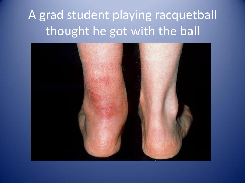
A grad student playing racquetball thought he got with the ball

Achilles Tendon Rupture
• Feels sudden “pop” or “being kicked”
• Weekend warriors in late 20’s, 30’s
• Medications
– Fluoroquinolones
– Steroids
• Missed 25% of time

Thompson Test
• Complete Rupture
– Abnormal Thompson
– Can’t stand on toes
• Partial Rupture
– Normal Thompson
– +/- Palpable defect
– Plantar flexion intact
– Patient can walk
– Tendon is not painful

Achilles Tendon Rupture
• X-rays not helpful
• Prompt ortho referral
• Non weight-bearing
• Aircast or posterior splint for comfort
– Slight plantar flexion

Sophomore joined the bowling team and dropped a ball on her foot

Metatarsal Fracture
• Mechanism = direct blow or twisting
• Edema, ecchymosis, pain, point tenderness
• Neurovascular exam
– Pain, pallor, paresthesia, pulselessness
• Lisfranc joint*
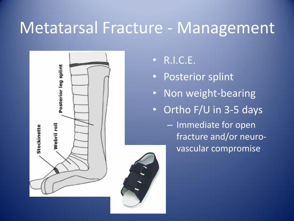
Metatarsal Fracture - Management
• R.I.C.E.
• Posterior splint
• Non weight-bearing
• Ortho F/U in 3-5 days
– Immediate for open fracture and/or neuro-vascular compromise
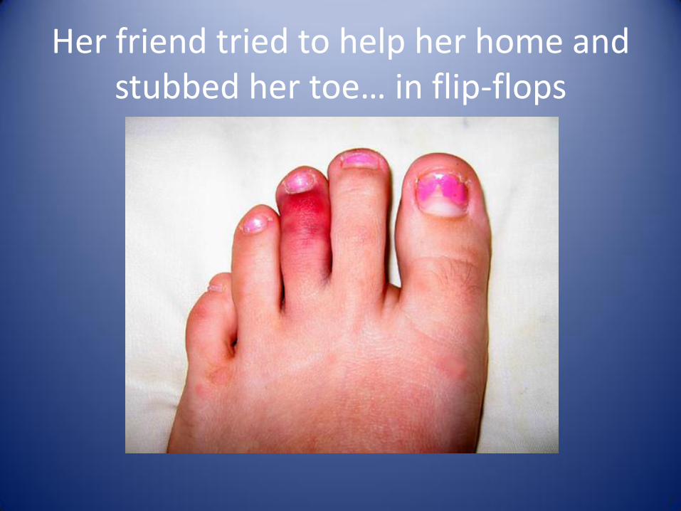
Her friend tried to help her home and stubbed her toe… in flip-flops

Toe Fractures
• Stubbing or direct blow
• Pain, edema, deformity, ecchymosis

Toe Fractures - Management
• Buddy taping
• Postop shoe prn pain
• R.I.C.E.
• Follow-up in 1-2 weeks

Toe Fracture – Referral?
1st toe fractures
• Fracture with dislocation
• Displaced intra-articular fx
• Intra-articular fx > 25% of joint space
• Unstable displaced fx
Lesser toe fractures
• Rarely
• Displaced intra-articular fx
• Irreducible fractures
• Open fractures
• Unstable displaced fractures

Basic Principles of splinting
• 1 joint above and below
• Clean, repair, and dress skin before application
• Clothing considerations
• Neurovascular status pre and post application
• R.I.C.E.
• Position of Function
• Pad between digits and bony prominences

Positions of Function for Splints
Splint Position
Volar Neutral forearm (thumb up), wrist slightly extended
Ulnar gutter Neutral forearm, wrist at 20 degrees extension, MCP at 50 degrees flexion, PIP in slight flexion (10 degrees), DIP in extension
Thumb spica Forearm neutral, wrist at 25 degrees extension, allowing thumb-index finger opposition and alignment of the thumb and forearm (“Can of Soda” position)
Finger Finger in slight flexion
Sugar Tong Elbow at 90 degrees flexion, neutral flexion, neutral wrist
Ankle posterior/stirrup Ankle at 90 degrees

Upper Extremity Splints
SPLINT INDICATION
Volar splint Wrist fractures or sprains, fractures of 2nd to 5th metacarpals, soft tissue injuries of the hand
Reverse sugar tong splint Wrist and distal forearm fractures
Ulnar gutter splint 5th metacarpal (Boxer’s) fractures
Thumb spica splint Scaphoid fractures, fractures of 1st (thumb) metacarpal, ulnar collateral ligament (Skier’s thumb) injuries
Volar finger splint Fractures of distal phalanges and interphalangeal joints
Buddy taping Finger phalanx fractures, finger dislocations (post-reduction)

Lower Extremity Splints
SPLINT INDICATION
Posterior splint Ankle, tarsal, and metatarsal fractures, severe sprains
Stirrup splint Ankle fractures
Buddy taping Toe phalanx fractures
Elastic wrap/AirCast Ankle sprains
Crutches As needed for pain with soft tissue injuries and until ortho follow-up for fractures requiring non weight-bearing
Postoperative shoe 5th metatarsal styloid avulsion fractures, 1st toe fractures and lesser toe fx prn pain

Take Home Points
• WHEN IN DOUBT, SPLINT IT!
• ALWAYS REMEMBER THE 5 P’s!
– pallor, pain, paresthesia, pulselessness, and paralysis
• DON’T LET THE X-RAY GET IN YOUR WAY

Management of Distal Extremity Injuries in College Health
John A. Vaughn, M.D.
The Ohio State University
614-292-2787