Embed Size (px)
Citation preview

3/14/2005
UNIVERSITY OF MINNESOTA BONE TUMOR BIOLOGY LABORATORY DEPARTMENT OF ORTHOPAEDIC SURGERY AND NCI DESIGNATED COMPREHENSIVE CANCER CENTER
Shared Confocal Microscope USER HANDBOOK:
POLICIES AND PRACTICES
Version
1.0


Confocal Microscope User Handbook FV500–BX61
i
Introduction
The goals of a shared instrument grant are to provide access to state-of-the-art technology that will facilitate interaction and enhance scientific productivity. A confocal microscope typifies that technology, which because of cost is usually beyond the scope of a single laboratory to either purchase or maintain.
This instrument will be shared between three major (75% of the time) and ten minor (25% of the time) investigators in the University of Minnesota's NCI designated Comprehensive Cancer Center, but not run as an internal service organization (ISO) or core service. Rather this is to be a cooperative venture to provide instrumentation and software in an informal, user-operated facility.
It is our hope that trained users will use this technology to analyze and document their own samples and thus enhance the scientific efforts of all the laboratories participating. We also hope that having this instrument will lead to further productive collaborations.

Confocal Microscope User Handbook FV500–BX61
Table of Contents
INTRODUCTION i
SECTION 1: Policies 1 Safety — Access — Microscope — Scanner — Computer —
Data Storage — Acknowledgement — Equitable Usage
SECTION 2: Training 4 Pre–training — Advanced training — User information
SECTION 3: BX 61 Microscope 6 Components — Objectives — Frame Controls — Fluorescence
— DIC — Automation — Stage Control
SECTION 4: Fluoview Software 10 Start-up — Microscope — Lasers — Scan Control — Saving &
Analyzing Images — Mass Storage
SECTION 5: Start-up & Shutdown 17 Checklist — Microscope — Scan Control — Finishing
SECTION 6: Troubleshooting 21
SECTION 7: Resources 22
SAMPLE RECORD SHEETS 23
Sign-in/out Log — On-line Reservations — Access paperwork

1 Section 1
Policies
Safety Users must provide documentation of Hazardous Chemical Waste
training to the confocal administrator. Hazardous Chemical Waste training is required for all Cancer Center access and needs to be updated annually.
It is recommended that users have Blood Borne Pathogen training. Users must take appropriate precautions when working with human derived samples or transfected animal tissues. Formalin fixation DOES NOT guarantee decontamination.
Samples containing biohazardous material(s), e.g. human derived tissue or retrovirus, must be noted in the sign-in/out log.
Users must dispose of their samples in their own laboratory.
Absolutely NO EATING OR DRINKING IS ALLOWED IN THE CONFOCAL MICROSCOPE ROOM [506 CCRB].
Although safety measures are in place to protect personnel from the potential hazards associated with the use of high energy lasers, UNDER
NO CIRCUMSTANCES are the covers to the laser housings to be removed, except by authorized service personnel.
Access [Sign-up] The confocal microscope is housed in Rm. 506 CCRB (Cancer Center
Research Bldg) in a secured room. In order to get in you will need to have ID card access. 1) Fill out PDF form with user information, proof of Hazardous Chemical Waste training and ID card number, 2) Turn in to the confocal administrator (Margaret, 470 CCRB, 322 CR or MMC 806), 3) Complete advanced training course given by Leeds Microscope or otherwise demonstrate familiarity with the FV500 system.
Sign up in advance using the on-line reservation schedule at http://www.cancer.umn.edu/cgi-bin/confocal/calweb.cgi Reservations are on a first-come, first-serve basis. If you must cancel, do so in advance.
Section
1
Advisory Committee:
Denis Clohisy Carol Lange Pat Mantyh
J im McCarthy Angela Mortari
Administrator : Margaret
Ramnaraine

Confocal Microscope User Handbook FV500–BX61
2 Section 1
If you are more than 15 minutes late for your reserved time, you forfeit that time IF someone else is waiting.
Report any problems in the "comments" column of the sign-in/out log, add a note to the event in the on-line calendar and inform Steve Riccio [at 612-209-1086], Ken Kilby [at 651-270-9379] (Leeds, imaging and microscope, respectively) and/or Margaret [at 6-3672, [email protected]] so the problem can be addressed and service can be scheduled, if needed.
Scheduled maintenance has priority sign-up, but you will have ample notification in the reservation schedule.
From time to time information will need to be passed on to the PIs and/or user. This will most often be by e-mail. It is therefore important to have your e-mail address and a contact phone number on file.
Microscope If others are signed up after you, leave the UV lamp and the automated
components (BX-UCB and stage controller power supply) on.
When you are finished wipe objectives clean of oil with lens paper and clean stage of any oil, glass chips or mounting media.
Leave the objectives on 4x when you finish and return the stage to non-escaped position.
Scanner If others are signed up after you, leave the laser(s) on.
Only turn on the laser(s) you need, not all of them, to save the tube life. [see dye list for laser used]
Computer There are two computers, one attached to the FV500 confocal system for
acquisition and one workstation for analysis. If people are waiting to acquire data, users should move to the second station to perform analysis.
Access to the computer is by secure log-in (by PI and PW). Access to the Fluoview program is by PI UN and will open the last settings used by that PI.
When finished you must: 1) exit the Fluoview program 2) log out of your session on the computer and 3) shut down the computer if there is no one signed up after you.

Confocal Microscope User Handbook FV500–BX61
3 Section 1
Data Storage Each PI is assigned 1.5 GB on the FV500 computer and 5 GB on the
workstation. You are responsible for maintaining your own section of the hard drive, no more space will be allocated.
It is suggested that only instrument setting and user profiles be stored on the FV500 computer hard drive and only the most current data on the workstation.
Data files should be archived on zip disk (100 or 250 MB), CD-R or DVD-R when acquisition and/or analysis is completed.
Acknowledgement This instrument was purchased through an NCRR SHARED
INSTRUMENTATION GRANT and should be acknowledged in any publications. The following statement is suggested: "We would like to acknowledge the use of confocal microscope made available through an NCRR SHARED INSTRUMENTATION GRANT (#1 S10 RR16851)."
Please provide a reprint of the paper so we can have it on file for our report.
Equitable Usage It is crucial that all users complete the sign-in/out log whenever using the confocal system. [blue folder on workstation desk]
Usage log will be used to compile report(s) to the funding agency.
The advisory committee will meet twice a year to make, review and update policy. It will also evaluate any problems and financial decisions.

4 Section 5
Training
Pre-training Pre-training materials and a copy of this handbook can be obtained
from Margaret. Read over and use for reference.
Formal training on confocal microscopes is available through BIPL. Contact Jerry Sedgewick (612-624-6607 or [email protected])
Safety.
Users must provide documentation of Hazardous Chemical Waste training to their safety officer with a copy to the confocal administrator. Documentation must be provided with access paperwork (PDF file). Card access will not be granted until documentation is received. Non-compliance with safety rules will result in revocation of user privileges.
Absolutely NO EATING OR DRINKING IS ALLOWED IN THE CONFOCAL MICROSCOPE ROOM [506 CCRB]. This includes waste from food or drink (i.e. wrappers or cups) in the trash.
Blood Borne Pathogen training is recommended, but not required.
Advanced training Vendor agrees to provide in-house training as part of the purchase.
It is recommended that your first session on the confocal be under the direction of Steve Riccio (Leeds) or someone who is familiar with the FV500 to ensure correct set-up of your settings.
A copy of this handbook will be available next to the instrument to act as a prompt to aid in using the confocal microscope system.
A PowerPoint slide presentation, used for training, is available for viewing as a refresher. To obtain e-mail Margaret.
Section
2
There are ISOs in place to provide an introduction to confocal microscopy. It is our intention to take full advantage of their expertise.

Confocal Microscope User Handbook FV500–BX61
5 Section 2
User information
Name:
Lab (PI)
Phone:
E-mail:
Lab Location:
Describe your experience with microscopy, particularly confocal:
Provide a brief description of the purpose for which you will use the confocal microscope:
A PDF file combining the user information and Cancer Center access needs to be filled out and all users must have this information on file with Margaret. All non-residents (of the CCRB) must also provide a copy of their Hazardous Waste training record. In addition, all users must complete advanced training, which is provided by Leeds or a designated representative, before using the confocal microscope.

6 Section 3
RSHT TL
BF
esc
4x
10x 20x
40x
notused 100x
60x
FITC TRITC DAPI CY5 DIC
Dichroic Mirror Setting [cube positions 1-6]
Stage escapeReflected light shutter OPEN/CLOSE
Transmitted light ON/OFF
Objectives
Microscope
Components After the system has initialized, the microscope is ready to use.
Motorized Features of the Microscope:
1. Objective Nosepiece 2. Fluorescent Mirror Cube Turret 3. Fluorescent Light Shutter 4. Condenser Turret 5. Condenser Top Lens 6. Reflected Light Aperture 7. Reflected Light Field Diaphragm
Various functions of the microscope are accessed manually, through the keypad or through the Fluoview Software.
Transmitted Light Source is external from the microscope and is either controlled manually or through Fluoview Software.
Objectives DO NOT move objectives by hand on the microscope, as this will
cause a software error.
The objectives are controlled through the keypad (right) or Fluoview Software.
Use <stage escape> when putting slide on stage or oil on sample.
4x and 10x are dry, 20x through 100x are oil objectives.
Section
3
This section will be tailored to the specifics of the purchased equipment. It is intended to provide an overview of the optical part of the system.

Confocal Microscope User Handbook FV500–BX61
7 Section 3
Microscope: Left View Right View
1
2 43
7
85 6
The top 3 plus 6 buttons of the keypad are duplicated on the microscope body and in the software controls. These buttons do not control objectives, but other functions, described elsewhere.
Frame Controls Microscope Frame Controls (below) when running FV500 Software: The
ONLY button not found elsewhere is 2 or 8. This is the switch (toggle) between fine and coarse focus. The stage escape, buttons 1 or 7, is also useful, but is duplicated on the keypad (previous page). Buttons 3 thru 6 are duplicated elsewhere and using the ones on the frame is not helpful.
Fluorescence / Brightfield Previewing your sample under fluorescence can be done manually, through the
keypad or through the Fluoview Software.
Manual settings for light path and filters are found at the top of the frame (above the objectives). Filter settings are found on the keypad, as well as the switch between transmitted (brightfield) and reflected light. For control through the Fluoview Software see Automation.
DIC DIC can be accessed manually (last filter on the frame), through the keypad (far
right button of 6) or through the Fluoview Software.
Settings are: [mirror unit] = DIC, [condenser] = none. Place the polarizing filter

Confocal Microscope User Handbook FV500–BX61
8 Section 5
(U-P110) in the light well, under the condenser. Rotate until extinction.
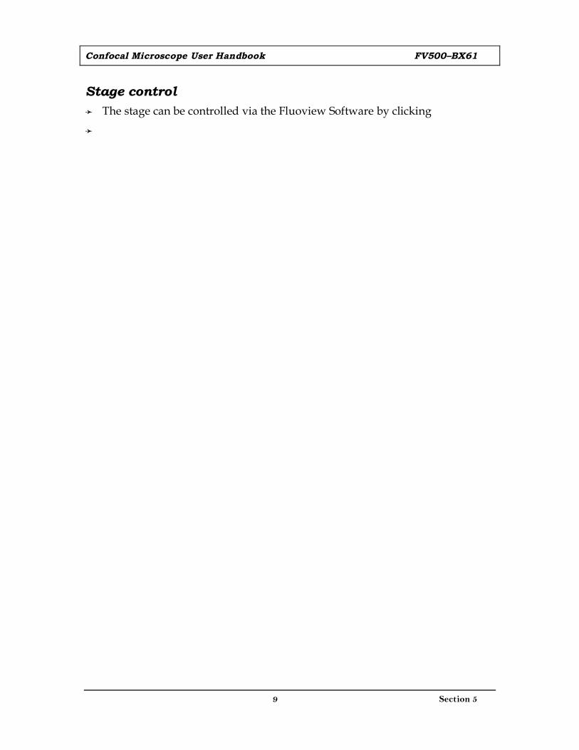
Automation The BX61 Microscope can be controlled via the Fluoview Software by clicking
the scope control button on the settings tab. The Graphic User Interface (GUI, below) will appear:
All functions of the microscope can be controlled here from the condenser to the filter cubes to the light sources. Just click on the diagram and the software will configure the microscope.
[Light Path] box selects the light path <BI> is for direct observation, <LSM> is for scanning, <TV> not used. [Mirror Unit] box clicking the cube automatically switches the turret. [Shutter] box click to open (9:00) / close (12:00) the EPI shutter. [Nosepiece] box click to choose the objective. [Top Lens] box clicking the engages (centered icon) or disengages (to side) the lens into the light path. [Condenser] box selects the condenser used. [Aperture Iris] box changes the AS value.
Confocal Microscope User Handbook FV500–BX61
9 Section 5
Stage control The stage can be controlled via the Fluoview Software by clicking

10 Section 4
Fluoview Software
Start-up After entering the Fluoview software the window below will open.
This software uses panel-type windows. Functions are executed by selecting the panel page tab (at the edge of the panel) of the function to be executed and the panel will move to the front.
The FLUOVIEW software is organized by two kinds of panels, the function panels (left, down side) and display panel (big one on right).
The function panels include the [Acquire], [File I/O], [Tile], [Process], [Analyze] and [Visualize] panels. The display panel shows either the [Live] panel or the panel image loaded from a file ([(filename)] panel).
Image files and observation methods are selected by means of “drag & drop”. [a complete layout of commands is found in section 5, page 18]
Section
4
This section will address the control capabilit ies of the system. It wil l also provide an overview of some of the analytic applications.

Confocal Microscope User Handbook FV500–BX61
11 Section 4
DYE [excitation/emission] Laser DYE [excitation/emission] Laser
Ca++ Crimson [590/615] Red HeNe eGFP [488/509] ArgonCa++ Green-1 [506/531] Argon FITC [494/518] ArgonCa++ Green-2 [503/536] Argon Fluo-3 [506/526] ArgonCa++ Green-5N [506/532] Gr HeNe Fura Red [472/657] Red HeNeCa++ Orange [549/576] Gr HeNe MitoTracker [490/516] ArgonCa++ Orange-5N [___/580] Gr HeNe PI [536/617] Gr HeNeCy 3 [550/565] Gr HeNe Rhodamine-Phalliodin [542/565] Gr HeNeCy 3.5 [581/596] Gr HeNe SNARF-1 (pH !) [488/530] Red HeNeCy 5 [650/670] Red HeNe Texas Red [595/615] Red HeNeCy 5.5 [675/695] Red HeNe TRITC [496/520] Gr HeNeDil [549/565] Gr HeNe YFP [514/527] Gr HeNeDiO [484/501] ArgonNOT on list:
AlexaFluor-1 [632/647] Red HeNe Calcein [494/517] ArgonAPC [650/660] Red HeNe DS Red [558/583] Red HeNeAPC/Cy7 [650;755/767] Red HeNe YoYo-1 [491/509] Argon
Microscope The BX61 Microscope is controlled via the Fluoview
Software by clicking the scope control button on the settings tab. The GUI will appear (see previous section).
Click on the diagram to the desired microscope configuration. Position and focus the specimen manually using the appropriate filter cube in <BI> mode (i.e. looking through the binocular eyepieces). Once the specimen is in focus, you are ready to scan.
Lasers There are 3 lasers: Argon [10mW, 488 nm], Green HeNe [1mW, 543 nm] and
Red HeNe [10mW, 633 nm]
The laser(s) used is dependent on the dyes. Below is a table listing the dyes in the <available dyes> list and some popular dyes not on the list. There is considerable overlap, however you should pick a laser close to either the excitation or emission max (usually they are close enough that it is obvious).
Scan Control Select the <LSM> button in the [Light Path] group box
of the settings panel.
Note: The <LSM> button looks pushed in to indicate that it is selected. (When scanning is started while the <BI> button is selected, the LSM light path is selected automatically. It is switched back to the visual observation automatically when scanning completes.)
Selecting the Dyeing Method
1. From the page tabs on the bottom right of the [Acquire] panel, select the [Dyes] sub-panel.

Confocal Microscope User Handbook FV500–BX61
12 Section 5
2. Select the specimen dyeing method by dragging desired dye in the [Available Dyes] list into the [Selected Dyes] group box to the field immediately above the list box.
3. Click the <Apply> button to apply the selected dyeing method to the [Ch] group box on the upper part of the [Acquire] panel.
Setting the Channels
1. In the [Ch] group box, make sure that the check boxes showing the applicable dyeing methods are checked-marked to indicate that the channels are ready for image acquisition.
2. If the check box of a channel is not check-marked, check it to make the channel ready.
Setting the XY Observation Mode
1. In the [Mode] group box in the [Acquire] panel, select the [Surface] option button.
2. In the [Mode] group box in the [Acquire] panel, select [800 by 600] from the [Size] drop-down list.

Confocal Microscope User Handbook FV500–BX61
13 Section 5
3. In the [Acquire] panel, select the XY observation mode option button (top left).
Select the <XY Repeat> button. The acquired image will be displayed in the [Live] panel. Note: this is your preview. The <stop scan> button will replace the <XY Repeat> button.
Setting the Cross-section to be Observed
After the preview image appears, move the Z stage to select the cross-section to be observed.
1. From the panel page tabs shown on the bottom right of the [Acquire] panel, select the [Z Stage] sub-panel (below).
2. Check the [Locked] check box in the [Z Stage] sub-panel. You have now transferred control of the z-stage to the computer. Do not turn the fine focus adjustment knob while the [Locked] check box is checked, for this may damage the Z motor.
3. While observing the image in the [Live] panel, locate the plane to be observed by displacing the stage using the <Z stage coarse adjustment> and <Z stage fine adjustment> buttons in the [Z Stage] sub-panel [not on the microscope].
Stopping Repeated Scanning
After the brightness and gain have been adjusted, select the <STOP SCAN> button in the [Acquire] panel to stop scanning temporarily
Acquiring Images
Select the <Once> button. The acquired image will be displayed in the [Live] panel.

Confocal Microscope User Handbook FV500–BX61
14 Section 5
Saving & Analyzing Images Display the [File I/O] panel.
When saving images acquired with more than one channel, it is possible to select whether images from more than one image are saved simultaneously or only one of the images is saved. Use the <Display channel switch> buttons to select the images to be saved. The selected images will be saved under the conditions set for each channel. Example — When only the image of Channel 1 is displayed, only the image of Channel 1 will be saved.
Click <Experiment> button in the [Save] group box. The [Save Experiment As] dialog box will open.
Enter the file name in the [File name:] text box.
Select “FLUOVIEW MultiTif” from [Save as type:]
Click the <Save> button
Saving, Opening and Shredding Images. Use the [File I/O] panel (top) to save, open or shred an image.
Shredding Images. Shredding, refers to removing it from the objects of processing including display. Shredding does not actually delete the image saved in the disk.
1. Click the <Experiment List> button in the toolbar at the bottom of the [File I/O] panel. The [Experiments in Memory] dialog box appears as shown.
2. In the [Experiments in Memory] dialog box, select the file name of the image to be shredded and click the <Shredder> button.
The file can also be shredded by placing the mouse pointer on it and dragging it to the <Shredder> button. The mouse pointer transforms to an image icon during dragging.

Confocal Microscope User Handbook FV500–BX61
15 Section 5
3. Click the <Done> button in the [Experiments in Memory] dialog box to close it. Multiple images displayed can be shredded simultaneously.
Displaying an Image in Simulated Colors
1. Display the [Display] panel of the image to be colored at the front.
2. Click the <LUT> button in the toolbar at the bottom left of the screen. The [Color Tool] dialog box appears.
3. When the image was acquired from more than one channel, select the channels to be colored using the [Ch1], [Ch2], [Ch3] option buttons. (The [Ch1], [Ch2] and [Ch3] option buttons are displayed only when an image acquired from more than one channel mode is displayed (selected).
4. From the [Standard Color LUTs] group box, select the desired color button. The selected LUT will be applied immediately to the image in the [Display] panel.
5. Click the <OK> button.
Image Annotation. Images can be annotated by clicking the Annotation button in the toolbar

Confocal Microscope User Handbook FV500–BX61
16 Section 5
Mass Storage There is no internet connection for the computer which runs the confocal,
therefore data must be transferred using zip disks, CD-R/RW, DVD-R/RW or removable USB drive.
Transfer to zip disks or removable USB drive is by a direct drag and drop.
Transfer to CD-R/RW or DVD-R/RW is by using a program found on the desktop “ “.

17 Section 5
Start-up and Shut-down
Checklist [System Start-up]
The FV500 Laser Confocal Microscope System consists of the following:
BX61 Upright Motorized Research Microscope FV500 Laser Scan Head Laser Combiner and Argon, Green HeNe and Red HeNe lasers Fiber Optic Delivery System Transmitted Light Detector Prior Motorized Stage Controller BX61 Handswitch, Stage Control Joystick, Transmitted Light Source Microscope Control Unit Argon, Green HeNe and Red HeNe Laser Power Supplies Mercury Burner Power Supply FV500 Control Unit and Power Supply FV500 Computer Computer Monitor and Surge Protected Power Outlet
1. Turn on the Surge Protected Power Outlet (Computer, monitor, FV500 Control Unit & LG-PS2). Press the POWER button on the tower to turn on the computer.
2. Turn on Laser Power Supplies ONLY as NEEDED (Argon, Green HeNe, Red HeNe) and “ON” for the Argon laser. The recommended warm-up is 10 min for the Argon and 30 min for the Gr HeNe.
3. Turn on the Mercury Burner.
4. Turn on the BX-UCB (Microscope Control Unit) [located on the top shelf].
5. Turn on the Prior Stage Controller Power Supply.
6. Logon to the computer using USER NAME and PASSWORD.
Section
5
This is intended to be a run-down on essential steps needed to initialize and shutdown the system.

Confocal Microscope User Handbook FV500–BX61
18 Section 5
7. Wait at least 2 min for the microscope systems to initialize then double-click the Fluoview Icon (right) to launch the confocal program. Note: it takes about 2 minutes to launch.
8. Sign in on the log sheet. [blue folder]
9. Turn the red lever on the nitrogen tank 90° counter clockwise to power the air table.
Microscope
The manual microscope controls are set as shown in the schema to the right.
Scan Control
The schematic to the left shows the steps for image acquisition in the Fluoview program
Light path selector
DIC slider
Filter cube display window
Filter selection buttons

Confocal Microscope User Handbook FV500–BX61
19 Section 5
Computer
The panel structure of the software is set-up according to the tree below.
When you are finished [System Shut-down]
1. Make sure objectives are clean of any residual oil using lens paper
2. Return microscope stage to non-escaped position and objectives to 4x.
3. Exit from the FV program. This may take awhile, however if you turn off the microscope before exiting, a system error will occur.
4. Transfer your files to removable media. If you have 1 GB or more in your folder on the hard drive, consider removing older files permanently (only 1.5 GB allowed).
5. If someone is signed up after you, do not turn things off. Log off (skip to #10), but do not shut down the computer (#11) and sign out on the log sheet (skip to #14). Otherwise:

Confocal Microscope User Handbook FV500–BX61
20 Section 5
6. Turn off the Prior Stage Controller Power Supply.
7. Turn off the BX-UCB (Microscope Control Unit) [located on the top shelf].
8. Turn off the Mercury Burner.
9. Turn off Laser Power Supplies (Argon, Gr HeNe, Red HeNe)
10. Select <shut down> on the computer and select LOG OFF from the drop-down menu. The computer will log your name out and return to the login menu box.
11. Select the shut down button to shut down the computer. Note: power will go off.
12. Turn off the Surge Protected Power Outlet (Computer, monitor, FV 500 Control Unit and LG-PS2). Do not turn the monitor, FV 500 Control Unit or LG-PS2 off.
13. Turn red lever on tank 90° clockwise to stop flow of gas to table.
14. Sign out on the log sheet.
15. Report any problems. In the log sheet if minor. Call Steve or Ken if major hardware problem. Let Margaret know if any service is needed or supplies run out.
Dispose of waste (e.g. used lens paper) in wastebasket and take your samples away to be disposed of according to hazardous waste/glass requirements. Cover microscope and turn off lights.

21 Section 6
Troubleshooting
Log-in problems
Can’t see an image
Image not focused
Bleed-thru from FTIC to Cy3
Section
6
Based on previous performance of this instrument, some of the more obvious problems are highlighted. What must be determined is: is it your fault or the machine's and do you need to call for help or service.

22 Section 7
Resources
People Steve Ricchio, Digital Imaging Specialist, Leeds Precision Instruments, Inc. Office 763-546-8575 • cel 612-209-1086 • [email protected]
Ken Kilby, Sales Representative (Microscopes), Leeds Precision Instruments, Inc. Office 763-546-8575 • cel 651-270-9379 • [email protected]
Angela Goodacre, Marketing Manager – Imaging Systems Applications, Olympus America, Inc. Voicemail 800-645-8100 ext 6004 • [email protected]
Jerry Sedgewick, Program Director, Biomedical Image Processing Lab (BIPL, www.bipl.ahc.umn.edu), 612-624-6607 • [email protected]
Margaret Ramnaraine, Scientist, Confocal Administrator, 612-626-3672 • [email protected]
User Manuals Olympus Fluoview FV500 Training, Presented by Leeds Precision Instruments, Inc. [version 2, 2/9/05] Powerpoint presentation in PDF
Olympus Fluoview FV500 Laser Scanning Confocal System Operating Instructions [version 1.1, 6/19/03] full version, PDF file
Olympus Fluoview FV500 Multi Point Time Lapse Software [Version 4.3a, 12/01/03] full version of the stage control manual, PDF file
Shared Confocal Microscope USER HANDBOOK: POLICIES AND PRACTICES [version 1, 3/15/05] PDF file
References Cell Biological Applications of Confocal Microscopy. Ed. B. Matsumoto.
Section
7
A handy list of ideas to pursue as users go on to more advanced applications.

Sign-in/out Log for Confocal Microscope [ Station #1 = FV500, Station #2 = workstation ]
Date User PI Station #1 / #2
Time In Out
Biohazard
YES / NO Comments

On-line Reservations for Confocal Microscope
Log in with ID and password Use the same as for log-in to the confocal
Select date and approximate time by clicking near time
Fill-in user information
NOTE: the USER ID will fill in the PI field and appear on the weekly calendar in the appropriate time slot.

UNIVERSITY OF MINNESOTA
BONE TUMOR BIOLOGY LABORATORYDEPARTMENT OF ORTHOPAEDIC SURGERY AND NCI DESIGNATED
COMPREHENSIVE CANCER CENTER
CONFOCAL MICROSCOPE ACCESS REQUEST FORM
PART I: User Information (please print)Name:
Lab (PI):
Phone:
E-mail:
Lab Location:
Describe your experience with microscopy, particularly confocal:
Provide a brief description of the purpose for which you will use the confocal microscope:
PART II: Cancer Center access (please print)
Resident of CCRB: New Request:
Non-resident CCRB: Update / change access:
Date: UCARD#: begins with 600953
Title: % effort:
Faculty or Administrative Supervisor:
Ucard Access: Other: Room 506 CCRB
* Is access required after 6pm and weekends (check):
Hazardous Chemical Waste Training Date: Anyone working in any capacity in the lab areas (including non-research staff) must provide documentation(<one year old) of Hazardous Chemical Waste Training (www.dehs.umn.edu)
RETURN COMPLETED FORM TO MARGARET RAMNARAINE. 322 CR, 470 CCRB or MMC 806.Approved Signature: [confocal manager] Date:
Approved:Signature: [Cancer Center] Date:


![INDEX [] duty trucks.pdf · ax76 4p5436 ax76 8m0760 ax77 7e6192 ax77 9y1956 bx61 6n6653 bx61 6n6655 bx61 6n6657 bx61 8n0539 bx68 4n8221 alternator bx75 6n7053 waterpump bx77 6215-61](https://img.pdfslide.us/doc/110x75/5b01ce967f8b9ab9598cdf8b/index-duty-truckspdfax76-4p5436-ax76-8m0760-ax77-7e6192-ax77-9y1956-bx61-6n6653.jpg)