Embed Size (px)
Citation preview

Cardiac Masses
Dennis A. Tighe, MD, FASE
University of Massachusetts Medical School
Worcester, MA

Cardiac Masses:
Considerations
• Definition of the mass– Nature
– Location
– Benign or malignant
• Presentation– Obstruction
– Direct myocardial involvement
– Embolization
– Constitutional or systemic symptoms
• Echocardiography is primary modality
• Multimodality imaging

Cardiac Masses:
Differential Diagnosis
• Anatomical variants
• Implanted devices
• Thrombus
• Vegetations
• Tumors
– Primary
– Metastatic
• Artifacts

Anatomical Variants• Left atrium
– Pectinate muscles
– Q-tip (“warfarin ridge”)
• Right atrium– Crista terminalis
– Eustachian valve
– Chiari network
• Ventricles– False bands
– Moderator band
– Hypertrophy
– Papillary muscles
– Non-compaction
• Pericardium– Pericardial cysts
– Pericardial fat
• Valves– Excresences
– Mitral annular calcification
• Lipomatous hypertrophy of interatrial septum
• Atrial septal aneurysm
• Hiatus hernias

Anatomical Variants
LAA
RA
Th

Implanted Devices
• Pacemaker leads
• Cardioverter-defibrillator leads
• Right heart catheters
• Occluder devices
• Prosthetic valves/clips
• Foreign bodies

Implanted Devices
LA
RA
CT

Implanted Devices
RA
wire
LALA

Thrombus
• Most common intra-cardiac mass
• Location often associated with cardiac pathology– LV thrombus
• Acute MI– Estimated 4-15% patients with anterior MI (LV apex)
• Dilated cardiomyopathy– DDx: false tendons, trabeculations, artifacts, apical hypertrophy,
tumors, non-compaction, Loeffler’s
– LA thrombus• Appendage
• Body
– RA thrombus• Catheter-related
• Pulmonary embolism
• Appendage

Thrombus
LV
RV
RA
LAA


Pulmonary Embolism Catheter-related
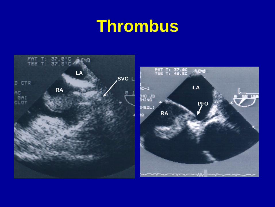
Thrombus
LA
RA LA
RA
PFO
SVC

Vegetations
• Locations:– Valve surfaces, areas of endocardium opposite intracardiac
shunts, or prosthetic materials
• Atrial surface mitral valve
• Ventricular surface of aortic valve
• Mobile, oscillating
• Tissue density differing from surrounding tissue
• Valvular regurgitation– Valvular stenosis (if large enough)
• Infective or non-infective
• May calcify if chronic/healed


Cardiac Tumors
• Primary Cardiac Tumors
– Rare
• 0.017% to 0.033% of autopsies
– Benign
• 75-85 % of primary cardiac tumors
– Malignant

Primary Cardiac Tumors

Cardiac Myxoma
• Most common primary cardiac tumor
• Most are sporadic– 10% familial
• Can recur and may be multicentric
• Female preponderance (60-70%)
• Most frequently discovered 3rd to 6th decades
• Can arise anywhere within the heart– About 75% occur in the left atrium near fossa ovalis
– Stalk
• Clinical presentation– Constitutional, embolic or obstructive symptoms
• Many detected asymptomatically

LA Myxoma
LV
LA
RV
RA
LA
RA
RV
IAS


LA Myxoma-Atypical Locations
LA
LV LV
RV

RA Myxoma
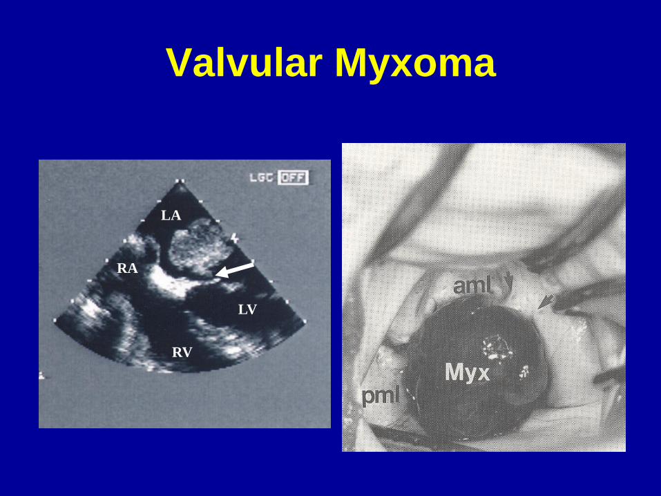
Valvular Myxoma
LA
LV
RA
RV

Papillary Fibroelastoma
• Second most common primary cardiac tumor– Most common tumor involving cardiac valves
• Aortic valve most common location– Both surfaces show equal prevalence
• May involve other cardiac structures/chambers (15-25%)
• Large majority found in left heart
• Avascular, papillary fronds, pedunculated
• Mid-portion of the valve– Usually do not cause valvular dysfunction
• Significant embolic potential recognized
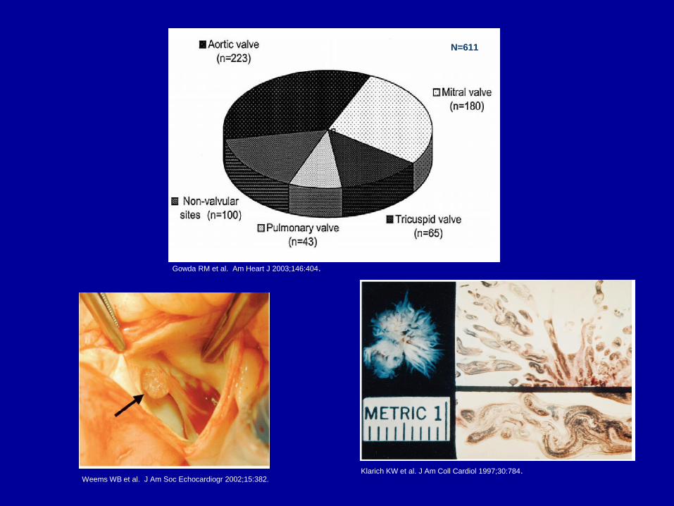
Klarich KW et al. J Am Coll Cardiol 1997;30:784.
Gowda RM et al. Am Heart J 2003;146:404.
Weems WB et al. J Am Soc Echocardiogr 2002;15:382.
N=611


Multiple tumors Right-sided

Lambl’s Excresences
• Common
– 70-80% adults
• Linear, filiform fronds
– Ventricular surface of semilunar valves
– Atrial surface of mitral valve
• Multiple
• Located at closure lines


Other Benign Primary Tumors
• Rhabdomyoma– Most common tumor in pediatric age group
– Muscular• May protrude into cavity
– Association with tuberous sclerosis
– Spontaneous regression
• Fibroma
• Lipoma
• Teratoma
• Angioma
• Paraganglioma
• Blood Cyst

Other Benign Primary Tumors
Rhabdomyomas Fibroma

Malignant Primary Cardiac Tumors
• Sarcomas (80%)– Angiosarcoma
• Usually found in right atrium
• Highly invasive
• Lung metastases common
– Other types (left atrium) • Undifferentiated sarcoma
• Rhabdomyosarcoma
• Fibrosarcoma
• Leiomyosaraoma
• Osteosarcoma (calification)
• Mesotheliomas (10%)– Arise from pericardium
– Rarely may involve conduction system
• Lymphomas (3-5%)
• Paragangliomas

Angiosarcoma
LA
RA

Primary Cardiac Lymphoma
Secondary (Metastatic)
Cardiac Tumors
• At least 20-to-40 times more common than
primary cardiac tumors
– 5-12% cancer patients
– Consider with known malignancy and occurrence
of new CV symptoms
• Breast and lung cancer encountered most
commonly
• Malignant melanoma highest propensity for
metastasis to the heart

Secondary (Metastatic)
Cardiac Tumors
• Pericardial involvement – Most common
• Pericardium > Myocardium > Endocardium
• Hematogenous/Lymphatic spread– Melanoma, lymphoma, breast
• Direct extension– Lung, breast, esophageal
• Invasion via venous structures– Vena cava
• Renal, Hepatocellular, Uterine
– Pulmonary veins• Lung, breast, thyroid


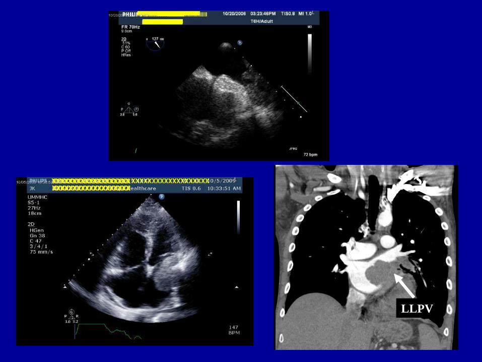
LLPV

Extra-cardiac Masses


Thank you for your attention