Embed Size (px)
Citation preview

University of Groningen
WNT pathway activation in COPD: a two way street between signalling and pathologyvan Dijk, Eline Margaretha
IMPORTANT NOTE: You are advised to consult the publisher's version (publisher's PDF) if you wish to cite fromit. Please check the document version below.
Document VersionPublisher's PDF, also known as Version of record
Publication date:2017
Link to publication in University of Groningen/UMCG research database
Citation for published version (APA):van Dijk, E. M. (2017). WNT pathway activation in COPD: a two way street between signalling andpathology. [Groningen]: Rijksuniversiteit Groningen.
CopyrightOther than for strictly personal use, it is not permitted to download or to forward/distribute the text or part of it without the consent of theauthor(s) and/or copyright holder(s), unless the work is under an open content license (like Creative Commons).
Take-down policyIf you believe that this document breaches copyright please contact us providing details, and we will remove access to the work immediatelyand investigate your claim.
Downloaded from the University of Groningen/UMCG research database (Pure): http://www.rug.nl/research/portal. For technical reasons thenumber of authors shown on this cover page is limited to 10 maximum.
Download date: 14-08-2019

512661-L-bw-Stam512661-L-bw-Stam512661-L-bw-Stam512661-L-bw-StamProcessed on: 21-8-2017Processed on: 21-8-2017Processed on: 21-8-2017Processed on: 21-8-2017 PDF page: 7PDF page: 7PDF page: 7PDF page: 7
Dutch Summary / Nederlandse Samenvatting
Chapter One General introduction

512661-L-bw-Stam512661-L-bw-Stam512661-L-bw-Stam512661-L-bw-StamProcessed on: 21-8-2017Processed on: 21-8-2017Processed on: 21-8-2017Processed on: 21-8-2017 PDF page: 8PDF page: 8PDF page: 8PDF page: 8
Chapter One
8
Preface
Chronic Obstructive Pulmonary Disease (COPD) is characterized by airway and parenchymal inflammation and remodelling, abnormal increase in air spaces (emphysema), bronchoconstriction, and mucus cell hyperplasia among others (1-3). The high levels of oxidative stress and protease-antiprotease imbalance present in COPD contribute to the development of emphysema by enhancing tissue destruction and impairing tissue repair of the lung parenchyma (1, 3-5). Together, these processes contribute to air trapping and a continuous decline of lung function. Available treatments can delay disease progression to some extent, but recovery or normalization of loss of lung function is not possible (6). In order to develop disease-modifying therapeutics it is crucial to unravel the mechanisms and signalling pathways underlying COPD. The WNT signalling pathway is a promising target. WNT ligands are growth factors that bind to FZD receptors and are involved in lung development and lung repair. This pathway is dysregulated in COPD (7-11). Previous work from our group demonstrated increased expression of the ligands WNT-5A and WNT-5B in lung fibroblasts of COPD patients (7). Furthermore, our group demonstrated a role for the WNT-5A and WNT-5B receptor Frizzled-8 (FZD8) in chronic bronchitis, airway inflammation, and profibrotic signalling (12-14). However, the exact underlying signalling mechanisms involved are not completely understood. Understanding of these mechanisms will contribute to the discovery of novel therapeutic targets in the treatment of COPD. Therefore, the objective of this thesis is to establish the role of WNT-5A/5B signalling in COPD. This thesis will focus on the role of WNT-5A and WNT-5B on inflammatory processes, and the effect of oxidative stress on WNT-5A and WNT-5B-mediated signalling. Furthermore, the role of parenchymal tissue disruption in enhanced airway narrowing will be discussed. Finally, this thesis will focus on the interaction between noncanonical WNT signalling, ageing and tissue damage to the lung, with a focus on WNT-5B and FZD8.
COPD
COPD is an incurable and life-threatening disease, which is characterized by progressive airflow limitation. Currently COPD is the fourth leading cause of death worldwide, and it is expected to be the third leading cause of death by

512661-L-bw-Stam512661-L-bw-Stam512661-L-bw-Stam512661-L-bw-StamProcessed on: 21-8-2017Processed on: 21-8-2017Processed on: 21-8-2017Processed on: 21-8-2017 PDF page: 9PDF page: 9PDF page: 9PDF page: 9
General Introduction
9
2020 (6). COPD has recently been characterized as a disease of accelerated lung ageing as oxidative stress, inflammation, cellular senescence, protease injury and apoptosis are all involved in the pathogenesis of COPD (2, 15). It is well-established that normal lung function declines with increasing age, and that smoking may enhance this age-related lung function decline (16). Decline in lung function is a slow process spanning several decades. The prevalence of COPD rises sharply from the age of 40, and most patients with COPD are in late middle age or older (17, 18). The major risk factor for developing COPD is long-term cigarette smoke exposure (19, 20). Smoking cessation will not recover lung function, but it does result in an almost normal pattern of lung function decline, albeit with a lower starting point (see Figure 1). Other risk factors that contribute to COPD are outdoor and indoor air pollution (the latter caused by for example biomass cooking), heating in poorly ventilated rooms, and occupational dust and chemicals (6, 21). Inhalation of these risk factors induces lung inflammation, a normal response that seems to be altered in individuals who develop COPD. This chronic inflammatory response may induce emphysema, peribronchial fibrosis, mucus cell hyperplasia and bronchoconstriction, among others (1-3). Together, these processes contribute to air trapping and the continuous decline of lung function. In addition, particularly in patients with severe disease, COPD involves several systemic features which have a major impact on survival and comorbid diseases (2).

512661-L-bw-Stam512661-L-bw-Stam512661-L-bw-Stam512661-L-bw-StamProcessed on: 21-8-2017Processed on: 21-8-2017Processed on: 21-8-2017Processed on: 21-8-2017 PDF page: 10PDF page: 10PDF page: 10PDF page: 10
Chapter One
10
Figure 1: The natural history of chronic airflow obstruction, adapted from Fletcher and Peto (16). FEV1 decreases gradually and continuously with increasing age. Smoking causes a steeper decline in lung function in susceptible smokers, but not in non-susceptible smokers. Smoking cessation at any age almost fully restores the natural pattern of lung function decline, although at a lower starting point (16).
Although most (80-90 %) of the COPD patients are (ex-)smokers, only 10-15 % of the smoking population develops COPD. Therefore, it is not surprising that studies from recent decades indicate that genetic susceptibility is underlying COPD development as well (22, 23). Furthermore, impaired lung development and growth has also been associated with an increased risk of COPD development later in life (24-26).
COPD is a heterogeneous disease, and individuals with COPD experience various symptoms, including shortness of breath, increased mucus production (resulting in productive cough), and wheezing (6). The severity of COPD is divided in four (1-4) grades by The Global Initiative for Chronic Obstructive Lung Disease (GOLD) guidelines (see Table 1) (6). To diagnose COPD, several factors are assessed such as spirometry, the patient’s level of symptoms, the identification of comorbidities, and exacerbation history. Airflow limitation is an important characteristic of COPD, and its measurement by spirometry is

512661-L-bw-Stam512661-L-bw-Stam512661-L-bw-Stam512661-L-bw-StamProcessed on: 21-8-2017Processed on: 21-8-2017Processed on: 21-8-2017Processed on: 21-8-2017 PDF page: 11PDF page: 11PDF page: 11PDF page: 11
General Introduction
11
required to make a confident diagnosis of COPD. To define airflow limitation, the fixed post-bronchodilator forced vital capacity/ forced expiratory volume in 1 second (FEV1/FVC) ratio of 0.7 is used (21). A ratio lower than 0.7 is considered airflow limitation. The lower the ratio, the more severe the airflow limitation.
Table 1: Grading of severity of airflow limitation in COPD (based on post-bronchodilator FEV1), adapted from Global Strategy for the Diagnosis, Management and Prevention of COPD, Global Initiative for Chronic Obstructive Lung Disease (GOLD) 2016 (6).
In patients with FEV1/FVC < 0.70: GOLD 1: Mild FEV1 ≥ 80% predicted GOLD 2: Moderate 50% ≤ FEV1 < 80% predicted GOLD 3: Severe 30% ≤ FEV1 < 50% predicted GOLD 4: Very severe FEV1 < 30% predicted Definition of abbreviation: COPD = chronic obstructive pulmonary disease; GOLD = Global Initiative for Chronic Obstructive Lung Disease
1. Inflammation
Chronic inflammation of the peripheral airways and lung parenchyma leads to the progressive irreversible airway obstruction and shortness of breath. Currently, this chronic inflammation is difficult to treat as this type of inflammation is largely resistant to treatment with corticosteroids (4). The observed inflammatory response in individuals with COPD comprises both the innate and adaptive immune response, which are linked to each other by dendritic cells (27). Various compononents present in cigarette smoke, and other inhaled noxious particles activate surface macrophages, airway epithelial cells, and dendritic cells via the activation of Toll-like receptors and via oxidative stress (4, 27). Activation of these cells induces release of several chemotactic mediators such as chemokines, and of damage-associated molecular patterns (DAMPs) (28). These all act as chemotactic factors attracting neutrophils and monocytes, as well as T lymphocytes, including T helper (Th1) cells, cytotoxic T lymphocytes and Th17 cells (4). In addition, the DAMPs can attract and activate immune cells upon binding to pattern recognition receptors, contributing to the neutrophilic airway inflammation. Together, these processes result in a characteristic pattern of increased numbers of neutrophils in the airway lumen and increased amounts of alveolar macrophages, T lymphocytes
512661-L-bw-Stam512661-L-bw-Stam512661-L-bw-Stam512661-L-bw-StamProcessed on: 21-8-2017Processed on: 21-8-2017Processed on: 21-8-2017Processed on: 21-8-2017 PDF page: 12PDF page: 12PDF page: 12PDF page: 12
Chapter One
12
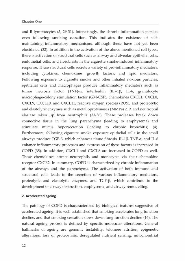
and B lymphocytes (5, 29-31). Interestingly, the chronic inflammation persists even following smoking cessation. This indicates the existence of self-maintaining inflammatory mechanisms, although these have not yet been elucidated (32). In addition to the activation of the above-mentioned cell types, there is activation of structural cells such as airway and alveolar epithelial cells, endothelial cells, and fibroblasts in the cigarette smoke-induced inflammatory response. These structural cells secrete a variety of pro-inflammatory mediators, including cytokines, chemokines, growth factors, and lipid mediators. Following exposure to cigarette smoke and other inhaled noxious particles, epithelial cells and macrophages produce inflammatory mediators such as tumor necrosis factor (TNF)-α, interleukin (IL)-1β, IL-6, granulocyte macrophage-colony stimulation factor (GM-CSF), chemokines CXCL1, CXCL8, CXCL9, CXCL10, and CXCL11, reactive oxygen species (ROS), and proteolytic and elastolytic enzymes such as metalloproteinases (MMPs) 2, 9, and neutrophil elastase taken up from neutrophils (33-36). These proteases break down connective tissue in the lung parenchyma (leading to emphysema) and stimulate mucus hypersecretion (leading to chronic bronchitis) (4). Furthermore, following cigarette smoke exposure epithelial cells in the small airways produce TGF-β, which enhances tissue fibrosis. IL-1β, TNF-α, and IL-6 enhance inflammatory processes and expression of these factors is increased in COPD (35). In addition, CXCL1 and CXCL8 are increased in COPD as well. These chemokines attract neutrophils and monocytes via their chemokine receptor CXCR2. In summary, COPD is characterized by chronic inflammation of the airways and the parenchyma. The activation of both immune and structural cells leads to the secretion of various inflammatory mediators, proteolytic and elastolytic enzymes, and TGF-β, which contribute to the development of airway obstruction, emphysema, and airway remodelling.
2. Accelerated ageing
The patology of COPD is chacaracterized by biological features suggestive of accelerated ageing. It is well established that smoking accelerates lung function decline, and that smoking cessation slows down lung function decline (16). The natural ageing process is defined by specific molecular alterations. General hallmarks of ageing are genomic instability, telomere attrition, epigenetic alterations, loss of proteostasis, deregulated nutrient sensing, mitochondrial

512661-L-bw-Stam512661-L-bw-Stam512661-L-bw-Stam512661-L-bw-StamProcessed on: 21-8-2017Processed on: 21-8-2017Processed on: 21-8-2017Processed on: 21-8-2017 PDF page: 13PDF page: 13PDF page: 13PDF page: 13
General Introduction
13
dysfunction, cellular senescence, stem cell exhaustion, and altered intracellular communication (37). These hallmarks are demonstrated to be involved in natural lung ageing (18, 38, 39). The ageing lung demonstrates a reduced elasticity (40). In chronic respiratory diseases such as COPD the hallmarks of ageing reach pathological levels and lung function decline is accelerated (15). For example, smokers and patients with COPD have increased oxidative damage to DNA in the lungs (41, 42). In addition, there is an increase in the number of double-stranded DNA breaks and a failure of DNA strand break repair in COPD (41). Circulating leukocytes from COPD patients and parenchymal lung cells from emphysematous lungs have been shown to have shorter telomeres than non-COPD controls (43-45). Epigenetic alterations take place in COPD, as differential DNA methylation has been detected in lymphocytes from COPD patients as compared to non-COPD controls. Notably, altered methylation is associated with COPD severity (46, 47). Next to DNA methylation changes in COPD, the antiaging factors histone deacetylase (HDAC) sirtuin and HDAC2 are suppressed in COPD which results in enhanced inflammation and increased cellular senescence (48-50). In addition, proteasome function is reduced in COPD patients and correlates inversely with the loss of lung function (51). During aging, nutrient sensing becomes dysregulated, which involves the PI3-AKT-mTOR pathway. This pathway integrates signals on nutrient availability to regulate cellular growth, and has an important role in cellular senescence (52). Inhibition of this pathway extends the lifespan of many species (52). There is evidence that the mTOR pathway is activated in lung epithelial cells of patients with COPD, possibly contributing to COPD pathogenesis (50). Mitochondrial dysfunction may contribute to ageing by enhancing production of ROS as part of the free radical theory of aging (53). In COPD, mitochondrial numbers are reduced, while mitochondrial ROS production is increased (54). It has been shown that cigarette smoke alters mitochondrial structure and function (55-57). Together, these findings suggest a role for mitochondria in the development of COPD. Another hallmark of ageing, cellular senescence, is most likely involved in COPD pathogenesis as well. Type II epithelial cells, endothelial cells, and fibroblasts from emphysematous lungs demonstrate enhanced senescence, and exposure of human epithelial cells to cigarette smoke results in cell senescence (43). A direct relationship has been demonstrated between the extent of p16-positive cell

512661-L-bw-Stam512661-L-bw-Stam512661-L-bw-Stam512661-L-bw-StamProcessed on: 21-8-2017Processed on: 21-8-2017Processed on: 21-8-2017Processed on: 21-8-2017 PDF page: 14PDF page: 14PDF page: 14PDF page: 14
Chapter One
14
senescence and the severity of inflammation in emphysematous lungs (58). Stem cell exhaustion is thought to have an important role in multiple age-related diseases (59). In COPD, basal and circulating progenitor cells show evidence of DNA damage and senescence which reduces their repair capacity, although there are conflicting reports (60-63). Finally, ageing is characterized by altered intracellular communication with an increase in low-grade systemic inflammation characterized by increasing higher levels of circulating pro-inflammatory cytokines in elderly individuals. This low-grade systemic inflammation may be responsible for the decline and onset of diseases in the elderly (64). COPD is characterized by alterations in both the innate and adaptive immune responses, and age-related changes in the immune system are thought to render COPD patients more prone to exacerbations (65). In addition to the altered general hallmarks of ageing, specific ageing-associated genes such as Klotho are also found to be dysregulated in the lungs of COPD patients (66, 67). In summary, COPD is characterized by accelerated ageing as defined by altered general hallmarks of ageing and specific dysregulated ageing-related mechanisms and genes.
3. Airway and parenchymal remodelling
Airway remodelling is a major feature of COPD. Under normal conditions the small airways (<2 mm in diameter) hardly contribute to airflow resistance in the healthy lung. However, as a result of remodelling these airways become the main site of resistance in COPD, contributing to airway obstruction (29, 68). Characteristics of airway remodelling that are present in COPD are thickening of the airway wall caused by peribronchial fibrosis and increased airway smooth muscle (ASM) mass, increased blood vessel density, enlargement of the submucosal glands, goblet cell metaplasia, epithelial cell metaplasia, loss of terminal and respiratory bronchioles, and enlargement and destruction of the alveoli (29, 69). Airway wall thickness is augmented due to increases in each of the airway wall constituents (epithelium, lamina propria, smooth muscle, and adventitia) (29). Of the mentioned remodelling characteristics, thickening of the airway wall was found to be the most strongly associated with disease progression from GOLD stage 0 to GOLD stage 4 (29). The increase in tissue between the epithelial surface and muscle layer is thought to have a role in nonspecific airway responsiveness, while the increase in connective tissue in the

512661-L-bw-Stam512661-L-bw-Stam512661-L-bw-Stam512661-L-bw-StamProcessed on: 21-8-2017Processed on: 21-8-2017Processed on: 21-8-2017Processed on: 21-8-2017 PDF page: 15PDF page: 15PDF page: 15PDF page: 15
General Introduction
15
adventitial compartment may contribute to fixed airway obstruction by preventing airway re-opening during lung inflation (29). In heathy lungs, the airway epithelium is the first barrier to protect the airways against inhaled pathogens and (noxious) particles. Chronic smoke exposure leads to epithelial remodelling and results in both squamous and mucous metaplasia, which contributes to airway obstruction in COPD patients (70, 71). In addition, the growth factors released by the epithelium could influence the underlying smooth muscle cells and fibroblasts, enhancing airway remodelling.
Small airway remodelling The area of the ASM layer in the small airways is increased by an average of 20 % in severe COPD patients (72-74). In contrast to the small airways, an increased ASM layer does not seem to contribute to the increased inner airway thickness of the large airways (75, 76). In contrast to what the name implies, the ASM layer is not composed entirely of ASM cells. Only 75 % of the ASM layer is ASM, with the remaining 25 % comprised of extracellular matrix (ECM) space, mast cells, and blood vessels (77, 78). The ECM within the ASM layer greatly contributes to the mechanical characteristics of the ASM and airway wall under normal and pathological conditions (79). Due to the presence of elastin fibers, the ECM maintains an elastic load which opposes ASM shortening (79, 80). Therefore, alterations in the composition or organization of ECM within the ASM compartment may influence elastic load and airway narrowing. Indeed, functional relationships between the fractional composition of the ECM within the ASM and airway narrowing have been demonstrated in asthmatics (81). Whether the ECM composition within the ASM layer also contributes to airway narrowing in the case of COPD is currently unknown.
Fibrosis Another key feature of small airway wall remodelling is fibrosis. Fibrosis is mainly characterized by alterations in the ECM deposition (82). The ECM consists of a large number of matrix proteins which are important for several processes, including maintenance of structural integrity, and cellular adhesion and migration (79). Main components of the lung ECM are collagen I, III and V, elastins, proteoglycans, biglycan, fibronectin, versican, decorin, lumican and tenascin, among others (83). Although the precise factors leading to small airway fibrosis and remodelling are poorly understood, it is clear that the

512661-L-bw-Stam512661-L-bw-Stam512661-L-bw-Stam512661-L-bw-StamProcessed on: 21-8-2017Processed on: 21-8-2017Processed on: 21-8-2017Processed on: 21-8-2017 PDF page: 16PDF page: 16PDF page: 16PDF page: 16
Chapter One
16
fibroblast plays a key role in these processes in COPD. Fibroblasts are crucial for regulating ECM turnover in the lungs, and dysregulation of fibroblast function may therefore lead to tissue fibrosis (84). Activation of fibroblasts by transforming growth factor β (TGF-β) results in ECM protein production and differentiation of fibroblasts into myofibroblasts (84, 85). Compared to fibroblasts, myofibroblasts have a more contractile profile with increased expression of contractile makers such as α-smooth muscle (sm)-actin (84, 86). Myofibroblasts are localized differentially in COPD lung tissue as compared to healthy lung tissue, with decreased expression in the bronchioles and alveoli and increased expression in the large airways (87). In addition the presence of myofibroblasts is associated with airway obstruction in the large airways in COPD (88).
Emphysema In addition to airway remodelling, individuals with COPD can demonstrate a variable degree of emphysema in the peripheral lung. Emphysema is characterized by a loss of the alveolar structure due to chronic inflammation and defective repair, resulting in an abnormal increase in air spaces (20). In contrast to the excessive activation of fibroblasts observed in the small airways, inadequate activation of fibroblasts in the peripheral lung may lead to emphysema (89). It has been shown that pulmonary fibroblasts from patients with emphysema fail to express elastin under all-trans retinoic acid (ATRA) stimulation, thereby contributing to the lack of alveolar repair in pulmonary emphysema (90).
A protease-antiprotease imbalance also contributes to the development of emphysema. Proteases such as matrix metallopeptidase/protease (MMP)-9 break down ECM proteins, while tissue inhibitors of MMP (TIMP) counteract this breakdown (91). An imbalance in factors driving the breakdown and production could therefore lead to alterations in ECM composition and emphysema (91). Indeed, it has been shown that the ECM and factors driving the ECM composition are altered in COPD (80, 91, 92). In a recent genome-wide gene expression study, upregulation of the elastogenesis-associated genes fibulin-5 (FBLN5), elastin (ELN), latent transforming growth factor β binding protein 2 (LTB2), and microfibrillar associated protein 4 (MFAP4) was demonstrated in lung tissue from COPD patients (93). Notably, these gene

512661-L-bw-Stam512661-L-bw-Stam512661-L-bw-Stam512661-L-bw-StamProcessed on: 21-8-2017Processed on: 21-8-2017Processed on: 21-8-2017Processed on: 21-8-2017 PDF page: 17PDF page: 17PDF page: 17PDF page: 17
General Introduction
17
expression findings were validated at mRNA and protein in the same study. In addition, FBLN5, ELN, and MFAP4 expression correlated negatively with lung function (93). Furthermore, polymorphisms of MMPs (MMP1, MMP2, MMP9, MMP12), ADAMs (ADAM33) and TIMPs (TIMP1, TIMP2) are associated with COPD (91).
The most noticeable alteration in the ECM in the alveolar compartment is decreased expression and disorganization of elastin fibers, leading to a loss of elastic recoil. Alterations in elastin expression are already present in mild to moderate COPD, and seen in both airways and alveoli of COPD patients (94). Studies on the total expression levels of collagen in COPD are inconsistent, but it has been observed that collagen fibers are more disorganized in severe COPD as compared to mild to moderate COPD (95). This disorganization of collagen may be caused by dysregulation of the small proteoglycan decorin. Decorin is important in collagen fibrillogenesis, and serves as a crosslinking molecule for adjacent collagen fibrils (96). In COPD, gene and protein expression levels of decorin are altered (97-99), and possibly contributes to the disorganization of collagen.
Other mechanisms induced by chronic smoke injury contributing to emphysema are increased apoptosis of alveolar epithelial cells, impaired phagocytosis function of macrophages, and increased oxidative stress (3, 100). Together, these mechanisms create an imbalance in the ECM turnover in the lungs, resulting in loss of functional lung tissue by elastin fiber degradation and loss of alveolar attachments leading to emphysema.
Tissue biomechanics The role of structural alterations in the parenchyma of COPD patients in enhanced airway narrowing is increasingly recognized (80). The parenchymal compartment is connected to the airways via parenchymal tethers, which transmit forces to the airways. Via this mechanism chest movements during inspiration are linked to airway opening (101-103). In healthy lung tissue, the parenchyma counteracts airway narrowing due to its elastic properties. Parenchymal tethers on the outside of an airway transmit trans-pulmonary pressure to the airway wall, thereby opposing the shortening of the airway smooth muscle (104). As a result, parenchymal tethering supports the relaxant

512661-L-bw-Stam512661-L-bw-Stam512661-L-bw-Stam512661-L-bw-StamProcessed on: 21-8-2017Processed on: 21-8-2017Processed on: 21-8-2017Processed on: 21-8-2017 PDF page: 18PDF page: 18PDF page: 18PDF page: 18
Chapter One
18
effect of deep inspiration and reduces bronchoconstriction (105). The parenchymal mechanical and structural properties therefore have a major impact on airway mechanics, and alterations of these properties are likely to influence airway narrowing (80). In COPD, the parenchyma and ECM are altered due to the presence of elastolytic enzymes and oxidative stress leading to a loss of elastic recoil (80, 92). Eventually the presence of elastolytic enzymes and oxidative stress alterations lead to damage of the parenchyma and ECM (3, 100), and likely affect airway mechanics in COPD enhancing bronchoconstriction. This role of the parenchyma in airway narrowing is especially interesting in mild or moderate COPD, as changes in the parenchymal compartment are not very profound yet in this stage, and possibly still reversible. In order to have a better understanding of enhanced airway narrowing in COPD, it is therefore important to investigate the role of the parenchymal compartment and to develop models in which the underlying pathological processes can be investigated. For this reason, we aimed to develop an ex vivo Precision Cut Lung Slice (PCLS) model in which we investigated the impact of low level elastase and oxidative stress exposure on airway narrowing and relaxation in relation to parenchymal and ECM structure, and alveolar epithelial repair (chapter 4).
4. Oxidative stress
Another key feature of COPD is an increased oxidative stress burden. Oxidative stress is defined by an imbalance between reactive oxygen species (ROS) and the ability of a biological system to either detoxify these ROS or repair the oxidant-induced damage. Oxidative stress may change proteins in a reversible or irreversible manner. Reversible changes are mostly found within cysteine residues and methionine, and can be repaired by specific enzymes, including thioredoxin and glutaredoxin or methionine sulfoxide reductase, respectively (106, 107). Such reversible alterations are known to modulate protein function and are involved in redox regulation (108). On the other hand, oxidative stress may lead to irreversible changes such carbonyl stress which leads to the production of highly reactive organic molecules that can modify proteins nonenzymatically (109, 110). COPD is characterized by oxidative and carbonyl stress, and particularly so during exacerbations. The lung is vulnerable to oxidative stress from the environment due to its anatomic structure. The lung is

512661-L-bw-Stam512661-L-bw-Stam512661-L-bw-Stam512661-L-bw-StamProcessed on: 21-8-2017Processed on: 21-8-2017Processed on: 21-8-2017Processed on: 21-8-2017 PDF page: 19PDF page: 19PDF page: 19PDF page: 19
General Introduction
19
exposed to both endogenous and exogenous sources of oxidative stress. Endogenous oxidative stress is generated by mitochondrial respiration and inflammatory responses to viral and bacterial infections within the lung. For example, it has been demonstrated that ROS are generated by several inflammatory and structural cells of the airways (111). Environmental sources of oxidative stress include oxidant gasses and ultrafine or nanoparticles from car exhaust fumes and industrial pollution. However, the main cause for environmentally derived ROS in COPD in the western world is cigarette smoke. Cigarette smoke-induced activation of antioxidants in airway leukocytes is absent in active smokers with COPD, and alveolar macrophages and activated peripheral blood neutrophils from COPD patients release more ROS (112-114). Other markers of oxidative stress and carbonyl stress in COPD include increased concentrations of nitrotyrosine and lipid peroxidation products, including 8-isoprostane, 4-hydroxy-2-nonenal, and malondialdehyde (MDA) (115-117). Furthermore, markers of oxidative stress including hydrogen peroxide, carbon monoxide, myeloperoxidase, and markers of oxidative tissue damage, such as 8-isoprostane and carbonyl stress in the form of MDA, are increased in exhaled breath or exhaled breath condensate from individuals with COPD (118-121). Finally, there are indications of systemic oxidative stress in COPD patients, as carbonyl adducts are found to be increased in the respiratory and skeletal muscle (116, 122). In addition to increased levels of ROS, endogenous levels of antioxidants are decreased in patients with COPD. For example, glutathione concentrations in bronchoalveolar lavage fluid (BALF) from COPD patients with frequent exacerbations are decreased as compared to BALF obtained from patients with stable COPD (123). Interestingly, while cigarette smoke exposure (CSE) drives the onset of COPD, smoking cessation does not stop the presence of oxidative stress or disease progression (124). The lasting oxidative stress burden probably stems from endogenous sources such as mitochondrial respiration (124). Oxidative stress induces damage by altering tissue and proteins directly, but it also induces damage indirectly by altering pathways which negatively impact disease progression. It has been demonstrated that oxidative stress results in enhanced inflammation in COPD as reflected by oxidative stress-mediated inflammatory gene expression, corticosteroid insensitivity, impaired resolution of inflammation, and corticosteroid insensitivity, among others (3). Furthermore, ROS can inhibit

512661-L-bw-Stam512661-L-bw-Stam512661-L-bw-Stam512661-L-bw-StamProcessed on: 21-8-2017Processed on: 21-8-2017Processed on: 21-8-2017Processed on: 21-8-2017 PDF page: 20PDF page: 20PDF page: 20PDF page: 20
Chapter One
20
elastin synthesis and repair, stimulate the release of proteases leading to the breakdown of the ECM (125) and inhibit antiproteases such as alpha1-antitrypsin (126). These ROS-induced processes therefore contribute to the development of emphysema. Taken together, there is mounting evidence of increased levels of oxidative stress in patients with COPD, caused by an imbalance between oxidants and antioxidants. This increased level of oxidative stress contributes to inflammation and the development of emphysema.
5. Therapeutic intervention
Available treatments can delay disease progression to some extent, but recovery or normalization of loss of lung function is not possible. Smoking cessation is the most effective and recommended treatment option, as smoking cessation slows down the accelerated lung decline in even severe cases of COPD (16, 20, 127). Pharmacological treatments of COPD consist of long-acting bronchodilators such as long-acting β2-adrenergic receptor agonists (LABAs) and long-acting muscarinic acetylcholine receptor antagonists (LAMAs), and anti-inflammatory agents (inhaled corticosteroids (ICS) and phosphodiesterase (PDE)4 inhibitors) (6, 19). Simultaneous treatment with LABAs and LAMAs has an additive effect both improving bronchoprotection/dilation and reducing symptoms, which has led to development of LABA-LAMA combination inhalers (4). While these treatments are effective in causing bronchoprotection, it is unclear whether they improve the underlying inflammation observed in patients with COPD. Treatment with ICS reduces the inflammation and the number of exacerbations mostly in a small subset of COPD patients who demonstrate an eosinophilic inflammatory response (128, 129). However, compelling evidence demonstrating that ICS treatment improves lung function decline is lacking (128). This lack of effect of ICS is most likely explained by the corticosteroid resistance observed in COPD patients. This corticosteroid resistance might be explained by the decrease in activity of histone deacetylase (HDAC)2, which is necessary to switch off inflammatory genes (130). In addition to ICS, PDE4 inhibitors are used in the treatment of patients with severe COPD (6). The efficacy of these inhibitors appears to be higher in a more severe disease phenotype defined by frequent exacerbations and mucus hypersecretion (131). In summary, none of the current pharmacological treatments slow down the loss of lung function in COPD patients. Therefore,

512661-L-bw-Stam512661-L-bw-Stam512661-L-bw-Stam512661-L-bw-StamProcessed on: 21-8-2017Processed on: 21-8-2017Processed on: 21-8-2017Processed on: 21-8-2017 PDF page: 21PDF page: 21PDF page: 21PDF page: 21
General Introduction
21
better insights into the mechanisms underlying COPD pathology are needed to develop novel and more effective therapies.
WNT signalling
The wingless/intergrase-1 (WNT) signalling pathway is an evolutionary conserved pathway that is crucial for stem cell maintenance, cell polarity and cell fate determination during embryonic development (132). For example, normal lung development is dependent on normal functioning of this pathway as embryos lacking specific WNT ligand expression exhibit complete lung agenesis (133). In addition, the WNT signalling pathway plays an important role in adult tissue where it contributes to tissue homeostasis trough maintaining stem cell pluripotency, inflammation, and repair (134, 135). Autocrine or paracrine secreted WNT ligands are cysteine-rich proteins which bind to Frizzled (FZD) receptors via disulfide bonds and subsequently induce various downstream signalling pathways in the cell (136, 137). WNT signalling can be divided into canonical (β-catenin dependent) or noncanonical WNT signalling (β-catenin independent). However, this division can only be used as a rough guide, as canonical and noncanonical signalling may overlap depending on the cellular context (138).
WNT ligands
WNT ligands are secreted cysteine residue (CR) rich proteins which are evolutionary conserved, and currently 19 human WNT ligands are known. Among vertebrate species, WNT gene sequence identity and gene structure are highly similar. Human WNT proteins share 27 % to 83 % amino-acid sequence identity and contain a conserved pattern of 23 or 24 CR (139). This conserved CR pattern is likely involved in proper protein folding via the formation of several disulfide bonds (136). In addition, all WNTs share a signal sequence for secretion, multiple highly charged amino-acid residues, and many potential glycosylation sites (137). WNT signalling between cells comprises of a series of steps, including posttranslational modification and secretion of WNT ligands, binding to receptors, activation of downstream signalling, and regulation of target gene transcription. Following their synthesis, WNTs are post translationally modified. For example, when WNTs are overexpressed in tissue

512661-L-bw-Stam512661-L-bw-Stam512661-L-bw-Stam512661-L-bw-StamProcessed on: 21-8-2017Processed on: 21-8-2017Processed on: 21-8-2017Processed on: 21-8-2017 PDF page: 22PDF page: 22PDF page: 22PDF page: 22
Chapter One
22
culture cells, multiple N-linked glycosylated intermediate WNT proteins are observed in cell lysates (136, 140, 141). This N-glycosylation is mediated by palmitoylation via the acyl-transferase porcupine (140), and is necessary for both WNT secretion (142, 143) and binding to the FZD receptor (144). In addition, WNT ligands undergo lipid modification with palmitoleic acid on a serine residue which renders WNTs hydrophobic (145, 146). Because of this hydrophobicity, WNTs are mostly bound to cell membranes and the ECM, and are associated with short range signalling (141). However, despite their hydrophobic nature, WNTs can signal over long distances as well via several mechanisms. To facilitate the extracellular movement, WNTs interact with heparan sulfate proteoglycans (HSPGs), consisting of a core protein to which heparan sulfate (HS) glycosaminoglycan (GAG) chains are attached (141). Furthermore, lipoprotein particles called argosomes can bind and transport the palmitoylated WNT ligands (147). Another mechanism involved in regulating extracellular WNT movement is transcytosis in endosomal trafficking vesicles. This mechanism appears to specifically increase long range WNT signalling (137, 148). Secretion of WNTs into the extracellular environment is dependent on Evenness interrupted (Evi/Wls), a multimembrane protein that shuttles WNTs from the Golgi apparatus to the plasma-membrane, and possibly further on (142, 149).
As mentioned above, the palmitoleic lipid modification is essential for WNT secretion, as this modification is involved in binding to Evi/Wls (150). Interestingly, the XWnt8-Fz8-cysteine rich domain (CRD) crystal structure demonstrated that one of the two interaction sites in this complex involved the palmitoylation at a serine residue (151), and mutants lacking this specific modification demonstrate reduced signalling capacity (152). Taken together, the regulation of extracellular WNT ligand movement is controlled in multiple ways, including palmitoleic lipid modification, interaction with HSPGs, and transcytosis via Evi/Wls.
Recently, XWnt8 was purified as a complex with mouse Frizzled-8 (Fz8) cysteine-rich domain (CRD) (151). In this XWnt8-Fz8-CRD complex XWnt8 grasps Fz8-CRD with two specific protein folds. These proteins folds are the “thumb” (an N-terminal α-helical domain (NTD) from residues ~32-250) and the “index” (a C-terminal cysteine-rich doman (CTD) from residues 261 to 338)

512661-L-bw-Stam512661-L-bw-Stam512661-L-bw-Stam512661-L-bw-StamProcessed on: 21-8-2017Processed on: 21-8-2017Processed on: 21-8-2017Processed on: 21-8-2017 PDF page: 23PDF page: 23PDF page: 23PDF page: 23
General Introduction
23
finger (151) (see Figure 2). This specific WNT-FZD interaction via a thumb and index finger was confirmed in different study using a functional assay with mouse Wnt3a and Fz8 (153). In Xwnt8 the 22 cysteine residues form 11 disulfide bonds in a pattern that is most likely the same among WNT proteins, as the partial structure of a highly divergent Drosophila WntD showed high similarity (154). The thumb region is crucial for WNT-FZD receptor interaction, as is demonstrated by the finding that mutations in the thumb region of Wnt3a are incompatible with secretion and/or activity of Wnt3a (155).
Figure 2: Overall structure of XWnt8 in complex with Fz8-CRD, adapted from Janda et al. (151). The complex XWnt8-Fz8-CRD resembles a hand where the ‘thumb’ and ‘index finger’ of XWnt8 bind to two opposite sites of Fz8-CRD. The ‘thumb’ is bound to Fz8-CRD via a palmitic of palmitoleic acid (PAM), whereas the ‘index finger’ is bound to Fz8-CRD by hydrophobic amino acid contacts (151).
Although FZDs are the main class of receptors to which WNTs bind, other WNT-binding receptors are known as well. Low-density-lipoprotein receptor-related protein (LRP)5/6 act as co-receptors in β-catenin-dependent signalling, in which they are crucial (156). The extracellular domain (ECD) of LRP6 mediates the interaction between WNT and FZD, which leads to a ternary complex. WNT by itself however does not bind well to LRP6. ROR1, ROR2 and RYK are transmembrane receptor tyrosine kinases. ROR proteins contain a CRD

512661-L-bw-Stam512661-L-bw-Stam512661-L-bw-Stam512661-L-bw-StamProcessed on: 21-8-2017Processed on: 21-8-2017Processed on: 21-8-2017Processed on: 21-8-2017 PDF page: 24PDF page: 24PDF page: 24PDF page: 24
Chapter One
24
similar to that observed in FZDs (157, 158), while RYK has a WNT binding domain (159). Both ROR and RYK proteins can act as co-receptors and either activate or inhibit β-catenin target gene transcription depending on the cellular context (158). While RYK can play a role in both the canonical and noncanonical WNT signalling pathway (159), ROR2 has been shown to activate noncanonical pathways (160). In addition to LRP5/6, ROR and RYK, protein tyrosine kinase 7 (PTK7) can act as a WNT co-receptor as well, activating the noncanonical pathways (161). Co-receptors are not essential for WNT signalling, as FZD receptor dimerization by itself is sufficient to induce downstream signalling (162). However, co-receptors are considered important regulators of downstream signalling.
FZD receptors
FZD receptors are classified as an independent class within the superfamily of G protein-coupled receptors (GPCRs) by the International Union of Basic and Clinical Pharmacology, and currently 10 distinct human FZDs are known (163, 164). Although FZDs share the GPCR architecture of seven-transmembrane segments, and demonstrate other functional features of GPCRs as well (165-169), the specific molecular mechanisms of FZD activation are currently unknown. As mentioned before, the WNT signalling pathway is a highly conserved pathway and therefore it is not surprising that FZD receptors are the most highly conserved seven transmembrane receptors among animal species, spanning species from worm to mammals (170). FZDs share a basic structure comprising an extracellular N terminus, a seven-transmembrane-spanning domain and an intracellular C terminus (171) (see Figure 3). The extracellular N-terminal signal sequence guarantees proper membrane insertion of the protein, and is followed by the cysteine-rich (CRD) FZD domain, which is implicated in specific binding to WNTs, although it is not indispensable (172-174). The CRD is connected to the seven transmembrane regions by a linker region of variable length. Finally, the transmembrane region gives rise to the C terminus, which has a variable length as well (164). The intracellular regions of FZDs provide an interaction surface for serine/threonine and certain tyrosine kinases, while these regions do not have enzymatic motifs themselves (164, 175). Therefore, signal transduction is dependent on the recruitment of proteins such as Dishevelled (DVL) which mediate downstream signalling. DVL is

512661-L-bw-Stam512661-L-bw-Stam512661-L-bw-Stam512661-L-bw-StamProcessed on: 21-8-2017Processed on: 21-8-2017Processed on: 21-8-2017Processed on: 21-8-2017 PDF page: 25PDF page: 25PDF page: 25PDF page: 25
General Introduction
25
involved in both canonical and noncanonical WNT signalling. In the canonical pathway, DVL prevents the constitutive degradation of cytosolic β-catenin, while in the noncanonical pathway DVL signals via the Daam1-Rho and Rac1 axis (176). Furthermore, DVL is in involved in WNT-calcium and WNT-RYK signalling as well, among others (176). FZD activation can be attenuated by the cell-surface transmembrane E3 ubiquitin ligase zinc and ringer (ZNRF)3 and its homologue ring finger protein (RNF)43 protein, as ZNRF3 promotes FZD and LRP6 turnover (177). Interestingly, DVL is required for ZNRF3/RNF43-mediated FZD break-down in a process where DVL serves as an adaptor protein and is necessary to bind ZNRF3/RNF43 to FZD (178). On the other hand, R-spondins, which can bind to FZDs, enhance WNT signalling by inhibiting ZNRF3 (177, 179).
Figure 3: Simplified scheme of FZD structure and extracellular and intracellular binding partners, adapted from Schulte (164). FZDs share a basic structure comprising an extracellular N terminus, a seven-transmembrane-spanning domain and an intracellular C terminus (164).
512661-L-bw-Stam512661-L-bw-Stam512661-L-bw-Stam512661-L-bw-StamProcessed on: 21-8-2017Processed on: 21-8-2017Processed on: 21-8-2017Processed on: 21-8-2017 PDF page: 26PDF page: 26PDF page: 26PDF page: 26
Chapter One
26
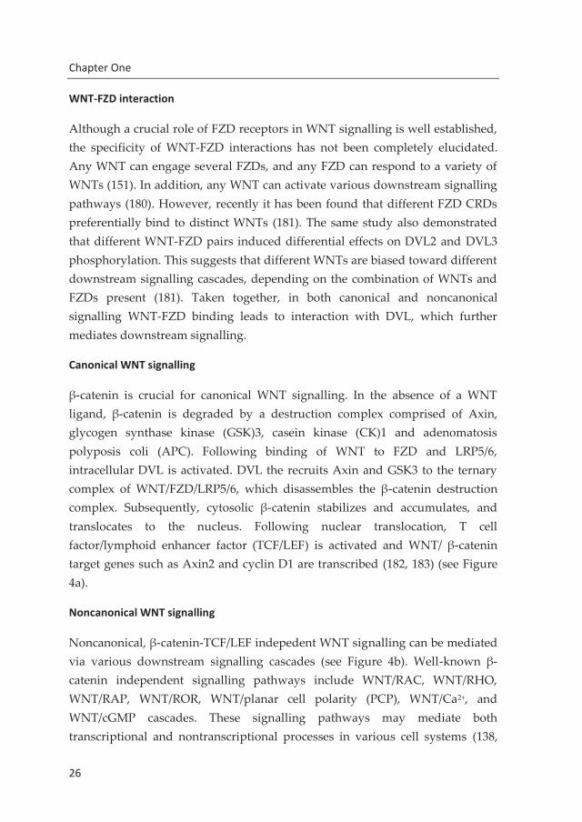
WNT-FZD interaction
Although a crucial role of FZD receptors in WNT signalling is well established, the specificity of WNT-FZD interactions has not been completely elucidated. Any WNT can engage several FZDs, and any FZD can respond to a variety of WNTs (151). In addition, any WNT can activate various downstream signalling pathways (180). However, recently it has been found that different FZD CRDs preferentially bind to distinct WNTs (181). The same study also demonstrated that different WNT-FZD pairs induced differential effects on DVL2 and DVL3 phosphorylation. This suggests that different WNTs are biased toward different downstream signalling cascades, depending on the combination of WNTs and FZDs present (181). Taken together, in both canonical and noncanonical signalling WNT-FZD binding leads to interaction with DVL, which further mediates downstream signalling.
Canonical WNT signalling
β-catenin is crucial for canonical WNT signalling. In the absence of a WNT ligand, β-catenin is degraded by a destruction complex comprised of Axin, glycogen synthase kinase (GSK)3, casein kinase (CK)1 and adenomatosis polyposis coli (APC). Following binding of WNT to FZD and LRP5/6, intracellular DVL is activated. DVL the recruits Axin and GSK3 to the ternary complex of WNT/FZD/LRP5/6, which disassembles the β-catenin destruction complex. Subsequently, cytosolic β-catenin stabilizes and accumulates, and translocates to the nucleus. Following nuclear translocation, T cell factor/lymphoid enhancer factor (TCF/LEF) is activated and WNT/ β-catenin target genes such as Axin2 and cyclin D1 are transcribed (182, 183) (see Figure 4a).
Noncanonical WNT signalling
Noncanonical, β-catenin-TCF/LEF indepedent WNT signalling can be mediated via various downstream signalling cascades (see Figure 4b). Well-known β-catenin independent signalling pathways include WNT/RAC, WNT/RHO, WNT/RAP, WNT/ROR, WNT/planar cell polarity (PCP), WNT/Ca2+, and WNT/cGMP cascades. These signalling pathways may mediate both transcriptional and nontranscriptional processes in various cell systems (138,
512661-L-bw-Stam512661-L-bw-Stam512661-L-bw-Stam512661-L-bw-StamProcessed on: 21-8-2017Processed on: 21-8-2017Processed on: 21-8-2017Processed on: 21-8-2017 PDF page: 27PDF page: 27PDF page: 27PDF page: 27
General Introduction
27
184). Three important FZD-mediated noncanonical pathways are the WNT/PCP, the WNT/Ca2+ (185), and the WNT/ cyclic guanosine 3’5’-monophosphate (cGMP) (186) signalling cascades.
In the WNT/PCP pathway, a ternary WNT/FZD complex with co-receptor RYK (187), ROR2 (160), PTK7 (188) or ROR2 and PTK7 (189) combined activates a cascade comprising the small GTPases RAC1 and Ras homolog gene family, member A (RhoA), and c-Jun N-terminal kinase (JNK). This cascade controls rearrangements in the cytoskeleton, affects the intracellular Ca2+ concentration, and regulates gene expression (184). As the name implies, this pathway regulates cell polarity in vertebrates, thereby mediating processes such as gastrulation and neural tube closure (190).
The ability of noncanonical WNT signalling to increase intracellular levels of Ca2+ was first demonstrated for the FZD2 receptor (191). This study indicated that FZD2 recruited the Gi/o family of heterotrimeric G proteins to communicate with phospholipases C (PLC). Activation of PLC increases the conversion of phosphatidylinositol 4,5-biphosphate (PIP2) to diacylglycerol (DAG) and inositol triphosphate (IP3) via hydrolysis. IP3 triggers Ca2+ release from the endoplasmatic reticulum, while DAG activates protein kinase C (PKC) which induces actin cytoskeleton remodelling. In addition, PLC activates effectors that mediate transcription of genes controlling cell fate and migration. Later, an additional route for FZD-mediated Ca2+ increase was discovered which is dependent on the activation of cGMP-selective phosphodiesterases (PDEs) (192). PDE6 is proposed to mediate this cascade, leading to drop in intracellular cGMP levels, which induces Ca2+ mobilization (193). It is still unclear whether the PLC and cGMP-PDE dependent Ca2+ pathways act in parallel and whether they are exclusive or complementary. In addition, it is unclear which factors cause the specific Ca2+ response for specific WNTs, although it has been shown that the WNT-induced Ca2+ responses are selective for pertussis toxin (PTX)-sensitive Gi/o proteins (164).

512661-L-bw-Stam512661-L-bw-Stam512661-L-bw-Stam512661-L-bw-StamProcessed on: 21-8-2017Processed on: 21-8-2017Processed on: 21-8-2017Processed on: 21-8-2017 PDF page: 28PDF page: 28PDF page: 28PDF page: 28
Chapter One
28
Figure 4a: Canonical WNT signalling, adapted from Sugimura et al. (214). In the absence of a WNT ligand, β-catenin is degraded by a destruction complex composed of Axin, APC, CK-1, and GSK3. Once WNT binds with FZD and LRP5/6 co-receptor, DVL scaffolds the β-catenin destruction complex resulting in accumulation of β-catenin in the cytosol and nucleus. In the nucleus, β-catenin forms a complex with TF to transcribe target genes (214).

512661-L-bw-Stam512661-L-bw-Stam512661-L-bw-Stam512661-L-bw-StamProcessed on: 21-8-2017Processed on: 21-8-2017Processed on: 21-8-2017Processed on: 21-8-2017 PDF page: 29PDF page: 29PDF page: 29PDF page: 29
General Introduction
29
Figure 4b: Noncanonical WNT signalling, adapted from Sugimura et al. (214). The activation of FZD by WNT is mediated by DVL or heterotrimeric G-proteins. Three important FZD-mediated noncanonical pathways are the WNT/PCP, the WNT/Ca2+ (204), and the WNT/ cyclic guanosine 3’5’-monophosphate (cGMP) (203) signalling cascades.
WNT signalling in COPD
β-catenin-mediated WNT signalling is central to mechanisms of lung repair and stem cell maintenance, and has been shown to regulate stem cell hierarchy in the renewal of lung epithelium (134, 194, 195). Alterations in WNT signalling in COPD are therefore likely to contribute to tissue remodelling and impaired repair. Indeed, dysregulation of WNT signalling is observed in COPD. For example, reduced canonical WNT signalling has been demonstrated in human airway epithelium and alveolar epithelial cells of patients with COPD, and microRNAs associated with WNT signalling are altered in COPD patients (196-198). One of the main pathological features of COPD is the loss of functional

512661-L-bw-Stam512661-L-bw-Stam512661-L-bw-Stam512661-L-bw-StamProcessed on: 21-8-2017Processed on: 21-8-2017Processed on: 21-8-2017Processed on: 21-8-2017 PDF page: 30PDF page: 30PDF page: 30PDF page: 30
Chapter One
30
alveolar tissue leading to emphysema. At the alveolar level, alveolar Type II cells are generally considered to be the facultative progenitor cells for the recovery of both the Type I and II pneumocyte pools, and are capable of differentiating into Type I cells following lung injury (199). Attenuated canonical WNT signalling in COPD has been linked to impaired alveolar repair. In COPD patients with emphysema, nuclear expression of β-catenin is decreased in alveolar Type II cells (197). In addition, therapeutic WNT/β-catenin activation by lithium chloride attenuates experimental emphysema in mice (197). Therefore, it is likely that decreased canonical WNT signalling has an important role in the parenchymal tissue destruction and impaired repair response observed in emphysema. A recent study provides more evidence for the hypothesis that decreased canonical WNT signalling is involved in emphysema. The family with sequence similarity 13, member A (FAM13A) gene has consistently been associated with COPD in genome-wide association studies (200-202), and Jiang et al. demonstrated that Fam13a(-/-) mice were resistant to elastase-induced emphysema due to reduced β-catenin degradation (203). They also demonstrated that protein levels of β-catenin and FAM13A were decreased and increased, respectively, in human COPD lungs (203). Together, these data suggest that FAM13A likely influences COPD susceptibility by enhancing β-catenin degradation.
Both canonical and noncanonical WNT signalling is altered in COPD. Dysregulated expression of noncanonical WNT-4, WNT-5A and WNT-5B has been observed in fibroblasts and airway epithelium of COPD patients as compared to controls and is associated with increased inflammatory processes, fibroblast activation, and airway remodelling (7-10, 13, 204). WNT-4 mRNA and protein levels were demonstrated to be higher in primary bronchial epithelial cells (PBECs) from COPD patients than non-smoker controls. Furthermore, exogenously added WNT-4 was shown to potentiate cigarette smoke exposure-induced upregulation of CXCL8 and vascular endothelial growth factor (8). WNT-5B protein was shown to be increased in airway epithelium from COPD patients, but not from healthy smokers or non-smokers (204). This indicates that the increase in WNT-5B expression is disease-related, and not smoking-related. In the same study it was shown that exogenously added WNT-5B increases the expression of remodeling related genes in BEAS-2B cells. This effect was

512661-L-bw-Stam512661-L-bw-Stam512661-L-bw-Stam512661-L-bw-StamProcessed on: 21-8-2017Processed on: 21-8-2017Processed on: 21-8-2017Processed on: 21-8-2017 PDF page: 31PDF page: 31PDF page: 31PDF page: 31
General Introduction
31
mediated by TGF-β/Smad3 signalling. Notably, it was demonstrated that WNT-5B upregulated the expression of remodeling-related genes in air liquid interface-cultured PBECs, particularly in PBECs from COPD patients (204). In addition, it was recently demonstrated that WNT-5A reduced WNT/β-catenin-driven alveolar epithelial cell wound healing and transdifferentiation in vitro (11). In this study, Baarsma et. al. showed that lung-specific WNT-5A (using SFT PC-rtTA TetO-WNT-5A mice) overexpression increased airspace enlargement in in vivo elastase-induced emphysema, and in vivo inhibition of WNT-5A attenuated lung tissue destruction. The reduced lung tissue destruction was accompanied by improved lung function, and restored expression levels of β-catenin-driven target genes and alveolar epithelial markers. Furthermore, the WNT-5A and WNT-5B receptor Frizzled-8 (FZD8) is related to chronic bronchitis, regulates airway inflammation, and is involved in TFG-β-induced profibrotic signalling (13, 14). Taken together, these results indicate that dysregulated noncanonical WNT signalling in COPD affects inflammatory processes, airway remodeling, and alveolar repair. Although it is clear that noncanonical WNT signalling is involved in COPD, the underlying processes are not fully understood. For example, WNT-5A and WNT-5B were shown to be associated with increased inflammatory processes, but the underlying signalling pathways remain to be elucidated. For this reason we aimed to assess the regulation of inflammatory cytokine release in response to WNT-5A/B signalling in human lung fibroblasts in chapter 2.
As described above, COPD is characterized as a disease of accelerated ageing. Recently, it was shown that WNT4 and WNT-5A expression are increased in the senile lung and contribute to myofibroblast-like differentiation (205). In addition, ageing of mouse hematopoietic stem cells (HSCs) was associated with a shift from canonical to noncanonical WNT signalling due to elevated expression of WNT-5A (206). Notably, in the same study it was demonstrated that WNT-5A treatment of young HSCs induces ageing-associated stem-cell apolarity, reduction of regenerative capacity and an ageing-like myeloid-lymphoid differentiation. In addition, WNT-5A haploinsufficiency attenuates HSC ageing, whereas stem-cell intrinsic reduction of WNT-5A expression rejuvenated aged HSCs (206). These findings indicate that a WNT signalling imbalance with decreased canonical and increased noncanonical signalling may

512661-L-bw-Stam512661-L-bw-Stam512661-L-bw-Stam512661-L-bw-StamProcessed on: 21-8-2017Processed on: 21-8-2017Processed on: 21-8-2017Processed on: 21-8-2017 PDF page: 32PDF page: 32PDF page: 32PDF page: 32
Chapter One
32
drive ageing. As mentioned above, such a WNT signalling imbalance is observed in COPD, and this imbalance may contribute to the accelerated ageing observed in COPD. To further investigate this, we aimed to investigate the interaction between noncanonical WNT signalling and ageing in chapter 5 using the PCLS model we developed in chapter 4.
Next to alterations in WNT ligands or FZD receptors, WNT antagonists are altered in COPD. Secreted frizzled-related protein (SFRP)2 is an extracellular regulator that suppresses WNT signalling, and SFRP2 was found to be upregulated in small airway epithelium of COPD patients (196). In addition, cigarette smoke exposure increases SFRP2 while decreasing WNT target genes in airway epithelial cells in vitro (196). As decreased WNT/β-catenin signalling is associated with emphysema, this implies that smoking may lead to emphysema by inhibiting WNT signalling. Taken together, both canonical and noncanonical WNT signalling is altered in COPD, leading to a WNT signalling imbalance. These alterations contribute to disease pathogenesis by increasing emphysema, inflammatory processes, and profibrotic signalling.
512661-L-bw-Stam512661-L-bw-Stam512661-L-bw-Stam512661-L-bw-StamProcessed on: 21-8-2017Processed on: 21-8-2017Processed on: 21-8-2017Processed on: 21-8-2017 PDF page: 33PDF page: 33PDF page: 33PDF page: 33
General Introduction
33
Scope of this thesis.
As described above, COPD is characterized by inflammation, increased levels of oxidative stress, parenchymal tissue destruction, and impaired repair. Dysregulated WNT expression contributes to the pathogenesis of COPD, but the underlying processes are not fully understood. Previous work from our group demonstrated a role for the noncanonical ligands WNT-5A, WNT-5B, and the receptor FZD8 in COPD. We hypothesized that noncanonical WNT signalling enhances COPD pathogenesis by increasing inflammatory processes and decreasing repair. Therefore, we aimed to further unravel the mechanisms by which noncanonical WNT signalling contributes to the development of COPD, with a focus on WNT-5A, WNT-5B, and FZD8.
There is increasing recognition of the potential importance of mesenchymal cells in inflammation (207). Recently, it has been demonstrated that pulmonary fibroblasts and airway smooth muscle cells may participate in the dysregulated inflammatory response seen in COPD (12, 72, 208). Noncanonical WNT signalling has been implicated in inflammation (7, 8, 209), and is involved in fibroblast activation (13). However, the effect of noncanonical WNT-5A/B signalling on inflammation in COPD is not well understood. For this reason we assessed the regulation of inflammatory cytokine release in response to WNT-5A/B signalling in human lung fibroblasts in chapter 2.
A possible link between oxidative stress and WNT signalling in the context of lung pathology has not been investigated yet. Although oxidative stress is known to alter WNT signalling in an indirect manner (210-213), it is not known whether oxidative stress alters WNT ligands directly. Therefore, we aimed to assess the effect of oxidative stress on WNT-5A and WNT-5B ligand-receptor interactions and the downstream signalling in chapter 3. WNT-5A and WNT-5B were oxidized using H2O2, and oxidation patterns were analysed using mass spectrometry. To determine ligand-receptor interactions, the xCELLigence real-time cell analysis system was used. Furthermore, MRC-5 pulmonary fibroblasts and mouse lung slices were treated with oxidized or non-oxidized WNT-5B and expression of several genes and proteins was analysed.

512661-L-bw-Stam512661-L-bw-Stam512661-L-bw-Stam512661-L-bw-StamProcessed on: 21-8-2017Processed on: 21-8-2017Processed on: 21-8-2017Processed on: 21-8-2017 PDF page: 34PDF page: 34PDF page: 34PDF page: 34
Chapter One
34
In COPD, the parenchyma and ECM are altered due to the presence of elastolytic enzymes and oxidative stress, leading to a loss of elastic recoil (3, 72, 80, 92). As the parenchyma around the airways is known to have an important role in airway narrowing (80), the increased levels of elastolytic enzymes and oxidative stress likely affect airway mechanics in COPD and enhance bronchoconstriction. In order to have a better understanding of enhanced airway narrowing in COPD, it is important to investigate the role of the parenchymal compartment. Therefore, in chapter 4 we aimed to study the impact of low level elastase and oxidative stress exposure on airway narrowing and relaxation in relation to parenchymal and ECM structure, and (in)activation of alveolar epithelial repair. We developed an ex vivo Precision Cut Lung Slice (PCLS) model to mimic these aspects of COPD pathophysiology.
Although several studies indicate a role of noncanonical WNT signalling in ageing and COPD, the precise role and the interaction between noncanonical WNTs, ageing and COPD remains poorly understood. In chapter 5 we studied this interaction, using aged mice in combination with the PCLS model that was developed in chapter 4.
Finally, in chapter 6, all the results presented in this thesis are discussed and placed into a broader perspective.

512661-L-bw-Stam512661-L-bw-Stam512661-L-bw-Stam512661-L-bw-StamProcessed on: 21-8-2017Processed on: 21-8-2017Processed on: 21-8-2017Processed on: 21-8-2017 PDF page: 35PDF page: 35PDF page: 35PDF page: 35
General Introduction
35
References
(1) Barnes PJ, Shapiro SD, Pauwels RA. Chronic obstructive pulmonary disease: molecular and cellular mechanisms. Eur Respir J 2003;22:672-688.
(2) Choudhury G, MacNee W. Role of Inflammation and Oxidative Stress in the Pathology of Ageing in COPD: Potential Therapeutic Interventions. COPD 2016:1-14.
(3) Kirkham PA, Barnes PJ. Oxidative stress in COPD. Chest 2013;144:266-273.
(4) Barnes PJ. New anti-inflammatory targets for chronic obstructive pulmonary disease. Nat Rev Drug Discov 2013;12:543-559.
(5) Barnes PJ. Inflammatory mechanisms in patients with chronic obstructive pulmonary disease. J Allergy Clin Immunol 2016;138:16-27.
(6) From the Global Strategy for the Diagnosis, Management and Prevention of COPD, Global Initiative for Chronic Obstructive Lung Disease (GOLD) 2016. Available from: http://goldcopd.org/. .
(7) Baarsma HA, Spanjer AI, Haitsma G, Engelbertink LH, Meurs H, Jonker MR, Timens W, Postma DS, Kerstjens HA, Gosens R. Activation of WNT/beta-catenin signaling in pulmonary fibroblasts by TGF-beta(1) is increased in chronic obstructive pulmonary disease. PLoS One 2011;6:e25450.
(8) Heijink IH, de Bruin HG, van den Berge M, Bennink LJ, Brandenburg SM, Gosens R, van Oosterhout AJ, Postma DS. Role of aberrant WNT signalling in the airway epithelial response to cigarette smoke in chronic obstructive pulmonary disease. Thorax 2013;68:709-716.
(9) Heijink I, de Bruin H, Dennebos R, van den Berge M, Gosens R, van Oosterhout A, Postma D. A role for WNT-5B in airway remodeling in COPD by activation of TGF-ß/Smad3 signaling? European Respiratory Journal 2013;42.
(10) van Dijk EM, Menzen MH, Spanjer AI, Middag LD, Brandsma CA, Gosens R. Noncanonical WNT-5B signaling induces inflammatory responses in human lung fibroblasts. Am J Physiol Lung Cell Mol Physiol 2016;310:L1166-76.
(11) Baarsma HA, Skronska-Wasek W, Mutze K, Ciolek F, Wagner DE, John-Schuster G, Heinzelmann K, Gunther A, Bracke KR, Dagouassat M, Boczkowski J, Brusselle GG, Smits R, Eickelberg O, Yildirim AO, Konigshoff M. Noncanonical WNT-5A signaling impairs endogenous lung repair in COPD. J Exp Med 2017;214:143-163.
(12) Spanjer AI, Menzen MH, Dijkstra AE, van den Berge M, Boezen HM, Nickle DC, Sin DD, Bosse Y, Brandsma CA, Timens W, Postma DS, Meurs H, Heijink IH, Gosens R. A pro-inflammatory role for the Frizzled-8 receptor in chronic bronchitis. Thorax 2016;71:312-322.
(13) Spanjer AI, Baarsma HA, Oostenbrink LM, Jansen SR, Kuipers CC, Lindner M, Postma DS, Meurs H, Heijink IH, Gosens R, Konigshoff M. TGF-beta-induced profibrotic signaling is regulated in part by the WNT receptor Frizzled-8. FASEB J 2016;30:1823-1835.
512661-L-bw-Stam512661-L-bw-Stam512661-L-bw-Stam512661-L-bw-StamProcessed on: 21-8-2017Processed on: 21-8-2017Processed on: 21-8-2017Processed on: 21-8-2017 PDF page: 36PDF page: 36PDF page: 36PDF page: 36
Chapter One
36
(14) Spanjer AIR, Menzen MH, Dijkstra AE, Brandsma CA, Postma DS, Timens W, Meurs H, Heijink HI, Gosens R. The role of the frizzled-8 receptor in COPD with chronic mucus hypersecretion [abstract]. European Respiratory Journal 2013;42: Suppl. 57:677s.
(15) Papaioannou AI, Rossios C, Kostikas K, Ito K. Can we delay the accelerated lung aging in COPD? Anti-aging molecules and interventions. Curr Drug Targets 2013;14:149-157.
(16) Fletcher C, Peto R. The natural history of chronic airflow obstruction. Br Med J 1977;1:1645-1648.
(17) Chapman KR, Mannino DM, Soriano JB, Vermeire PA, Buist AS, Thun MJ, Connell C, Jemal A, Lee TA, Miravitlles M, Aldington S, Beasley R. Epidemiology and costs of chronic obstructive pulmonary disease. Eur Respir J 2006;27:188-207.
(18) Ito K, Barnes PJ. COPD as a disease of accelerated lung aging. Chest 2009;135:173-180.
(19) Pauwels RA, Buist AS, Ma P, Jenkins CR, Hurd SS, GOLD Scientific Committee. Global strategy for the diagnosis, management, and prevention of chronic obstructive pulmonary disease: National Heart, Lung, and Blood Institute and World Health Organization Global Initiative for Chronic Obstructive Lung Disease (GOLD): executive summary. Respir Care 2001;46:798-825.
(20) Rabe KF, Hurd S, Anzueto A, Barnes PJ, Buist SA, Calverley P, Fukuchi Y, Jenkins C, Rodriguez-Roisin R, van Weel C, Zielinski J, Global Initiative for Chronic Obstructive Lung Disease. Global strategy for the diagnosis, management, and prevention of chronic obstructive pulmonary disease: GOLD executive summary. Am J Respir Crit Care Med 2007;176:532-555.
(21) Vestbo J, Hurd SS, Agusti AG, Jones PW, Vogelmeier C, Anzueto A, Barnes PJ, Fabbri LM, Martinez FJ, Nishimura M, Stockley RA, Sin DD, Rodriguez-Roisin R. Global strategy for the diagnosis, management, and prevention of chronic obstructive pulmonary disease: GOLD executive summary. Am J Respir Crit Care Med 2013;187:347-365.
(22) Marciniak SJ, Lomas DA. Genetic susceptibility. Clin Chest Med 2014;35:29-38.
(23) Lee JH, Cho MH, Hersh CP, McDonald ML, Crapo JD, Bakke PS, Gulsvik A, Comellas AP, Wendt CH, Lomas DA, Kim V, Silverman EK, COPDGene and ECLIPSE Investigators. Genetic susceptibility for chronic bronchitis in chronic obstructive pulmonary disease. Respir Res 2014;15:113-014-0113-2.
(24) Duijts L, Reiss IK, Brusselle G, de Jongste JC. Early origins of chronic obstructive lung diseases across the life course. Eur J Epidemiol 2014;29:871-885.
(25) Postma DS, Bush A, van den Berge M. Risk factors and early origins of chronic obstructive pulmonary disease. Lancet 2015;385:899-909.
(26) Carraro S, Scheltema N, Bont L, Baraldi E. Early-life origins of chronic respiratory diseases: understanding and promoting healthy ageing. Eur Respir J 2014;44:1682-1696.
(27) Freeman CM, Curtis JL. Lung Dendritic Cells: Shaping Immune Responses Throughout COPD Progression. Am J Respir Cell Mol Biol 2016.

512661-L-bw-Stam512661-L-bw-Stam512661-L-bw-Stam512661-L-bw-StamProcessed on: 21-8-2017Processed on: 21-8-2017Processed on: 21-8-2017Processed on: 21-8-2017 PDF page: 37PDF page: 37PDF page: 37PDF page: 37
General Introduction
37
(28) Pouwels SD, Heijink IH, ten Hacken NH, Vandenabeele P, Krysko DV, Nawijn MC, van Oosterhout AJ. DAMPs activating innate and adaptive immune responses in COPD. Mucosal Immunol 2014;7:215-226.
(29) Hogg JC, Chu F, Utokaparch S, Woods R, Elliott WM, Buzatu L, Cherniack RM, Rogers RM, Sciurba FC, Coxson HO, Pare PD. The nature of small-airway obstruction in chronic obstructive pulmonary disease. N Engl J Med 2004;350:2645-2653.
(30) Barnes PJ. Immunology of asthma and chronic obstructive pulmonary disease. Nat Rev Immunol 2008;8:183-192.
(31) Brusselle GG, Joos GF, Bracke KR. New insights into the immunology of chronic obstructive pulmonary disease. Lancet 2011;378:1015-1026.
(32) Gamble E, Grootendorst DC, Hattotuwa K, O'Shaughnessy T, Ram FS, Qiu Y, Zhu J, Vignola AM, Kroegel C, Morell F, Pavord ID, Rabe KF, Jeffery PK, Barnes NC. Airway mucosal inflammation in COPD is similar in smokers and ex-smokers: a pooled analysis. Eur Respir J 2007;30:467-471.
(33) Gao W, Li L, Wang Y, Zhang S, Adcock IM, Barnes PJ, Huang M, Yao X. Bronchial epithelial cells: The key effector cells in the pathogenesis of chronic obstructive pulmonary disease? Respirology 2015;20:722-729.
(34) Barnes PJ. The cytokine network in chronic obstructive pulmonary disease. Am J Respir Cell Mol Biol 2009;41:631-638.
(35) Barnes PJ. The cytokine network in asthma and chronic obstructive pulmonary disease. J Clin Invest 2008;118:3546-3556.
(36) Russell RE, Thorley A, Culpitt SV, Dodd S, Donnelly LE, Demattos C, Fitzgerald M, Barnes PJ. Alveolar macrophage-mediated elastolysis: roles of matrix metalloproteinases, cysteine, and serine proteases. Am J Physiol Lung Cell Mol Physiol 2002;283:L867-73.
(37) Lopez-Otin C, Blasco MA, Partridge L, Serrano M, Kroemer G. The hallmarks of aging. Cell 2013;153:1194-1217.
(38) Blasco MA. Telomeres and human disease: ageing, cancer and beyond. Nat Rev Genet 2005;6:611-622.
(39) Navarro S, Driscoll B. Regeneration of the Aging Lung: A Mini-Review. Gerontology 2016.
(40) Sharma G, Goodwin J. Effect of aging on respiratory system physiology and immunology. Clin Interv Aging 2006;1:253-260.
(41) Caramori G, Adcock IM, Casolari P, Ito K, Jazrawi E, Tsaprouni L, Villetti G, Civelli M, Carnini C, Chung KF, Barnes PJ, Papi A. Unbalanced oxidant-induced DNA damage and repair in COPD: a link towards lung cancer. Thorax 2011;66:521-527.
(42) Aoshiba K, Zhou F, Tsuji T, Nagai A. DNA damage as a molecular link in the pathogenesis of COPD in smokers. Eur Respir J 2012;39:1368-1376.
(43) Tsuji T, Aoshiba K, Nagai A. Alveolar cell senescence in patients with pulmonary emphysema. Am J Respir Crit Care Med 2006;174:886-893.
(44) Savale L, Chaouat A, Bastuji-Garin S, Marcos E, Boyer L, Maitre B, Sarni M, Housset B, Weitzenblum E, Matrat M, Le Corvoisier P, Rideau D, Boczkowski J, Dubois-Rande JL,

512661-L-bw-Stam512661-L-bw-Stam512661-L-bw-Stam512661-L-bw-StamProcessed on: 21-8-2017Processed on: 21-8-2017Processed on: 21-8-2017Processed on: 21-8-2017 PDF page: 38PDF page: 38PDF page: 38PDF page: 38
Chapter One
38
Chouaid C, Adnot S. Shortened telomeres in circulating leukocytes of patients with chronic obstructive pulmonary disease. Am J Respir Crit Care Med 2009;179:566-571.
(45) Houben JM, Mercken EM, Ketelslegers HB, Bast A, Wouters EF, Hageman GJ, Schols AM. Telomere shortening in chronic obstructive pulmonary disease. Respir Med 2009;103:230-236.
(46) Vucic EA, Chari R, Thu KL, Wilson IM, Cotton AM, Kennett JY, Zhang M, Lonergan KM, Steiling K, Brown CJ, McWilliams A, Ohtani K, Lenburg ME, Sin DD, Spira A, Macaulay CE, Lam S, Lam WL. DNA methylation is globally disrupted and associated with expression changes in chronic obstructive pulmonary disease small airways. Am J Respir Cell Mol Biol 2014;50:912-922.
(47) Qiu W, Baccarelli A, Carey VJ, Boutaoui N, Bacherman H, Klanderman B, Rennard S, Agusti A, Anderson W, Lomas DA, DeMeo DL. Variable DNA methylation is associated with chronic obstructive pulmonary disease and lung function. Am J Respir Crit Care Med 2012;185:373-381.
(48) Rajendrasozhan S, Yang SR, Kinnula VL, Rahman I. SIRT1, an antiinflammatory and antiaging protein, is decreased in lungs of patients with chronic obstructive pulmonary disease. Am J Respir Crit Care Med 2008;177:861-870.
(49) Ito K, Ito M, Elliott WM, Cosio B, Caramori G, Kon OM, Barczyk A, Hayashi S, Adcock IM, Hogg JC, Barnes PJ. Decreased histone deacetylase activity in chronic obstructive pulmonary disease. N Engl J Med 2005;352:1967-1976.
(50) MacNee W. Is Chronic Obstructive Pulmonary Disease an Accelerated Aging Disease? Ann Am Thorac Soc 2016;13:S429-S437.
(51) Chen ZH, Lam HC, Jin Y, Kim HP, Cao J, Lee SJ, Ifedigbo E, Parameswaran H, Ryter SW, Choi AM. Autophagy protein microtubule-associated protein 1 light chain-3B (LC3B) activates extrinsic apoptosis during cigarette smoke-induced emphysema. Proc Natl Acad Sci U S A 2010;107:18880-18885.
(52) Johnson SC, Rabinovitch PS, Kaeberlein M. mTOR is a key modulator of ageing and age-related disease. Nature 2013;493:338-345.
(53) Balaban RS, Nemoto S, Finkel T. Mitochondria, oxidants, and aging. Cell 2005;120:483-495.
(54) Sureshbabu A, Bhandari V. Targeting mitochondrial dysfunction in lung diseases: emphasis on mitophagy. Front Physiol 2013;4:384.
(55) Ballweg K, Mutze K, Konigshoff M, Eickelberg O, Meiners S. Cigarette smoke extract affects mitochondrial function in alveolar epithelial cells. Am J Physiol Lung Cell Mol Physiol 2014;307:L895-907.
(56) Hara H, Araya J, Ito S, Kobayashi K, Takasaka N, Yoshii Y, Wakui H, Kojima J, Shimizu K, Numata T, Kawaishi M, Kamiya N, Odaka M, Morikawa T, Kaneko Y, Nakayama K, Kuwano K. Mitochondrial fragmentation in cigarette smoke-induced bronchial epithelial cell senescence. Am J Physiol Lung Cell Mol Physiol 2013;305:L737-46.
(57) Hoffmann RF, Zarrintan S, Brandenburg SM, Kol A, de Bruin HG, Jafari S, Dijk F, Kalicharan D, Kelders M, Gosker HR, Ten Hacken NH, van der Want JJ, van Oosterhout

512661-L-bw-Stam512661-L-bw-Stam512661-L-bw-Stam512661-L-bw-StamProcessed on: 21-8-2017Processed on: 21-8-2017Processed on: 21-8-2017Processed on: 21-8-2017 PDF page: 39PDF page: 39PDF page: 39PDF page: 39
General Introduction
39
AJ, Heijink IH. Prolonged cigarette smoke exposure alters mitochondrial structure and function in airway epithelial cells. Respir Res 2013;14:97-9921-14-97.
(58) Tsuji T, Aoshiba K, Nagai A. Alveolar cell senescence exacerbates pulmonary inflammation in patients with chronic obstructive pulmonary disease. Respiration 2010;80:59-70.
(59) Signer RA, Morrison SJ. Mechanisms that regulate stem cell aging and life span. Cell Stem Cell 2013;12:152-165.
(60) Shaykhiev R, Crystal RG. Basal cell origins of smoking-induced airway epithelial disorders. Cell Cycle 2014;13:341-342.
(61) Ryan DM, Vincent TL, Salit J, Walters MS, Agosto-Perez F, Shaykhiev R, Strulovici-Barel Y, Downey RJ, Buro-Auriemma LJ, Staudt MR, Hackett NR, Mezey JG, Crystal RG. Smoking dysregulates the human airway basal cell transcriptome at COPD risk locus 19q13.2. PLoS One 2014;9:e88051.
(62) Paschalaki KE, Starke RD, Hu Y, Mercado N, Margariti A, Gorgoulis VG, Randi AM, Barnes PJ. Dysfunction of endothelial progenitor cells from smokers and chronic obstructive pulmonary disease patients due to increased DNA damage and senescence. Stem Cells 2013;31:2813-2826.
(63) Janssen WJ, Yunt ZX, Muldrow A, Kearns MT, Kloepfer A, Barthel L, Bratton DL, Bowler RP, Henson PM. Circulating hematopoietic progenitor cells are decreased in COPD. COPD 2014;11:277-289.
(64) Cevenini E, Monti D, Franceschi C. Inflamm-ageing. Curr Opin Clin Nutr Metab Care 2013;16:14-20.
(65) Curtis JL, Freeman CM, Hogg JC. The immunopathogenesis of chronic obstructive pulmonary disease: insights from recent research. Proc Am Thorac Soc 2007;4:512-521.
(66) Li L, Wang Y, Gao W, Yuan C, Zhang S, Zhou H, Huang M, Yao X. Klotho Reduction in Alveolar Macrophages Contributes to Cigarette Smoke Extract-induced Inflammation in Chronic Obstructive Pulmonary Disease. J Biol Chem 2015;290:27890-27900.
(67) Gao W, Yuan C, Zhang J, Li L, Yu L, Wiegman CH, Barnes PJ, Adcock IM, Huang M, Yao X. Klotho expression is reduced in COPD airway epithelial cells: effects on inflammation and oxidant injury. Clin Sci (Lond) 2015;129:1011-1023.
(68) van den Berge M, ten Hacken NH, Cohen J, Douma WR, Postma DS. Small airway disease in asthma and COPD: clinical implications. Chest 2011;139:412-423.
(69) Hogg JC, Macklem PT, Thurlbeck WM. Site and nature of airway obstruction in chronic obstructive lung disease. N Engl J Med 1968;278:1355-1360.
(70) Papi A, Casoni G, Caramori G, Guzzinati I, Boschetto P, Ravenna F, Calia N, Petruzzelli S, Corbetta L, Cavallesco G, Forini E, Saetta M, Ciaccia A, Fabbri LM. COPD increases the risk of squamous histological subtype in smokers who develop non-small cell lung carcinoma. Thorax 2004;59:679-681.
(71) Williams OW, Sharafkhaneh A, Kim V, Dickey BF, Evans CM. Airway mucus: From production to secretion. Am J Respir Cell Mol Biol 2006;34:527-536.
512661-L-bw-Stam512661-L-bw-Stam512661-L-bw-Stam512661-L-bw-StamProcessed on: 21-8-2017Processed on: 21-8-2017Processed on: 21-8-2017Processed on: 21-8-2017 PDF page: 40PDF page: 40PDF page: 40PDF page: 40
Chapter One
40
(72) Chung KF. The role of airway smooth muscle in the pathogenesis of airway wall remodeling in chronic obstructive pulmonary disease. Proc Am Thorac Soc 2005;2:347-54; discussion 371-2.
(73) Saetta M, Di Stefano A, Turato G, Facchini FM, Corbino L, Mapp CE, Maestrelli P, Ciaccia A, Fabbri LM. CD8+ T-lymphocytes in peripheral airways of smokers with chronic obstructive pulmonary disease. Am J Respir Crit Care Med 1998;157:822-826.
(74) Kuwano K, Bosken CH, Pare PD, Bai TR, Wiggs BR, Hogg JC. Small airways dimensions in asthma and in chronic obstructive pulmonary disease. Am Rev Respir Dis 1993;148:1220-1225.
(75) Tiddens HA, Pare PD, Hogg JC, Hop WC, Lambert R, de Jongste JC. Cartilaginous airway dimensions and airflow obstruction in human lungs. Am J Respir Crit Care Med 1995;152:260-266.
(76) Bosken CH, Wiggs BR, Pare PD, Hogg JC. Small airway dimensions in smokers with obstruction to airflow. Am Rev Respir Dis 1990;142:563-570.
(77) James AL, Elliot JG, Jones RL, Carroll ML, Mauad T, Bai TR, Abramson MJ, McKay KO, Green FH. Airway smooth muscle hypertrophy and hyperplasia in asthma. Am J Respir Crit Care Med 2012;185:1058-1064.
(78) Thomson RJ, Bramley AM, Schellenberg RR. Airway muscle stereology: implications for increased shortening in asthma. Am J Respir Crit Care Med 1996;154:749-757.
(79) Bonnans C, Chou J, Werb Z. Remodelling the extracellular matrix in development and disease. Nat Rev Mol Cell Biol 2014;15:786-801.
(80) Bidan CM, Veldsink AC, Meurs H, Gosens R. Airway and Extracellular Matrix Mechanics in COPD. Front Physiol 2015;6:346.
(81) Yick CY, Ferreira DS, Annoni R, von der Thusen JH, Kunst PW, Bel EH, Lutter R, Mauad T, Sterk PJ. Extracellular matrix in airway smooth muscle is associated with dynamics of airway function in asthma. Allergy 2012;67:552-559.
(82) Salazar LM, Herrera AM. Fibrotic response of tissue remodeling in COPD. Lung 2011;189:101-109.
(83) Parameswaran K, Willems-Widyastuti A, Alagappan VK, Radford K, Kranenburg AR, Sharma HS. Role of extracellular matrix and its regulators in human airway smooth muscle biology. Cell Biochem Biophys 2006;44:139-146.
(84) McAnulty RJ. Fibroblasts and myofibroblasts: their source, function and role in disease. Int J Biochem Cell Biol 2007;39:666-671.
(85) Bartram U, Speer CP. The role of transforming growth factor beta in lung development and disease. Chest 2004;125:754-765.
(86) White ES. Lung extracellular matrix and fibroblast function. Ann Am Thorac Soc 2015;12 Suppl 1:S30-3.
(87) Karvonen HM, Lehtonen ST, Harju T, Sormunen RT, Lappi-Blanco E, Makinen JM, Laitakari K, Johnson S, Kaarteenaho RL. Myofibroblast expression in airways and alveoli is affected by smoking and COPD. Respir Res 2013;14:84-9921-14-84.

512661-L-bw-Stam512661-L-bw-Stam512661-L-bw-Stam512661-L-bw-StamProcessed on: 21-8-2017Processed on: 21-8-2017Processed on: 21-8-2017Processed on: 21-8-2017 PDF page: 41PDF page: 41PDF page: 41PDF page: 41
General Introduction
41
(88) Lofdahl M, Kaarteenaho R, Lappi-Blanco E, Tornling G, Skold MC. Tenascin-C and alpha-smooth muscle actin positive cells are increased in the large airways in patients with COPD. Respir Res 2011;12:48-9921-12-48.
(89) Plantier L, Boczkowski J, Crestani B. Defect of alveolar regeneration in pulmonary emphysema: role of lung fibroblasts. Int J Chron Obstruct Pulmon Dis 2007;2:463-469.
(90) Plantier L, Rochette-Egly C, Goven D, Boutten A, Bonay M, Leseche G, Fournier M, Crestani B, Boczkowski J. Dysregulation of elastin expression by fibroblasts in pulmonary emphysema: role of cellular retinoic acid binding protein 2. Thorax 2008;63:1012-1017.
(91) Mocchegiani E, Giacconi R, Costarelli L. Metalloproteases/anti-metalloproteases imbalance in chronic obstructive pulmonary disease: genetic factors and treatment implications. Curr Opin Pulm Med 2011;17 Suppl 1:S11-9.
(92) Eurlings IM, Dentener MA, Cleutjens JP, Peutz CJ, Rohde GG, Wouters EF, Reynaert NL. Similar matrix alterations in alveolar and small airway walls of COPD patients. BMC Pulm Med 2014;14:90-2466-14-90.
(93) Brandsma CA, van den Berge M, Postma DS, Jonker MR, Brouwer S, Pare PD, Sin DD, Bosse Y, Laviolette M, Karjalainen J, Fehrmann RS, Nickle DC, Hao K, Spanjer AI, Timens W, Franke L. A large lung gene expression study identifying fibulin-5 as a novel player in tissue repair in COPD. Thorax 2015;70:21-32.
(94) Black PN, Ching PS, Beaumont B, Ranasinghe S, Taylor G, Merrilees MJ. Changes in elastic fibres in the small airways and alveoli in COPD. Eur Respir J 2008;31:998-1004.
(95) Tjin G, Xu P, Kable SH, Kable EP, Burgess JK. Quantification of collagen I in airway tissues using second harmonic generation. J Biomed Opt 2014;19:36005.
(96) Gubbiotti MA, Vallet SD, Ricard-Blum S, Iozzo RV. Decorin interacting network: A comprehensive analysis of decorin-binding partners and their versatile functions. Matrix Biol 2016;55:7-21.
(97) Noordhoek JA, Postma DS, Chong LL, Menkema L, Kauffman HF, Timens W, van Straaten JF, van der Geld YM. Different modulation of decorin production by lung fibroblasts from patients with mild and severe emphysema. COPD 2005;2:17-25.
(98) van Straaten JF, Coers W, Noordhoek JA, Huitema S, Flipsen JT, Kauffman HF, Timens W, Postma DS. Proteoglycan changes in the extracellular matrix of lung tissue from patients with pulmonary emphysema. Mod Pathol 1999;12:697-705.
(99) Hallgren O, Nihlberg K, Dahlback M, Bjermer L, Eriksson LT, Erjefalt JS, Lofdahl CG, Westergren-Thorsson G. Altered fibroblast proteoglycan production in COPD. Respir Res 2010;11:55-9921-11-55.
(100) Chung KF, Adcock IM. Multifaceted mechanisms in COPD: inflammation, immunity, and tissue repair and destruction. Eur Respir J 2008;31:1334-1356.
(101) Suki B, Bates JH. Extracellular matrix mechanics in lung parenchymal diseases. Respir Physiol Neurobiol 2008;163:33-43.
(102) Suki B, Bates JH. Lung tissue mechanics as an emergent phenomenon. J Appl Physiol (1985) 2011;110:1111-1118.

512661-L-bw-Stam512661-L-bw-Stam512661-L-bw-Stam512661-L-bw-StamProcessed on: 21-8-2017Processed on: 21-8-2017Processed on: 21-8-2017Processed on: 21-8-2017 PDF page: 42PDF page: 42PDF page: 42PDF page: 42
Chapter One
42
(103) Lauzon AM, Bates JH, Donovan G, Tawhai M, Sneyd J, Sanderson MJ. A multi-scale approach to airway hyperresponsiveness: from molecule to organ. Front Physiol 2012;3:191.
(104) Ma B, Bates JH. Mechanical interactions between adjacent airways in the lung. J Appl Physiol (1985) 2014;116:628-634.
(105) An SS, Bai TR, Bates JH, Black JL, Brown RH, Brusasco V, Chitano P, Deng L, Dowell M, Eidelman DH, Fabry B, Fairbank NJ, Ford LE, Fredberg JJ, Gerthoffer WT, Gilbert SH, Gosens R, Gunst SJ, Halayko AJ, Ingram RH, Irvin CG, James AL, Janssen LJ, King GG, Knight DA, Lauzon AM, Lakser OJ, Ludwig MS, Lutchen KR, Maksym GN, Martin JG, Mauad T, McParland BE, Mijailovich SM, Mitchell HW, Mitchell RW, Mitzner W, Murphy TM, Pare PD, Pellegrino R, Sanderson MJ, Schellenberg RR, Seow CY, Silveira PS, Smith PG, Solway J, Stephens NL, Sterk PJ, Stewart AG, Tang DD, Tepper RS, Tran T, Wang L. Airway smooth muscle dynamics: a common pathway of airway obstruction in asthma. Eur Respir J 2007;29:834-860.
(106) Herrero E, Ros J, Tamarit J, Belli G. Glutaredoxins in fungi. Photosynth Res 2006;89:127-140.
(107) Vogt W. Oxidation of methionyl residues in proteins: tools, targets, and reversal. Free Radic Biol Med 1995;18:93-105.
(108) Herrero E, Ros J, Belli G, Cabiscol E. Redox control and oxidative stress in yeast cells. Biochim Biophys Acta 2008;1780:1217-1235.
(109) Cabiscol E, Tamarit J, Ros J. Protein carbonylation: proteomics, specificity and relevance to aging. Mass Spectrom Rev 2014;33:21-48.
(110) Dean RT, Fu S, Stocker R, Davies MJ. Biochemistry and pathology of radical-mediated protein oxidation. Biochem J 1997;324 ( Pt 1):1-18.
(111) Yao H, Rahman I. Current concepts on oxidative/carbonyl stress, inflammation and epigenetics in pathogenesis of chronic obstructive pulmonary disease. Toxicol Appl Pharmacol 2011;254:72-85.
(112) Dove RE, Leong-Smith P, Roos-Engstrand E, Pourazar J, Shah M, Behndig AF, Mudway IS, Blomberg A. Cigarette smoke-induced induction of antioxidant enzyme activities in airway leukocytes is absent in active smokers with COPD. Eur Clin Respir J 2015;2:10.3402/ecrj.v2.27837. eCollection 2015.
(113) Schaberg T, Klein U, Rau M, Eller J, Lode H. Subpopulations of alveolar macrophages in smokers and nonsmokers: relation to the expression of CD11/CD18 molecules and superoxide anion production. Am J Respir Crit Care Med 1995;151:1551-1558.
(114) Vaitkus M, Lavinskiene S, Barkauskiene D, Bieksiene K, Jeroch J, Sakalauskas R. Reactive oxygen species in peripheral blood and sputum neutrophils during bacterial and nonbacterial acute exacerbation of chronic obstructive pulmonary disease. Inflammation 2013;36:1485-1493.
(115) Ichinose M, Sugiura H, Yamagata S, Koarai A, Shirato K. Increase in reactive nitrogen species production in chronic obstructive pulmonary disease airways. Am J Respir Crit Care Med 2000;162:701-706.

512661-L-bw-Stam512661-L-bw-Stam512661-L-bw-Stam512661-L-bw-StamProcessed on: 21-8-2017Processed on: 21-8-2017Processed on: 21-8-2017Processed on: 21-8-2017 PDF page: 43PDF page: 43PDF page: 43PDF page: 43
General Introduction
43
(116) Rahman I, van Schadewijk AA, Crowther AJ, Hiemstra PS, Stolk J, MacNee W, De Boer WI. 4-Hydroxy-2-nonenal, a specific lipid peroxidation product, is elevated in lungs of patients with chronic obstructive pulmonary disease. Am J Respir Crit Care Med 2002;166:490-495.
(117) Kluchova Z, Petrasova D, Joppa P, Dorkova Z, Tkacova R. The association between oxidative stress and obstructive lung impairment in patients with COPD. Physiol Res 2007;56:51-56.
(118) Montuschi P. Exhaled breath condensate analysis in patients with COPD. Clin Chim Acta 2005;356:22-34.
(119) Paredi P, Kharitonov SA, Barnes PJ. Analysis of expired air for oxidation products. Am J Respir Crit Care Med 2002;166:S31-7.
(120) Paredi P, Kharitonov SA, Leak D, Ward S, Cramer D, Barnes PJ. Exhaled ethane, a marker of lipid peroxidation, is elevated in chronic obstructive pulmonary disease. Am J Respir Crit Care Med 2000;162:369-373.
(121) Bartoli ML, Novelli F, Costa F, Malagrino L, Melosini L, Bacci E, Cianchetti S, Dente FL, Di Franco A, Vagaggini B, Paggiaro PL. Malondialdehyde in exhaled breath condensate as a marker of oxidative stress in different pulmonary diseases. Mediators Inflamm 2011;2011:891752.
(122) Barreiro E, Peinado VI, Galdiz JB, Ferrer E, Marin-Corral J, Sanchez F, Gea J, Barbera JA, ENIGMA in COPD Project. Cigarette smoke-induced oxidative stress: A role in chronic obstructive pulmonary disease skeletal muscle dysfunction. Am J Respir Crit Care Med 2010;182:477-488.
(123) Drost EM, Skwarski KM, Sauleda J, Soler N, Roca J, Agusti A, MacNee W. Oxidative stress and airway inflammation in severe exacerbations of COPD. Thorax 2005;60:293-300.
(124) Louhelainen N, Rytila P, Haahtela T, Kinnula VL, Djukanovic R. Persistence of oxidant and protease burden in the airways after smoking cessation. BMC Pulm Med 2009;9:25-2466-9-25.
(125) Cantin A, Crystal RG. Oxidants, antioxidants and the pathogenesis of emphysema. Eur J Respir Dis Suppl 1985;139:7-17.
(126) Sayyed AK, Despande KH, Suryakar AN, Ankush RD, Katkam RV. Oxidative stress and serum alpha(1) - Antitrypsin in smokers. Indian J Clin Biochem 2008;23:375-377.
(127) Godtfredsen NS, Lam TH, Hansel TT, Leon ME, Gray N, Dresler C, Burns DM, Prescott E, Vestbo J. COPD-related morbidity and mortality after smoking cessation: status of the evidence. Eur Respir J 2008;32:844-853.
(128) Barnes PJ. Inhaled corticosteroids in COPD: a controversy. Respiration 2010;80:89-95.
(129) Barnes NC, Sharma R, Lettis S, Calverley PM. Blood eosinophils as a marker of response to inhaled corticosteroids in COPD. Eur Respir J 2016;47:1374-1382.
(130) Barnes PJ. Role of HDAC2 in the pathophysiology of COPD. Annu Rev Physiol 2009;71:451-464.

512661-L-bw-Stam512661-L-bw-Stam512661-L-bw-Stam512661-L-bw-StamProcessed on: 21-8-2017Processed on: 21-8-2017Processed on: 21-8-2017Processed on: 21-8-2017 PDF page: 44PDF page: 44PDF page: 44PDF page: 44
Chapter One
44
(131) Rennard SI, Calverley PM, Goehring UM, Bredenbroker D, Martinez FJ. Reduction of exacerbations by the PDE4 inhibitor roflumilast--the importance of defining different subsets of patients with COPD. Respir Res 2011;12:18-9921-12-18.
(132) Logan CY, Nusse R. The Wnt signaling pathway in development and disease. Annu Rev Cell Dev Biol 2004;20:781-810.
(133) Goss AM, Tian Y, Tsukiyama T, Cohen ED, Zhou D, Lu MM, Yamaguchi TP, Morrisey EE. Wnt2/2b and beta-catenin signaling are necessary and sufficient to specify lung progenitors in the foregut. Dev Cell 2009;17:290-298.
(134) Reya T, Clevers H. Wnt signalling in stem cells and cancer. Nature 2005;434:843-850.
(135) Clevers H, Loh KM, Nusse R. Stem cell signaling. An integral program for tissue renewal and regeneration: Wnt signaling and stem cell control. Science 2014;346:1248012.
(136) Mason JO, Kitajewski J, Varmus HE. Mutational analysis of mouse Wnt-1 identifies two temperature-sensitive alleles and attributes of Wnt-1 protein essential for transformation of a mammary cell line. Mol Biol Cell 1992;3:521-533.
(137) Mikels AJ, Nusse R. Wnts as ligands: processing, secretion and reception. Oncogene 2006;25:7461-7468.
(138) Niehrs C. The complex world of WNT receptor signalling. Nat Rev Mol Cell Biol 2012;13:767-779.
(139) Miller JR. The Wnts. Genome Biol 2002;3:REVIEWS3001.
(140) Tanaka K, Kitagawa Y, Kadowaki T. Drosophila segment polarity gene product porcupine stimulates the posttranslational N-glycosylation of wingless in the endoplasmic reticulum. J Biol Chem 2002;277:12816-12823.
(141) Reichsman F, Smith L, Cumberledge S. Glycosaminoglycans can modulate extracellular localization of the wingless protein and promote signal transduction. J Cell Biol 1996;135:819-827.
(142) Gross JC, Boutros M. Secretion and extracellular space travel of Wnt proteins. Curr Opin Genet Dev 2013;23:385-390.
(143) Herr P, Basler K. Porcupine-mediated lipidation is required for Wnt recognition by Wls. Dev Biol 2012;361:392-402.
(144) Tang X, Wu Y, Belenkaya TY, Huang Q, Ray L, Qu J, Lin X. Roles of N-glycosylation and lipidation in Wg secretion and signaling. Dev Biol 2012;364:32-41.
(145) Willert K, Brown JD, Danenberg E, Duncan AW, Weissman IL, Reya T, Yates JR,3rd, Nusse R. Wnt proteins are lipid-modified and can act as stem cell growth factors. Nature 2003;423:448-452.
(146) Takada R, Satomi Y, Kurata T, Ueno N, Norioka S, Kondoh H, Takao T, Takada S. Monounsaturated fatty acid modification of Wnt protein: its role in Wnt secretion. Dev Cell 2006;11:791-801.
(147) Greco V, Hannus M, Eaton S. Argosomes: a potential vehicle for the spread of morphogens through epithelia. Cell 2001;106:633-645.
512661-L-bw-Stam512661-L-bw-Stam512661-L-bw-Stam512661-L-bw-StamProcessed on: 21-8-2017Processed on: 21-8-2017Processed on: 21-8-2017Processed on: 21-8-2017 PDF page: 45PDF page: 45PDF page: 45PDF page: 45
General Introduction
45
(148) Coudreuse DY, Roel G, Betist MC, Destree O, Korswagen HC. Wnt gradient formation requires retromer function in Wnt-producing cells. Science 2006;312:921-924.
(149) Bartscherer K, Pelte N, Ingelfinger D, Boutros M. Secretion of Wnt ligands requires Evi, a conserved transmembrane protein. Cell 2006;125:523-533.
(150) Coombs GS, Yu J, Canning CA, Veltri CA, Covey TM, Cheong JK, Utomo V, Banerjee N, Zhang ZH, Jadulco RC, Concepcion GP, Bugni TS, Harper MK, Mihalek I, Jones CM, Ireland CM, Virshup DM. WLS-dependent secretion of WNT3A requires Ser209 acylation and vacuolar acidification. J Cell Sci 2010;123:3357-3367.
(151) Janda CY, Waghray D, Levin AM, Thomas C, Garcia KC. Structural basis of Wnt recognition by Frizzled. Science 2012;337:59-64.
(152) Franch-Marro X, Wendler F, Griffith J, Maurice MM, Vincent JP. In vivo role of lipid adducts on Wingless. J Cell Sci 2008;121:1587-1592.
(153) Kumar S, Zigman M, Patel TR, Trageser B, Gross JC, Rahm K, Boutros M, Gradl D, Steinbeisser H, Holstein T, Stetefeld J, Ozbek S. Molecular dissection of Wnt3a-Frizzled8 interaction reveals essential and modulatory determinants of Wnt signaling activity. BMC Biol 2014;12:44-7007-12-44.
(154) Chu ML, Ahn VE, Choi HJ, Daniels DL, Nusse R, Weis WI. structural Studies of Wnts and identification of an LRP6 binding site. Structure 2013;21:1235-1242.
(155) MacDonald BT, Hien A, Zhang X, Iranloye O, Virshup DM, Waterman ML, He X. Disulfide bond requirements for active Wnt ligands. J Biol Chem 2014;289:18122-18136.
(156) He X, Semenov M, Tamai K, Zeng X. LDL receptor-related proteins 5 and 6 in Wnt/beta-catenin signaling: arrows point the way. Development 2004;131:1663-1677.
(157) Mikels A, Minami Y, Nusse R. Ror2 receptor requires tyrosine kinase activity to mediate Wnt5A signaling. J Biol Chem 2009;284:30167-30176.
(158) Green JL, Kuntz SG, Sternberg PW. Ror receptor tyrosine kinases: orphans no more. Trends Cell Biol 2008;18:536-544.
(159) Lu W, Yamamoto V, Ortega B, Baltimore D. Mammalian Ryk is a Wnt coreceptor required for stimulation of neurite outgrowth. Cell 2004;119:97-108.
(160) Oishi I, Suzuki H, Onishi N, Takada R, Kani S, Ohkawara B, Koshida I, Suzuki K, Yamada G, Schwabe GC, Mundlos S, Shibuya H, Takada S, Minami Y. The receptor tyrosine kinase Ror2 is involved in non-canonical Wnt5a/JNK signalling pathway. Genes Cells 2003;8:645-654.
(161) Peradziryi H, Kaplan NA, Podleschny M, Liu X, Wehner P, Borchers A, Tolwinski NS. PTK7/Otk interacts with Wnts and inhibits canonical Wnt signalling. EMBO J 2011;30:3729-3740.
(162) Carron C, Pascal A, Djiane A, Boucaut JC, Shi DL, Umbhauer M. Frizzled receptor dimerization is sufficient to activate the Wnt/beta-catenin pathway. J Cell Sci 2003;116:2541-2550.
(163) Foord SM, Bonner TI, Neubig RR, Rosser EM, Pin JP, Davenport AP, Spedding M, Harmar AJ. International Union of Pharmacology. XLVI. G protein-coupled receptor list. Pharmacol Rev 2005;57:279-288.

512661-L-bw-Stam512661-L-bw-Stam512661-L-bw-Stam512661-L-bw-StamProcessed on: 21-8-2017Processed on: 21-8-2017Processed on: 21-8-2017Processed on: 21-8-2017 PDF page: 46PDF page: 46PDF page: 46PDF page: 46
Chapter One
46
(164) Schulte G. International Union of Basic and Clinical Pharmacology. LXXX. The class Frizzled receptors. Pharmacol Rev 2010;62:632-667.
(165) Dijksterhuis JP, Petersen J, Schulte G. WNT/Frizzled signalling: receptor-ligand selectivity with focus on FZD-G protein signalling and its physiological relevance: IUPHAR Review 3. Br J Pharmacol 2014;171:1195-1209.
(166) Kilander MB, Dijksterhuis JP, Ganji RS, Bryja V, Schulte G. WNT-5A stimulates the GDP/GTP exchange at pertussis toxin-sensitive heterotrimeric G proteins. Cell Signal 2011;23:550-554.
(167) Egger-Adam D, Katanaev VL. The trimeric G protein Go inflicts a double impact on axin in the Wnt/frizzled signaling pathway. Dev Dyn 2010;239:168-183.
(168) Egger-Adam D, Katanaev VL. Trimeric G protein-dependent signaling by Frizzled receptors in animal development. Front Biosci 2008;13:4740-4755.
(169) Luchtenborg AM, Solis GP, Egger-Adam D, Koval A, Lin C, Blanchard MG, Kellenberger S, Katanaev VL. Heterotrimeric Go protein links Wnt-Frizzled signaling with ankyrins to regulate the neuronal microtubule cytoskeleton. Development 2014;141:3399-3409.
(170) Schioth HB, Fredriksson R. The GRAFS classification system of G-protein coupled receptors in comparative perspective. Gen Comp Endocrinol 2005;142:94-101.
(171) Vinson CR, Conover S, Adler PN. A Drosophila tissue polarity locus encodes a protein containing seven potential transmembrane domains. Nature 1989;338:263-264.
(172) Xu YK, Nusse R. The Frizzled CRD domain is conserved in diverse proteins including several receptor tyrosine kinases. Curr Biol 1998;8:R405-6.
(173) Chen CM, Strapps W, Tomlinson A, Struhl G. Evidence that the cysteine-rich domain of Drosophila Frizzled family receptors is dispensable for transducing Wingless. Proc Natl Acad Sci U S A 2004;101:15961-15966.
(174) Baarsma HA, Konigshoff M, Gosens R. The WNT signaling pathway from ligand secretion to gene transcription: molecular mechanisms and pharmacological targets. Pharmacol Ther 2013;138:66-83.
(175) Dale TC. Signal transduction by the Wnt family of ligands. Biochem J 1998;329 ( Pt 2):209-223.
(176) Gao C, Chen YG. Dishevelled: The hub of Wnt signaling. Cell Signal 2010;22:717-727.
(177) Hao HX, Xie Y, Zhang Y, Charlat O, Oster E, Avello M, Lei H, Mickanin C, Liu D, Ruffner H, Mao X, Ma Q, Zamponi R, Bouwmeester T, Finan PM, Kirschner MW, Porter JA, Serluca FC, Cong F. ZNRF3 promotes Wnt receptor turnover in an R-spondin-sensitive manner. Nature 2012;485:195-200.
(178) Jiang X, Charlat O, Zamponi R, Yang Y, Cong F. Dishevelled promotes Wnt receptor degradation through recruitment of ZNRF3/RNF43 E3 ubiquitin ligases. Mol Cell 2015;58:522-533.

512661-L-bw-Stam512661-L-bw-Stam512661-L-bw-Stam512661-L-bw-StamProcessed on: 21-8-2017Processed on: 21-8-2017Processed on: 21-8-2017Processed on: 21-8-2017 PDF page: 47PDF page: 47PDF page: 47PDF page: 47
General Introduction
47
(179) Koo BK, Spit M, Jordens I, Low TY, Stange DE, van de Wetering M, van Es JH, Mohammed S, Heck AJ, Maurice MM, Clevers H. Tumour suppressor RNF43 is a stem-cell E3 ligase that induces endocytosis of Wnt receptors. Nature 2012;488:665-669.
(180) Mikels AJ, Nusse R. Purified Wnt5a protein activates or inhibits beta-catenin-TCF signaling depending on receptor context. PLoS Biol 2006;4:e115.
(181) Dijksterhuis JP, Baljinnyam B, Stanger K, Sercan HO, Ji Y, Andres O, Rubin JS, Hannoush RN, Schulte G. Systematic mapping of WNT-FZD protein interactions reveals functional selectivity by distinct WNT-FZD pairs. J Biol Chem 2015;290:6789-6798.
(182) Clevers H, Nusse R. Wnt/beta-catenin signaling and disease. Cell 2012;149:1192-1205.
(183) Lustig B, Jerchow B, Sachs M, Weiler S, Pietsch T, Karsten U, van de Wetering M, Clevers H, Schlag PM, Birchmeier W, Behrens J. Negative feedback loop of Wnt signaling through upregulation of conductin/axin2 in colorectal and liver tumors. Mol Cell Biol 2002;22:1184-1193.
(184) Semenov MV, Habas R, Macdonald BT, He X. SnapShot: Noncanonical Wnt Signaling Pathways. Cell 2007;131:1378.
(185) Wang HY, Malbon CC. Wnt-frizzled signaling to G-protein-coupled effectors. Cell Mol Life Sci 2004;61:69-75.
(186) Wang HY. WNT-frizzled signaling via cyclic GMP. Front Biosci 2004;9:1043-1047.
(187) Macheda ML, Sun WW, Kugathasan K, Hogan BM, Bower NI, Halford MM, Zhang YF, Jacques BE, Lieschke GJ, Dabdoub A, Stacker SA. The Wnt receptor Ryk plays a role in mammalian planar cell polarity signaling. J Biol Chem 2012;287:29312-29323.
(188) Peradziryi H, Tolwinski NS, Borchers A. The many roles of PTK7: a versatile regulator of cell-cell communication. Arch Biochem Biophys 2012;524:71-76.
(189) Martinez S, Scerbo P, Giordano M, Daulat AM, Lhoumeau AC, Thome V, Kodjabachian L, Borg JP. The PTK7 and ROR2 Protein Receptors Interact in the Vertebrate WNT/Planar Cell Polarity (PCP) Pathway. J Biol Chem 2015;290:30562-30572.
(190) Gomez-Orte E, Saenz-Narciso B, Moreno S, Cabello J. Multiple functions of the noncanonical Wnt pathway. Trends Genet 2013;29:545-553.
(191) Slusarski DC, Corces VG, Moon RT. Interaction of Wnt and a Frizzled homologue triggers G-protein-linked phosphatidylinositol signalling. Nature 1997;390:410-413.
(192) Ahumada A, Slusarski DC, Liu X, Moon RT, Malbon CC, Wang HY. Signaling of rat Frizzled-2 through phosphodiesterase and cyclic GMP. Science 2002;298:2006-2010.
(193) Wang H, Lee Y, Malbon CC. PDE6 is an effector for the Wnt/Ca2+/cGMP-signalling pathway in development. Biochem Soc Trans 2004;32:792-796.
(194) Konigshoff M, Eickelberg O. WNT signaling in lung disease: a failure or a regeneration signal? Am J Respir Cell Mol Biol 2010;42:21-31.
(195) Reynolds SD, Zemke AC, Giangreco A, Brockway BL, Teisanu RM, Drake JA, Mariani T, Di PY, Taketo MM, Stripp BR. Conditional stabilization of beta-catenin expands the pool of lung stem cells. Stem Cells 2008;26:1337-1346.

512661-L-bw-Stam512661-L-bw-Stam512661-L-bw-Stam512661-L-bw-StamProcessed on: 21-8-2017Processed on: 21-8-2017Processed on: 21-8-2017Processed on: 21-8-2017 PDF page: 48PDF page: 48PDF page: 48PDF page: 48
Chapter One
48
(196) Wang R, Ahmed J, Wang G, Hassan I, Strulovici-Barel Y, Hackett NR, Crystal RG. Down-regulation of the canonical Wnt beta-catenin pathway in the airway epithelium of healthy smokers and smokers with COPD. PLoS One 2011;6:e14793.
(197) Kneidinger N, Yildirim AO, Callegari J, Takenaka S, Stein MM, Dumitrascu R, Bohla A, Bracke KR, Morty RE, Brusselle GG, Schermuly RT, Eickelberg O, Konigshoff M. Activation of the WNT/beta-catenin pathway attenuates experimental emphysema. Am J Respir Crit Care Med 2011;183:723-733.
(198) Ezzie ME, Crawford M, Cho JH, Orellana R, Zhang S, Gelinas R, Batte K, Yu L, Nuovo G, Galas D, Diaz P, Wang K, Nana-Sinkam SP. Gene expression networks in COPD: microRNA and mRNA regulation. Thorax 2012;67:122-131.
(199) Mason RJ. Biology of alveolar type II cells. Respirology 2006;11 Suppl:S12-5.
(200) Cho MH, Boutaoui N, Klanderman BJ, Sylvia JS, Ziniti JP, Hersh CP, DeMeo DL, Hunninghake GM, Litonjua AA, Sparrow D, Lange C, Won S, Murphy JR, Beaty TH, Regan EA, Make BJ, Hokanson JE, Crapo JD, Kong X, Anderson WH, Tal-Singer R, Lomas DA, Bakke P, Gulsvik A, Pillai SG, Silverman EK. Variants in FAM13A are associated with chronic obstructive pulmonary disease. Nat Genet 2010;42:200-202.
(201) Guo Y, Lin H, Gao K, Xu H, Deng X, Zhang Q, Luo Z, Sun S, Deng H. Genetic analysis of IREB2, FAM13A and XRCC5 variants in Chinese Han patients with chronic obstructive pulmonary disease. Biochem Biophys Res Commun 2011;415:284-287.
(202) Kim WJ, Lee SD. Candidate genes for COPD: current evidence and research. Int J Chron Obstruct Pulmon Dis 2015;10:2249-2255.
(203) Jiang Z, Lao T, Qiu W, Polverino F, Gupta K, Guo F, Mancini JD, Naing ZZ, Cho MH, Castaldi PJ, Sun Y, Yu J, Laucho-Contreras ME, Kobzik L, Raby BA, Choi AM, Perrella MA, Owen CA, Silverman EK, Zhou X. A Chronic Obstructive Pulmonary Disease Susceptibility Gene, FAM13A, Regulates Protein Stability of beta-Catenin. Am J Respir Crit Care Med 2016;194:185-197.
(204) Heijink IH, de Bruin HG, Dennebos R, Jonker MR, Noordhoek JA, Brandsma CA, van den Berge M, Postma DS. Cigarette smoke-induced epithelial expression of WNT-5B: implications for COPD. Eur Respir J 2016;48:504-515.
(205) Kovacs T, Csongei V, Feller D, Ernszt D, Smuk G, Sarosi V, Jakab L, Kvell K, Bartis D, Pongracz JE. Alteration in the Wnt microenvironment directly regulates molecular events leading to pulmonary senescence. Aging Cell 2014;13:838-849.
(206) Florian MC, Nattamai KJ, Dorr K, Marka G, Uberle B, Vas V, Eckl C, Andra I, Schiemann M, Oostendorp RA, Scharffetter-Kochanek K, Kestler HA, Zheng Y, Geiger H. A canonical to non-canonical Wnt signalling switch in haematopoietic stem-cell ageing. Nature 2013;503:392-396.
(207) Alkhouri H, Poppinga WJ, Tania NP, Ammit A, Schuliga M. Regulation of pulmonary inflammation by mesenchymal cells. Pulm Pharmacol Ther 2014.
(208) Zhang J, Wu L, Qu JM, Bai CX, Merrilees MJ, Black PN. Pro-inflammatory phenotype of COPD fibroblasts not compatible with repair in COPD lung. J Cell Mol Med 2012;16:1522-1532.

512661-L-bw-Stam512661-L-bw-Stam512661-L-bw-Stam512661-L-bw-StamProcessed on: 21-8-2017Processed on: 21-8-2017Processed on: 21-8-2017Processed on: 21-8-2017 PDF page: 49PDF page: 49PDF page: 49PDF page: 49
General Introduction
49
(209) Jung YS, Lee HY, Kim SD, Park JS, Kim JK, Suh PG, Bae YS. Wnt5a stimulates chemotactic migration and chemokine production in human neutrophils. Exp Mol Med 2013;45:e27.
(210) Kajla S, Mondol AS, Nagasawa A, Zhang Y, Kato M, Matsuno K, Yabe-Nishimura C, Kamata T. A crucial role for Nox 1 in redox-dependent regulation of Wnt-beta-catenin signaling. FASEB J 2012;26:2049-2059.
(211) Funato Y, Miki H. Redox regulation of Wnt signalling via nucleoredoxin. Free Radic Res 2010;44:379-388.
(212) Zhang X, Abreu JG, Yokota C, MacDonald BT, Singh S, Coburn KL, Cheong SM, Zhang MM, Ye QZ, Hang HC, Steen H, He X. Tiki1 is required for head formation via Wnt cleavage-oxidation and inactivation. Cell 2012;149:1565-1577.
(213) Sandieson L, Hwang JT, Kelly GM. Redox regulation of canonical Wnt signaling affects extraembryonic endoderm formation. Stem Cells Dev 2014;23:1037-1049.
(214) Sugimura R, Li L. Noncanonical Wnt signaling in vertebrate development, stem cells, and diseases. Birth Defects Res C Embryo Today 2010;90:243-256.

512661-L-bw-Stam512661-L-bw-Stam512661-L-bw-Stam512661-L-bw-StamProcessed on: 21-8-2017Processed on: 21-8-2017Processed on: 21-8-2017Processed on: 21-8-2017 PDF page: 50PDF page: 50PDF page: 50PDF page: 50