Embed Size (px)
Citation preview

University of Groningen
Viral neuroinvasionSips, George
IMPORTANT NOTE: You are advised to consult the publisher's version (publisher's PDF) if you wish to cite fromit. Please check the document version below.
Document VersionPublisher's PDF, also known as Version of record
Publication date:2014
Link to publication in University of Groningen/UMCG research database
Citation for published version (APA):Sips, G. (2014). Viral neuroinvasion: causes, mechanisms and potential consequences. [S.l.]: s.n.
CopyrightOther than for strictly personal use, it is not permitted to download or to forward/distribute the text or part of it without the consent of theauthor(s) and/or copyright holder(s), unless the work is under an open content license (like Creative Commons).
Take-down policyIf you believe that this document breaches copyright please contact us providing details, and we will remove access to the work immediatelyand investigate your claim.
Downloaded from the University of Groningen/UMCG research database (Pure): http://www.rug.nl/research/portal. For technical reasons thenumber of authors shown on this cover page is limited to 10 maximum.
Download date: 27-07-2019

Copyright © Marijke Jansen, www.marijkejansenphotoart.nl


79
http://onlinelibrary.wiley.com/doi/10.1002/rmv.712/abstract;jsessionid=D24D00DAD976F61EAB50DEBE426CED2F.f01t02
Published in: Reviews in Medical Virology 2012;22:69-87Copyright © 2011 John Wiley & Sons, Ltd.
G.J. Sips, J. Wilschut, J.M. Smit
NEUROINVASIVE FLAVIVIRUS INFECTIONS
CHAPTER 3

80
chapter 3
Neuroinvasive flavivirus infections
Gregorius J. Sips, Jan Wilschut and Jolanda M. Smit* Department of Medical Microbiology, Molecular Virology Section, University Medical Center Groningen, University of Groningen, The Netherlands *Corresponding author: Dr. Jolanda M. Smit, Department of Medical Microbiology, Molec-ular Virology Section, University Medical Center Groningen, Antonius Deusinglaan 1, 9713 AV Groningen, The Netherlands. E-mail: [email protected] Received: 11 May 2011; Revised: 5 August 2011; Accepted: 9 August 2011
SUMMARYFlaviviruses, including Dengue, West Nile, Japanese encephalitis, and Tick-borne
encephalitis virus, are major emerging human pathogens, affecting millions of individuals worldwide. Many clinically important flaviviruses elicit CNS diseases in infected hosts, including traditional “hemorrhagic” viruses, such as Dengue. This review focuses on the epidemiology, symptomatology, neuropathology, and, specifically, neuropathogenesis of flavivirus-induced human CNS disease. A detailed insight into specific factors priming towards neuroinvasive disease is of clear clinical significance, as well as importance to the development of antiviral therapies and identification of key mechanisms involved in the (re)emergence of specific flaviviruses, including potentially novel or previously unrecog-nized ones, as neuroinvasive pathogens.
ABBREVIATIONS USEDAFP, acute flaccid paralysis; BBB, blood-brain barrier; C, capsid; CCL-2, chemokine (C-C
motif) ligand-2; CCR5, C-C chemokine receptor type 5; CNS, central nervous system; DENV, dengue virus; DHF, dengue hemorrhagic fever; DSS, dengue shock syndrome; E, envelope; GAG, glycosaminoglycan; Hsp, heat shock protein; ICAM-1, intercellular adhesion mole-cule-1; JEV, japanese encephalitis virus; LIV, louping ill virus; MIF, macrophage migration inhibitory factor; MMP9, matrix metallopeptidase 9;MVEV, murray valley encephalitis virus; NCR, noncoding region; NO, nitric oxide; OAS, 2-5 oligoadenylate synthethase; POWV, powassan virus; prM, premembrane; ROCV, rocio virus; SLEV, saint louis encephalitis virus; TBEV, tick-borne encephalitis virus; TBEV-Eu, european tick-borne encephalitis virus; TBEV-FE, far eastern tick-borne encephalitis virus; TBEV-Sib, siberian tick-borne encepha-litis virus; WNV, west nile virus; YFV, yellow fever virus.

81
Neuroinvasive flavivirus infections
3
INTRODUCTION
Because of rapid changes in climate and demography, vector-transmitted arboviral dis-eases pose an increasing threat to global health and welfare [1,2]. Among the most severe arboviral infections known to affect human race are those caused by members of the Flavivirus genus of the Flaviviridae. The genus comprises over 70 different members and includes major human pathogens such as Yellow Fever virus (YFV), Dengue virus (DENV), Japanese encephalitis virus (JEV), Tick-borne encephalitis virus (TBEV), and West Nile virus (WNV) [3–5].
Flaviviruses are single-stranded, positive-sense RNA viruses, whose genome encodes three structural and seven NS proteins [6]. All flaviviruses circulate in transmission cycles consisting of vertebrate hosts and insect vectors, in which humans mostly act as dead-end hosts [7]. Natural cases of human infection almost invariably follow the bite of an infected tick or mosquito, although incidental cases related to other transmission mechanisms, including the use of infected blood products and organ transplants or, in case of TBEV, oral transfer through consumption of unpasteurized milk (products) have, infrequently, been reported as well [4,5]. A spectrum of distinct clinical syndromes is known to complicate flavi-virus infections in humans, ranging from relatively mild fever and arthalgia to severe hem-orrhagic and encephalitic manifestations [5]. In contrast to the systemic syndromes, the development of encephalitic pathology relies upon the ability of the virus to gain entry to the CNS, a process known as viral neuroinvasiveness, and to infect neural cells, a phenom-enon known as neurovirulence [8]. Interestingly, both abilities seem to be widely dispersed among various members of the Flavivirus genus (Figure 1, red). Neuroinvasive infections frequently occur upon infection with emerging viruses such as JEV, WNV, and TBEV, which have a global distribution range (Figure 2) and affect hundreds of thousands of individuals worldwide, annually [5,9]. Recently, however, they have also increasingly been reported in the setting of viruses that otherwise mostly cause hemorrhagic disease such as DENV[10–12]. This phenomenon of neuroinvasive dengue was, until recent years, largely unrecognized and interestingly suggests the existence of a continuum concerning the pathogenesis of flaviviral CNS infections and potential emergence of more neurovirulent dengue strains.
This review aims to synthesize current knowledge on flavivirus-induced CNS disease, including the emerging concept of neurological dengue. Apart from discussing their epi-demiology, symptomatology, and neuropathology, we will specifically focus on the neu-ropathogenesis of highly prevalent emerging viruses associated with human CNS disease. A profound understanding of the pathogenesis of these syndromes is of clear importance with respect to the development of effective therapeutic strategies. Furthermore, the identification of specific factors involved in flaviviral neuroinvasiveness, such as distinct viral proteins or host factors, might help to explain the (re)emergence of specific viruses,
82
chapter 3
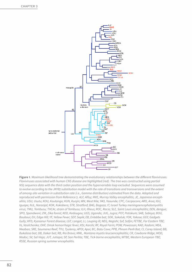
Figure 1. Maximum likelihood tree demonstrating the evolutionary relationships between the different flaviviruses. Flaviviruses associated with human CNS disease are highlighted (red). The tree was constructed using partial NS5 sequence data with the third codon position and the hypervariable loop excluded. Sequences were assumed to evolve according to the JKY85 substitution model with the rate of transitions and transversions and the extent of among-site variation in substitution rate (i.e., Gamma distribution) estimated from the data. Adapted and reproduced with permission from Reference 5. ALF, Alfuy; MVE, Murray Valley encephalitis; JE, Japanese enceph-alitis; USU, Usutu; KOU, Koutango; KUN, Kunjin; WN, West Nile; YAO, Yaounde; CPC, Cacipacore; ARO, Aroa; IGU, Iguape; NJL, Naranjal; KOK, Kokobera; STR, Stratford; BAG, Bagaza; IT, Israel Turkey meningoencephalomyelitis virus; TMU, Tembusu; THCAr, strain of Tembusu; ILH, Ilheus; ROC, Rocio; SLE, Saint Louis encephalitis; DEN, dengue; SPO, Spondweni; ZIK, Zika forest; KED, Kedougou; UGS, Uganda; JUG, Jugra; POT, Potiskum; SAB, Saboya; BOU, Bouboui; EH, Edge Hill; YF, Yellow Fever; SEP, Sepik; EB, Entebbe bat; SOK, Sokoluk; YOK, Yokose; GGY, Gadgets Gully; KFD, Kyasanur Forest disease; LGT, Langat; LI, Louping ill; NEG, Negishi; Sof, Sofjin; FETBE, Far Eastern TBE; Vs, Vasilchenko; OHF, Omsk hemorrhagic fever; KSI, Karshi; RF, Royal Farm; POW, Powassan; KAD, Kadam; MEA, Meaban; SRE, Saumarez Reef; TYU, Tyuleniy; APOI, Apoi; BC, Batu Cave; PPB, Phnom Penh Bat; CI, Carey Island; BB, Bukalasa bat; DB, Dakar bat; RB, Rio Bravo; MML, Montana myotis leucoencephalitis; CR, Cowbone Ridge; MOD, Modoc; SV, Sal Vieja; JUT, Jutiapa; SP, San Perlita; TBE, Tick-borne encephalitis; WTBE, Western European TBE; RSSE, Russian spring summer encephalitis.

83
Neuroinvasive flavivirus infections
3
including previously unrecognized or potentially novel ones, as neuroinvasive pathogens. We will start with a discussion of JEV which, historically, has been the best studied neuro-virulent flavivirus and is the cause of annual large-sized epidemics in many Eastern Asian countries.
JAPANESE ENCEPHALITIS VIRUS
Epidemiology
Japanese encephalitis virus constitutes the most significant cause of mosquito-borne encephalitis worldwide and is endemic throughout large parts of Central and Southeastern Asia, where it causes a massive total of about 30,000–50,000 reported cases of infection, annually [9,13–15]. In endemic areas, JEV is estimated to have an asymptomatic/symptomatic disease ratio of about 25–1000/1 and about 20%–30% of all symptomatic infections are fatal [9,13–15]. Symptomatic infections mostly present as febrile syndromes that commonly progress into the multifocal CNS disorders that characterize the disease [9,13,14]. It has been estimated that approximately 70% of symptomatic infections clinically manifest as enceph-
Figure 2. Approximate geographic distribution of major emerging flaviviruses associated with human CNS disease. Based on data from References 9, 14, 51–54, 89, 90 and 131

84
chapter 3
alitis, whereas an additional 10% present as meningitis [9,13,14]. JEV mostly affects children and nonimmune adults and treatment remains largely supportive [16]. Effective vaccination schemes have been developed but their implementation in high-risk areas has, thus far, proven difficult for financial and logistical reasons [9,17,18].
Central nervous system disease
Neurological symptoms typically develop after an incubation period of 5–15 days and commonly include alterations of consciousness, seizures, and, specifically, the develop-ment of Parkinsonian movement disorders and dystonias, which have been reported to occur in up to 60% of symptomatic patients [13]. Another 5%–20% of patients will, addition-ally, present with poliomyelitis-like pyramidal motor pathology, characterized by multi-focal paralysis or paresis [13]. After an acute episode of illness, about 50% of survivors retain permanent (neuro)psychiatric sequelae, which manifest as persisting cognitive or move-ment disorders [13]. Although these sequelae could result from acute disease processes, subacute and chronic forms of JEV infection have been reported as well. In this respect, one report has, interestingly, described relapses associated with the recovery of infectious virus from peripheral blood leukocytes, whereas another study described the prolonged persistence of IgM and viral antigens in the CSF of about 5% of patients studied [19,20].
Neuropathology
Autopsy studies have identified gray matter areas of the thalamus, midbrain/substantia nigra, hippocampus, cerebral cortices, and anterior horns of the medulla oblongata and cervical spinal cord as primary JEV-affected brain regions [21–25]. General neuropathological alterations consist of edema, hemorrhage, vascular congestion, and widespread perivas-cular inflammatory infiltrates. Characteristic of the disease are distinct foci of acellular necrotic ‘plaques’ confined to gray matter areas. Astrogliosis and the formation of micro-glial nodules, often in close proximity to affected regions, have been described as well. Furthermore, examinations of the previously mentioned subacute and chronic cases of infection have demonstrated diffuse calcium deposits as well as binucleated nerve cells [21–25]. Neuronal cells, particularly pyramidal (motor) neurons, clearly constitute the main cellular target population of JEV in vivo. On microscopic evaluation, many neurons within affected areas display clear degenerative changes and contain viral antigen [21–25] (Figure 3). Infection of vascular endothelial cells as well as occasional ependymal cells and astrocytes have, albeit infrequently, been reported as well [21,22].
Neuropathogenesis
With respect to exact CNS entry mechanisms of JEV, both intraneural transport through the olfactory nerve, following intranasal inoculation, as well as hematogenous transport have been described [26–29]. The first method of inoculation is unlikely to occur in vivo, but a

85
Neuroinvasive flavivirus infections
3
more indirect mechanism of entry through the olfactory nerve following initial systemic rep-lication and subsequent hematogenous spread to the olfactory mucosa has, interestingly, been described in studies with the closely related flaviviruses Saint Louis encephalitis virus (SLEV) and Murray Valley encephalitis virus (MVEV) [30,31]. In animal models, a cytokine-me-diated increased permeability of the blood-brain barrier (BBB) has been demonstrated, which likely precedes and facilitates viral transport across the BBB [27]. Several mechanisms of transport across the BBB have been described, including direct infection of vascular endothelial cells, transcellular transport, and within infected monocytes, the so-called “Trojan-horse” mechanism [26–29]. Within the CNS, JEV has, notably, been shown to display a particular tropism for developing neurons and neuroprogenitor cells which might help to explain the viral predilection for specific brain regions, such as the hippocampus, as well as the severity of JEV infections and their outcome in children [32,33]. Although this distinct tropism might point towards the existence of specific neuronal JEV-receptors, few studies have examined the presence of such receptors on cells of neural origin. So far, only one study, using mouse neuroblastoma cells, has been published, suggesting that heat shock protein 70 (Hsp70) mediates viral entry into neurons and further studies defining the nature of potential neuronal JEV-receptors are warranted [34]. Downstream of cellular entry, both virus-induced apoptosis as well as necrosis, mediated by an uncontrolled overactivation of microglia and release of reactive oxygen species, TNFα, and nitric oxide (NO), leading towards “bystander” damage to neuronal cells, have been demonstrated in vitro [35–37]. Furthermore, microglia and JEV-infected leukocytes have been identified as possible viral reservoirs and could play a role in the pathogenesis of subacute and chronic infections, as well as the neurological sequelae, that have been reported following JEV-infection [38,39]. The pathogenesis of these chronic forms of JEV infection, however, has, thus far, not been widely investigated and is certainly in need of further study.
A large number of studies have, historically, been performed to address specific viral determinants of neuroinvasiveness and neurovirulence. These studies have shown that a large proportion, and likely the vast majority, of epitopes that govern JEV neuroinvasive-ness and neurovirulence are located within relatively limited sections of the viral enve-lope (E) protein. Particularly, these include areas within the lateral surface of domain III as well as base of domain II of E, which are believed to play crucial roles in cellular receptor binding and fusion with target cells, respectively [8,40–45]. Apart from these studies clearly indicating an important role for the E protein, a relatively limited number of recent studies have demonstrated the involvement of other viral proteins in governing viral neuroinva-siveness and neurovirulence. Several studies have, in this respect, indicated the effect of mutations in the viral capsid (C) and premembrane (prM) proteins in limiting viral neuro-virulence [46,47]. Also, it has recently been shown that production of the NS1’ protein, which occurs as a result of ribosomal frame-shifting in members of the JE-serocomplex, but not in most other flaviviruses, increases viral neuroinvasiveness [48].

86
chapter 3
WEST NILE VIRUS
Epidemiology
West Nile virus, a mosquito-borne member of the JE-serocomplex, which has historically been endemic throughout large parts of Africa, Asia, Australia, and Europe, caused a mas-sive outbreak of human disease in the New York area in 1999 and, since then, has rapidly spread throughout the North American continent [49–53]. Serological studies have indicated the circulation of the virus in a number of Latin American countries as well, but reports on human infection have thus far remained sparse [52,54].
Following the 1999 epidemic, WNV has become the leading cause of arboviral enceph-alitis in the USA and, here alone, a total of about 30,000 cases of infection have been reported during the last decade, of which approximately 1,200 (4%) have been fatal [55]. It is estimated that about 80% of infections are asymptomatic, whereas symptomatic infec-tions mostly give rise to the development of a self-limited febrile syndrome known as West Nile fever (WNF) [49,50]. About 1/150 patients develop CNS complications, which are usually grouped together under the term West Nile neuroinvasive disease (WNND) [49,50]. Unlike some other flaviviruses, WNV, notably, mostly appears to affect the elderly and immuno-compromised, even when introduced into largely naïve populations as occurred in 1999 [49,50]. Currently, treatment remains largely supportive, although extensive scientific efforts are being made to develop therapies for future clinical use [56].
CENTRAL NERVOUS SYSTEM DISEASEWest Nile neuroinvasive disease can be subclassified into three main clinical syndromes
of meningitis, encephalitis, and acute flaccid paralysis/poliomyelitis [49,50]. Of these syn-dromes, the clinical picture of acute flaccid paralysis (AFP) is the most distinctive as well as best characterized entity. In contrast to what holds true for most other arboviral encephalitides, neuromuscular weakness constitutes a prominent finding in WNND, occur-ring in up to 50% of patients, sometimes in the absence of other disease symptoms [49]. AFP typically presents as monoplegia, asymmetric upper or lower extremity weakness, or generalized asymmetric tetraplegia or quadriplegia. Additionally, in about 70% of patients with AFP, there is involvement of one or more of the cranial nerve(s) and a large number of patients require intubation or ventilation because of respiratory failure [49,50]. Although most patients with WNV meningitis without focal neurological deficits tend to recover fully, the prognosis is much worse in cases of encephalitis or AFP, which are characterized by a 10%–20% mortality rate [49]. Up to 70%–75% of survivors of WNND, furthermore, retain per-manent neurological sequelae [49]. Recently, subacute and relapsing forms of AFP, as well as long-term persistence of WNV associated with viral shedding in urine, have been reported in subsets of patients, which might provide clues with respect to possible mechanisms of flavivirus persistence as well as the frequent occurrence of postinfectious sequelae [57,58].

87
Neuroinvasive flavivirus infections
3
Neuropathology
Histologically, WNND is characterized by a pattern of microglial nodules, perivascular inflammatory infiltrates and reactive astrogliosis combined with neuronal loss, necrotic foci, and neuronophagia [59–61]. Topographically, there is a clear predilection for gray matter areas of the brainstem, particularly the medulla, and spinal cord [59–61]. Brain areas that might, additionally, be affected include the cerebellum, temporal lobes, basal ganglia, and thalamus [59–61]. This predilection in terms of affected brain areas has been confirmed by immunohistochemical studies [59–61] (Figure 3). The virus primarily infects neurons, mostly pyramidal motor neurons of the anterior horns and cerebellar Purkinje cells, although there have been occasional reports of infection of astroglial and monocytic cells as well [59–62].
Figure 3. Immunohistochemical stains of flavivirus-positive brain tissue samples. A Immunohistochemical stain demonstrating JEV-positivity of hippocampal pyramidal neurons in a case of JE (anti-JEV antibody, x50). Repro-duced with kind permission from Reference 22. B Immunohistochemical stain demonstrating WNV-positivity of pontine neurons in a case of WNE (anti-flavivirus polyclonal antibody, H/E counterstain, x50). Courtesy of Dr. J.Guarner (Department of Pathology and Laboratory Medicine, Emory University School of Medicine, Atlanta, GA, USA). C Immunohistochemical stain demonstrating DENV-positivity of cortical neurons in a case of dengue infection (anti-DENV antibody). Reproduced with kind permission from Reference 109. D Immunohistochemical stain demon-strating TBEV-positivity of cerebellar neurons in a case of TBE (anti-TBEV antibody, H/E counterstain, x40). Courtesy of Dr. E. Gelpi and Prof.H. Budka (Institute of Neurology, Medical University of Vienna, Austria)

88
chapter 3
Neuropathogenesis
The recent North American epidemics have greatly fuelled WNV-related research and, as a consequence, led to a vast increase in our knowledge of its neuropathogenesis as well as, potentially, that of other neuroinvasive flaviviruses. Importantly, several adverse effects of innate and adaptive systemic antiviral immune responses have, during recent years, been described, which lead towards increased permeability of the BBB and, hence, likely facili-tate viral entry into the CNS [63]. Specifically, increases in brain endothelial capillary perme-ability have been reported, induced by the TLR3-mediated release of TNFα, as well as by macrophage migration inhibitory factor (MIF), intercellular adhesion molecule-1 (ICAM-1), and matrix metallopeptidase 9 (MMP9). Dysregulations of TLR3-responses have specifically been demonstrated to compromise BBB-integrity in the elderly [64–68]. Following this break-down, the virus has been suggested to cross the BBB via several mechanisms, including transcellular transport, paracellular transport, direct infection of endothelial cells, or Tro-jan-horse mechanisms of entry [69]. Another pathway, which was shown to directly induce AFP in animal models of infection, includes retrograde axonal transport through peripheral motor nerves [70].
Upon entry of the CNS, WNV displays a particular tropism for (anterior horn motor) neu-rons. Not many studies have, so far, been undertaken to identify possible neuronal WNV-re-ceptors, although one study, interestingly, described the presence of a plasma membrane glycoprotein of Mr 105,000 that facilitated viral entry in murine neuroblastoma cells [71]. Once infected, neural cells have been demonstrated to undergo various mechanisms of apoptosis [72–75]. Bystander damage, resulting from immunopathological effects of the CD8+ T-cell response as well as the recruitment of inflammatory monocytes mediated by chemokine (C-C motif) ligand-2 (CCL-2), has been described, as well [76,77]. A number of recent studies have investigated the pathogenesis of persistent WNV infections. These studies have demonstrated persistence of WNV in the CNS and peripheral organs, par-ticularly the urinary tract, and correlated neurological sequelae to persistent viral disease activity in the CNS [78–80].
As for JEV, the E protein appears to be particularly important in governing the neuroin-vasiveness and neurovirulence of WNV [40,81–83]. Additional roles for the C as well as several NS proteins have been described in governing viral neurovirulence, but further studies addressing their roles in flaviviral neuropathogenesis would be required [62,84].
Interestingly, a number of predisposing host factors have now been identified. Of these, genetic risk factors include mutations in the C—C chemokine receptor type 5 (CCR5) and 2–5 oligoadenylate synthethase (OAS) genes, which play important roles in antiviral immune responses [85,86]. Furthermore, a number of acquired, age-specific, T-cell defects in both CD4 as well as CD8 subsets have been described, which, at least in animal models, greatly increase host susceptibility to severe and neuroinvasive WNV infection [87].

89
Neuroinvasive flavivirus infections
3
DENGUE VIRUS
Epidemiology
The mosquito-borne DENV is by far the most important arbovirus known to affect man-kind and constitutes a significant public health problem, particularly in the developing world. According to the WHO, DENV is now endemic in over 100 different countries, where some 2.5 billion people are at risk of getting infected. In these regions, approximately 50–100 million cases of DENV infection occur annually, about 250,000 to 500,000 of which are cases of severe dengue hemorrhagic fever (DHF) [88,89]. Outside of the more traditional regions, a re-emergence of autochtonous DENV transmission within Europe has, after a long period of absence, recently, been described [90]. Approximately 50% of infections are asymptomatic, whereas symptomatic infections can present with a variety of clinical syndromes, ranging from aspecific or mild-febrile disease to the aforementioned DHF or dengue shock syndromes (DSS) [88,89,91]. Treatment of DENV remains supportive and there is a strong and urgent need for effective therapeutic and vaccination strategies [88,89,91,92].
Central nervous system disease
Apart from hemorrhagic disease symptoms, dengue can also present with a number of less typical symptoms and, in this respect, there has, interestingly, been a recent re-ap-praisal of its neurological complications [10,11,93]. Although a large proportion of neurological complications probably result from the consequences of systemic infection, and have been termed “dengue encephalopathy”, it has, during recent years, become apparent that DENV indeed also causes true neuroinvasive disease in subsets of infected individuals [10,11,93]. According to various large epidemiological studies, neurological manifestations make up part of the clinical picture of approximately 1%–5% of all cases of symptomatic DENV infec-tion and, in endemic areas, DENV might represent a significant and potentially underre-ported cause of viral encephalitis [12,94–101]. Notably, these figures are roughly comparable to those of WNV in the Western hemisphere and the large numbers of individuals annually affected by DENV turn the concept of DENV-induced CNS disease into a potentially world-wide phenomenon that is of considerable clinical significance.
In general, neurological dengue can present with a wide variety of CNS manifestations, which commonly include non-specific alterations of consciousness, seizures, headache, and meningeal signs but, in analogy to JEV and WNV, may also include paralytic or Par-kinsonian symptoms [10,11,93]. Generally, neurological dengue is associated with a poor out-come. Risk factors for the disease include infection with serotypes 2 or 3 and the age of the patient, younger children carrying a higher risk of developing neurological disease than older ones [12,93].

90
chapter 3
Neuropathology
Little is known about the exact pathology of neurological dengue. Relatively few studies have addressed this phenomenon, although their number has vastly increased during recent years. A number of early autopsy studies have reported general neuropathological alterations, such as edema, vascular congestion, and perivascular lymphocytic infiltration, in the CNS of patients with dengue [102,103]. These studies, furthermore, reported distinct neuronal abnormalities, many neurons being acidophilic or displaying a clear shrinkage of cytoplasm. A number of recent studies have demonstrated high positivity rates of CSF sam-ples for DENV RNA and DENV-specific IgM or IgG in patients with neurological dengue, indi-cating that direct neuroinvasion might occur in a considerable fraction of these patients [99,104,105]. Another study on patients with neurological dengue interestingly reported infil-tration of both gray and white matter areas with DENV-positive macrophages that were often found in close proximity to neurons demonstrating clear cytopathic alterations [106]. Additionally, various studies have demonstrated the presence of DENV antigens in neu-rons, astrocytes, microglia, endothelial and perivascular cells, or recovered viral RNA by RT-PCR from brain tissue samples [94,107–109] (Figure 3).
Neuropathogenesis
The concept of neuroinvasive dengue has arisen relatively recently and, as a conse-quence, its neuropathogenesis largely remains elusive. Much of our present knowledge on this topic, interestingly, comes from animal models that were originally aimed at studying hemorrhagic disease. In many of these models, DENV was shown to induce neurological instead of hemorrhagic syndromes and, therefore, they have been very successful in identi-fying a number of possible neuropathogenic mechanisms and underlying virus-host inter-actions [110]. With respect to entry into the CNS, a cytokine-mediated breakdown of the BBB and Trojan-horse mechanism of entry have been suggested [106,111]. Furthermore, a distinct viral tropism for neurons of the anterior horns, hippocampus, cerebral cortex, and olfac-tory bulb has been demonstrated in vivo, and DENV-triggered apoptosis has been shown to occur in human and murine neurons both in vivo and in vitro [112–117]. Interestingly, as was, in a slightly different way, suggested for JEV, Hsp70, together with Hsp90, has been shown to form a candidate receptor complex governing DENV entry in human monocytes as well as neuroblastoma cells [118]. Additionally, a possible DENV receptor of Mr 65,000 has been iden-tified on human and murine neuroblastoma cells as well, although the role of both pro-teins as potential neuronal dengue receptors in vivo requires further elucidation [119]. As for WNV and JEV, mutations within several domains of the DENV E protein have been shown to mediate DENV neuroinvasiveness and neurovirulence in animal models of infection [120–125].

91
Neuroinvasive flavivirus infections
3
OTHER MOSQUITO-BORNE CAUSES OF CENTRAL NERVOUS SYSTEM DISEASE
Apart from the viruses discussed so far, two other members of the Japanese encephalitis serocomplex, SLEV and MVEV, as well as a member of the Ntaya serocomplex, Rocio virus (ROCV), have been associated with the development of human CNS disease [5]. Although these viruses have caused considerable epidemics in the past, they have not done so during recent decades, the reasons for which are not understood. Occasional cases of human infection, particularly for SLEV, continue to be reported and proof of the contin-uous circulation of all of these viruses in various vertebrate hosts in wildlife exists [126–129]. Factors and mechanisms explaining why these viruses apparently have not, in recent years, re-emerged as major human pathogens, whereas closely related flaviviruses have, are important topics of further study and will likely provide more general insight into flavivirus ecology and virus-host interactions.
TICK-BORNE ENCEPHALITIS VIRUS
Epidemiology
Tick-borne encephalitis virus is the most common cause of arboviral encephalitis in Europe and, in terms of annual morbidity, second only to JEV among the neurovirulent fla-viviruses [130–132]. Historically, TBEV has been endemic in many parts of Central Europe, the former Soviet Union and Asia, but, more recently, has emerged in an increasing number of Western European countries as well [130–133]. Phylogenetically, European (TBEV-Eu), Siberian (TBEV-Sib), and Far-Eastern (TBEV-FE) TBEV subtypes are recognized, which together have accounted for annual averages of about 9000 reported cases of infection during the past two decades [130–133]. It is estimated that about 70%–95% of all cases of TBEV infection occur asymptomatically [132]. Neurological disease manifests as meningitis in 50% and (meningo)encephalitis in the other 50% of symptomatic cases [132]. Mortality rates have been reported to range from 0.1%–4%, upon infection with TBEV-Eu, to up to 20%–40% following infec-tion with TBEV-FE [130–132]. Like WNV, TBEV mostly affects the elderly [131]. A number of anti-viral vaccines are available and large-scale vaccination programs have, so far with varying rates of success, been implemented in a number of countries where TBEV is endemic [133].
Central nervous system disease
Common neurological symptoms have been reported to include ataxia and tremors. Approximately 10%–15% of symptomatic cases are complicated by the development of poliomyelitic pathology that most commonly affects the upper limbs [131]. Neurological sequelae develop in about 20%–50% of survivors and chronic forms of TBEV have been reported as well [130–132]. Most cases of chronic TBEV infection have been linked to specific TBEV (TBEV-Sib) subtypes, possibly pointing towards the importance of specific viral fac-tors in the pathogenesis of chronic flaviviral CNS disease [134].

92
chapter 3
Neuropathology
Tick-borne encephalitis virus induces widespread inflammatory changes, characterized by diffuse inflammatory infiltrates in combination with astrogliosis, the formation of micro-glial nodules, neuronophagia, and varying degrees of neuronal loss [135]. In general, the most frequently affected areas, in declining order, include the anterior horns of the spinal cord, brainstem, cerebellum, and basal ganglia [135]. Immunohistochemical experiments have demonstrated that many large neurons within affected areas contain viral antigen, although, interestingly, an inverse correlation between the number of infected neurons and magnitude of the infiltrating immune response was observed, suggesting underlying immunopathogenic mechanisms [135] (Figure 3).
Neuropathogenesis
Very little is known about the exact route of entry of TBEV into the CNS. This is likely mostly hematogenous, because a high level of peripheral viremia appears to be a prereq-uisite for the development of neurological symptoms [136]. Individual reports, however, cor-relating tick bites of the upper trunk to the development of localized shoulder girdle paral-ysis and paresis, suggest direct entry via peripheral nerves might take place as well [137]. Within the CNS, neurons are the most affected cell types and a number of human neural cell lines have been demonstrated to undergo apoptosis as well as necrosis upon infection with TBEV in vitro [138]. Autopsy studies, however, have been inconsistent in demonstrating the occurrence of neuronal apoptosis in vivo and, furthermore, imbalances between viral loads and the magnitude of the infiltrating immune responses have, as mentioned previ-ously, been demonstrated, indicating potentially underlying immunopathogenic mecha-nisms [135]. Indeed, a more detailed examination of the anti-TBEV immune response in post-mortem tissue sections indicated that CD8+ granzyme B-releasing cytotoxic T-cells might significantly contribute to neuronal damage in vivo via the induction of bystander damage [139]. This pathological role of CD8+ T cells, as well as adverse effects of an elevated TNFα response, was later confirmed by in vivo animal experiments, suggesting that both viral as well as immunological factors determine the eventual outcome of TBEV-infections [140,141]. A number of specific viral and host factors have now been identified. Important host factors, as for WNV, have been demonstrated to include genetic alterations in the CCR5, OAS, and TLR3 genes, which play crucial roles in antiviral immune responses [142–144]. Compromised T-cell responses have been suggested to significantly contribute to the development of chronic TBEV infections, and, furthermore, in some of these chronic cases autoantibodies against axonal neurofilaments were found which were absent in acute cases of TBEV [145,146]. A number of mutations in the viral genome have been demonstrated to mediate viral neuroinvasiveness and/or neurovirulence. Most prominently, these include mutations within the lateral region of domain III of the TBEV E protein, as is the case for many mos-quito-borne flaviviruses discussed so far as well [147–149]. Additionally, mutations within the

93
Neuroinvasive flavivirus infections
3
3’-noncoding region (NCR) of the TBEV genome, probably affecting viral RNA replication, as well as the viral C protein, interfering with virus assembly, have been shown to alter viral neuroinvasiveness as well as neurovirulence in animal models [150]. A recent study, further-more, provided evidence that TBE viruses naturally exist as quasispecies populations and that attenuation of the viral virulence profile depends upon selection out of this pre-ex-isting pool of viruses rather than upon random mutagenesis. The nonstructural NS2B and NS3 proteins have been suggested to play an important role in this selection process [151]. Furthermore, another nonstructural protein, NS1, of particular strains of TBEV-Sib has, interestingly, been demonstrated to play a role in the development of chronic TBEV [134].
OTHER TICK-BORNE CAUSES OF CENTRAL NERVOUS SYSTEM DISEASEApart from TBEV, two other members of the TBE-serocomplex, Powassan virus (POWV)
and Louping ill virus (LIV), have been associated with the development of human CNS disease [5]. As is true for the less common mosquito-borne viruses, these viruses have not caused any large human outbreaks recently, despite their continuous circulation among various types of vertebrate hosts in wildlife [152,153].
CONCLUSIONS AND FUTURE PERSPECTIVESAs demonstrated in this review, the Flavivirus genus of the family Flaviviridae consists
of a group of highly important human pathogens, many of which possess the capacity to induce a range of specific CNS diseases in infected hosts. Here, we have reviewed the epi-demiology, symptomatology, pathology, and, specifically, pathogenesis of neuroinvasive flavivirus infections, combining and comparing current knowledge of all major emerging flaviviruses associated with human CNS disease (summarized in Tables 1 and 2). In this respect, an interesting and, thus far, not much-studied phenomenon is the pathogenesis of neurological dengue. DENV is an example of a highly prevalent flavivirus, which, under most circumstances, displays a relatively low tendency to cause clinically overt CNS infec-tions but appears to do so in a subset of cases, when specific conditions are met. This sug-gests the existence of a kind of continuum with respect to the pathogenesis of flavivirus-in-duced CNS disease. A number of common themes, both in terms of the neuropathogenesis as well as neuropathology of neuroinvasive flaviviruses, can, indeed, be identified and pro-vide interesting avenues for future research (Figure 4, Table 2).
Synthesis of the reviewed data reveals that all neuroinvasive flaviviruses infect a rela-tively limited number of highly specific brain regions involved in motor control, including the thalamus, basal ganglia, brainstem, and anterior horns of the spinal cord, resulting in distinct neurological disease symptoms. It, therefore, remains of clear interest to study these as well as other viruses that specifically target these brain areas with respect to the etiology and pathogenesis of, especially transient, sporadic, or idiopathic cases of, motor disorders of unknown origin in which identical brain regions are affected [154].

94
chapter 3
Viral entry into the nervous system plays a key role in the pathogenesis of flavivirus-in-duced CNS disease. (Severe) systemic infections, resulting in a mass release of inflamma-tory factors and cytokines, might pave the way for CNS infections by compromising BBB-in-tegrity. In this process, host factors governing antiviral immune responses might, as has now been demonstrated, play an important role. Another interesting mode of viral entry into the CNS is provided by axonal transport, where viruses hitchhike along existing neu-ronal transport pathways. Viral spread within the CNS might occur via these axonal path-ways as well, as many affected brain areas have been demonstrated to be interconnected structures involved in motor functioning. Indeed, flaviviruses have been demonstrated to possess a specific tropism for (motor)neurons, which have been demonstrated to undergo various mechanisms of apoptosis and/or necrosis following infection. Although specific neuronal receptors have thus far not clearly been identified, the flaviviral E protein, which is directly involved in cellular receptor recognition, has repeatedly been demonstrated to be highly important in governing viral neuroinvasiveness and neurovirulence (Figure 5).
Increased knowledge of flaviviral neuropathogenesis, as reviewed here, has significantly contributed to stronger evidence-based preventive and therapeutic options and has con-siderably improved our insight into the structural and genetical mechanisms that have enabled these viruses to (re)emerge as neuroinvasive pathogens. Mutation studies revealing the structural regions within the viral genome that determine viral neuroinvasive-ness and neurovirulence, including the lateral surface of domain III as well as the base of domain II of the E protein, have, in this respect, been highly important for the development of safe, attenuated vaccine strains, many of which are now being investigated in (pre)clin-ical trials [17,92]. Furthermore, the natural occurrence of mutations leading to successfully replicating wild-type strains of increased neuroinvasiveness might explain why certain fla-
Table 1. Overview of the epidemiology, symptomatology and current treatment options of CNS diseases caused by major emerging flaviviruses associated with human CNS disease
Virus Endemic areas CNS disease(% symptomatic disease)
Sequelae (%)
Risk groups endemic areas
Therapies
JEV Central and SE Asia [13-15]
Encephalitis (70%)Parkinsonism/dystonia (60%)Meningitis (10%)[9,13,14]
50%[13]
ChildrenNonimmune adults[13,14]
Vaccination Supportive [16-18]
WNV Worldwide (diffuse)[49-54]
WNND (<1%)AFP (50% of WNND)[49,50]
70-75%[49]
Elderly[49,50]
Supportive[56]
DENV Tropical regions world-wide[88,89,91]
Neurological Dengue (1-5%)Encephalopathy (?)Encephalitis (?)[12,94-98]
? Children?[12]
Supportive[91,92]
TBEV Europe, Asia[130-132]
Meningitis (50%)Encephalitis (50%)[132]
20-50%[130-132]
Elderly[131]
VaccinationSupportive[133]

95
Neuroinvasive flavivirus infections
3
viviruses have evolved and (re)emerged as specific neuroinvasive pathogens. Host and ecological factors have probably played a role in this process as well, because closely related viruses that do have neurovirulent potential are probably, for other reasons, not circulating to a sufficient extent in the human population, while, at the same time, not all individuals infected with widely prevalent neurovirulent viruses eventually develop CNS symptoms. The association of dengue with neuroinvasive disease and emergence of WNV as the cause of WNND in the Western hemisphere are, in light of the (re)emergence of neu-roinvasive flaviviral strains, interesting examples, as WNV had historically mostly been associated with relatively “mild” and dengue, in its severe manifestations, with “hemor-rhagic” disease. A particular insight into the neuropathogenesis of neurological dengue is, in this respect, warranted.Table 2. Overview of the neuropathology and neuropathegenesis of CNS diseases caused by major emerging flavi-viruses associated with human CNS disease
Virus Primarily affected brain regions
Suggested CNS entry mecha-nisms
Suggested pathogenesis CNS disease
Suggested pathogenesis chronic disease/ sequelae
Viral neuroin-vasiveness and neuro-virulence determinats
JEV Thalamus, Midbrain, Hippocampus[21-25]
Hematogenous[27-29]
Axonal transport [26]
Apoptosis [36]
-particular tro-pism developing neurons and neuroprogenitor cells [32,33]
Bystander damage [35,37]
Persistent infec-tion of microglia/ leukocytes?[38,39]
E protein [40-44]
WNV Brainstem, Spinal cord[59-61]
Hematogenous [69]
Axonal transport [70]
Apoptosis [72-75]
Bystander damage [76,77]
Host factors- CCR5 and OAS mutations [85,86]
- TLR3 dysregu-lation (elderly) [64,65]
- CD4 and CD8 T-cell defects (elderly) [87]
Persistent infection urinary tract? [80]
Persistent viral activity CNS? [78,79]
E protein [40,81-83]
C, NS' proteins [62,84]
DENV ? Hematogenous? [106,111]
Apoptosis [115-117] ? E protein [120-125]
TBEV Brainstem, Spinal cord[135]
Hematogenous [136]
Axonal trans-port? [137]
Apoptosis [138]
Bystander damage [139]
Host factors- CCR5, OAS and TLR3 mutations [142-144]
Viral factors- TBEV-Sib / NS1 protein [134]
Host factors-compromised immune response [145,146]
E protein [147-1493'NCR, C, NS proteins [150,151]

96
chapter 3
Increased knowledge on flaviviral neuropathogenesis will be crucial for the development of therapeutic approaches aimed at mitigating serious neurological disease complications as well. A specifically important prerequisite of these therapies should be their ability to cross the BBB and become locally available within the CNS. In this respect, as an impor-tant example, humanized monoclonal antibodies have now been described for WNV that were not only able to prevent, but also treat neurological complications once infection of the CNS had established [155]. Despite their hematogenous administration, these antibodies were, furthermore, able to prevent neuronal spread and the subsequent development of AFP in animal models of infection. In light of flaviviral neuropathogenesis, this is highly relevant as various flaviviruses might spread towards, and within, the CNS through axons, potentially rendering purely peripherally acting treatment methods ineffective [70,156]. Trials with these antibodies have now been started and it will be interesting to analyze their effectivity in the clinical setting [56]. Additionally, again fuelled by a detailed knowledge of neuropathogenic mechanisms, several approaches aimed at maintaining the integrity of the BBB, possibly by directly acting on compromising factors such as MMP9, as well as inhibiting different mechanisms of neuronal apoptosis, are currently being investigated in in vitro as well as in vivo models of neuroinvasive infection [73,157–160]. It will be interesting to pursue these and similar lines of research further and examine whether they can poten-tially be extrapolated to other (neuroinvasive) flaviviruses or combined in order to develop more effective treatments.
Further research into flaviviral neuropathogenesis is, in light of these therapeutic efforts as well as the unparalleled prevalence and impact of the respective viruses, of major global importance. It should specifically include the study of, thus far, less intensively investi-gated topics, including the potential existence of specific neuronal receptors, prevalence,
Figure 4. Overview of different entry mechanisms (A) as well as commonly affected brain areas (B) in flavirus-in-duced CNS disease

97
Neuroinvasive flavivirus infections
3
potential clinical significance, and pathogenesis of chronic and persistent flaviviral CNS infections, and exact characteristics of neurological dengue. Taking into account their full zoonotic spectrum as well as potential to (re)emerge, future research should not be limited to major causes of human encephalitis but should include the study of other, even cur-rently less prevalent or significant, flaviviral causes of CNS disease in humans and other vertebrates alike.
Figure 5. Structural overview of the envelope of a mature flavivirusvirion (A) as well as several molecular determi-nants of neuroinvasiveness and neurovirulence located within specific sections of the E protein (B). Figure A illus-trates the organization of E proteins on the viral envelope. Groups of three parallel homodimers are clustered within dictinct rafts which, together, form a typical herringbone pattern on the viral surface. A single raft is highlighted and symmetry axes as well as the respective domains of the E proteins are indicated (domain I: red; domain II: yellow; domain III: blue and fusion loop: green) Figure B provides a structural close-up of an individual homodimer in which several amino acids that have, experimentally, been shown to alter neuroinvasiveness and neurovirulence are highlighted (numbered arrows). Note that many of the indicated mutations map to the lateral surface of domain III or the base of domain II of E (circles), indicating the potential role of specific cellular receptors or attachment factors in the pathogenesis of flavivirus-induced CNS disease. Adapted and reproduced with kind permission from Reference 45. The amino acid numbers as well as their approximate positions, as depicted in Figure 5 B, are based on data from Reference 8
98
chapter 3
CONFLICT OF INTEREST
The authors have no competing interest.
ACKNOWLEDGEMENTSThe authors would like to thank dr. J. Guarner (Department of Pathology and Laboratory
Medicine, Emory University School of Medicine, Atlanta, GA, USA) as well as dr. E. Gelpi and prof. H. Budka (Institute of Neurology, Medical University of Vienna, Austria) for kindly pro-viding us with representative immunohistochemical stains of WNV-infected and TBEV-in-fected human brain tissue samples, respectively (as shown in Figure 3). Furthermore, we would like to thank all other authors and publishers who have kindly allowed us to use previously published materials, in part or in whole, as part of this manuscript and apolo-gize to those colleagues whose work could not be included in this review because of length constraints.

99
Neuroinvasive flavivirus infections
3
REFERENCES
1. Morens DM, Folkers GK, Fauci AS. The chal-lenge of emerging and re-emerging infec-tious diseases. Nature 2004; 430: 242–249. DOI: 10.1038/nature02759
2. Mackenzie JS, Gubler DJ, Petersen LR. Emerging flaviviruses: the spread and resurgence of Japanese encephalitis, West Nile and dengue viruses. Nature Medicine
2004; 10: S98-109. DOI: 10.1038/nm11443. Weissenbock H, Hubalek Z, Bakonyi T, et
al. Zoonotic mosquito-borne flaviviruses: worldwide presence of agents with proven pathogenicity and potential candidates of future emerging diseases. Veterinary Micro-
biology 2010; 140: 271–280. DOI: 10.1016/j.
vetmic.2009.08.0254. Dobler G. Zoonotic tick-borne flaviviruses.
Veterinary Microbiology 2010; 140: 221–228. DOI: 10.1016/j.vetmic.2009.08.024
5. Gould EA, Solomon T. Pathogenic flavi-viruses. Lancet 2008; 371: 500–509. DOI: 10.1016/S0140-6736(08)60238-X
6. Mukhopadhyay S, Kuhn RJ, Rossmann MG. A structural perspective of the flavivirus life cycle. Nature Reviews Microbiology 2005; 3: 13–22. DOI: 10.1038/nrmicro1067
7. Weaver SC, Barrett AD. Transmission cycles, host range, evolution and emer-gence of arboviral disease. Nature Reviews
Microbiology 2004; 2: 789–801. DOI: 10.1038/nrmicro1006
8. McMinn PC. The molecular basis of viru-lence of the encephalitogenic flaviviruses. Journal of General Virology 1997; 78(Pt 11): 2711–2722.
9. Solomon T. Flavivirus encephalitis. The New England Journal of Medicine 2004; 351: 370–378. DOI: 10.1056/NEJMra030476
10. Varatharaj A. Encephalitis in the clinical spectrum of dengue infection. Neurology
India 2010; 58: 585–591. DOI: 10.4103/0028-3886.68655
11. Murthy JM. Neurological complication of dengue infection. Neurology India 2010; 58: 581–584. DOI: 10.4103/0028-3886.68654
12. Solomon T, Dung NM, Vaughn DW, et al. Neurological manifestations of dengue infection. Lancet 2000; 355: 1053–1059. DOI: 10.1016/S0140-6736(00)02036-5
13. Misra UK, Kalita J. Overview: Japanese encephalitis. Progress in Neurobiology 2010; 91: 108–120. DOI: 10.1016/j.pneu-robio.2010.01.008
14. Ghosh D, Basu A. Japanese encephalitis-a pathological and clinical perspective. PLoS
Neglected Tropical Diseases 2009; 3: e437. DOI: 10.1371/journal.pntd.0000437
15. World Health Organization. http://www.who.int/vaccine_research/diseases/vector/en/index2.html [1 February 2011].
16. Gould EA, Solomon T, Mackenzie JS. Does antiviral therapy have a role in the con-trol of Japanese encephalitis? Antiviral
Research 2008; 78: 140–149. DOI: 10.1016/j.antiviral.2007.10.005
17. Wilder-Smith A, Halstead SB. Japanese encephalitis: update on vaccines and vac-cine recommendations. Current Opinion in
Infectious Diseases 2010; 23: 426–431. DOI: 10.1097/QCO.0b013e32833c1d01
18. World Health Organization. WHO position paper on Japanese encephalitis vaccines. WHO Weekly Epidemiological Record 2006; 81: 331–340.

100
chapter 3
19. Ravi V, Desai AS, Shenoy PK, et al. Persis-tence of Japanese encephalitis virus in the human nervous system. Journal of Medical
Virology 1993; 40: 326–329.20. Sharma S, Mathur A, Prakash V, et al. Jap-
anese encephalitis virus latency in periph-eral blood lymphocytes and recurrence of infection in children. Clinical and Experi-
mental Immunology 1991; 85: 85–89.21. German AC, Myint KS, Mai NT, et al. A
preliminary neuropathological study of Japanese encephalitis in humans and a mouse model. Transactions of the Royal
Society of Tropical Medicine and Hygiene
2006; 100: 1135–1145. DOI: 10.1016/j.trstmh.2006.02.008
22. Desai A, Shankar SK, Ravi V, et al. Japanese encephalitis virus antigen in the human brain and its topographic distribution. Acta
Neuropathologica 1995; 89: 368–373.23. Johnson RT, Burke DS, Elwell M, et al. Jap-
anese encephalitis: immunocytochemical studies of viral antigen and inflammatory cells in fatal cases. Annals of Neurology 1985; 18: 567–573. DOI: 10.1002/ana.410180510
24. Ishii T, Matsushita M, Hamada S. Character-istic residual neuropathological features of Japanese B encephalitis. Acta Neuropatho-
logica 1977; 38: 181–186.25. Zimmerman HM. The pathology of Japa-
nese B encephalitis. American Journal of
Pathology 1946; 22: 965–991.26. Myint KS, Raengsakulrach B, Young GD,
et al. Production of lethal infection that resembles fatal human disease by intra-nasal inoculation of macaques with Jap-anese encephalitis virus. The American
Journal of Tropical Medicine and Hygiene 1999; 60: 338–342.
27. Liu TH, Liang LC, Wang CC, et al. The blood-brain barrier in the cerebrum is the initial site for the Japanese encephalitis virus entering the central nervous system. Journal of Neurovirology 2008; 14: 514–521. DOI: 10.1080/13550280802339643
28. Liou ML, Hsu CY. Japanese encephalitis virus is transported across the cerebral blood vessels by endocytosis in mouse brain. Cell and Tissue Research 1998; 293: 389–394.
29. Hase T, Dubois DR, Summers PL. Compar-ative study of mouse brains infected with Japanese encephalitis virus by intracere-bral or intraperitoneal inoculation. Inter-national Journal of Experimental Pathology 1990; 71: 857–869.
30. McMinn PC, Dalgarno L,Weir RC. A com-parison of the spread of Murray Valley encephalitis viruses of high or low neuroin-vasiveness in the tissues of Swiss mice after peripheral inoculation. Virology 1996; 220: 414–423. DOI: 10.1006/viro.1996.0329
31. Monath TP, Cropp CB, Harrison AK. Mode of entry of a neurotropic arbovirus into the central nervous system. Reinvestigation of an old controversy. Laboratory Investiga-
tion 1983; 48: 399–410.32. Das S, Basu A. Japanese encephalitis
virus infects neural progenitor cells and decreases their proliferation. Journal of
Neurochemistry 2008; 106: 1624–1636. DOI: 10.1111/j.1471-4159.2008.05511.x
33. Ogata A, Nagashima K, Hall WW, et al. Jap-anese encephalitis virus neurotropism is dependent on the degree of neuronal maturity. Journal of Virology 1991; 65: 880–886.

101
Neuroinvasive flavivirus infections
3
34. Das S, Laxminarayana SV, Chandra N, et al. Heat shock protein 70 on Neuro2a cells is a putative receptor for Japanese enceph-alitis virus. Virology 2009; 385: 47–57. DOI: 10.1016/j.virol.2008.10.025
35. Ghoshal A, Das S, Ghosh S, et al. Proinflam-matory mediators released by activated microglia induces neuronal death in Jap-anese encephalitis. Glia 2007; 55: 483–496. DOI: 10.1002/glia.20474
36. Su HL, Liao CL, Lin YL. Japanese enceph-alitis virus infection initiates endoplasmic reticulum stress and an unfolded protein response. Journal of Virology 2002; 76: 4162–4171.
37. Raung SL, Kuo MD, Wang YM, et al. Role of reactive oxygen intermediates in Japa-nese encephalitis virus infection in murine neuroblastoma cells. Neuroscience Letters 2001; 315: 9–12.
38. Thongtan T, Cheepsunthorn P, Chai-worakul V, et al. Highly permissive infection of microglial cells by Japanese encephalitis virus: a possible role as a viral reservoir. Microbes and Infection 2010; 12: 37–45. DOI: 10.1016/j.micinf.2009.09.013
39. Yang KD, Yeh WT, Chen RF, et al. A model to study neurotropism and persistency of Japanese encephalitis virus infection in human neuroblastoma cells and leuko-cytes. Journal of General Virology 2004; 85: 635–642.
40. Lee E, Hall RA, Lobigs M. Common E pro-tein determinants for attenuation of gly-cosaminoglycan-binding variants of Jap-anese encephalitis and West Nile viruses. Journal of Virology 2004; 78: 8271–8280. DOI: 10.1128/JVI.78.15.8271-8280.2004
41. Ni H, Barrett AD. Molecular differences between wild-type Japanese encephalitis virus strains of high and low mouse neu-roinvasiveness. Journal of General Virology
1996; 77(Pt 7): 1449–1455.42. Sumiyoshi H, Tignor GH, Shope RE. Charac-
terization of a highly attenuated Japanese encephalitis virus generated from molecu-larly cloned cDNA. Journal of Infectious Dis-
eases 1995; 171: 1144–1151.43. Hasegawa H, Yoshida M, Shiosaka T, et
al. Mutations in the envelope protein of Japanese encephalitis virus affect entry into cultured cells and virulence in mice.
Virology 1992; 191: 158–165.44. Cecilia D, Gould EA. Nucleotide changes
responsible for loss of neuroinvasiveness in Japanese encephalitis virus neutraliza-tion-resistant mutants. Virology 1991; 181: 70–77.
45. Zhang Y, Zhang W, Ogata S, et al. Confor-mational changes of the flavivirus E glyco-protein. Structure 2004; 12: 1607–1618. DOI: 10.1016/j.str.2004.06.019
46. Kim JM, Yun SI, Song BH, et al. A single N-linked glycosylation site in the Japanese encephalitis virus prM protein is critical for cell type-specific prM protein biogenesis, virus particle release, and pathogenicity in mice. Journal of Virology 2008; 82: 7846–7862. DOI: 10.1128/JVI.00789-08
47. Mori Y, Okabayashi T, Yamashita T, et al. Nuclear localization of Japanese encepha-litis virus core protein enhances viral repli-cation. Journal of Virology 2005; 79: 3448–3458. DOI: 10.1128/JVI.79.6.3448-3458.2005
48. Melian EB, Hinzman E, Nagasaki T, et
al. NS1’ of flaviviruses in the Japanese encephalitis virus serogroup is a product

102
chapter 3
of ribosomal frameshifting and plays a role in viral neuroinvasiveness. Journal of
Virology 2010; 84: 1641–1647. DOI: 10.1128/JVI.01979-09
49. Gyure KA. West Nile virus infections. Journal of Neuropathology and Experi-
mental Neurology 2009; 68: 1053–1060. DOI: 10.1097/NEN.0b013e3181b88114
50. Kramer LD, Li J, Shi PY. West Nile virus. Lancet Neurology 2007; 6: 171–181. DOI: 10.1016/S1474-4422(07)70030-3
51. Roth D, Henry B, Mak S, et al. West Nile virus range expansion into British Columbia. Emerging Infectious Diseases 2010; 16: 1251–1258.
52. Artsob H, Gubler DJ, Enria DA, et al. West Nile Virus in the New World: trends in the spread and proliferation of West Nile Virus in the Western Hemisphere. Zoonoses
and Public Health 2009; 56: 357–369. DOI: 10.1111/j.1863-2378.2008.01207.x
53. Hubalek Z. Mosquito-borne viruses in Europe. Parasitology Research 2008; 103 Suppl 1: S29-43. DOI: 10.1007/s00436-008-1064-7
54. Pauvolid-Correa A, Morales MA, Levis S, et
al. Neutralising antibodies for West Nile virus in horses from Brazilian Pantanal. Memórias do Instituto Oswaldo Cruz 2011; 106: 467–474.
55. US Centers for Disease Control and Preven-tion. http://www.cdc.gov/ncidod/dvbid/westnile/index.htm [1 February 2011].
56. Diamond MS. Progress on the develop-ment of therapeutics against West Nile virus. Antiviral Research 2009; 83: 214–227. DOI: 10.1016/j.antiviral.2009.05.006
57. Sejvar JJ, Davis LE, Szabados E, et al. Delayed-onset and recurrent limb weak-ness associated with West Nile virus infec-tion. Journal of Neurovirology 2010; 16: 93–100. DOI: 10.3109/13550280903586378
58. Murray K, Walker C, Herrington E, et al. Per-sistent infection with West Nile virus years after initial infection. Journal of Infectious
Diseases 2010; 201: 2–4. DOI: 10.1086/64873159. Guarner J, Shieh WJ, Hunter S, et al. Clinico-
pathologic study and laboratory diagnosis of 23 cases with West Nile virus enceph-alomyelitis. Human Pathology 2004; 35: 983–990.
60. Sampson BA, Armbrustmacher V. West Nile encephalitis: the neuropathology of four fatalities. Annals of the New York Academy
of Sciences 2001; 951: 172–178.61. Shieh WJ, Guarner J, Layton M, et al. The
role of pathology in an investigation of an outbreak of West Nile encephalitis in New York, 1999. Emerging Infectious Diseases
2000; 6: 370–372.62. van Marle G, Antony J, Ostermann H, et al.
West Nile virus-induced neuroinflamma-tion: glial infection and capsid protein-me-diated neurovirulence. Journal of Virology
2007; 81: 10933–10949. DOI: 10.1128/JVI.02422-06
63. Samuel MA, Diamond MS. Pathogenesis of West Nile Virus infection: a balance between virulence, innate and adaptive immunity, and viral evasion. Journal of
Virology 2006; 80: 9349–9360. DOI: 10.1128/JVI.01122-06

103
Neuroinvasive flavivirus infections
3
64. Kong KF, Delroux K, Wang X, et al. Dysregu-lation of TLR3 impairs the innate immune response to West Nile virus in the elderly. Journal of Virology 2008; 82: 7613–7623. DOI: 10.1128/JVI.00618-08
65. Wang T, Town T, Alexopoulou L, et al. Toll-like receptor 3 mediates West Nile virus entry into the brain causing lethal enceph-alitis. Nature Medicine 2004; 10: 1366–1373. DOI: 10.1038/nm1140
66. Arjona A, Foellmer HG, Town T, et al. Abro-gation of macrophage migration inhibitory factor decreases West Nile virus lethality by limiting viral neuroinvasion. The Journal of
Clinical Investigation 2007; 117: 3059–3066. DOI: 10.1172/JCI32218
67. Dai J, Wang P, Bai F, et al. Icam-1 partici-pates in the entry of west nile virus into the central nervous system. Journal of
Virology 2008; 82: 4164–4168. DOI: 10.1128/JVI.02621-07
68. Wang P, Dai J, Bai F, et al. Matrix metallopro-teinase 9 facilitates West Nile virus entry into the brain. Journal of Virology 2008; 82: 8978–8985. DOI: 10.1128/JVI.00314-08
69. Verma S, Lo Y, Chapagain M, et al. West Nile virus infection modulates human brain microvascular endothelial cells tight junc-tion proteins and cell adhesion molecules: Transmigration across the in vitro blood-brain barrier. Virology 2009; 385: 425–433. DOI: 10.1016/j.virol.2008.11.047
70. Samuel MA, Wang H, Siddharthan V, et al. Axonal transport mediates West Nile virus entry into the central nervous system and induces acute flaccid paralysis. Proceed-
ings of the National Academy of Sciences
of the United States of America 2007; 104: 17140–17145. DOI: 10.1073/pnas.0705837104
71. Chu JJ, Ng ML. Characterization of a 105-kDa plasma membrane associated glyco-protein that is involved in West Nile virus binding and infection. Virology 2003; 312: 458–469.
72. Yang MR, Lee SR, Oh W, et al. West Nile virus capsid protein induces p53-medi-ated apoptosis via the sequestration of HDM2 to the nucleolus. Cellular Microbi-
ology 2008; 10: 165–176. DOI: 10.1111/j.1462-5822.2007.01027.x
73. Samuel MA, Morrey JD, Diamond MS. Caspase 3-dependent cell death of neu-rons contributes to the pathogenesis of West Nile virus encephalitis. Journal of
Virology 2007; 81: 2614–2623. DOI: 10.1128/JVI.02311-06
74. Shrestha B, Gottlieb D, Diamond MS. Infec-tion and injury of neurons by West Nile encephalitis virus. Journal of Virology 2003; 77: 13203–13213.
75. Yang JS, Ramanathan MP, Muthumani K, et al. Induction of inflammation by West Nile virus capsid through the caspase-9 apoptotic pathway. Emerging Infectious
Diseases 2002; 8: 1379–1384.76. King NJ, Getts DR, Getts MT, et al. Immuno-
pathology of flavivirus infections. Immu-
nology and Cell Biology 2007; 85: 33–42. DOI: 10.1038/sj.icb.7100012
77. Getts DR, Terry RL, Getts MT, et al. Ly6c + “inflammatory monocytes” are micro-glial precursors recruited in a pathogenic manner in West Nile virus encephalitis. The Journal of Experimental Medicine 2008; 205: 2319–2337. DOI: 10.1084/jem.20080421

104
chapter 3
78. Appler KK, Brown AN, Stewart BS, et al. Persistence of West Nile virus in the cen-tral nervous system and periphery of mice. PloS One 2010; 5: e10649. DOI: 10.1371/journal.pone.0010649
79. Siddharthan V, Wang H, Motter NE, et al. Persistent West Nile virus associated with a neurological sequela in hamsters iden-tified by motor unit number estimation. Journal of Virology 2009; 83: 4251–4261. DOI: 10.1128/JVI.00017-09
80. Tesh RB, Siirin M, Guzman H, et al. Persis-tent West Nile virus infection in the golden hamster: studies on its mechanism and possible implications for other flavivirus infections. Journal of Infectious Diseases 2005; 192: 287–295. DOI: 10.1086/431153
81. Beasley DW, Davis CT, Whiteman M, et al. Molecular determinants of virulence of West Nile virus in North America. Archives
of Virology, Supplement 2004; (18): 35–41.82. Shirato K, Miyoshi H, Goto A, et al. Viral
envelope protein glycosylation is a molec-ular determinant of the neuroinvasiveness of the New York strain of West Nile virus. Journal of General Virology 2004; 85: 3637–3645. DOI: 10.1099/vir.0.80247-0
83. Chambers TJ, Halevy M, Nestorowicz A, et al. West Nile virus envelope proteins: nucleotide sequence analysis of strains differing in mouse neuroinvasiveness. Journal of General Virology 1998; 79(Pt 10): 2375–2380.
84. Botha EM, Markotter W, Wolfaardt M, et al. Genetic determinants of virulence in path-ogenic lineage 2 West Nile virus strains. Emerging Infectious Diseases 2008; 14: 222–230.
85. Glass WG, McDermott DH, Lim JK, et al. CCR5 deficiency increases risk of sympto-matic West Nile virus infection. The Journal
of Experimental Medicine 2006; 203: 35–40. DOI: 10.1084/jem.20051970
86. Lim JK, Lisco A, McDermott DH, et al. Genetic variation in OAS1 is a risk factor for initial infection with West Nile virus in man. PLoS Pathogens 2009; 5: e1000321. DOI: 10.1371/journal.ppat.1000321
87. Brien JD, Uhrlaub JL, Hirsch A, et al. Key role of T cell defects in age-related vul-nerability to West Nile virus. The Journal
of Experimental Medicine 2009; 206: 2735–2745. DOI: 10.1084/jem.20090222
88. World Health Organization. http://www.who.int/mediacentre/factsheets/fs117/en/ [1 February 2011].
89. US Centers for Disease Control and Preven-tion. http://www.cdc.gov/dengue [1 February
2011].90. Gould EA, Gallian P, De Lamballerie X, et al.
First cases of autochthonous dengue fever and chikungunya fever in France: from bad dream to reality! Clinical Microbiology
and Infection 2010; 16: 1702–1704. DOI: 10.1111/j.1469-0691.2010.03386.x
91. Halstead SB. Dengue. Lancet 2007; 370: 1644–1652. DOI: 10.1016/S0140-6736(07) 61687-0
92. Webster DP, Farrar J, Rowland-Jones S. Progress towards a dengue vaccine. The
Lancet Infectious Diseases 2009; 9: 678–687. DOI: 10.1016/S1473-3099(09)70254-3
93. Gulati S, Maheshwari A. Atypical manifesta-tions of dengue. Tropical Medicine & Inter-
national Health 2007; 12: 1087–1095. DOI: 10.1111/j.1365-3156.2007.01891.x

105
Neuroinvasive flavivirus infections
3
94. Nogueira RM, Schatzmayr HG, de Filippis AM, et al. Dengue virus type 3, Brazil, 2002. Emerging Infectious Diseases 2005; 11: 1376–1381.
95. Cam BV, Fonsmark L, Hue NB, et al. Pro-spective case-control study of encephalop-athy in children with dengue hemorrhagic fever. The American Journal of Tropical Med-
icine and Hygiene 2001; 65: 848–851.96. Pancharoen C, Thisyakorn U. Neurological
manifestations in dengue patients. The
Southeast Asian Journal of Tropical Medi-
cine and Public Health 2001; 32: 341–345.97. Thisyakorn U, Thisyakorn C, Limpitikul
W, et al. Dengue infection with central nervous system manifestations. The South-
east Asian Journal of Tropical Medicine and
Public Health 1999; 30: 504–506.98. Hendarto SK, Hadinegoro SR. Dengue
encephalopathy. Acta Paediatrica Japonica
1992; 34: 350–357.99. Kumar R, Tripathi S, Tambe JJ, et al.
Dengue encephalopathy in children in Northern India: clinical features and com-parison with non dengue. Journal of the
Neurological Sciences 2008; 269: 41–48. DOI: 10.1016/j.jns.2007.12.018
100. Srey VH, Sadones H, Ong S, et al. Etiology of encephalitis syndrome among hospi-talized children and adults in Takeo, Cam-bodia, 1999–2000. The American Journal
of Tropical Medicine and Hygiene 2002; 66: 200–207.
101. Kankirawatana P, Chokephaibulkit K, Puthavathana P, et al. Dengue infection presenting with central nervous system manifestation. Journal of Child Neurology
2000; 15: 544–547.
102. Bhamarapravati N, Tuchinda P, Boonyapa-knavik V. Pathology of Thailand haemor-rhagic fever: a study of 100 autopsy cases. Annals of Tropical Medicine and Parasi-
tology 1967; 61: 500–510.103. Chimelli L, Hahn MD, Netto MB, et al.
Dengue: neuropathological findings in 5 fatal cases from Brazil. Clinical Neuro-
pathology 1990; 9: 157–162.104. Domingues RB, Kuster GW, Onuki-Castro
FL, et al. Involvement of the central nervous system in patients with dengue virus infection. Journal of the Neurological
Sciences 2008; 267: 36–40. DOI: 10.1016/j.jns.2007.09.040
105. Puccioni-Sohler M, Soares CN, Papa-iz-Alvarenga R, et al. Neurologic dengue manifestations associated with intrath-ecal specific immune response. Neu-
rology 2009; 73: 1413–1417. DOI: 10.1212/WNL.0b013e3181bd8258
106. Miagostovich MP, Ramos RG, Nicol AF, et al. Retrospective study on dengue fatal cases. Clinical Neuropathology 1997; 16: 204–208.
107. Balsitis SJ, Coloma J, Castro G, et al. Tro-pism of dengue virus in mice and humans defined by viral nonstructural protein 3-specific immunostaining. The American
Journal of Tropical Medicine and Hygiene 2009; 80: 416–424.
108. de Araujo JM, Schatzmayr HG, de Fil-ippis AM, et al. A retrospective survey of dengue virus infection in fatal cases from an epidemic in Brazil. Journal of Virological
Methods 2009; 155: 34–38. DOI: 10.1016/j.jvi-romet.2008.09.023

106
chapter 3
109. Nogueira RM, Filippis AM, Coelho JM, et
al. Dengue virus infection of the central nervous system (CNS): a case report from Brazil. The Southeast Asian Journal of Trop-
ical Medicine and Public Health 2002; 33: 68–71.
110. Yauch LE, Shresta S. Mouse models of dengue virus infection and disease. Antiviral Research 2008; 80: 87–93. DOI: 10.1016/j.antiviral.2008.06.010
111. Chaturvedi UC, Dhawan R, Khanna M, et
al. Breakdown of the blood–brain barrier during dengue virus infection of mice. Journal of General Virology 1991; 72(Pt 4): 859–866.
112. An J, Zhou DS, Kawasaki K, et al. The pathogenesis of spinal cord involvement in dengue virus infection. Virchows Archiv 2003; 442: 472–481. DOI: 10.1007/s00428-003-0785-3
113. Cole GA, Wisseman CL, Jr, Nathanson N. Pathogenesis of type 1 dengue virus infec-tion in suckling, weaning and adult mice. II. Immunofluorescent and histological studies. Journal of Comparative Pathology 1973; 83: 243–252.
114. Sanchez-Burgos G, Hernandez-Pando R, Campbell IL, et al. Cytokine production in brain of mice experimentally infected with dengue virus. Neuroreport 2004; 15: 37–42.
115. Jan JT, Chen BH, Ma SH, et al. Potential dengue virus-triggered apoptotic pathway in human neuroblastoma cells: arachidonic acid, superoxide anion, and NF-kappaB are sequentially involved. Journal of Virology
2000; 74: 8680–8691.
116. Despres P, Frenkiel MP, Ceccaldi PE, et al. Apoptosis in the mouse central nervous system in response to infection with mouse-neurovirulent dengue viruses. Journal of Virology 1998; 72: 823–829.
117. Despres P, Flamand M, Ceccaldi PE, et al. Human isolates of dengue type 1 virus induce apoptosis in mouse neuroblastoma cells. Journal of Virology 1996; 70: 4090–4096.
118. Reyes-Del Valle J, Chavez-Salinas S, Medina F, et al. Heat shock protein 90 and heat shock protein 70 are components of dengue virus receptor complex in human cells. Journal of Virology 2005; 79: 4557–4567. DOI: 10.1128/JVI.79.8.4557-4567.2005
119. Ramos-Castaneda J, Imbert JL, Barron BL, et al. A 65-kDa trypsin-sensible mem-brane cell protein as a possible receptor for dengue virus in cultured neuroblas-toma cells. Journal of Neurovirology 1997; 3: 435–440.
120. Bordignon J, Strottmann DM, Mosimann AL, et al. Dengue neurovirulence in mice: identification of molecular signatures in the E and NS3 helicase domains. Journal
of Medical Virology 2007; 79: 1506–1517. DOI: 10.1002/jmv.20958
121. Lee E, Wright PJ, Davidson A, et al. Viru-
lence attenuation of Dengue virus due to augmented glycosaminoglycan-binding affinity and restriction in extraneural dis-semination. Journal of General Virology
2006; 87: 2791–2801. DOI: 10.1099/vir.0.82164-0
122. Bray M, Men R, Tokimatsu I, et al. Genetic determinants responsible for acquisition of dengue type 2 virus mouse neurovirulence. Journal of Virology 1998; 72: 1647–1651.

107
Neuroinvasive flavivirus infections
3
123. Hiramatsu K, Tadano M, Men R, et al. Muta-tional analysis of a neutralization epitope on the dengue type 2 virus (DEN2) envelope protein: monoclonal antibody resistant DEN2/DEN4 chimeras exhibit reduced mouse neurovirulence. Virology 1996; 224: 437–445. DOI: 10.1006/viro.1996.0550
124. Sanchez IJ, Ruiz BH. A single nucleotide change in the E protein gene of dengue virus 2 Mexican strain affects neuroviru-lence in mice. Journal of General Virology
1996; 77 (Pt 10): 2541–2545.125. Kawano H, Rostapshov V, Rosen L, et al.
Genetic determinants of dengue type 4 virus neurovirulence for mice. Journal of
Virology 1993; 67: 6567–6575.126. Mackenzie JS, Williams DT. The Zoonotic
Flaviviruses of Southern, South-Eastern and Eastern Asia, and Australasia: The Potential for Emergent Viruses. Zoonoses
and Public Health 2009; DOI: 10.1111/j.1863-2378.2008.01208.x
127. Figueiredo LT. The Brazilian flaviviruses. Microbes and Infection 2000; 2: 1643–1649.
128. Mondini A, Cardeal IL, Lazaro E, et al. Saint Louis encephalitis virus, Brazil. Emerging
Infectious Diseases 2007; 13: 176–178.129. Spinsanti LI, Diaz LA, Glatstein N, et al.
Human outbreak of St. Louis encepha-litis detected in Argentina, 2005. Journal of Clinical Virology 2008; 42: 27–33. DOI: 10.1016/j.jcv.2007.11.022
130. Mansfield KL, Johnson N, Phipps LP, et al. Tick-borne encephalitis virus – a review of an emerging zoonosis. Journal of General
Virology 2009; 90: 1781–1794. DOI: 10.1099/vir.0.011437-0
131. Lindquist L, Vapalahti O. Tick-borne encephalitis. Lancet 2008; 371: 1861–1871. DOI: 10.1016/S0140-6736(08)60800-4
132. Gritsun TS, Lashkevich VA, Gould EA. Tick-borne encephalitis. Antiviral Research
2003; 57: 129–146.133. Kunze U, ISW TBE. Tick-borne encephalitis:
the impact of epidemiology, changing life-style, and environmental factors. Confer-ence report of the 12th Annual Meeting of the International Scientific Working Group on Tick-Borne Encephalitis (ISWTBE). Vac-
cine 2011; 29: 1355–1356. DOI: 10.1016/j.vac-cine.2010.12.048
134. Gritsun TS, Frolova TV, Zhankov AI, et al. Characterization of a siberian virus isolated from a patient with progressive chronic tick-borne encephalitis. Journal of Virology
2003; 77: 25–36. 135. Gelpi E, Preusser M, Garzuly F, et al. Visu-
alization of Central European tick-borne encephalitis infection in fatal human cases. Journal of Neuropathology and Experi-
mental Neurology 2005; 64: 506–512.136. Mandl CW. Steps of the tick-borne enceph-
alitis virus replication cycle that affect neu-ropathogenesis. Virus Research 2005; 111: 161–174. DOI: 10.1016/j.virusres.2005.04.007
137. Gaidamovich SY. Tick-Borne Flavivirus Infections. In Exotic Viral Infections, Por-terfield JS (eds). Chapman & Hall: London, 1995; 203–221.
138. Ruzek D, Vancova M, Tesarova M, et al. Mor-phological changes in human neural cells following tick-borne encephalitis virus infection. Journal of General Virology 2009; 90: 1649–1658. DOI: 10.1099/vir. 0.010058-0
108
chapter 3
139. Gelpi E, Preusser M, Laggner U, et al. Inflam-matory response in human tick-borne encephalitis: analysis of postmortem brain tissue. Journal of Neurovirology 2006; 12: 322–327. DOI: 10.1080/13550280600848746
140. Ruzek D, Salat J, Palus M, et al. CD8+ T-cells mediate immunopathology in tick-borne encephalitis. Virology 2009; 384: 1–6. DOI: 10.1016/j.virol.2008.11.023
141. Hayasaka D, Nagata N, Fujii Y, et al. Mor-tality following peripheral infection with tick-borne encephalitis virus results from a combination of central nervous system pathology, systemic inflammatory and stress responses. Virology 2009; 390: 139–150. DOI: 10.1016/j.virol.2009.04.026
142. Kindberg E, Mickiene A, Ax C, et al. A dele-tion in the chemokine receptor 5 (CCR5) gene is associated with tickborne enceph-alitis. Journal of Infectious Diseases 2008; 197: 266–269. DOI:10.1086/524709
143. Barkhash AV, Perelygin AA, Babenko VN, et al. Variability in the 2’-5’-oligoadeny-late synthetase gene cluster is associated with human predisposition to tick-borne encephalitis virus-induced disease. Journal
of Infectious Diseases 2010; 202: 1813–1818. DOI: 10.1086/657418
144. Kindberg E, Vene S, Mickiene A, et al. A func-tional Toll-like receptor 3 gene (TLR3) may be a risk factor for tick-borne encephalitis virus (TBEV) infection. Journal of Infectious
Diseases 2011; 203: 523–528. DOI: 10.1093/infdis/jiq082
145. Naslednikova IO, Ryazantseva NV, Novitskii VV, et al. Chronic tick-borne encephalitis virus antigenemia: possible pathogenesis pathways. Bulletin of Experimental Biology
and Medicine 2005; 139: 451–454.
146. Fokina GI, Roikhel VM, Magaznik SS, et al. Development of antibodies to axonal neu-rofilaments in the progression of chronic tick-borne encephalitis. Acta Virologica 1991; 35: 458–463.
147. Mandl CW, Allison SL, Holzmann H, et
al. Attenuation of tick-borne encepha-litis virus by structure-based site-spe-cific mutagenesis of a putative flavivirus receptor binding site. Journal of Virology
2000; 74: 9601–9609.148. Holzmann H, Stiasny K, Ecker M, et al. Char-
acterization of monoclonal antibody-es-cape mutants of tick-borne encephalitis virus with reduced neuroinvasiveness in mice. Journal of General Virology 1997; 78(Pt 1): 31–37.
149. Holzmann H, Heinz FX, Mandl CW, et al. A single amino acid substitution in envelope protein E of tick-borne encephalitis virus leads to attenuation in the mouse model. Journal of Virology 1990; 64: 5156–5159.
150. Mandl CW, Holzmann H, Meixner T, et al. Spontaneous and engineered deletions in the 3’ noncoding region of tick-borne encephalitis virus: construction of highly attenuated mutants of a flavivirus. Journal
of Virology 1998; 72: 2132–2140.151. Ruzek D, Gritsun TS, Forrester NL, et al.
Mutations in the NS2B and NS3 genes affect mouse neuroinvasiveness of aWestern European field strain of tick-borne enceph-alitis virus. Virology 2008; 374: 249–255. DOI: 10.1016/j.virol.2008.01.010
152. Ebel GD. Update on Powassan virus: emergence of a North American tick-borne flavivirus. Annual Review of Ento-
mology 2010; 55: 95–110. DOI: 10.1146/annurevento-112408-085446

109
Neuroinvasive flavivirus infections
3
153. Davidson MM, Williams H, Macleod JA. Louping ill in man: a forgotten disease. Journal of Infection 1991; 23: 241–249.
154. Jang H, Boltz DA, Webster RG, et al. Viral parkinsonism. Biochimica et Biophysica Acta 2009; 1792: 714–721. DOI: 10.1016/j.bbadis.2008.08.001
155. Oliphant T, Engle M, Nybakken GE, et al. Development of a humanized mono-clonal antibody with therapeutic potential against West Nile virus. Nature Medicine
2005; 11: 522–530. DOI: 10.1038/nm1240156. Morrey JD, Siddharthan V, Wang H, et
al. West Nile virus-induced acute flaccid paralysis is prevented by monoclonal anti-body treatment when administered after infection of spinal cord neurons. Journal
of Neurovirology 2008; 14: 152–163. DOI: 10.1080/13550280 801958930
157. Dutta K, Mishra MK, Nazmi A, et al. Minocy-cline differentially modulates macrophage mediated peripheral immune response fol-lowing Japanese encephalitis virus infec-tion. Immunobiology 2010; 215: 884–893. DOI: 10.1016/j.imbio.2009. 12.003
158. Mishra MK, Dutta K, Saheb SK, et al. Under-standing the molecular mechanism of blood-brain barrier damage in an exper-imental model of Japanese encephalitis: correlation with minocycline administra-tion as a therapeutic agent. Neurochem-
istry International 2009; 55: 717–723. DOI: 10.1016/j.neuint.2009.07.006
159. Mishra MK, Basu A. Minocycline neuro-protects, reduces microglial activation, inhibits caspase 3 induction, and viral rep-lication following Japanese encephalitis. Journal of Neurochemistry 2008; 105: 1582–1595. DOI: 10.1111/j.1471-4159.2008.05238.x
160. Michaelis M, Kleinschmidt MC, Doerr HW, et al. Minocycline inhibits West Nile virus replication and apoptosis in human neu-ronal cells. Journal of Antimicrobial Chemo-
therapy 2007; 60: 981–986. DOI: 10.1093/jac/dkm307