Embed Size (px)
Citation preview

University of Groningen
FDG-PET/CT in staging and treatment of esophageal cancerSchreurs, Liesbeth Maria Antonia
IMPORTANT NOTE: You are advised to consult the publisher's version (publisher's PDF) if you wish to cite fromit. Please check the document version below.
Document VersionPublisher's PDF, also known as Version of record
Publication date:2014
Link to publication in University of Groningen/UMCG research database
Citation for published version (APA):Schreurs, L. M. A. (2014). FDG-PET/CT in staging and treatment of esophageal cancer. Groningen: s.n.
CopyrightOther than for strictly personal use, it is not permitted to download or to forward/distribute the text or part of it without the consent of theauthor(s) and/or copyright holder(s), unless the work is under an open content license (like Creative Commons).
Take-down policyIf you believe that this document breaches copyright please contact us providing details, and we will remove access to the work immediatelyand investigate your claim.
Downloaded from the University of Groningen/UMCG research database (Pure): http://www.rug.nl/research/portal. For technical reasons thenumber of authors shown on this cover page is limited to 10 maximum.
Download date: 15-03-2020

67
Chapter 3
Value of EUS in Determining Curative Resectability in
Reference to CT and FDG-PET: The Optimal Sequence
in Preoperative Staging of Esophageal Cancer?
Liesbeth M.A. Schreurs
Cecile A.C.J.W. Janssens
Henk Groen
Paul Fockens
Hendrik M. van Dullemen
Mark I. van Berge Henegouwen
Gerrit W. Sloof
Jan Pruim
Jan J.B. van Lanschot
Ewout W. Steyerberg
John Th.M. Plukker

PET/CT superior in staging esophageal cancer
68
Abstract
Background
The separate value of endoscopic ultrasonography (EUS), multidetector
computed tomography (CT), and (18)F-fluorodeoxyglucose positron emission
tomography (FDG-PET) in the optimal sequence in staging esophageal cancer
has not been investigated adequately.
Methods
The staging records of 216 consecutive operable patients with esophageal cancer
were reviewed blindly. Different staging strategies were analyzed, and the
likelihood ratio (LR) of each module was calculated conditionally on individual
patient characteristics. A logistic regression approach was used to determine the
most favorable staging strategy.
Results
Initial EUS results were not significantly related to the LRs of initial CT and
FDG-PET results. The positive LR (LR+) of EUS-fine-needle aspiration (FNA)
was 4, irrespective of CT and FDG-PET outcomes. The LR+ of FDG-PET varied
from 13 (negative CT) to 6 (positive CT). The LR+ of CT ranged from 3-4
(negative FDG-PET) to 2-3 (positive FDG-PET). Age, histology, and tumor
length had no significant impact on the LRs of the three diagnostic tests.
Conclusions
This study argues in favor of PET/CT rather than EUS as a predictor of curative
resectability in esophageal cancer. EUS does not correspond with either CT or
FDG-PET. LRs of FDG-PET were substantially different between subgroups of
negative and positive CT results and vice versa.

PET/CT superior in staging esophageal cancer
69
Introduction
Accurate preoperative staging in esophageal cancer is important in the choice of
treatment, preventing unnecessary toxic preoperative chemoradiation and/or
surgical explorations. Moreover, it is essential to determine optimal treatment
and to monitor treatment response after neoadjuvant therapy.1-3 Radical
surgery with curative intent is only possible if distant metastases (M1) and
infiltration of the primary tumor into adjacent vital structures (T4b) are absent.
If present, primary (chemo)radiation, brachytherapy or stent placement are
more adequate and less invasive alternatives as palliative treatment.4-7
Currently, preoperative staging of esophageal cancer includes endoscopic
ultrasonography (EUS) with or without fine-needle aspiration (FNA) of
suspicious lymph nodes, 16–64 multidetector/slice computed tomography (CT),
external ultrasound (US) of the cervical region, and bronchoscopic examination,
if indicated, in mid/upper thoracic tumors. To detect distant nodal and systemic
metastases, whole-body positron emission tomography with 18F-
fluordeoxyglucose (FDG-PET) or PET/CT is widely used.3 These staging
methods are used in different sequences, according to the guidelines employed.
Despite these dedicated staging techniques, surgical resection is still abandoned
in 10–50% of all cases due to excessive locoregional tumor extent or presence of
distant metastases.3,8
Assessment of resectability is based on both local and distant criteria. Imaging
techniques are more or less complementary, but outcome may also depend on
the sequence of the preoperative workup. Furthermore, a recent study showed
significant but small differences in perceived patient burden between PET and
CT compared with EUS.9 Therefore, it is important to know the adequate
sequences of these different diagnostic methods and when to use PET/CT or only
CT (upfront), followed by EUS, and vice versa. Several studies found that FDG-
PET combined with EUS-FNA improved preoperative staging of esophageal
cancer.3, 10-13 Fusion of FDG-PET and CT images also provided an increase in

PET/CT superior in staging esophageal cancer
70
preoperative management from 6 to 25%.14-16 The optimal staging strategy,
however, remains unclear, and the additional value of combined PET/CT has
not been determined adequately yet.
Therefore, we used a logistic regression approach to determine the extent to
which the individual value of each diagnostic staging technique depends on the
order in which the procedure is applied and to determine if this staging method
adds useful information to what is already known, either because of individual
characteristics or on the basis of preliminary staging results.17 Three routine
diagnostic staging techniques (EUS, CT, and FDG-PET) were tested in terms of
curatively intended resectability of esophageal cancer. For this purpose, we
compared the likelihood ratios (LRs) in different staging strategies, calculated
at the level of the individual patient.

PET/CT superior in staging esophageal cancer
71
Patients and Methods
Study Design
The medical records from a multicenter (Academic Medical Center, Amsterdam
and University Medical Center, Groningen) prospective cohort staging
improvement study lasting from October 2002 to October 2004 were used.18 The
study consisted of 258 consecutive patients with biopsy-proven cancer of the
thoracic esophagus or gastroesophageal junction (GEJ). Exclusion criteria were
age <18 years, inability to undergo major surgery, pregnancy, and history of
another malignancy in the previous 5 years. According to the above-mentioned
criteria, 216 operable patients were eligible for participation. Informed consent
was obtained from all 216 patients who formed our study population. Patient
and tumor characteristics are listed in Table 1.
All these patients underwent thoracic and abdominal CT, EUS with FNA on
indication, and whole-body FDG-PET within a time period of 6 weeks. All
PET/CT and EUS were performed and reviewed independently by well-trained
and experienced investigators in both highly qualified centers. Interpretation of
each modality was blinded, and investigators were unaware of other clinical or
diagnostic data.18 Resectability was determined by local tumor invasion of vital
structures, excluding non-curatively resectable group of unresectable tumor
(T4b), unresectable conglomerate of nonregional nodal disease, or distant
metastases (M1). Distant metastases included lymph node metastases in the
cervical area or at the celiac axis depending upon primary tumor location or
hematogenous metastases, usually to liver and lungs and bone metastasis. To
exclude pathological cervical lymph nodes, external ultrasonography of the neck
with FNA was performed on indication. All potential sites of incurable disease
were confirmed pathologically or were followed with additional imaging during
at least 12 months. All records, including histology achieved from biopsy,
surgical explorations, and resections were registered and available for analysis.

PET/CT superior in staging esophageal cancer
72
Pathological confirmation or any progression of unconfirmed suspicious lesion
during 6-month follow-up was considered as gold standard.
Table 1. Clinicopathological characteristics and univariate analysis of coefficients
N=216 % Resectable
(n=150)
Unsesectable
(n=66)
p-Value
Gender
Male 181 83.8 84.7% 81.8% 0.60
Female 35 16.2
Age (years) 0.11
Median (range) 63 29-82 63.4 (9.26) 61.17 (10.05)
Localizationa 0.14
High 23 10.6 12 (8.0%) 11 (16.7%)
Low 139 64.4 101 (67.3%) 38 (56.6%)
GEJ 54 25.0 37 (24.7%) 17 (25.8%)
Tumor length (cm) 0.001
Median (range) 5.0 0-18 5.47 7.36
Histological type 0.058
AC 168 77.8 122 (81.3%) 46 (69.7%)
SCC 48 22.2 28 (18.7%) 20 (30.3%)
Test outcomes
EUS outcome U 2 (1.3%) 8 (12.1%) n.a.
CT outcome U 9 (6.0%) 26 (39.4%) n.a.
FGD-PET outcome U 5 (3.3%) 30 (45.5%) n.a.
Clinical stage
T1 9 4.2
T2 22 10.4
T3 171 80.7
T4 10 4.7
Missing value 4 -

PET/CT superior in staging esophageal cancer
73
Staging based on total staging (EUS-FNA, CT, FDG-PET, and additional investigations, such as external
sonography of the neck and bronchoscopy). GEJ: gastroesophageal junction, tumor length: length of the tumor
on EUS, AC: adenocarcinoma, SCC: squamous cell carcinoma, MWU: Mann–Whitney U-test, χ2: Pearson chi-
square test, grouping variable: irresectability. a High: above the carina, Low: below the carina, U:
unresectable.
Computed Tomography
A 16 or 64 multidetector row spiral CT scanner (Philips MX 8000; Philips
Medical Systems, Best, The Netherlands or Somatom Sensation; Siemens
Medical Systems, Erlangen, Germany) was used for CT imaging. CT scans
(collimation 16 × 1.5 mm) were performed with both intravenous and oral
contrast fluid and achieved in craniocaudal direction from the neck to the upper
abdomen including the liver. Images had 3 mm reconstructed slice thickness
with 1.5 mm effective section thickness. Round lymph nodes with low
attenuation and lymph nodes with a size cutoff of 10 mm in smallest diameter
were suspected to be pathologic.
Endoscopic Ultrasound
EUS was performed with a radial scanner (GF-UM 130 or GF-UM160, 5–20
MHz; Olympus Medical Systems, Tokyo, Japan), and EUS-guided FNA of
suspected lymph nodes was obtained via a separate linear-array echoendoscope
(GIF-UC140P; Olympus Medical Systems, Tokyo, Japan or FGUX-36, 5–7.5
MHz; Pentax, Benelux, Breda, The Netherlands). A 22-gauge needle was used
for aspiration (Echo tip; Wilson-Cook Medical Inc., Winston-Salem, NC). If
passage of a standard echoendoscope was not feasible because of stenosis, a
small-caliber probe (MH-908, 7.5 MHz; Olympus Medical Systems, Tokyo,
Japan) was used in an attempt to pass the tumor. EUS was performed with the
patient in left decubitus position under sedation using 2.5–10 mg midazolam
intravenously.

PET/CT superior in staging esophageal cancer
74
Positron Emission Tomography with 18F-Fluorodeoxyglucose
All patients were fasted for at least 4 h before FDG-PET imaging. FDG-PET
was performed with an ECAT 951/31 or an ECAT HR+ positron camera
(Siemens/CTI, Knoxville, TN, USA). Depending on body weight, a mean dose of
400–580 MBq FDG was administered intravenously. Data acquisition started in
whole-body mode 90 min after injection, for 5 min per bed position from the
skull to the mid femur.
Statistical Analyses
The results of EUS, CT, and FDG-PET together with the results of surgical
exploration and pathological evaluation of the resection specimen were
converted into a final gold-standard dichotomous outcome: resectable with
curative intent (“resectable” hereinafter), or incurable/unresectable. Baseline
variables were divided into three groups: (1) individual patient characteristics
including age and gender, (2) tumor characteristics of the primary tumor
including histological type, location, and length measured on EUS, and (3)
staging characteristics including EUS outcome, CT outcome, and FDG-PET
outcome. Resectable and unresectable tumors were compared by using the
Pearson chi-square test (χ2) for ordinal/nominal variables and the Mann–
Whitney U-test (MWU) for continuous variables.
To estimate the probability of curative surgery (resectable) versus palliative
treatment (unresectable), logistic regression analyses were used according to a
recently developed regression approach.17 In this approach, the regression
equation for the likelihood ratio (LR) of the test results (logistic regression
model) is obtained by taking the difference in coefficients between prior and
posterior odds. The prior odds model included all covariates that were
significantly related to the resectability of the esophageal malignancy. The
posterior odds model included all variables from the prior odds model plus the
results of one or two of the additional imaging tests. In this way, the LR of a

PET/CT superior in staging esophageal cancer
75
resectable tumor is calculated for individual risk profiles. In the conventional
log-odds formulation of the Bayes rule, the natural logarithm (ln) of LR is the
difference between ln(posterior odds) and ln(prior odds).17,19 Although we
performed regression analyses for the three diagnostic imaging modalities in
various sequences, only the four scenarios which are clinically relevant are
presented (Figure 1, order A to D).
Descriptive statistics were obtained using SPSS 14.0 for Windows. The
multivariable logistic regression analyses for prior and posterior odds models
and the LR were programmed in S-PLUS (V6; Insightful Corp., Seattle, WA).
Values of p less than 0.05 were considered statistically significant.
Figure 1. Order A to D; four different staging scenarios
White box: one method: first staging procedure,
Grey box: second method: two staging procedures,
Dark grey: third method: three staging procedures.
EUS + CT + PET
PET + EUS + CT
PET + CT + EUS
CT + EUS + PET
A
B
D
C

PET/CT superior in staging esophageal cancer
76
Results
The length of the tumor was a statistically significant risk factor for
irresectability (p = 0.001). Not surprisingly, all staging characteristics were also
significantly related with curative resectability. Age, gender, tumor location,
and histological type were not significantly correlated with curative resection
(Table 1). For each scenario (Figure 1, order A to D), the coefficients of the
ln(prior odds), ln(posterior odds), and ln(LR) regression models are presented in
Table 2. Age, histological type, and tumor length did not significantly contribute
to the LRs of the involved diagnostic tests in any of the models (p > 0.05).
Impact of Different Staging Tests in Different Staging Scenarios
Figure 2 illustrates the difference a test outcome (Figure 2a, 2b and 2c) and
histological type (Figure 2d) made on the LR+ of a following test. We chose
tumor length as the X-variable only to spread out our dot plot rather than
because of its correlation with the outcomes of the investigated modalities. In
Figure 2a and 2b, there is a significant difference between negative and positive
outcomes of the preceding test. In Figure 2c, the effect of EUS after PET or CT
is not significant, although there is a tendency towards a visually apparent
clustering into four groups. This is based on the combination of positive/negative
CT results and histological type (adenocarcinoma/squamous cell carcinoma;
Figure 2d).
In Table 2, the outcomes of EUS were not significantly related with either the
LRs of the CT results or with the LRs of the FDG-PET results (order A; p = 0.49
and p = 0.91, respectively). CT results were strongly related to the LRs of the
FDG-PET results (Table 2, order A; p = 0.03). The negative regression

PET/CT superior in staging esophageal cancer
77
Table 2. Logistic regression models for the likelihood ratio of CT, FDG-PET, and EUS
conditional on age, tumor length, and histological type
Stage Test Covariate Logistic regression Likelyhood ratio
Coeff SE p Coeff SE p
Order A: EUS – CT – FDG-PET
I EUS – 1.79 0.86 0.04 – – –
II EUS 1.42 0.97 0.14 -0.37 0.53 0.49
CT 2.42 0.46 <0.001 2.42 0.60 <0.001
III EUS 1.48 1.01 0.14 0.06 0.53 0.91
CT 1.69 0.53 0.001 -0.73 0.33 0.03
FDG-PET 2.91 0.57 <0.001 2.91 0.92 0.001
Order B: FDG-PET – EUS – CT
I FDG-PET – 3.37 0.55 <0.001 – – –
II FDG-PET 3.36 0.55 <0.001 -0.01 0.07 0.95
EUS 1.81 0.93 0.05 1.81 2.73 0.51
III FDG-PET 2.91 0.57 <0.001 -0.45 0.19 0.02
EUS 1.48 1.01 0.14 -0.33 0.49 0.50
CT 1.69 0.53 0.001 1.69 0.65 0.01
Order C: FDG-PET – CT – EUS
I FDG-PET – 3.37 0.55 <0.001 – – –
II FDG-PET 2.90 0.57 <0.001 -0.46 0.19 0.01
CT 1.78 0.52 0.001 1.78 0.54 0.001
III FDG-PET 2.91 0.57 <0.001 0.01 0.10 0.92
CT 1.69 0.53 0.001 -0.08 0.11 0.45
EUS 1.48 1.01 0.14 1.48 2.62 0.57
Order D: CT – EUS – FDGPET
I CT – 2.47 0.45 <0.001 – – –
II CT 2.42 0.46 <0.001 0.05 0.06 0.44
EUS 1.42 0.97 0.14 1.42 2.61 0.59
III CT 1.69 0.53 0.03 -0.73 0.33 0.03
EUS 1.48 1.01 0.14 0.06 0.53 0.91
FDG-PET 2.91 0.92 0.001 2.91 0.92 0.001
Coeff: coefficient, SE: standard error, p: p-value, I: one and first staging procedure, II: second method/two
staging procedures, III: third method/three staging procedures

PET/CT superior in staging esophageal cancer
78
coefficient for CT (Table 2, order D; coeff. = −0.73) indicates that LR+ and LR−
of FDG-PET were lower when CT was also positive compared with negative CT
findings. Vice versa, FDG-PET results were strongly related with the LRs of CT
(Table 2, model C; p = 0.01). EUS had no impact as a test for incurability, if it
was performed after FDG-PET and CT in the staging workup (Table 2, order C;
p = 0.57), nor was there a significant relation between FDG-PET + CT results
and the LRs of EUS (Table 2, order C; p = 0.92 and p = 0.45, respectively). There
was also no significant relation between FDG-PET imaging and the LRs of EUS-
FNA (Table 2, order B; p = 0.95). However, in the workup with FDG-PET and
EUS-FNA, PET was strongly related to the LRs of CT imaging, but EUS-FNA
was not (Table 2, order B; p = 0.02 and p = 0.50, respectively).
Figure 2. Positive likelihood ratio of CT, FDG-PET and EUS conditional on tumor
length (a to c) and on histological cell type (d) stratified to negative and positive test
results of both other tests.

PET/CT superior in staging esophageal cancer
79
Results were not corrected for the effects of the third test because in these models there was no third test as
co-variable. Test outcome 0: resectable with curative intent, 1: incurable/unresectable disease. Histology 1:
adenocarcinoma, 2: squamous cell carcinoma.
Optimal Preoperative Workup
In Figure 1, it is already obvious that a staging scenario with a PET scan
upfront followed by CT and EUS will yield the highest ratio of true-negative test
outcomes of all one- and two-step strategies.
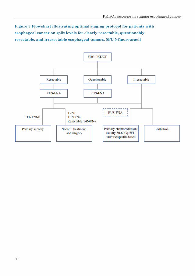
According to all of the above-described models, we composed an idealized
protocol for optimal staging workup using EUS, CT, and FDG-PET on split
levels for patients with clearly resectable, questionably resectable, and
irresectable esophageal tumors (Figure 3). In this flowchart, we recommend
performance of PET/CT upfront in every patient, followed by EUS in those with
clearly resectable disease to identify patients with locally advanced disease, as
they may benefit from neoadjuvant chemoradiation before surgery. When there
is disagreement about resectability with curative intent based on the location of
suspect lymph nodes or because of tumor depth, we advise EUS-FNA for
pathological examination of FDG-avid sites and/or suspicious lesions on PET/CT
imaging in advanced, questionably resectable disease. In patients with primary
irresectable disease that could possibly be managed curatively by definitive
chemoradiation, EUS-FNA should be performed on indication. However, EUS
can be omitted in patients with clearly incurable disease, so they can be referred
immediately for palliative treatment.
PET/CT superior in staging esophageal cancer
80
Figure 3 Flowchart illustrating optimal staging protocol for patients with
esophageal cancer on split levels for clearly resectable, questionably
resectable, and irresectable esophageal tumors. 5FU 5-fluorouracil

PET/CT superior in staging esophageal cancer
81
Discussion
In this study a validated reformulated logistic regression approach was used to
calculate the likelihood ratios of CT, FDG-PET, and EUS in order to determine
the resectability with curative intent for different patient, tumor, and staging
characteristics.17 Given the outcomes of one or two diagnostic tests, we were
able to determine the value added by each test in the staging workup of
esophageal cancer patients in predicting a dichotomous outcome (curative
resectability versus irresectability). It was not possible to make subdivisions
based on age, histological type, or tumor length in deciding whether to perform
a test or not.
According to the results of this logistic regression approach, PET/CT has to be
recommended as the first staging procedure, reserving EUS for limited cases
and candidates with curable disease (Figure 3). CT and FDG-PET outcomes
strongly overlap and strengthen each other. FDG-PET reaches the highest LR+
when CT is negative (LR+ = 12.5–13.0), and vice versa the LR+ of CT reaches
4.4. EUS is insensitive with respect to resectability, with nonsignificant LRs
when the results are expressed as a dichotomous outcome. This finding is not in
line with the generally allotted role of EUS in esophageal cancer staging. One
explanation might be that criteria based on nodal status and depth of tumor
invasion alone are not strong enough to preclude surgical resection. Even
though EUS is a powerful test for detecting lymph node metastases and tumor
depth, these outcomes have almost no influence on decision-making when
incurability/irresectability is the only parameter to be assessed. Only when EUS
clearly identifies patients with a T4b tumor or cytologically proven nonregional
nodes is it helpful for the exclusion of patients from potentially curative surgery.
In the current study, only ten tumors (10/216; 5%) were considered as not
curatively resectable on EUS as they were staged as T4b tumors. Usually the
endoscopist will stage a tumor as T4 if he or she is clearly convinced of tumor
invasion into surrounding structures precluding radical surgery. However, if

PET/CT superior in staging esophageal cancer
82
invasion is not clear or is doubtful, the tumor will probably be staged as T3 and
the patient, in most cases, will be offered neoadjuvant therapy as standard
treatment. Based on these results, EUS has limited impact beyond PET/CT on
staging advanced esophageal tumors in terms of curative resectability. EUS
seems to be more valuable as an additional long-term prognostic factor rather
than a potential predictor of irresectability at time of diagnosis.
Currently, neoadjuvant chemoradiation is being increasingly applied in the
treatment of locally advanced esophageal cancer in an effort to improve
microscopic radical resectability and survival by downstaging the tumor process
and reducing local recurrence rates. In this way, staging has major
consequences on treatment selection and also when comparing outcomes
between studies and institutes. Furthermore, EUS is an invasive diagnostic
procedure and not always applicable because of stenosis or use of a less accurate
miniprobe in up to 30% of cases, which may lead to inadequate assessment of
depth invasion and nodal staging.20 Moreover, in a previous study the perceived
patient burden of EUS in assessment of the preoperative tumor stage was
relatively high compared with CT and or PET/CT. Both EUS and FDG-PET
have relatively good accuracy in restaging esophageal cancer after neoadjuvant
therapy. Although both imaging methods have their limitations in assessing
response to neoadjuvant chemoradiation, the accuracy rate of CT alone is poor.21
The more tests used in a preoperative staging program, the higher the chance of
a correct outcome. However, one must balance likelihoods and certitude against
costs, radiation, and inconvenience for the patient. Difficulties arise when test
outcomes are contradictory. This study offers a new perspective on the
performance of current diagnostic tests in the staging workup for esophageal
cancer patients. It indicates the individual impact of each test on medical
decision-making and the congruence between them. These results strongly
argue for use of PET/CT as the first staging procedure, reserving EUS-FNA for

PET/CT superior in staging esophageal cancer
83
those cases with uncertainty or disagreement about the location of positive
lymph nodes (regional versus nonregional nodes) or tumor depth, which may
affect curative resectability. Biopsies of FDG-avid sites at time of EUS will
actually increase the yield of pathological proof from initial EUS without
scheduling a separate EUS to prove irresectable disease.
Acknowledgment
This study was supported by a ZonMw program for Health Care Efficiency
Research.

PET/CT superior in staging esophageal cancer
84
Reference List
(1) Messa C, Bettinardi V, Picchio M, et al. PET/CT in diagnostic oncology. Q J Nucl
Med Mol Imaging. 2004;48:66–75.
(2) Reinartz P, Wieres FJ, Schneider W, Schur A, Buell U. Side-by-side reading of PET
and CT scans in oncology: which patients might profit from integrated PET/CT?
Eur J Nucl Med Mol Imaging. 2004;31:1456–61.
(3) Van Westreenen HL, Heeren PAM, van Dullemen HM et al. Positron emission
tomography with F-18-fluorodeoxyglucose in a combined staging strategy of
esophageal cancer prevents unnecessary surgical explorations. J Gastrointest Surg.
2005;9:54–61.
(4) Pera M, Pera M. Recent changes in the epidemiology of esophageal cancer. Surg
Oncol. 2001;10:81–90.
(5) Pera M, Manterola C, Vidal O, Grande L. Epidemiology of esophageal
adenocarcinoma. J Surg Oncol. 2005;92:151–9.
(6) Siesling S, van Dijck JA, Visser O, Coebergh JW; Working Group of The
Netherlands Cancer Registry. Trends in incidence of and mortality from cancer in
The Netherlands in the period 1989–1998. Eur J Cancer. 2003;39:2521–30.
(7) Steyerberg EW, Homs MYV, Stokvis A, Essink-Bot ML, Siersema PD. Stent
placement or brachytherapy for palliation of dysphagia from esophageal cancer: a
prognostic model to guide treatment selection. Gastrointest Endosc. 2005;62:333–
40.
(8) Clements DM, Bowrey DJ, Havard TJ. The role of staging investigations for
oesophago-gastric carcinoma. Eur J Surg Oncol. 2004;30:309–12.
(9) Westerterp M, van Westreenen HL, Deutekom M, et al. Patients’ perception of
diagnostic tests in the preoperative assessment of esophageal cancer. Patient Prefer
Adherence 2008:2;157–62.
(10) Flamen P, Lerut A, van Custem E, et al. The utility of positron emission
tomography for the diagnosis and staging of recurrent esophageal cancer. J Thorac
Cardiovasc Surg. 2000;120:1085–92.

PET/CT superior in staging esophageal cancer
85
(11) Heeren PA, Jager PL, Bongaerts F, van Dullemen H, Sluiter W, Plukker JT.
Detection of distant metastases in esophageal cancer with (18)F-FDG PET. J Nucl
Med. 2004;45:980–7.
(12) Luketich JD, Friedman DM, Weigel TL, et al. Evaluation of distant metastases in
esophageal cancer: 100 consecutive positron emission tomography scans. Ann
Thorac Surg. 1999; 68:1133–6; discussion 1136-7.
(13) Van Westreenen HL, Westerterp M, Bossuyt PM, et al. Systematic review of the
staging performance of 18F-fluorodeoxyglucose positron emission tomography in
esophageal cancer. J Clin Oncol. 2004;22:3805–12.
(14) Bar-Shalom R, Guralnik L, Tsalic M, et al. The additional value of PET/CT over
PET in FDG imaging of oesophageal cancer. Eur J Nucl Med Mol Imaging.
2005;32:918–24.
(15) Jadvar H, Henderson RW, Conti PS. 2-Deoxy-2-[F−18]fluoro-d-glucose-positron
emission tomography/computed tomography imaging evaluation of esophageal
cancer. Mol Imaging Biol. 2006;8:193–200.
(16) Munden RF, Macapinlac HA, Erasmus JJ. Esophageal cancer: The role of
integrated CT-PET in initial staging and response assessment after preoperative
therapy. J Thorac Imaging. 2006;21:137–45.
(17) Janssens AC, Deng Y, Borsboom GJ, Eijkemans MJ, Habbema JD, Steyerberg EW.
A new logistic regression approach for the evaluation of diagnostic test results. Med
Decis Making. 2005;25:168–77.
(18) Van Westreenen HL, Westerterp M, Sloof GW, et al. Limited additional value of
positron emission tomography in staging oesophageal cancer. Br J Surg.
2007;94:1515–20.
(19) Janssens AC, Steyerberg EW, Jiang Y, Habbema JD, Van Duijn CM, Criswell LA.
Value of the HLA-DRB1 shared epitope for predicting radiographic damage in
rheumatoid arthritis depends on the individual patient risk profile. J Rheumatol.
2006;33:2383–9.
(20) Wallace MB, Hawes RH, Sahai AV, Van Velse A, Hoffman BJ. Dilation of
malignant esophageal stenosis to allow EUS guided fine-needle aspiration: safety
and effect on patient management. Gastrointest Endosc. 2000; 51:309–13.

PET/CT superior in staging esophageal cancer
86
(21) Westerterp M, van Westreenen HL, Reitsma JB, et al. Esophageal cancer: CT,
endoscopic US, and FDG PET for assessment of response to neoadjuvant therapy—
systematic review. Radiology. 2005;236:841–51.

87
Part III FDG-PET/CT in Radiotherapy Planning
![Progress and Promise of FDG-PET Imagingfor Cancer Patient ... · Management and Oncologic Drug Development ... DG-PET (2-[18F]Fluoro-2-deoxyglucose positron emission tomography) is](https://img.pdfslide.us/doc/110x75/5ea78ca90dcaec79e2683d24/progress-and-promise-of-fdg-pet-imagingfor-cancer-patient-management-and-oncologic.jpg)
![FDG-PET in Large Vessel Vasculitis...FDG-PET in Large Vessel Vasculitis 61 5. [18 F]FDG-PET and [18 F]FDG-PET/CT [18 F]FDG-PET is an operator-independent, non- invasive imaging modality](https://img.pdfslide.us/doc/110x75/5f6c13132f0609183b646bce/fdg-pet-in-large-vessel-vasculitis-fdg-pet-in-large-vessel-vasculitis-61-5.jpg)


![ORIGINAL RESEARCH Open Access F]FDG-PET imaging is an ... · Background: Positron emission tomography (PET) with [2-18 F]-2-fluoro-2-deoxy-D-glucose ([18 F]FDG-PET) was acquired at](https://img.pdfslide.us/doc/110x75/5fffd04346622d24ad6dffc9/original-research-open-access-ffdg-pet-imaging-is-an-background-positron-emission.jpg)