Embed Size (px)
Citation preview

UNIVERSITÀ’ DEGLI STUDI DI TRIESTE
XXV CICLO DEL DOTTORATO DI RICERCA IN
NANOTECNOLOGIA
Nanotechnology Applications in Quantitative
Neuroscience: Proteomic Analysis of Malignant Gliomas
Settore Disciplinare: FIS/03
DOTTORANDO
Mario Ganau
COORDINATORE
Prof Maurizio Fermeglia
SUPERVISORE DI TESI
Prof Giacinto Scoles
TUTOR
Dott.ssa Loredana Casalis
ANNO ACCADEMICO 2011 / 2012

2
To all those who constantly supported me
during those challenging years

3
Nanotechnology Applications in Quantitative Neuroscience:
Proteomic Analysis of Malignant Gliomas
Outline
Abstract (En) pag 5
Abstract (It) pag 6
Acknowledgement pag 7
Chapter 1 Introduction pag 9
1.1 Background pag 10
1.2 Gliomas pag 10
1.3 GFAP pag 12
1.4 From Micro- to Nanobiosensors for protein recognition: state of the art pag 14
1.4 a) Optical read-out pag 15
1.4 b) Radio-labeled read-out pag 17
1.4 c) Mass-detection read-out pag 18
1.4 d) Mechanical-sensing read-out pag 19
1.5 Micro- and Nanofabrication strategies: overview pag 21
1.5 a) Phololithography pag 21
1.5 b) Microcontact Printing pag 22
1.5 c) Dip Pen Nanolithography pag 24
1.5 d) AFM Nanografting pag 25
1.5 e) DNA-Directed Immobilization pag 27
1.6 Aims of the research pag 28
Chapter 2 Experimental session pag 31
2.1 Fabrication and functionalization of microwells pag 32
2.2 Immobilization of antibodies pag 33
2.2 a) Fabrication of patches of biotin terminated alkanethiol pag 33
2.2 b) Preparation of DDI of biontinylated antibodies pag 35
2.3 Immobilization of GFAP pag 37
2.3 a) Immobilization of GFAP in PBS pag 37
2.3 b) Immobilization of GFAP in multicells’ lysate pag 38
2.4 Signal to noise ratio and roughness analysis of SAMs and NAMs pag 38
2.5 Benchmark tests with ELISA pag 39

4
Chapter 3 Results pag 40
3.1 Patterning of living cells pag 41
3.1 a) Tests with Prokaryotes pag 42
3.1 b) Tests with Eukaryotes pag 43
3.2 Optimizing the recognition of GFAP pag 44
3.2 a) Antibodies’ immobilization pag 45
3.2 b) GFAP in PBS pag 52
3.3 c) GFAP in multicells’ lysate pag 53
3.3 Challenging the sensitivity of ELISA pag 56
Chapter 4 Discussion pag 59
4.1 Clinical Considerations pag 60
4.1 a) Genetic and Proteomic Features of Gliomas pag 60
4.2 Methodological constraints and opportunities pag 62
4.2 a) DDI-based proteomic assays pag 62
4.2 b) Comparison with ELISA-based proteomic assays pag 64
4.2 c) Trends in single cells DNA barcode analysis pag 65
Chapter 5 Conclusions and Outlook pag 67
5.1 Optimization of protocols for protein recognition pag 68
5.2 Toward Quantitative Neuroscience pag 69
Abbreviation List pag 71
References pag 73

5
Abstract (En)
The current limit of knowledge advancement in proteomic analysis of gliomas, the most
common primary malignant brain tumors, is related to the high sensitivity required to detect specific
biomarkers within few cells volumes. To address this problem we developed a quantitative
approach to eventually enable precise, high throughput and low cost analysis of glial cells with
potential capability of real-time pathological screening and subtyping of brain tumors.
A device consisting in micro-fabricated wells capable to isolate and host living astrocytes
was designed and functionalized. Then for the fabrication of a nanobiosensor, able to detect in small
volumes the presence of specific biomarkers, ideally for multiplexing assays and meant to fit within
the small dimensions of this microdevice, an approach consisting in DNA-directed-immobilization
(DDI) of biotinylated antibodies (Abs) on a single stranded DNA (ssDNA) nanoarray, produced by
Atomic Force Microscopy (AFM) nanografting, was carefully optimized. The proof of concept was
realized with Abs specific for Glial Fibrillary Acidic Protein (GFAP), a biomarker which belongs to
the family of intermediate filaments and is crucial in cell’s differentiation, within a platform ready
for parallelization.
Nanosized patches of thiol modified ssDNA were prepared by AFM-based nanografting
inside a matrix of self assembled monolayers (SAM) of alkanethiol-modified gold surfaces.
Subsequently a complementary DNA strand (cDNA) conjugated to streptavidin (STV) was allowed
to covalently bind to the patch by sequence specific DNA hybridization. Finally the biotin binding
sites of STV were exploited to immobilize biotinylated monoclonal GFAP Abs (already in use for
ELISA assays) on the top of those nanopatches. The efficiency of those nano-immuno arrays was
tested by successfully obtaining the immobilization of purified recombinant GFAP protein, down to
a concentration of 4 nM, firstly in standard PBS then in multicells’ lysate obtained from U87 glial
cultures. The immobilization was detected by means of AFM measuring step by step the increases
in the height of the patches and excluding modification of the roughness of both the SAM and the
nanopatches after incubation with the cells’ lysate through a signal to noise ratio analysis. Titration
curves for a comparison of sensitivity between this technique and the conventional ELISA assays
are provided; they indeed confirm that the sensitivity of our nanosensors is at least that of ELISA,
with the advantage of the scalability of the device.

6
Abstract (It)
L’attuale limite di avanzamento dello stato dell’arte dell’analisi proteomica dei gliomi
cerebrali, la classe istologica di tumori cerebrali più frequente ed aggressiva, è legato alla difficoltà
di individuare specifici biomarkers in piccoli volumi cellulari. Per superare questo limite si è deciso
di sviluppare un approccio nanoquantitativo che consenta un’analisi proteomica precisa, ad alta
sensibilità e basso costo, degli astrociti tumorali, con potenzialità di screening in tempo reale e
sottotipizzazione di tumori cerebrali. Previa fabbricazione e funzionalizzazione di micro pozzetti
idonei ad ospitare cellule astrocitarie, ci si è dedicati alla realizzazione di biosensori in grado di
riconoscere specifici biomarkers e di essere accoppiati ai micro pozzetti. Al fine di immobilizzare
anticorpi specifici per proteine di interesse in ambito neuroncologico, è stato scelto un approccio
basato sul nanografting con Microscopio a Forza Atomica (AFM) e sull’immobilizzazione diretta
sul DNA di anticorpi (DDI). In particolare la prova concettuale è stata condotta con anticorpi
specifici per la Glial Fibrillary Acidic Protein (GFAP), un marcatore della differenziazione
astrocitaria appartenente alla famiglia dei filamenti intermedi intracellulari, su una piattaforma atta
ad una successiva parallelizzazione.
I nanocostrutti responsabili del riconoscimento della proteina d’interesse, sono stati realizzati
partendo da molecole di DNA a singola elica (ssDNA) graftate in una matrice di monostrati
autoassemblati (SAM) di superfici d’oro alchiltiolo modificato. Al fine di sfruttare la capacità della
streptavidina (STV) di legarsi ad anticorpi biotinilati è stata successivamente indotta l’ibridazione di
un filamento di DNA complementare (cDNA) a quello precedentemente immobilizzato sulla
superficie nanoassemblata che presentasse anche una coda di STV. I siti di legame per la biotina
intrinseci al tetramero di STV sono quindi stati sfruttati per immobilizzare sulla superficie dei
nanocostrutti degli anticorpi monoclonali biotinilati specifici per GFAP (già in uso per i protocolli
ELISA). L’efficienza dei nano-immuno costrutti così ottenuti è stata testata ottenendo
l’immobilizzazione di GFAP ricombinante anche a basse concentrazioni (fino a 4nM), sia in
presenza di standard PBS, sia in presenza di un lisato multicellulare ottenuto da colture gliali di
cellule U87. L’immobilizzazione di GFAP è stata confermata dall’incremento in altezza dei
nanocostrutti misurato all’AFM escludendo modificazioni del rapporto segnale/rumore sia del SAM
che dei nanocostrutti prima e dopo aggiunta di lisato multicellulare. Il limite di sensibilità del
prototipo così ottenuto è stato confrontato con quello raggiungibile con protocolli standard ELISA,
mostrando una sensibilità almeno comparabile all’ELISA a fronte di un maggiore potenziale
diagnostico legato alla sua scalabilità.

7
Acknowledgements
My sincere and infinite gratitude goes to my supervisor Prof Giacinto Scoles and my tutor
Dr Loredana Casalis because by accepting me in their laboratory three years ago, they gave me a
precious, once in a lifetime opportunity. Their enormous encouragement and genuine help made me
feeling comfortable in such a technical nano-world, so far away from my clinical-surgical daily
practice.
I would like to express my deepest thanks to Dr Alessandro Bosco, Dr Pietro Parisse, Dr
Barbara Sanavio and Dr Denis Scaini for their passionate help in teaching me the fundamentals of
AFM and nanografting process. All those fruitful hours spent together contributed to increase my
understanding of the complexity surrounding proteomic assays. Indeed, they have been invaluable
in helping me to overcome the many unsuccessful experiments and to keep progressing towards the
optimization of the most convenient strategy for nano-immobilization of proteins.
My huge appreciation goes to my colleagues at the NanoInnovation Lab, Stefania
Corvaglia, Luca Ianeselli and Maryse Nkoua who cared about supporting me in several parallel
procedures carried out during this PhD project, and for offering me their excellent skills during
hard tasks such as the microwells fabrication and their effective functionalization. As well I would
like to thank Anita Palma for her precious efforts to enabling an effective comparison in terms of
sensitivity between our methodology and the standard ELISA assays.
I would like to thank Dr Daniela Cesselli for the thorough encouragement and meaningful
discussions on single cells proteomics and its correlation with practical pathological needs, her
expert guidance helped us in understanding the potentialities of the device developed in those years
and formed the basis for its further clinical characterization.
I am also particularly thankful to all those that enabled a tight collaboration with the
partner labs where some experiments have been realized or part of them made possible: the IOM
INFM @ TASC for the support provided in the fabrication of microwells, the Structural Biology Lab @
Elettra and Spinal Biophysics Lab @ UniTS for the opportunity to test living prokaryotes and eukaryotes
within the microwells, and the Dept of Medical and Biological Sciences @ UniUD for the benchmark testing
with ELISA protocols.

8
Finally, I am immensely grateful to my fiancée Lara, my parents Francesco and Marinella
and my sister Laura for their endless love, enduring support and constant guidance during those
long and fruitful years here in Trieste.

9
Chapter 1:
Introduction

10
1.1 Background
Based upon the latest genetic and proteomic insights into cancer’ biology, which opened
new avenues for novel applied clinical research, trends in oncology highlight that molecular
characterization of the tumorigenesis process will be essential in tomorrow’s clinical practice to
predict prognosis and guide therapy. Noteworthy, the promise of individualized molecular
medicine, which is particularly relevant to oncology because even similarly classified tumors can
follow quite different clinical outcomes, could be realized by identifying molecular targets for
therapy and by measuring tangible response or regression in clinical trials.
Specific patterns of protein expression in tumors and matched normal tissues can now be
reliably analyzed using quantitative proteomic techniques. Among them the most effective are
enzyme-linked immunosorbent assay (ELISA), two-dimensional gel electrophoresis (2DGE) and
matrix-assisted laser desorption ionization time of flight (MALDI-TOF) mass spectrometry, which
allow to simultaneously identify and characterize differentially expressed proteins. Nevertheless,
arrays of proteins with well-defined feature size and spacing already demonstrated the potential to
boost the detection of key biomolecules by favoring the study of surface-cellular interaction. In fact
among the current limits of knowledge advancement in oncology the main one is probably related
to the high sensitivity required to accurately monitor protein-protein interactions, which are relevant
to follow changes in cellular pathways due to different kinds of external perturbations. Such caveat
explain why a new quantitative approach based on the nano-immuno-arrays technology, could be
highly effective in enabling a precise, high throughput and low cost in vitro analysis of tumor cells’
lysates, or in vivo studies of their secretome (down to the single cells level).
Focusing our attention to neuro-oncology we identified the remarkable need for more
sophisticated diagnostic tools with potential capability of real-time pathological screening and
subtyping of glial tumors.
1.2 Gliomas
Gliomas (see Fig 1.1) are primary brain tumors classified according to the histological
classification of the World Health Organization in: Glioblastomas (Grade IV), Anaplastic
Astrocytomas (Grade III), Low Grade Astrocytomas and Oligodendrogliomas (Grade II). Among
them the most aggressive ones, namely Glioblastomas and Anaplastic Astrocytomas present an
incidence of 3.5-2.8 and 0.3-1.2 new cases per 100.000 per year respectively (Ohgaki et al, 2005;
Deorah et al, 2006).

11
Fig 1.1: T1-weighted MRI image showing a left temporal high-grade glioma
Gliomas are characterized by rapid growth, high level of cellular heterogeneity (see Fig 1.2)
due to genetic alteration, and infiltrative behaviour, nevertheless those tumors are generally
confined in the brain parenchyma, and do not show any tendency to bony calvarium or extracranial
invasion. It is recognized that more complete resections lead to better prognosis, accordingly
microscopic local resection is the current neurosurgical end point. Despite that, tumor infiltration is
unfortunately widespread and recurrence may occur near resection margin as well as far away from
it in both cerebral hemispheres, rarely in the cerebellum and even more rarely in the spinal cord.
Fig 1.2: Hematoxylin and eosin stain of a high-grade glioma showing the high degree of cellularity and
neovascularisation, typical features of those tumors
12
Radiotherapy is therefore central in the treatment of high grade gliomas, since limited field
fractionated radiation, covering the MRI enhanced lesion as well as peritumoral edema, improves
patients survival: the standard dose is 60 Gy (Fine et al, 1993).
Temozolomide (TMZ) received Food and Drug Administration approval in 1999 for
refractory Anaplastic Astrocytomas, followed by the first line indication for Glioblastoma. The
international EORTC/NCIC trial in 2005 demonstrated a substantial improvement in median
survival, representing the first of such improvements since the introduction of radiation therapy in
the mid-1970s (Stupp et al, 2005). Local intraoperative therapy with antineoplastic wafers
(impregnated carmustine implants) has demonstrated a further survival advantage although only in
patients with O(6)-methylguanine-DNA methyltransferase (MGMT) promoter methylation
(Westphal et al, 2003; Lechapt-Zalcman et al, 2012).
To date, complete microscopic excision followed by adjuvant radio- or chemotherapy
represents the standard of care, nevertheless tumor recurrence generally occurs within few months
due to the widespread neoplastic infiltration (Smoll et al, 2012). In fact, primary brain tumors
invade widely spreading single cells anywhere within the brain parenchyma, through infiltration of
blood vessel walls, subpial glial spaces, or white matter tracts. These mechanisms lead to the
development of tumor satellites that escape resection and treatment, eventually serving as reservoirs
for tumor recurrences. Beside parenchymal infiltration, another challenge in the management of
gliomas is the nearly universal propensity of these neoplasms to present an intratumoral and
intertumoral molecular heterogeneity allowing tumoral reservoirs to be even more resistent to
further treatment than the primary lesion (Furnari et al, 2007).
Despite continuous refinements in therapeutic strategies, the prognosis for patients with
high-grade gliomas remains dismal, and second-time surgical resection along with local
intraoperative positioning of antineoplastic wafers represent the only treatment choices for patients
with recurrent gliomas (Westphal et al, 2003). As a result, the median survival of high-grade
gliomas is 14-month for patients undergoing surgery plus chemotherapy, and 22-month for those
treated with surgery plus chemotherapy plus carmustine wafers; so that only 2.2% of patients are
expected to survive 3 years or more after diagnosis of a Glioblastoma (Ohgaki et al, 2005; Smoll et
al, 2012).
1.3 GFAP
At present, gliomas are diagnosed by histopathological criteria, and known robust prognostic
factors for most of these tumors are limited to tumor grade and patient age.
13
While there has been progress in understanding some clinical aspects of these tumors
(Prados and Levin, 2000; Kitange et al, 2003), new molecular markers are required to better define
prognosis and response to therapy
The widespread acceptance that genetic signatures such as losses on chromosomes 1p and
19q are of prognostic value in Oligodendrogliomas (Cairncross et al., 1998) has spurred interest in
developing molecular markers to predict outcome and response to treatment across a broader
population of gliomas. In fact, while numerous genetic alterations have been described in
Glioblastomas (von Deimling et al, 1995; Watanabe et al, 1996), such markers have proved to be of
marginal utility in predicting outcome or guiding decisions about disease management.
Importantly, recent expression profiling studies have revealed that proteomic patterns could
be of prognostic value (Freije et al, 2004; Nutt et al, 2003), and a systematic review of multiple
independent proteomic analyses of gliomas published on PubMed since 2008 has demonstrated
alterations of almost 100 different proteins. Among them we have chosen the Glial Fibrillary Acidic
Protein (GFAP), a biomarker belonging to the family of intermediate filaments (IF) for our proof of
concept.
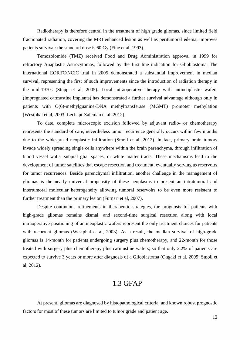
Fig 1.3: Fluorescent micrographs (red stains for GFAP+ astrocytes, whereas turquoise and green stain for
oligodendrocyte O4 and β-tubulin markers respectively) showing high-grade glioma cells growing as: a)
spheres in culture (more aggressive behavior), b) monolayer in culture (better differentiated tumors).
Photos from: North Central Cancer Treatment Group at Mayo Clinic (USA)
Firstly isolated, characterized and described by Lawrence F. Eng in 1969, GFAP is a type III
IF protein that maps in humans to 17q21 (Bongcam-Rudloff et al, 1991; Eng et al, 2000). Being
closely related to its non-epithelial family members, such as vimentin, desmin, and peripherin,

14
which are all involved in the structure and function of the cell’s cytoskeleton, GFAP is thought to
help maintaining astrocytes’ mechanical strength and shape.
Noteworthy, GFAP is necessary for many critical roles in the central nervous system (CNS):
it is pivotal during mitosis, as confirmed by the increase in the amount of phosphorylated GFAP
during this phase of cellular life, it plays a role in astrocyte-neuron interactions as well as cell-cell
communication, and has been linked to multiple degenerative processes including abnormal
myelination, white matter structure deterioration, and functional/structural impairment of the blood–
brain barrier (Liedtke et al, 1996).
Finally, GFAP is a potentially informative plasma biomarker for gliomas being detected in
plasma samples of every patient with high-grade glioma (Husain et al, 2012). Because of its role in
cell’s differentiation, only well differentiated cells retain the ability to express it (see Fig 1.3), while
the most aggressive ones lose this typical feature at a certain point in their dedifferentiation process
(Singh et al, 2003).
Beside GFAP, other proteins of interest are now being advocated for early diagnosis or
monitoring of glioma. To this regard collection or panels of such proteins as opposed to a single
biomarker will be necessary for a reliable diagnostic improvement in terms of accuracy (Rasooly et
al, 2006). In general, to realize the full potential of clinical biomarkers, new bioanalytical
technologies are being developed with the aim to achieve an accurate detection at low volumes (till
to pg/ml) of panels of biomarkers (Kingsmore et al, 2006)
1.4 From Micro- to Nanobiosensors for protein recognition:
state of the art
A biosensor is a device that combines a biological component with a physicochemical
detector used for the recognition of an analyte at a micro- or nanoscale. An indispensable
requirement for any biosensor is an excellent specificity and sensitivity for biomarkers detection,
the former can be defined as the ability of the assay to rule out a condition when a specific
biomarker is absent, while the latter is defined as the ability of the assay to identify a condition
when it is present (Rusling et al, 2010). Those clinical specificity and sensitivity parameters are
closely linked to the method used for measurements, and need to be high (>90%) to avoid false
positive or false negative results (Moncada et al, 2008).
The sensitive biological elements of biosensor, generally represented by biologically derived
materials or biomimetic components (i.e. tissue, microorganisms, enzymes, antibodies, nucleic
15
acids, etc.), interact throughout specific binding to, or recognition of, the analyte under study. The
detector element or transducer, which works in a physicochemical way (i.e. optical, piezoelectric,
electrochemical, etc.), transform the signal resulting from the interaction of the analyte with the
biological element into another signal that can be more easily measured and quantified.
Detection elements play a key role in analyte recognition in biosensors: therefore detection
elements with high analyte specificity and binding strength are required. While antibodies (Abs)
have been increasingly used as detection elements in biosensors, some key challenges remain: their
immobilization on the biosensor surface and the optimal method for identifying the antigen-
antibody interaction.
According to the array-based optical, mass-detection or radio-labeled read out, micro and
nanobiosensors can be classified as follows:
1.4 a) Optical read-out
Optical biosensors account for the most known and widespread devices and protocols for
detection of bioanalytes, including enzyme-linked immunosorbent assay (ELISA), which is
particularly useful for a plate-based detection and quantification of substances such as peptides,
proteins, antibodies and hormones.
In an ELISA assay, an antigen must be immobilized to a solid surface and then complexed
with an antibody that is linked to an enzyme; the detection is accomplished by assessing the
conjugated enzyme activity via incubation with a substrate to produce a measureable product, which
generally is a detectable fluorescence signal. Therefore the most crucial element of the detection
strategy is a highly specific antibody-antigen interaction.
ELISAs are typically performed in polystyrene plates, which will passively bind Abs and
proteins; having the reactants of the ELISA immobilized to the microplate surface makes it easy to
separate bound from nonbound material during the assay. This ability to wash away nonspecifically
bound materials makes the ELISA a powerful tool for measuring specific analytes within a crude
preparation.
ELISAs can be performed with a number of modifications to the basic procedure (see Fig
1.4): the key step, immobilization of the antigen of interest, can be accomplished by direct
adsorption to the assay plate or indirectly, via a capture antibody that has been attached to the plate.
The antigen is then detected either directly (labeled primary antibody) or indirectly (labeled
secondary antibody).

16
The most powerful ELISA assay format is the sandwich assay. This type of capture assay is
called a “sandwich” assay because the analyte to be measured is bound between two primary Abs:
the capture antibody and the detection antibody.
The sandwich format is often the preferred one because it is more robust and equally
sensitive then direct or indirect assays.
Fig 1.4: Schematic representation of the most common ELISA formats.
Drawing from: Thermo Fisher Scientific Inc
ELISA is nearly always performed using 96-well or 384-well polystyrene plates and
samples in solution (i.e., biological fluids, culture media or cell lysates); however, other variants of
ELISA exist:
- ELISPOT (enzyme-linked immunospot assay) refers to ELISA-like capture and
measurement of proteins secreted by cells that are plated in polyvinylidene difluoride
(PVDF)-membrane-backed microplate wells. It is a "sandwich" assay in which the
proteins are captured locally as they are secreted by the plated cells, and detection is with
a precipitating substrate. ELISPOT is like a Western blot in that the result is spots on a
membrane surface (Czerkinsky et al, 1983)
- In-cell ELISA is performed with cells that are plated and cultured overnight in standard
microplates. After the cultured cells are fixed, they undergo a permeabilization and
blocking processes, and finally target proteins are detected with Abs. This is an indirect
assay, not a sandwich assay. The secondary Abs are either fluorescent, for direct
measurement by a fluorescent plate reader or a microscope; or enzyme-conjugated, for
detection with a soluble substrate using a plate reader.

17
1.4 b) Radio-labeled read-out
The measurement of radiolabels by scintillation counting has long been one of the most
reliable methods for accurate, quantitative measurement in biochemical experiments (Lees et al,
1999).
Today it has been supplanted by the ELISA method, where as previously said the antigen-
antibody reaction is measured using colorimetric instead of radioactive signals; however, because of
their robustness, consistent results and relatively low price per test, radio-ladeled read-out methods
are again becoming popular (Godovac-Zimmermann et al, 2005).
The concepts of radio-labeled read-out have been employed in the context of proteomics,
where they offered gains in absolute sensitivity and dynamic range: for instance multi-photon
detection methodology, proposed as a tool to routinely and quantitatively detect radioactive labels
on two-dimensional gels, has several characteristics that are advantageous for functional protein
detection (Kleineret al, 2008):
- First of all, by using single particle detectors, the sensitivity for detection of radiolabels
can be improved dramatically;
- Secondly, because single particle detectors can differentiate the particle energies
produced by different decay processes, it is possible to choose combinations of
radioisotopes that can be detected and quantified individually on the same 2D gel;
- Thirdly, this technology is essentially linear over 6 to 7 orders of magnitude (i.e. it is
possible to accurately quantify radiolabeled proteins over a range from at least 60
zeptomoles to 60 femtomoles).
In principle, the implementation of chemical radiolabeling methods could provide a 100-
fold decrease in the amount of biological material needed for proteomics experiments, while
reducing imaging times 10–100-fold, with total amounts of radioactivity far below legal limits
(Kleineret et al, 2008).
Overall, the quest for ultra-high sensitivity and quantitative precision is providing new
impetus to proteomics studies: both micro- and nanoarrays hold the promise of high selectivity and
sensitivity, ease of use reasonable costs per assay and good possibilities for future automation.
Nevertheless several drawbacks still limit the diffusion of radio-labeled read-out: the most
important ones are certainly related to the special facility, precautions and licensing required: since
radioactive substances are used a gamma counter is essential to measure the radiations emitted by
the radionuclide, while security issues impose strict protocols for their stocking and disposal.

18
1.4 c) Mass-detection read-out
Mechanical interactions are fundamental to biology, in fact on one hand mechanical forces
of chemical origin determine motility and adhesion on the cellular scale, and govern transport and
affinity on the molecular scale; on the other biological sensing in the mechanical domain provides
unique opportunities to measure forces, displacements and mass changes from cellular and
subcellular processes (Arlett et al, 2011).
The advances in micro- and nanofabrication technologies have enabled the preparation of
increasingly smaller mechanical transducers, so that nowadays a promising family of biosensors is
represented by micro- and nanomechanical systems, which are basically cantilever-like sensors:
they are particularly well matched in size with molecular interactions, and provide a basis for
biological probes with single-molecule sensitivity, indeed (Yang et al, 2006; Zougagh et al, 2009).
Recently, detection of mass in the zeptogram range and sensitivity in liquid to the fraction of nM
concentration in real time has been demonstrated (von Muhlen et al, 2010).
Despite biosensors based on nanomechanical systems have gained considerable relevance in
the last decade, several theoretical and experimental studies, reporting the influence of the mass
transport on antibody biosensors as a function of analyte concentration and incubation time,
concluded that pushing the sensitivity to the limit of single molecule detection may not bring the
expected benefit to the overall performance (Nair et al, 2006; Sheehan et al, 2005).
In fact mass transport can significantly lower the practical sensitivity of a device by
reducing the number of binding events (Tamayo et al, 2013). Moreover, especially at low
concentration, which is typical of biomolecular experiments, the interaction between target
molecules and the biosensor can play as critical a role as the chemical reaction itself in governing
the binding rate (Kusnezow et al, 2006).
In the attempt to overcome these limitation Melli and colleagues developed a
micromechanical sensor (see Fig 1.5) based on vertically oriented oscillating beams (or pillars)
which make it possible to locate the sensitive area at the free end of the oscillators (Melli et al,
2011).
Practically, an array of such pillars (3μm x 8μm in plane, and 15μm in height) behaves as
an array of isolated nanosized sensors embedded in a quasi-infinite analyte solution: while the top
face of the pillars represents the nanosized active area, the pillars themselves can be operated as
mass detectors. In particular these three-dimensional structures with dimensions comparable to the
diffusion length of the target molecules have proved to increase the reaction speed by 3 orders of
magnitude, while attaining improvement also in concentration sensitivity.

19
Fig 1.5: Micromechanical pillars. Photo from: Melli M et al, ACS Nano 2011
1.4 d) Mechanical-sensing read-out
To conclude this digression on micro- and nanobiosensors it is also useful to cite that in the
last decade, the quest for protein detection in smaller volumes, along with continuous efforts to
monitor specific interactions between Abs and antigens employed in an immunoassay system have
led to the development of several ELISA like biosensors, including Atomic Force Microscopy
(AFM) based arrays.
AFM is a very high-resolution type of scanning probe microscopy, consisting of a cantilever
with a sharp tip (probe) at its end that is used to scan the specimen surface, a laser to measure any
probe deflection and reflect it from the top surface of the cantilever into an array of photodiodes;
the tip-sample intermolecular forces are detected, as a function of the distance between the two
(Picas et al, 2012).
The AFM can be operated in a number of modes, depending on the application: basically
AFM imaging may be divided into static mode (also called contact) and a variety of dynamic modes
(non-contact or "tapping") where the cantilever is oscillated. The stiffness of the cantilever
determines the ratio between the distance moved and the force exerted by the surface, therefore this
parameter is most relevant for determining the tip-sample interaction during the majority of AFM
operation modes.

20
Fig 1.6: Schematic illustration of the AFM: the tip is attached to a cantilever, and is raster-scanned over a
surface. The cantilever deflection due to tip-surface interactions is monitored by a photodiode sensitive to
laser light reflected at the tip backside; the position of the reflected beam is kept centered in the diode
through feedback-controlled z-changes in the stage.
Drawing from: Dept of Pharmaceutical Physical Chemistry, Uppsala University (Sweden)
For instance while medium stiffness cantilevers are well suited for patterning of surfaces
(i.e. nanografting); on the other hand, since to obtain quality images it is critical that the AFM tip
not damage the surface being scanned, softer cantilevers (< 0.04N/m) allowing to image surfaces
with very low forces, are the most indicated to avoid undesirable surface modifications.
AFM is a powerful technique for investigating surfaces by visualizing their topographic
characteristics: even very low number of target molecules can be reliably detected using height
and/or compressibility measurements. Starting from the works of Liu and colleagues it has been
consistently demonstrated that accurate height measurements of nanopatches before and after
sequence specific hybridization of DNA oligomers allow for reliable, sensitive and label-free
detection of hybridization itself (Liu et al, 2005; Liu et al, 2008, Mirmomtaz et al, 2008).
Moreover, AFM probes may also be functionalized: in 1997, Allen and colleagues were the
first to use AFM probes functionalized with ferritin to monitoring the adhesive forces between the
probe and anti-ferritin antibody-coated substrates (Allen et al, 1997).
Recently, Volkov and colleagues demonstrated that a reliable reading of the immunosignal
(a suggested dimensionless combination of brush length and grafting density) can be obtained from

21
an area as small as ∼3μm2: approximately 4 million times smaller compared to typical ELISA
sensors (Volkov et al, 2010). Intriguingly, they found that AFM could reliably distinguish between
having the immunosignal from either antibody and from both Abs together: attaining a new
detection limit that was impossible to obtain by using standard optical methods.
1.5 Micro- and Nanofabrication strategies: overview
The basic concept of several techniques for fabrication and biopatterning of surfaces at the
micro- and nanometer scale, including photolithography, microcontact printing, dip pen
nanolithography, AFM nanografting will be described in the following pages. To conclude, special
attention will be drawn to the DNA-directed immobilization (DDI), a bottom up
biofunctionalization technique particularly suitable for a high-sensitivity determination of panels of
biomarkers.
1.5 a) Photolithography
Photolithography is a very cost-effective process used in microfabrication to pattern thin
films: in other words it allows to transfer a geometric pattern from a photomask to a light-sensitive
chemical photoresist on the substrate. A series of chemical treatments is then used to enable
deposition of a new material in the desired pattern upon the material underneath the photoresist.
Photolithography enables scientists to create extremely small patterns (down to a few tens of
nm in size), while maintaining exact control over the shape and size of the objects created.
Photolithography requires a clean room and combines several steps in sequence (see Fig 1.7)
for the microfabrication of masters and the subsequent process of replica molding of the masters in
poly-dimethylsiloxane (PDMS).
- Organic or inorganic contaminations are firstly removed by wet chemical treatment
(generally with solutions containing hydrogen peroxide) and wafers are used as
substrates to fabricate the master molds.
- The wafers are then heated to a temperature sufficient to drive off any moisture that may
be present on their surface, and covered with the photoresist by spin coating.
Immediately after spinning, the wafers must be baked to drive off excess of photoresist
solvent, and afterwards exposed to a UV dose of 375 to 240mJ/cm2 (depending on the
thickness of the SU 8) on a contact aligner.

22
Fig 1.7: Microfabrication process: PDMS prepolymer is poured onto a photolithographically patterned
master; after curing PDMS is peeled away and microwells are ready to be functionalized
for deposition of living cells. Drawing adapted from: Dept Biomedical Engineering, UCIrvive (USA)
- The masters are further treated to render the exposed surface very inert, which facilitate
the release of the PDMS mold after curing.
- The PDMS is ready to be degassed and poured onto the master, then degassed again and
cured in a 65° oven for 1 to 16h. Once cured, the PDMS is manually peeled off the
master, and the resulting structure (i.e. the microwells) are ready to be functionalized.
1.5 b) Microcontact Printing
For a while photografting of proteins was obtained from classical photolithographic
techniques; in 1993 a novel approach, called microcontact printing, was introduced by Whitesides
and colleagues for patterning SAM of alkanethiols onto gold substrates (Kumar et al, 1993; López
et al 1993).
To date, microcontact printing is widely used for generating micropatterns of nanomaterials
such as organic molecules and biomolecules over large surface areas (>cm2). In the microcontact
printing process, a microstructured elastomer stamp is coated with a solution of a nanomaterial and
applied to a substrate of choice, then after a given period of time in conformal contact (generally

23
overnight), the stamp is removed leaving a replica of the stamp pattern on the substrate surface (see
Fig 1.8).
The elastomer stamps are made typically from PDMS by curing liquid prepolymers of
PDMS on a lithographically prepared master. The elastomeric properties of stamps made of PDMS
ensure conformal contact (intimate contact) with various substrates.
Fig 1.8: Scheme of microcontact printing: a conventionally fabricated PDMS stamp serves as the vehicle to
transfer the ink of choice, in this case proteins, upon brief contact; then the transfer of those proteins occurs
only at the sites of conformal contact between stamp and substrate.
Drawing from: Bernard A et al, Adv Mater 2000
As the stamps can be structured with almost any pattern, conformal contact can be achieved
in many different geometrically conformed ways.
Moreover, since this technique is carried out under room temperature, different
biomolecules may directly be transferred in a controlled way onto a variety of substrates while
retaining their biological activity. However the deposition of liquid samples followed by drying
could be very complex, leading to ill-defined patterns, protein aggregation and loss of biological
activity.

24
Noteworthy, this problem has recently been overcame by extending these concepts to the
nanoscale dimensions in a process referred to as nanocontact printing, so that, features as small as
40nm can now be fabricated by this way (Li et al, 2003). Nanocontact printing has been achieved
by decreasing the feature sizes in the PDMS stamp and diluting the nanomaterial inks, utilising
special variants of PDMS stamps or employing new polymeric material stamps. Another important
factor on obtaining high resolution prints at the 100nm level relates with the ink utilised, to this
regard biomolecules are attractive nanocontact printing inks since their high molecular weight
prevents diffusion during the printing step, resulting in high-resolution features.
As opposed to the parallel conventional photolithographic process, nanocontact printing is
not diffraction limited and makes possible to pattern surfaces with molecular sized features. A
significant advantage of nanocontact printing lithography compared to serial techniques such as dip-
pen nanolithography (DPN) is that large areas can be nanopatterned rapidly.
Nevertheless, multicomponent biomolecule nanopatterning is still very problematic with this
technology due to the practical difficulties in accurately aligning multiple flexible stamps over a
large area while maintaining a nanoscale resolution, and thus further development is required to
solve this problem.
1.5 c) Dip Pen Nanolithography
While studying a process through which molecules could be transferred to a wide variety of
surfaces to create stable chemically-adsorbed monolayers in a high resolution lithographic process,
Mirkin and colleagues termed DPN a scanning probe lithography technique where an AFM tip was
used to transfer alkane thiolates to a gold surface (Mirkin et al, 1996; Piner et al, 1999).
DPN allows surface patterning on scales of under 100nm, and is the nanotechnology analog
of the dip pen (also called the quill pen), where the tip of an AFM cantilever acts as a "pen," which
is coated with a chemical compound or mixture acting as an "ink," and put in contact with a
substrate, the "paper." DPN enables direct deposition of nanoscale materials onto a substrate in a
flexible manner.
Recent advances have demonstrated massively parallel patterning using two-dimensional
arrays of 55,000 tips. Applications of this technology currently range through chemistry, materials
science, and the life sciences, and include fabrication of ultra high density biological nanoarrays
(Chai et al, 2011).

25
1.5 d) AFM Nanografting
Nanopatterning of surfaces for biomedical applications has been of growing interest in
recent years, from both scientific and technological points of view: indeed diverse biological and
medical applications can be envisioned such as biochips and biosensors.
To this regard nanotechnology not only offers the reward of smaller dimensions with more
reaction sites, but also smaller test sample volumes and potentially higher sensitivity and
throughput screening for molecular diagnostics.
Beside its uses as imaging tool, the AFM can also be exploited for a fine nanopatterning of
surfaces: if compared to other methods of nanofabrication, nanografting allows more precise control
over the size and geometry of patterned features and their location on the surface. The technique of
nanografting is usually used on self assembled monolayers (SAMs) and is achieved in the presence
of a second replacement surfactant molecule with a greater affinity for the surface, or concentration
in the grafting solution than the molecule being removed by the AFM tip (see Fig 1.9). Therefore,
once the pre-formed SAM is removed from the desired area by the AFM tip, it will be replaced with
a second surfactant to form a new SAM in the patterned area.
Fig 1.9: Representation of AFM based nanografting: once the pre-formed SAM is removed from the desired
area by the AFM tip, it will be replaced with a second surfactant to form a new SAM in the patterned area.
Drawing from: Xu S et al, Langmuir 1997

26
Noteworthy, some criteria need to be met: the SAM must be readily removable with the
force applied by the AFM tip, but more importantly, the second surfactant must form the new SAM
rapidly. It is for these reasons that thiol SAMs on gold are usually the system of choice for
nanografting experiments, due to the way in which thiols rapidly form homogenous monolayers on
exposed gold surfaces. This strategy may be used for the production of nanometer-sized protein
patterns on gold surfaces by exploiting the affinity of biomolecules towards different SAMs (Liang
et al, 2012).
AFM based lithography methods are attractive nano-arraying techniques and have shown
many potentialities in generating arrays with significantly reduced amount of capture materials,
such as DNA, peptides and Abs. Further, these methods exploit the AFM tip (radius of curvature
below 10nm) to selectively pattern complex structures on the surface and can offer high sensitivity
and resolution.
By varying fabrication parameters, as the number of scanning lines at high tip load set in a
given surface area, the speed of the AFM tip, the concentration of molecules in solution, etc the
numbers of molecules released to the surface can be appropriately tuned. In the pioneering work of
Mirmomtaz, our group showed that by nanografting DNA nanostructures patches of predetermined
different height could be reproducibly created (Mirmomtaz et al, 2008).
Moreover recent investigations demonstrated the correlation between patch height and DNA
molecules surface density in the range of 1012
- 3x10¹³
mol cm-2
(Castronovo et al, 2011; Bosco et
al, 2012)
The great advantage of AFM patterning is that the same technique may be used for both
patterning and imaging a SAM as several physical and mechanical properties can be measured all at
once.
The topographic height of the patches is used to infer any change at any step of the
nanoassay, and, concertedly with the measurement of the roughness, within and outside the patch,
constitute a unique method not only to quantify the bio-recognition events, but also to rule out the
presence of unspecific molecular adsorption.
Other advantages of this technique include (Bano et al, 2009; Sanavio et al, 2010):
- the possible identification of molecular orientation by measuring the molecular height
with high precision (order of Angstroms) with respect to a supporting substrate,
- the well defined patterning of homogeneously oriented molecules,
- the possibility of printing multiple features in array format, where different molecules
are placed selectively at different sites.

27
1.5 e) DNA-Directed Immobilization
Diagnostic immunoassays and DNA sensing are driving efforts to miniaturize biological
assays and to conduct them in parallel; specifically, DNA-based arrays are becoming the leading
technology for high integration and miniaturization of bioassays (Schena et al, 1995; Bernard et al,
2000).
The use of DNA microarrays technology for proteomics known as DDI was introduced in
1994 by Niemayer and colleagues showing that covalent DNA-streptavidin conjugates could be
utilized for the reversible and site-selective immobilization of various biotinylated enzymes and
antibodies (Niemayer et al, 1994).
Their pioneering experiments demonstrated that enzymes, such as biotinylated alkaline
phosphatase, beta-galactosidase, or horseradish peroxidase, as well as antibodies, such as
biotinylated anti-mouse or anti-rabbit immunoglobulins, could be coupled to the DNA-streptavidin
adapters by simple, two-component incubation, and that the resulting preconjugates could be
exploited to hybridize complementary oligonucleotides by surface-bound (Niemayer et al, 1999;
Niemayer, 2002; Niemayer, 2012).
DDI proceeds with a higher immobilization efficiency than conventional immobilization
techniques because of the reversible formation of the rigid, double-stranded DNA spacer between
the surface and the proteins.
The simultaneous immobilization of different compounds using microstructured
oligonucleotide arrays as immobilization matrices demonstrates that DDI proceeds with site
selectivity due to the unique specificity of Watson-Crick base pairing; moreover, it allows for a
reversible functionalization of the sensor surfaces with the proteins of interest.
Since DDI technologies and DNA nanoconstruction essentially depend on similar pre-
requisites, which in particular are: large and uniform hybridization efficiencies, combined with low
nonspecific cross-reactivity between individual sequences, this microarray approach has emerged
along the last years as a promising tool for chip-based immunoassay meant to multiplex antigen
detection.
In fact, it is well known that the self-assembly of semi-synthetic DNA-protein conjugates in
so called nano-assembled monolayers (NAMs) makes it easy to generate unlimited reproducible,
configurable nanoarrays exploiting precise and reliably proteins/Abs detection methods (see Fig
1.10).

28
Fig 1.10: Schematic representation of the three steps involved in DDI: A) ssDNA grafting, B) immobilization
of the antibody of interest by hybridization with the complementary strand of DNA, C) immobilization of the
protein of interest. Drawing adapted from Sanavio B et al, ACS Nano 2010
DDI allows for highly economical use of antibody materials (at least 100-fold lower than the
amount needed for preparing an array by direct spotting), therefore taking into account the greater
versatility and convenience of handling of the self-assembly approach, DDI proved to be an
advantageous alternative to conventional techniques for generating versatile and robust protein
arrays.
The DDI strategy bears the potential for relatively rapid high sensitivity determination of
limited panels of biomarkers with good precision and accuracy.
Despite its potential to revolutionize protein diagnostics, the major problems in the
fabrication of such antibody arrays concern the initial efforts required to reproduce homogeneously
the attachment of the antibody on the DNA substrate.
To this regard, protein recognition could eventually be carried out in a single step by directly
grafting the double strand DNA already bound with the antibody of interest onto the SAM, such
advancement could significantly reduce both the procedural steps needed and relative handling
time, as well as the costs of analysis in the near future.
1.6 Aim of the research
In this research we have chosen to exploit all the potentialities of microfrabrication,
nanofunctionalization and imaging described in the previous pages to develop a new diagnostic tool
(see Fig 1.11).

29
Fig 1.11: Schematic representation of the device.
Drawings adapted from Bano F et al, NanoLett 2009
In fact, to fulfil the abovementioned goal of enabling a precise, high throughput and low cost
in vitro analysis of tumor cells’ lysate, or, in vivo studies, also of their secretome, down to the single
cells level, we developed a device consisting in micro-fabricated wells capable to isolate and host
living astrocytes (ideally, one cell per micro-well).
Fig 1.12: Schematic drawing of the cells immobilized in each microwell and faced to the
functionalized nano-immuno arrays

30
Beside tailoring the processes of microfabrication to create wells suitable for hosting living
cells, the utmost attention was paid to the optimization of the multiple steps required to
functionalize those microwells: an indispensable pre-requisite for enabling cell adhesion, especially
when dealing with eukaryotes.
Then, to create an effective biosensor, able to detect in such small volumes the presence of
the proteins of interest, ideally for multiplexing assays and meant to fit within the small dimensions
of this microdevice, we challenged the optimization of protocols for immobilization of antibodies
onto surface tethered, spatially constrained DNA patches.
Accordingly, we prepared nanosized immuno patches specific for the recognition of GFAP
on a SAM of top-terminated oligo-ethyleneglicol alkanethiol-modified (TOEG) gold surface, in
order to use the solid matrix with nanopatches as the ceiling of each microwell (see Fig 1.12).
Toward this path, we focused toward the sensitivity implementation of the nanobiosensor, and tried
to achieve that of current benchmark assays such as ELISA.

31
Chapter 2:
Experimental Session

32
2.1 Fabrication and functionalization of microwells
Microwells were obtained from a membrane of PDMS, by spin coating and replica molding
(see Fig 2.1) from a reusable master with positive pillar-like relief structure (traditional
photolithography using photoresist SU8 forms).
Fig 2.1: Schematic representation of microwells’ fabrication
The functionalization of the floor and the walls of each well was obtained by staining the
PDMS structure with a polyornithine solution (0.01% of polyornithine plus 5% of lucifer yellow)
for 1h at room temperature. The excess of polyorthinine outside each microwell was removed
through microcontact printing run overnight in order to clean up the PDMS within the wells.
Aiming to test the feasibility of living cells patterning within the PDMS microwells, a series
of tests were conducted with different cells populations:
- On a first series of experiments the ability of prokaryotes, such as E.Coli (kindly
provided by Structural Biology Lab @ Elettra), to adhere and survive with and without
administration of growth medium, for at least 1h, within the microwells was tested.
- Subsequently a similar set of experiments was conducted in order to understand whether
complex and fragile eukaryotes, such as hyppocampal cells (kindly provided by Spinal
Biophysics Lab @ UniTS), could adhere and survive onto a PDMS surface with or
without functionalization with polyornithine.

33
2.2 Immobilization of antibodies
For the fabrication of nanoarrays, meant to immobilize Abs specific for the protein of
interest (i.e. GFAP), an approach based on AFM nanografting techniques was applied. Whilst it
would be ideally desirable to directly functionalize a biosensor’s surface with specifics Abs, the
chemical modification to which they would be subject could affect their specificity; therefore it is
still complex presently to avoid intermediate steps for Abs immobilization.
To this regard, the most common strategy is to exploit the high affinity of the streptavidin-
biotin binding, working with commercially available biotinylated Abs (see Fig 2.2).
Fig 2.2: Schematic exploitation of the biotin-streptavidin affinity
for specific immobilization of biotinylated Abs
Two main strategies were adopted: the first one based on fabrication of NAMs of biotin-
streptavidin complexes; the others based on optimized DDI strategies.
2.2 a) Fabrication of patches of biotin terminated alkanethiol
Nanosized patches of biotin terminated alkanethiol were prepared by tip-assisted AFM
nanografting inside a matrix of SAM of alkanethiol-modified gold surfaces; then streptavidin (STV)
was exploited to recognize the biotin and act as an antibody binding protein for biotinylated Abs.

34
STV (see Fig 2.3) is a protein extensively used in molecular biology and bionanotechnology
due to its extraordinarily high affinity for biotin (Kd = 10-14
to 10-15
M), and the strong streptavidin-
biotin complex's resistance to organic solvents, denaturants, detergents, proteolytic enzymes, and
extremes of temperature or pH (Deng et al. 2012).
Fig 2.3: Tetrameric structure of Streptavidin. Drawing modified from Protein Data Bank
STV in its tetrameric structure presents 4 biotin binding sites, which can be exploited to both
recognize the nanosized patches of biotin terminated alkanethiol, and subsequently to immobilize
biotinylated monoclonal GFAP Abs on top of them (see Fig 2.4).
Fig 2.4: Schematic representation of the three steps involved in the fabrication of patches of biotin
terminated alkanethiol: A) biotin grafting, B) hybridization of the NAM of biotin with STV and subsequent
immobilization of biotinylated Abs, C) recognition of the protein of interest

35
A commercial AFM (Park XE-100) was utilized to obtain small patches, sized 1μm x 1μm,
with an inter-patches distance of at least 2.5μm. Nanografting was conducted in a solution of biotin
5μl (50mM) in H2O plus ethanol 150μl.
The biotin patches were obtained promoting the replacement of the TOEG6 molecules with the
biotin terminated alkanethiol by scanning the predetermined area of 1μm x 1μm with cantilever
characterized by a stiffness of 0.6 N/m (NSC 19 MikroMasch).
During nanografting the tip was always applied at a large force (about 100nN) with a scan rate
of 2000nm/s.
- This first grafting step was followed by a three series of washing with
Tris(hydroxymethyl)aminomethane-Ethylene-diamine-tetraacetic acid (Tris-EDTA or
TE) buffer to remove any physically adsorbed molecules before the start of the imaging
session.
- The second step consisted in the incubation of the NAMs of biotin with STV by specific
linking, obtained at the concentration of 500pM in Phosphate Buffered Saline (PBS) and
an incubation time of 1h at room temperature.
Even this second step was routinely followed by a thorough SAM washing (3 times with
PBS) before conducting the subsequent analysis of the sample by means of AFM
topographic height imaging.
- The third step consisted in the immobilization of the biotinylated monoclonal GFAP Abs
onto the nanopatches (Synaptic System). A solution of 100μl of PBS containing GFAP
Abs (Synaptic System) was incubated over the NAM for 1h at room temperature with a
dilution ratio of 1:100.
After a routine washing of the SAM, repeated for three times in a row with PBS the
NAMs’ height finally achieved was measured by the standardized AFM topographic
analysis performed after each previous step.
2.2 b) Preparation of DDI of biotinylated antibodies
Multiple NAMs of thiol modified single strand DNA (ssDNA) were prepared by AFM-
based nanografting inside a matrix of SAM of TOEG3 or TOEG6 surfaces.
Subsequently the preparation of multiple nanopatches, meant to act as bionanosensors, was
obtained by optimizing the previously described DDI strategy (see Fig 2.5).

36
Fig 2.5: Schematic representation of the three main options to immobilize biotinylated antibodies
onto DDI fabricated NAMs: the cDNA utilized to covalently bind to the ssDNA may be functionalized with a
tyrosine binder-tag, necessary for the subsequent conjugation with STV, or directly with a STV-tag
The size of each patch was 1μm x 1μm, whereas the inter-patches distance chosen was 2.5
μm. Nanografting was conducted in a solution of NaCl 1M with various strands of DNA modified
with a thiol linker, (CH2)6SH (also named C6), at the 3’ termination. The length of the C6 linker is
about 1.2nm. C6 has the double function to link covalently the DNA to the gold surface while
making the DNA molecules away from the same surface and available to hybridization with the
complementary strand.
Three different strands of ssDNA were utilized:
- cF4: 5’-ctt att tta ttg tta tac gcc c-3’ and its cDNA conjugated with a tyrosine binder-tag,
- cF9: 5’-ctt gat tgc cac ttt cca c-3’ and its cDNA already conjugated with a STV-tag,
- cF5: 5’-ctt atc tta tga ccg gac c-3’ and its cDNA already conjugated with a STV-tag.
The concentration of ssDNA utilized in each set of experiments was 5μM in TE buffer 1M
NaCl; the DNA patches were obtained promoting the replacement of the TOEG molecules with the
oligonucleotides by scanning an area of 1μm x 1μm with the AFM tip applying a large force (about
100nN) with a scan rate of 2000nm/s. TOEG is composed by an alkanethiol with 16 carbons plus 6
(TOEG6) or 3 (TOEG3) ethyleneglicol units, noteworthy the former is much more water soluble
than the latter, making it easier for the TOEG6 to be removed during the nanografting procedure by
varying the fabrication parameter (number of scanning lines in a given surface area).
- This first grafting step was followed by a three series of washing with TBS to remove
any physically adsorbed molecules before the start of the imaging session.
37
- The second step consisted in the hybridization of the NAM of ssDNA patches with their
complementary DNA strands (cDNA) by sequence specific DNA linking. The
hybridization was conducted at the concentration of 500nM in TE buffer 1M NaCl with
an incubation time of 1h at room temperature. Even this second step was routinely
followed by a thorough SAM washing, 3 times with Tris Buffered Saline (TBS), before
conducting the subsequent analysis of the sample by means of AFM topographic
imaging.
- The third step consisted in the immobilization of the antibodies specific for the chosen
protein of interest (always GFAP) on the top of the NAMs with or without blocking
buffer solutions. Blocking buffer solutions are routinely used during antibody staining
procedures (i.e. Western Blotting, direct and sandwich ELISA assays or immune-
histochemistry-based protocols) to reduce background or unspecific staining; for the
purposes of this experimental session two different blocking solutions were tested: Skim
Milk Powder in TBS blocking buffer or BSA blocking buffer.
As in the experimental session with patches of biotin terminated alkanethiol, biotinylated
monoclonal GFAP Abs (Synaptic System, GmbH) were tested onto the nanopatches. A solution of
100μl of blocking buffer containing GFAP Abs at a dilution of 1:100 was incubated over the SAM
for 1h at room temperature. After a routine washing of the SAM, repeated for three times in a row
with TBS (set of experiments without blocking buffer) or TBS containing 0.05% Tween 20 (TBST)
plus three more times with TBS (set of experiments with blocking buffer), the NAMs’ height
afterward achieved was measured by AFM topographic analysis to confirm the immobilization of
the GFAP Abs onto the NAMs.
2.3 Immobilization of GFAP
Aiming to further characterize a protocol suitable for multiplexing analysis, the series of
experiments with DDI of biotinylated Abs were the only ones finally brought to the next level of
protein immobilization.
2.3 a) Immobilization of GFAP in PBS
To avoid risks of unspecific bindings, the immobilization of GFAP onto the NAMs was
initially conducted only by incubation with recombinant GFAP (Synaptic System) at a

38
concentration of 40nM for 1h at room temperature in a solution of blocking buffer. The subsequent
AFM imaging was performed after three washes with TBST followed by three more washes with
TBS.
2.3 b) Immobilization of GFAP in multicells’ lysate
The suitability of those nano-immuno arrays to selectively recognize GFAP in the cells’
lysate, which contains several different proteins potentially responsible for unspecific bindings over
the NAMs or over the surrounding SAM, was finally tested by incubation of the SAM in a solution
of 100μl of multicells’ lysate (obtained from U87 cells at different concentration of 105cells/cc
down to 104cells/cc) containing GFAP at a known concentration (from 40nM). The incubation was
conducted at room temperature for 1h and, after a preventive washing protocol similar to those
conducted after incubation in standard conditions, the height of the NAMs was analyzed by AFM to
validate the experimental session.
2.4 Signal to noise ratio and roughness analyses
of the SAMs and NAMs
The three preliminary steps (grafting of ssDNA, hybridization of cDNA+STV,
immobilization of GFAP Abs) needed to realize the nano-immuno arrays above described, along
with the subsequent immobilization of GFAP in standard conditions and in the multicells’ lysate
were confirmed by topographic analysis performed with AFM.
The fabricated nanopatches were imaged after resetting the value of the perpendicular force
load to the smallest possible, still detectable, value. Each imaging session was conducted using a
cantilever characterized by a stiffness of 0.03 N/m (CSC 38, MikroMasch) in a 300μl solution of
TE or TBS or PBS (as specified above), with the following parameters: pixel size: 256 x 256, and
scan rate: 1Hz. For each step not only the relative height reached by the NAMs, but also their
roughness with respect to the surrounding TOEG SAM were measured by scanning side by side the
patches within the SAM at a minimum force (always lower than 0nN, and in any case as much close
as possible to values of set point loss) and constant speed (1000nm/s).
The imaging parameters were appropriately chosen to avoid any misleading compression of
the tip over the scanned area, thus respecting characteristics acquired by the surface after each
single step. All experiments were performed at least in triplicate, and the given values and errors

39
correspond to the mean values and standard deviation of height obtained from at least three
independent patches. The evidence of a progressive NAMs’ height increase was meant to confirm
their selectivity in recognizing step by step the appropriate ligand.
2.5 Benchmark tests with ELISA
Being ELISA the most standardized method for protein detection, it served as a benchmark
to compare the sensitivity acquired with the above-described nano-immuno array and a diagnostic
tool daily used in the clinical practice.
Microtiter plates were coated with 200ng of GFAP anti-mouse IgG (Synaptic System) in 0.1
M sodium carbonate (pH 9.6) overnight at 4°C and were blocked with 3% Bovine Serum Albumine
(BSA) (Sigma) in Phosphate Buffered Saline containing 0.05% Tween 20 (PBST) for 12 hours.
After three washes with PBST, plates were transferred at room temperature and incubated with
100μl cells’ lysates and different concentrations of GFAP protein (Synaptic System) diluted in
100μl blocking solution for 2h.
Plates were washed three times with PBST and incubated for 1h with a 1:1000 dilution of
biotinylated GFAP Abs. This was followed by three washes with PBST, and an incubation of 1h
with the HorseRadish Peroxidase (HRP)-conjugated Streptavidin (Millipore), in a 1:5000 dilution in
blocking buffer.
Finally, plates were washed three times with PBST and developed with TMB reagent
(Sigma) for 5 min before the reaction was stopped by the addition of 1M H2SO4. Absorbance at
450nm was measured in a plate reader (Titertek Multiskan MCC/340).

40
Chapter 3:
Results
Acknowledgements:
Luca Ianeselli for his help in the fabrication of PDMS microwells
Stefania Corvaglia for her pivotal role in the functionalization of PDMS microwells
Dr Alessandro Bosco for invaluable contribution with the optimization of the DDI strategy
Dr Anita Palma for having run the benchmark tests with ELISA

41
3.1 Patterning of living cells
The fabrication and functionalization of microwells was operationally straightforward: in
fact it requires no special equipment but a clean room, and the latest step for PDMS curing and
peeling can be carried out in a conventional laboratory on an inexpensive optically transparent
polymeric support (see Fig 3.1).
Fig 3.1: PDMS microwells
The main advantage of the PDMS substrates is that they theoretically allow to pattern any
cell within microwells of the desired diameters, in order to confirm this assumption we initially
tested them trying to pattern prokaryotes with microfabricated wells of very small dimensions:
10µm in diameter and 1µm in height (see Fig 3.2).
Fig 3.2: 3-D imaging of PDMS-molding microwells fabricated at IOM INFM @ TASC, Trieste

42
3.1 a) Tests with Prokaryotes
This set of experiments, carried out with E.Coli (see Fig 3.3), confirmed that those
prokaryotes are able to adhere and survive with or without administration of growth medium, for at
least 1h even within non-functionalized PDMS microwells (see Fig 3.4).
Fig 3.3: Non-contact AFM micrograph in air of E.Coli deposited onto a mica surface
Fig 3.4: Inverted optical microscope image showing adhesion, survival and replication of E.Coli
even within non-functionalized microwells
Although not operationally deemed for the prototyping of the device tested in this project,
those high informative results open the way to further researches aimed at patterning living E.Coli.
Noteworthy those bacteria are commonly utilized for several bioengineering purposes, therefore an

43
assessment of genetically modified strains of E.Coli within similar devices, appropriately created
for their patterning and proteomic analysis, could be easily conducted in the near future.
3.1 b) Tests with Eukaryotes
Eukaryotic cells are typically much larger than those of prokaryotes: they have a variety of
internal membranes and structures, called organelles, and a cytoskeleton composed of microtubules,
microfilaments, and intermediate filaments, which play an important role in defining the cell's
organization and shape. According to the final purposes of astrocytes’ analysis, PDMS microwells
were finally fabricated with appropriate dimensions of 100µm in diameter x 100µm in height.
Analyzed with an inverted optical microscope, a series of initial tests over non-functionalized
PDMS surfaces rapidly showed that their tailored functionalization with adhesion substrates, such
as polyornithine, is a mandatory step to successfully achieve adhesion and survival of eukaryotes,
such as hyppocampal cells, within microwells (see Fig 3.5).
Fig 3.5: Patterning of hyppocampal cells on non-functionalized (left)
and functionalized (right) PDMS surfaces.
The microcontact printing strategy exploited to functionalize the PDMS microwells yielded
to a successful deposition of polyornithine only within the microwells: in fact the use of lucifer
yellow to mark its presence nicely demonstrated their effective functionalization, as indicated by the
fluorescent signal only in the well’s floor and walls (see Fig 3.6).

44
Fig 3.6: Functionalization of PDMS microwells with polyornithine stained in green
thanks to lucifer yellow
In conclusion, these series of tests demonstrated that the microfabrication and
functionalization processes are easily performed, and that they allow for a feasible and effective
patterning of living CNS cells within PDMS microwells of the appropriate dimensions.
3.2 Optimizing the recognition of GFAP
As a first test meant to rule out any unspecific alteration of the TOEG SAMs (TOEG3 as
well as TOEG6) after incubation with biological samples, 100μl of cells’ lysate obtained firstly
from prokaryotes (E.Coli) and lately from eukaryotes (HeLa cells) were deposited onto the SAMs,
the incubation time lasted 1h at room temperature. Pre- and post-incubation imaging sessions were
carried out throughout AFM-based roughness analysis in a solution of PBS 300μl with soft
cantilevers (CSC 38, MikroMasch) and the following parameters: scan rate of 2000nm/s, pixel size
256 x 256.
These initial tests did not show any significant difference in terms of roughness of the SAMs
as calculated before and after cell’s lysate deposition, confirming the suitability of TOEG SAMs for
their promising functionalization oriented toward effective nano-immuno recognition of the proteins
of interest (see Table 3.1).
100 μm

45
Scan Size
Roughness Analysis
PBS Cells’ Lysate (1h)
1μm 155.7pm 143.1pm
TOEG3 5μm 139.7pm 132.8pm
10μm 135.7pm 143.0pm
1μm 145.2pm 132.0pm
TOEG6 5μm 158.3pm 145.2pm
10μm 147.5pm 148.5pm
Table 3.1: Physical properties (roughness) of TOEG surfaces are not altered
after incubation with cell’s lysate
3.2 a) Antibodies’ immobilization
The fabrication of nanosized patches of biotin terminated alkanelthiol by AFM-based
nanografting inside a matrix of SAM of alkanethiol-modified gold surfaces, rapidly yielded to a
successful immobilization of biotinylated monoclonal GFAP Abs. On average the final NAMs’
height was 11 ± 1nm, as confirmed by topographic imaging conducted in TBS with the AFM (see
Fig 3.7).
Fig 3.7: Successful immobilization of GFAP Abs onto NAMs of biotin terminated alkanethiol

46
Taking into account that the height of the TOEG6 monolayer, well known in our laboratory
from extensive sets of previous measurements, ranges from 1.9nm to 2.1nm, each progressive
increase, step by step observed, in the NAMs’ relative height (Δh biotin+STV ≈ 6 nm and Δh GFAP
Abs ≈ 3 nm) is compatible with crystallographic data (biotin+STV: 3nm x 6nm x 6nm; GFAP Abs
4nm x 2.5nm x 3nm) sorted from the literature according to Protein Data Bank
(www.wwpdb.org).Noteworthy, the initial height of the biotin-modified alkanethiol (C16)
nanopatches was only ≈ 0.2nm, as calculated from the difference between the assumed TOEG6
height of 2.2nm and the measured difference of 2.0nm between TOEG6 and the alkanethiol patch,
and is certainly far too low for a standing-up alkanethiol monolayer. Nevertheless, since
nanografting was performed in a water solution, and the used alkanethiol is not very soluble in
water, what might have happened is that only few alkanethiol molecules were released in the
nanografted area, probably adopting a laying-down conformation. Moreover, being the van der
Waals radius of C ≈ 0.17nm, we should have expected to measure a 0.34nm absolute height after
biotin terminated alkanethiol nanografting, so that a possible reason for this low initial height of the
biotin-modified alkanethiol could probably be related to an excessive tip compression.
Although this strategy confirmed to be very effective and straightforward its unsuitability
for multiplexing assays fostered the subsequent trials meant to optimize the DDI protocols for
antibody/protein arrays. The first experiments testing the DDI strategy for immobilization of
biotinylated Abs were carried out with cF4 ssDNA (5’-ctt att tta ttg tta tac gcc c-3’) along with its
cDNA already functionalized with a tyrosine binder-tag. Those tests led to progressive increase in
the NAMs’ height till to the hybridization of ssDNA with the STV-cDNA complex. In fact the
binding between the tyrosin binder tag of the cF4 cDNA conjugate, and one of the 6 tyrosin chains
of STV was obtained in solution before incubation with the ssDNA patches. As a result after
immobilization of ssDNA and formation of the dsDNA+STV the measured relative height with
regard to the TOEG monolayer were 6.8 ± 1.2nm and 13 ± 2.7nm respectively. Taking into account
that the height of the TOEG monolayer ranges from 1.9nm to 2.1nm, we obtained an absolute
height of the ssDNA of ≈ 8.7nm. The fully stretched length of a 24 nucleotides DNA, as the one
used in this experiment, is expected to be ≈ 8.2nm + 1.2nm = 9.4nm, having taken into account the
length of the C6 alkanethiol linker (1.2nm) attached to the ssDNA 3’ termination. Following the
study performed by Mirmomtaz and colleagues on variable density DNA patches, which correlates
DNA patch height to DNA surface density (Mirmomtaz et al, 2008), we can conclude that our
nanopatches had a concentration of molecules in the upper end of 1012
mol cm-2
range (probably. 7 -
9), and is definitely below saturation (3 x 10¹³
mol cm-2
). This condition corresponds to optimal

47
hybridization efficiency, and explains the correct formation of dsDNA-STV complexes (see Fig
3.8).
Fig 3.8: Successful hybridization of STV onto NAMs of cDNA with a tyrosin binder tag
Unfortunately none of the trials conducted with cF4 with the explained strategy yielded to a
successful immobilization of biotinylated GFAP Abs: because of an uncontrollable lack of stability
of those NAMs, the experiments were subsequently carried out with cF9 ssDNA (5’-ctt gat tgc cac
ttt cca c-3’) and its cDNA already functionalized with a STV-tag.
Nevertheless, those trials did not allow, initially, to obtain the hybridization between ssDNA
and its conjugated-cDNA, instead a dramatic decrease in NAMs’ height was always observed (see
Fig 3.9).
To overcome this problem a similar strand of DNA, cF5 ssDNA (5’-ctt atc tta tga ccg gac c-
3’), along with its cDNA already functionalized with a STV-tag was tried (see fig 3.10).
Even with this DNA strand the hybridization did not lead to an increase in NAMs’ height as
expected; instead those experiments confirmed as in the previous set with cF9 a decrease below
initial ssDNA values. To understand the reasons for those failures each single step of the DDI
protocol, its multiple parameters and every buffer utilized were independently assessed multiple
times. Lastly, while struggling against these problems, concerns have been raised on the integrity of
both the cDNA strands (cF9 and cF5) conjugated with STV.

48
Fig 3.9: Decrease in the NAM’s height following the tentative hybridization between cF9 ssDNA and its
cDNA directly conjugated with STV
Fig 3.10: Decrease in the NAM’s height following the tentative hybridization between cF5 ssDNA and its
cDNA directly conjugated with STV

49
To test this hypothesis a Sodium Dodecyl Sulphate - PolyAcrylamide Gel Electrophoresis
(SDS PAGE) assay, usually the first choice to assess protein purity due to its high reliability was
conducted at the Structural Biology Lab @ Elettra.
Surprisingly, this SDS PAGE revealed a remarkable degradation of the STV tag, whose
molecular weight is expected to be ≈ 60kDa (see Fig 3.11).
Fig 3.11: SDS PAGE of a DNA strand already functionalized with a STV-tag (right, note the absence of the
expected STV stacking at 60kDa) versus control STV (left)
This result supported the internal speculations raised about the responsibility of the cDNA-
STV construct, their possible degradation and consequent poor stability, for the failure of such set
of experiments.
At the same time comforted by this SDS PAGE assay, we ruled out any doubt concerning
the protocol itself.

50
The DDI strategy was therefore applied again, without changing the already optimized
parameters but using a completely brand new batch of cF5 (5’-ctt att tta ttg tta tac gcc c-3’) ssDNA
and its freshly conjugated STV-cDNA (see Fig 3.12).
Fig 3.12: Successful immobilization of GFAP Abs onto DDI NAMs (imaging in TE)
Fig 3.13: Progressive increase in NAMs’ height in blocking buffer only (imaging in TE)

51
Those experiments yielded to a successful progressive increase in the relative NAM’s
height, on average from 5nm (ssDNA), to 10nm (cDNA+STV) till to 13nm (cDNA plus GFAP
Abs) with respect to TOEG6.
Before proceeding to the experimental session dedicated to the recognition of GFAP, the
behaviour of those NAMs was tested with biological samples in a multicells’ lysate obtained from a
eukaryote cell line (U87 glial culture). Accordingly the nano-immuno arrays were tested before and
after functionalization with GFAP Abs by depositing on top of them 100μl of cells’ lysate each time
for 1h at room temperature.
Pre- and post-incubation imaging sessions were routinely carried out throughout AFM-based
roughness analysis in a solution of PBS 300μl with soft cantilevers (CSC 38, MikroMasch) and the
following parameters were applied: scan rate 2000nm/s, pixel size 256 x 256.
This experiment confirmed that the roughness of both NAMs and SAMs did not change
significantly before and after incubation with cell’s lysate, while the STV-tag of those NAMs still
retain the specificity to appropriately immobilize biotinylated Abs (see Table 3.2).
Table 3.2: Roughness Analysis with/without (w/wo) cell’s lysate

52
After a long and continuous optimization of the DDI protocols, a better understanding of this
strategy was finally achieved, developing a promising expertise in such a bottom up nanofabrication
and nanofunctionalization technique.
Nevertheless, despite such encouraging results many aspects needed to be addressed: 1) the
role of blocking buffers on the selectivity of the antigen antibody recognition, and 2) the ability of
those NAMs to recognize the protein of interest in a cells’ lysate.
3.2 b) GFAP in PBS
To verify the efficacy of blocking buffers to reduce the background noise or to prevent
unspecific staining in presence of the ligand, the subsequent imaging sessions were always
conducted in PBS, which is much more tailored for imaging of biologically active biosensors.
Noteworthy, when the experiment was repeated step by step, utilizing the abovementioned
TBS buffer instead than TE (≈ 150mM), the progressive increases in relative height resulted slightly
higher (see Figs 3.14 and 3.15). This result is conceivable since the two buffers have different ionic
strengths: the higher is the ionic strength, the higher the screening of the electrostatic charge, the
more the molecules adopt a compact conformation (i.e. lower height).
Fig 3.14: Successful immobilization of GFAP Abs onto DDI NAMs (imaging in TBS)

53
Fig 3.15: progressive increase in NAMs’ height in blocking buffer (imaging in TBS)
3.2 c) GFAP in multicells’ lysate
The suitability of those nano-immuno arrays to selectively recognize the protein of interest
not only in presence of recombinant GFAP alone but also in the cells’ proteome, which contains
several different proteins potentially responsible for unspecific bindings over the NAMs or over
the surrounding SAM, was finally tested by incubation of the SAM in a solution of 100μl of
blocking buffer containing multicells’ lysate (obtained from U87 cells at different concentration
of 105cells/cc down to 10
4cells/cc) and recombinant GFAP at a known concentration of 40nM.
Starting from a relatively small, known concentration of 40nM, we observed that the final
NAMs height corresponded to 32 ± 2.8nm, almost similar to the 29.4 ± 2.2nm registered in a
solution of 100μl of blocking buffer containing only recombinant GFAP at a known
concentration of 40nM (see Figs 3.16 and 3.17).

54
Fig 3.16: progressive increase in NAMs’ height in blocking buffer
plus multicells’ lysate (imaging in TBS)
Fig 3.17: Final height of the nanopatches after incubation with recombinant GFAP in PBS and after
incubation in the cells’ proteome at the same concentration of GFAP (40nM)

55
Indeed, the evidence of a progressive NAMs’ height increase confirmed the functionalized
NAMs retain their selectivity in recognizing step by step the appropriate ligand. Moreover the 3-D
images of the nano-immuno arrays, routinely obtained at the end of every step to check at a glance
that the area surrounding each NAM was clean, suggested that the multicells’ lysate did not
influence nor the antigen-antibody recognition nor the subsequent height analysis (see Fig 3.18).
Fig 3.18: Successful recognition and immobilization of GFAP in multicells’ lysate: both SAMs
and NAMs are not affected by binding of unspecific proteins contained in the cells’ lysate
Actually, the results herein presented confirm that on one hand, nanografting has unique
capabilities for controlling density and conformation of patterned biomolecules at the nanometer
scale; on the other, the inner characteristics of DDI, such as high efficiency of adsorption, site-
selectivity and reversibility, allow for selective immobilization of a specific protein of interest on
the generated DNA patterns using semisynthetic protein-DNA conjugates.
Once the proof of concept was obtained, a determination of the sensitivity of those nano-
immuno arrays remained to be realized: for this reason we focused toward increasing the
experimental data to realize a titration curve and compare it with that of ELISA.

56
3.3 Challenging the sensitivity of ELISA
Once optimized the antibody detection using a cells’ lysate, we also decided to show the
possibility to quantitatively determine the concentration of GFAP in the sample. We decided to
obtain the affinity curve for our nanobiosensor assay by measuring the variation of the height on the
biosensor after the incubation with a solution containing a fixed concentration of the protein of
interest in blocking buffer. We therefore monitored the height variation on the patches topped with
the GFAP Abs ranging from 200pM to 100nM showing a sigmoidal behaviour as expected for the
binding of an antigen to antibody (see Fig 3.19).
Fig 3.19: Titration curve of the nano-immuno arrays’ sensitivity for GFAP. In black the experimental values
(from 200pM to 100nM) and in red the fit with the Hill equation. From the fit we obtained: hmin= 0.78 ±
0.28nm, hmax = 5.07 ± 0.75nm, n = 1.43 ± 0.82 and KD = 6.6 ± 3.8nM

57
In order to quantitatively describe the recognition of GFAP via our nanobiosensor assay, we
utilized a standard sigmoidal curve (Hill equation) with the following formula:
where hmin is the minimum height variation expected, hmax is the height variation upon saturation of
our biosensor, KD is the microscopic dissociation constant and n is the Hill coefficient that quantify
the cooperativity of the binding.
From the fit, we obtained a dissociation constant in the nanomolar range, with a dissociation
constant equal to KD = 6.6 ± 3.8nM, and a slightly cooperative effect (n >1). However the large
error in the Hill coefficient could mask the fact that the system has also a negative cooperative
effect: meaning that once an antigene binds to an antibody, then the affinity of the antibody for
another antigene decreases. In fact on our biosensor we expect that the steric hindrance between
proximal Abs on the patches could contribute decreasing the cooperativity.
In order to study the binding affinity curve of our nanobiosensor assay with a standard
technique, we produced an ELISA assay for GFAP using the same Abs (see Fig 3.20) and we
analogously fit the values of absorbance with Hill equation.
The parameters obtained are n = 1.51 ± 0.26 that is compatible with a cooperative effect of
the recognition event, and a dissociation constant equal to KD = 8.30 ± 1.04nM that is perfectly in
agreement with the value found for the nanosensor assay.
On a global perspective the results herein obtained are very promising: those data in
particular allow envisioning the possibility for our nanodevice to be a valid alternative to ELISA
assays.

58
Fig 3.20: Sensibility of ELISA in GFAP recognition from 200pM to 100nM. In black the
experimental values and in red the fit with the Hill equation. From the fit we obtained: Absmin= 0.00 ± 0.01,
Absmax = 1.55 ± 0.10, n = 1.51 ± 0.26 and KD = 8.30 ± 1.04nM

59
Chapter 4:
Discussion

60
4.1 Clinical Considerations
Clinicians are nowadays at a crossroad where therapeutic choices are being made not only
considering conventional histological diagnosis but also the latest insights from molecular biology
(Odreman et al, 2005). A significant body of evidence demonstrates that even genetically identical
cells can exhibit significant functional heterogeneity, accordingly molecular cancer diagnostics is
rapidly moving beyond genomics to proteomics, with the aim to identify those post-translational
modifications expressed under pathologic conditions (Krutzik et al, 2004). The proteome and
secretome by definition are dynamics and change both in physiologic and pathologic conditions; the
ultimate goal of determining them is to characterize the flow of information within the cells,
through the intercellular protein circuitry that regulates the extracellular microenvironment. Indeed,
the study of proteomics and molecular biomarkers already allows to identify direct or indirect
predictive factors, and soon it will hopefully determine which affected pathway could become a
selective therapeutic target. Accordingly, nanotechnology-based approaches are being extensively
explored to discover, identify and quantify clinically useful molecular signatures for early detection,
diagnosis and prognosis of several tumors.
4.1 a) Genetic and Proteomic Features of Gliomas
One of the insidious biological features of gliomas is their potential to extensively invade
normal brain tissue, yet molecular mechanisms that dictate this locally invasive behavior remain
poorly understood (Ohgaki et al, 2005; Maruo et al, 2012).
To date many chromosomal and genetic aberrations involved in the genesis of high grade
gliomas have been outlined, showing interesting relationships between survival, pathobiology, and
molecular signature responsible for the formation of primary (de novo) and secondary (progressive)
Glioblastomas (Mendoza-Maldonado et al, 2011) Although histologically indistinguishable, these
WHO Grade IV tumors occur in different age groups and present distinct genetic alterations
affecting similar molecular pathways (Brandes et al, 2010).
For example the inactivation of p53, a proapoptotic protein, may occur due to direct
mutation in progressive Glioblastomas, or amplification of E3 ubiquitin-protein ligase Mdm2
(MDM2), a negative regulator of the p53 tumor suppressor gene, in de novo ones. Similarly,
activation of the phosphonositide 3-kinase (PI3K) pathway, a family of enzymes involved in
cellular functions such as cell growth, proliferation, differentiation, motility, survival and

61
intracellular trafficking, is induced by several cooperative mechanisms including: mutation of the
tumor suppressor gene phosphatase and tensin homolog (PTEN), as well as epidermal growth factor
receptor (EGFR) amplification and mutation (Furnari et al, 2007).
Immunohistochemical markers are therefore important and rapidly evolving tools in the
classification and neuropathological diagnosis of malignant gliomas. Currently, among the markers
for classification of gliomas, the most clinically useful and specific are GFAP and oligodendrocyte
transcription factor 2 (OLIG2). In particular, GFAP is universally expressed in astrocytic and
ependymal tumors and only rarely in oligodendroglial lineage tumors, thus serving as an effective
tool for unequivocal identification of gliomas and their distinction from non-CNS tumors, while
aiding the pathologist in the distinction of different glioma classes (Furnari et al, 2007).
The above described scenario gives an idea of the cellular and molecular complexity of the
gliomas’ microenvironment suggesting the importance to understand intracellular and intercellular
signaling. Recently, an expanded collection of novel markers has emerged from numerous avenues
of research and holds potential to be deployed to improve classification and inform the potential
clinical course of glioma patients. Among them, newly discovered stem and progenitor cell markers
are of particular interest because once clinically validated, they might aid in the differential
diagnosis of these tumors as well as in the monitoring of their responses to therapy (see Fig 4.1).
Fig 4.1: Role of stem cells in tumor recurrencies.
Drawing adapted from: Rahman M et al, Future Oncol 2012

62
Intensive research efforts are recently attempting to uncover agents that may target
subpopulations of these cells with high tumorigenic potential and increased resistance to current
therapies. Along these lines, the cell surface marker, CD133, and other markers of stem cells, such
as Nestin, have been shown to negatively correlate with outcome parameters (Mao et al, 2007). To
this regard Singh and colleagues showed that CD133+ brain tumor cells can self-renew and undergo
lineage-specific differentiation (Singh et al, 2003). Certainly, these newly discovered markers
suggest that pathologists will soon have at their disposal highly useful tools for improving the
clinical diagnosis and classification of gliomas.
4.2 Methodological constraints and opportunities
Different ways to immobilize DNA molecules or specific proteins of interest have been
subject of intensive research over the last years: the strongest efforts being put into the optimization
of nano- to femtomolar detection of bioanalytes onto functionalized arrays surfaces. Understanding
the structure and function of each protein and the complexities of protein–protein interactions will
be critical for shaping the most effective proteomic instruments in the future. In general, among the
complex aspects characterizing the development of biosensors the most intriguing one is certainly
the individual conditioning of the different elements that must be assembled: proteins, DNA and
antibodies must in fact maintain their functional conformation throughout the assay procedure,
despite relevant differences in electrical charge, hydrophobicity, post-translational modification and
folding (Ling et al, 2007). To date a number of techniques have emerged as effective tools for the
discovery of key biomarkers, nevertheless a combination of multiple techniques is mandatory to
attain the goal of measuring multiple parameters in a single living cell, and only the hybridization of
proteomic methods with protocols and devices for cells patterning is finally yielding to the
development of arrays for few cells proteomics.
4.2 a) DDI-based proteomic assays
Our approach utilized DNA self-assembly as a means to array antibodies specific for tumor
biomarkers with well-defined size and spacing features. Such very general sensitive, reproducible,
label free, miniaturized diagnostic platform seems particularly suitable for a parallel detection of 5
to 8 circulating tumor biomarkers, including cell-surface and intracellular biomarkers obtained in

63
vitro from tumor’s lysate, or cytokines and other cell-signaling proteins detected in vivo from their
secretome, in a quantitative manner and at a low cost (see Fig 4.2).
Due to the miniaturization and to its array format, our device can be easily made to face a
microfabricated array of microwells with potentiality for single cells secretome analysis.
Fig 4.2: Intended functionalization of adjacent nano-immuno arrays for multiplexing analysis
Based on previous results obtained in our laboratory, we optimized the fabrication of DNA
nano-immuno arrays as a first step towards the immobilization of Abs specific for certain proteins
of interest, through an approach consisting in DDI of DNA-conjugated Abs. The device read-out
consisted in AFM differential topographic height detection: upon binding of the target, the
nanopatch height is expected to change, as a function of the number of biomolecules captured by
the Abs on the nanospot. The feasibility of such kind of approach to detect, as a proof of principle,
anti-streptavidin (STV) and anti-GoX (glucose oxidase) Abs was already experimentally
demonstrated few years ago (Fruk et al, 2007; Bano et al, 2009), but only recently, we achieved the
necessary expertise to successfully compare these nanodevices against standard ELISA assays on
GFAP. With respect to ELISA, where the protein capture molecules are physisorbed on the surface
and therefore randomly oriented, our assay shows unique capabilities for controlling and optimize
density, binder directionality and to preserve its structural native conformation at the nanometer
scale, with a strong impact on device sensitivity and reproducibility.

64
The binding affinity (titration) curves obtained to date from our nanobiosensor assay by
measuring the variation of the height at the Abs-containing nanospot after the incubation with a
solution containing a fixed concentration of the protein of interest in blocking buffer (following
exactly the same protocols used for ELISA), confirms that the sensitivity of our nanosensors is at
least that of ELISA, with the advantage of the scalability of the device.
Reducing the diffusion time of the solute, by exposing only the active area of the device to
the solution (going to microliter to nanoliter), we could expect to easily increase the sensitivity of at
least one order of magnitude. At this point, the optimization of the surface density of the DNA
capture molecules on the device will be crucial. Experimental evidence that the hybridization limit
of such nanodevices could be primarily and intrinsically controlled by molecular crowding are in
fact arising.
4.2 b) Comparison with ELISA-based proteomic assays
The current standard for immunoassays and emerging molecular diagnostics is to analyze
one analyte only: specifically, one aliquot of a patient sample (serum, plasma or tissue specimens)
is processed and tested for one analyte at a time. Pressure for more accurate tests supported the
extensive research to achieve state of the art performance of relatively simple assays.
As a reflection, ELISA is one of the most important bio-chemical techniques used mainly to
detect the presence of Abs or antigens in a sample based on antibody-antigen immunoreactions (Jia
et al, 2009).
Due to its simplicity, low cost, easy reading, acceptability and safety (Engvall and Perlman,
1971; Lequin et al 2005) ELISA is widely used for detection of cancer protein markers, pathogen,
and other proteins relative to various diseases with detection limit from 0.1 ng to 1 μg mL-1
(Engvall
and Perlman, 1971; Koppelman et al, 2004).
However, the knowledge of specific tumor protein markers is still limited, especially for
certain tumors such as high grade gliomas; on the other hand the level of those markers is very low
at the early stage of several cancers and therefore beyond the detection limit of the state-of-the art
diagnostic techniques.
Moreover, the demand for parallel, multiplex analysis of protein biomarkers from ever
smaller biospecimens, such as those obtained with stereotaxis biopsies or after cell sorting of more
abundant neoplastic specimens meant to identify tumor stem cells (i.e. CD133 glial cells), is an
increasing trend for both fundamental biology and clinical diagnostics.

65
The need for increasing the current detection sensitivity, while searching for key proteins in
smaller sample volumes, is therefore among the main triggering factors that led to the development
of methods for single cells analyses. Attempts included miniaturization and mimicking of
conventional proteomic protocols as well as exploration of novel ideas and techniques that enable
new types of experiments, expanding the scientific field of “cells on a chip” in “single cells on a
chip” (Lindstrom et al, 2011).
4.2 c) Trends in single cells DNA barcode analysis
The demand for parallel, multiplex analysis of protein biomarkers from even smaller
biospecimens is an increasing trend for both fundamental biology and clinical diagnostics (Heath et
al, 2008).
To date, the most highly multiplex protein assays rely on spatially encoded antibody
microarrays, this approach capitalizes on the chemical robustness of DNA oligomer strands and on
the reliable assembly of DNA labeled structures via complementary hybridization (Shin et al,
2010).
Recently the miniaturization of conventional techniques let to the development of DNA
barcode-type arrays at 10-times higher density than standard spotted microarrays, potentially
enabling for high-throughput and low-cost measurements.
Generally speaking, the immunoassay region of the chip should ideally be a microscopic
barcode customized for the detection of many proteins and/or for the quantization of a single or few
proteins over a broad concentration range.
Accordingly, using the DNA-encoded antibody library technique, Fan and colleague
developed and optimized an antibody array applied towards the measurement of a highly multiplex
panel of proteins from small whole blood specimens (range of μl obtained through finger prick).
The versatility of their barcode immunoassay is demonstrated by the ability to stratify cancer
patients via multiple measurements of a dozen blood protein biomarkers for each patient (Fan et al,
2008).
This technique was further applied by coupling it with the immobilization of living cells,
with an outlook for multiplex assay of citoplasmic proteins. Shin and colleagues for example, were
able to detect simultaneously not only enzymes, such as phospho-extracellular signal regulated
kinase (ERK), but also receptors, such as EGFR, both key nodes of the PI3K signaling pathway of
glioblastomas, at concentrations of 10 to 1 ng/ml (Shin et al, 2010; Shi et al, 2012).

66
The coupling of DNA barcode analysis with immobilization and characterization of few to
single cells represented a remarkable boost for cancer proteomics, in fact among the advantages of
single cells analysis the main one is certainly the possibility to foster qualitative protein
measurements, but in a quantitative fashion (Shi et al, 2012). One example relates to the
interrogation of cross talk between signaling pathways within a cellular population as a paradigm to
understand the overall tumor architecture: in their experiments Wang and colleagues assessed how
cell-cell contacts and soluble factor signaling influence interaction among a glioma cell line (Wang
et al, 2012).
In particular they interrogated the activity associated with PI3K signaling in a model of
glioma cancer (U87 cells line) as a function of cell-cell separation: their results indicated that only a
subpopulation of cells presented a constitutive activation of EGFR while the majority did not. This
finding not only confirms that such approach allows for a thorough quantitative in vitro analysis of
the proteome of few living cells, but also for a simulation of their hypothetical behavior in vivo: in
line with the hypothesis that the expression of EGFR in a subpopulation of cells represents a trigger
for parenchymal invasion, its expression if the majority of the entirety of the tumor would not
enhance tumorigenicity instead would create a self-inhibiting state.
Hopefully, all these methodologies will soon increase the understanding of cellular
pathways involved in high grade gliomas, so that tremendous advances might be expected within
few years from now. With specific regard to the forthcoming steps of our research those advances
could come on one hand from the exploitation of the potentialities for multiplexing analysis,
demonstrated by our nanobiosensors, with concomitant detection of key biomarkers such as p53,
nestin, EGF-R, OLIG2 or PTEN; on the other hand, aiming to increase the statistical value of the
assays and therefore its clinical meaning, from the incubation of a relevant number of cells
(hopefully ≈ 100, one for each microwell) obtained from homogeneous cell populations (perhaps
sorted according to their positivity for CD 133).

67
Chapter 5:
Conclusions and Outlook

68
5.1 Optimization of protocols for protein recognition
The future of healthcare relies heavily on diagnostics: ideally, very early detection of unique
molecular or protein patterns would allow for tailored treatments and management of many
degenerative and neoplastic diseases (Stegh et al, 2013).
There are two main challenges in the development of new analytical methods for detection
of proteins of interest: the first one is the technological, the second purely qualitative:
- The first technical challenge lies in reducing the quantities of biological specimens
required for diagnostic assays, while scaling down the minimum amount of DNA or
proteins that can be directly detected.
- The second consists in increasing the overall knowledge in the field of protein
characterization and its clinical usefulness by identifying specific pathologic biomarkers
and clarifying the correlation between over-expression or under-expression of certain
proteins and the subsequent clinical course.
Indeed, the choice to optimize the DDI strategy, over the one consisting in the fabrication of
nanosized patches of biotin terminated alkanethiol, was supported by the quest for multiplexing
assays meant to overcome the current limits of single analyte assays. To this regard, the
combination of nanografting with DDI meant for an easy, highly selective and precise arrangement
of biomolecules, was already tested in a complex biological mixture represented by human serum
(Bano et al, 2009); furthermore we have herein proved the ability of antibodies immobilized on the
top of our nano-immuno arrays to preserve their specificity in the presence of a multicells’ lysate.
Noteworthy, this strategy for realizing nano-immuno arrays seems particularly advantageous
in terms of multiplexing analysis since the limiting factor for the number of protein tested is only
the number of different DNA strands and antibody binding protein used. This technical feature
could easily open the doors to future clinical applications of the diagnostic approach herein
described. Especially because the accuracy achieved so far is a promising starting point to reach and
eventually outreach the detection limits of ELISA protocols, while yielding to sensitivity
maximization in term of sample quantities that are hardly accessible with conventional proteomic
techniques.
Moreover, the inherent capacity of nucleic acids to self-assemble, exploited here, would also
permit novel approaches based on the spontaneous formation of water-soluble, programmable
nanoassemblies with defined shape, size and functionalities, as for DNA origami (Rothemund et al,
2006).

69
By introducing into our system a sandwich assay in which a second conjugated antibody,
recognizing an independent epitope of the same protein, is combined via double-strand DNA
interaction to a DNA-origami molecule modified with a fluorophore, we could combine the AFM
topographic read-out to a fluorescence read-out. This approach could allow for fast screening and,
perhaps for a further increase in the sensitivity of the system.
Also, fluorescence can be gauged in order to equalize the dynamic range of the fluorescence
signals corresponding to biomarkers having different dynamic range of expression. Fluorescence-
positive spots could then be analyzed by means of AFM topography read-out to check for eventual
unspecific binding.
The main drawback of our technique could be seen then in the time needed for the process of
nanografting itself. Nanografting is in fact a serial process that requires few hours to produce an
ideal mesh of, for instance, 5x5 spots of 1μm2, meaning 5 different DNA to load 5 different
antibodies, each one repeated 5 times for statistical purposes.
However, the DNA matrix produced by nanografting can be easily reproduced in multiple
copies, by means of stamping techniques as SupraMolecular NanoStamping (Akbulut et al, 2007),
with the possibility of producing from one master some tens of copies of (negative) DNA matrixes.
Finally, in our system the technical ability of conjugate antibodies with DNA strand plays a
major role, while the assay sensitivity is ultimately limited by the affinity of the chosen protein
binder, i.e. the specific antibody-biomarker affinity. By choosing recombinant, single domain camel
antibodies (in VHH format), having a mass of 15kDa (Aliprandi et al, 2010), which is 10-times
smaller than a conventional IgG, more easy to functionalize or conjugate, we could prepare active
surfaces with a significantly higher density, and we could identify Abs with optimal binding
capacity for the in vivo receptor conformations, with the optimization of specific features (KD and
stability).
5.2 Toward Quantitative Neuroscience
The next generation of nanotechology-based devises hold the promise for improved
multiplexed sensing and therefore for a better diagnostic accuracy meant to eventually improve the
outcome of oncological patients. The results herein described represent an initial step to test the
sensitivity and, in line with the expectation of a forthcoming patient-tailored medicine, the
specificity of nano-immuno arrays designed for the detection of pivotal biomarkers, such as GFAP
involved in the pathogenesis of gliomas. To this regard the characterization of this DDI strategy will

70
soon allow for the concomitant detection of several proteins of interest, making this technique a
valid alternative for the current benchmark of ELISA-based proteomic.
Assaying a large panel of different proteins would not be possible without a high density
antibody arrays, not just because they should be spatially constrained but also because of the high
sensitivity required, therefore a consistent DNA loading across the nanoarray represents a
meaningful strategy. The approach described of label-free nano-immuno detection, due to the
promising results obtained in a multicells’ lysate, confirms its suitability to effectively work in a
complex environment without being influenced by unspecific bindings with the many elements
included in a cell’s proteome.
The biosensor configuration and the possibility to pattern living cells within functionalized
microwells allows to easily enhance common proteomic strategies, with a successful clinical
downstream. To this regard an appropriate design of the DNA nanospot in terms of probe density,
length, etc, to maximize the hybridization efficiency with the DNA-protein conjugates, could be
made with the aid of molecular simulations. Although further studies are warranted to better
characterize the multiplexing strategy, this step seems easily reachable in the next future; a further
clinical challenge will then be to expand this platform toward the analysis of surgical specimens,
eventually including the comparison with conventional clinical and pathological data.

71
Abbreviation List
2DGE: Two-dimensional gel electrophoresis
Abs: Antibodies
AFM: Atomic Force Microscopy
cDNA: Complementary DNA strand
CNS: Central Nervous System
DDI: DNA-Directed-Immobilization
DPN: Dip Pen Nanolithography
EDTA: Ethylene-diamine-tetraacetic acid
EGFR: Epidermal Growth Factor Receptor
ELISA: Enzyme-linked immunosorbent assay
GFAP: Glial Fibrillary Acidic Protein
HRP: HorseRadish Peroxidase
IF: Intermediate Filaments
MALDI-TOF: Matrix-assisted laser desorption ionization time of flight mass spectrometry
MDM2: E3 ubiquitin-protein ligase Mdm2
MGMT: O(6)-methylguanine-DNA methyltransferase
NAM: Nano Assembled Monolayer
OLIG2: Oligodendrocyte Transcription Factor 2
PBS: Phosphate Buffered Saline
PBST: PBS + Tween 20

72
PDMS: Poly-dimethylsiloxane
PI3K: Phosphonositide 3-kinase
PTEN: Phosphatase and Tensin Homolog
PVDF: Polyvinylidene difluoride
SAM: Self Assembled Monolayer
ssDNA: Single Strand DNA
STV: Streptavidine
TBS: Tris Buffered Saline
TBST: TBS + Tween 20
TE: Tris-EDTA
TMZ: Temozolomide
TOEG: Alkanethiol-modified Gold
Tris: Tris(hydroxymethyl)aminomethane

73
References
Akbulut O, Jung JM, Bennett RD, Hu Y, Jung HT, Cohen RE, Mayes AM, Stellacci F. Application
of supramolecular nanostamping to the replication of DNA nanoarrays. Nano Lett 2007; 7(11):
3493-3498
Aliprandi M, Sparacio E, Pivetta F, Ossolengo G, Maestro R, de Marco A. The availability of a
recombinant anti-SNAP antibody in VHH format amplifies the application flexibility of SNAP-
tagged proteins. J Biomed Biotechnol 2010; 2010: 658954
Allen S, Chen X, Davies J, Davies MC, Dawkes AC, Edwards JC, Roberts CJ, Sefton J, Tendler SJ,
Williams PM. Detection of antigen-antibody binding events with the atomic force microscope.
Biochemistry 1997; 36(24): 7457-7463.
Arlett JL, Myers EB, Roukes ML. Comparative Advantages of Mechanical Biosensors. Nat.
Nanotechnol 2011; 6: 203-215.
Bano F, Fruk L, Sanavio B, Glettenberg M, Casalis L, Niemeyer CM, Scoles G. Toward
multiprotein nanoarrays using nanografting and DNA directed immobilization of proteins. Nano
Lett 2009; 9: 2614-2618.
Bernard M, Renault JP, Michel B, Bosshard HR, Delamarche E. Microcontact printing of proteins.
Adv Mater 2000; 12: 1067-1070
Bongcam-Rudloff E, Nistér M, Betsholtz C, Wang JL, Stenman G, Huebner K, Croce CM,
Westermark B. Human glial fibrillary acidic protein: complementary DNA cloning, chromosome
localization, and messenger RNA expression in human glioma cell lines of various phenotypes.
Cancer Res 1991; 51: 1553-1560
Bosco A, Bano F, Parisse P, Casalis L, DeSimone A, Micheletti C. Hybridization in nanostructured
DNA monolayers probed by AFM: theory versus experiment. Nanoscale 2012; 4(5): 1734-1741

74
Brandes AA, Franceschi E, Tosoni A, Bartolini S, Bacci A, Agati R, Ghimenton C, Turazzi S,
Talacchi A, Skrap M, Marucci G, Volpin L, Morandi L, Pizzolitto S, Gardiman M, Andreoli A,
Calbucci F, Ermani M. O(6)-methylguanine DNA-methyltransferase methylation status can change
between first surgery for newly diagnosed glioblastoma and second surgery for recurrence: clinical
implications. Neuro Oncol 2010; 12(3): 283-288
Cairncross JG, Ueki K, Zlatescu MC, Lisle DK, Finkelstein DM, Hammond RR, Silver JS, Stark
PC, Macdonald DR, Ino Y. Specific genetic predictors of chemotherapeutic response and survival
in patients with anaplastic oligodendrogliomas. J. Natl. Cancer Inst 1998; 90: 1473-1479.
Castronovo M, Scaini D. The atomic force microscopy as a lithographic tool: nanografting of DNA
nanostructures for biosensing applications. Methods Mol Biol 2011; 749: 209-221
Chai J, Wong LS, Giam L, Mirkin CA. Single-molecule protein arrays enabled by scanning probe
block copolymer lithography. Proc Natl Acad Sci USA 2011; 108(49): 19521-19525
Czerkinsky CC, Nilsson LA, Nygren H, Ouchterlony O, Tarkowski A. A solid-phase enzyme-
linked immunospot (ELISPOT) assay for enumeration of specific antibody-secreting cells. J
Immunol Methods 1983; 65(1-2): 109-121
Deng L, Broom A, Kitova EN, Richards MR, Zheng RB, Shoemaker GK, Meiering EM, Klassen
JS. Kinetic stability of the streptavidin-biotin interaction enhanced in the gas phase. J Am Chem
Soc. 2012; 134(40): 16586-16596.
Eng LF, Ghirnikar RS, Lee YL. Glial fibrillary acidic protein: GFAP-thirty-one years (1969-2000).
Neurochem Res 2000; 25: 1439-1451.
Fan R, Vermesh O, Srivastava A, Yen BK, Qin L, Ahmad H, Kwong GA, Liu CC, Gould J, Hood
L, Heath JR. Integrated barcode chips for rapid, multiplexed analysis of proteins in microliter
quantities of blood. Nat Biotechnol 2008; 26(12): 1373-1378.
Fine HA, Dear KB, Loeffler JS et al. Meta-analysis of radiation therapy with and without adjuvant
chemotherapy for malignant gliomas in adults. Cancer 1993; 71: 2585-97

75
Freije WA, Castro-Vargas FE, Fang Z, Horvath S, Cloughesy T, Liau LM, Mischel PS, Nelson SF.
Gene expression profiling of gliomas strongly predicts survival. Cancer Res 2004; 64: 6503-6510.
Fruk L, Müller J, Weber G, Narváez A, Domínguez E, Niemeyer CM. DNA-directed
immobilization of horseradish peroxidase-DNA conjugates on microelectrode arrays: towards
electrochemical screening of enzyme libraries. Chemistry 2007; 13(18): 5223-5231
Furnari FB, Fenton T, Bachoo RM, Mukasa A, Stommel JM, Stegh A, Hahn WC, Ligon KL,
Louis DN, Brennan C, Chin L, DePinho RA, Cavenee WK. Malignant astrocytic glioma: genetics,
biology, and paths to treatment. Genes Dev 2007; 21: 2683-2710
Godovac-Zimmermann J, Kleiner O, Brown LR, Drukier AK. Perspectives in spicing up
proteomics with splicing. Proteomics 2005; 5(3): 699-709
Heath JR, Davis ME. Nanotechnology and cancer. Annu Rev Med 2008; 59: 251-265.
Husain H, Savage W, Grossman SA, Ye X, Burger PC, Everett A, Bettegowda C, Diaz LA Jr, Blair
C, Romans KE, Holdhoff M. Pre- and post-operative plasma glial fibrillary acidic protein levels in
patients with newly diagnosed gliomas. J Neurooncol 2012; 109: 123-127.
Kingsmore SF. Multiplexed protein measurement: technologies and applications of protein and
antibody arrays. Nat Rev Drug Discov 2006; 5(4): 310-320.
Kitange GJ, Templeton KL, and Jenkins RB. Recent advances in the molecular genetics of primary
gliomas. Curr. Opin. Oncol 2003; 15: 197-203.
Kleiner O, Price DA, Ossetrova N, Osetrov S, Volkovitsky P, Drukier AK, Godovac-Zimmermann
J. Ultra-high sensitivity multi-photon detection imaging in proteomics analyses. Proteomics 2005;
5(9): 2322-2330
Krutzik PO, Irish JM, Nolan GP, Perez OD. Analysis of protein phosphorylation and cellular
signaling events by flow cytometry: techniques and clinical applications. Clin Immunol 2004;
110(3): 206-221

76
Kumar A, Whitesides GM. Features of gold having micrometer to centimeter dimensions can be
formed through a combination of stamping with an elastomeric stamp and an alkanethiol ink
followed by chemical etching. App. Phys. Lett. 1993; 63: 2002-2004
Kusnezow W, Syagailo YV, Goychuk I, Hoheisel JD, Wild DG. Antibody Microarrays: The
Crucial Impact of Mass Transport on Assay Kinetics and Sensitivity. Expert Rev Mol Diagn 2006;
6: 111-124
Lechapt-Zalcman E, Levallet G, Dugué AE, Vital A, Diebold MD, Menei P, Colin P, Peruzzy P,
Emery E, Bernaudin M, Chapon F, Guillamo JS. O(6) -methylguanine-DNA methyltransferase
(MGMT) promoter methylation and low MGMT-encoded protein expression as prognostic markers
in glioblastoma patients treated with biodegradable carmustine wafer implants after initial surgery
followed by radiotherapy with concomitant and adjuvant temozolomide. Cancer 2012; 118(18):
4545-4554
Lees JE, Richards PG. Rapid, high-sensitivity imaging of radiolabeled gels with microchannel plate
detectors. Electrophoresis 1999; 20(10): 2139-2143
Li HW, Muir BVO, Fichet G, Huck WTS. Nanocontact Printing: A Route to Sub-50-nm-Scale
Chemical and Biological Patterning. Langmuir 2003; 19: 1963-1965
Liang J, Castronovo M, Scoles G. DNA as invisible ink for AFM nanolithography. J Am Chem Soc
2012; 134(1): 39-42.
Liedtke W, Edelmann W, Bieri PL, Chiu FC, Cowan NJ, Kucherlapati R, Raine CS. GFAP is
necessary for the integrity of CNS white matter architecture and long-term maintenance of
myelination. Neuron 1996; 17: 607-615.
Ling MM, Ricks C, Lea P. Multiplexing molecular diagnostics and immunoassays using emerging
microarray technologies. Expert Rev Mol Diagn 2007; 7(1): 87-98.
Liu M, Amro NA, Liu GY. Nanografting for surface physical chemistry. Annu Rev Phys Chem
2008; 59: 367-386

77
Liu M, Liu GY. Hybridization with nanostructures of single-stranded DNA. Langmuir 2005; 21(5):
1972-1978
López GP, Biebuyck HA, Frisbie CD, Whitesides GM. Imaging of features on surfaces by
condensation figures. Science 1993; 260(5108): 647-649
Mao Y, Zhou L, Zhu W, Wang X, Yang G, Xie L, Mao X, Jin K. Proliferative status of tumor stem
cells may be correlated with malignancy grade of human astrocytomas. Front Biosci 2007; 12:
2252-2259
Maruo T, Ichikawa T, Kanzaki H, Inoue S, Kurozumi K, Onishi M, Yoshida K, Kambara H,
Ouchida M, Shimizu K, Tamaru S, Chiocca EA, Date I. Proteomics-based analysis of invasion-
related proteins in malignant gliomas. Neuropathology 2012; doi: 10.1111/j.1440-
1789.2012.01361.x. [Epub ahead of print]
Melli M, Scoles G, Lazzarino M. Fast detection of biomolecules in diffusion-limited regime using
micromechanical pillars. ACS Nano 2011; 5(10): 7928-7935
Mendoza-Maldonado R, Faoro V, Bajpai S, Berti M, Odreman F, Vindigni M, Ius T, Ghasemian
A, Bonin S, Skrap M, Stanta G, Vindigni A. The human RECQ1 helicase is highly expressed in
glioblastoma and plays an important role in tumor cell proliferation. Mol Cancer 2011; 10: 83
Mirkin CA, Letsinger RL, Mucic RC, Storhoff JJ. A DNA-based method for rationally assembling
nanoparticles into macroscopic materials. Nature 1996; 382(6592): 607-609
Mirmomtaz E, Castronovo M, Grunwald C, Bano F, Scaini D, Ensafi AA, Scoles G, Casalis L.
Quantitative study of the effect of coverage on the hybridization efficiency of surface-bound DNA
nanostructures. Nano Lett 2008; 8(12): 4134-4139
Moncada V, Srivastava S. Biomarkers in oncology research and treatment: early detection research
network: a collaborative approach. Biomark Med 2008; 2(2): 181-195.
Nair PR, Alam MA. Performance Limits of Nanobiosensors. Appl Phys Lett 2006; 88: 233120

78
Niemeyer CM, Sano T, Smith CL, Cantor CR. Oligonucleotide-directed self-assembly of proteins:
semisynthetic DNA--streptavidin hybrid molecules as connectors for the generation of macroscopic
arrays and the construction of supramolecular bioconjugates. Nucleic Acids Res 1994; 22(25): 5530-
5539
Niemeyer CM, Boldt L, Ceyhan B, Blohm D. DNA-Directed immobilization: efficient, reversible,
and site-selective surface binding of proteins by means of covalent DNA-streptavidin conjugates.
Annal Biochem. 1999; 268(1): 54-63.
Niemeyer CM. The developments of semisynthetic DNA-protein conjugates. Trends Biotechnol
2002; 20(9): 395-401
Niemeyer CM. Semisynthetic DNA-protein conjugates for biosensing and nanofabrication. Angew
Chem Int Ed Engl 2010; 49(7): 1200-1216
Nutt CL, Mani DR, Betensky RA, Tamayo P, Cairncross JG, Ladd C, Pohl U, Hartmann C,
McLaughlin ME, Batchelor TT. Gene expression-based classification of malignant gliomas
correlates better with survival than histological classification. Cancer Res 2003; 63: 1602-1607
Odreman F, Vindigni M, Gonzales ML, Niccolini B, Candiano G, Zanotti B, Skrap M, Pizzolitto
S, Stanta G, Vindigni A. Proteomic studies on low- and high-grade human brain astrocytomas. J
Proteome Res 2005; 4(3): 698-708
Ohgaki H, Dessen P, Jourde B et al. Genetic pathways to glioblastoma: a population based study.
Cancer Res 2005; 64: 6892-99
Ohgaki H, Kleihues P. Population based studies on incidence, survival rates, and genetic alterations
in astrocytic and oligodendroglial gliomas. J Neuropathol Exp Neurol 2005; 64: 479-89
Picas L, Milhiet PE, Hernández-Borrell J. Atomic force microscopy: a versatile tool to probe the
physical and chemical properties of supported membranes at the nanoscale. Chem Phys Lipids
2012; 165(8): 845-860

79
Piner RD, Zhu J, Xu F, Hong S, Mirkin CA. "Dip-Pen" nanolithography Science 1999;
283(5402):661-663
Prados MD, Levin V. Biology and treatment of malignant glioma. Semin. Oncol 2000; 27: 1-10.
Rahman M, Hoh B, Kohler N, Dunbar EM, Murad GJ. The future of glioma treatment: stem cells,
nanotechnology and personalized medicine. Future Oncol 2012; 8(9): 1149-1156.
Rasooly A, Jacobson J. Development of biosensors for cancer clinical testing. Biosens Bioelectron
2006; 21(10): 1851-1858.
Rothemund PW. Folding DNA to create nanoscale shapes and patterns. Nature 2006; 440(7082):
297-302.
Rusling JF, Kumar CV, Gutkind JS, Patel V. Measurement of biomarker proteins for point-of-care
early detection and monitoring of cancer. Analyst 2010; 135(10): 2496-2511.
Sanavio B, Scaini D, Grunwald C, Legname G, Scoles G, Casalis L. Oriented immobilization of
prion protein demonstrated via precise interfacial nanostructure measurements. ACS Nano 2010;
4(11): 6607-6616.
Schena M, Shalon D, Davis RW, Brown PO. Quantitative monitoring of gene expression patterns
with a complementary DNA microarray. Science 1995; 270(5235): 467-470.
Sheehan PE, Whitman LJ. Detection Limits for Nanoscale Biosensors. Nano Lett 2005; 5: 803-807
Shi Q, Qin L, Wei W, Geng F, Fan R, Shin YS, Guo D, Hood L, Mischel PS, Heath JR. Single-cell
proteomic chip for profiling intracellular signaling pathways in single tumor cells. Proc Natl Acad
Sci U S A 2012; 109(2): 419-424.
Shin YS, Ahmad H, Shi Q, Kim H, Pascal TA, Fan R, Goddard WA 3rd, Heath JR. Chemistries for
patterning robust DNA microbarcodes enable multiplex assays of cytoplasm proteins from single
cancer cells. ChemPhysChem 2010; 11(14): 3063-3069.

80
Singh SK, Clarke ID, Terasaki M, Bonn VE, Hawkins C, Squire J, Dirks PB. Identification of a
cancer stem cell in human brain tumors. Cancer Res 2003; 63; 5821-5828
Stegh AH. Toward personalized cancer nanomedicine - past, present, and future. Integr Biol
(Camb) 2013; 5(1): 48-65
Stupp R, Mason WP, Van den Bent MJ et al. Radiotherapy plus conventional and adjuvant
temozolomide for glioblastoma. N Eng J Med 2005; 352: 987-96
Tamayo J, Kosaka PM, Ruz JJ, San Paulo Á, Calleja M. Biosensors based on nanomechanical
systems. Chem Soc Rev 2013; 42(3): 1287-1311
Volkov D, Strack G, Halámek J, Katz E, Sokolov I. Atomic force microscopy study of
immunosensor surface to scale down the size of ELISA-type sensors. Nanotechnology 2010;
21(14): 145503.
von Deimling A, Louis DN, Wiestler OD. Molecular pathways in the formation of gliomas. Glia
1995; 15: 328-338.
von Muhlen MG, Brault ND, Knudsen SM, Jiang S, Manalis SR. Label-Free Biomarker Sensing in
Undiluted Serum with Suspended Microchannel Resonators. Anal Chem 2010; 82: 1905-1910
Wang J, Tham D, Wei W, Shin YS, Ma C, Ahmad H, Shi Q, Yu J, Levine RD, Heath JR.
Quantitating cell-cell interaction functions with applications to glioblastoma multiforme cancer
cells. Nano Lett 2012; 12(12): 6101-6106.
Watanabe K, Tachibana O, Sata K, Yonekawa Y, Kleihues P, Ohgaki H. Overexpression of the
EGF receptor and p53 mutations are mutually exclusive in the evolution of primary and secondary
glioblastomas. Brain Pathol 1996; 6: 217-223.
Westphal M, Hilt DC, Bortley E et al. A phase 3 trial of local chemotherapy with biodegradable
carmustine (BCNU) wafers (Gliadel wafers) in patients with primary malignant glioma. Neuro
Oncol 2003; 5: 79-88

81
Xu S, Liu GY. Nanometer-Scale Fabrication by Simultaneous Nanoshaving and Molecular Self-
Assembly. Langmuir 1997; 13: 127–129
Yang YT, Callegari C, Feng XL, Ekinci KL, Roukes ML. Zeptogram-Scale Nanomechanical Mass
Sensing. Nano Lett 2006; 6: 583-586
Zougagh M, Ríos A. Micro-electromechanical sensors in the analytical field. Analyst 2009; 134(7):
1274-1290

82