Embed Size (px)
Citation preview

UNIVERSITA’ DEGLI STUDI DI PALERMO
PhD Programme in Experimental and Applied Medical Sciences and Biotechnology:
Genomics and Proteomics applied to Oncological and Endocrine- Metabolic research
Department of Biological, Chemical and Pharmaceutical Science and Technology
(STEBICEF)
SSD BIO/18
MECHANISMS OF CHROMOSOMAL INSTABILITY:
RELATIONSHIP BETWEEN TUMOR SUPPRESSORS
AND SAC GENES
PhD Thesis by: Director of PhD Programme:
Dr.ssa Lorena Veneziano Prof.ssa Carla Giordano
Supervisor:
Prof. Aldo Di Leonardo
CICLO XXVI
2016

CONTENTS
1
To my parents:
I couldn’t have achieved this
goal without their help.
This PhD is also theirs.
CONTENTS
2
CONTENTS
CONTENTS ....................................................................................................................................... 2
ABSTRACT ....................................................................................................................................... 4
LIST OF PAPERS ............................................................................................................................. 5
CHAPTER 1: INTRODUCTION ...................................................................................................... 6
1.1 SPINDLE ASSEMBLY CHECKPOINT ............................................................................ 6
1.1.1 SAC Activation ............................................................................................................ 7
1.1.2 Mitotic Arrest Deficient (MAD2) .............................................................................. 13
1.1.3 Centromere Associated Protein E (CENP-E) ............................................................. 15
1.2 ANEUPLOIDY AND CANCER ....................................................................................... 19
1.2.1 Origins of Aneuploidy ............................................................................................... 20
1.2.2 Proliferation and Physiology of Aneuploid Cells ...................................................... 24
1.2.3 Aneuploidy and Chromosome Instability in Cancer .................................................. 28
1.3 THE IMPORTANCE OF GENETIC BACKGROUND IN ANEUPLOIDY ................... 30
1.3.1 INK4/ARF locus ........................................................................................................ 30
1.3.2 p14ARF
Tumor Suppressor and Cancer ....................................................................... 32
1.3.3 p53-dependent p14ARF
tumor suppression.................................................................. 33
1.3.4 p53-indipendent function of p14ARF
........................................................................... 34
1.3.5 Role of Tumor Suppressors in Aneuploidy ................................................................ 36
CHAPTER 2: AIM OF RESEARCH ............................................................................................... 38
CHAPTER 3: RESULTS ................................................................................................................. 39
3.1 p14ARF
PREVENTS PROLIFERATION OF ANEUPLOID CELLS BY INDUCING
p53-DEPENDENT APOPTOSIS ...................................................................................... 39
3.1.1 Ectopic expression of p14ARF
in MAD2 post-transcriptional silenced HCT116
cells induced slowing down of proliferation .............................................................. 39
3.1.2 Ectopic expression of p14ARF
reduced aneuploid cells and mitotic abnormalities
caused by MAD2 depletion ....................................................................................... 42
3.1.3 p14ARF
ectopic expression induces apoptosis and not premature cellular
senescence in aneuploid cells ..................................................................................... 44
3.2 CENP-E DEPLETION INDUCES ANEUPLOIDY THAT IS REDUCED BY THE
TUMOR SUPPRESSOR p14ARF
....................................................................................... 49
3.2.1 CENP-E post-transcriptional silencing has no effect on cell proliferation ................ 50
3.2.2 CENP-E depletion induces different aneuploidy levels in HCT116 cells and
IMR90 primary fibroblast .......................................................................................... 51
3.2.3 Aneuploid IMR90 cells return normal at long time after CENP-E depletion but
not HCT116 cells. ...................................................................................................... 54
3.2.4 p14ARF
counteract aneuploidy development. .............................................................. 55

CONTENTS
3
3.3 ANEUPLOIDY IS NOT TOLERATE IN HCT116 CELLS EXPRESSING p14ARF
........ 57
3.3.1 Cloning of p14ARF
c-DNA into pBPSTR1 vector ...................................................... 57
3.3.2 Generation of pBPSTR1-p14ARF HCT116 cells ...................................................... 62
3.3.3 p14ARF
counteract aneuploidy induced by CENP-E posttranscriptional silencing ..... 64
CHAPTER 4: DISCUSSION ........................................................................................................... 67
CHAPTER 5: MATERIALS AND METHODS .............................................................................. 72
5.1 Cells and cell culture ......................................................................................................... 72
5.2 Cells Transfection .............................................................................................................. 72
5.3 Stable expression of p14ARF
in HCT116 cells .................................................................... 73
5.3.1 Purification of p14ARF
c-DNA from pcDNA3.1 and “Fill in” protocol ..................... 73
5.3.2 Digestion, de-phosphorylation and purification of pBPSTR1 ................................... 74
5.3.3 Quantification of p14ARF
c-DNA and pBPSTR1 vector ............................................. 74
5.3.4 Ligation reaction between p14ARF
c-DNA and pBPSTR1 vector .............................. 74
5.3.5 Transformation of E. Coli with pBPSTR1-p14ARF construct .................................. 75
5.3.6 Phoenix cells: protocols ............................................................................................. 76
5.3.7 Inducible expression of p14ARF
in HCT116 cells ....................................................... 76
5.4 Cell viability ...................................................................................................................... 76
5.5 Real time qRT-PCR ........................................................................................................... 77
5.6 Western Blotting ................................................................................................................ 77
5.7 Determination of ploidy ..................................................................................................... 78
5.8 Immunofluorescence microscopy ...................................................................................... 78
5.9 Senescence-associated b-galactosidase activity assay ....................................................... 78
5.10 Acridine Orange/Ethidium Bromide Assay ....................................................................... 79
5.11 Statistical analysis .............................................................................................................. 79
CHAPTER 6: ACKNOWLEDGMENT .......................................................................................... 80
REFERENCES................................................................................................................................. 81
ABSTRACT
4
ABSTRACT
The majority of solid tumors are characterized by aneuploidy that is believed to be the
consequence of chromosomal instability (CIN). The mechanisms leading to aneuploidy
and the pathway (s) that allows its tolerance are not completely understood. The Spindle
Assembly Checkpoint (SAC) is a cellular surveillance mechanism that works to maintain
the genomic balance in mitosis. Alterations of SAC components can induce aneuploidy but
it is not clear if these defects are sufficient for tumorigenesis. In this process the genetic
background of the cell plays an important role. It is known that p53 defects allow cells to
quickly proliferate tolerating CIN. On the contrary, activation of wt-p53 counteracts
aneuploidy. Less is known about the role of the p14ARF
tumor suppressor to counteract
aneuploidy.
In this thesis I investigate the relationship between some of the SAC genes that if depleted
induce aneuploidy and tumor suppressor genes. First, to investigate the role of p14ARF
to
counteract aneuploidy it was ectopically expressed in HCT116 cells (near diploid) after
MAD2 depletion a crucial component of the SAC. MAD2 posttranscriptional silencing
induced high levels of aneuploid cells and aberrant mitosis that decreased when p14ARF
was simultaneously expressed. In addition, p14ARF
ectopic expression in MAD2-depleted
cells induced apoptosis associated with increased p53 protein levels. This response was not
detected in HCT116 p53KO cells suggesting that p14ARF
counteracts aneuploidy activating
apoptosis p53-dependent. Second, I wanted to probe the relationship between the motor
protein CENP-E, which works only in the SAC signaling, and aneuploidy in human cells.
To this aim I used two types of cells, human primary fibroblasts (IMR90) and near diploid
cells (HCT116) lacking p14ARF
, and analyzed the effects of CENP-E depletion up to four
weeks. These experiments showed a different response for the two cell types. Aneuploidy
was tolerated for longer times in cells lacking p14ARF
expression rather than in primary
cells. In addition the observations that the reduction of aneuploidy in IMR90 cells was
proportional to the increase of p14ARF
gene expression, and that its ectopic expression in
HCT116 cells reduced aneuploidy confirm the ability of p14ARF
to counteract aneuploidy.
Third, to improve these results I generated HCT116 cells expressing a functional p14ARF
to
assess the effects of CENP-E depletion. Collectively, these results suggest that the tumor
suppressor p14ARF
may have an important role to contrast aneuploidy activating a p53-
dependent apoptosis pathway and that it is generally involved to counterbalance
aneuploidy induced by different stimuli.

LIST OF PAPERS
5
LIST OF PAPERS
This thesis consists of two publications and one paper in process to be submitted to
an international scientific journal as listed below:
1. Lentini L., Piscitello D., Veneziano L., Di Leonardo A. Simultaneous
reduction of MAD2 and BUBR1 expression induces mitotic spindle alterations associated
with p53 dependent cell cycle arrest and death. Cell Biol Int. 2014 Aug. 38(8):933-41.
2. Veneziano L., Lentini L., Barra V., Spatafora S., Di Leonardo A. p14(ARF)
Prevents Proliferation of Aneuploid Cells by Inducing p53-Dependent Apoptosis. J Cell
Physiol. 2016 Feb. 231(2):336-44.

CHAPTER 1: INTRODUCTION
6
CHAPTER 1: INTRODUCTION
For all living organisms transferring hereditary information from mother cell to
daughter cell is a crucial step and highly controlled. Eukaryotes store their hereditary
information on separate chromosomes within their nuclei. Diploid organisms contain two
of each chromosome type (2N). A normal human cells exhibits 23 pairs of chromosomes,
22 pairs of autosomes and one pair of heterosomes (XX and XY) that determine the sex.
During cell division, cells duplicate their DNA which is equally distributed onto the
daughter cells, thus generating two cells with exactly the same genetic information.
Genome integrity is maintained by different checkpoints activated in the various phases of
the cell cycle. Mitosis, one of the two main phases that are part of the cell cycle, forms the
basis for cellular proliferation and normal development of any organism. During this
process many mitotic events are involved to control a perfect coordination between several
signaling cascades and more than a hundred different proteins that work together to ensure
fidelity of cell division. The most important mitotic checkpoint is the Spindle Assembly
Checkpoint (SAC) that prevents wrong chromosome segregations which could lead to
form daughter cells with an abnormal chromosome numbers and/or structurally deformed
chromosomes, an event known as aneuploidy.
1.1 SPINDLE ASSEMBLY CHECKPOINT
Mitosis is the most vulnerable stage of the cell cycle because DNA damage cannot
be repaired when the chromosomes are condensed and because the kinetochore attacks to
microtubules is a stochastic nature event (Rieder & Maiato 2004). Consequently, all sister
chromatids are not captured simultaneously by the spindle fibers so eukaryotic cells have
developed a control point called Spindle Assembly Checkpoint (SAC), which is active
both in the absence of a proper strength of kinetochore-microtubule tension and when the
kinetochore is not properly attached to the spindle fibers. The SAC aims to maintain
genomic balance by facilitating equal segregation of chromosomes between the two
daughter cells (Musacchio 2015). SAC dysfunctions can cause numerical chromosome
changes that is an event linked with stimulation of malignant cell growth (Kops et al.
2005).
CHAPTER 1: INTRODUCTION
7
1.1.1 SAC Activation
SAC is activated in early mitosis to monitor the attachments between microtubules
and chromosomes kinetochores working to prevent aneuploidy caused by improper sister
chromatid separations (Musacchio 2015). The improper or absent attachment of even a
single chromosome with the spindle microtubules generates a STOP signal called "wait
anaphase" which activates the SAC and prevents the metaphase-anaphase transition to help
the cell to provide the time needed for all kinetochores are captured by the spindle fibers
with the development of proper tension (Silva et al. 2011). However, the SAC activation in
a cell depends on the presence of at least one of the following: the number of unattached
kinetochores (Dick & Gerlich 2013), the amount of mitotic arrest deficient 2 (Mad2)
protein at the unattached kinetochores, the amount of Mitotic Checkpoint Complex (MCC)
formed (Collin et al. 2013).
The proteins involved in the SAC pathway are divided into two groups: the first
includes the proteins that form the so-called "bona fide SAC components" (i.e. Mad1,
Mad2, Bub1, BubR1, Bub3, Mps1) while the second includes the proteins involved in the
regulation of APC/C and in the interaction with proteins of the SAC (Khodjakov & Rieder
2001). Most of these proteins are believed to act as catalyst for the accumulation of the
SAC effector and a reversible protein phosphorylation is a crucial regulator of the SAC
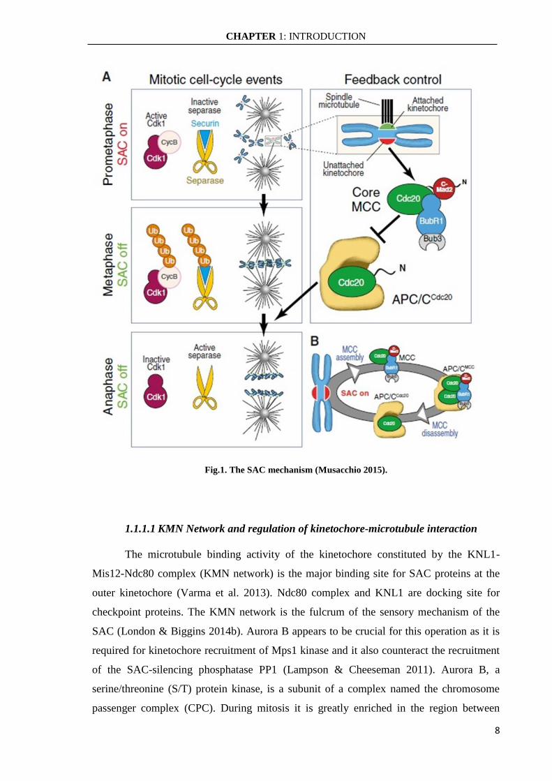
signaling. The SAC effector is named the Mitotic Checkpoint Complex (MCC) that targets
the anaphase-promoting complex or cyclosome (APC/C) (Fig.1). This complex can bind
stably to APC/C thereby inhibiting its E3 ubiquitin ligase activity necessary to target
several mitotic proteins for degradation. MCC complex is involved in the inhibition of
Cdc20 protein, a co-activator of APC/C complex. By inhibiting Cdc20, the SAC efficiently
inhibits the APC/C and halts mitotic progression by preventing the degradation of two
mitotic key substrates: securin and cyclin B1. Securin is an inhibitor of separase, a protease
that cleaves a cohesin subunit allowing sister chromatid separation, and cyclin B1 is an
activator of CDK1, the major mitotic kinase. The kinetochore plays a crucial role in the
activation of SAC pathway because all SAC components are recruited to unattached
kinetochores where they exhibit a rapid turnover and where the signal is generated. In fact,
Cdc20 is recruited to kinetochores and incorporated into the inhibitory complex MCC.
Once all kinetochores are correctly attached to microtubules the MCC complex
disassembles and the APC/C-Cdc20 complex become active and triggers anaphase entry
(Foley & Kapoor 2013; London & Biggins 2014b).
CHAPTER 1: INTRODUCTION
8
Fig.1. The SAC mechanism (Musacchio 2015).
1.1.1.1 KMN Network and regulation of kinetochore-microtubule interaction
The microtubule binding activity of the kinetochore constituted by the KNL1-
Mis12-Ndc80 complex (KMN network) is the major binding site for SAC proteins at the
outer kinetochore (Varma et al. 2013). Ndc80 complex and KNL1 are docking site for
checkpoint proteins. The KMN network is the fulcrum of the sensory mechanism of the
SAC (London & Biggins 2014b). Aurora B appears to be crucial for this operation as it is
required for kinetochore recruitment of Mps1 kinase and it also counteract the recruitment
of the SAC-silencing phosphatase PP1 (Lampson & Cheeseman 2011). Aurora B, a
serine/threonine (S/T) protein kinase, is a subunit of a complex named the chromosome
passenger complex (CPC). During mitosis it is greatly enriched in the region between

CHAPTER 1: INTRODUCTION
9
kinetochores from which it can phosphorylate kinetochore substrates, including
centromeric protein A (CENP-A) in the inner kinetochore and the subunits of the KMN
network in the outer kinetochore (Carmena et al. 2012). Aurora B-dependent
phosphorylation of kinetochore substrates is strictly linked to the state of kinetochore-
microtubule attachment, and declines when bi-orientation ensues (Emanuele et al. 2008).
The basic tails of Ndc80 contains numerous phosphorylation sites for the Aurora B kinase
and these are phosphorylated in response to improper kinetochore-microtubule
interactions, thereby destabilizing the interaction. As Aurora B is concentrated at the
centromere region this establishes a gradient of Aurora B activity and proper kinetochore-
microtubule attachments move the KMN network away from Aurora B activity thus
stabilizing the binding (Welburn et al. 2010). Furthermore, Aurora B makes a crucial
contribution to SAC signal because it is essential for recruitment of Mps1 to kinetochore
whose activity is required for kinetochore recruitment of all downstream component of
MCC complex. Instead Mps1 and Aurora B kinase constitute the most upstream
components of the checkpoint (Heinrich et al. 2012; Saurin et al. 2011). Mps1
phosphorylates the phosphodomain of KNL1 (MELT motifs), thus creating docking sites
for the recruitment of additional SAC proteins, including Bub3, Bub1, BubR1 (known as
MAD3 in yeast), MAD1, MAD2, and Cdc20, which play a crucial role in the assembly of
MCC, either as MCC subunits or by supporting MCC assembly (Fig.2) (Musacchio 2015).
Fig.2. Recruitment of MCC complex (Musacchio 2015).

CHAPTER 1: INTRODUCTION
10
1.1.1.2 Mitotic Checkpoint Complex (MCC)
The MCC complex serves as an effector for the SAC because it physically interacts
with APC/C to negatively affect its ubiquitin ligase activity in response to the presence of
unattached kinetochores in a mitotic cell (Hein & Nilsson 2014). The individual
components of MCC are known to dynamically exchange between the cytosol and
unattached kinetochores (Howell et al. 2004; Shah et al. 2004). However, the precise
molecular mechanisms controlling this protein trafficking are not known, and the exact
composition of MCC in time and cellular space is also poorly understood. The MCC is
formed by the association of MAD2, BubR1 and Bub3 with Cdc20. BubR1 is the largest
protein in the MCC and is able to interact with two Cdc20 molecules via its KEN1 and
KEN2 motifs (Izawa & Pines 2015; Ibrahim 2015). It can also bind Bub3 via its GLEBS
motif (Overlack et al. 2015), dimerize with budding uninhibited by benzimidazole-1
(Bub1) using extended loop helix (Lischetti et al. 2014) and interact with PP2A
phosphatase through a kinetochore attachment regulatory (KARD) domain (Suijkerbuijk et
al. 2012). MAD2 is a SAC protein that exists in two structural conformations, namely
open-MAD2 (O-MAD2) and closed-MAD2 (C-MAD2), to control APC/C activity (Luo &
Yu 2008; Mapelli & Musacchio 2007). Bub3 contains a WD40 β-propeller domain and it
forms a complex with Bub1 and BubR1 to recruit them to the kinetochores (Taylor et al.
1998). Cdc20 has a wide range of binding partners like APC/C subunits, MAD2, BubR1,
Bub1 and several mitotic substrates of APC/C (Musacchio 2015). The binding of MCC to
Cdc20 prevents Cdc20 binding to mitotic substrates proteins (Chao et al. 2012).
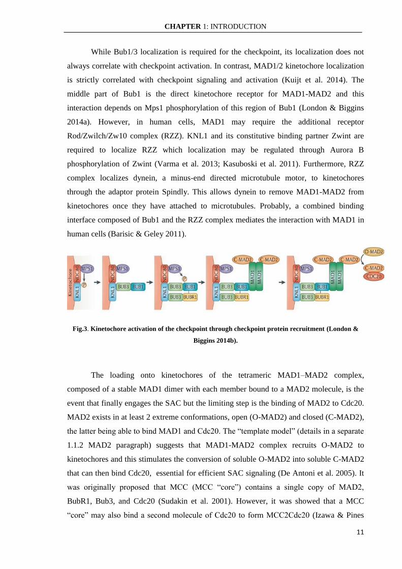
The assembly of MCC at unattached kinetochores is a step-wise process (Fig.3).
Once Mps1 is located and active at kinetochore, it stimulates recruitment of the Bub1-
Bub3 complex, which is needed for recruitment of BubR1-Bub3 and MAD1-MAD2
(London & Biggins 2014b). Initially it is important Mps1 phosphorylation of outer
kinetochore KNL1 protein which works as a receptor of Bub1 and BubR1 through two
distinct KI motifs located in the N-terminal region. These KI motifs make contact
specifically with the TRP domains of Bub1 and BubR1 (Kiyomitsu et al. 2011). However
for Bub1 and BubR1 localization is dispensable an additional mechanism contributes. For
Bub1 kinetochore localization is required that Mps1 phosphorylates MELT motifs in
KNL1 and binding to them depends on Bub3 which acts as a signaling adaptor to form a
complex with BubR1 and Bub1 (Musacchio 2015). Thus, the Bub3:Bub1 complex recruits
BubR1:Bub3.
CHAPTER 1: INTRODUCTION
11
While Bub1/3 localization is required for the checkpoint, its localization does not
always correlate with checkpoint activation. In contrast, MAD1/2 kinetochore localization
is strictly correlated with checkpoint signaling and activation (Kuijt et al. 2014). The
middle part of Bub1 is the direct kinetochore receptor for MAD1-MAD2 and this
interaction depends on Mps1 phosphorylation of this region of Bub1 (London & Biggins
2014a). However, in human cells, MAD1 may require the additional receptor
Rod/Zwilch/Zw10 complex (RZZ). KNL1 and its constitutive binding partner Zwint are
required to localize RZZ which localization may be regulated through Aurora B
phosphorylation of Zwint (Varma et al. 2013; Kasuboski et al. 2011). Furthermore, RZZ
complex localizes dynein, a minus-end directed microtubule motor, to kinetochores
through the adaptor protein Spindly. This allows dynein to remove MAD1-MAD2 from
kinetochores once they have attached to microtubules. Probably, a combined binding
interface composed of Bub1 and the RZZ complex mediates the interaction with MAD1 in
human cells (Barisic & Geley 2011).
Fig.3. Kinetochore activation of the checkpoint through checkpoint protein recruitment (London &
Biggins 2014b).
The loading onto kinetochores of the tetrameric MAD1–MAD2 complex,
composed of a stable MAD1 dimer with each member bound to a MAD2 molecule, is the
event that finally engages the SAC but the limiting step is the binding of MAD2 to Cdc20.
MAD2 exists in at least 2 extreme conformations, open (O-MAD2) and closed (C-MAD2),
the latter being able to bind MAD1 and Cdc20. The “template model” (details in a separate
1.1.2 MAD2 paragraph) suggests that MAD1-MAD2 complex recruits O-MAD2 to
kinetochores and this stimulates the conversion of soluble O-MAD2 into soluble C-MAD2
that can then bind Cdc20, essential for efficient SAC signaling (De Antoni et al. 2005). It
was originally proposed that MCC (MCC “core”) contains a single copy of MAD2,
BubR1, Bub3, and Cdc20 (Sudakin et al. 2001). However, it was showed that a MCC
“core” may also bind a second molecule of Cdc20 to form MCC2Cdc20 (Izawa & Pines

CHAPTER 1: INTRODUCTION
12
2015). This is possible thanks to two KEN domains of BubR1 that allow it to interact with
Cdc20 and MAD2 protein is required for this bond, so Mad2 and BubR1 synergize to
inhibit Cdc20-mediated activation of APC/C (Fig.4) (Burton & Solomon 2007; Fang
2002).
Fig.4. MCC complex (Musacchio 2015)
1.1.1.3 SAC Silencing
Recent studies have shown that rather than the intra-kinetochore tension, the stable
microtubule attachments at the kinetochores is a major driver for SAC inactivation
(Tauchman et al. 2015; Etemad et al. 2015). The rapid activation of APC/C–Cdc20 in
response to attachment of the last kinetochore suggests that Cdc20 is quickly liberated
from inhibition. The generation of active Cdc20 consists of at least 2 steps, namely killing
of the kinetochore signal and disassembly of existing MCC complexes.
Silencing the kinetochore signal requires the removal of the checkpoint proteins
from the kinetochore. At least two independent mechanisms lead to MAD1-MAD2
kinetochore dissociation dynein-mediated: a) stripping of checkpoint proteins and b)
reversal of activating phosphorylation through phosphatase activity. The first mechanism
consists of the motor protein dynein that localizes to the kinetochore through RZZ/Spindly.
MAD1-MAD2 and BubR1 are also removed by dynein during this process, coupling
microtubule binding to stripping of kinetochore checkpoint proteins (Gassmann et al. 2010;
Barisic & Geley 2011). The MAD1-MAD2 complex is also inhibited by “capping” of C-
MAD2 by p31comet
, which prevents binding of O-MAD2 to the complex once it is removed
from kinetochores (Fava et al. 2011). The second mechanism involves protein
phosphatase1 (PP1) recruitment for SAC silencing and his activity requires its interaction

CHAPTER 1: INTRODUCTION
13
with kinetochores. KNL1 contains a conserved PP1 binding site close to the region of
KNL1 that interacts with microtubules, and the link of PP1 to KNL1 contributes to SAC
silencing. A model for PP1-mediated SAC silencing is thus dephosphorization of MELT
motifs to remove Bub1 and BubR1 from kinetochores (Rosenberg et al. 2011; Espeut et al.
2012). The PP1 binding site on KNL1 contains a phosphorylation site for Aurora B, which
when phosphorylated prevents PP1 binding. Thus, PP1 and Aurora B antagonize each
other on the outer kinetochore and when microtubules bind the balance tips toward PP1
binding and SAC silencing (Liu et al. 2010).
MCC and APC/C–MCC are stable complexes and their disassembly is an active
process but the exact mechanism is not clear. At least 2 distinct pathways have been
suggested to remove MAD2 from Cdc20: a p31comet
-catalyzed mechanism and APC/C-
mediated ubiquitination of Cdc20. p31comet
binds to C-MAD2 present in MCC functioning
as an endogenous MAD2 inhibitor and also promotes the Cdk-dependent phosphorylation
of Cdc20 to release Cdc20 from MCC (Chao et al. 2012; Varetti et al. 2011). To facilitate
efficient C-MAD2 removal p31comet
might collaborate with the AAA-ATPase TRIP13
(Eitan et al. 2014). A second proposed mechanism of MCC dissociation, which would be
specific for APC/C bound MCC, is ubiquitination of Cdc20 by the APC/C, a process
regulated by APC15 (Mansfeld et al. 2011).
1.1.2 Mitotic Arrest Deficient (MAD2)
The heart of the SAC is MAD2 (Mitotic Arrest Deficient 2), a conserved ∼200-
residue protein, highly conserved in eukaryotes. Its main task is to safeguard the event of
chromosomes segregation from possible errors during mitosis. The MAD2 protein has a
central role in generation of the MCC and co-ordination of the early mitotic events with the
SAC activity. It is a component of Mitotic Arrest Deficient proteins, that include also
MAD1 and MAD3 (BubR1 in humans), initially identified in yeast as important regulators
of the mitotic checkpoint and then confirmed in humans (Li & Benezra 1996).
As said MAD2 exists in at least 2 extreme conformations, open O-MAD2 and
closed C-MAD2, differing in the position of secondary structure elements in the N- and C-
terminal regions (Fig.5). The C-MAD2 conformation is able to bind MAD1 and Cdc20 to
form the MCC complex. The structural properties of O-MAD2 do not permit the formation
of O-MAD2:O-MAD2 homodimer and O-MAD2:Cdc20 heterodimer. Conversely, C-
MAD2 is able to form a homodimer with C-MAD2 and heterodimers with O-MAD2,
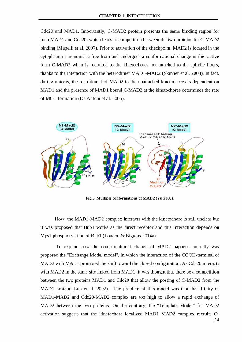
CHAPTER 1: INTRODUCTION
14
Cdc20 and MAD1. Importantly, C-MAD2 protein presents the same binding region for
both MAD1 and Cdc20, which leads to competition between the two proteins for C-MAD2
binding (Mapelli et al. 2007). Prior to activation of the checkpoint, MAD2 is located in the
cytoplasm in monomeric free from and undergoes a conformational change in the active
form C-MAD2 when is recruited to the kinetochores not attached to the spindle fibers,
thanks to the interaction with the heterodimer MAD1-MAD2 (Skinner et al. 2008). In fact,
during mitosis, the recruitment of MAD2 to the unattached kinetochores is dependent on
MAD1 and the presence of MAD1 bound C-MAD2 at the kinetochores determines the rate
of MCC formation (De Antoni et al. 2005).
Fig.5. Multiple conformations of MAD2 (Yu 2006).
How the MAD1-MAD2 complex interacts with the kinetochore is still unclear but
it was proposed that Bub1 works as the direct receptor and this interaction depends on
Mps1 phosphorylation of Bub1 (London & Biggins 2014a).
To explain how the conformational change of MAD2 happens, initially was
proposed the "Exchange Model model", in which the interaction of the COOH-terminal of
MAD2 with MAD1 promoted the shift toward the closed configuration. As Cdc20 interacts
with MAD2 in the same site linked from MAD1, it was thought that there be a competition
between the two proteins MAD1 and Cdc20 that allow the posting of C-MAD2 from the
MAD1 protein (Luo et al. 2002). The problem of this model was that the affinity of
MAD1-MAD2 and Cdc20-MAD2 complex are too high to allow a rapid exchange of
MAD2 between the two proteins. On the contrary, the “Template Model” for MAD2
activation suggests that the kinetochore localized MAD1–MAD2 complex recruits O-

CHAPTER 1: INTRODUCTION
15
MAD2 to kinetochores through dimerization with C-MAD2 bound to MAD1 and this
stimulates the conversion of soluble O-MAD2 into soluble C-MAD2 that can then bind
Cdc20 (De Antoni et al. 2005). This model identified MAD1-bound C-MAD2 as a
template for O-MAD2 conversion into a Cdc20-bound C-MAD2 copy. The conformational
dimerization of MAD2 appears to be a transient interaction between an O-MAD2 subunit
and a spatially localized rigid scaffold, the MAD1:C-MAD2 complex (Mapelli &
Musacchio 2007).
These interactions might be directly regulated by Mps1 at kinetochores as Mps1
stimulates O-MAD2 recruitment in human cells (Hewitt et al. 2010). Indeed, O-MAD2
activation has been reconstituted with purified proteins but the low rates measured in vitro
might suggest that kinetochores provide additional layers of catalysis and one of these is
probably MAD1 (Simonetta et al. 2009; Kruse et al. 2014). The silencing of the pathway is
due to the intervention of p31comet
protein which promotes the exit from mitosis by binding
to C-MAD2, and obtaining the release of Cdc20 ready to active the APC /C complex (De
Antoni et al. 2005).
In addition, it should be noted that also in interphase cells, the MAD1:C-MAD2
complex forms at the nuclear envelope to produce Cdc20 inhibitor complex during
interphase to avoid the premature degradation of Cyclin B and securin that upon entry to M
phase leads to precocious anaphase (Rodriguez-Bravo et al. 2014).
Under normal growth conditions, functional inactivation of MAD2 induces cellular
senescence and causes defects in the response of the cells to DNA damage (Lentini et al.
2012; Lawrence et al. 2015). In addition, the deregulation of MAD2 generates significant
mitotic anomalies as the deletion of even one of MAD2 allele increases the chromosomal
instability of the cell (Musacchio & Salmon 2007). It was found that MAD2 deregulation
in the cells induces premature sister chromatids separation and aneuploidy (Meraldi et al.
2004; Lentini et al. 2012; Veneziano et al. 2016).
1.1.3 Centromere Associated Protein E (CENP-E)
Accurate chromosome segregation during mitosis requires the bipolar attachment of
kinetochore of duplicated chromosomes to spindle microtubules emanating from opposite
poles (Cleveland et al. 2003). Microtubule capture by the kinetochore is a stochastic
process. Although some chromosomes achieve biorientation without being transported to
the spindle pole, dynein-mediated transport is an important mechanism to collect

CHAPTER 1: INTRODUCTION
16
chromosomes to a common microtubule-dense region, where kinetochores have a greater
chance of promoting efficient chromosome alignment.
In this process an important role is mediated by CENP-E (Centromere Associated
Protein-E), a plus-end directed kinesin-7 motor protein of 312kDa (in human cells)
required for chromosome segregation in both mitosis and meiosis ( Yen et al. 1992; Schaar
et al. 1997; Kim et al. 2008) . CENP-E protein accumulates in late G2, functions during
mitosis, and is degraded at the end of mitosis as quantitatively as cyclin B (Brown et al.
1994). During mitosis, CENP-E localizes to kinetochores, where it is one of a number of
proteins that serve as linkers between chromosomes and the microtubules of the mitotic
spindle (Gudimchuk et al. 2013). It has a long coiled-coil region separating the motor
domain near its N-terminus from a C-terminal domain that contains sites responsible for
association with the kinetochore. The process of capturing spindle microtubules by
kinetochores is prone to errors. Undesirable attachment frequently occurs in early
prometaphase, with a single kinetochore capturing microtubules from both spindle poles
(merotelic attachment), or both sister kinetochores attached to the same pole (syntelic
attachment) (Cimini & Degrassi 2005). It was found that CENP-E possesses a highly
flexible and very long coiled-coil that raises the possibility that it may also contribute, in
part, to the inappropriate attachments of kinetochores (Kim et al. 2008). These improper
kinetochore attachments, if not resolved, can lead to chromosome missegregation and
aneuploidy (Holland & Cleveland 2009).
CENP-E also functions in the spindle assembly checkpoint (SAC) to prevent
chromosome missegregation and aneuploidy (Abrieu et al. 2000; Weaver et al. 2003).
CENP-E association with the kinetochore has been reported to be mediated by a large
number of kinetochore-associated proteins with which it interacts, including the
centromeric protein F (CENP-F), NUF2, and SKAP (Huang et al. 2012; Liu et al. 2007).
Also CENP-E binds and, in the absence of bound microtubules, activates the SAC kinase
BubR1 forming a stable ternary complex (spindle microtubule/CENP-E/BubR1) and
producing checkpoint signaling that is silenced either by spindle microtubule capture or the
tension developed at kinetochores (Mao et al. 2005).
It was identified that CENP-E can be multiply phosphorylated during mitosis
(Nousiainen et al. 2006). However, the significance of all of these phosphorylations has not
been established but some of these can regulate CENP-E functions. Phosphorylation of the
C-terminal tail of CENP-E by Cdk1, MAPK, or Mps1 has been proposed either to regulate
CHAPTER 1: INTRODUCTION
17
CENP-E motor activity prior to its binding to kinetochores or inhibit a microtubule binding
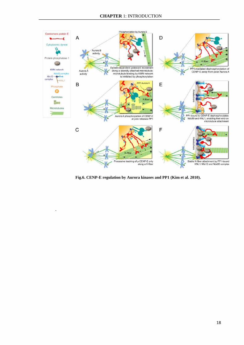
site in the tail (Espeut et al. 2008). Furthermore, it was discovered an Aurora/PP1
phosphorylation switch that is required not only for congression of polar chromosomes
through modulation of the intrinsic motor properties of CENP-E, but also for subsequent
stable biorientation of those chromosomes (Kim et al. 2010). In the regulation of CENP-E,
the Aurora kinase activity is opposed by Protein Phosphatase 1 (PP1) functions (Liu et al.
2010). It has been shown that PP1 can localize at outer of kinetochore and it can stabilize
kinetochore-microtubule attachment by counteracting Aurora B kinase activity. In
particular, Aurora kinases, both A and B, phosphorylate a single conserved residue close to
the CENP-E motor domain while PP1 has a docking domain that overlaps the site of
phosphorylation so the PP1 bind to CENP-E is disrupted by Aurora mediated
phosphorylation. Aurora A phosphorylates CENP-E near the spindle poles, releasing PP1
from CENP-E. CENP-E phosphorylated is active and able to bind the microtubules to
KMN network on the kinetochore thus to carry the chromosomes the spindle equator along
the K-fiber of an already bioriented chromosome (Kapoor et al. 2006). As chromosomes
congress, kinetochores move away from the Aurora A gradient concentrated at the spindle
poles and CENP-E is dephosphorylated and recruits a high local concentration of PP1 to
the outer kinetochores of chromosomes so it has translocated away from a pole. CENP-E
delivered PP1 and dephosphorylation of kinetochore key components, such as Ndc80 and
KNL1, is essential for stable kinetochore-microtubule interactions (Fig.6) (Kim et al.
2010).
CHAPTER 1: INTRODUCTION
18
Fig.6. CENP-E regulation by Aurora kinases and PP1 (Kim et al. 2010).
.

CHAPTER 1: INTRODUCTION
19
1.2 ANEUPLOIDY AND CANCER
Cancer is the result of cells that have undergone unregulated growth as a
consequence of defects in one or more regulatory signaling pathways. The development of
cancer is usually a multistep process and one of its fundamental features is tumor clonality,
the development of tumors from single cells that begin to proliferate abnormally. One of
the hallmarks of cancer, especially solid tumor, is aneuploidy but its role in the process of
carcinogenesis is not yet well defined.
A functional mitotic checkpoint can avoid aberrant chromosome content which can
generate aneuploidy or polyploidy. Polyploidy describes a chromosome content which is a
multiple of the parental euploid karyotype and it is a common phenomenon in nature
(Hollister 2015). In contrast, aneuploidy is a change in chromosomes number or structure
that are not a multiple of the whole chromosome set and it is usually not well-tolerated by
cells. Exception trisomy 21 (Down syndrome), trisomy 13 and 18, no other aneuploid
disorders were found to be compatible with live and most aneuploidies affecting the whole
organism are lethal. Aneuploidy can arise either by gain or loss of few intact chromosomes
(chromosomal aneuploidy) or by chromosome rearrangements such as translocations,
inversions, amplifications and deletions (segmental or structural aneuploidy) (Janssen et al.
2011).
More than a century ago, Theodar Boveri (Boveri, 1914) led to a hypothesis that
aberrant mitosis would generate cells with different chromosome content that might
contribute to tumorigenesis. In recent years, different results have clarified the molecular
mechanisms involved in this process although many of these are conflicting results about
the role of aneuploidy in carcinogenesis (Weaver et al. 2007; Torres et al. 2008).
Remembering the famous incipit of the book “Anna Karenina” we could be say that the
aneuploid tumors are like unhappy families of Tolstoj: each tumor has its particular
content of abnormal chromosomes and consequently their atypical features (Pellman
2007).
It is important to separate aneuploidy from chromosomal instability (CIN) which is
as well as aneuploidy a frequent hallmark of cancer. Whereas aneuploidy describes the
present state of the genomic material, chromosomal instability describes ongoing
missegregation of different chromosomes (Holland & Cleveland 2009). Every cell with
CIN can be aneuploid whereas not every aneuploid cell is genomically unstable, as shown
in cells from Down syndrome patients (Gordon et al. 2012).

CHAPTER 1: INTRODUCTION
20
1.2.1 Origins of Aneuploidy
Chromosome missegregation can either happen randomly or due to mutations
leading to defects in spindle assembly checkpoint (SAC), sister chromatid cohesion,
aberrant mitosis or merotelic attachment (Fig.7). In contrast, structural aneuploidies can
result from errors in DNA replication and repair.
Fig.7. Origins of chromosome aneuploidy (Siegel & Amon 2012)
1.2.1.1 Spindle Assembly Checkpoint Defects
Several studies have reported induction of aneuploidy and tumor formation in cell
culture and mouse models as a consequence of deregulation of SAC proteins. A complete
knockout of many spindle checkpoint genes, such as genes encoding for Bub3 and MAD2,
were shown to be embryonic lethal (Dobles et al. 2000; Kalitsis et al. 2000). However,
mice with impaired SAC survive and their cells can divide even though the chromosomes
are not aligned properly leading to chromosomally instable cells (Fig. 7A). This was shown
by Michel and colleagues that generated MAD2 haplo-insufficient mice developing lung
tumors at high rates and respective cells displayed high levels of chromosomally instable
aneuploid cells (Michel et al. 2001). Similarly, in mouse models reduced levels of CENP-E
CHAPTER 1: INTRODUCTION
21
and BubR1 have been reported to increase the frequency of aneuploidy and cause
formation of spontaneous or induced tumors in vivo (Weaver et al. 2003; Dai et al. 2004;
Weaver et al. 2007). Likewise, was found that MAD2 or BubR1 haploinsufficiency
condition can induce aneuploidy and mitotic alterations in human primary fibroblasts
(IMR90) and in near stable colon cancer cells (HCT116) (Lentini et al. 2012; Lentini et al.
2014). In human tumors, mutations in BUBR1 have been linked to induction of mosaic
variegated aneuploidy (MVA), a rare disorder due to constitutional aneuploidy that
predisposes these individuals to develop cancer (Hanks et al. 2004). Moreover, MVA
patient-derived cell lines exhibit reduced BubR1 expression which was associated with low
SAC activity and defects in chromosome alignment (Suijkerbuijk et al. 2010).
Consequently, deregulation of SAC proteins in cancer cell lines can lead to incorrect
chromosome segregation, which provides one potential explanation for the aneuploidy
induction in cancer cells.
1.2.1.2 Cohesion defects
Sister chromatid cohesion defects result in chromosomal missegregation. During
cell divisions, two newly replicated sister chromatids are kept together by conserved
protein complexes called cohesins. This cohesin complex protects the sister chromatids
from premature segregation which may result in aneuploidy and thus is implicated in
control of genomic stability (Fig. 7B). The cohesin rings are cleaved by separase at
anaphase onset. Sister chromatid cohesion is necessary to create tension by keeping the
sister chromatids together while they are pulled by microtubules that depart from the two
poles (Peters 2012). Inactivation of proteins working in the cohesion pathway, such as
STAG2, has been reported to cause aneuploidy in human cells (Solomon et al. 2011). In
line, separase levels were found to be increased in breast cancer tissues (Zhang et al. 2008)
as well as other members of the cohesion pathway as for example WALP (Oikawa et al.
2004) and securing (Zou et al. 1999). Furthermore, it was shown that sister chromatid
cohesion is important to prevent merotelic attachment (Cimini 2008).
1.2.1.3 Merotelic kinetochore-microtubules attachment
The Spindle Assembly Checkpoint discriminates proper from improper
kinetochore-micotubules attachment because proper attachment generates tension that
results in intra-kinetochore stretching and this kinetochore state is unable to bind SAC
proteins (Maresca & Salmon 2010). A particular type of improper attachment that is not

CHAPTER 1: INTRODUCTION
22
detected by the checkpoint is the merotelic attachment, in which the same kinetochore
binds to microtubules from both poles. Merotelic attachments generate tension and are
therefore not sensed as erroneous. This attachment defects result in lagging chromosomes
(chromosomes that are not properly aligned) and consequently chromosome
missegregation and aneuploidy (Fig. 7C) (Cimini et al. 2001). Several defects are found to
generate merotelic attachments such as defects in condensin (Samoshkin et al. 2009),
induction of multipolar spindles (Ganem et al. 2009) and hyper stable kinetochore-
microtubule interactions (Kabeche & Compton 2012).
1.2.1.4 Multi-polar mitotic spindles
The spindle poles contribute to the formation of a bipolar mitotic spindle in early
mitosis. The centrosome forms the poles of the mitotic spindle and cells possessing extra
centrosomes can form multipolar spindles. If not corrected, a multipolar anaphase can
produce three or more highly aneuploid daughter cells that are likely to be inviable (Fig.
7D). Defects involving centrosome changes in number, organization, and behavior, have
been found in a variety of solid tumors (Brinkley 2001). However, multipolar mitotic
divisions are rare because in most cases extra centrosomes are clustered into two groups
allowing bipolar spindles to form. Nevertheless, in these cells merotelic attachments are
frequent and the consequent lagging chromosomes cause low-level aneuploidy (Ganem et
al. 2009). Interestingly, it was found that centrosome amplification alone is not sufficient
to induce chromosomal instability in colon cancer cells with a MIN phenotype because
clustered extra centrosomes form a pseudo bipolar spindle. To generate aneuploidy cells is
necessary that centrosome amplification has to be associated to alterations in genes
regulating mitosis progression such as Aurora-A/STK15 to trigger chromosomal instability
(Lentini et al. 2007).
Furthermore, experiments with cultured cells have shown that progression of
mitosis in the presence of multiple spindle poles leads to defects in chromosome
segregation (Ganem et al. 2009; Maiato & Logarinho 2014).
1.2.1.5 Structural Aneuploidy
Many aneuploid cells can display numerical aneuploidies but also structural
aneuploidies due to mitotic mistakes (Janssen et al. 2011; Ganem & Pellman 2012). For
example, the MAD2 overexpression in a mouse model led to the induction of loss or gain
CHAPTER 1: INTRODUCTION
23
of whole chromosomes, as well as caused structural defects in the chromosomes and
initiated tumorigenesis (Sotillo et al. 2007).
In aneuploid cells, the origin of these gross chromosomal rearrangements (GCRs)
such as translocations, deletion of a chromosome arm, interstitial deletions or inversions, is
mostly unclear. It is known that DNA damaging agents such as γ-irradiation and
hydroxyurea induce chromosomal rearrangements suggesting that rearrangements might be
a result of improper repair of DNA lesions (Suzuki et al. 2003). In addition, the prolonged
mitotic arrest is also a source of DNA damage. For example, depletion of CENP-E by
RNAi in colon cancer cells induces a transient mitotic arrest and DNA breaks (Dalton et al.
2007).
The presence of damaged DNA and chromosome breakage, or lagging
chromosomes may result in the formation of aberrant nuclear structures called micronuclei
after cell division (Cimini et al. 2001). Micronuclei can contain a chromosomal fragment
or an entire chromosome. Replication of this trapped DNA is often defective and results in
a pulverization of the chromosome which is later reassembled leading to structural
aneuploidy (Crasta et al. 2012). In pRb-depleted primary human fibroblasts, that possess
an intact spindle checkpoint, the generation of micronuclei is responsible of aneuploidy
development (Amato et al. 2009). In these cells, pRb loss promotes aneuploidy mainly by
inducing CENPA overexpression that in turn might induce micronuclei caused by mis-
attached kinetochores which could trigger chromosome segregation errors (Amato et al.
2009).
1.2.1.6 Polyploidy
Polyploid cells are those containing more than two paired (homologous) sets of
chromosomes. In other mammals, tetraploidy causes early lethality and spontaneous
abortion or resorption of the embryo. In contrast, it is frequent in plants and many insects.
In human it was observed that some kinds of cells are polyploid as megakaryocytes,
hepatocytes, osteoclasts and skeletal muscle cells. However, polyploidy is even present in
some cancer cells (Davoli & de Lange 2011).
In tumors, the chromosome numbers are distributed into two peaks, one
representing tumors that are near-diploid and one representing tumors with a chromosome
number between a triploid and a tetraploid genome (Storchova & Kuffer 2008). This
bimodal distribution underlines that aneuploidy in cancer cannot be explained with a single

CHAPTER 1: INTRODUCTION
24
mechanism underlying aneuploidy. To account for aneuploidy with high chromosome
numbers it was proposed that these cancers originate from an unstable tetraploid
intermediate (Shackney et al. 1989). Tetraploid cells are known to frequently missegregate
chromosomes due to their supernumerary centrosomes (Ganem et al. 2009) and they will
therefore readily generate subclones with the hypotetraploid or hypertriploid chromosome
numbers observed in cancer.
At the molecular level several mechanisms have been found to facilitate induction
of polyploidy: cell fusion, failure in cytokinesis or other steps in mitosis and endo-
reduplication. Cell fusion generates a bi-nucleate intermediate that can produce daughter
cells with single 4N nuclei in G1. Experimentally induced fusion of primary human
fibroblasts has been shown to enhance their in vitro transformation with potent oncogenes
(Duelli et al. 2007). Several types of failure mitosis can give rise to a cell with double the
chromosome number. In cells with MAD2 overexpression was observed induction of
tetraploidy through cytokinesis failure (Sotillo et al. 2007). Furthermore, failure in
cytokinesis is associated with overexpression of Aurora A, a kinase critical for mitosis
(Zhang et al. 2004).
Tetraploidization and its associated aneuploidy are particularly well-suited to
accelerate tumor genome evolution because allow tumor cells to sustain a higher incidence
of mutations thereby increasing the probability of adaptive changes (Davoli & de Lange
2011).
1.2.2 Proliferation and Physiology of Aneuploid Cells
Maintenance of a balanced euploid genome is a key requisite for the success of all
multicellular organisms. Possession of an equal number of each chromosome ensures a
balanced genome where genes on different chromosomes are present in equal numbers. In
contrast, aneuploidy, results in an unbalanced genome with different copy numbers for
genes on different chromosomes. This is generally not well tolerated in nature.
Aneuploidy that arises during gamete formation or during the early embryonic
divisions results in entire organisms with an aberrant karyotype that is frequently lethal
early in development. For example, in mouse all autosomal aneuploidies are embryonic
lethal with the exception of Trisomy 19, which is the smallest mouse autosome and even
these mice die shortly after birth. Even in human, the best-known and most studied
organismal aneuploidy is the genetic condition responsible for Down syndrome (Trisomy
CHAPTER 1: INTRODUCTION
25
21) whose individuals display frequently stunted growth. Down syndrome individuals do,
however, exhibit a decreased risk of solid tumors (Satgé et al. 2003). Two other trisomies
survive to birth in humans; these are Trisomy 18 (Edwards syndrome) and Trisomy 13
(Patau syndrome). Only approximately 10% of children born with these syndromes live to
one year of age (Rasmussen et al. 2003). In summary, it is clear that aneuploidy causing
growth retardation and developmental abnormalities in most organisms.
It is not clear if the aneuploidy effects on cell physiology are due to the mere
presence of additional DNA in the form of chromosomes or lack of chromosomes, or
whether they are due to changes in gene expression levels. In some organisms aneuploid
chromosomes are expressed according to gene copy number (e.g. yeast, MEFs and Down
syndrome) while in other organisms (plants and Drosophila) exists a compensatory
mechanisms that attempt to “balance out” gene copy number imbalances caused by
aneuploidy (Torres et al. 2007; Williams et al. 2008; Mao et al. 2003; Larsson et al. 2004;
Stenberg & Larsson 2011). It appears that species-specific differences exist in the ability to
attenuate gene expression from aneuploid chromosomes, both at the RNA and protein
levels, and these differences affect the aneuploid effects on cell physiology.
1.2.2.1 General detrimental consequences of aneuploidy in the cells
Beyond the effects of aneuploidy on gene expression, cellular studies have revealed
that aneuploidy causes several general effects. These are largely detrimental and appear to
be conserved across species but, in rare circumstances, were observed beneficial effects of
aneuploidy. Among the key phenotypes shared by aneuploid cells is their slower
proliferation compared to euploid cells. This effect was observed in aneuploid mouse
embryonic fibroblasts (MEFs) trisomic for either chromosome 1, 13, 16, or 19 (Williams et
al. 2008) that exhibit proliferation defects, as do cells harboring random aneuploidies
caused by impaired SAC function. Also in primary human fibroblasts (IMR90), aneuploidy
caused by MAD2 depletion triggers slowdown proliferation and activation of cellular
senescence by the p53 pathway (Lentini et al. 2012).
Beyond proliferation rates, aneuploid cells also exhibit a number of other
phenotypes that can be broadly summarized as an “aneuploidy stress response” (Fig.8).
The major effects of aneuploidy observed in cells are metabolic alterations, protein
imbalance, genomic instability and death. Metabolic changes were found in MEF cultures
containing the aneuploidy-inducing Cdc20AAA
mutation which exhibit increased glucose
CHAPTER 1: INTRODUCTION
26
uptake and increased production of lactate and reactive oxygen species which led to the
activation of p53 through activation of ATM pathway (Li et al. 2010). In addition to
energy stress, proteotoxic stress, i.e. physiological strain accrued from an abundance of
misfolded proteins, is present in aneuploidy yeast cells (Torres et al. 2007). Evidence for
proteotoxic stress also exists in aneuploid mammalian cells. Trisomic MEFs harbor
increased rates of autophagy and increased basal levels of the inducible chaperone Hsp72
(Tang et al. 2011). It is evident that aneuploid cells display marked growth defects, which
are probably based on an increased need in energy due to transcription, translation and
degradation of extra protein.
Aneuploidy has been shown to increase genomic instability and the rate of
chromosome missegregations that can results in DNA damage. Merotelically attached
chromosomes, induced by compounds that interfere with mitotic spindle formation, remain
in the center of the cell during anaphase and are broken during cytokinesis (Janssen et al.
2011). Lagging chromosomes can also form micronuclei, which then experience
substantial DNA damage during subsequent replication (Crasta et al. 2012). Janssen et al.
found that lagging chromosomes suffer DNA damage during cytokinesis, which activates
p53 through the ATM pathway while a second study using the same treatment found no
evidence for DNA damage in lagging chromosomes but p53 was still activated and halted
cell cycle progression through a p38 kinase dependent stress response, presumably
triggered by aneuploidy-induced stresses such as metabolic alterations and protein
imbalances (Thompson & Compton 2010). In conclusion, there is indication that aneuploid
cells display a common aneuploidy specific cellular response, which may be conserved.
However, this hypothesis is mainly based on correlative studies and detailed experiments
elucidating underlying mechanisms are yet missing.

CHAPTER 1: INTRODUCTION
27
Fig.8. Observed characteristics of aneuploidy in mammalian cells (Siegel & Amon 2012).
1.2.2.2 “Beneficial” effects of aneuploidy
Despite the clearly detrimental effects of aneuploidy on cellular fitness, this
condition can, in rare cases of strong selective pressures, give cells a competitive edge.
Potential beneficial effects of aneuploidy under extreme selective pressure have been
observed in several systems. For example, in budding yeast, specific aneuploidies have
been shown to provide resistance to toxic agents (Pavelka et al. 2010). In human
fibroblasts, the introduction of an additional copy of chromosome 8 caused loss of contact
inhibition, but cells still proliferated more slowly than diploid cells (Nawata et al. 2011).
Probably, aneuploidy can provide an effective means of quickly adapting to a selective
pressure. However, this selective advantage comes at a price for the cells because changes
in gene copy number of an entire chromosome induced by aneuploidy disrupt protein and
energy homeostasis and cause proliferation defects in addition to chromosome specific
detrimental effects. Naturally, in presence of mutations that mitigate the adverse effects of
aneuploidy, the full adaptive and genome-instability inducing potential of aneuploidy
comes into play as suggested by aneuploidy-tolerating mutations found in yeast (Torres et
al. 2010). Loss of p53 also increases the proliferative abilities of aneuploid mammalian
cells (Li et al. 2010; Thompson & Compton 2010; Janssen et al. 2011). Thus, identification
of genetic alterations that ameliorate the adverse effects of aneuploidy could yield dramatic
insight into tumorigenesis.

CHAPTER 1: INTRODUCTION
28
1.2.3 Aneuploidy and Chromosome Instability in Cancer
Aneuploidy has been implicated in several diseases but the most striking correlation
between aneuploidy and disease can be found in cancer. The occurrence of aneuploidy in
cancer has long been known. David van Hansemann first noted that tumors have
unbalanced mitoses over 120 years ago (Hansemann 1890). This work influenced Theodor
Boveri to expand upon his earlier characterization of aneuploidies in sea urchins to suggest
that a single aneuploid cell might cause cancer (Boveri 1914). More than ninety percent of
solid tumors and seventy-five percent of blood cancers show some degree of aneuploidy
(Weaver & Cleveland 2006). Many different types of chromosomal abnormalities are
observed in cancer cells underlying the complexity and elusive relationship between
aneuploidy and tumorigenesis. It is difficult assert if aneuploidy can promote cancer
although cancer and aneuploidy cells show similar characteristics, like changes in
metabolism or elevated proteotoxic stress and genomic instability (Hanahan & Weinberg
2011). The situation is, however, not simple as “aneuploidy causes cancer” suggested by
Boveri almost 100 years ago. It appears that aneuploidy sometimes promotes
tumorigenesis, sometimes seems inconsequential, and sometimes inhibits disease initiation
and progression.
The major evidences that aneuploidy contributes to tumorigenesis comes from the
study of animal models of chromosomal instability. Chromosomal instability (CIN) refers
to an increased rate of chromosome missegregation due to errors in mitosis (Geigl et al.
2008). There are many events leading to CIN: multipolar spindles, improper chromosome
condensation or cohesion, defective mitotic checkpoint, improper kinetochore-
microtubules attachments etc. (Thompson et al. 2010). In mouse models, many studies
refer a link between CIN and tumorigenesis and most of them are focused on partial
inactivation of proteins which are involved in the spindle assembly checkpoint (SAC).
Generally a weakened SAC results either in increased chromosome mis-alignments or in
the inability to resolve them, thus leading to chromosome missegregation and subsequent
formation of aneuploidy. Deletion of one copy of MAD2 induces aneuploidy in vitro and
in vivo and leads to a high frequency of mice with papillary lung adenocarcinomas, a tumor
that is extremely rare in wild-type mice (Michel et al. 2001). At the same time
heterozygosis of MAD1, BubR1 or CENP-E increase constitutive tumors in mouse while
overexpression of MAD2 or Bub1 induces CIN and cancer (Dai et al. 2004; Iwanaga et al.
2007; Weaver et al. 2007; Sotillo et al. 2007; Sotillo et al. 2010; Ricke et al. 2011). Mice
CHAPTER 1: INTRODUCTION
29
with one Cdc20AAA
allele that cannot be inhibited by the SAC have an increased tumor
incidence (Li et al. 2009). Similar results are observed when the SAC is hyper-activated by
overexpression of the outer kinetochore component Hec1 (Diaz-Rodríguez et al. 2008) or
by overexpressing the SAC kinase Bub1 (Ricke et al. 2011). Both result in aneuploidies in
vitro and cause an increase in tumor incidence and alteration of tumor spectra in vivo. In
summary, both weakening and hyper-activating the SAC is sufficient to generate
aneuploidy and to induce tumorigenesis.
However, although tumorigenesis is elevated, this increase is modest in many cases,
particularly in mice with loss-of-function mutations in SAC genes (e.g. only 20% of
CENP-E+/-
mouse develop tumors (Weaver et al. 2007). Consistently, haploinsufficiency of
Bub3 or Rae1 did not result in increased tumorigenesis and some mouse models even
showed decreased tumor formation when challenged with carcinogens (Babu et al. 2003;
Kalitsis et al. 2005). Furthermore, homozygous knockouts of CENP-E or of any other SAC
signaling genes (MAD2, MAD1, BubR1 etc.) result in massive chromosome segregation
defects and early embryonic lethality suggesting that the relationship between impaired
SAC signaling, aneuploidy and tumor onset is complex (Giam & Rancati 2015). Indeed,
aneuploidy can also act as a tumor suppressor. Aneuploidy induced by the loss of one copy
of CENP-E inhibited tumorigenesis in some tissues. Furthermore, introducing additional
aneuploidy can prevent tumorigenesis, presumably by inducing sufficient levels of
aneuploidy to cause cell death (Weaver et al. 2007; Silk et al. 2013). At the same time
simultaneous reduction of MAD2 and BubR1 expression induces elevate rate of
aneuploidy and mitotic alterations that result in p53 dependent cell cycle arrest of tumor
cells (Lentini et al. 2014).
The observation that aneuploidy has been associated with defective development
and lethality in multicellular organisms (Siegel & Amon 2012) and that, in mice and
humans, all autosomal monosomies and almost all trisomies result in embryonic lethality
reinforce the idea that aneuploidy alone may not induce tumorigenesis. Moreover,
individuals with trisomy 21 have a lower likelihood of developing solid tumors (Hasle et
al. 2000; Satgé et al. 2003), as do mouse models of the disease. However, since cancer
genomes are highly complex and contain additional mutations besides chromosome copy
number changes, it remains controversial whether aneuploidy acts as a driving force or as a
foe of tumorigenesis.

CHAPTER 1: INTRODUCTION
30
1.3 THE IMPORTANCE OF GENETIC BACKGROUND IN
ANEUPLOIDY
As described in the previous sections, aneuploidy hinders cell proliferation in most
cases and hardly any direct effects of aneuploidy are enough to induce tumorigenesis. At
the same time the aneuploidy induced by haploinsufficiency of SAC genes has mixed
effects on the development of tumors in mouse models (see paragraph 2.3). These
observations can be explained by different options: 1. Aneuploidy is a consequence of CIN
and the degree of CIN frequently correlates with karyotypic complexity. 2. SAC genes
have other non-mitotic functions that make it difficult to dissect which function is
associated to increased cancer susceptibility. 3. Aneuploidy has been implicated in several
diseases but the most striking correlation between aneuploidy and disease can be found in
cancer. aneuploidy needed additional oncogenic mutations (such as presence of oncogenes
or inactivation of tumor-suppressor genes) (Giam & Rancati 2015).
It has been suggested that the range in severity of the phenotypes observed in cells
with CIN differs depending on the number of processes that will be affected when such a
mutation is incurred. If mutating a factor only affects one cellular process that promotes
tumorigenesis, the effect will be less severe than if multiple tumor-promoting pathways are
affected by a single alteration (Ricke et al. 2008).
1.3.1 INK4/ARF locus
The human INK4/ARF (CDKN2A) locus, located on human chromosome 9q21,
encodes two completely unrelated proteins p16INK4a
and p14ARF
(p19ARF
in mouse), both
potent inhibitors of cell proliferation. The mechanism by which these two proteins are
produced is quite unusual: each transcript has a specific 5’ exon, E1α or E1β for p16INK4a
and p14ARF
respectively, which are spliced to a common exon 2. This exon contains two
overlapped ORFs, therefore the two proteins encoded share no amino acid sequence
identity (Fig.9) (Quelle et al. 1995). The alpha transcript encoding p16INK4a
was the first to
be identified so the protein produced by the alternative beta transcript was named ARF,
where ARF stands for Alternative Reading Frame. In addition, it has been found in the
CDKN2a locus the presence of the INK4b gene (also known as CDKN2B) that is
generated from the tandem duplication of INK4 gene and encodes the kinase inhibitor
p15INK4b
(Sharpless & DePinho 1999). Both proteins interact and inhibit the kinase activity
of cyclin dependent kinases 4 and 6 (CDKs) which in turn affect the pRb pathway and led
CHAPTER 1: INTRODUCTION
31
to E2F repression and growth arrest. The binding with p16INK4a
and p15 INK4b
prevents the
interaction of the CDKs 4 and 6 with the D-Cyclins, required for their catalytic activity and
for the cell cycle progression from G1 to S phase (Russo et al. 1998; Serrano et al. 1993;
Sherr 2006).
The β transcript results in a polypeptide of 132 amino acids and 14 kDa named
p14ARF
and regulates the induction of cell cycle arrest and/or apoptosis by p53-dependent
and independent pathways (Ozenne et al. 2010; Sherr 2006). Under normal conditions, the
p14ARF
gene (and the other genes in the locus) is repressed by the action of Polycomb
proteins (PcG), which inhibit the expression of specific genes by chromatin modifications.
BMI (B lymphoma Mo-MLV insertion region 1) is one of the main PcG components that
repress p14ARF
expression. In fact, murine embryonic fibroblasts (MEFs) BMI-1-/-
show a
detected upregulation of the expression of p14ARF
and p16INK4a
(Jacobs et al. 1999). The
gene silencing PcG-mediated is also the molecular mechanism by which the p53 tumor
suppressor represses the p14ARF
expression. Indeed, p53 binds the promoter of p14ARF
and
recruits the complex histone deacetylation (HDAC) and PcG proteins (Zeng et al. 2011).
p14ARF
transcription is upregulated in response to a host of oncogenic signals
including c-Myc, Ras, E2F-1, E1A, and v-Abl to induce cell cycle arrest (Sherr 2001).
Interestingly, Ras-induced ARF-mediate cell cycle arrest is not immediate. Wild-type
MEFs transduced with oncogenic RasV12 accumulate ARF protein over time and do not
succumb to ARF-mediated cell cycle arrest for approximately 5 days. This data suggests
that a threshold level of ARF protein must accumulate before cell cycle arrest can be
achieved (Groth et al. 2000).
Since p14ARF
is normally activated as a result of oncogenic signals to stabilize p53
tumor suppressor and p16INK4a
or p15INK4b
proteins are involved in cellular pathways
controlled by pRB, it is intuitive to think that mutations that fall in a common region of
exon 2 can alter simultaneously two pathways that have a synergistic effect on the control
and block cell proliferation (Sharpless 2005). Mutations within exon 2 that affect both
p14ARF
and p16Ink4a
are found in many cancers (del Arroyo & Peters 2005; Gardie et al.
1998).

CHAPTER 1: INTRODUCTION
32
Fig.9. INK4/ARF locus.
1.3.2 p14ARF
Tumor Suppressor and Cancer
The genes in the INK4/ARF locus are frequently mutated or silenced in cancer cells
since they encode proteins that prevent tumorigenesis (Fig.9). A direct contribution of
p14ARF
to tumor formation has been documented using genetic analysis of tumors,
molecular and cell biology methods and animal models (Muniz et al. 2011; Shimizu et al.
2010). It has been reported that ARF-null mice are highly cancer prone. Particularly, ARF
knock out mice die after 1 year from spontaneous tumor development. Moreover,
heterozygous mice also develop tumors after a longer latency than ARF-null mice (Maggi
et al. 2014). Although alterations of INK4a-ARF locus are not common in humans, they
were found in roughly 30% of human tumors such as glioblastoma, melanoma, pancreatic
adenocarcinoma (Maggi et al. 2014; Sherr 1998; Sharpless & DePinho 1999). In most
cases of human cancer, all three proteins of the IINK4/ARF locus are lost, making it
difficult to determine their individual roles in human tumor suppression. However, there
are specific examples in which only p14ARF
appears to be affected in human cancer, and
these cases appear to be most common in melanoma patients. Gene deletions in families
with melanoma neural system tumor syndrome occur specifically in exon 1β (Randerson-
Moor et al. 2001). It has also described many p14ARF
-specific alterations in other cancers
as Barrett's adenocarcinoma, breast cancer, colorectal carcinoma, epithelial ovarian, gastric
cancer, osteosarcoma and etc. (Maggi et al. 2014). Furthermore, the p14ARF
promoter
contains a CpG island, and p14ARF
expression is consequently downregulated by promoter
methylation (Robertson & Jones 1998; Zheng et al. 2000; Gonzalez & Serrano 2006).
Taken together, these evidences clearly demonstrate the importance of p14ARF
tumor

CHAPTER 1: INTRODUCTION
33
suppression in human cancers. Because of the multiple roles played by p14ARF
protein, it is
conceivable to think that the alteration of its functions have a key role in the development
of tumors.
1.3.3 p53-dependent p14ARF tumor suppression
One of the most well defined function of p14ARF
protein is to suppress aberrant cell
growth in response to oncogene insults by activating the transcription factor p53 that
trigger the expression of many apoptosis inducers and cell cycle inhibitory genes (Ozenne
et al. 2010). Among the many proteins counteracting genomic instability by ensuring
genome surveillance and maintenance is the tumor suppressor p53, nicknamed the
“Guardian of the Genome” (Vousden & Lane 2007). p53 critically determines the fate of
cells experiencing DNA damage, activating cell cycle arrest, senescence or apoptosis
depending on the severity of the insult (Bieging et al. 2014). TP53 is mutated in
approximately half of all human cancers and the frequently genetic alterations are missense
mutations that disrupt p53's ability to act as a transcriptional activator (Kato et al. 2003;
Junttila & Evan 2009). It is well known that p53, for its important role, is subjected to
stringent multi-level regulation. It has been widely reported that MDM2 (or HDM2)
interacts with p53, blocks p53-mediated transactivation and, thanks to its E3 ubiquitin
ligase function, targets the p53 protein for rapid degradation (Chen et al. 1995; Kubbutat et
al. 1997; Levine 1997). Furthermore, p53 itself stimulates the transcription of MDM2
binding its promoter; these determines the activation of a negative feedback system of p53
shutting down (Marine & Lozano 2010).
In this regulation pathway of p53 is involved p14ARF
protein. In presence of
oncogenic stimuli p14ARF
binds the C-terminal domain of MDM2 and keep it in the
nucleolus where usually resides p14ARF
because it has a specific amino acid sequence
called NOLS. This event prevents the interaction between MDM2 and p53 and the
transport in the cytoplasm of p53 and degradation MDM2-mediated (Pomerantz et al.
1998; Weber et al. 1999; Ozenne et al. 2010). By using deletion mutants of p14ARF
protein
(able or not to localize to the nucleolus) it has been shown that both binding to MDM2 and
the localization of p14ARF
protein in the nucleolus are necessary for p14ARF
-induced p53
stabilization, p53 activation and cell cycle arrest. In particular, the interaction between
p14ARF
and MDM2 sequestered MDM2 in the granular region of the nucleolus (Weber et
al. 2000). Also p14ARF
is able to inhibit the ARF-BP1/Mule protein, another E3 ubiquitin
CHAPTER 1: INTRODUCTION
34
ligase that targets p53 (Chen et al. 2005). A study highlights the importance of p14ARF
in
p53's tumor suppressive role in response to oncogenic stimuli using mice with an extra
copy of p53 (“supra p53” mouse) that are completely protected from oncogenic stress-
induced tumorigenesis. However, this protection is completely abrogated in ARF-deficient
“supra p53” mice (Efeyan et al. 2006). Keeping in mind that p14ARF
transcription is
negatively regulated by p53, yet another negative feedback loop exists to limit p53
activation.
In addition to the known function of MDM2 regulation by p14ARF
, a recent study
describes a new mechanism through which MDM2 can in turn regulate p14ARF
levels
during the tumorigenic process. It was shown that MDM2 overexpression in various cancer
cell lines causes p14ARF
reduction inducing its degradation through the proteasome thanks
to p14ARF
phosphorylation PKC-mediated (Vivo et al. 2015).
1.3.4 p53-indipendent function of p14ARF
Although p14ARF
is undoubtedly a critical component of the p53 pathway, there are
some evidences that p14ARF
has also the ability to restrain cell growth independently of
p53. Mice lacking ARF, p53 and MDM2 are more tumor prone that those lacking only p53
and MDM2. Furthermore, ARF-/-
and ARF+/-
mice develop a broader spectrum of tumors
than p53-null mice (Weber et al. 2000). In line with this Weber and colleagues (2000)
showed that ARF overexpression can induces a G1 arrest in cells lacking p53. In particular,
in cells deficient for ARF/p53/MDM2 (derived from triple knockout or TKO mice), they
observed that the reintroduction of wild type ARF was able to prevent S phase entry and/or
trigger apoptosis by mechanisms that did not require the expression of wild-type p53
protein. They also demonstrated a significant reduction of colony formation in ARF
infected TKO mice. Moreover, it has also been reported that p14ARF
induces cell cycle
arrest in a p53-independent manner in human lung tumor cells (Eymin et al. 2003). In
particular, p14ARF
expression determined a G2 arrest followed by apoptosis both in ‘‘in
vitro’’ and ‘‘in vivo’’ models. In this latter case, upon p14ARF
overexpression a decrease of
tumor growth and induction of lung tumors regression was observed in xenograft models.
Other studies support the idea that p14ARF
also stimulates important pathways to
maintain the genomics integrity and stability. In fact, in response to DNA damage caused
by ionizing radiation, UV and genotoxic treatments, p14ARF
intervenes either through a
p53-dependent that p53-indipedent pathways. Together with p53, activated by kinases

CHAPTER 1: INTRODUCTION
35
ATM and ATR, it can induce cell cycle arrest and possibly apoptosis. However, it was
seen that the activation of ATM/ATR signaling cascade may be upstream triggered from
p14ARF
protein which stabilizes the Tip60 protein, a histone acetyltransferase that activates
ATM by acetylation (Eymin et al. 2006). Taken together these results support a role of
p14ARF
in mediating p53- independent tumor suppressive functions and suggest that p14ARF
also acts independently of the MDM2-p53 axis in tumor surveillance.
In line with this, it has been reported that p14ARF
can interacts with a multitude of
different cellular partners: proteins involved in transcriptional control (E2Fs, DP1, p63, c-
Myc, Hif1α), nucleolar proteins such as nucleophosmin (NPM/B23), viral proteins (HIV-
1Tat), mitochondrial protein (p32) and many others (Sherr 2006). The variety of the p14ARF
interactors strongly suggested that p14ARF
has a wider role to protect the cell upon different
types of insults. For example p14ARF
interacts and antagonizes the transcriptional functions
of Myc and E2F1, powerful oncogenes required for cell cycle progression, inducing their
capture in the nucleolus or preventing the recruitment of their transcriptional coactivators
(Eymin et al. 2001). Instead, other p14ARF
partners like B23/NPM are degraded by the
proteasome in an ubiquitin-dependent manner or few others like Tip60 or TOPO I become
activated or stabilized (Pollice et al. 2008).
In addition, it has been described that p14ARF
is able to promote sumoylation of
some of its interactors. This modification can affects an high variety of phenomena such as
protein stability, transport, modulation of gene expression (up-regulation or down-
regulation), ubiquitination, DNA repair, and centromeric chromatid cohesion(Tago et al.
2005; Ozenne et al. 2010). In particular, it has been reported that p14ARF
interacts with the
Myc-associated zinc finger protein Miz1 and, by inducing its sumoylation, facilitates the
assembly of the Myc-Miz1 complex that cause the switch from G1 arrest to apoptosis
(Herkert et al. 2010). It has been shown that p14ARF
can induce sumoylation of both
MDM2 and nucleophosmin NPM/B23 (Maggi et al. 2014). Although the precise
mechanism underlying this p14ARF
function is currently unknown it has been suggested
that it explicates this function through a direct interaction with the sumo-conjugating
enzyme Ubc9. Additionally, p14ARF
also inhibits the function of a de-sumoylating protein,
SENP3 (Haindl et al. 2008). Although p14ARF
involvement in the sumoylation process is
well documented, the biological meaning of p14ARF
mediated sumoylation is still unclear.
As p14ARF
is mainly localized in the nucleolus this led to the hypothesis that it
might play a role in the ribosomal biogenesis (Saporita et al. 2007). In fact, there are data
CHAPTER 1: INTRODUCTION
36
that show inhibition of rRNA processing following the downregulation of NPM mRNA. In
addition, it has been shown by blocking NPM nucleo-cytoplasmic shuttling that p14ARF
is
able to interfere with the ribosome export resulting in a delayed rRNA transcription and
processing (Itahana et al. 2003; Sherr 2006).
1.3.5 Role of Tumor Suppressors in Aneuploidy
As discussed above SAC weakening is sufficient to generate aneuploidy. However
it has been demonstrated that aneuploidy have deleterious effects on cellular fitness in both
yeast and mammalian cells (Torres et al. 2007; Siegel & Amon 2012). The aneuploidy
induced by SAC gene alteration, in many case, generates a modest increase of
tumorigenesis because the cell active control pathways to limit aneuploidy proliferation.
For example, MAD2 depletion generate aneuploidy in primary human fibroblasts that
active a senescence cellular pathway p53/p21-mediated (Lentini et al. 2012). This anti-
proliferative effect can be mitigated by genetic alterations that allow cells to tolerate the
adverse effects of aneuploidy, and by mutating genes that restrict proliferation of aneuploid
cells, such as p53. Two of the most recurrent cytogenetic abnormalities observed among
different types of cancers were gain of chromosome 8q (encoding the MYC oncogene) and
loss of 17p (where is localized the TP53 tumor suppressor gene) suggesting that
aneuploidy could underlie transformation by amplification of oncogenes or loss of tumor
suppressors (Nicholson & Cimini 2013).
In support of this hypothesis, it was observed that aneuploidy caused by MAD1 or
MAD2 depletion enhances tumorigenesis of cells with a genetic background p53-/-
(Holland & Cleveland 2009). However, loss of p53 allows highly aneuploid cells to
proliferate in vitro (Li et al. 2010; Thompson & Compton 2010; Janssen et al. 2011), but
does not directly cause euploid cells to become aneuploid (Bunz et al. 2002). At the same
time, CENP-E heterozygous and p19/ARF null mice developed much more easily and with
greater frequency spontaneous tumors compared to mice heterozygous for CENP-E but
p19ARF wt (Weaver et al. 2007). Perhaps, loss of a tumor suppressor such as p53 is a
prerequisite for the development of aneuploidy in human tumors, or an event required
immediately after aneuploidy induction to promote tolerance to the aneuploid state.
However the relationship between aneuploidy, CIN and tumorigenesis is not so
simple. It was proposed that a moderately elevated rate of CIN could potentially allow
transformation while too much or too little CIN would have no effect or even inhibit the

CHAPTER 1: INTRODUCTION
37
carcinogenesis process (Weaver & Cleveland 2007). Silk et al. showed that exacerbating
the level of CIN in CENP-E+/−
mice by crossing them to MAD2+/−
or p19ARF−/−
mice or by
treating them with the chemical carcinogen DMBA resulted in enhanced cell death and
reduced tumor incidence (Silk et al. 2013). A possible explanation of these observations is
that eukaryotic cells have acquired surveillance mechanisms that actively prevent the
propagation of highly aneuploid cells (Giam & Rancati 2015). In this case, while too
much CIN could activate these protection mechanisms and target the cell to death or arrest,
a moderate level of CIN might allow aberrant cells to keep proliferating. Accordingly, the
tumor suppressor p53 is upregulated upon aneuploidization and has been shown to limit
the proliferation of aneuploid cells in culture (Thompson & Compton 2010; Li et al. 2010;
Lentini et al. 2012; Veneziano et al. 2016). A possible stress pathway that could play a role
is the p38/p53 pathway that limits proliferation of aneuploid cells (Thompson & Compton
2010). Recently, alternative pathways were proposed to reduce proliferation of aneuploid
cells through activation of p14ARF
/p53 apoptosis or cellular senescence p53/p21 mediated
(Veneziano et al. 2016; Lentini et al. 2012). These results suggested that p14ARF
could be a
potential target that aneuploid cells use to overcome restriction mechanisms of cell
proliferation. Further data showed that p14ARF
is involved in the control of genomic
stability in p53-/-
cells; p53-/-
MEFs and also ARF-/-
MEFs and ARF-/-
/p53-/-
double
knockout MEFs had defects in chromosome segregations that were restored by p14ARF
with the cooperation of Aurora B (Britigan et al. 2014).
In summary, CIN and aneuploidy have tumor-promoting abilities that are limited
by anti-proliferative effects associated with aneuploidy. When these anti-proliferative
effects are suppressed through aneuploidy-tolerating mutations, such as tumor suppressor
genes, the full tumorigenic potential of the condition is unleashed.

CHAPTER 2: AIM OF RESEARCH
38
CHAPTER 2: AIM OF RESEARCH
The alteration of the Spindle Assembly Checkpoint genes can lead to errors in
chromosome segregations (CIN) and aneuploidy. These chromosome numeric aberrations
are commonly observed in human cancer. Many studies described that the alteration of
SAC genes is involved in aneuploidy generation and that this process can be implicated in
tumorigenesis (Giam & Rancati 2015). However, it was also observed that aneuploidy is
better tolerated in cells that show alteration in tumor suppressor genes (Siegel & Amon
2012). Consequently, it is important to understand which genes and pathway are normally
involved to prevent the aneuploidy proliferation.
At the same time, Weaver’s group suggested a new role of p14ARF
in the control of
aneuploidy in MEFs, where the tumor suppressor gene limits aneuploidy in a way p53-
indipendent with Aurora B collaboration (Britigan et al. 2014; Silk et al. 2013).
In this study, I wanted to investigate the relationship between some of the SAC
genes, that if depleted induce aneuploidy, and tumor suppressor genes. My aim was, first,
to investigate the role of p14ARF
in counteracting aneuploidy by inducing its ectopically
expression in HCT116 cells, in which p14ARF
is not functional because of mutations in
both the alleles (promoter hypermethylation and gene mutation) (Burri et al. 2001), after
MAD2 depletion. I evaluated the level of aneuploidy and mitotic alterations in cells
expressing or not expressing p14ARF
and the possible pathway(s) (cellular senescence or
apoptosis) and gene partners activated by p14ARF
to limit aneuploidy. I used post-
transcriptional silencing of CENP-E, which works only in the SAC signaling, to induce
aneuploidy in HCT116 cells and in IMR90 cells to investigate the involvement of p14ARF
on aneuploid cell proliferations. To refine these results, I generate HCT116 cells
expressing a functional p14ARF
to assess the ability of p14ARF
to counteract aneuploidy
induced by CENP-E depletion.
Determination of pathways that are deregulated in all aneuploid cells will lead to
further research analyzing common patterns of genes involved in the maintaining of
aneuploidy. Identifying these differences is crucial for understanding the impact of
aneuploidy on tumorigenesis.

CHAPTER 3: RESULTS
39
CHAPTER 3: RESULTS
3.1 p14ARF
PREVENTS PROLIFERATION OF ANEUPLOID CELLS
BY INDUCING p53-DEPENDENT APOPTOSIS
Aneuploidy could potentially increase the risk of neoplastic transformation but it
seems that this process occur when it is associated with mutations of tumor suppressor
genes. Previously, it has suggested that p14ARF
could be one of the genes able to counteract
the development of aneuploid cells (Lentini et al. 2012). In fact, aneuploidy caused by
MAD2 haploinsufficiency activated a p53-dependent senescence pathway in IMR90
human fibroblasts to counteract aneuploidy deleterious effects. On the contrary, aneuploidy
promoted by MAD2 post-transcriptional silencing appeared to be well tolerated in
MCF10A epithelial cells where the ARF gene coding for p14ARF
is deleted (Lentini et al.
2012). In addition, it was shown that primary human fibroblasts (IMR90) perceived
DNMT1 absence that would result in DNA hypomethylation, as a stress signal that
activated a p14ARF
/p53 pathway inducing G1 arrest. When this pathway was not working,
cells progressed incorrectly in the cell cycle with altered DNA methylation
(hypomethylation) that affected chromosomal segregation thus resulting in aneuploidy
(Barra et al. 2012).
To investigate further the putative role of p14ARF
dysfunction in aneuploid
tolerance, I used as a system model near diploid HCT116 cells in which p14ARF
is not
functional (Burri et al. 2001). I expressed ectopically the ARF gene in these cells where
depletion of MAD2 by RNAi triggered aneuploidy and visualized that p14ARF
ectopic
expression induced p53-dependent apoptosis of aneuploid cells.
3.1.1 Ectopic expression of p14ARF
in MAD2 post-transcriptional silenced
HCT116 cells induced slowing down of proliferation
To understand if p14ARF
is able to counteract aneuploidy caused by MAD2
haploinsufficiency, p14ARF
was ectopically expressed in HCT116 cells that have one
p14ARF
allele mutated and the other allele is silenced by DNA hypermethylation of the
promoter (Burri et al. 2001). HCT116 cells were chosen because they are an established
near-diploid cell line that maintains a stable karyotype (Lengauer et al. 1997). In order to

CHAPTER 3: RESULTS
40
mimic MAD2 haploinsufficiency, I utilized the RNA interference strategy by using two
different small interfering RNA (siRNAs) targeting the MAD2 transcript (siMAD2 #1,
siMAD2 #2). Real-time qRT-PCR analysis showed that the siMAD2 #1 was able to reduce
the amount of MAD2 transcript just about 50%. Thus, I decided to conduct all subsequent
experiments using the siMAD2#1 (Fig. 10 A). Western blot experiments confirmed that
RNA interference reduced to about 50% the MAD2 protein in HCT116 transfected cells
(Fig. 10 B).
Fig.10. p14ARF ectopic expression and MAD2 post-transcriptional silencing in HCT116 cells.
A: Real Time RT-PCR analysis showing RNA interference of siMAD2 #2, siMAD2 #1 and siMAD2-
scramble at 72 h after transfection. B: Western Blot analysis at 72 h post transfection showing that RNA
interference induced the reduction of MAD2 protein level in siMAD2 (2) and siMAD2/pcDNA3.1-p14 (4)
HCT116 cells in comparison to WT (1) and pcDNA3.1-p14 (3) HCT116 cells, below the WB panel is
showed the densitometry analysis to quantitate the MAD2 protein as normalized to β-Tubulin. C: Western
blot analysis confirmed p14ARF protein increase 72 h after pcDNA3.1-p14 plasmid transient transfection in
HCT116 pcDNA3.1-p14 (1) and HCT116 siMAD2/pcDNA3.1-p14 (2) compared to HCT116 pcDNA3.1 (3)
and HCT116 siMAD2 (4); the graph above shows p14ARF
protein levels normalized to β-Tubulin.

CHAPTER 3: RESULTS
41
To express ectopically p14ARF
, HCT116 cells were transfected with the plasmid
pcDNA3.1 carrying the p14ARF
c-DNA (Ayrault et al. 2006). By Western blot analysis, the
p14ARF
expression was confirmed in all samples transfected with the pcDNA3.1-p14
plasmid when compared to WT-HCT116 cells. Early effects of p14ARF
ectopic expression
and MAD2 depletion were estimated by evaluating the cellular density/dish 48 h and 72 h
post-transfection. While there were no statistically significant differences in proliferation
between cells transfected with siMAD2 and siMAD2-scramble siRNAs, ectopic expression
of p14ARF
induced a decrease of cell number in MAD2 silenced cells, as displayed by
microscopic observation (Fig. 11 A). This result seems to be caused by p14ARF
ectopic
expression, since cells transfected with control cells (siMAD2 scramble/ pcDNA3.1-p14 or
siMAD2/pcDNA3.1) did not show reduction in cell numbers at 72 h post transfection (Fig.
11 B and C).
Fig.11. p14ARF
ectopic expression reduces proliferation of MAD2-depleted HCT116 cells. A:
Pictures showing the cell density/dish of siMAD2 and siMAD2/pcDNA3.1-p14 HCT116 cells at 0 and 72 h.
B: The graph shows HCT116 cell proliferation at 0, 48, and 72 h after transfection of siRNA targeting MAD2
transcript (siMAD2), scrambled-siMAD2, and pcDNA3.1, pcDNA3.1-p14. C: The graph shows cell
proliferation of siMAD2/pcDNA3.1, siMAD2/pcDNA3.1-p14, and siMAD2 scramble/pcDNA3.1-p14
HCT116 cells at 0, 48, and 72 h post transfection. The experiment was repeated twice. (Student’s t-test *
P<0.05; ** P<0.01, n=50).
.

CHAPTER 3: RESULTS
42
3.1.2 Ectopic expression of p14ARF
reduced aneuploid cells and mitotic
abnormalities caused by MAD2 depletion
In agreement with previous reports (Thompson & Compton 2010; Lentini et al.
2012) analysis of mitotic cells by cytogenetics revealed the presence of more than 90% of
aneuploid cells after partial depletion of MAD2. The majorities (75%) of these mitotic
cells were hypodiploid and only 16% were hyperdiploid (Fig. 12 A). Thus, weakening the
SAC by MAD2 depletion induced aneuploidy in near diploid HCT116 cells. Previously, it
was suggested that p14ARF
could play some roles in aneuploidy (Barra et al. 2012; Silk et
al. 2013). To get additional clues on this aspect I estimated aneuploidy in HCT116 cells
depleted of MAD2 and with transient expression of ectopic p14ARF
. Both in siMAD2-
scramble and in pcDNA3.1-p14 HCT116 cells the aneuploidy level changed slightly in
comparison to control. As expected the number of aneuploid cells dropped to 60% (56%
hypodiploid and 4% hyperdiploid) after transient expression of ectopic p14ARF
in MAD2-
depleted HCT116 cells. On the contrary, MAD2-depleted HCT116 cells and then
transfected with the pcDNA3.1-empty vector still showed a high percentage of aneuploid
cells (79%) similar to that shown by cells transfected with siMAD2 alone (Fig. 12 A).
Since aneuploidy may be associated with aberrant mitosis, I evaluated the presence
of mitotic spindle alterations after p14ARF
ectopic expression and MAD2 depletion in
HCT116 cells. Detection of b-tubulin of cells in mitosis revealed mitotic alterations as
follows: monopolar spindles, all/ few chromosome outside of the spindle, and wrong
orientation of the spindle. These altered mitosis were especially found in MAD2-depleted
HCT116 cells (61%), and were reduced (40%) when p14ARF
was ectopically expressed
(Fig. 3 B and C). These results are similar to those reported by the Weaver group where
haploinsufficiency of CENP-E, induced mitotic spindle alterations (Silk et al. 2013). The
finding that the percentage of altered metaphases is reduced in cells re-expressing p14ARF
in comparison to HCT116 siMAD2/pcDNA3.1, suggested that p14ARF
re-expression
promotes the elimination of aneuploid cells caused by MAD2 depletion (Fig. 12 B and C).

CHAPTER 3: RESULTS
43
Fig.12. Aneuploidy and mitotic alteration are reduced in HCT116 siMAD2 cells following
p14ARF
ectopic expression. A: Representative pictures of aneuploid metaphases (left) found in siMAD2,
siMAD2/pcDNA3.1-p14 HCT116 cells and euploid metaphases of pcDNA3.1, and siMAD2 scramble
HCT116 cells (right). On the right the graph showing the percentages of aneuploid cells in HCT116
pcDNA3.1 (1) (Mitotic Index (MI):15), siMAD2-scramble (2) (MI:14), pcDNA3.1-p14 (3) (MI:13),
siMAD2-scramble/pcDNA3.1-p14 (4) (MI:13), siMAD2 (5) (MI:6), siMAD2/pcDNA3.1 (6) (MI:11), and
siMAD2/pcDNA3.1-p14 (7) (MI:9). B: On the right representative images of mitotic alterations detected by
b-tubulin staining in all samples analyzed (DNA was stained with DAPI); on the left the percentages of
normal and altered metaphases in pcDNA3.1 (1), siMAD2-scramble (2), pcDNA3.1-p14 (3), siMAD2-
scramble/pcDNA-p14 (4), siMAD2 (5), siMAD2/pcDNA3.1 (6), and siMAD2/ pcDNA3.1-p14 (7) HCT116
cells. C: The graph summarizes the percentages of specific mitotic spindle alterations detected. All
experiments were repeated at least twice. (Student’s t-test * P<0.05; ** P<0.01, n=50 metaphases).
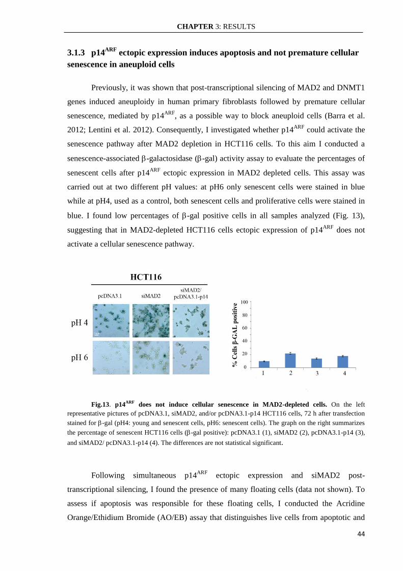
CHAPTER 3: RESULTS
44
3.1.3 p14ARF
ectopic expression induces apoptosis and not premature cellular
senescence in aneuploid cells
Previously, it was shown that post-transcriptional silencing of MAD2 and DNMT1
genes induced aneuploidy in human primary fibroblasts followed by premature cellular
senescence, mediated by p14ARF
, as a possible way to block aneuploid cells (Barra et al.
2012; Lentini et al. 2012). Consequently, I investigated whether p14ARF
could activate the
senescence pathway after MAD2 depletion in HCT116 cells. To this aim I conducted a
senescence-associated -galactosidase (-gal) activity assay to evaluate the percentages of
senescent cells after p14ARF
ectopic expression in MAD2 depleted cells. This assay was
carried out at two different pH values: at pH6 only senescent cells were stained in blue
while at pH4, used as a control, both senescent cells and proliferative cells were stained in
blue. I found low percentages of -gal positive cells in all samples analyzed (Fig. 13),
suggesting that in MAD2-depleted HCT116 cells ectopic expression of p14ARF
does not
activate a cellular senescence pathway.
Fig.13. p14ARF
does not induce cellular senescence in MAD2-depleted cells. On the left
representative pictures of pcDNA3.1, siMAD2, and/or pcDNA3.1-p14 HCT116 cells, 72 h after transfection
stained for -gal (pH4: young and senescent cells, pH6: senescent cells). The graph on the right summarizes
the percentage of senescent HCT116 cells (-gal positive): pcDNA3.1 (1), siMAD2 (2), pcDNA3.1-p14 (3),
and siMAD2/ pcDNA3.1-p14 (4). The differences are not statistical significant.
Following simultaneous p14ARF
ectopic expression and siMAD2 post-
transcriptional silencing, I found the presence of many floating cells (data not shown). To
assess if apoptosis was responsible for these floating cells, I conducted the Acridine
Orange/Ethidium Bromide (AO/EB) assay that distinguishes live cells from apoptotic and
CHAPTER 3: RESULTS
45
necrotic cells (Fig. 14 A). I found a high percentage of apoptotic (36%) and necrotic cells
(17%) following p14ARF
ectopic expression and MAD2 depletion. By applying the
Student’s t-test, these percentages of apoptotic and necrotic cells were statistically
significant in comparison to the percentages found in siMAD2/pcDNA3.1 (22% apoptotic
and 6% necrotic) and siMAD2-scramble/pcDNA3.1 (16% apoptotic and 3% necrotic)
samples. In contrast, a small percentage of apoptotic cells (11% and 17%) detected in
HCT116 MAD2-depleted cells and in siMAD2 scramble/ pcDNA3.1-p14 cells,
respectively, was not statistically significant by the Student’s t-test (Fig. 14 B). The
similarity between the percentage of apoptotic cells and the reduction of aneuploid cells
after p14ARF
ectopic expression suggests apoptosis as a major mechanism to eliminate
aneuploid cells (Fig. 14 B). Since the p14ARF
gene can act in p53-dependent and p53-
independent apoptotic pathways activated following DNA damage (Ozenne et al. 2010), I
investigated whether p14ARF
cooperated with p53 activating apoptosis in response to
aneuploidy. To this aim, I conducted a Western Blot analysis to evaluate the p53 protein
levels. MAD2 post-transcriptional silencing and p14ARF
ectopic expression induced a
significant increase of p53 protein levels compared to control. These findings suggest a
possible collaboration between p14ARF
and p53 to counteract aneuploidy (Fig. 14 C).

CHAPTER 3: RESULTS
46
Fig.14. p14ARF
ectopic expression induces apoptosis and increase of p53 protein levels in
MAD2-depleted HCT116 cells. A: Examples of HCT116 cells stained with Orange Acridine and Ethidium
Bromide 72 h after transfection; the orange arrow (merge panel, bottom left) points to live cells, yellow
(white) and blue (gray) arrows points to necrotic and apoptotic cells (merge panel, bottom right),
respectively. B: The graph summarizes the percentage of live, apoptotic and necrotic HCT116 cells
transfected with pcDNA3.1 (1), pcDNA3.1-p14 (2), siMAD2 (3), siMAD2-scramble (4),
siMAD2/pcDNA3.1 (5), siMAD2-scramble/pcDNA3.1-p14 (6), siMAD2/pcDNA3.1-p14 (7). The
experiment was repeated at least twice. (Student’s t-test * P<0.05; ** P<0.01, n=150). C: Western blot
analysis showing the increase of p53 protein levels after p14ARF
ectopic expression, in HCT116 pcDNA3.1-
p14 (2), HCT116 siMAD2 (3), and in HCT116 siMAD2/pcDNA3.1-p14 (4) cells when compared to HCT116
control cells (1); on the right is showed the densitometric analysis of the WB to quantitate the p53 protein
level that is normalized to -Tubulin.
CHAPTER 3: RESULTS
47
To clarify the role of p53 in the induction of apoptosis after p14ARF
ectopic
expression in aneuploid cells, I used HCT116 p53KO cells transfected with siMAD2 and
pcDNA3.1-p14. The AO/EB assay in HCT116 p53KO cell, following p14ARF
ectopic
expression showed that the percentage of apoptotic cells (20%) found in HCT116
siMAD2/pcDNA3.1-p14 cells was comparable with the percentage found in cells
transfected with siMAD2 and the pcDNA3.1 empty vector (18%). Following MAD2 post-
transcriptional silencing and p14ARF
ectopic expression, I detected a decrease of apoptotic
cells in the absence of p53. This result suggests that cells were eliminated through a p53-
dependent apoptotic pathway where p14ARF
had a key role (Fig. 15 A). In agreement with
this result cytogenetic analyses showed that siMAD2 induced higher number of aneuploid
cells in HCT116 p53KO cells (61%) than siMAD2-scramble cells (21%). On the contrary,
HCT116 p53KO cells that expressed p14ARF
ectopically showed a percentage of aneuploid
cells (66%) similar to the percentage showed by HCT116 p53KO siMAD2/pcDNA3.1
cells (77%) and HCT116-siMAD2 cells (61%). These differences were not statistical
significant by the Student’s t-test. Thus, in the absence of p53, p14ARF
did not induce the
elimination of aneuploid cells caused by MAD2 depletion (Fig. 6B). Real Time qRT-PCR
analyses confirmed that siMAD2#1 induced MAD2 gene post-transcriptional silencing of
about 50% compared to siMAD2-scramble and p53KO HCT116 cells (Fig. 15 C).

CHAPTER 3: RESULTS
48
Fig.15. p14ARF
does not induce apoptosis in HCT116 p53KO cells. A: On the left are showed
representative pictures of the indicated cells stained with Orange Acridine and Ethidium Bromide 72 h after
transfection; the graph on the right summarizes the percentages of live, apoptotic, and necrotic cells in
pcDNA3.1 (1), pcDNA3.1-p14 (2), siMAD2 (3), siMAD2/pcDNA3.1 (4), and siMAD2/pcDNA3.1-p14 (5) -
HCT116 cells. B: The graph shows the percentages of aneuploid cells in p53 KO pcDNA3.1 (1), pcDNA3.1-
p14 (2), siMAD2-scramble (3), siMAD2 (4), siMAD2/pcDNA3.1 (5) and siMAD2/pcDNA3.1-p14 (6)
HCT116 cells. All experiments were repeated at least twice. (Student’s t-test * P<0.05; ** P<0.01, n=150).
C: Real time RT-PCR analysis showing RNA interference of siMAD2#1 and siMAD2 scramble in HCT116
p53KO cells at 72 h after transfection.
CHAPTER 3: RESULTS
49
3.2 CENP-E DEPLETION INDUCES ANEUPLOIDY THAT IS
REDUCED BY THE TUMOR SUPPRESSOR p14ARF
It was shown that mouse model can develop tumors when CIN is induced by
haploinsufficiency of SAC genes but, on the other hand, sometimes the weakening of the
SAC only is not enough and need additional mutations to induce carcinogenesis (Giam &
Rancati 2015). To reconcile these observations we have to consider that only specific
aneuploid karyotypes favor tumorigenesis. Alternatively, the observed differences in
cancer susceptibility could be due to different levels of CIN in the various mouse models
where some tissues accumulate different levels of CIN and are prone to transformation
than other. Another possibility stems from the observation that SAC genes have other non-
mitotic functions, as an example Mad2 can be involved in the DNA replication checkpoint
in yeast (Sugimoto I. et al. 2004), and making it difficult to disentangle which function is
associated to increased cancer susceptibility. Thus, I decided to look at CENP-E, a motor
protein required for stable spindle microtubule capture at kinetochores (Yen et al. 1992;
Schaar et al. 1997; Gudimchuk et al. 2013) which functions only in SAC signaling (Vitre
et al. 2014) .
It was reported that CENP-E homozygous knockout (CENPE-/-
) mice experience
massive chromosome segregation defects and embryonic lethality (Putkey et al. 2002). On
the contrary, heterozygous knockout (CENPE+/-
) mice are viable even though they exhibit
elevated levels of aneuploidy and develop spleen and lung cancer (Weaver et al. 2007; Silk
et al. 2013). However, it unknown if CENP-E heterozygosity could have effects on ploidy
in not tumorigenic (normal) human cells similar to those observed in mice.
Previously, I reported that p14ARF
counteract aneuploidy of HCT116 siMAD2 cells
(Veneziano et al. 2016) and it was also reported that loss of the ARF tumor suppressor
gene in mouse is sufficient to increase the number of near tetraploid cells (Britigan et al.
2014).
I decided then to induce CENP-E partial depletion in human primary fibroblasts
IMR90 and in HCT116 cells lacking of p14ARF
expression and follow aneuploidy
generation up to four weeks. In line, CENP-E depletion is an excellent strategy to induce
aneuploidy and study the role of p14ARF
on the fate of these aneuploid cells.

CHAPTER 3: RESULTS
50
3.2.1 CENP-E post-transcriptional silencing has no effect on cell proliferation
To assess if CENP-E depletion induces aneuploidy I used the RNA interference
strategy targeting transiently CENP-E to generate primary human fibroblasts (IMR90) and
near-diploid cells (HCT116) (Lengauer et al. 1997) with a CENP-E haploinsufficiency
condition (~50% of gene expression).
Initially I transfected the HCT116 cells with two different small interfering RNA
(siRNAs) targeting the CENP-E transcript (siCENP-E #1, siCENP-E #2) and with
unspecific siRNA targeting the green fluorescent protein (siGFP) as a control. Real-Time
RT-PCR analysis showed decreased levels of CENP-E transcripts in transfected HCT116
cells, demonstrating the efficacy of post-transcriptional silencing of both siRNAs. In
comparison to WT cells, CENP-E transcriptional levels were reduced by ~50% and by
~40% after transfection with siCENP-E #1 and siCENP-E #2, respectively. Because the
goal is to obtain a haploinsufficiency condition I decided to conduct all subsequent
experiments in HCT116 cells using the siCENP-E #1 (Fig.16 A). The same experiment
was done in IMR90 cells and Real-Time RT-PCR analysis showed decreased levels of
CENP-E transcripts when cells were transfected with siCENP-E #2 (~50%) and not with
siCENP-E #1 (~97%). Accordingly, I decided to conduct all subsequent analysis in IMR90
cells using siCENPE#2 (Fig.16 C).
Early effects on cell proliferation of IMR90 and HCT116 siCENP-E cells were
estimated by evaluating the cellular density/dish 24 48 and 72 hours post-transfection. In
HCT116 cells I found a slowing down of proliferation at 48h after transfection of siCENP-
E. However, cells resumed normal growth levels at 72h in comparison to siGFP HCT116
cells (Fig.16 B). This result suggests that the partial depletion of the checkpoint protein has
no relevant negative effects on cell proliferation. When IMR90 cells were transfected with
the siRNA targeting CENP-E we did not observe differences on cell proliferation at 24, 48
and 72 hours after transfection: siCENP-E and siGFP IMR90cells show a similar growth
profile (Fig.16 D). Consequently, weakening the mitotic checkpoint does not seem to
affect the proliferation of both IMR90 and HCT116 cells.

CHAPTER 3: RESULTS
51
Fig.16. CENP-E depletion does not affect cell proliferation in IMR90 and HCT116 cells. A:
Real Time RT-PCR analysis showing RNA interference of siCENP-E #2 (4), siCENP-E #1 (3), siGFP (2)
and WT (1) HCT116 cells at 72h after transfection. B: The graph shows HCT116 cell proliferation at 0, 48,
and 72 hours after transfection of siRNA targeting CENP-E transcript (siCENP-E) and GFP transcript
(siGFP) used as a control. C: Real Time RT-PCR analysis showing RNA interference of siCENP-E #2 (4),
siCENP-E #1 (3), siGFP (2) and WT (1) IMR90 cells at 72h after transfection. D: Cell proliferation graph of
IMR90 cells after siCENP-E and siGFP silencing at 0, 48, and 72 hours. The experiment was repeated at
least twice. (Student’s t-test: * P<0.05; ** P<0.01).
3.2.2 CENP-E depletion induces different aneuploidy levels in HCT116 cells
and IMR90 primary fibroblast
In order to assess whether weakening the SAC by CENP-E post transcriptional
silencing induces the same levels of aneuploidy in normal diploid cells (human primary
fibroblasts IMR90) and in a near-diploid cell line that maintains a stable karyotype
(HCT116) (Lengauer et al. 1997), I evaluated the presence of aneuploid cells with classical
cytogenetics.
Analysis of mitotic cells displayed significant levels of aneuploid cells (about 80%)
72h after partial depletion (haploinsufficient condition) of CENP-E in HCT116 cells that
do not express a p14ARF
functional protein (Burri et al. 2001). The majority of these mitotic
CHAPTER 3: RESULTS
52
cells were hypodiploid (78%) and only a small part were hyperdiploid (5%) compared to
control (Fig.17A). Since aneuploidy may be associated with aberrant mitosis, I evaluated
the presence of mitotic spindle and chromosome alterations after CENP-E depletion in
HCT116 cells. I detected many abnormal mitosis and, in particular, monopolar spindle
(20%) and lagging chromosome (37%). These mitotic alterations could be the cause of the
aneuploid cells development (Fig.17 B). This finding is in line with what was previously
observed by others (Silk et al. 2013).
At the same time, I made a post-transcriptional silencing of CENP-E in human
primary fibroblasts IMR90 and analyzed the early effects on aneuploidy development. At
72 hour after transfection I found about 50% of aneuploidy cells and, in particular, 30.5%
of cells were hypodiploid and 15% hyperdiploid compared to siGFP control cells (20% of
hypodiploid and 3% of hyperdiploid) (Fig.17 C). Comparing aneuploidy levels of two
different cell types I observed a dissimilar answer. In cells with a normal genetic
background (IMR90) only half became aneuploid while in cells lacking p14ARF
protein
(HCT116) the levels of aneuploidy were much higher (80%) (Fig. 17 D). These results
suggest that the presence or not of p14ARF
may affect the development of aneuploidy.

CHAPTER 3: RESULTS
53
Fig.17. Post transcriptional silencing of CENP-E induces aneuploidy. A: Representative pictures
of aneuploidy and euploid metaphases found in siCENP-E and siGFP HCT116 cells after 72h of transfection;
the graph shows the percentages of aneuploidy (hypodiploid and hyperdiploid) cells in siGFP and siCENP-E
HCT116 cells. B: Representative images of mitotic alterations (green: β-tubulin blue: DNA stained with
DAPI); below, the graph shows the percentage of normal and altered metaphases (lagging chromosomes and
monopolar spindle). C: Representative pictures of aneuploidy and euploid metaphases found in siCENP-E
and siGFP IMR90 cells after 72h of transfection; below, the graph showing the percentages of aneuploid
cells (hypodiploid and hyperdiploid) in siGFP and siCENP-E IMR90 cells. D: The graph summarizes and
compares the percentage of siGFP (1) and siCENP-E (2) aneuploid cells found both IMR90 that HCT116
cells. All experiments were repeated at least twice. (Student’s t-test * P<0.05; ** P<0.01, n=50 metaphases).
CHAPTER 3: RESULTS
54
3.2.3 Aneuploid IMR90 cells return normal at long time after CENP-E
depletion but not HCT116 cells.
It has been reported that aneuploidy induced in response to a malfunction of the
mitotic checkpoint is a condition that, sometimes, is not well tolerated by the cells. As a
result, the cells selected to go forward in the proliferation are mainly diploid, while the
aneuploid cells are subjected to negative selection (Siegel & Amon 2012; Weaver &
Cleveland 2007; Giam & Rancati 2015). As aneuploidy has negative consequences for the
fitness of normal cells, I wanted to evaluate proliferation up to four weeks of IMR90 and
HCT116 cells CENP-E depleted. To this aim, cells were analyzed at different time (72
hours, two weeks and four weeks) after transfection of CENP-E siRNA by cytogenetics.
I found about 50% of aneuploidy cells in human primary fibroblasts 72 hours after
CENP-E depletion and they are maintained up to two weeks. In detail, two weeks after
siRNA transfection, I found 46% of aneuploidy cells (38% hypodiploid cells and 8% of
hyperdiploid cells) compared to control IMR90 siGFP cells. However, aneuploidy cells
dropped to 22.5% in IMR90 cells after four weeks by post-transcriptional silencing of
CENP-E (20% hypodiploid and 2.5% hyperdiploid) similar to the percentage of found in
control cells (20%). At two weeks, it seems that siCENP-E cells maintain aneuploidy,
especially hypodiploid, while at four weeks it decreased to normal levels (Fig.18 A).
Regarding the proliferation of HCT116 aneuploid cells lacking of p14ARF
protein, I
found about 80% of aneuploidy after 72 hours of CENP-E depletion and about 50% of
aneuploidy after two weeks compared to control. After four weeks from CENP-E depletion
HCT116 cells showed 35% of aneuploidy that is stably maintained compared to siGFP
control. Though aneuploidy tends to decrease with time, sign that it has toxic effects on
cells, a part of HCT116 cells keeps up a good level of aneuploidy for four weeks in
absence of p14ARF
functional protein. These results suggest that cells with a normal genetic
background tolerate aneuploidy only for a short period of time and then return to normal
levels, while tumor cells lacking p14ARF
expression can tolerate aneuploidy longer (Fig.18
B).

CHAPTER 3: RESULTS
55
Fig.18. Aneuploidy decreases in IMR90 cells but not in cells lacking of p14ARF
(HCT116). A:
The graph shows the percentage of aneuploid and euploid siGFP (1) and siCENP-E (2) IMR90 cells at 72
hours, 2 weeks and 4 weeks after siRNA transfection. B: Percentage of aneuploid and euploid siGFP (1) and
siCENP-E (2) HCT116 cells (lacking of p14ARF
protein) displayed by the graph at 72 hours, 2 weeks and 4
weeks after siRNA transfection. All experiments were repeated at least twice. (Student’s t-test * P<0.05; **
P<0.01, n=50 metaphases).
3.2.4 p14ARF
counteract aneuploidy development.
The different response to aneuploidy could be due to different p14ARF
protein levels
between the two cell types used (HCT116 and IMR90 cells). To explore this hypothesis, I
wanted to visualize the p14ARF
expression in IMR90 cells partially depleted for CENP-E at
72 hours, 2 weeks and 4 weeks. The level of p14ARF
mRNA in IMR90 cells at 72 hours
after the posttranscriptional silencing of CENP-E was similar to p14ARF
expression in the
control (siGFP). However, p14ARF
expression was increased at two weeks and especially at
four weeks from the CENP-E silencing. It should be noted that the increase of the p14ARF
expression matches with the decrease of the aneuploid cells in IMR90 cells suggesting an
involvement of the tumor suppressor in the control of cellular ploidy (Fig. 19 A).
In line with this hypothesis, I estimated aneuploidy in HCT116 cells CENP-E
depleted that were transiently transfected with the plasmid pcDNA3.1 coding for the
p14ARF
c-DNA (Ayrault et al. 2006). This plasmid allows me to obtain transient expression
of ectopic p14ARF
, as I have done previously in HCT116 cells partially depleted for MAD2
transcript.
In pcDNA3.1-p14 HCT116 cells the aneuploidy level changed slightly in
comparison to control (empty vector pcDNA3.1). As expected the number of aneuploid

CHAPTER 3: RESULTS
56
cells dropped from 80% to 56% (46% hypodiploid and 10% hyperdiploid) after transient
expression of p14ARF
in CENP-E depleted HCT116 cells. On the contrary, CENP-E
depleted HCT116 cells and then transfected with the pcDNA3.1-empty vector still showed
a high percentage of aneuploid cells (73%) similar to that shown by cells transfected with
siCENP-E alone (Fig.19 B). This result is similar to the result obtained in HCT116
siMAD2 cells but it allows me to reinforce the idea that p14ARF
re-expression counteracts
proliferation of aneuploid cells caused by different stimuli. Consequently, I can generalize
the role of p14ARF
gene to maintain stable the ploidy of the cells.
Fig. 19. Involvement of p14ARF
in aneuploidy control. A: On the left, Real Time RT-PCR analysis
showing p14ARF
mRNA expression levels in IMR90 siCENP-E cells at 72 hours, 2 weeks and 4 weeks after
transfection compared to control. On the right, graph showing aneuploidy levels in siCENP-E IMR90 cells
(2) from 72 hours up to 4 weeks compared to control siGFP IMR90 cells (1) (described in 4.2.3 paragraph).
B: The graph displays the percentage of aneuploidy in pcDNA3.1 (1), pcDNA3.1-p14 (2), siCENP-E (3),
siCENP-E/pcDNA3.1 (4), siCENP-E/pcDNA3.1-p14 (5) HCT116 cells.
CHAPTER 3: RESULTS
57
3.3 ANEUPLOIDY IS NOT TOLERATE IN HCT116 CELLS
EXPRESSING p14ARF
Previous results suggest that cells lacking of the tumor suppressor p14ARF
respond
differently to the induction of aneuploidy. It is conceivable that the genetic background
plays a key role in the control of aneuploidy generation. For example, alteration of CENP-
E gene induces aneuploidy in MEFs and the simultaneous absence of ARF significantly
increases its levels. In addition it was shown that MEFs ARF-/-
become aneuploid
suggesting a role of the p14ARF
tumor suppressor in the control of aneuploidy (Silk et al.
2013). In line with these results, I observed that in human cells depletion of SAC genes
(MAD2 and CENP-E) induced aneuploidy that is increased in cells lacking of p14ARF
expression (HCT116) compared to normal cells (IMR90) suggesting the idea that p14ARF
is
involved to limit aneuploidy development.
In order to evaluate if different p14ARF
expression levels may modulate the effect
induced by CENP-E depletion in the cells, I engineered a tetracycline-regulated retroviral
vector (pBPSTR1) for the inducible expression of p14ARF
that has been transfected in
nearly diploid HCT116 tumor cells that does not have a functional p14ARF
(Burri et al.
2001).
3.3.1 Cloning of p14ARF
c-DNA into pBPSTR1 vector
In order to obtain the inducible expression of p14ARF
in the HCT116 cells, the
coding sequence of p14ARF
was cloned in the pBPSTR1 retroviral vector (Fig. 20 A). The
pBPSTR1 vector is a modified version of the pBABEpuro vector in which were cloned the
two components of the inducible system “Tet-off”: tTA and TetO (Fig.20 C) (Pear et al.
1993). The p14ARF
c-DNA was isolated from pcDNA3.1-p14 vector (kindly provided by
Prof. S. Gazzeri, University J. Fourier, La Tronche, France) where it was cloned between
the EcoRI e NotI restriction sites. To assure the correct extraction of the p14ARF
insert the
restriction enzymes BamHI e XbaI cutting upstream and downstream of the insertion site
were used (Fig. 20 B). Gel electrophoresis confirmed the extraction of the full length c-
DNA as revealed by the ≈ 500bp band (Fig. 20 B). The c-DNA was then purified from the
agarose gel for the cloning in the pBPSTR1 vector. Cuts made by BamHI e XbaI are not
compatible with the restriction sites present in the polylinker of pBPSTR1 and it was not

CHAPTER 3: RESULTS
58
possible to clone the p14ARF
insert directly in the pBPSTR1 vector. To override this
problem the p14ARF
insert was cloned blunt in the vector and the correct orientation was
then checked. To make a blunt-blunt ligation of the p14ARF
c-DNA and the pBPSTR1
plasmid, the 5'-protruding cohesive ends of the p14ARF
c-DNA, following BamHI/XbaI
digestion, were treated with the Klenow fragment of DNA polymerase I, that lacks of the
5'-3' exonuclease activity, in a process called fill-in. The pBPSTR1 plasmid was digested
with PmeI to generate the blunt ends, and gel electrophoresis confirmed the linearized
pBPSTR1 vector (7022bp) by presence of the band of ≈7000bp (Fig. 20 C). The pBPSTR1
vector was then, dephosphorylated with alkaline phosphatase to prevent its circularization
in the next step of ligation.
The ligation mix (ratio of 1:5 plasmid/insert) was used to transform E. coli
competent cells. To verify the presence of the pBPSTR1-p14ARF construct the positive
colonies were analyzed by the PCR colony assay with primers matching outside of the
pBPSTR1 polylinker (Fig. 20 D). Two bacterial colonies containing the pBPSTR1-
p14ARF construct (Fig 20 D) were detected.

CHAPTER 3: RESULTS
59
Fig.20. Engineering of the retroviral vector pBPSTR1 with the p14ARF
c-DNA. A: Experiment
design scheme. B: Map of the vector pcDNA3.1 containing the p14ARF
cDNA. Boxed is the region where the
cDNA was inserted; on the right, gel electrophoresis showing: not-digested pcDNA3.1-p14 (lane1) and
BamHI/XbaI double-digested pcDNA3.1-p14 (lane2) identified with the marker λHindIII (lane M1) and
marker 100bp ladder (lane M2). C: Map of the retroviral vector pBPSTR1. Boxed are the polylinker region
and the site where the p14ARF
cDNA was inserted. On the right, gel electrophoresis showing: not-digested
pBPSTR1 (lane1), PmeI digested pBPSTR1 (lane2) and marker λHindIII (lane M). D: Map of pBPSTR1
vector with the primers mapping outside of the polylinker used for the PCR colony (highlighted in green). On
the right gel electrophoresis of the amplicons of the PCR Colony assay showing: (lanes 1-3) positive colonies
“1” and “3” as detected by the band at about 700bp; (lane 2) negative bacterial colony “2” detected by a band
of 200bp; (lane 4) empty vector; (lane 5) no template; marker 100bp ladder (lane M).

CHAPTER 3: RESULTS
60
Since the insert was cloned blunt to obtain the p14ARF
inducible expression, it is
important to verify the appropriate 5'-3 'orientation of the insert cloned into the plasmid
that must be with the 5’end downstream of the CMV promoter and the operator TetO (Fig.
21). To this aim, I used three different methods to verify the correct orientation of the
insert following blunt-blunt ligation. 1) PCR colony was set up with one primer designed
on an insert sequence and the other one designed on the plasmid sequence adjacent to the
polylinker site. The pairs of primers were chosen to obtain an amplification product only if
the insert was cloned in the correct orientation (Fig.21 A). The gel electrophoresis showed
that the bacterial colonies 1 and 3 contained the pBPSTR1 vector with the correct
orientation of the p14ARF
insert (Fig.21 A). 2) The plasmid DNA extracted by the colonies
1 and 3 was digested with EcoRI that has a single cutting site in the vector pBPSTR1 and a
second site in the sequence of the insert, in order to discriminate the correct orientation
according to the length of the fragments obtained. (Fig.21 B). 3) The plasmid DNA of the
colony 1 was sequenced (BMR Genomics, Padua, Italy). The primers used for the reaction
mapped upstream and downstream of the cloning site and were previously used in the PCR
colony reactions. The DNA sequencing confirmed the presence of the insert in the correct
orientation (Fig. 21 C).

CHAPTER 3: RESULTS
61
Fig.21. Colony screening to verify the correct orientation of the insert. A. Top: map of
pBPSTR1 where the p14ARF
c-DNA was cloned (red arrow indicates the 5’-3’ insert orientation) and position
of the primers used for the Colony PCR assay. Bottom: agarose gel electrophoresis of colonies 1 and 3 PCR
products of pBPSTR1-p14ARF (M=marker 100bp ladder) using primers 1 and 3 (green & yellow arrows)
(lanes 1-2), primers 4 and 2 (lanes 3-4), primers 1 and 4 (lanes 5-6) and with no template (lane 7). B. Top:
map of pBPSTR1-p14ARF where the red arrow indicates the 5’-3’ insert orientation and the black rectangles
indicate the EcoRI sites. Bottom: agarose gel electrophoresis of colony 1(lanes 1-2-3) and colony 3 (lanes 4-
5-6) after EcoRI digestion; pBPSTR1-p14ARF vector uncut (lane 1-4), pBPSTR1-p14ARF linearized with
BamH1 (lane 2-5), pBPSTR1-p14ARF cut with EcoRI (lane 3-6) and marker λHindIII (lane M). C:
Sequencing (both directions) with vector specific primers confirmed the presence of the cloned p14ARF
cDNA
in the pBPSTR1 expression vector.

CHAPTER 3: RESULTS
62
3.3.2 Generation of pBPSTR1-p14ARF HCT116 cells
The retroviruses containing the pBPSTR1-p14ARF construct were produced in the
Phoenix Amphotropic packaging cell line (Pear et al. 1993). The Phoenix cells were
previously cultured for a week in medium containing Hygromycin-B (300μg/ml), because
gag and pol viral genes are inserted in the cells along with the gene for Hygromycin-B
resistance, and then they were transfected with pBPSTR1-p14ARF vector. After 72 hours
of transfection, the medium was collected, filtered, and Polybrene (a cationic polymer) was
added to increase the efficiency of infection in HCT116 cells. Then HCT116 cell was
selected using Puromycin (2µg/mL) for at least ten days. Preliminary, to assess the Tet-Off
functionality it was done an experiment by transfecting the pBPSTR1-H2BGFP vector
expressing the H2BGFP protein (the H2B histone fused with the Green Fluorescent
Protein) in HCT116 cells. The analysis of these transfected cells by fluorescence
microscopy indicated that the treatment with Doxycycline (2µg/mL) for 96 hours was
sufficient to reduce the expression of the H2BGFP fusion protein (Fig. 22 A).
To evaluate both the presence of the ectopic p14ARF
and the amount of its transcript
level, it was done a PCR reaction with specific primers mapping in the pBPSTR1-p14ARF
sequence (showed in Fig.21A) in stably transfected HCT116 cells. Agarose gel
electrophoresis confirmed the presence of the p14ARF
insert in pBPSTR1-p14 HCT116
cells after its amplification from genomic DNA (Fig.22 B). The Real Time RT-PCR assay
showed the presence of highest levels of p14ARF
in pBPSTR1-p14ARF HCT116 cells
compared to wild type HCT116 and to HCT116 cells harboring the pBPSTR1-H2BGFP
plasmid (Fig. 22 C).
To exclude any side effects of the presence of ectopic p14ARF
it was necessary to
evaluate that its re-expression did not alter normal cellular proliferation in HCT116 cells.
The effects on cell proliferations of the re-expression of p14ARF
were estimated by
evaluating the cellular density/dish for 96 hours in pBPSTR1-p14ARF HCT116 cells. Not
significant differences between the proliferation rate of HCT116 cells expressing p14ARF
and H2BGFP or WT control cells were observed (Fig. 22 D). This result indicates that the
re-expression of p14ARF
gene in HCT116 cells does not affect cell proliferation.
In addition the pBPSTR1-p14ARF HCT116 cells showed level of aneuploid cells
similar to that found in pBPSTR1-H2BGFP or WT-HCT116 cells suggesting that re-
expression of p14ARF
has not effects on cellular ploidy (Fig. 22 E).

CHAPTER 3: RESULTS
63
Fig.22. Engineering of HCT116 cells with the retroviral construct pBPSTR1-p14ARF
and initial
characterization. A: Evaluation of the functionality of the Tet-Off System by treating the stably infected
H2BGFP-HCT116 cells with Doxycycline for 96 hours showed a remarkable decline in the number of
H2BGFP positive cells. B: Correct integration of the retroviral vector in genomic DNA as revealed by PCR
using specific primers mapping in the vector. C: Real time RT-PCR showing overexpression of p14ARF
in
stably infected HCT116 cells compared to controls. (Student’s t-test * P<0.05; ** P<0.01, n=4) D:
Proliferation assay showed normal cell proliferation in HCT116 cells that re-express p14ARF
tumor
suppressor gene compared to H2BGFP and WT HCT116 control cells. E: Cytogenetic analysis showed no
ploidy changes between pBPSTR1-p14ARF and control cells.
CHAPTER 3: RESULTS
64
3.3.3 p14ARF
counteract aneuploidy induced by CENP-E posttranscriptional
silencing
Once confirmed that p14ARF
re-expression does not affect the normal proliferation
and the ploidy of HCT116 cells, I used these engineered cells to corroborate the hypothesis
that p14ARF
gene has an important role in limiting the proliferation of aneuploidy cells. To
this aim, CENP-E was posttranscriptional silenced to induce aneuploidy in pBPSTR1-
p14ARF HCT116 cells. The percentage of induced aneuploid cells was then estimated by
modulating p14ARF
expression levels by using Doxycycline, analogue of Tetracycline,
which prevents the binding of the trans-activator (tTA) to the promoter.
First of all, I checked the siRNA targeting CENP-E transcript (siCENP-E),
previously used for the WT HCT116 cells, was able to induce CENP-E haploinsufficiency
in pBPSTR1-p14ARF HCT116 cells. The qRT-PCR confirmed that the RNA interference
strategy reduced of about 40% the transcription levels of CENP-E (Fig. 23 A).
As said (4.1.1 paragraph), the p14ARF
ectopic expression reduced the proliferation
of MAD2 silenced HCT116 cells so I wanted to assess whether the presence of the tumor
suppressor induced a reduced proliferation even in pBPSTR1-p14ARF HCT116 cells
CENP-E depleted. To this aim, I estimated the cellular proliferation by evaluating the
cellular density/dish 24, 48 and 72 hours after CENP-E transfection. Likewise, I found a
significant reduction of cell proliferations at 48 and 72 hours after CENP-E silencing in
HCT116 cells engineered for p14ARF
expression compared to control (Fig.23 B).
Previously, I observed that p14ARF
could reduce aneuploidy levels of 25-30% both
in siMAD2 and in siCENP-E HCT116 cells (paragraph: 4.1.2 and 4.2.4). To explore if the
stable p14ARF
expression had the same effects in pBPSTR1-p14ARF HCT116 cells, I
conducted a cytogenetic analysis to evaluate the ploidy after CENP-E depletion. The re-
expression of p14ARF
greatly reduced the percentage of aneuploid cells (Fig. 23 C).
Since the pBPSTR1-p14ARF inducible vector allows modulation of p14ARF
expression I evaluated the number of aneuploid cells induced by CENP-E depletion after
treating the pBPSTR1-p14ARF HCT116 cells with doxycycline that decreases p14ARF
level.
As shown in the Figure 23D, the stable expression of p14ARF
in HCT116 cells has
reduced the number of aneuploid cells induced by CENP-E depletion (≈22%) in
comparison to that showed by the siCENP-E HCT116 cells (≈80%) and similarly with the

CHAPTER 3: RESULTS
65
percentage of siGFP HCT116 cells (≈10%). On the contrary, the pBPSTR1-p14ARF cells
silenced for CENP-E showed more aneuploid cells (≈45%) following treatment with
doxycycline, which no had effect on pbpSTR1-p14ARF HCT116 cells transfected with the
siGFP control (≈10% of aneuploid cells) (Fig.23D).
These results suggest that cells with reduced expression of p14ARF
cannot properly
counteract aneuploidy confirming and extending the crucial role played by p14ARF
in the
maintenance of genomic stability.
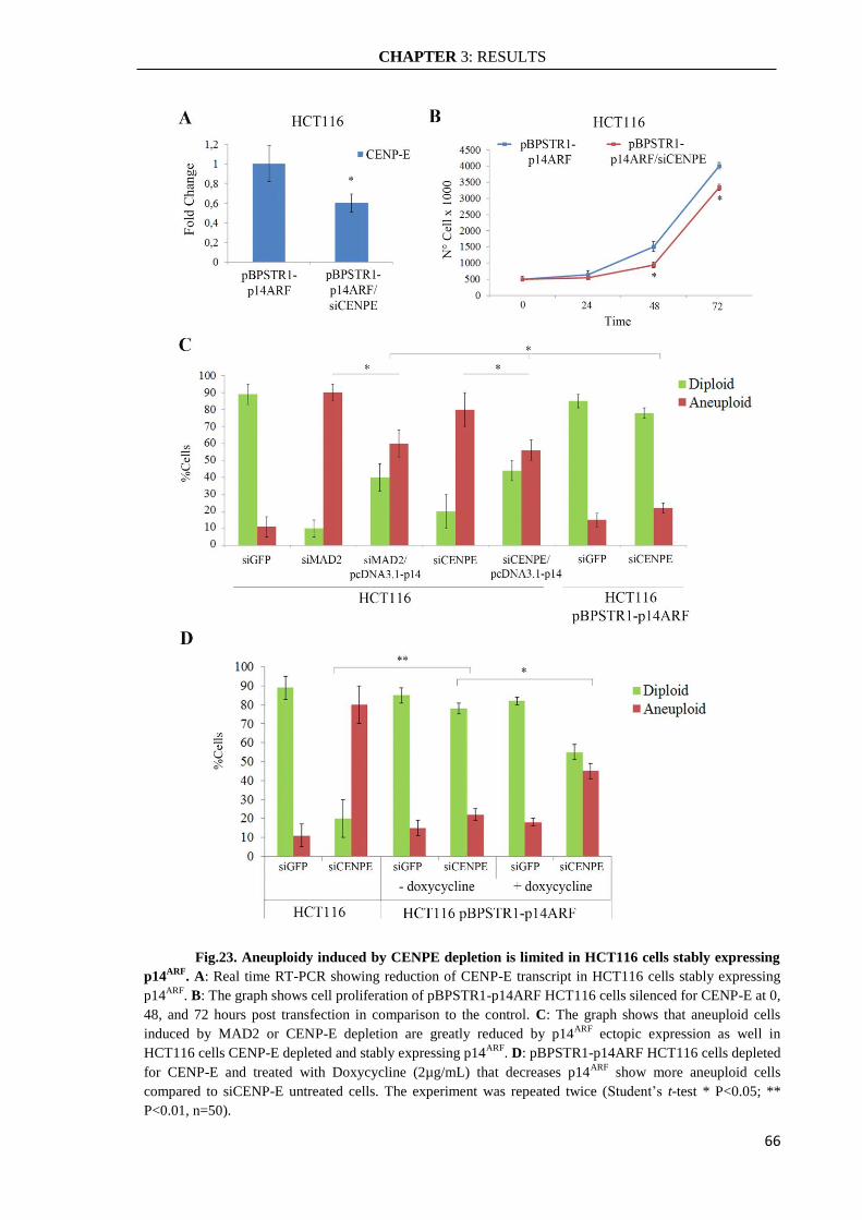
CHAPTER 3: RESULTS
66
Fig.23. Aneuploidy induced by CENPE depletion is limited in HCT116 cells stably expressing
p14ARF
. A: Real time RT-PCR showing reduction of CENP-E transcript in HCT116 cells stably expressing
p14ARF
. B: The graph shows cell proliferation of pBPSTR1-p14ARF HCT116 cells silenced for CENP-E at 0,
48, and 72 hours post transfection in comparison to the control. C: The graph shows that aneuploid cells
induced by MAD2 or CENP-E depletion are greatly reduced by p14ARF
ectopic expression as well in
HCT116 cells CENP-E depleted and stably expressing p14ARF
. D: pBPSTR1-p14ARF HCT116 cells depleted
for CENP-E and treated with Doxycycline (2µg/mL) that decreases p14ARF
show more aneuploid cells
compared to siCENP-E untreated cells. The experiment was repeated twice (Student’s t-test * P<0.05; **
P<0.01, n=50).
CHAPTER 4: DISCUSSION
67
CHAPTER 4: DISCUSSION
Cells with altered chromosome numbers are frequently found in cancerous tissues,
miscarriage embryos as well as in disabled born children such as trisomy 21 patients
(Holland & Cleveland 2012). Many studies on aneuploid tissues and embryos documented
that changes in chromosome number lead to growth retardation and in most cases to
embryonic lethality (Siegel & Amon 2012). In contrast, many cancerous tissues display
high levels of cells with altered chromosome number suggesting that these cells could
benefit from altered protein expression levels due to changes in chromosome number and
outcompete cells with normal diploid chromosome content (Pellman 2007). Genetic
analyses have hypothesized that for malignant transformation are needed at least six gene
mutations (Vogelstein 1996) but it is rare for a single cell to acquire all necessary
mutations considering the mutation rate in humans. Thus, it was born the idea that
destabilizing the genome is necessary for cancer development and the chromosomal
instability (CIN), which results in gains or losses of whole chromosomes or translocation
of chromosome segments, can be a strategy to accumulate the mutant alleles essential for
malignant proliferation (Gordon et al. 2012). Although the phenotypes of cells and tissues
with altered chromosome numbers were often described, underlying mechanisms that lead
to the aneuploidy phenotype remain unknown. Consequently, it is difficult identify key
pathways that could prevent aneuploidy development.
Cancer cells can have gross abnormalities in their chromosome numbers, most as a
result of defects in the quality control of sister chromatid separation (Weaver et al. 2007).
During mitosis the proper chromosomes segregation is controlled by a specific checkpoint,
the Spindle Assembly Checkpoint (SAC). If any of SAC components is mutated or its
expression is reduced, miss-segregation events would not be correctly reported, promoting
aneuploidy development (Silva et al. 2011). In most cases, aneuploidy cells are often
eliminated by protecting mechanisms. Enhanced proteasomal degradation has been
suggested as one aneuploidy tolerating mechanisms (Torres et al. 2010). Also, the p38-p53
axis has been suggested as a pathway limiting the proliferation of aneuploid cells
(Thompson & Compton 2010). It was also suggested that heightened energy metabolism in
aneuploidy cells may be behind the surge in ROS levels. Increased ROS cause DNA
damage that activates the ATM/P53 pathway, and this depends on the severity of
aneuploidy (Li et al. 2010). Aneuploidy could potentially increase the risk of neoplastic

CHAPTER 4: DISCUSSION
68
transformation but it seems that this process occur when it is associated with mutations in
tumor suppressor genes.
In this thesis, I propose an alternative protective mechanism carried out by the
tumor suppressor p14ARF
against aneuploidy development. The p14ARF
protein plays
several biological functions in the cell with the purpose to suppress aberrant cell growth.
The well-defined function of p14ARF
is to stabilize and activate p53 by neutralizing the
inhibitory effects of the E3 ubiquitin ligase hMdm2 in response to oncogenic stress
(hyperproliferative signals) (Sharpless 2005). Though the p14ARF
protein is a key player in
this p53 pathway, there are evidences that it could promote a p53-independent cell cycle
arrest and/or apoptosis (Weber et al. 2000; Müer et al. 2012). p14ARF
expression is
generally lost by deletion or hypermethylation of CpG islands localized at its promoter
region (Badal et al. 2008). Hypermethylation of p14ARF
promoter has been reported in
many tumors, and this loss occurs at early stages of tumorigenesis in some tumors such as
colorectal, gastric, prostate, and breast cancer (Ozenne et al. 2010). In many human
cancers, deregulation of the p53 pathway usually occurs by inactivation of the TP53 gene
through point mutations. Moreover, inactivation of the p14ARF
gene has been proposed as a
mechanism that disrupts p53 activity in tumors with wild-type TP53 gene (Nyiraneza et al.
2012).
In agreement with other data (Amato et al. 2009), I showed that weakening the
SAC by MAD2 post-transcriptional silencing increased aneuploidy in HCT116 cells
(lacking of p14ARF
). However, when the p14ARF
functionality was restored by ectopic
expression of p14ARF
, aneuploidy and the proliferation rate was reduced in these aneuploid
cells (4.1.1 and 4.1.2 paragraphs). At the same time, mitotic abnormalities observed after
MAD2 post-transcriptional silencing decreased as a result of p14ARF
ectopic expression.
These preliminary findings suggest that p14ARF
is able to prevent proliferation of aneuploid
cells caused by reduction of SAC activity (paragraph: 4.1.2).
In line with other results where human primary fibroblast activated a premature
cellular senescence response p53-mediated after induction of aneuploidy by MAD2 post-
transcriptional silencing to halt aneuploidy cells proliferation (Lentini et al. 2012), I
wanted to investigate if p14ARF
triggered this pathway even in MAD2 depleted HCT116
cells. By contrast, I found that premature cellular senescence was not activated in response
to aneuploidy in HCT116 tumor cells where p14ARF
was ectopically expressed. Instead, the
presence of apoptotic cells in MAD2-depleted HCT116 tumor cells, in response to p14ARF
CHAPTER 4: DISCUSSION
69
ectopic expression, suggested induction of apoptosis as the mechanism adopted by p14ARF
to counteract aneuploid cells proliferation. At this point, I wanted to investigate on possible
partners of p14ARF
that collaborate to activate apoptosis in response to aneuploidy.
Apoptosis was associated with the increase of p53 protein, suggesting that the transient re-
expression of p14ARF
in these aneuploid cells induces a p53-dependent apoptosis. The
existence of a p14ARF
-p53 axis is confirmed by experiments done in HCT116 p53KO cells,
where cells interfered for MAD2 become aneuploid but did not show increased number of
aoptotic cells when p14ARF
was re-expressed (paragraph: 4.1.3).
Previously, it has been shown that aneuploidy caused by MAD2 haploinsufficiency
increased both the frequency and the number of tumors in a p53-/-
background (Holland &
Cleveland 2009). Consistent with this hypothesis, it was reported that a p38 kinase-
dependent stress response activates p53 to induce the p21waf1
in response to chromosome
missegregation in HCT116 cells (Thompson & Compton 2010). Likely, the cellular signal
triggering this pathway relies on the presence of DNA damage that could be caused by
chromosomal mechanical stress (Janssen et al. 2011). On the contrary, the results of this
thesis suggest that, after p14ARF
re-expression, apoptosis activation p53-dependent hampers
aneuploid cell proliferation.
It has been documented that MAD2 as well as others SAC genes have no-mitotic
functions making it difficult to identify which specific alteration could activate the p14ARF
response (Giam & Rancati 2015). For example, it was seen that MAD2 may be involved in
the DNA replication checkpoint in yeast (Sugimoto et al. 2004). Thus, it becomes
important to verify that the p14ARF
response is indeed linked only to the aneuploid
stimulus. I choose then to induce aneuploidy using the post-transcriptional silencing of
CENP-E gene which is required only in mitosis for stable spindle microtubule capture at
kinetochores (Yen et al. 1992; Schaar et al. 1997; Gudimchuk et al. 2013) and evaluate the
effects both in near diploid cells lacking of p14ARF
HCT116 cells that in human primary
fibroblasts. Until now, it is known that the reduction of CENP-E induces aneuploidy in
MEFs that potentially could promote tumorigenesis in mouse (Weaver et al. 2007; Silk et
al. 2013). Accordingly, I found that CENP-E depletion caused aneuploidy both in human
primary fibroblasts IMR90 and in HCT116 cells. However, the number of aneuploid cells
was higher in HCT116 cells than in IMR90 cells and could be explained by the lack of
p14ARF
functionality in the HCT116 cells (paragraph: 4.2.2). These results are consistent
with MEFs CENPE+/-
/ARF-/-
showing high rate of aneuploidy unlike MEFs CENPE+/-
/ARF+/+
(Weaver et al. 2007; Silk et al. 2013).

CHAPTER 4: DISCUSSION
70
CENP-E depletion has not relevant influence on cell proliferation of both kind of
cells (paragraph4.2.1) but induced mitotic abnormalities like monopolar spindle and
lagging chromosomes that could explain the aneuploidy generation as seen in mouse (Silk
et al. 2013). It was reported that aneuploidy cells are promptly outcompeted by normal
cells in culture (Thompson & Compton 2010) and do not exist data about the fate of
induced aneuploid cells at longer time from induction than 72 hours. Also in this case, the
two different cell types have responded differently. At four weeks after CENP-E depletion
aneuploidy returned at normal levels in human primary fibroblasts IMR90 cells while
HCT116 cells showed still aneuploid cells although the percentage of aneuploidy
decreased when compared to that at 72 hours. The finding that aneuploid cells were found
up to two weeks in IMR90 cells suggests that a threshold level of p14ARF
protein must be
reached to activate a pathway that block aneuploidy cells proliferation (Groth et al. 2000).
Also, it is interesting to notice that IMR90 cells were still aneuploid at two weeks after
CENP-E depletion but with a different profile showing a marked decrease of hyperdiploid
cells (paragraph: 4.2.3). Probably, the tumor suppressor p14ARF
is able to counteract
hyperdiploidy in agreement with other data showing that loss of ARF is sufficient to
increase the number of near tetraploid cells in the mouse (Britigan et al. 2014).
The increased p14ARF
expression levels at 72 hours, 2weeks and 4weeks after
CENP-E silencing and the simultaneous aneuploidy decrease in IMR90 cells suggested the
involvement of the tumor suppressor to limited aneuploidy proliferation. Similarly, the
p14ARF
ectopic expression in CENP-E depleted HCT116 cells induced a decrease of the
percentage of aneuploid cells (paragraph: 4.2.4).
Interestingly, aneuploidy induced by CENP-E depletion was not tolerated in
pBPSTR1-p14ARF HCT116 cells. This finding strongly suggests that the re-expression of
p14ARF
blocked aneuploidy development. The level of aneuploidy in these cells resulted
lower than cell expressing transiently p14ARF
both in siMAD2 and in siCENP-E HCT116
cells (paragraph: 4.3.3). Moreover, the post-transcriptional silencing of CENP-E in
HCT116 cells stably expressing p14ARF
negatively influenced the cellular proliferation
confirming that the tumor suppressor limited aneuploidy. The reduction of p14ARF
trough
Doxycycline treatment increased aneuploid cell numbers suggesting that aneuploidy
development is influenced by p14ARF
expression levels (paragraph: 4.3.3).
Generally, gene expression correlates with gene copy number. Aneuploidy
unbalancing genomic material could then induce a signal resembling hyperproliferative

CHAPTER 4: DISCUSSION
71
stress typically sensed by p14ARF
. Likely, p14ARF
could signal the presence of gene
expression imbalance resulting from chromosome missegregation and activate apoptosis
p53-dependent, as previously described (Williams et al. 2008).
Taken together, these results reinforce the idea that the abolition of p14ARF
expression or p14ARF
related partners that control genomic stability is one of the strategies
adopted by human tumor cells to tolerate aneuploidy.
CHAPTER 5: MATERIALS AND METHODS
72
CHAPTER 5: MATERIALS AND METHODS
5.1 Cells and cell culture
Human primary fibroblasts (IMR90, ATCC) were cultured in EMEM supplemented
with: 10% FBS (GIBCO, Invitrogen, Monza Italy), 100units/ml penicillin and 0, 1 mg/ml
streptomycin, 1% NEAA; Colon cancer cells HCT116 with MIN phenotype (near-diploid
cells) and p53-/-
HCT116 cells (kindly provided by Dr. B. Vogelstein, John Hopkins
University, Baltimore, MD) were cultured in D-MEM with 10% FBS (GIBCO, Invitrogen,
Monza, Italy), 100 U/ml penicillin and 0.1 mg/ml streptomycin. Cells were cultured in a
humidified atmosphere of 4% CO2 in air at 37° C.
5.2 Cells Transfection
For siRNAs transfection 1,5x10⁵ IMR90 cells and 2,5x10
5 HCT116 cells were
plated in 6-well dishes and incubated at 37°C. Specific siRNAs duplex were mixed with
Lipofectamine2000 Reagent (Invitrogen), according to manufacturer’s recommendation
and added to the cells. After 6 hours at 37°C, the transfection medium was replaced with
fresh medium. Twenty-four hours after plating, HCT116 cells were transfected with
siRNAs targeting MAD2 siRNA n°1 (5’-AUACGGACUCACCUUUTT-3’), MAD2
siRNA n°2 (5’ –AAGUGGUGAGGUCCUGGAATT– 3’) or with control MAD2 scramble
siRNA (5’- CAGUCGCGUUUGCGACUGG- 3’), siRNA GFP
(5’GGCUACGUCCAGGAGCGCACC-3’), CENP-E siRNA n°1 (5’-
AAGCAGAGAGAAGGGUGAACC-3’) at a final concentration of 60 nM. After
additionally five hours these cells were transfected with the pcDNA3.1 empty plasmid or
harbouring the p14ARF
c-DNA (kindly provided by S. Gazzeri, University J. Fourier, La
Tronche, France). IMR90 cells were transfected with control siRNA GFP and CENP-E
siRNA n°2 (5’–AACGAAGAGUUACUUGGUGCC–3’) at a final concentration of 40nM.
The day of transfection the siRNA or the plasmid DNA and the transfection reagent
(Lipofectamine 2000, Invitrogen, Monza, Italy) were diluted separately in Opti-MEM
(Invitrogen, Monza, Italy) mixed gently and then incubated for 5 min at room temperature.
After incubation the siRNA and the plasmid DNA were mixed gently with Lipofectamine
2000 (Invitrogen, Monza, Italy), allowed to sit 20 min at room temperature to allow
complex formation, and added to the plates with 2ml of Medium for 72 h.
CHAPTER 5: MATERIALS AND METHODS
73
5.3 Stable expression of p14ARF
in HCT116 cells
To generate HCT116 cells expressing p14ARF
protein, the c-DNA of p14ARF
from
pcDNA3.1 was cloned in inducible retroviral vector pBPSTR1. Then using Phoenix
packaging cells, the recombinant retroviral was used to infect HCT116 cells. Using the
TET-off system, the expression of p14ARF
was modulated by doxycycline at the
concentration of 2µg/mL.
5.3.1 Purification of p14ARF
c-DNA from pcDNA3.1 and “Fill in”
protocol
An aliquot of pcDNA3.-p14 plasmid was subjected to double digestion using the
restriction enzymes BamHI and XbaI to isolate the c-DNA. The reaction mixture (Vf
=100µL) contained:
20μg pcDNA3.1-p14
5μl BamHI (10u/μl)
5μl XbaI (10u/μl)
10μl Buffer Tango 10x
60μl H2O DNAsi/RNAsi free
The mixture was incubated at 37°C for 2 hours.
Electrophoresis on 1% agarose gel, with ethidium bromide (0,5μg / ml) in TAE
buffer (40mM Tris-acetate, 1mM EDTA), was used to purify the correspondent band of
p14ARF
c-DNA (≈ 500bp) with PureLink Quick Gel Extraction Kit extraction PCR
Purification COMBO kit (Invitrogen). Then the p14ARF
cDNA was subjected to “Fill in”
reaction:
20μg DNA
3μl Buffer NEB2
3μl dNTP 1mM (Cf = 0,1mM)
1,5μl BSA (1μg/μl)
0,5μl frammento di Klenow (Roche)
The mixture was incubated at 37°C for 30 minutes.
CHAPTER 5: MATERIALS AND METHODS
74
5.3.2 Digestion, de-phosphorylation and purification of pBPSTR1
An aliquot of pBPSTR1 retroviral vector was subjected to digestion using the
restriction enzymes PmeI to obtain blunt end for 2 hours at 37°C. The reaction mixture was
performed with:
3,5μg DNA
1μl PmeI
2μl Buffer B
13,5μl H2O
Then the vector was precipitate with NaAc 0,3M ed EtOH, suspended in 20 µl of
TE buffer and dephosphorylated with alkaline phosphatases of calf intestine (CIAP). The
mixture was incubated at 50°C for 10 minutes and then at 68°C for 10 minutes. The vector
was purified with phenol-chloroform protocol and precipitated with NaAC 0.3M and
absolute EtOH.
5.3.3 Quantification of p14ARF
c-DNA and pBPSTR1 vector
pBPSTR1 plasmid and p14ARF
c-DNA was subjected to electrophoresis on 1%
agarose gel, with ethidium bromide (0,5μg / ml) in TAE buffer (40mM Tris-acetate, 1mM
EDTA), and quantized using 2-Log DNA Ladder 100ng/μl (New England Biolabs).
5.3.4 Ligation reaction between p14ARF
c-DNA and pBPSTR1 vector
The ligation mix was performed in 20µl using a molar ratio of 1:5 vector to insert:
0,5µl pBPSTR1
6µl of c-DNA BamHI filled / XbaI filled
2µl di Buffer
0,5µl di Ligase
11µl H2O
Reaction incubated at 12°C o.n.

CHAPTER 5: MATERIALS AND METHODS
75
5.3.5 Transformation of E. Coli with pBPSTR1-p14ARF construct
The ligase mixture was used for transformation of competent cells of E. coli
(XL1Blue strain) using thermal shock protocol. The suspension was seeded in a Petri dish
containing agar supplemented with ampicillin 100μg / ml and incubated overnight at 37 °
C in order to select the transformed bacteria. From the transformation several colonies
were obtained, subsequently subjected to a colony PCR reaction with primers specific for
the vector sequences and the insert.
For the colony PCR, part of the colony was picked from the plate and dissolved in
sterile H2O in a PCR tube. After the first step at 95 ° C for 3 minutes, to take place lysis of
bacterial cells, it was added 20µl of reaction mixture:
12,5µl REDTaq ReadyMix PCR Reaction Mix con MgCl2 (Sigma Aldrich
Inc.)
1,25µl primer Fw 4µM
1,25µl primer Rv 4µM
5µl H2O
Thermic profile:
Oligo Sequence 5’-3’
pBPSTR1-Fw CCAGACGACGAGGCTTGC
pBPSTR1-Rev GACGCCATCCACGCTGTTTTG
P14-Fw GGTTTTCGTGGTTCACATCCCGC
P14-Rev CAGGAAGCCCTCCCGGGCAGC
CHAPTER 5: MATERIALS AND METHODS
76
5.3.6 Phoenix cells: protocols
Twenty four hours before transfection of retroviral vector, PHOENIX cells were
seeded in 100mm Petri dish in number 3.000.000, in order to have at the time of
transfection a confluence of 70%, optimally to ensure high transfection efficiency. The day
of transfection, the plasmid DNA and the transfection reagent (Lipofectamine 2000,
Invitrogen, Monza, Italy) were diluted separately in Opti-MEM (Invitrogen, Monza, Italy)
mixed gently and then incubated for 5 min at room temperature. After incubation the
plasmid DNA were mixed gently with Lipofectamine 2000 (Invitrogen, Monza, Italy),
allowed to sit 20 min at room temperature to allow complex formation, and added to the
plates with 8ml of Medium. After 72 hours, the medium with the viral particles produced
was collected from the Petri dish and filtered through a syringe filter with 0,45μm; 4ml of
solution, supplemented with Polybrene 4μg/ml, were used for the infection of HCT116
cells, seeded the day before in a plate from 100mm to 3.000.000. In the plate it was added
1 ml of DMEM to obtain a total volume of 5ml. After 5 hours 3ml of DMEM was added at
the plate to obtain volume of 8ml.
After 24 hours post infection, the culture medium was removed and fresh medium
was added (8 ml).
5.3.7 Inducible expression of p14ARF
in HCT116 cells
After 72h post infection of pBPSTR1-p14 retroviral vector of HCT116 was added
Puromycin (1μg/ml) in the culture medium, for the selection of stably infected HCT116
cells. The selection was carried out for a week.
To modulate the p14ARF
expression we used doxycycline at the final concentration
of 2µg/mL to obtain the reduction of p14ARF
(TET OFF system).
5.4 Cell viability
To assess cell viability cells were transfected with the specific siRNA (siMAD2,
siMAD2 scramble, siGFP, siCENPE) and the plasmids pcDNA3.1 (empty) and
pcDNA3.1-p14 for 72 h, harvested by trypsinization and collected in a tube with 4ml of
CHAPTER 5: MATERIALS AND METHODS
77
phosphate buffered saline (PBS). Cell suspensions (100 ml) were mixed with 100 ml of
Trypan Blue (Sigma–Aldrich, Milan, Italy) and 10ml were placed in a Burker chamber for
cell counting.
5.5 Real time qRT-PCR
Primers to be used in Real-Time qRT-PCR experiments were designed with Primer
Express software (Applied Biosystems, Monza, Italy) choosing amplicons of 70–100 bp.
The selected sequences were tested against public databases (BLAST) to confirm the
identity of the genes. Total RNA was extracted from cells by using the RNAeasy Mini kit
according to the manufacturer’s instruction (Qiagen, Milan, Italy). RNA was reverse-
transcribed in a final volume of 40ml using the High Capacity c-DNA Archive kit (Applied
Biosystems) for 10 min at 25°C and 2 h at 37°C. Real-Time qRT-PCR reaction was
performed as previously described (Barra et al. 2012). Real-Time qRT-PCR was done in a
final volume of 20ml comprising 1X Master Mix SYBR Green (Applied Biosystems) and
0.3mM of forward and reverse primers for MAD2 (Fwd: 50-
GCCGAGTTTTTCTCATTTGG-30; Rev 50-CCGATTCTTCCCACTTTTCA-30), CENP-
E (Fwd: 5’-GTGGGACCAGTTCAGCCTGATA-3’; Rev: 5’-
CCAAGTGATTCTCTCTGCTGTTC-3’), GAPDH (Fwd:5’-
CTCATGACCACAGTCCATGCC-3’; Rev:5’-CAATCCACAGTCTTCTGGGT-3’),
p14ARF
(Fwd:5’- CGAGAACATGGTGCGCAGGT-3’; Rev: 5’-
GATGTGAACCACGAAAACCCTC-3’).
5.6 Western Blotting
Protein concentration was measured using the Bio-RadProtein Assay (Bio-Rad,
Milan, Italy). Proteins (50mg) were separated by 10% SDS-PAGE containing 0.1% SDS
and transferred to Hybond-C nitrocellulose membranes (Amersham Life Science,
LittleChalfont, England) by electroblotting. The membranes were sequentially incubated
with primary antibodies against p53 (mouse, ab1101 Abcam, Cambridge, UK), MAD2
(goat, C19-Santa Cruz, Heidelberg, Germany), p14ARF
(goat, C18-Santa Cruz, Heidelberg,
Germany) and HRP-conjugated mouse (ab6789, Abcam, Cambridge, UK), or goat
(ab97110, Abcam, Cambridge, UK) as secondary antibodies. The target protein was
detected with enhanced chemiluminescence Western blotting detection reagents (Pierce,
Milan, Italy). Membranes were stained by Ponceau-Red to confirm equivalent loading of
CHAPTER 5: MATERIALS AND METHODS
78
total protein in all lanes. We used antibody against b-tubulin (mouse, SIGMA,Milan, Italy,
1:10,000) to confirm proteins loading. The WB bands were quantified with “Image Lab”
software (Bio-Rad, Milan, Italy).
5.7 Determination of ploidy
Cells were treated with 0.2mg/ml colcemid (Demecolcine, Sigma–Aldrich, Milan,
Italy) for 4 h. Cells were harvested by trypsinization, swollen in 75mM KCl at 37°C, fixed
with 3:1 methanol/acetic acid (v/v), and dropped onto clean, ice-cold glass microscope
slides. The slides were air dried and stained with 3% GIEMSA a in phosphate-buffered
saline for 10 min. Chromosome numbers were evaluated by looking at least 50 metaphases
for each sample using a Zeiss Axioskop microscope under a 63X objective. The
experiment was repeated at least twice. The statistical analysis was done by using the
Student’s t-test.
5.8 Immunofluorescence microscopy
To visualize b-tubulin cells were grown on rounded glass coverslips and then fixed
with Ethanol/Acetic acid 95:5 for 10 min, permeabilized with 0.1% TritonX (Sigma-
Aldrich, Milan, Italy) in PBS for 15 min and blocked with 0.1% Bovine Serum Albumin
(BSA) for 30 min, both at room temperature. Coverslips were incubated with a mouse
monoclonal antibody against β-tubulin mouse (1:200, Sigma–Aldrich, Milan, Italy) over-
night at 4° C, followed by a goat anti-mouse IgG-FITC secondary antibody (Sigma–
Aldrich, Milan, Italy, diluted 1:100 in PBS) for 1 h at 37° C. Nuclei were visualized with 1
mg/ml of 40,6-Diamidino-2-phenylindole (DAPI) and examined on a Zeiss Axioskop
microscope equipped for fluorescence, images were captured with a CCD digital camera
(AxioCam, Zeiss, Milan, Italy) and then transferred to Adobe PhotoShop for printing. We
evaluated at least 100 mitoses for each sample. The experiment was repeated twice.
5.9 Senescence-associated b-galactosidase activity assay
Senescence-Associated βGalactosidase (SA-βGal) activity was measured 72 h after
siMAD2 RNA transfection, cells were washed in PBS, fixed for 3min (room temperature)
in 2% paraformaldehyde, washed, and incubated for 24 h at 37°C (no CO2) with fresh SA-

CHAPTER 5: MATERIALS AND METHODS
79
βGal stain solution: 1 mg/ml 5-bromo-4-chloro-3-indyl-D-galactopylanoside (X-Gal,
SIGMA), 5mmol/L potassium ferrocyanide, 5 mmol/L potassium ferricyanide,150
mmol/L NaCl, 2 mmol/L MgCl2, 0.01% sodium deoxycholate, and 0.02% Nonidet-40
(Lentini et al. 2012). Senescent cells were evaluated using a Zeiss Axioskop microscope
under a 20X objective (100 cells/sample). The experiment was repeated twice.
5.10 Acridine Orange/Ethidium Bromide Assay
Acridine orange (AO) permeates all cells and makes the nuclei to appear green.
Ethidium bromide (EB) is only taken up by cells when cytoplasmic membrane integrity is
lost and stains the nucleus in red. Thus, live cells have a normal green nucleus, apoptotic
cells have bright green nucleus with condensed or fragmented chromatin; cells died from
direct necrosis have a structurally normal orange–red nucleus. Floating cells were collected
in a 15ml tube, adherent cells were harvested by trypsinization and added to the same 15
ml tube. Cells were centrifuged and resuspended in AO/ EB solution then dropped onto
glass microscope slides. Apoptotic cells were evaluated by using a Zeiss Axioskop
microscope with a 20X objective. At least 150 cells for each sample were scored and the
experiment was repeated twice.
5.11 Statistical analysis
All experiments were repeated at least twice and statistically analyzed by the
Student’s t-test. In the figures the symbol *** indicates a Pvalue<0.001, ** indicates a P
value <0.01 and the symbol * indicates a P value <0.05.

CHAPTER 6: ACKNOWLEDGMENT
80
CHAPTER 6: ACKNOWLEDGMENT
First, I would like to thank my PhD Supervisor Prof. Aldo Di Leonardo for his
skillful supervision and guidance during my PhD training. He relied on my work and
supported me in difficult moments. Our discussions were always useful to enhance my
work and helped me grow finding my professional independence.
I also want to thank Dr. Laura Lentini for being first a guide, then an excellent
colleague who solved all my doubts and uncertainties and finally a good friend to share
funny moments. Furthermore, I would like to thank Dr. Danilo Cilluffo for his patience
and availability to help me in the last experiments of my thesis. In these years he has
become a generous friend and colleague. I would like to thank the whole ADL lab that
includes colleagues like Giuseppe Costa, Viviana Barra, Sergio Spatafora and others
graduating students with whom I shared many funny moments, jokes, evenings outside and
lots of laughs both inside and outside the laboratory.
Thank you all for your friendship and the time we had here in the lab. All of you
helped me create wonderful memories that I will keep for a lifetime.
Additionally, my thanks go to Prof. Salvatore Feo for his collaborations and advice
in these years. Thanks also to the Director of PhD Programme Prof. Carla Giordano.
Great and warm thanks to my family, especially to my parents who always
supported me in my life. They were my strength in all moments when I thought I did not
get enough courage. It was only with their help that I was able to complete this PhD. Of
course, I would like to thank my brother Fabio and his wife Daniela, who are very
important to me and have been close to me all these years. Special thanks to my nephew
Gabriele who is my joy and the best “Jew’s harp” of my life.
Finally, last but not least, I would like to say thank you to Vincenzo, who gave me
his ears for everything, his support and his love.
With infinite emotion, I close this “pathway” of my life leaving a piece of my heart
in the lab so I have a reason to come back again.

REFERENCES
81
REFERENCES
Abrieu, A. et al., 2000. CENP-E as an essential component of the mitotic checkpoint in
vitro. Cell, 102(6), pp.817–26. Available at:
http://www.ncbi.nlm.nih.gov/pubmed/11030625 [Accessed November 23, 2016].
Amato, A. et al., 2009. CENPA overexpression promotes genome instability in pRb-
depleted human cells. Molecular cancer, 8, p.119. Available at:
http://www.ncbi.nlm.nih.gov/pubmed/20003272 [Accessed November 23, 2016].
De Antoni, A. et al., 2005. The Mad1/Mad2 complex as a template for Mad2 activation in
the spindle assembly checkpoint. Current biology : CB, 15(3), pp.214–25. Available
at: http://www.ncbi.nlm.nih.gov/pubmed/15694304 [Accessed November 22, 2016].
del Arroyo, A.G. & Peters, G., 2005. The Ink4a/Arf network--cell cycle checkpoint or
emergency brake? Advances in experimental medicine and biology, 570, pp.227–47.
Available at: http://www.ncbi.nlm.nih.gov/pubmed/18727503 [Accessed November
25, 2016].
Ayrault, O. et al., 2006. Human tumor suppressor p14ARF negatively regulates rRNA
transcription and inhibits UBF1 transcription factor phosphorylation. Oncogene,
25(58), pp.7577–7586. Available at:
http://www.nature.com/doifinder/10.1038/sj.onc.1209743 [Accessed November 29,
2016].
Babu, J.R. et al., 2003. Rae1 is an essential mitotic checkpoint regulator that cooperates
with Bub3 to prevent chromosome missegregation. The Journal of cell biology,
160(3), pp.341–53. Available at: http://www.ncbi.nlm.nih.gov/pubmed/12551952
[Accessed November 23, 2016].
Badal, V. et al., 2008. Regulation of the p14ARF promoter by DNA methylation. Cell
Cycle, 7(1), pp.112–119. Available at:
http://www.ncbi.nlm.nih.gov/pubmed/18196972 [Accessed December 17, 2016].
Barisic, M. & Geley, S., 2011. Spindly switch controls anaphase: spindly and RZZ
functions in chromosome attachment and mitotic checkpoint control. Cell cycle
(Georgetown, Tex.), 10(3), pp.449–56. Available at:
http://www.ncbi.nlm.nih.gov/pubmed/21252629 [Accessed November 22, 2016].
Barra, V. et al., 2012. Bypass of cell cycle arrest induced by transient DNMT1 post-
transcriptional silencing triggers aneuploidy in human cells. Cell division, 7(1), p.2.
Available at: http://www.ncbi.nlm.nih.gov/pubmed/22305267 [Accessed November
26, 2016].
Bieging, K.T., Mello, S.S. & Attardi, L.D., 2014. Unravelling mechanisms of p53-
mediated tumour suppression. Nature reviews. Cancer, 14(5), pp.359–70. Available
at: http://www.ncbi.nlm.nih.gov/pubmed/24739573 [Accessed November 25, 2016].
Brinkley, B.R., 2001. Managing the centrosome numbers game: from chaos to stability in
cancer cell division. Trends in cell biology, 11(1), pp.18–21. Available at:
http://www.ncbi.nlm.nih.gov/pubmed/11146294 [Accessed November 23, 2016].
Britigan, E.M.C. et al., 2014. The ARF tumor suppressor prevents chromosomal instability

REFERENCES
82
and ensures mitotic checkpoint fidelity through regulation of Aurora B. Molecular
biology of the cell, 25(18), pp.2761–73. Available at:
http://www.ncbi.nlm.nih.gov/pubmed/25057018 [Accessed November 24, 2016].
Brown, K.D. et al., 1994. Cyclin-like accumulation and loss of the putative kinetochore
motor CENP-E results from coupling continuous synthesis with specific degradation
at the end of mitosis. The Journal of cell biology, 125(6), pp.1303–12. Available at:
http://www.ncbi.nlm.nih.gov/pubmed/8207059 [Accessed November 22, 2016].
Bunz, F. et al., 2002. Targeted inactivation of p53 in human cells does not result in
aneuploidy. Cancer research, 62(4), pp.1129–33. Available at:
http://www.ncbi.nlm.nih.gov/pubmed/11861393 [Accessed November 25, 2016].
Burri, N. et al., 2001. Methylation silencing and mutations of the p14ARF and p16INK4a
genes in colon cancer. Laboratory investigation; a journal of technical methods and
pathology, 81(2), pp.217–29. Available at:
http://www.ncbi.nlm.nih.gov/pubmed/11232644 [Accessed November 28, 2016].
Burton, J.L. & Solomon, M.J., 2007. Mad3p, a pseudosubstrate inhibitor of APCCdc20 in
the spindle assembly checkpoint. Genes & development, 21(6), pp.655–67. Available
at: http://www.ncbi.nlm.nih.gov/pubmed/17369399 [Accessed November 22, 2016].
Carmena, M. et al., 2012. The chromosomal passenger complex (CPC): from easy rider to
the godfather of mitosis. Nature Reviews Molecular Cell Biology, 13(12), pp.789–
803. Available at: http://www.nature.com/doifinder/10.1038/nrm3474 [Accessed
November 22, 2016].
Chao, W.C.H. et al., 2012. Structure of the mitotic checkpoint complex. Nature,
484(7393), pp.208–13. Available at: http://www.ncbi.nlm.nih.gov/pubmed/22437499
[Accessed November 22, 2016].
Chen, D. et al., 2005. ARF-BP1/Mule is a critical mediator of the ARF tumor suppressor.
Cell, 121(7), pp.1071–83. Available at:
http://www.ncbi.nlm.nih.gov/pubmed/15989956 [Accessed November 25, 2016].
Chen, J., Lin, J. & Levine, A.J., 1995. Regulation of transcription functions of the p53
tumor suppressor by the mdm-2 oncogene. Molecular medicine (Cambridge, Mass.),
1(2), pp.142–52. Available at: http://www.ncbi.nlm.nih.gov/pubmed/8529093
[Accessed November 25, 2016].
Cimini, D., 2008. Merotelic kinetochore orientation, aneuploidy, and cancer. Biochimica et
biophysica acta, 1786(1), pp.32–40. Available at:
http://www.ncbi.nlm.nih.gov/pubmed/18549824 [Accessed November 23, 2016].
Cimini, D. et al., 2001. Merotelic kinetochore orientation is a major mechanism of
aneuploidy in mitotic mammalian tissue cells. The Journal of cell biology, 153(3),
pp.517–27. Available at: http://www.ncbi.nlm.nih.gov/pubmed/11331303 [Accessed
November 23, 2016].
Cimini, D. & Degrassi, F., 2005. Aneuploidy: a matter of bad connections. Trends in cell
biology, 15(8), pp.442–51. Available at:
http://www.ncbi.nlm.nih.gov/pubmed/16023855 [Accessed November 22, 2016].
Cleveland, D.W., Mao, Y. & Sullivan, K.F., 2003. Centromeres and kinetochores: from

REFERENCES
83
epigenetics to mitotic checkpoint signaling. Cell, 112(4), pp.407–21. Available at:
http://www.ncbi.nlm.nih.gov/pubmed/12600307 [Accessed November 22, 2016].
Collin, P. et al., 2013. The spindle assembly checkpoint works like a rheostat rather than a
toggle switch. Nature cell biology, 15(11), pp.1378–85. Available at:
http://www.ncbi.nlm.nih.gov/pubmed/24096242 [Accessed November 23, 2016].
Crasta, K. et al., 2012. DNA breaks and chromosome pulverization from errors in mitosis.
Nature, 482(7383), pp.53–8. Available at:
http://www.ncbi.nlm.nih.gov/pubmed/22258507 [Accessed November 23, 2016].
Dai, W. et al., 2004. Slippage of mitotic arrest and enhanced tumor development in mice
with BubR1 haploinsufficiency. Cancer research, 64(2), pp.440–5. Available at:
http://www.ncbi.nlm.nih.gov/pubmed/14744753 [Accessed November 23, 2016].
Dalton, W.B. et al., 2007. Human cancer cells commonly acquire DNA damage during
mitotic arrest. Cancer research, 67(24), pp.11487–92. Available at:
http://www.ncbi.nlm.nih.gov/pubmed/18089775 [Accessed November 23, 2016].
Davoli, T. & de Lange, T., 2011. The causes and consequences of polyploidy in normal
development and cancer. Annual review of cell and developmental biology, 27,
pp.585–610. Available at: http://www.ncbi.nlm.nih.gov/pubmed/21801013 [Accessed
November 23, 2016].
Diaz-Rodríguez, E. et al., 2008. Hec1 overexpression hyperactivates the mitotic checkpoint
and induces tumor formation in vivo. Proceedings of the National Academy of
Sciences of the United States of America, 105(43), pp.16719–24. Available at:
http://www.ncbi.nlm.nih.gov/pubmed/18940925 [Accessed November 23, 2016].
Dick, A.E. & Gerlich, D.W., 2013. Kinetic framework of spindle assembly checkpoint
signalling. Nature cell biology, 15(11), pp.1370–7. Available at:
http://www.ncbi.nlm.nih.gov/pubmed/24096243 [Accessed November 23, 2016].
Dobles, M. et al., 2000. Chromosome missegregation and apoptosis in mice lacking the
mitotic checkpoint protein Mad2. Cell, 101(6), pp.635–45. Available at:
http://www.ncbi.nlm.nih.gov/pubmed/10892650 [Accessed November 23, 2016].
Duelli, D.M. et al., 2007. A virus causes cancer by inducing massive chromosomal
instability through cell fusion. Current biology : CB, 17(5), pp.431–7. Available at:
http://www.ncbi.nlm.nih.gov/pubmed/17320392 [Accessed November 23, 2016].
Efeyan, A. et al., 2006. Tumour biology: Policing of oncogene activity by p53. Nature,
443(7108), p.159. Available at: http://www.ncbi.nlm.nih.gov/pubmed/16971940
[Accessed November 25, 2016].
Emanuele, M.J. et al., 2008. Aurora B kinase and protein phosphatase 1 have opposing
roles in modulating kinetochore assembly. The Journal of Cell Biology, 181(2),
pp.241–254. Available at: http://www.jcb.org/lookup/doi/10.1083/jcb.200710019
[Accessed November 22, 2016].
Espeut, J. et al., 2012. Microtubule binding by KNL-1 contributes to spindle checkpoint
silencing at the kinetochore. The Journal of cell biology, 196(4), pp.469–82.
Available at: http://www.ncbi.nlm.nih.gov/pubmed/22331849 [Accessed November
22, 2016].

REFERENCES
84
Espeut, J. et al., 2008. Phosphorylation relieves autoinhibition of the kinetochore motor
Cenp-E. Molecular cell, 29(5), pp.637–43. Available at:
http://www.ncbi.nlm.nih.gov/pubmed/18342609 [Accessed November 23, 2016].
Etemad, B., Kuijt, T.E.F. & Kops, G.J.P.L., 2015. Kinetochore-microtubule attachment is
sufficient to satisfy the human spindle assembly checkpoint. Nature communications,
6, p.8987. Available at: http://www.ncbi.nlm.nih.gov/pubmed/26621779 [Accessed
November 22, 2016].
Eymin, B. et al., 2001. Human ARF binds E2F1 and inhibits its transcriptional activity.
Oncogene, 20(9), pp.1033–41. Available at:
http://www.ncbi.nlm.nih.gov/pubmed/11314038 [Accessed November 25, 2016].
Eymin, B. et al., 2006. p14ARF activates a Tip60-dependent and p53-independent
ATM/ATR/CHK pathway in response to genotoxic stress. Molecular and cellular
biology, 26(11), pp.4339–50. Available at:
http://www.ncbi.nlm.nih.gov/pubmed/16705183 [Accessed November 25, 2016].
Eymin, B. et al., 2003. p14ARF induces G2 arrest and apoptosis independently of p53
leading to regression of tumours established in nude mice. Oncogene, 22(12),
pp.1822–35. Available at: http://www.ncbi.nlm.nih.gov/pubmed/12660818 [Accessed
November 25, 2016].
Fang, G., 2002. Checkpoint protein BubR1 acts synergistically with Mad2 to inhibit
anaphase-promoting complex. Molecular biology of the cell, 13(3), pp.755–66.
Available at: http://www.ncbi.nlm.nih.gov/pubmed/11907259 [Accessed November
22, 2016].
Fava, L.L. et al., 2011. Probing the in vivo function of Mad1:C-Mad2 in the spindle
assembly checkpoint. The EMBO journal, 30(16), pp.3322–36. Available at:
http://www.ncbi.nlm.nih.gov/pubmed/21772247 [Accessed November 22, 2016].
Foley, E.A. & Kapoor, T.M., 2013. Microtubule attachment and spindle assembly
checkpoint signalling at the kinetochore. Nature reviews. Molecular cell biology,
14(1), pp.25–37. Available at: http://www.ncbi.nlm.nih.gov/pubmed/23258294
[Accessed November 22, 2016].
Ganem, N.J., Godinho, S.A. & Pellman, D., 2009. A mechanism linking extra centrosomes
to chromosomal instability. Nature, 460(7252), pp.278–82. Available at:
http://www.ncbi.nlm.nih.gov/pubmed/19506557 [Accessed November 23, 2016].
Ganem, N.J. & Pellman, D., 2012. Linking abnormal mitosis to the acquisition of DNA
damage. The Journal of cell biology, 199(6), pp.871–81. Available at:
http://www.ncbi.nlm.nih.gov/pubmed/23229895 [Accessed November 23, 2016].
Gardie, B. et al., 1998. Genomic alterations of the p19ARF encoding exons in T-cell acute
lymphoblastic leukemia. Blood, 91(3), pp.1016–20. Available at:
http://www.ncbi.nlm.nih.gov/pubmed/9446664 [Accessed November 25, 2016].
Gassmann, R. et al., 2010. Removal of Spindly from microtubule-attached kinetochores
controls spindle checkpoint silencing in human cells. Genes & development, 24(9),
pp.957–71. Available at: http://www.ncbi.nlm.nih.gov/pubmed/20439434 [Accessed
November 22, 2016].

REFERENCES
85
Geigl, J.B. et al., 2008. Defining “chromosomal instability”. Trends in genetics : TIG,
24(2), pp.64–9. Available at: http://www.ncbi.nlm.nih.gov/pubmed/18192061
[Accessed November 23, 2016].
Giam, M. & Rancati, G., 2015. Aneuploidy and chromosomal instability in cancer: a
jackpot to chaos. Cell division, 10, p.3. Available at:
http://celldiv.biomedcentral.com/articles/10.1186/s13008-015-0009-7.
Gonzalez, S. & Serrano, M., 2006. A new mechanism of inactivation of the INK4/ARF
locus. Cell cycle (Georgetown, Tex.), 5(13), pp.1382–4. Available at:
http://www.ncbi.nlm.nih.gov/pubmed/16855387 [Accessed November 25, 2016].
Gordon, D.J., Resio, B. & Pellman, D., 2012. Causes and consequences of aneuploidy in
cancer. Nature reviews. Genetics, 13(3), pp.189–203. Available at:
http://www.ncbi.nlm.nih.gov/pubmed/22269907 [Accessed November 23, 2016].
Groth, A. et al., 2000. Oncogenic Ras induces p19ARF and growth arrest in mouse embryo
fibroblasts lacking p21Cip1 and p27Kip1 without activating cyclin D-dependent
kinases. The Journal of biological chemistry, 275(35), pp.27473–80. Available at:
http://www.ncbi.nlm.nih.gov/pubmed/10842176 [Accessed November 25, 2016].
Gudimchuk, N. et al., 2013. Kinetochore kinesin CENP-E is a processive bi-directional
tracker of dynamic microtubule tips. Nature cell biology, 15(9), pp.1079–88.
Available at: http://www.ncbi.nlm.nih.gov/pubmed/23955301 [Accessed November
23, 2016].
Haindl, M. et al., 2008. The nucleolar SUMO-specific protease SENP3 reverses SUMO
modification of nucleophosmin and is required for rRNA processing. EMBO reports,
9(3), pp.273–9. Available at: http://www.ncbi.nlm.nih.gov/pubmed/18259216
[Accessed November 25, 2016].
Hanahan, D. & Weinberg, R.A., 2011. Hallmarks of cancer: the next generation. Cell,
144(5), pp.646–74. Available at: http://www.ncbi.nlm.nih.gov/pubmed/21376230
[Accessed November 23, 2016].
Hanks, S. et al., 2004. Constitutional aneuploidy and cancer predisposition caused by
biallelic mutations in BUB1B. Nature genetics, 36(11), pp.1159–61. Available at:
http://www.ncbi.nlm.nih.gov/pubmed/15475955 [Accessed November 23, 2016].
Hasle, H., Clemmensen, I.H. & Mikkelsen, M., 2000. Risks of leukaemia and solid
tumours in individuals with Down’s syndrome. Lancet (London, England),
355(9199), pp.165–9. Available at: http://www.ncbi.nlm.nih.gov/pubmed/10675114
[Accessed November 23, 2016].
Hein, J.B. & Nilsson, J., 2014. Stable MCC binding to the APC/C is required for a
functional spindle assembly checkpoint. EMBO reports, 15(3), pp.264–272. Available
at: http://embor.embopress.org/cgi/doi/10.1002/embr.201337496 [Accessed
November 22, 2016].
Heinrich, S. et al., 2012. Mph1 kinetochore localization is crucial and upstream in the
hierarchy of spindle assembly checkpoint protein recruitment to kinetochores. Journal
of Cell Science, 125(20), pp.4720–4727. Available at:
http://jcs.biologists.org/cgi/doi/10.1242/jcs.110387 [Accessed November 22, 2016].

REFERENCES
86
Herkert, B. et al., 2010. The Arf tumor suppressor protein inhibits Miz1 to suppress cell
adhesion and induce apoptosis. The Journal of cell biology, 188(6), pp.905–18.
Available at: http://www.ncbi.nlm.nih.gov/pubmed/20308430 [Accessed November
25, 2016].
Hewitt, L. et al., 2010. Sustained Mps1 activity is required in mitosis to recruit O-Mad2 to
the Mad1-C-Mad2 core complex. The Journal of cell biology, 190(1), pp.25–34.
Available at: http://www.ncbi.nlm.nih.gov/pubmed/20624899 [Accessed November
22, 2016].
Holland, A.J. & Cleveland, D.W., 2009. Boveri revisited: chromosomal instability,
aneuploidy and tumorigenesis. Nature reviews. Molecular cell biology, 10(7), pp.478–
87. Available at: http://www.ncbi.nlm.nih.gov/pubmed/19546858 [Accessed
November 22, 2016].
Holland, A.J. & Cleveland, D.W., 2012. Chromoanagenesis and cancer: mechanisms and
consequences of localized, complex chromosomal rearrangements. Nature Medicine,
18(11), pp.1630–1638. Available at: http://www.ncbi.nlm.nih.gov/pubmed/23135524
[Accessed December 17, 2016].
Hollister, J.D., 2015. Polyploidy: adaptation to the genomic environment. The New
phytologist, 205(3), pp.1034–9. Available at:
http://www.ncbi.nlm.nih.gov/pubmed/25729801 [Accessed November 23, 2016].
Howell, B.J. et al., 2004. Spindle Checkpoint Protein Dynamics at Kinetochores in Living
Cells. Current Biology, 14(11), pp.953–964. Available at:
http://linkinghub.elsevier.com/retrieve/pii/S0960982204003860 [Accessed November
22, 2016].
Huang, Y. et al., 2012. CENP-E kinesin interacts with SKAP protein to orchestrate
accurate chromosome segregation in mitosis. The Journal of biological chemistry,
287(2), pp.1500–9. Available at: http://www.ncbi.nlm.nih.gov/pubmed/22110139
[Accessed November 23, 2016].
Ibrahim, B., 2015. Spindle assembly checkpoint is sufficient for complete Cdc20
sequestering in mitotic control. Computational and structural biotechnology journal,
13, pp.320–8. Available at: http://www.ncbi.nlm.nih.gov/pubmed/25977749
[Accessed November 22, 2016].
Itahana, K. et al., 2003. Tumor suppressor ARF degrades B23, a nucleolar protein involved
in ribosome biogenesis and cell proliferation. Molecular cell, 12(5), pp.1151–64.
Available at: http://www.ncbi.nlm.nih.gov/pubmed/14636574 [Accessed November
25, 2016].
Iwanaga, Y. et al., 2007. Heterozygous deletion of mitotic arrest-deficient protein 1
(MAD1) increases the incidence of tumors in mice. Cancer research, 67(1), pp.160–
6. Available at: http://www.ncbi.nlm.nih.gov/pubmed/17210695 [Accessed November
23, 2016].
Izawa, D. & Pines, J., 2015. The mitotic checkpoint complex binds a second CDC20 to
inhibit active APC/C. Nature, 517(7536), pp.631–4. Available at:
http://www.ncbi.nlm.nih.gov/pubmed/25383541 [Accessed November 22, 2016].
Jacobs, J.J. et al., 1999. The oncogene and Polycomb-group gene bmi-1 regulates cell

REFERENCES
87
proliferation and senescence through the ink4a locus. Nature, 397(6715), pp.164–8.
Available at: http://www.ncbi.nlm.nih.gov/pubmed/9923679 [Accessed November 25,
2016].
Janssen, A. et al., 2011. Chromosome Segregation Errors as a Cause of DNA Damage and
Structural Chromosome Aberrations. Science, 333(6051), pp.1895–1898. Available
at: http://www.sciencemag.org/cgi/doi/10.1126/science.1210214 [Accessed
November 23, 2016].
Junttila, M.R. & Evan, G.I., 2009. p53--a Jack of all trades but master of none. Nature
reviews. Cancer, 9(11), pp.821–9. Available at:
http://www.ncbi.nlm.nih.gov/pubmed/19776747 [Accessed November 25, 2016].
Kabeche, L. & Compton, D.A., 2012. Checkpoint-independent stabilization of
kinetochore-microtubule attachments by Mad2 in human cells. Current biology : CB,
22(7), pp.638–44. Available at: http://www.ncbi.nlm.nih.gov/pubmed/22405866
[Accessed November 23, 2016].
Kalitsis, P. et al., 2000. Bub3 gene disruption in mice reveals essential mitotic spindle
checkpoint function during early embryogenesis. Genes & development, 14(18),
pp.2277–82. Available at: http://www.ncbi.nlm.nih.gov/pubmed/10995385 [Accessed
November 23, 2016].
Kalitsis, P. et al., 2005. Increased chromosome instability but not cancer predisposition in
haploinsufficient Bub3 mice. Genes, chromosomes & cancer, 44(1), pp.29–36.
Available at: http://www.ncbi.nlm.nih.gov/pubmed/15898111 [Accessed November
23, 2016].
Kapoor, T.M. et al., 2006. Chromosomes can congress to the metaphase plate before
biorientation. Science (New York, N.Y.), 311(5759), pp.388–91. Available at:
http://www.ncbi.nlm.nih.gov/pubmed/16424343 [Accessed November 23, 2016].
Kasuboski, J.M. et al., 2011. Zwint-1 is a novel Aurora B substrate required for the
assembly of a dynein-binding platform on kinetochores. Molecular biology of the cell,
22(18), pp.3318–30. Available at: http://www.ncbi.nlm.nih.gov/pubmed/21775627
[Accessed November 22, 2016].
Kato, S. et al., 2003. Understanding the function-structure and function-mutation
relationships of p53 tumor suppressor protein by high-resolution missense mutation
analysis. Proceedings of the National Academy of Sciences of the United States of
America, 100(14), pp.8424–9. Available at:
http://www.ncbi.nlm.nih.gov/pubmed/12826609 [Accessed November 25, 2016].
Khodjakov, A. & Rieder, C.L., 2001. Centrosomes enhance the fidelity of cytokinesis in
vertebrates and are required for cell cycle progression. The Journal of cell biology,
153(1), pp.237–42. Available at: http://www.ncbi.nlm.nih.gov/pubmed/11285289
[Accessed November 23, 2016].
Kim, Y. et al., 2010. Aurora kinases and protein phosphatase 1 mediate chromosome
congression through regulation of CENP-E. Cell, 142(3), pp.444–55. Available at:
http://www.ncbi.nlm.nih.gov/pubmed/20691903 [Accessed November 25, 2016].
Kim, Y. et al., 2008. CENP-E combines a slow, processive motor and a flexible coiled coil
to produce an essential motile kinetochore tether. The Journal of cell biology, 181(3),
REFERENCES
88
pp.411–9. Available at: http://www.ncbi.nlm.nih.gov/pubmed/18443223 [Accessed
November 22, 2016].
Kiyomitsu, T., Murakami, H. & Yanagida, M., 2011. Protein interaction domain mapping
of human kinetochore protein Blinkin reveals a consensus motif for binding of spindle
assembly checkpoint proteins Bub1 and BubR1. Molecular and cellular biology,
31(5), pp.998–1011. Available at: http://www.ncbi.nlm.nih.gov/pubmed/21199919
[Accessed November 22, 2016].
Kops, G.J.P.L., Weaver, B.A.A. & Cleveland, D.W., 2005. On the road to cancer:
aneuploidy and the mitotic checkpoint. Nature reviews. Cancer, 5(10), pp.773–85.
Available at: http://www.ncbi.nlm.nih.gov/pubmed/16195750 [Accessed November
23, 2016].
Kruse, T. et al., 2014. A direct role of Mad1 in the spindle assembly checkpoint beyond
Mad2 kinetochore recruitment. EMBO reports, 15(3), pp.282–90. Available at:
http://www.ncbi.nlm.nih.gov/pubmed/24477933 [Accessed November 22, 2016].
Kubbutat, M.H., Jones, S.N. & Vousden, K.H., 1997. Regulation of p53 stability by
Mdm2. Nature, 387(6630), pp.299–303. Available at:
http://www.ncbi.nlm.nih.gov/pubmed/9153396 [Accessed November 25, 2016].
Kuijt, T.E.F. et al., 2014. Conditional targeting of MAD1 to kinetochores is sufficient to
reactivate the spindle assembly checkpoint in metaphase. Chromosoma, 123(5),
pp.471–80. Available at: http://www.ncbi.nlm.nih.gov/pubmed/24695965 [Accessed
November 22, 2016].
Lampson, M.A. & Cheeseman, I.M., 2011. Sensing centromere tension: Aurora B and the
regulation of kinetochore function. Trends in Cell Biology, 21(3), pp.133–140.
Available at: http://linkinghub.elsevier.com/retrieve/pii/S0962892410002369
[Accessed November 22, 2016].
Larsson, J. et al., 2004. Painting of fourth in genus Drosophila suggests autosome-specific
gene regulation. Proceedings of the National Academy of Sciences of the United
States of America, 101(26), pp.9728–33. Available at:
http://www.ncbi.nlm.nih.gov/pubmed/15210994 [Accessed November 23, 2016].
Lawrence, K.S., Chau, T. & Engebrecht, J., 2015. DNA damage response and spindle
assembly checkpoint function throughout the cell cycle to ensure genomic integrity.
PLoS genetics, 11(4), p.e1005150. Available at:
http://www.ncbi.nlm.nih.gov/pubmed/25898113 [Accessed November 22, 2016].
Lengauer, C., Kinzler, K.W. & Vogelstein, B., 1997. Genetic instability in colorectal
cancers. Nature, 386(6625), pp.623–627. Available at:
http://www.nature.com/doifinder/10.1038/386623a0 [Accessed November 29, 2016].
Lentini, L. et al., 2012. MAD2 depletion triggers premature cellular senescence in human
primary fibroblasts by activating a p53 pathway preventing aneuploid cells
propagation. Journal of cellular physiology, 227(9), pp.3324–32. Available at:
http://www.ncbi.nlm.nih.gov/pubmed/22170163 [Accessed November 23, 2016].
Lentini, L. et al., 2007. Simultaneous Aurora-A/STK15 overexpression and centrosome
amplification induce chromosomal instability in tumour cells with a MIN phenotype.
BMC cancer, 7, p.212. Available at: http://www.ncbi.nlm.nih.gov/pubmed/17999753

REFERENCES
89
[Accessed November 25, 2016].
Lentini, L. et al., 2014. Simultaneous reduction of MAD2 and BUBR1 expression induces
mitotic spindle alterations associated with p53 dependent cell cycle arrest and death.
Cell Biology International, 38(8), pp.933–941.
Levine, A.J., 1997. p53, the cellular gatekeeper for growth and division. Cell, 88(3),
pp.323–31. Available at: http://www.ncbi.nlm.nih.gov/pubmed/9039259 [Accessed
November 25, 2016].
Li, M. et al., 2009. Loss of spindle assembly checkpoint-mediated inhibition of Cdc20
promotes tumorigenesis in mice. The Journal of cell biology, 185(6), pp.983–94.
Available at: http://www.ncbi.nlm.nih.gov/pubmed/19528295 [Accessed November
23, 2016].
Li, M. et al., 2010. The ATM-p53 pathway suppresses aneuploidy-induced tumorigenesis.
Proceedings of the National Academy of Sciences of the United States of America,
107(32), pp.14188–93. Available at: http://www.ncbi.nlm.nih.gov/pubmed/20663956
[Accessed November 23, 2016].
Li, Y. & Benezra, R., 1996. Identification of a human mitotic checkpoint gene: hsMAD2.
Science (New York, N.Y.), 274(5285), pp.246–8. Available at:
http://www.ncbi.nlm.nih.gov/pubmed/8824189 [Accessed November 22, 2016].
Lischetti, T. et al., 2014. The internal Cdc20 binding site in BubR1 facilitates both spindle
assembly checkpoint signalling and silencing. Nature communications, 5, p.5563.
Available at: http://www.ncbi.nlm.nih.gov/pubmed/25482201 [Accessed November
23, 2016].
Liu, D. et al., 2007. Human NUF2 interacts with centromere-associated protein E and is
essential for a stable spindle microtubule-kinetochore attachment. The Journal of
biological chemistry, 282(29), pp.21415–24. Available at:
http://www.ncbi.nlm.nih.gov/pubmed/17535814 [Accessed November 23, 2016].
Liu, D. et al., 2010. Regulated targeting of protein phosphatase 1 to the outer kinetochore
by KNL1 opposes Aurora B kinase. The Journal of cell biology, 188(6), pp.809–20.
Available at: http://www.ncbi.nlm.nih.gov/pubmed/20231380 [Accessed November
22, 2016].
London, N. & Biggins, S., 2014a. Mad1 kinetochore recruitment by Mps1-mediated
phosphorylation of Bub1 signals the spindle checkpoint. Genes & development, 28(2),
pp.140–52. Available at: http://www.ncbi.nlm.nih.gov/pubmed/24402315 [Accessed
November 22, 2016].
London, N. & Biggins, S., 2014b. Signalling dynamics in the spindle checkpoint response.
Nature Reviews Molecular Cell Biology, 15(11), pp.736–748. Available at:
http://www.nature.com/doifinder/10.1038/nrm3888 [Accessed November 22, 2016].
Luo, X. et al., 2002. The Mad2 spindle checkpoint protein undergoes similar major
conformational changes upon binding to either Mad1 or Cdc20. Molecular cell, 9(1),
pp.59–71. Available at: http://www.ncbi.nlm.nih.gov/pubmed/11804586 [Accessed
November 22, 2016].
Luo, X. & Yu, H., 2008. Protein metamorphosis: the two-state behavior of Mad2. Structure

REFERENCES
90
(London, England : 1993), 16(11), pp.1616–25. Available at:
http://www.ncbi.nlm.nih.gov/pubmed/19000814 [Accessed November 22, 2016].
Maggi, L.B. et al., 2014. ARF tumor suppression in the nucleolus. Biochimica et
Biophysica Acta (BBA) - Molecular Basis of Disease, 1842(6), pp.831–839. Available
at: http://linkinghub.elsevier.com/retrieve/pii/S0925443914000325 [Accessed
November 22, 2016].
Maiato, H. & Logarinho, E., 2014. Mitotic spindle multipolarity without centrosome
amplification. Nature cell biology, 16(5), pp.386–94. Available at:
http://www.ncbi.nlm.nih.gov/pubmed/24914434 [Accessed November 23, 2016].
Mansfeld, J. et al., 2011. APC15 drives the turnover of MCC-CDC20 to make the spindle
assembly checkpoint responsive to kinetochore attachment. Nature cell biology,
13(10), pp.1234–43. Available at: http://www.ncbi.nlm.nih.gov/pubmed/21926987
[Accessed November 22, 2016].
Mao, R. et al., 2003. Global up-regulation of chromosome 21 gene expression in the
developing Down syndrome brain. Genomics, 81(5), pp.457–67. Available at:
http://www.ncbi.nlm.nih.gov/pubmed/12706104 [Accessed November 23, 2016].
Mao, Y., Desai, A. & Cleveland, D.W., 2005. Microtubule capture by CENP-E silences
BubR1-dependent mitotic checkpoint signaling. The Journal of cell biology, 170(6),
pp.873–80. Available at: http://www.ncbi.nlm.nih.gov/pubmed/16144904 [Accessed
November 23, 2016].
Mapelli, M. et al., 2007. The Mad2 conformational dimer: structure and implications for
the spindle assembly checkpoint. Cell, 131(4), pp.730–43. Available at:
http://www.ncbi.nlm.nih.gov/pubmed/18022367 [Accessed November 22, 2016].
Mapelli, M. & Musacchio, A., 2007. MAD contortions: conformational dimerization
boosts spindle checkpoint signaling. Current opinion in structural biology, 17(6),
pp.716–25. Available at: http://www.ncbi.nlm.nih.gov/pubmed/17920260 [Accessed
November 22, 2016].
Maresca, T.J. & Salmon, E.D., 2010. Welcome to a new kind of tension: translating
kinetochore mechanics into a wait-anaphase signal. Journal of cell science, 123(Pt 6),
pp.825–35. Available at: http://www.ncbi.nlm.nih.gov/pubmed/20200228 [Accessed
November 23, 2016].
Marine, J.-C. & Lozano, G., 2010. Mdm2-mediated ubiquitylation: p53 and beyond. Cell
death and differentiation, 17(1), pp.93–102. Available at:
http://www.ncbi.nlm.nih.gov/pubmed/19498444 [Accessed November 25, 2016].
Meraldi, P., Draviam, V.M. & Sorger, P.K., 2004. Timing and checkpoints in the
regulation of mitotic progression. Developmental cell, 7(1), pp.45–60. Available at:
http://www.ncbi.nlm.nih.gov/pubmed/15239953 [Accessed November 22, 2016].
Michel, L.S. et al., 2001. MAD2 haplo-insufficiency causes premature anaphase and
chromosome instability in mammalian cells. Nature, 409(6818), pp.355–9. Available
at: http://www.ncbi.nlm.nih.gov/pubmed/11201745 [Accessed November 23, 2016].
Müer, A. et al., 2012. p14(ARF)-induced apoptosis in p53 protein-deficient cells is
mediated by BH3-only protein-independent derepression of Bak protein through
REFERENCES
91
down-regulation of Mcl-1 and Bcl-xL proteins. The Journal of biological chemistry,
287(21), pp.17343–52. Available at:
http://www.jbc.org/cgi/doi/10.1074/jbc.M111.314898 [Accessed December 17,
2016].
Muniz, V.P. et al., 2011. The ARF tumor suppressor inhibits tumor cell colonization
independent of p53 in a novel mouse model of pancreatic ductal adenocarcinoma
metastasis. Molecular cancer research : MCR, 9(7), pp.867–77. Available at:
http://www.ncbi.nlm.nih.gov/pubmed/21636682 [Accessed November 25, 2016].
Musacchio, A., 2015. The Molecular Biology of Spindle Assembly Checkpoint Signaling
Dynamics. Current biology : CB, 25(20), pp.R1002-18. Available at:
http://www.ncbi.nlm.nih.gov/pubmed/26485365 [Accessed November 23, 2016].
Musacchio, A. & Salmon, E.D., 2007. The spindle-assembly checkpoint in space and time.
Nature reviews. Molecular cell biology, 8(5), pp.379–93. Available at:
http://www.ncbi.nlm.nih.gov/pubmed/17426725 [Accessed November 22, 2016].
Nawata, H. et al., 2011. Dysregulation of gene expression in the artificial human trisomy
cells of chromosome 8 associated with transformed cell phenotypes. PloS one, 6(9),
p.e25319. Available at: http://www.ncbi.nlm.nih.gov/pubmed/21980425 [Accessed
November 23, 2016].
Nicholson, J.M. & Cimini, D., 2013. Cancer karyotypes: survival of the fittest. Frontiers in
oncology, 3, p.148. Available at: http://www.ncbi.nlm.nih.gov/pubmed/23760367
[Accessed November 25, 2016].
Nousiainen, M. et al., 2006. Phosphoproteome analysis of the human mitotic spindle.
Proceedings of the National Academy of Sciences of the United States of America,
103(14), pp.5391–6. Available at: http://www.ncbi.nlm.nih.gov/pubmed/16565220
[Accessed November 23, 2016].
Nyiraneza, C. et al., 2012. Hypermethylation of the 5’ CpG island of the p14ARF flanking
exon 1β in human colorectal cancer displaying a restricted pattern of p53
overexpression concomitant with increased MDM2 expression. Clinical epigenetics,
4(1), p.9. Available at:
http://clinicalepigeneticsjournal.biomedcentral.com/articles/10.1186/1868-7083-4-9
[Accessed December 17, 2016].
Oikawa, K. et al., 2004. Expression of a novel human gene, human wings apart-like
(hWAPL), is associated with cervical carcinogenesis and tumor progression. Cancer
research, 64(10), pp.3545–9. Available at:
http://www.ncbi.nlm.nih.gov/pubmed/15150110 [Accessed November 23, 2016].
Overlack, K. et al., 2015. A molecular basis for the differential roles of Bub1 and BubR1
in the spindle assembly checkpoint. eLife, 4, p.e05269. Available at:
http://www.ncbi.nlm.nih.gov/pubmed/25611342 [Accessed November 22, 2016].
Ozenne, P. et al., 2010. The ARF tumor suppressor: structure, functions and status in
cancer. International journal of cancer, 127(10), pp.2239–47. Available at:
http://www.ncbi.nlm.nih.gov/pubmed/20549699 [Accessed November 25, 2016].
Pavelka, N. et al., 2010. Aneuploidy confers quantitative proteome changes and phenotypic
variation in budding yeast. Nature, 468(7321), pp.321–5. Available at:

REFERENCES
92
http://www.ncbi.nlm.nih.gov/pubmed/20962780 [Accessed November 23, 2016].
Pear, W., Nolan, G. & Scott, M., 1993. Production of high-titer helper-free retroviruses by
transient transfection. Proceedings of the. Available at:
http://www.pnas.org/content/90/18/8392.short [Accessed December 9, 2016].
Pellman, D., 2007. Cell biology: Aneuploidy and cancer. Nature, 446(7131), pp.38–39.
Available at: http://www.nature.com/doifinder/10.1038/446038a [Accessed
November 29, 2016].
Peters, J.-M., 2012. The many functions of cohesin--different rings to rule them all? The
EMBO journal, 31(9), pp.2061–3. Available at:
http://www.ncbi.nlm.nih.gov/pubmed/22491011 [Accessed November 23, 2016].
Pollice, A., Vivo, M. & La Mantia, G., 2008. The promiscuity of ARF interactions with the
proteasome. FEBS letters, 582(23–24), pp.3257–62. Available at:
http://www.ncbi.nlm.nih.gov/pubmed/18805416 [Accessed November 25, 2016].
Pomerantz, J. et al., 1998. The Ink4a tumor suppressor gene product, p19Arf, interacts with
MDM2 and neutralizes MDM2’s inhibition of p53. Cell, 92(6), pp.713–23. Available
at: http://www.ncbi.nlm.nih.gov/pubmed/9529248 [Accessed November 25, 2016].
Putkey, F.R. et al., 2002. Unstable kinetochore-microtubule capture and chromosomal
instability following deletion of CENP-E. Developmental cell, 3(3), pp.351–65.
Available at: http://www.ncbi.nlm.nih.gov/pubmed/12361599 [Accessed December 1,
2016].
Quelle, D.E. et al., 1995. Alternative reading frames of the INK4a tumor suppressor gene
encode two unrelated proteins capable of inducing cell cycle arrest. Cell, 83(6),
pp.993–1000. Available at: http://www.ncbi.nlm.nih.gov/pubmed/8521522 [Accessed
November 23, 2016].
Randerson-Moor, J.A. et al., 2001. A germline deletion of p14(ARF) but not CDKN2A in
a melanoma-neural system tumour syndrome family. Human molecular genetics,
10(1), pp.55–62. Available at: http://www.ncbi.nlm.nih.gov/pubmed/11136714
[Accessed November 25, 2016].
Rasmussen, S.A. et al., 2003. Population-based analyses of mortality in trisomy 13 and
trisomy 18. Pediatrics, 111(4 Pt 1), pp.777–84. Available at:
http://www.ncbi.nlm.nih.gov/pubmed/12671111 [Accessed November 23, 2016].
Ricke, R.M., Jeganathan, K.B. & van Deursen, J.M., 2011. Bub1 overexpression induces
aneuploidy and tumor formation through Aurora B kinase hyperactivation. The
Journal of cell biology, 193(6), pp.1049–64. Available at:
http://www.ncbi.nlm.nih.gov/pubmed/21646403 [Accessed November 23, 2016].
Ricke, R.M., van Ree, J.H. & van Deursen, J.M., 2008. Whole chromosome instability and
cancer: a complex relationship. Trends in genetics : TIG, 24(9), pp.457–66. Available
at: http://www.ncbi.nlm.nih.gov/pubmed/18675487 [Accessed November 23, 2016].
Rieder, C.L. & Maiato, H., 2004. Stuck in division or passing through: what happens when
cells cannot satisfy the spindle assembly checkpoint. Developmental cell, 7(5),
pp.637–51. Available at: http://www.ncbi.nlm.nih.gov/pubmed/15525526 [Accessed
November 23, 2016].
REFERENCES
93
Robertson, K.D. & Jones, P.A., 1998. The human ARF cell cycle regulatory gene promoter
is a CpG island which can be silenced by DNA methylation and down-regulated by
wild-type p53. Molecular and cellular biology, 18(11), pp.6457–73. Available at:
http://www.ncbi.nlm.nih.gov/pubmed/9774662 [Accessed November 25, 2016].
Rodriguez-Bravo, V. et al., 2014. Nuclear pores protect genome integrity by assembling a
premitotic and Mad1-dependent anaphase inhibitor. Cell, 156(5), pp.1017–31.
Available at: http://www.ncbi.nlm.nih.gov/pubmed/24581499 [Accessed November
22, 2016].
Rosenberg, J.S., Cross, F.R. & Funabiki, H., 2011. KNL1/Spc105 recruits PP1 to silence
the spindle assembly checkpoint. Current biology : CB, 21(11), pp.942–7. Available
at: http://www.ncbi.nlm.nih.gov/pubmed/21640906 [Accessed November 22, 2016].
Russo, A.A. et al., 1998. Structural basis for inhibition of the cyclin-dependent kinase
Cdk6 by the tumour suppressor p16INK4a. Nature, 395(6699), pp.237–43. Available
at: http://www.ncbi.nlm.nih.gov/pubmed/9751050 [Accessed November 23, 2016].
Samoshkin, A. et al., 2009. Human condensin function is essential for centromeric
chromatin assembly and proper sister kinetochore orientation. PloS one, 4(8),
p.e6831. Available at: http://www.ncbi.nlm.nih.gov/pubmed/19714251 [Accessed
November 23, 2016].
Saporita, A.J. et al., 2007. Therapeutic targets in the ARF tumor suppressor pathway.
Current medicinal chemistry, 14(17), pp.1815–27. Available at:
http://www.ncbi.nlm.nih.gov/pubmed/17627519 [Accessed November 25, 2016].
Satgé, D., Sasco, A.J. & Lacour, B., 2003. Are solid tumours different in children with
Down’s syndrome? International journal of cancer, 106(2), pp.297–8. Available at:
http://www.ncbi.nlm.nih.gov/pubmed/12800210 [Accessed November 23, 2016].
Saurin, A.T. et al., 2011. Aurora B potentiates Mps1 activation to ensure rapid checkpoint
establishment at the onset of mitosis. Nature Communications, 2, p.316. Available at:
http://www.nature.com/doifinder/10.1038/ncomms1319 [Accessed November 22,
2016].
Schaar, B.T. et al., 1997. CENP-E function at kinetochores is essential for chromosome
alignment. The Journal of cell biology, 139(6), pp.1373–82. Available at:
http://www.ncbi.nlm.nih.gov/pubmed/9396744 [Accessed November 22, 2016].
Serrano, M., Hannon, G.J. & Beach, D., 1993. A new regulatory motif in cell-cycle control
causing specific inhibition of cyclin D/CDK4. Nature, 366(6456), pp.704–7.
Available at: http://www.ncbi.nlm.nih.gov/pubmed/8259215 [Accessed November 23,
2016].
Shackney, S.E. et al., 1989. Model for the genetic evolution of human solid tumors.
Cancer research, 49(12), pp.3344–54. Available at:
http://www.ncbi.nlm.nih.gov/pubmed/2720687 [Accessed November 23, 2016].
Shah, J. V et al., 2004. Dynamics of Centromere and Kinetochore Proteins. Current
Biology, 14(11), pp.942–952. Available at:
http://linkinghub.elsevier.com/retrieve/pii/S0960982204003811 [Accessed November
22, 2016].

REFERENCES
94
Sharpless, N.E., 2005. INK4a/ARF: a multifunctional tumor suppressor locus. Mutation
research, 576(1–2), pp.22–38. Available at:
http://www.ncbi.nlm.nih.gov/pubmed/15878778 [Accessed November 25, 2016].
Sharpless, N.E. & DePinho, R.A., 1999. The INK4A/ARF locus and its two gene products.
Current opinion in genetics & development, 9(1), pp.22–30. Available at:
http://www.ncbi.nlm.nih.gov/pubmed/10072356 [Accessed November 23, 2016].
Sherr, C.J., 2006. Divorcing ARF and p53: an unsettled case. Nature reviews. Cancer,
6(9), pp.663–73. Available at: http://www.ncbi.nlm.nih.gov/pubmed/16915296
[Accessed November 23, 2016].
Sherr, C.J., 2001. The INK4a/ARF network in tumour suppression. Nature reviews.
Molecular cell biology, 2(10), pp.731–7. Available at:
http://www.ncbi.nlm.nih.gov/pubmed/11584300 [Accessed November 25, 2016].
Sherr, C.J., 1998. Tumor surveillance via the ARF-p53 pathway. Genes & development,
12(19), pp.2984–91. Available at: http://www.ncbi.nlm.nih.gov/pubmed/9765200
[Accessed November 25, 2016].
Shimizu, T. et al., 2010. c-MYC overexpression with loss of Ink4a/Arf transforms bone
marrow stromal cells into osteosarcoma accompanied by loss of adipogenesis.
Oncogene, 29(42), pp.5687–99. Available at:
http://www.ncbi.nlm.nih.gov/pubmed/20676132 [Accessed November 25, 2016].
Siegel, J.J. & Amon, A., 2012. New insights into the troubles of aneuploidy. Annual review
of cell and developmental biology, 28, pp.189–214. Available at:
http://www.ncbi.nlm.nih.gov/pubmed/22804579 [Accessed November 23, 2016].
Silk, A.D. et al., 2013. Chromosome missegregation rate predicts whether aneuploidy will
promote or suppress tumors. Proceedings of the National Academy of Sciences,
110(44), pp.E4134–E4141. Available at:
http://www.pnas.org/cgi/doi/10.1073/pnas.1317042110 [Accessed November 22,
2016].
Silva, P. et al., 2011. Monitoring the fidelity of mitotic chromosome segregation by the
spindle assembly checkpoint. Cell proliferation, 44(5), pp.391–400. Available at:
http://www.ncbi.nlm.nih.gov/pubmed/21951282 [Accessed November 23, 2016].
Simonetta, M. et al., 2009. The influence of catalysis on mad2 activation dynamics. PLoS
biology, 7(1), p.e10. Available at: http://www.ncbi.nlm.nih.gov/pubmed/19143472
[Accessed November 22, 2016].
Skinner, J.J. et al., 2008. The Mad2 partial unfolding model: regulating mitosis through
Mad2 conformational switching. The Journal of cell biology, 183(5), pp.761–8.
Available at: http://www.ncbi.nlm.nih.gov/pubmed/19029339 [Accessed November
22, 2016].
Solomon, D.A. et al., 2011. Mutational Inactivation of STAG2 Causes Aneuploidy in
Human Cancer. Science, 333(6045), pp.1039–1043. Available at:
http://www.sciencemag.org/cgi/doi/10.1126/science.1203619 [Accessed November
23, 2016].
Sotillo, R. et al., 2010. Mad2-induced chromosome instability leads to lung tumour relapse

REFERENCES
95
after oncogene withdrawal. Nature, 464(7287), pp.436–40. Available at:
http://www.ncbi.nlm.nih.gov/pubmed/20173739 [Accessed November 23, 2016].
Sotillo, R. et al., 2007. Mad2 overexpression promotes aneuploidy and tumorigenesis in
mice. Cancer cell, 11(1), pp.9–23. Available at:
http://www.ncbi.nlm.nih.gov/pubmed/17189715 [Accessed November 23, 2016].
Stenberg, P. & Larsson, J., 2011. Buffering and the evolution of chromosome-wide gene
regulation. Chromosoma, 120(3), pp.213–25. Available at:
http://www.ncbi.nlm.nih.gov/pubmed/21505791 [Accessed November 23, 2016].
Storchova, Z. & Kuffer, C., 2008. The consequences of tetraploidy and aneuploidy.
Journal of cell science, 121(Pt 23), pp.3859–66. Available at:
http://www.ncbi.nlm.nih.gov/pubmed/19020304 [Accessed November 23, 2016].
Sudakin, V., Chan, G.K. & Yen, T.J., 2001. Checkpoint inhibition of the APC/C in HeLa
cells is mediated by a complex of BUBR1, BUB3, CDC20, and MAD2. The Journal
of cell biology, 154(5), pp.925–36. Available at:
http://www.ncbi.nlm.nih.gov/pubmed/11535616 [Accessed November 22, 2016].
Sugimoto, I. et al., 2004. DNA replication checkpoint control mediated by the spindle
checkpoint protein Mad2p in fission yeast. The Journal of biological chemistry,
279(45), pp.47372–8. Available at: http://www.ncbi.nlm.nih.gov/pubmed/15347659
[Accessed November 23, 2016].
Suijkerbuijk, S.J.E. et al., 2010. Molecular causes for BUBR1 dysfunction in the human
cancer predisposition syndrome mosaic variegated aneuploidy. Cancer research,
70(12), pp.4891–900. Available at: http://www.ncbi.nlm.nih.gov/pubmed/20516114
[Accessed November 23, 2016].
Suijkerbuijk, S.J.E. et al., 2012. The vertebrate mitotic checkpoint protein BUBR1 is an
unusual pseudokinase. Developmental cell, 22(6), pp.1321–9. Available at:
http://www.ncbi.nlm.nih.gov/pubmed/22698286 [Accessed November 22, 2016].
Suzuki, K. et al., 2003. Radiation-induced DNA damage and delayed induced genomic
instability. Oncogene, 22(45), pp.6988–93. Available at:
http://www.ncbi.nlm.nih.gov/pubmed/14557802 [Accessed November 23, 2016].
Tago, K., Chiocca, S. & Sherr, C.J., 2005. Sumoylation induced by the Arf tumor
suppressor: a p53-independent function. Proceedings of the National Academy of
Sciences of the United States of America, 102(21), pp.7689–94. Available at:
http://www.ncbi.nlm.nih.gov/pubmed/15897463 [Accessed November 25, 2016].
Tang, Y.-C. et al., 2011. Identification of aneuploidy-selective antiproliferation
compounds. Cell, 144(4), pp.499–512. Available at:
http://www.ncbi.nlm.nih.gov/pubmed/21315436 [Accessed November 23, 2016].
Tauchman, E.C., Boehm, F.J. & DeLuca, J.G., 2015. Stable kinetochore-microtubule
attachment is sufficient to silence the spindle assembly checkpoint in human cells.
Nature communications, 6, p.10036. Available at:
http://www.ncbi.nlm.nih.gov/pubmed/26620470 [Accessed November 22, 2016].
Taylor, S.S., Ha, E. & McKeon, F., 1998. The human homologue of Bub3 is required for
kinetochore localization of Bub1 and a Mad3/Bub1-related protein kinase. The
REFERENCES
96
Journal of cell biology, 142(1), pp.1–11. Available at:
http://www.ncbi.nlm.nih.gov/pubmed/9660858 [Accessed November 23, 2016].
Thompson, S.L., Bakhoum, S.F. & Compton, D.A., 2010. Mechanisms of chromosomal
instability. Current biology : CB, 20(6), pp.R285-95. Available at:
http://www.ncbi.nlm.nih.gov/pubmed/20334839 [Accessed November 23, 2016].
Thompson, S.L. & Compton, D.A., 2010. Proliferation of aneuploid human cells is limited
by a p53-dependent mechanism. The Journal of cell biology, 188(3), pp.369–81.
Available at: http://www.ncbi.nlm.nih.gov/pubmed/20123995 [Accessed November
25, 2016].
Torres, E.M. et al., 2007. Effects of aneuploidy on cellular physiology and cell division in
haploid yeast. Science (New York, N.Y.), 317(5840), pp.916–24. Available at:
http://www.ncbi.nlm.nih.gov/pubmed/17702937 [Accessed November 23, 2016].
Torres, E.M. et al., 2010. Identification of aneuploidy-tolerating mutations. Cell, 143(1),
pp.71–83. Available at: http://www.ncbi.nlm.nih.gov/pubmed/20850176 [Accessed
November 23, 2016].
Torres, E.M., Williams, B.R. & Amon, A., 2008. Aneuploidy: cells losing their balance.
Genetics, 179(2), pp.737–46. Available at:
http://www.ncbi.nlm.nih.gov/pubmed/18558649 [Accessed November 23, 2016].
Varetti, G. et al., 2011. Homeostatic control of mitotic arrest. Molecular cell, 44(5),
pp.710–20. Available at: http://www.ncbi.nlm.nih.gov/pubmed/22152475 [Accessed
November 22, 2016].
Varma, D. et al., 2013. Spindle assembly checkpoint proteins are positioned close to core
microtubule attachment sites at kinetochores. The Journal of Cell Biology, 202(5),
pp.735–746. Available at: http://www.jcb.org/lookup/doi/10.1083/jcb.201304197
[Accessed November 22, 2016].
Veneziano, L. et al., 2016. p14ARF Prevents Proliferation of Aneuploid Cells by Inducing
p53-Dependent Apoptosis. Journal of Cellular Physiology, 231(2), pp.336–344.
Vitre, B. et al., 2014. Kinetochore-microtubule attachment throughout mitosis potentiated
by the elongated stalk of the kinetochore kinesin CENP-E. Molecular biology of the
cell, 25(15), pp.2272–81. Available at:
http://www.ncbi.nlm.nih.gov/pubmed/24920822 [Accessed November 23, 2016].
Vivo, M. et al., 2015. MDM2-mediated degradation of p14ARF: a novel mechanism to
control ARF levels in cancer cells. PloS one, 10(2), p.e0117252. Available at:
http://www.ncbi.nlm.nih.gov/pubmed/25723571 [Accessed November 25, 2016].
Vousden, K.H. & Lane, D.P., 2007. p53 in health and disease. Nature reviews. Molecular
cell biology, 8(4), pp.275–83. Available at:
http://www.ncbi.nlm.nih.gov/pubmed/17380161 [Accessed November 25, 2016].
Weaver, B.A.A. et al., 2007. Aneuploidy acts both oncogenically and as a tumor
suppressor. Cancer cell, 11(1), pp.25–36. Available at:
http://www.ncbi.nlm.nih.gov/pubmed/17189716 [Accessed November 23, 2016].
Weaver, B.A.A. et al., 2003. Centromere-associated protein-E is essential for the
mammalian mitotic checkpoint to prevent aneuploidy due to single chromosome loss.
REFERENCES
97
The Journal of cell biology, 162(4), pp.551–63. Available at:
http://www.ncbi.nlm.nih.gov/pubmed/12925705 [Accessed November 23, 2016].
Weaver, B.A.A. & Cleveland, D.W., 2007. Aneuploidy: instigator and inhibitor of
tumorigenesis. Cancer research, 67(21), pp.10103–5. Available at:
http://www.ncbi.nlm.nih.gov/pubmed/17974949 [Accessed November 25, 2016].
Weaver, B.A.A. & Cleveland, D.W., 2006. Does aneuploidy cause cancer? Current
opinion in cell biology, 18(6), pp.658–67. Available at:
http://www.ncbi.nlm.nih.gov/pubmed/17046232 [Accessed November 23, 2016].
Weber, J.D. et al., 2000. Cooperative signals governing ARF-mdm2 interaction and
nucleolar localization of the complex. Molecular and cellular biology, 20(7),
pp.2517–28. Available at: http://www.ncbi.nlm.nih.gov/pubmed/10713175 [Accessed
November 25, 2016].
Weber, J.D. et al., 1999. Nucleolar Arf sequesters Mdm2 and activates p53. Nature cell
biology, 1(1), pp.20–6. Available at: http://www.ncbi.nlm.nih.gov/pubmed/10559859
[Accessed November 25, 2016].
Welburn, J.P.I. et al., 2010. Aurora B Phosphorylates Spatially Distinct Targets to
Differentially Regulate the Kinetochore-Microtubule Interface. Molecular Cell, 38(3),
pp.383–392. Available at:
http://linkinghub.elsevier.com/retrieve/pii/S1097276510003138 [Accessed November
22, 2016].
Williams, B.R. et al., 2008. Aneuploidy affects proliferation and spontaneous
immortalization in mammalian cells. Science (New York, N.Y.), 322(5902), pp.703–9.
Available at: http://www.ncbi.nlm.nih.gov/pubmed/18974345 [Accessed November
23, 2016].
Yen, T.J. et al., 1992. CENP-E is a putative kinetochore motor that accumulates just before
mitosis. Nature, 359(6395), pp.536–9. Available at:
http://www.ncbi.nlm.nih.gov/pubmed/1406971 [Accessed December 1, 2016].
Yu, H., 2006. Structural activation of Mad2 in the mitotic spindle checkpoint: the two-state
Mad2 model versus the Mad2 template model. The Journal of cell biology, 173(2),
pp.153–7. Available at: http://www.ncbi.nlm.nih.gov/pubmed/16636141 [Accessed
November 22, 2016].
Zeng, Y. et al., 2011. p53 binds to and is required for the repression of Arf tumor
suppressor by HDAC and polycomb. Cancer research, 71(7), pp.2781–92. Available
at: http://www.ncbi.nlm.nih.gov/pubmed/21447739 [Accessed November 25, 2016].
Zhang, D. et al., 2004. Cre-loxP-controlled periodic Aurora-A overexpression induces
mitotic abnormalities and hyperplasia in mammary glands of mouse models.
Oncogene, 23(54), pp.8720–30. Available at:
http://www.ncbi.nlm.nih.gov/pubmed/15480417 [Accessed November 23, 2016].
Zhang, N. et al., 2008. Overexpression of Separase induces aneuploidy and mammary
tumorigenesis. Proceedings of the National Academy of Sciences of the United States
of America, 105(35), pp.13033–8. Available at:
http://www.ncbi.nlm.nih.gov/pubmed/18728194 [Accessed November 23, 2016].

REFERENCES
98
Zheng, S. et al., 2000. Correlations of partial and extensive methylation at the p14(ARF)
locus with reduced mRNA expression in colorectal cancer cell lines and
clinicopathological features in primary tumors. Carcinogenesis, 21(11), pp.2057–64.
Available at: http://www.ncbi.nlm.nih.gov/pubmed/11062168 [Accessed November
25, 2016].
Zou, H. et al., 1999. Identification of a vertebrate sister-chromatid separation inhibitor
involved in transformation and tumorigenesis. Science (New York, N.Y.), 285(5426),
pp.418–22. Available at: http://www.ncbi.nlm.nih.gov/pubmed/10411507 [Accessed
November 23, 2016].