Embed Size (px)
Citation preview

Università degli Studi di Padova
Dipartimento di Scienze Chirurgiche, Oncologiche e Gastroenterologiche,
Sezione di Oncologia e Immunologia
Scuola di Dottorato di Ricerca in Oncologia e Oncologia Chirurgica
Ciclo XXVII
Study of Immune Senescence and Premature Aging in Two Populations
at Higher Risk of Cancer: Elderly People and Perinatally Human
Immunodeficiency Virus (HIV)-Infected Children
Direttore della Scuola: Ch.ma Prof.ssa Paola Zanovello
Supervisore: Ch.ma Prof.ssa Anita De Rossi
Dottoranda: Ketty Gianesin


3
INDICE
Sommario
Page 5
Summary
Page 9
Chapter 1
Page 13
Introduction
Chapter 2
Page 33
Immune Senescence and Cancer in Elderly Patients: Results from an
Exploratory Study and Preliminary Results of Long-term Follow-up
Chapter 3
Page 71
Premature Aging and Immune Senescence in HIV-Infected Children
Chapter 4
Page 103
General Conclusions
References
Page 109
Publications
Page 119

4

5
Sommario
L’immunosenescenza e l’invecchiamento prematuro rappresentano importanti fattori di
rischio per l’insorgenza di tumore; il deterioramento delle funzioni immunitarie e
l’accumulo di danni al DNA con l’avanzare dell’età determinano un generale
peggioramento dello stato di salute e predispongono alla malattia neoplastica. Come
risultato della maggiore aspettativa di vita, è previsto che il numero di tumori nella
popolazione anziana aumenterà significativamente nei prossimi anni. E’ sempre più
importante, pertanto, trovare dei marcatori prognostici per l’insorgenza di cancro e/o di
risposta alla terapia, anche al fine di migliorare il trattamento di questa popolazione più
vulnerabile. Va inoltre sottolineato che certe condizioni, come l’infezione da HIV,
sembrano accelerare il processo di invecchiamento e di immunosenescenza; l’attivazione
cronica del sistema immunitario potrebbe giocare un ruolo critico in queste disfunzioni,
che, oltre all’immunodepressione, potrebbero costituire un importante fattore di rischio
per l’insorgenza dei tumori associati ad HIV. L’alta incidenza di tumore fra gli individui
HIV-infetti, rispetto alla popolazione generale di pari età, anche dopo l’introduzione della
terapia antiretrovirale (ART) che riduce i livelli di HIV e parzialmente ripristina il
sistema immunitario, va a sostegno dell’ipotesi che altre condizioni, oltre
all’immunodepressione, possano giocare un ruolo nel processo di cancerogenesi nelle
persone HIV-infette. Questo è di particolare rilievo nei bambini con infezione perinatale
da HIV, nei quali il sistema immunitario co-evolve dalla nascita con il virus. Dopo
l’introduzione dell’ART, la maggior parte dei bambini HIV-infetti diventa adolescente ed
entra nell’età adulta; nonostante la terapia, questi rimangono ad alto rischio di tumore.
Il mio progetto di Dottorato si è focalizzato sullo studio dell’immunosenescenza e
dell’invecchiamento prematuro in due popolazioni ad alto rischio di cancro: i soggetti
anziani e i bambini con infezione perinatale da HIV.
6
1. Immunosenescenza e cancro negli anziani
In questo studio sono stati analizzati l’output timico, il profilo immunofenotipico dei
linfociti CD4+ e CD8
+ e la lunghezza del telomero (TL) nel sangue periferico di pazienti
con cancro di età superiore ai 70 anni. Sono stati arruolati 52 soggetti anziani con cancro
e 39 controlli di pari età senza storia di cancro. Le percentuali di cellule C8+ naive e CD8
+
di recente uscita timica e i livelli di TREC (“T-cell receptor excision circles”, marker
molecolare per valutare l’output timico) erano significativamente più bassi nei pazienti
con cancro che nei controlli (16.7% [9.3-25.2] vs 24.6% [14.7-33.5], p=0.003; 34.2%
[24.7-46.4] vs 44.9% [36.0-50.7], p=0.004; 16.0 [7.7-31.5] vs 25.0 [14.0-56.0] TREC
copies/105 PBMC, p=0.031; rispettivamente). La TL nelle cellule mononucleari di sangue
periferico (PBMC) era significativamente più corta nei pazienti con cancro che nei
controlli (p=0.046) e correlava con l’età solo in quest’ultimi (r=-0.354, p=0.031). Il
profilo telomeri corti(≤mediana)/bassi livelli di TREC (≤mediana) era associato con un
alto rischio di cancro (OR=3.68 [95%CI 1.22-11.11]; p=0.021). E’ in corso un sottostudio
longitudinale al fine di valutare l’impatto di questi marcatori sulla risposta alla
chemioterapia e/o sul decorso della malattia. Dati preliminari hanno mostrato, che dopo il
trattamento chemioterapico, vi è un aumento delle cellule CD8+ differenziate allo stadio
terminale (p=0.031), e che l’accorciamento del telomero durante il follow-up tende ad
essere maggiore nei pazienti con malattia di stadio III rispetto a quelli di stadio II. Inoltre,
l’analisi del danno al DNA nei PBMC, prima e dopo l’esposizione ad una sorgente di
raggi gamma, ha suggerito che i pazienti con cancro hanno una ridotta efficienza nei
processi di riparo del DNA. In generale, questi risultati indicano che l’immunosenescenza
è significativamente più grave nei pazienti anziani con cancro rispetto ai controlli di pari
età. Il basso output timico e i telomeri più corti nelle cellule di sangue periferico

7
potrebbero riflettere una condizione pre-esistente, che facilita l’insorgenza di tumore nelle
persone anziane e che potrebbe influenzare la risposta alla chemioterapia.
2. Invecchiamento prematuro nei bambini con infezione perinatale da HIV
Sono stati studiati 71 bambini HIV-infetti (HIV+) nati da madre HIV-infetta, dai 0 ai 5
anni di età, 65 bambini esposti non infetti (HEU) nati da madre HIV-infetta e 56 non
esposti non infetti (HUU) di pari età. Il 42% dei bambini HIV+ non era in ART. La TL
era significativamente più corta nei bambini HIV+ rispetto ai bambini HEU e HUU
(p<0.0001, dopo aggiustamento per età); inoltre, i bambini HIV+ ART-naive mostravano
una TL più corta rispetto ai bambini in ART (mediana 2.11 [range interquartilico (IQR)
1.75-2.37 vs 2.46 [2.07-2.68]; p=0.0029, dopo aggiustamento per età). Le cellule CD8+ di
recente uscita timica (CD45RA+CD31
+) e i livelli di TREC erano significativamente più
bassi nei bambini HIV+ rispetto ai bambini HEU e HUU (p=0.005 e p=0.0249,
rispettivamente), mentre le percentuali delle cellule CD8+ di memoria effettrici
(CD45RA-CD27
-) e differenziate allo stadio terminale (CD45RA
+CD27
-) erano maggiori
nei primi (p=0.033 e p<0.001, rispettivamente). Le cellule CD8+ senescenti (CD28
-
CD57+) erano maggiori negli HIV+ che nei bambini HEU e HUU (25.8% [12.4-43.2] vs
8.5% [6.8-16.7] vs 9.7% [3.3-27.3]; p=0.004), così come le cellule CD8+ attivate
(CD38+HLA-DR
+) (7.0% [5.2-12.2] vs 3.5% [3.9-7.6] vs 3.7 [2.4-6.7]; p<0.001) e le
cellule CD8+PD-1
+ (7.1% [5.0-12.4] vs 3.5% [2.1-5.9] vs 3.7% [2.4-5.3]; p<0.001).
All’interno della sottopopolazione di cellule CD4+, la percentuale delle cellule senescenti
non era significativamente diversa fra HIV+ e controlli, sebbene l’espressione di PD-1
tendesse ad essere maggiore negli HIV+ (p=0.050). Nell’insieme, questi dati
suggeriscono che i bambini HIV-infetti mostrano un invecchiamento prematuro e uno

8
stato di immunosenescenza accelerato che colpisce per lo più la sottopopolazione delle
cellule CD8+. L’infezione da HIV per sé sembra influenzare il processo di
invecchiamento, piuttosto che l’esposizione all’ART per profilassi o trattamento. I
meccanismi attraverso i quali HIV potrebbe influenzare queste funzioni rimangono
ancora da chiarire; comunque, l’attivazione cronica del sistema immunitario dovuta alla
persistenza degli antigeni virali potrebbe promuovere la rapida proliferazione delle cellule
CD8+ causando l’erosione dei telomeri e determinando il fenotipo immunosenescente.
Conclusioni
Nel complesso questi studi suggeriscono che la lunghezza dei telomeri, l’output timico e
l’analisi immunofenotipica rappresentano dei marcatori utili per monitorare
l’invecchiamento prematuro e l’immunosenescenza. Nei pazienti anziani, il basso output
timico e i telomeri corti sono significativamente associati al tumore e potrebbero
influenzare la risposta alla chemioterapia. Pertanto, questi marcatori potrebbero essere
utili nella popolazione anziana per predire l’insorgenza di neoplasia e pianificare strategie
terapeutiche. Nel contesto dell’infezione pediatrica da HIV, il nostro studio indica
l’importanza di analizzare, in aggiunta agli esami correnti, come la plasmaviremia HIV e
il profilo delle cellule CD4+, la lunghezza del telomero e le sottopopolazioni delle cellule
CD8+, al fine di valutare lo stato di invecchiamento prematuro e l’immunosenescenza in
questa popolazione ad alto rischio di cancro.

9
Summary
Immune senescence and premature aging are important tumor risk factors. Decline in
immune functions and accumulation of DNA damages influence overall health and
predispose to malignancies. As a result of longer life expectancy, the number of cancers
in the elderly population is expected to increase significantly over the next years.
Considering the effect of the growing geriatric population on cancer care, the need to
develop prognostic markers for cancer onset and/or chemotherapy response for improved
treatment in this frail population is becoming greater. Furthermore, several conditions,
such as HIV infection, also appear to accelerate aging and immune senescence; chronic
immune activation may play a critical role in these dysfunctions which, besides immune
depression, may constitute important risk factors for the onset of HIV-associated tumors.
The persistence of a higher incidence of malignancies in HIV-infected individuals
compared with the age-matched general population, even after the introduction of
antiretroviral therapy (ART), which can reduce HIV levels and partially restore the
immune functions, indicates that other conditions than immune depression play a role in
the cancerogenesis occurring in HIV-infected patients. This is of particular importance in
the perinatally HIV-infected children in whom the immune system co-evolves with HIV
from birth. After the introduction of ART, most perinatally HIV-infected children are
entering into adolescence and young adulthood and, despite ART, they remain at high
risk of malignancies.
My PhD project focuses on the study of immune senescence and premature aging in two
populations at higher risk of cancer: elderly people and perinatally HIV-infected children.

10
1. Immune senescence and cancer in elderly people
This study analyzed thymic output, immunophenotypic profile of CD4+ and CD8
+
lymphocytes and peripheral blood telomere length (TL) in cancer patients 70≥ years old.
Fifty-two elderly cancer patients and 39 age-matched controls without personal history of
cancer were enrolled. The percentages of CD8+ naïve and CD8
+ recent thymic emigrant
cells (RTE) cells and levels of TREC (T-cell receptor excision circles, a molecular marker
to evaluate thymic output) were significantly lower in cancer patients than in controls
(16.7% [9.3-25.2] vs 24.6% [14.7-33.5], p=0.003; 34.2% [24.7-46.4] vs 44.9% [36.0-
50.7], p=0.004; 16.0 [7.7-31.5] vs 25.0 [14.0-56.0] TREC copies/105 PBMC, p=0.031;
respectively). TLs in peripheral blood mononuclear cells (PBMC) were significantly
shorter in cancer patients than they did in controls (p=0.046) and did not correlate with
age in patients, whereas it did in controls (r=-0.354, p=0.031). Short
telomere(≤median)/low TREC(≤median) profile was associated with a higher risk of
cancer (OR=3.68 [95%CI 1.22-11.11]; p=0.021). A longitudinal substudy is in progress
to evaluate the effect of these markers on chemotherapy response and/or disease outcome.
Preliminary data from this longitudinal substudy showed that, after chemotherapy
treatment, there was an increase in CD8+ terminally differentiated cells (p=0.031), and
telomere shortening at follow-up tended to be greater in stage III patients than in those at
stage II. In addition, analysis of DNA-damage in PBMC, before and after exposure to a
source of y-radiation, suggested that the DNA repair process is less efficient in cancer
patients than in controls. Taken together, these findings indicate that immune senescence
is significantly worse in elderly cancer patients than in age-matched controls. The low
thymic output and the short telomeres in peripheral blood cells of cancer patients may
reflect a pre-existing condition which facilitates the onset of malignancies in elderly
people and may affect the response to chemotherapy and disease outcome.

11
2. Premature aging in perinatally HIV-infected children
Seventy-one HIV-infected (HIV+) children born to HIV-infected mothers, aged from 0-5
years, 65 HIV-exposed-uninfected (HEU) born to HIV-infected mothers and 56 HIV-
unexposed-uninfected (HUU) age-matched children were studied. 42% of the HIV+
children were not on ART. TLs were significantly shorter in HIV+ than in HEU and
HUU children (overall p<0.0001, adjusted for age); in addition, HIV+ ART-naïve
children had shorter TLs than children on ART (median 2.11 [interquartile range (IQR)
1.75-2.37] vs 2.46 [2.07-2.68]; p=0.0029 adjusted for age). CD8+ RTE (CD45RA
+CD31
+)
and TREC levels were significantly lower in the HIV+ group than in the HEU and HUU
groups (overall, p=0.005 and p=0.0249, respectively), whereas percentages of CD8+
effector memory (CD45RA-CD27-) and terminally differentiated cells (CD45RA+CD27
-)
were higher in the former (overall, p=0.033, and p<0.001, respectively). CD8+ senescent
cells (CD28-CD57
+) were higher in HIV+ than in HEU and HUU children (25.8% [12.4-
43.2] vs 8.5% [6.8-16.7] vs 9.7% [3.3-27.3]; p=0.004), as were CD8+ activated cells
(CD38+HLA-DR
+) (7.0% [5.2-12.2] vs 3.5% [3.9-7.6] vs 3.7% [2.4-6.7]; p<0.001) and
CD8+PD-1
+ cells (7.1% [5.0-12.4] vs 3.5% [2.1-5.9] vs 3.7% [2.4-5.3]; p<0.001). Within
the CD4+ cell subset, percentages of senescent cells did not differ between HIV+ and
controls, although PD-1 expression tended to be up-regulated in HIV+ children (overall,
p=0.050). Overall, these data suggest that HIV-infected children exhibit premature aging
and accelerated immune senescence, which particularly affects the CD8+ cell subset. HIV
infection per se seems to influence the aging process, rather than exposure to ART for
prophylaxis or treatment. The mechanism(s) by means of which HIV may affect these
functions remain to be investigated; however, chronic immune activation due to viral
antigen persistence may promote rapid proliferation of CD8+ cells, resulting in erosion of
telomeres and immunosenescent phenotype.
12
Conclusions
Taken together these studies indicate that TL, TREC and immunophenotypic analysis are
useful markers in monitoring premature aging and immune senescence. In elderly
patients, low thymic output and short telomeres are significantly associated with tumor
and may affect the response to chemotherapy. Thus, these markers may be useful in the
elderly population to predict tumor onset and to plan therapeutic strategies. In the context
of pediatric HIV infection, in addition to current assays, i.e. HIV plasmaviremia and
CD4+ cell profile, our study indicates the need to analyze TL and CD8
+ cell subsets to
assess the status of premature aging and immune senescence in this population at high
risk of cancer.

13
Chapter 1
Introduction

14
15
1. INTRODUCTION
1.1 Aging and Cancer
1.1.1 Aging and elderly people
Aging represents the accumulation of changes in a person over time and coincides with a
gradual decline in the functional reserve of multiple organ system, thus leading to
impaired function and increased vulnerability to death. The progressive loss of
physiological integrity that occurs with aging is the primary risk factor for major human
chronic diseases, including cancer.
Advances in medicine and socioeconomic development have substantially reduced
mortality and morbidity rates due to infectious conditions and, to some extent, non-
communicable diseases. As a result of both longer life expectancy and declining fertility
rates, the proportion of people aged over 60 years is growing faster than any other age
group in almost every country. Between 2000 and 2050, the proportion of the world's
population over 60 years will double from about 11% to 22% [United Nations, 2013]. The
absolute number of people aged 60 years and over is expected to increase from 605
million to 2 billion over the same period, and the number of people aged 80 years and
over will have almost quadrupled between 2000 and 2050 (Figure 1). During the next 5
years, for the first time in the world’s history, people aged 65 years and older will
outnumber children aged younger than 5 years [United Nations, 2013].

16
Figure 1. Proportion of population aged 60 years and over between 1950-2050. [From United Nations,
Department of Economic and Social Affairs 2013].
These demographic and epidemiological changes are accompanied by an increase of age-
associated diseases, where cardiovascular diseases and cancers (15.1%) are the leading
contributors to disease burden in older people. In particular, the burden of malignancies in
elderly people is forecasted to increase by 69% to 2030 [Prince MJ, Lancet 2014] (Figure
2).
Figure 2. Leading contributors to burden of disease in people aged 60 years and older [From Prince
MJ et al, Lancet 2014].

17
1.1.2 Biological aging and cellular senescence
The aging process is an open question. However, the improvement of knowledge allowed
to identify a set of distinctive characteristics of aging, and nine hallmarks have been
recently advanced: genomic instability, telomere attrition, epigenetic alterations,
mitochondrial dysfunction, loss of protein homeostasis, deregulated nutrient sensing,
cellular senescence, stem cell exhaustion and altered intercellular communication [López-
Otín C et al, Cell 2013] (Figure 3).
Figure 3. The hallmarks of aging. Nine common denominators of aging characterizing the mammalian
aging: genomic instability, telomere attrition, epigenetic alterations, loss of protein homeostasis,
deregulated nutrient sensing, mitochondrial dysfunction, cellular senescence, stem cell exhaustion and
altered intercellular communication. [From López-Otín C et al, Cell 2013].
The genomic damages include point mutations, translocations, chromosomal alterations
and gene copy number variations [Moskalev AA et al, Ageing Res Rev 2013]. These all
can be generated by exposure to genotoxic agents and lack of efficiency of DNA repair
mechanisms [Neri S et al, J Gerontol A Biol Sci Med Sci 2005; Engels WR et al, Cell
Cycle 2007; St Laurent G et al, Mech Ageing Dev 2010].

18
In addition to the genetic lesions, also the epigenetic alterations (i.e. variation in DNA
methylation or acetylation patterns) can contribute to the aging process, as these changes
may influence chromatin remodeling and DNA stability [Fraga MF et al, Proc Natl Acad
Sci USA 2005; Calvanese V et al, Ageing Res Rev 2009; Esteller M et al, N Engl J Med
2008]. Despite the accumulation of DNA-damage occurs randomly in the genome,
telomeres are the region more susceptible to age-related damage [Blasco MA, Nat Rev
Genet 2005]. Since they are progressively shortened during each cell division, due to end-
replication problems of DNA polymerase, they represent a mitotic counter that
determines the proliferative capacity of any normal somatic cell. Defects in telomere
length have been implicated in the pathogenesis of several age-related diseases, including
cancer (see section 1.1.3) [Blasco MA, Nat Rev Genet 2005]. Mitochondrial DNA
represents another target more susceptible to injury, so that mitochondrial dysfunction has
been hypothesized be a central mechanism driving mammalian aging [Kujoth GC et al,
Science 2005] and capable to modulate risk of several age-associated diseases [Kenney
MC et al, BMC Med Genet 2013; Lezi E & Swerdlow RH, Adv Exp Med Biol 2012;
Ashar FN et al, J Mol Med 2014].
Cellular senescence plays a key role in the aging process. It was originally described
more than 40 years ago as a process that limited the proliferation (growth) of human
fibroblast after a cultivation period [Hayflick L & Moorhead PS, Exp Cell Res 1961].
Cellular senescence defines a condition of permanent cell-cycle arrest, in response to
various stress beyond telomere shortening (replicative senescence), that are oncogenic
RAS activation (oncogene-induced senescence), non-telomeric DNA-damage or
perturbation to chromatin organization [Campisi J and d'Adda di Fagagna F, Nat Rev Mol
Cell Biol 2007]. The cellular senescent phenotype includes a permanent arrest of cell
proliferation, resistance to apoptosis (in some cells), and an altered pattern of genes’

19
expression. Senescent cells accumulate in aged tissues [Dimri GP et al, Proc Natl Acad
Sci USA 1995; Price JS et al, Aging Cell 2002; Sis B et al, Kidney Int 2007; Takayama K
et al, J Orthop Res 2014].
Along with the accumulation of senescent cells, the regenerative potential of tissue is a
distinctive marker of aging. Indeed, it has been demonstrated that, with age, the bone
regeneration capacity [Gruber R et al, Exp Gerontol 2006] as well as T-cell
lymphopoiesis and B-cell lymphopoiesis are compromised [Linton PJ & Dorshkind K,
Nat Immunol 2004; Miller JP & Allman D, Semin Immunol 2005; Wang J et al, Curr
Opin Immunol 2011]. Adult stem cells experience many stressful insults in the course of
a lifetime of tissue repair, and thus the decrease of their function with aging may play an
important role in the development of aging-related disease [Stenderup K et al, Bone 2003;
Wagner W et al, PLoS One 2009; Mansilla E et al, Stem Cells Int 2011; Chakkalakal JV
et al, Nature 2012].
The altered intercellular communication represents another mechanism that contributes to
the aging process and to the determination of aging phenotype. As described above,
senescent cells acquire widespread modifications in gene expression including changes in
cell-cycle inhibitors or activators, and also in genes encoding secreted proteins, such as
proinflammatory cytokines and growth factors that alter the neighboring (non senescent)
cells and the tissue microenvironment [Parrinello S et al, J Cell Sci 2005; Bavik et al C,
Cancer Res 2006]. This phenomenon of chronic, low-grade inflammation is defined as
“inflammaging” [Franceschi C & Campisi J, J Gerontol A Biol Sci Med Sci 2014].
Senescent cells display a unique phenotype, which has been termed "senescence-
associated secretory phenotype" (SASP) [Erusalimsky JD & Kurz DJ, Exp Gerontol
2005; Acosta JC et al, Cell 2008; Coppé JP et al, PLoS One 2010; Salminen A et al, Eur J
Neurosci 2011]. Senescent cells have been shown to disrupt normal tissue structures and
20
differentiated functions in complex cell culture models [Tsai KK et al, Cancer Res 2005],
thus resulting in an important driver of age-related diseases [Gorenne I et al, Cardiovasc
Res 2006; Burton DG et al, Exp Gerontol 2010; Salminen A et al, Eur J Neurosci 2011;
Kumar M et al, Am J Respir Cell Mol Biol 2014].
Finally, the loss of protein homeostasis and the deregulation of nutrient sensing are
another two common features of aging, since remarkable examples of genetic
manipulation of the signaling pathways that regulate proteolytic system and metabolic
system showed that these can modulate the aging process and contribute to diseases [Hipp
MS et al, Trends Cell Biol 2014; Fontana L et al, Pro Natl Acad Sci USA 2004; Colman
RJ et al, Nat Commun 2014].

21
1.1.3 Cellular senescence and cancer
The link between cell senescence and cancer has been deeply investigated to understand
the parallel and opposite forces that drive aging and cancer. On the one hand, cancer is
characterized by an aberrant gain of cellular fitness and aging by a loss of fitness; on the
other hand, both cancer and aging are different manifestations of the accumulation of
cellular damage.
As described before, cell senescence is not simply a halt to cell proliferation, but
coincides also with a senescent phenotype (SASP), frequently characterized by secretion
of degradative enzymes, cytokines and growth factors that may have deleterious effect on
function and/or homeostasis of neighboring tissues (Figure 4).
Figure 4. Tissue damage responses and potential deleterious effect of senescent cells. [From Campisi J
& d’Adda di Fadagna F, Nat Rev Mol Cell Biol 2007].
As briefly depicted before, the cellular proliferative span is determined by telomere
shortening. Telomeres are the repetitive DNA sequences, bound by a complex of proteins
(the shelterin complex) at the end of linear chromosomes [Blackburn EH et al, Nat Med
2006], that progressively shorten at each division due to the end-replication problem in
human somatic cells that do not express telomerase. This enzyme is a telomere-specific

22
ribonucleoprotein reverse transcriptase that adds single-stranded telomeric repeats to the
chromosomal 3’ end, preventing telomere shortening in germ-line and in many cancer
cells. When telomeres become critically short, they are no longer protected by the
shelterin complex and are recognized as DNA double strand breaks [d'Adda di Fagagna F
et al, Nature 2003]. Telomeric DNA damage causes persistent DNA-damage response
(DDR) activation [Fumagalli M et al, Nat Cell Biol 2012], that leads to replicative
cellular senescence and/or apoptosis [Fumagalli M et al, PLoS One 2014]. p53 and p16-
retinoblastoma (pRB) proteins are involved in the principal pathways engaged in the
DDR response after senescent-inducing signals and also in the major tumor suppressor
pathways. Dysfunctional telomeres, recognized as double strand breaks, activates p53, a
crucial upstream inducer of the apoptotic pathway. Mutations that dampen the p53 or
p16/pRB pathways cause the loss of their tumor suppressor activity, conferring resistance
to senescence or apoptosis. Progressive shortening of telomeres increases genetic
instability and risk of cancer [Popov N & Gil J, Epigenetics 2010; Muller PA & Vousden
KH, Nat Cell Biol 2013].
Key components of the DDR, such as (Ataxia Telangiectasia Mutated) ATM, (Nijmegen
Breakage Syndrome) NBS1 and (Checkpoint Kinase-2) CHK2, that are the first proteins
to initiate and maintain the response to DNA-damage, drive the secretion of potent
inflammatory cytokines, as IL-6 and IL-8, that can enforce senescence but also play a
tumor-promoting role by favoring tumor growth, cancer cell invasiveness and
angiogenesis [Coppé JP et al, PLoS Biol 2008; Rodier F, Nat Cell Biol 2009].
23
1.2 Human Immunodeficiency Virus (HIV) Infection and Aging
1.2.1 Pediatric HIV Infection
Today, approximately 35 million people are living with HIV worldwide; more than three
million are children [UNAIDS, Global Report 2013]. The number of new infections in
children has decreased in the last years [UNAIDS, 2013 progress report], due to increased
access to antiretroviral treatment (ART) to prevent mother-to-child transmission (MTCT).
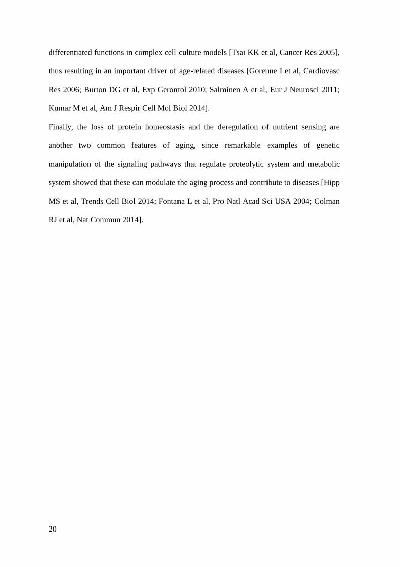
However, 240000 new pediatric HIV infections still occurred worldwide in 2013, because
treatment coverage remains suboptimal in many countries [WHO, UNAIDS, UNICEF
2013] (Table 1).
Table 1. Global summary of the AIDS epidemic in 2013
Number of people
living with HIV in
2013 Total 35.0 [33.1-37.2] million
Adults 31.8 [30.1-33.7] million
Children (<15 years) 3.2 [2.9-3.5] million
People newly
infected with HIV
in 2013
Total 2.1 [1.9-2.4] million
Adults 1.9 [1.7-2.1] million
Children (<15 years) 240000 [210000-280000]
AIDS deaths in 2013
Total 1.5 [1.4-1.7] million
Adults 1.3 [1.2-1.5] million
Children (<15 years) 190000 [210000-280000]
[From WHO, UNAIDS, UNICEF 2013]
MTCT is the main source of pediatric HIV-infection and, without prevention of MTCT,
the overall risk of transmission is up to 40%. Most of MTCT occurs around the time of
delivery, but it may occur also in utero; breastfeeding is an additional mode of MTCT
[The Working Group on MTCT of HIV, J Acquir Immune Defic Syndr Hum Retrovirol

24
1995]. MTCT is a multifactorial event in which high maternal plasma HIV RNA level
represents the major risk factor; mode of delivery and gestational age are also important
factors [The European Collaborative Study, AIDS 1999]. Moreover, a body of evidence
has established that also host genetic factors are important determinants of MTCT and
HIV infection outcome [Ometto L et al, J Infect Dis 2001; Ricci E et al, J Acquir Immune
Defic Syndr 2009; Ricci E et al, J Transl Med 2010; Freguja R et al, New Microbiol
2012; Gianesin K et al, PLoS One 2012].
The course of disease progression in infants markedly differs from that in adults. The
median time to AIDS and death in ART-untreated adults is approximately 10 years from
infection, but in children the course of infection is faster [Collaborative Group on AIDS
Incubation and HIV Survival, Lancet 2000; Blanche S et al, New Engl J Med 1994;
Barnhart HX et al, Pediatrics 1996] (Figure 5). In ART-untreated children, progression of
HIV infection follows two patterns: about 15-20% develop rapid progression to AIDS,
whereas the others progress more slowly [Blanche S et al, Am J Dis Child 1990]. In a
recent meta-analysis conducted in 12112 infants born from HIV-infected mothers, it was
estimated that survival from acquisition of infection postnatally in breast-feeding children
was higher than in those with infection acquired around delivery: 1-year mortality was
26% among children infected through breast milk compared to 52% for peri-partum
infected children [Becquet R et al, PLoS One 2012]. Furthermore, in a study that
distinguished in utero, intrapartum and postnatal infections, it was estimated that median
time from infection to death was 208, 380, and >500 days, respectively [Marinda E et al,
Pediatr Infect Dis J 2007]. These differences in survival time are partly due to the
immunological immaturity at the time of infection [Chakraborty R, Curr HIV Res 2005].
The levels of plasma HIV RNA are generally higher in vertically infected children than in
adults, persist at high levels and decline slowly with age [De Rossi A et al, J Clin Invest
25
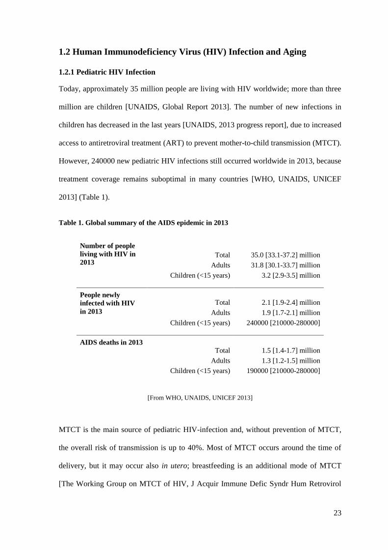
1996; McIntosh K et al, Pediatr Infect Dis J 1996] (Figure 5). Conversely to adults, the
relationship between CD4+ cell count and plasma HIV RNA is quite complex in children.
High levels of plasmaviremia may be observed despite normal CD4+ lymphocyte counts,
since CD4+ cell counts physiologically fall with age and plasma HIV RNA declines non-
linearly up to 5 years of life [Mofenson LM et al, J Infect Dis 1997; PENTA, AIDS
1998].
Figure 5. Dynamics of HIV RNA and CD4+ levels in adults and children. Relative levels of HIV RNA
(red) and CD4+ cells (blue) in adults (dotted lines) and children (solid lines) in the years following
acquisition of HIV. Gray boxes highlight the median time to AIDS/Death for infants by region and route of
infection in comparison to adults. (US: United States). [From Tobin NH & Aldrovandi GM, Immunol Rev
2013].

26
1.2.2 Pediatric HIV infection and antiretroviral therapy
The introduction of the ART has changed the natural history of pediatric HIV-1 infection,
reducing the rate of MTCT of HIV to less than 2% [Cooper ER et al, J Acquir Immune
Defic Syndr 2002] and resulting in a substantial decline in mortality rates and in an
improved quality of life in HIV-infected children [Gibb DM et al, BMJ 2003; Judd A et
al, Clin Infect Dis 2007]. ART has transformed pediatric HIV infection into a chronic
disease and now infected infants and children often survive to adolescence and adulthood
[Deeks SG et al, Lancet 2013; de Martino M et al, JAMA 2000; Chiappini E et al, AIDS
2007]. Early ART leads to a sustained viral suppression and allows the recovery of
thymic function, which in children is a pivotal event in immune reconstitution, and
normalization of immunologic responses to non-HIV antigens [Luzuriaga K et al, J Virol
2000; Chiappini E et al, AIDS 2006; Zanchetta M et al, Antivir Ther 2008; Goetghebuer
T et al, AIDS 2009]. However, despite early ART treatment controls HIV replication and
reduces viral load, it does not significantly prevent the establishment of a reservoir of
latently infected cells that precludes virus eradication [Zanchetta M et al, Antivir Ther
2008]. Therefore, perinatal infected children require life-long ART with the risk of
important complications, including drug toxicities and adverse clinical events, consistent
with those experienced by an aging population, including malignancies.

27
1.2.3 Cancer risk in children with perinatal HIV-infection
HIV-infected children and adults have an higher cancer risk compared to the general
population; in particular, Kaposi sarcoma (KS), non-Hodgkin lymphoma (NHL), and
invasive cervical cancer (ICC) represent the cancers with the higher incidence rates
among people with HIV/AIDS and are defined as AIDS-defining malignancies (ADM)
[CDC; Morb Mortal Wkly Rep 1992]. The long-term HIV-induced immunodeficiency
together with co-infection with oncogenic viruses (KS-associated herpes virus (KSHV),
Epstein-Barr virus (EBV) and Human Papilloma Virus (HPV), etiological agents of KS,
NHL and ICC, respectively) represent the explanation for the increased risk of ADM
[Grulich AE et al, Lancet 2007].
The impact of ART on cancer incidence in adulthood has been well described [Engels EA
et al, AIDS 2006; Biggar RJ et al, J Natl Cancer Inst 2007; Grulich AE et al, Lancet 2007;
Crum-Cianflone N et al, AIDS 2009; Simard EP et al, Cancer 2011], whereas it has been
evaluated only by few studies in childhood [Kest H et al, Pediatr Infect Dis J 2005;
Chiappini E et al, J Clin Oncol 2007; Alvaro-Meca A et al, Pediatr Infect Dis J 2011;
Simard EP et al, Cancer Epidemiol Biomarkers Prev 2012]. Overall, epidemiological data
suggest that with the widespread of ART, the incidence of ADM significantly decreased
in both adults and children, due likely to partial immune restoration associated with an
improved immune surveillance against oncogenic viruses [Biggar RJ et al, J Natl Cancer
Inst 2007; Simard EP et al, Cancer Epidemiol Biomarkers Prev 2012]. In an Italian
pediatric population of 1190 perinatal HIV-infected children, the ADM incidence
decreased significantly from pre-ART to ART era [Chiappini E et al, J Clin Oncol 2007]
(Table 2). The same trend was observed in an US study that analyzed cancer incidence
between the pre-ART (1980-1995) and ART eras (1996-2007) in people diagnosed with
AIDS during childhood (0-14 years): KS and NHL declined by 87% and 60%,
28
respectively in the ART era [Simard EP et al, Cancer Epidemiol Biomarkers Prev 2012].
Furthermore, a Spanish study that evaluated epidemiological trends of cancer diagnosed
through 3 calendar periods (early-period ART: 1997-1999, middle-period ART: 2000-
2002, and late-period ART: 2003-2008), found that the rate of ADM diagnoses decreased
significantly (from 9.1 to 3.6 to 1.0 per 1000 children/year) during the study period
[Alvaro-Meca A et al, Pediatr Infect Dis J 2011] (Table 2). However, despite ART
reduced ADM, the overall incidence of malignancies in HIV-infected children do not
significantly differ between pre-ART and ART-era [Chiappini E et al, J Clin Oncol 2007;
Alvaro-Meca A et al, Pediatr Infect Dis J 2011; Simard EP et al, Cancer Epidemiol
Biomarkers Prev 2012] (Table 2). The improvement in long-term life expectancy has led
to change the spectrum of cancer among people living with HIV, shifting from ADM to
non-ADM. In the Spanish study cited before, the rate of overall non-ADM increased from
0.6 to 5.0 to 8.7 cases of cancer per 1000 children/year during the early, middle and late
ART eras [Alvaro-Meca A et al, Pediatr Infect Dis J 2011]. Malignant neoplasm of bone
and articular cartilage, Hodgkin lymphoma (HD) and leiomyosarcoma were the cancers
with the highest incidence [Simard EP et al, Cancer Epidemiol Biomarkers Prev 2012].
The pathogenesis for the increased non-ADM incidence remains to be fully elucidated.
Immunodepression was long considered to play pivotal role in the etiology of cancer;
however, given the ART-associated immune recovery, the immunodeficiency is not
sufficient to explain this excess risk. The dysfunction of immune system due to persistent
immune activation, premature aging and immune senescence may be the drivers of non-
ADM [Deeks SG et al, Curr Opin Immunol 2012; Phillips AN et al, AIDS 2008].

29
Table 2. Epidemiologic data on cancer rate in HIV-infected children over time
Incidence of cancer*
Study population Study period All cancer ADM non-ADM
5840 1980-1995 5.59 4.70 0.65
(United States)1 1996-2007 2.13 1.50 0.63
1190 1985-1995 4.49 2.97 1.69
(Italy)2 1996-1999 4.09 2.60 1.48
2000-2004 0.76 0.76 -
1307 1997-1999 9.70 9.10 0.60
(Spain)3 2000-2002 8.70 3.60 5.00
2003-2008 9.70 1.00 8.70
*Incidence rates are per 1000 children/year
From: 1 Simard EP et al, Cancer Epidemiol Biomarkers Prev 2012
2 Chiappini E et al, J Clin Oncol 2007
3 Alvaro-Meca A et al, Pediatr Infect Dis J 2011

30
1.2.4 Premature aging and immune senescence in HIV-infection
Accumulating evidence suggest that HIV-infected people undergoing long-term ART
tend to show an accelerated aging phenotype, that involves the immune system and leads
to the increase in the frequency of non-AIDS-related complications (i.e. cardiovascular,
liver and kidney diseases, non-ADM) [Deeks SG, Annu Rev Med 2011; Chiappini E et al,
Cancer Lett 2014].
The immunological alterations that characterized immune senescence both in
physiological aging and in HIV infection have several common features, and in both
conditions immune activation/inflammation play a crucial role in promoting the gradual
decline of the functionality of the immune system [Desai S and Landay A, Curr
HIV/AIDS Rep 2010]. These processes affect all aspects of immunity, but the T-cell
compartment seems to be the most damaged. These changes are characterized by a
reduction of number and function of hematopoietic stem cells, thymic involution, loss of
naïve cells, progressive enrichment of terminally differentiated T-cells, shrinkage of the
T-cell repertoire, reversed ratio of CD4+ to CD8
+ cells and increased levels of pro-
inflammatory cytokines [Deeks SG, Annu Rev Med 2011].
In the context of HIV infection, the virus replicates and releases virions into circulation.
A low-grade, chronic and systemic replication occurs despite ART and causes a
continuous stimulation of immune system and inflammatory response. In addition, the
injury to the immune component of gastrointestinal mucosal surface, along with damage
to the intestinal epithelial microenvironment with its antimicrobial functions, may affect
systemic immune activation during the chronic phase of HIV infection through the release
of microbial products into blood circulation [Brenchley JM et al, Nat Med 2006], a
phenomenon defined as microbial translocation. The strong pressure on the immune
system, exerted by viral antigens, leads to the clonal expansion of activated cells,

31
resulting in differentiation and accumulation of senescent cells with loss of effector
functions and proliferative capacity [Desai S & Landay A, Curr HIV/AIDS Rep 2010;
Khaitan A & Unumatz D, Curr HIV/AIDS Rep 2011] (Figure 6). Finally, the
accumulation of senescent cells secreting proinflammatory factors results in a chronic
inflammation status that leads to the development of age-related pathologies [Ovadya Y
& Krizhanovsky V, Biogerontology 2014]. Indeed, the progressive decline of immune
function may result in an impaired immune surveillance against tumor antigen favoring
cancer onset. The chronic immune activation may also induce B-cell stimulation leading
to an expansion of EBV-infected cells, thus increasing the risk of developing EBV-
associated malignancies [Petrara MR et al, Front Microbiol 2013]; both leiomyosarcoma
and HD, two of the non-AIDS-defining cancers with higher incidence in the ART-era, are
EBV-associated cancers [Bhatia K et al, Curr Opin Oncol 2012].
Figure 6. Accelerated aging model in HIV infection. [From Desai S & Landay A. Curr HIV/AIDS Rep
2010]
32
Since telomeres have been postulated as a universal biological clock that shorten in
parallel with aging of cells and whose attrition reflects cellular replication history and
cellular senescence, shortening telomere length is considered the best marker of
biological aging. Telomere length in peripheral blood mononuclear cells (PBMC) is
representative of that of many tissues; indeed, intra-individual correlation between
telomere lengths in different tissue is high [Daniali L et al, Nat Commun 2013]. Short
telomere length has been associated with older age [Barrett EL and Richardson DS,
Aging Cell 2011], age-related diseases and low survival in the general population
[Brouilette SW et al, Lancet 2007; Cawthon RM et al, Lancet 2003; Heidinger BJ et al,
Proc Natl Acad Sci U S A 2012]. Recently, short telomere length has been associated
with HIV infection [Côté HC et al, PLoS One 2012; Pathai S et al, AIDS 2013].
Moreover, considering the capability of nucleoside reverse transcriptase inhibitors
(NRTIs) to repress not only HIV reverse transcriptase but also human telomerase, it has
been suggested that ART may be a potential factor contributing to HIV-associated
accelerated shortening of telomere and premature aging [Liu X et al, Nucleic Acids Res
2007; Hukezalie KR et al, PLoS One 2012; Leeansyah E et al, J Infect Dis 2013].

33
Chapter 2

34

35
Immune Senescence and Cancer in Elderly Patients: Results from an
Exploratory Study and Preliminary Results of Long-term Follow-up
Ketty Gianesin1, Bergamo F
2, Falci C
3, Sergi G
4, Giunco S
5, De Ronch I
4, Valpione S
1,
Soldà C2, Fiduccia P
2, Lonardi S
2, Zanchetta M
5, Keppel S
5, Brunello A
2, Cascarilla R
2,
Zafferri V2, Manzato E
4, Zagonel V
2, De Rossi A
1,5
1 Section of Oncology and Immunology, Department of Surgery, Oncology and
Gastroenterology-DiSCOG, University of Padova, Padova, Italy
2
Medical Oncology Unit I, Istituto Oncologico Veneto (IOV), IRCCS, Padova, Italy
3
Medical Oncology Unit II, Istituto Oncologico Veneto (IOV), IRCCS, Padova, Italy
4 Department of Medicine-DiMED, Geriatric Section, University of Padova, Padova, Italy
5 Immunology and Molecular Oncology, Istituto Oncologico Veneto (IOV), IRCCS,
Padova, Italy
The results of the case-control study have been published in :
“Falci C, Gianesin K, Sergi G, Giunco S, De Ronch I, Valpione S, Soldà C, Fiduccia P,
Lonardi S, Zanchetta M, Keppel S, Brunello A, Zafferri V, Manzato E, De Rossi A,
Zagonel V. Immune senescence and cancer in elderly patients: results from an
exploratory study. Exp Gerontol 2013; 48:1436-42”
The longitudinal substudy is in progress.

36
ABSTRACT
Background. The challenge of immune senescence has never been addressed in elderly
cancer patients. This study compares the thymic output and peripheral blood telomere
length in 70≥ years old cancer patients. A longitudinal substudy is in progress to evaluate
the impact of these markers on chemotherapy response and disease outcome.
Patients and Methods. Fifty-two elderly cancer patients and 39 age-matched controls
without personal history of cancer were enrolled. All patients underwent a
Comprehensive Geriatric Assessment. Peripheral blood samples were studied for naïve
and recent thymic emigrant (RTE) CD4+ and CD8
+ cells by flow cytometry. T-cell
receptor rearrangement excision circle (TREC) levels, telomere length and telomerase
activity in peripheral blood cells were quantified by real-time PCR. Patients with
colorectal cancer has been enrolled in the longitudinal substudy.
Results. The percentages of CD8+ naïve and CD8
+ RTE cells and TREC levels were
significantly lower in cancer patients than in controls (p=0.003, p=0.004, p=0.031,
respectively). Telomere lengths in peripheral blood cells were significantly shorter in
cancer patients than in controls (p=0.046) and did not correlate with age in patients,
whereas it did in controls (r=-0.354, p=0.031). Short telomere(≤median)/low
TREC(≤median) profile was associated with higher risk of cancer (OR=3.68 [95%CI
1.22-11.11]; p=0.021). Preliminary data from the longitudinal substudy suggested that
after chemotherapy there was an increase of CD8+ terminally differentiated cells
(p=0.031), and shortening of telomeres tended to higher in patients with stage III than in
those with stage II.
37
Conclusions. Immune senescence is significantly worse in elderly cancer patients than in
age-matched controls. The low thymic output and the shorter telomeres in peripheral
blood cells of cancer patients may reflect a pre-existing condition which facilitates the
onset of malignancies in elderly people and may impact the response to chemotherapy
and the disease outcome.

38
1. INTRODUCTION
People over 65 years old are the fastest-growing age bracket in the population and will
account for an estimated 20% of Americans and 25% of Europeans by the year 2030
[Fries JF, Ann Intern Med 2003]. The incidence of malignancies increases with age, so
the number of cancers in the elderly is expected to increase significantly in years to come
[American Cancer Society, 2012]. Several studies have shown that elderly patients are
less likely to be treated according to guidelines, and their under-treatment may be
detrimental to both survival and quality of life [Sargent DJ et al, N Engl J Med 2001;
Bouchardy C et al, J Clin Oncol 2003; Dale DC, J Support Oncol 2003; Ng R et al, Clin
Lung Cancer 2005]. Elderly cancer patients may benefit from chemotherapy just as much
as younger adults, but at a higher risk of hematological toxicity [Muss HB et al, JAMA
2005; Hurria A et al, J Natl Compr Canc Netw 2012; Muss HB et al, J Clin Oncol 2007].
Better understanding of the physiological and functional changes that occur with aging
will enable to improve strategies for treating elderly cancer patients. Since the aging
process coincides with a gradual decline in the functional reserve of multiple organ
systems [Balducci L, J Support Oncol 2003] the search for laboratory markers of
biological aging and organ reserve should be a priority of clinical research in the field of
geriatric oncology.
Over a lifetime, the immune system undergoes profound remodeling process with major
impact on health and survival [Grubeck-Loebenstein B et al, Aging Clin Exp Res 2009;
Fulop T et al, Ann N Y Acad Sci 2010]. Thymic involution and diminished output of T
lymphocytes are thought to be among the major factors contributing to the loss of
immune function with age [Berzins SP et al, J Exp Med 1998]. T-cell output begins to
decline exponentially from early in life, and at 75 years of age the immune repertoire

39
appears to be severely impaired [Douek DC et al, Nature 1998; Naylor K et al, J Immunol
2005]. However, recent data suggest that the thymus may remain active even late in life,
supplying functional T cells to the periphery [Nasi M et al, Aging Cell 2006; Mitchell
WA et al, Clin Exp Immunol 2010].
Measuring T-cell receptor rearrangement excision circle (TREC) levels in peripheral
blood lymphocytes has been suggested as a method for quantifying thymic output [Douek
DC et al, Nature 1998; Zhang L et al, J Exp Med 1999; Ometto L et al, AIDS 2002; De
Rossi A et al, J Infect Dis 2002]. TRECs are generated by T-cell receptor gene
rearrangement and maintained in thymic emigrant cells as DNA episomes. Because
TRECs are not duplicated during mitosis, their concentration is diluted out with each cell
division. The frequency of recent thymic emigrant (RTE) cells in peripheral blood,
identified by the marker CD31+ among the CD45RA
+ naïve T cells [Kimmig S et al, J
Exp Med 2002], decreases with aging and correlates well with the decline in TREC levels
[Kohler et al, Eur J Immunol 2005; Junge S et al, Eur J Immunol 2007]. Very little is
known about the relationship between TRECs and cancer, especially in elderly patients.
One study on head and neck cancer patients, including just a few ≥70 years old, showed
that the age-associated decrease of TREC numbers and naïve T lymphocytes was
significantly greater in cancer patients than in controls, suggesting altered lymphocyte
homeostasis in the former [Kuss I et al, Clin Immunol 2005].
The immune system function depends largely on its capacity for extensive cell division
and clonal lymphocyte expansion. Telomere length, and its regulation by telomerase have
attracted considerable attention, due to their potential roles in controlling cell replication
[Blackburn E et al, Nat Med 2006]. Telomeres are capping end structures of eukaryotic

40
chromosomes essential for protecting chromosome integrity [Blasco MA, Nat Rev Genet
2005]. Telomeres are progressively shortened during each cell division due to end-
replication problems of DNA polymerase; when a critical length is reached, the cell
undergoes cycle arrest and apoptosis [Blasco MA, Nat Rev Genet 2005]. Permanent cell
growth relies on telomere maintenance, and certain human cell subsets, as well as most
cancer cells, have telomerase activity which enables telomere elongation [Dolcetti R &
De Rossi A, Med Res Rev 2012]. Despite their telomerase activity, most tumor cells have
shorter telomeres than the corresponding normal tissues, and there is a relationship
between short telomeres and genetic instability [Garcia-Aranda C et al, Cancer 2006;
Rampazzo E et al, Br J Cancer 2010]. Since telomere shortening reflects cell turnover and
exposure to oxidative and inflammatory damage, which are crucial processes of
biological aging, it has been suggested that telomere length serves as an indicator of aging
process [Wong JMY & Collins K, Lancet 2003; Aviv A, J Gerontol A Biol Sci Med Sci
2006; Baird DM, Exp Gerontol 2006]. Telomere shortening in peripheral blood cells has
been associated with a number of chronic diseases, such as coronary heart disease,
hypertension, dementia, obesity, insulin resistance, and osteoporosis. However, two
clinical trials failed to confirm any relationship between telomere length and frailty
syndrome in elderly non-cancer patients [Woo J et al, Mech Ageing Dev 2008; Collerton
J et al, Mech Ageing Dev 2012].
Several studies have investigated the relationship between telomere length in peripheral
blood cells and cancer risk and outcome. Although few manuscript report that longer
telomere length is a risk factor [Lan Q et al, PLoS One 2013; Svenson U et al, Cancer Res
2008], most of the studies indicate that short telomere lengths are associated with a cancer
risk and/or worse prognosis [Bojesen SE et al, Nat Genet 2013; Martinez-Delgado B et al,

41
J Med Genet 2012; Riegert-Johnson DL et al, Int J Biol Markers 2012; Shao L et al, J
Urol 2007; Wu X et al, J Natl Cancer Inst 2003; Qu F et al, Mol Oncol 2014; Chen Y et
al, Ann Oncol 2014]. To date, however, telomere length in peripheral blood cells has been
measured in elderly non-cancer patients and younger cancer patients, but no data are
available for elderly cancer patients.
In this study, we present the results of a prospective observational study providing the
first description of immune senescence markers and frailty scores in elderly cancer
patients and age-matched controls. Furthermore, we report the preliminary results of the
ongoing longitudinal substudy planned to evaluate the impact of these markers on
chemotherapy response and/or risk of tumor relapse.
2. MATERIAL and METHODS
2.1 Study design and study population
Patients enrolled in this prospective observational study were aged ≥70 years, with stage
I-III breast or colorectal cancer, diagnosed during the previous 2 months and radically
resected, consecutively admitted to the Medical Oncology Units from September 2010 to
March 2012. Controls included patients ≥70 years old with no personal history of cancer,
consecutively admitted to the Geriatric Clinic. For both groups, the exclusion criteria
were: any hematological disorders, chronic diseases requiring immunosuppressive
treatment, prior immunodeficiency, blood transfusion ≤4 weeks before blood sampling,
active infectious diseases, extremely severe comorbidities suggesting a life expectancy <6
months, or severe cognitive impairment hampering communication with the physician.
A subgroup of patients with colorectal cancer have been enrolled in the longitudinal
substudy.

42
The study was approved by the institutional Ethics Committee and conducted in
accordance with the Helsinki Declaration and Good Clinical Practice guidelines. Written
informed consent was obtained from all patients.
2.2 Clinical assessment
Complete demographic and clinical details were collected at baseline for each patient
(Table 1). At the first visit, both cases and controls underwent traditional comprehensive
geriatric assessment (CGA) administered by a multidisciplinary team, including medical
oncologist, geriatrician and psychologist. As reported elsewhere [Basso U & Monfardini
S, Eur J Cancer Care 2004], the CGA included Activities of Daily Living (ADL),
Instrumental Activities of Daily Living (IADL), Short Portable Mental Status
Questionnaire (SPMSQ), and the Mini Mental Status Examination (MMSE).
Comorbidities and their severity were studied according to the Cumulative Illness Rating
Scale-Comorbidity Index (CIRS-CI) and Cumulative Illness Rating Scale-Severity Index
(CIRS-SI) [Conwell Y et al, J Am Geriatric Soc 1993]. Affective status was assessed with
the Geriatric Depression Scale (GDS) [Yesavage JA et al, J Psychiatr Res 1983].
Nutritional status was explored with the Mini Nutritional Assessment (MNA) [Guigoz Y
& Vellas B, Nutr Workshop Ser Clin Perform Programme 1999]. Social aspects included
household composition, institutionalization, amount of assistance provided by a caregiver.
The number of drugs taken by patients for concomitant diseases was also recorded. The
CGA results were interpreted as previously reported by Balducci [Balducci L, J Support
Oncol 2003], classifying patients as fit, vulnerable or frail. A multidimensional prognostic
index (MPI) score was also calculated based on findings for the ADL, IADL, SPMSQ,
CIRS-CI, CIRS-SI, MNA, number of drugs and social conditions, assuming a low,

43
moderate and high mortality risk for MPI ≤0.33, 0.34-0.66, >0.66, respectively [Pilotto A
et al, Rejuvenation Res 2008; Giantin V et al, J Geriatric Oncol 2013].
2.3 Biomarker analyses
Peripheral blood samples were collected at the time of enrollment, prior to any
oncological medical treatment (endocrine therapy, chemotherapy, radiotherapy or
immune therapy). In the longitudinal substudy, samples were collected at the end of
adjuvant chemotherapy treatment and/or at tumor relapse; the median period of follow-up
was 22 [20-28] months. Samples were analyzed for the standard blood parameters (Table
1S, Supplementary data), for Cytomegalovirus (CMV) and for the following tests.
2.4 Flow cytometry
Peripheral blood mononuclear cells (PBMC) were isolated from peripheral blood by
centrifugation on a Ficoll-Paque gradient [Freguja R et al, Clin Exp Immunol 2011]. Cells
were stained with the following labeled monoclonal antibodies (mAbs): anti-CD3 (FITC),
anti-CD4 (PerCP), anti-CD8 (PerCP), anti-CD31 (PE) and anti-CD45RA (APC).
Appropriate isotypic controls (mouse IgG1-PE and mouse IgG2b-APC) were used to
assess non-specific staining. All samples were analysed by four-color flow cytometry on
a fluorescence-activated cell sorter (FACS)Calibur (Becton-Dickinson) [Freguja R et al,
Clin Exp Immunol 2011]. Data were processed with CellQuest Pro Software (Becton-
Dickinson) and analysed using Kaluza® Analysis Software v.1.2 (Beckman Coulter).
Anti-CD45RA and anti-CD27 were used to identify, within CD3+CD4
+ and CD3
+CD8
+
gates, naïve (CD45RA+CD27
+), central memory (CD45RA
-CD27
+), effector memory
(CD45RA-CD27
-) and terminally differentiated memory (CD45RA
+CD27
-) cells.
Moreover, anti-CD45RA and anti-CD31 were used to identify recent thymic emigrant

44
(RTE) (CD45RA+CD31
+), and anti-CD28 and anti-CD57 to identify senescent cells
(CD28-CD57
+).
2.5 TREC quantification
Thymic output in PBMC was studied by measuring TREC levels by real-time polymerase
chain reaction (PCR), exactly as described previously [Ometto L et al, AIDS 2002; De
Rossi A et al, J Infect Dis 2002]. TREC levels were expressed as the number of TREC
copies per 105
PBMC [De Rossi A et al, J Infect Dis 2002; Anselmi A et al, Clin Exp
Immunol 2007].
2.6 Telomere length measurement by quantitative real-time PCR
Telomere length in PBMC was determined by real-time PCR exactly as described
elsewhere [Rampazzo E et al, Br J Cancer 2010; Rampazzo E et al, Haematologica 2012],
and values were expressed as relative telomere/single-copy gene (T/S) ratio [Rampazzo E
et al, Br J Cancer 2010].
2.7 Telomerase activity quantification
Telomerase activity was quantified by real-time PCR, as described elsewhere [Rampazzo
E et al, Haematologica 2012] and expressed in relative units (RU).
2.8 Irradiation of PBMC and detection of DNA-damage by flow cytometry
After isolation of PBMC by Ficoll-Paque gradient, cells (~8*106) were cultured in RPMI
medium supplemented with 10% fetal calf serum (FCS, Life Technologies), 10 ng/ml IL-
2 (Roche), 2mM L-glutamine (Gibco) and 50 ug/ml Gentamycin (Sigma) (standard
medium) at 37°C in 5% CO2, in 6-well plate for 12 hours before y-irradiation. Following
the removal of ~106
PBMC (untreated control), the remaining cells were irradiated to a

45
dose of 2.5 Gy using y-radiation from a 137
Cesium source (CisBIO International, France)
at room temperature and were immediately incubated in 12-well plate in standard medium
at 37°C in 5% CO2; aliquots of ~106
PBMC cells were taken 1, 5 and 24 h post-
irradiation. Cells were fixed in 4% paraformaldehyde (Sigma) for 10 min at 37°C, chilled
on ice for 1 min, washed in PBS, permeabilized with 90% methanol in 0.5% bovine
serum albumin (BSA; Sigma)-PBS, and kept at -20°C for 1 h. Cells were washed three
times with 0.5% BSA-PBS and incubated in blocking buffer [PBS supplemented with 8%
FCS, 0.1 g/L RNAseA (Life Technologies), 10 mM NaF (Sigma) and 1 mM NaVO3
(Sigma), 0.25 g/L salmon sperm DNA (Sigma), 0.2% Triton X-100 (Roche)] for 1 h prior
to y-H2AX staining with the AlexaFluor-488 mouse anti-H2AX (pS139) antibody (BD
Pharmigen) diluted in blocking buffer for 1 h at room temperature. Cells were washed
twice in 0.5% BSA-PBS and resuspended in PBS. Isotype antibody was included as a
background reference. A minimum of 100000 cells were acquired using a FACSCalibur
(Becton-Dickinson). Cellular debris were excluded from the analysis by gating a forward
scatter versus side scatter plot. Analysis of flow cytometry data was conducted using the
CellQuest Pro Software (Becton-Dickinson). Percentage of y-H2AX positive cells and
median fluorescence intensity were calculated at each time point.
2.9 Statistical analysis
Differences in geriatric parameters between groups were examined with the Mann-
Whitney test for non-parametric data and Pearson’s χ2 test with odds ratios (OR) and 95%
confidence intervals (CI) for nominal data (CGA, MPI, ADL, IADL). The Mann-Whitney
test was used to compare subsets of lymphocytes, TREC levels and telomerase activity.
Student’s t-test was used to compare the telomere lengths. Correlations between age and
TREC levels, or telomere length in both groups were analysed with Pearson’s χ2
test.

46
Spearman’s rank correlation was used to analyse associations between geriatric
parameters and TREC levels, telomere lengths or telomerase activity. TREC levels and
telomere length were analysed also as dichotomous variables (cut-off: ≤median) and OR
were estimated using logistic regression model. All statistical analyses were performed
using SPSS software, version 19 (SPSS Inc.). All p-values were two-tailed, and were
considered significant when lower than 0.05.
3. RESULTS
3.1 Characteristics of study population
Ninety-one patients, 52 with cancer (26 breast and 26 colorectal cancer) and 39 controls,
were enrolled in the study. Their demographic and clinical characteristics are listed in
Table 1. There were more females in the control group (82.1%) than among the cancer
patients (63.5%); the latter had better Karnofsky performance status than controls. As
established by the study protocol, before enrollment, cancer patients underwent a
thorough radiological assessment to rule out metastases. According to the TNM staging
system, 16 patients (30.8%) had stage I, 33 (63.5%) had stage II, and 3 (5.7%) had stage
III disease. Thirty-one patients were prescribed the same adjuvant therapy as for younger
adults, according to good clinical practice, 18 patients received an adapted treatment, due
to problems detected at the CGA, and 3 were given no adjuvant therapy, due to frailty
detected at the CGA and were only followed up.
3.2 Comprehensive geriatric assessment
All cancer patients and controls underwent the full CGA; 17.3% of the former and 43.6%
of the latter were classified as frail (OR=0.29, 95% CI 0.10-0.82), while the proportion of
vulnerable patients in the two groups did not differ significantly (Table 2). 5.8% of cancer

47
patients and 23.1% of controls had a moderate MPI (OR=0.20, 95% CI 0.05-0.79), and
only one control had a severe MPI score. When MPI was calculated as a continuous
variable, the median score was significantly lower for cancer patients than for controls
(Table 2). In ADL and IADL, cancer patients tended to have preserved greater physical
autonomy than controls, although the difference was not statistically significant (Table 2).
Controls had better cognitive status in both the MMSE and SPMSQ, but their burden of
associated diseases (as shown by CIRS-CI and CIRS-SI) was higher, as was their use of
drugs for these comorbidities (Table 2).
3.3 Standard hematological and biochemical parameters
The hematological parameters of cancer patients and controls differed only in their
platelet count (Table 1S, Supplementary data). Controls had significantly lower levels of
nutritional markers, such as total plasma proteins, triglycerides, and LDL-cholesterol;
CRP and IL-6 were significantly higher in controls than in cancer patients (Table 1S,
Supplementary data). The overall prevalence of CMV-IgG seropositive individuals was
96.7%. Antibodies against CMV were found in 49 (94.2%) of cancer patients and 39
(100%) of controls. No statistical difference was found between CMV antibody titers
between the groups (p=0.498) (Table 1S, Supplementary data).
3.4 Immune senescence markers
Cancer patients and controls had comparable percentages of total CD4+ lymphocytes,
naïve CD4+CD45RA
+ and RTE CD4
+CD45RA
+CD31
+ cells (Table 3). By contrast, the
median (interquartile) percentages of naïve CD8+CD45RA
+CD27
+ and RTE
CD8+CD45RA
+CD31
+ cells were significantly lower in cancer patients than in controls
(16.7% [9.3-25.2] versus 24.6% [14.7-33.5]; p=0.003) and (34.2% [24.7-46.4] versus

48
44.9% [36.0-50.7]; p=0.004), respectively (Table 3). Notably, the lower percentage of
CD8+ naïve cells in cancer patients was compensated by a higher expansion of CD8
+
memory cells; thus, cancer patients and controls did not differ in terms of their
percentages of total CD8+ cells (Table 3). In particular, among the CD8
+ cell subsets, the
CD45RA-CD27
- (effector memory) cells were found to be significantly higher in cancer
patients than in controls (21.7% [12.7-31.7] versus 16.3% [10.4-25.0]; p=0.042). The
imbalance in favor of the CD8+ memory cell subset in cancer patients was evident even
when the absolute cell count was considered (not shown). For a few subjects with
available frozen samples (11 cancer patients and 8 controls), markers of immune
senescence (CD28-CD57
+) were investigated. Cancer patients and controls exhibited
percentages of CD4+CD28
-CD57
+, but CD8
+ cells with senescent phenotype tended to be
higher in the former (p=0.082) (Table 3S, Supplementary data).
TREC levels in PBMC were significantly higher in controls than in cancer patients (25.0
[14.0-56.0] versus 16.0 [7.7-31.5] TREC copies/105 PBMC; p=0.031) (Table 3). TREC
levels decreased significantly with increasing age in cancer patients (r=-0.478; p<0.001),
while did not correlate with age in controls (r=0.071; p=0.677) (Figure 1A).
The mean telomere length in PBMC was significantly lower in cancer patients than in
controls; the former had a mean (± standard deviation (SD)) telomere length of 1.09±0.31
T/S versus 1.22±0.30 T/S in controls, p=0.046 (Table 3). Telomere length correlated
inversely with age in controls (r=-0.354, p=0.031), but not in cancer patients (r=-0.011,
p=0.938) (Figure 1B). Telomerase activity was significantly higher in cancer patients,
being detected in 38/52 patients (73.1%; median value 14.6 RU) and 20/38 controls
(52.6%; median value 1.4 RU); p=0.003. Telomerase activity was not influenced by age
in either group.

49
TREC was defined as high and low, and telomere length as long or short according to
their value above and below the median, respectively. Subjects with TREC low/telomere
short profile were at higher risk of cancer than subjects with only low TREC or short
telomere (Table 4).
3.5 Relationship between TREC levels, telomere length and geriatric characteristics
In the control group, the CIRS-SI was the only tool which revealed a significant positive
association with TREC level (r=0.45, p=0.01) and telomere length (r=0.35, p=0.03),
whereas it had a significant negative correlation with the number of drugs taken for
concomitant diseases (r=-0.39, p=0.02) (Table 2S, Supplementary data). No relationship
emerged between telomere length, thymic output and geriatric features of cancer patients
(Table 2S, Supplementary data). Neither a classification of unfitness at CGA (not shown)
nor the MPI score (Table 2S, Supplementary data) correlated significantly with the
thymic output or telomere length in either group. Among the controls, TREC levels
correlated positively with IL-6 (r=0.34, p=0.04), and negatively with total plasma protein
levels (r=-0.38, p=0.02). Neither of these correlations emerged in the cancer patients (data
not shown).
3.6 Preliminary results from the longitudinal substudy
Twenty-two patients have been already enrolled in this substudy. Eight patients received
capecitabine plus oxaliplatin, 1 capecitabine plus bevacizumab, 8 capecitabine alone for 6
months while 5, all with stage II disease, received no adjuvant chemotherapy. Four
patients suspended therapy before the deadline due to poor tolerability and/or onset of
cardiovascular complications. Three patients had a relapse: one with stage II disease (no

50
adjuvant chemotherapy) and two with stage III (both with a complete chemotherapy
treatment).
Overall, telomere length tended to decrease during the follow-up period (T/Sbaseline
1.23±0.28 versus T/Sfollow-up 1.14±0.25; p=0.201), even if at not significant level. At
baseline, telomere length was not associated with tumor staging, being T/S of 1.20±0.26
and 1.27±0.32 for stage II and III respectively (p=0.300), but their shortening at follow-
up tended to be higher in patients with stage III than in those with stage II (∆baseline-follow-up
T/S 0.13 versus 0.07). The ΔT/S of 3 patients with tumor relapse were: 0.15, 0.05 and -
0.14, respectively; the low number and the heterogeneity (stage disease and treatment) did
not allow to draw any conclusions. TREC levels tended to be lower in samples at follow-
up compared to baseline (6 [3-18] versus 10 [6-22] TREC copies/105
PBMC; p=0.161).
These results are in agreement with phenotypic analysis of peripheral blood cells.
Frequencies of CD4+ and CD8
+ naïve T cell did not change during follow-up period.
Conversely, within the CD8+ memory cell subset, we found a shift of cells from effector
(CD45RA-CD27
-) to terminally differentiated (CD45RA
+CD27
-) phenotype (Figure 2).
Moreover, senescent cells tended to be more expanded at follow-up than at baseline
(63.0% [57.9-73.5] versus 58.5% [46.5-64.0]; p=0.099). The higher frequencies of these
subsets may explain the tendency of shorter telomere length in samples at follow-up.
The higher levels of memory cells in elderly cancer patients, both before and after
treatment, is important. It is known that DNA-damage recognition and repair efficiency
varies between memory and naïve cells [Scarpaci S et al, Mech Ageing Dev 2003;
Cossarizza A et al, Mech Ageing Dev 1996]. In a preliminary experiment, we analyzed
the DNA-damage repair in PBMC exposed to a dose of 2.5 Gy using y-radiation from a
137Cesium source. By flow cytometric assay, we found that the level of y-H2AX, a robust

51
marker of cellular double strand breaks [d'Adda di Fagagna F et al, Nature 2003], was
higher in untreated PBMC from cancer patients than in those from controls, suggesting a
more unstable genome in the former. Soon after irradiation, the level of y-H2AX
increased in both groups, but at 24 h it remained higher in cancer patients than in controls
(Figure 3), suggesting a lower efficiency in DNA repair process in the former [Banáth JP
et al, BMC Cancer 2010].
4. DISCUSSION
This is the first study aiming to shed light on two essential aspects of the aging process
(thymic output and telomere length in peripheral blood cells) in elderly cancer patients.
We found that 70- to 92-year-old cancer patients had significantly lower TREC levels,
lower percentages of naïve and RTE CD8+ T lymphocytes, and a more expanded CD8
+
memory cell subset than age-matched controls. These results are partially consistent with
those of a small trial in which head and neck cancer patients (most of them under 70-
years-old) revealed lower TREC levels than controls [Kuss I et al, Clin Immunol 2005].
The expansion of memory CD8+
cells, particularly those with effector phenotype, is also
consistent with the findings of two recent studies conducted on breast cancer patients
[Hueman MT et al, Cancer Immunol Immunother 2007; Poschke I et al, Int J Cancer
2012]. The shift from naïve T cells to memory cells was probably due to greater
stimulation by tumor antigens; as CD8+ T cells are the key components of tumor immune
surveillance, the tumor-induced dysfunction may be more evident in the CD8-cell subset
than in the CD4+
T-cell compartment [Klebanoff CA et al, Immunol Rev 2006; Williams
MA & Bevan MJ, Annu Rev Immunol 2007].

52
Our findings confirm reports by other authors that thymic activity does not stop
completely beyond 70 years of age [Nasi M et al, Aging Cell 2006; Mitchell WA et al,
Clin Exp Immunol 2010]. Notably, while TREC levels in cancer patients dropped
significantly with increasing age, they remained relatively constant in controls. Thymic
output may compensate for the loss of peripheral blood lymphocytes in elderly patients,
but this homeostatic phenomenon seems to disappear in cancer patients. In our study, the
control group included a higher proportion of frail patients, with higher average MPI
scores and more chronic diseases and comorbidities than the cancer patients. Levels of
pro-inflammatory cytokine IL-6 were also significantly higher in controls than in cancer
patients. Systemic inflammation and loss of peripheral blood cells may stimulate thymic
output, which plays an important role in immunological homeostasis. In fact, TREC
levels did correlate significantly with CIRS-CI in controls group, but not in cancer
patients. The imbalance between the two groups in terms of their geriatric conditions
reinforces the magnitude of the difference observed for TREC levels between cancer
patients and controls. While the hypothesis of immune homeostasis may justify a higher
thymic output in controls, the lower age-related TREC levels seen in cancer patients may
point to a pre-existing condition, favoring immune escape and the onset of malignant
disease.
In agreement with the lower percentage of CD8+
naïve and RTE cells in cancer patients
than in controls, the former tended to have a higher percentage of CD8+
immune
senescent cells. Several data indicate that latent CMV infection leads to significant
changes in the CD8+
repertoire, and it may be an important driver of immune senescence
[Almanzar G et al, J Virol 2005; Solana R et al, Immun Ageing 2012]. The above results
53
are unlikely to be due to CMV infection, because all except three cancer patients were
CMV-positive and with similar titers of anti-CMV antibodies.
As regards telomere length, there are many data available on the relationship between
telomere length and aging-associated changes and chronic [Wong JMY & CollinsK ,
Lancet 2003; Aviv A, J Gerontol A Biol Sci Med Sci 2006; Baird DM, Exp Gerontol
2006], but data are lacking for elderly cancer patients. Our study showed that cancer
patients’ telomeres in PBMC are significantly shorter than those seen in controls. In
addition, while control patients’ telomeres become shorter with age, as expected
according to previous studies [Blasco MA, Nat Rev Genet 2005; Der G et al, PLoS One
2012; Steenstrup T et al, Eur J Epidemiol 2013;, no such relationship between age and
telomere length was seen in our cancer patients. This difference cannot be explained by
lower telomerase activity, because it was detected in the PBMC of most cancer patients.
The fact that controls scored worse on the severity and comorbidity indexes than cancer
patients clearly highlights the magnitude of the difference in telomere length between the
two groups.
Several studies have found shorter telomeres in tumor cells than in surrounding non-
cancer cells [Rampazzo E et al, Br J Cancer 2010; Bisoffi M et al, Int J Cancer 2006], but
the intriguing finding of shorter telomeres in PBMC cannot be explained by the presence
of cancer cells in peripheral blood. Our cancer patients had radically-resected early breast
or colorectal cancer, and had undergone through radiological assessment to exclude both
persistent local disease and distant metastases. As reported elsewhere [Franken B et al,
Breast Cancer Res 2012; Shimada R et al, Oncol Rep 2012], the number of epithelial cells
detectable in the peripheral blood on patients with early breast or colorectal cancer is so
small that it cannot influence telomere length or telomerase activity. We surmised that our
control patients’ longer telomeres are related to their higher proportion of naïve CD8+

54
cells. To explore this issue, we estimated telomere length separately in CD8+ naïve and
CD8+ memory cells from 3 cancer patients: the two subsets had a similar telomere length,
indicating that the shorter telomeres cannot be explained by an excessive
replication/expansion of the memory cell subset (not shown). We therefore suggest that
telomere length in peripheral blood cells reflects the more advanced biological aging of
the immune system in cancer patients than in controls, a pre-existing condition which
facilitates the onset of cancer in the former. In addition, the analysis of y-H2AX
expression in PBMC, before and after exposure to a source of y-radiation, revealed that
cancer patients, have a higher baseline level of DNA-damage and a lower capacity to
repair it compared to controls. Because genomic instability is a driver of tumorigenic
process, subjects with higher genomic instability are at a greater risk for developing
cancer and relapse.
In summary, from the exploratory study emerged that elderly cancer patients have
significantly lower thymic output and shorter telomeres in their peripheral blood cells
than age-matched non-cancer patients; these results indicate that immune senescence may
hamper the ability of the immune system to prevent cancer initiation and/or control tumor
progression. Moreover, considering that the effective immune responses are dependent
upon optimal lymphocyte division and clonal expansion, and that immune senescence
may contribute to chemotherapy response and outcome of disease, the search of these
markers may be useful to predict tumor onset and treatment response.
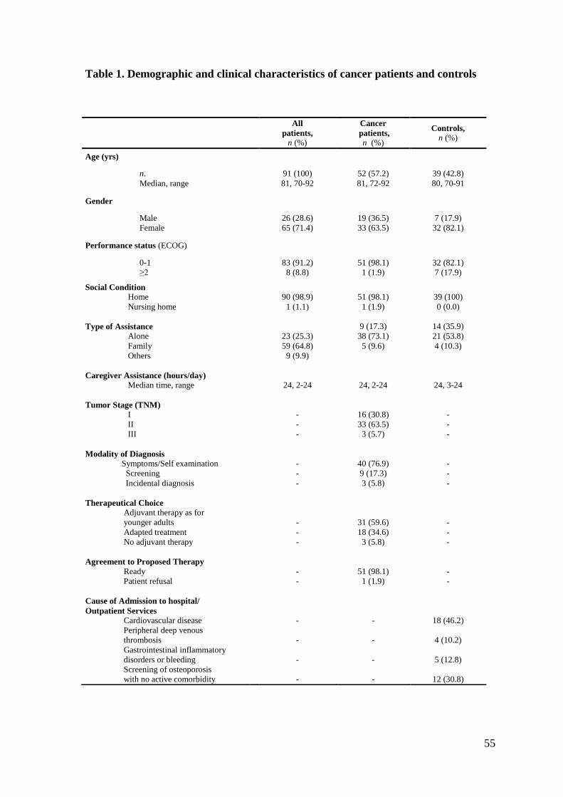
55
Table 1. Demographic and clinical characteristics of cancer patients and controls
All
patients,
n (%)
Cancer
patients,
n (%)
Controls,
n (%)
Age (yrs)
n.
Median, range
91 (100)
81, 70-92
52 (57.2)
81, 72-92
39 (42.8)
80, 70-91
Gender
Male
Female
26 (28.6)
65 (71.4)
19 (36.5)
33 (63.5)
7 (17.9)
32 (82.1)
Performance status (ECOG)
0-1
≥2
83 (91.2)
8 (8.8)
51 (98.1)
1 (1.9)
32 (82.1)
7 (17.9)
Social Condition
Home
Nursing home
90 (98.9)
1 (1.1)
51 (98.1)
1 (1.9)
39 (100)
0 (0.0)
Type of Assistance
Alone
Family
Others
23 (25.3)
59 (64.8)
9 (9.9)
9 (17.3)
38 (73.1)
5 (9.6)
14 (35.9)
21 (53.8)
4 (10.3)
Caregiver Assistance (hours/day)
Median time, range
24, 2-24
24, 2-24
24, 3-24
Tumor Stage (TNM)
I
II
III
-
-
-
16 (30.8)
33 (63.5)
3 (5.7)
-
-
-
Modality of Diagnosis
Symptoms/Self examination
Screening
Incidental diagnosis
-
-
-
40 (76.9)
9 (17.3)
3 (5.8)
-
-
-
Therapeutical Choice
Adjuvant therapy as for
younger adults
Adapted treatment
No adjuvant therapy
-
-
-
31 (59.6)
18 (34.6)
3 (5.8)
-
-
-
Agreement to Proposed Therapy
Ready
Patient refusal
-
-
51 (98.1)
1 (1.9)
-
-
Cause of Admission to hospital/
Outpatient Services
Cardiovascular disease
Peripheral deep venous
thrombosis
Gastrointestinal inflammatory
disorders or bleeding
Screening of osteoporosis
with no active comorbidity
-
-
-
-
-
-
-
-
18 (46.2)
4 (10.2)
5 (12.8)
12 (30.8)

56
Table 2. Scores of geriatric tools in cancer patients and controls
Geriatric Tool All patients Cancer patients Controls OR (95% CI) p-value
CGA, n (%)
Fit
Vulnerable
Frail
MPI, n (%)
Low
Moderate
Severe
MPI
Median, range
Dependencies in ADL, n (%)
No
≥1
Dependencies in IADL, n (%)
No
≥1
MMSE
Median [IQR]
SPMSQ
Median [IQR]
CIRS-CI Median [IQR]
CIRS-SI
Median [IQR]
GDS
Median [IQR]
BMI Median [IQR]
Drugs for concomitant diseases
Median [IQR]
37 (40.7)
28 (30.7)
26 (28.6)
78 (85.7)
12 (13.2)
1 (1.1)
0.25, 0.00-0.69
71 (78.0)
20 (22.0)
57 (62.6)
34 (37.4)
27.1 [24.4-28.4]
1.0 [0.0-2.0]
3.0 [1.0-4.0]
1.5 [1.3-1.6]
3.0 [1.0-6.0]
25.7 [24.0-27.4]
5 [3-6]
24 (46.2)
19 (36.5)
9 (17.3)
49 (94.2)
3 (5.8)
0 (0.0)
0.19, 0.00-0.44
44 (84.6)
8 (15.4)
36 (69.2)
16 (30.8)
26.2 [24.4-27.3]
1.0 [0.0-2.0]
2.0 [1.0-3.0]
1.4 [1.2-1.5]
3.0 [1.2-6.0]
25.8 [24.2-26.9]
4 [2-5]
13 (33.3)
9 (23.1)
17 (43.6)
29 (74.3)
9 (23.1)
1 (2.6)
0.25, 0.06-0.69
27 (69.2)
12 (30.8)
21 (53.8)
18 (46.2)
28.3 [26.4-29.3]
0.0 [0.0-2.0]
3.0 [2.0-4.0]
1.6 [1.4-1.8]
3.0 [1.0-6.0]
25.0 [23.0-28.6]
5 [4-8]
1
1.14 (0.40-3.24)
0.29 (0.10-0.82)
1
0.20 (0.05-0.79)
<0.01
-
1
0.41 (0.15-1.14)
1
0.52 (0.22-1.23)
-
-
-
-
-
-
-
0.801 a
0.020 a
0.022 a
ns a
0.001 a
0.790 a
0.133 a
0.001 b
0.013 b
0.032 b
<0.001 b
0.888 b
0.911b
0.003 b
a Pearson's χ
2 or Fisher test;
b Mann-Whitney U test
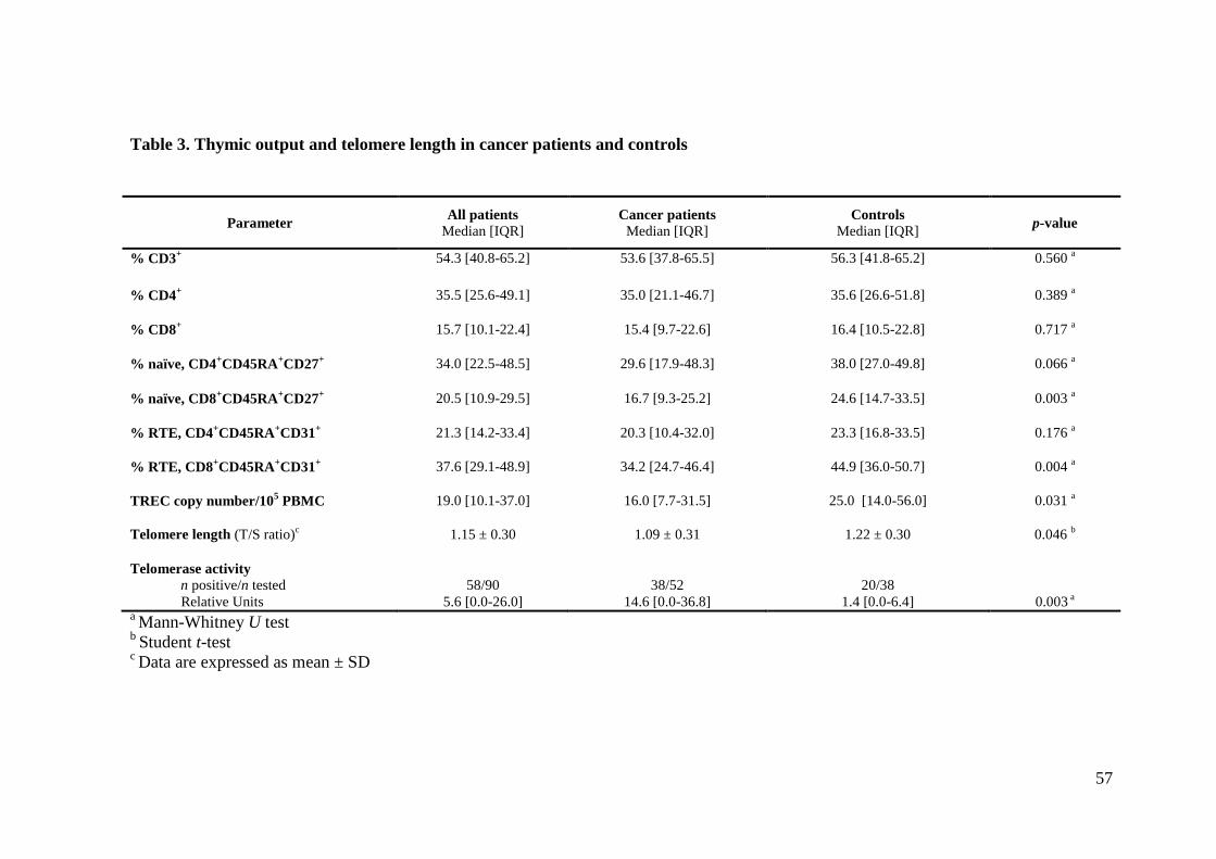
57
Table 3. Thymic output and telomere length in cancer patients and controls
Parameter
All patients
Median [IQR] Cancer patients
Median [IQR] Controls
Median [IQR]
p-value
% CD3+
54.3 [40.8-65.2] 53.6 [37.8-65.5] 56.3 [41.8-65.2] 0.560 a
% CD4+
35.5 [25.6-49.1] 35.0 [21.1-46.7] 35.6 [26.6-51.8] 0.389 a
% CD8+
15.7 [10.1-22.4] 15.4 [9.7-22.6] 16.4 [10.5-22.8] 0.717 a
% naïve, CD4+CD45RA
+CD27
+
34.0 [22.5-48.5] 29.6 [17.9-48.3] 38.0 [27.0-49.8] 0.066 a
% naïve, CD8+CD45RA
+CD27
+ 20.5 [10.9-29.5] 16.7 [9.3-25.2] 24.6 [14.7-33.5] 0.003
a
% RTE, CD4+CD45RA
+CD31
+
21.3 [14.2-33.4]
20.3 [10.4-32.0]
23.3 [16.8-33.5]
0.176 a
% RTE, CD8+CD45RA
+CD31
+
37.6 [29.1-48.9]
34.2 [24.7-46.4]
44.9 [36.0-50.7]
0.004 a
TREC copy number/105 PBMC
19.0 [10.1-37.0]
16.0 [7.7-31.5]
25.0 [14.0-56.0]
0.031 a
Telomere length (T/S ratio)c
1.15 ± 0.30 1.09 ± 0.31 1.22 ± 0.30 0.046 b
Telomerase activity n positive/n tested
Relative Units
58/90
5.6 [0.0-26.0]
38/52
14.6 [0.0-36.8]
20/38
1.4 [0.0-6.4]
0.003 a
a Mann-Whitney U test
b Student t-test
c Data are expressed as mean ± SD

58
References
Fries JF. Measuring and monitoring success in compressing morbidity. Ann Intern Med 2003;
139:455-9
American Cancer Society. Cancer Facts & Figures 2012. Atlanta: American Cancer Society 2012,
1-65
Sargent DJ, Goldberg RM, Jacobson SD, Macdonald JS, Labianca R, Haller DG, Shepherd LE,
Seitz JF, Francini G. A pooled analysis of adjuvant chemotherapy for resected colon cancer in
elderly patients. N Engl J Med 2001; 345:1091-7
Bouchardy C, Rapiti E, Fioretta G, Laissue P, Neyroud-Caspar I, Schäfer P, Kurtz J, Sappino AP,
Vlastos G. Undertreatment strongly decreases prognosis of breast cancer in elderly women. J Clin
Oncol 2003; 21:3580-7
Dale DC. Poor prognosis in elderly patients with cancer: the role of bias and undertreatment. J
Support Oncol 2003; 1:11-7
Ng R, de Boer R, Green MD. Undertreatment of elderly patients with non-small-cell lung cancer.
Clin Lung Cancer 2005; 7:168-74
Muss HB, Woolf S, Berry D, Cirrincione C, Weiss RB, Budman D, Wood WC, Henderson IC,
Hudis C, Winer E, Cohen H, Wheeler J, Norton L; Cancer and Leukemia Group B. Adjuvant
chemotherapy in older and younger women with lymph node-positive breast cancer. JAMA 2005;
293:1073-81
Hurria A, Browner IS, Cohen HJ, Denlinger CS, deShazo M, Extermann M, Ganti AK, Holland
JC, Holmes HM, Karlekar MB, Keating NL, McKoy J, Medeiros BC, Mrozek E, O'Connor T,
Petersdorf SH, Rugo HS, Silliman RA, Tew WP, Walter LC, Weir AB 3rd, Wildes T. Senior
adult oncology. J Natl Compr Canc Netw 2012; 10:162-209
Muss HB, Berry DA, Cirrincione C, Budman DR, Henderson IC, Citron ML, Norton L, Winer
EP, Hudis CA; Cancer and Leukemia Group B Experience. Toxicity of older and younger patients
treated with adjuvant chemotherapy for node-positive breast cancer: the Cancer and Leukemia
Group B Experience. J Clin Oncol 2007; 25:3699-704
Balducci L. New paradigms for treating elderly patients with cancer: the comprehensive geriatric
assessment and guidelines for supportive care. J Support Oncol 2003; 1:30-7
Grubeck-Loebenstein B, Della Bella S, Iorio AM, Michel JP, Pawelec G, Solana R.
Immunosenescence and vaccine failure in the elderly. Aging Clin Exp Res 2009; 21:201-9
Fulop T, Kotb R, Fortin CF, Pawelec G, de Angelis F, Larbi A. Potential role of
immunosenescence in cancer development. Ann N Y Acad Sci 2010; 1197:158-65
59
Berzins SP, Boyd RL, Miller JF. The role of the thymus and recent thymic migrants in the
maintenance of the adult peripheral lymphocyte pool. J Exp Med 1998; 187:1839-48
Douek DC, McFarland RD, Keiser PH, Gage EA, Massey JM, Haynes BF, Polis MA, Haase AT,
Feinberg MB, Sullivan JL, Jamieson BD, Zack JA, Picker LJ, Koup RA. Changes in thymic
function with age and during the treatment of HIV infection. Nature 1998; 396:690-5
Naylor K, Li G, Vallejo AN, Lee WW, Koetz K, Bryl E, Witkowski J, Fulbright J, Weyand CM,
Goronzy JJ. The influence of age on T cell generation and TCR diversity. J Immunol 2005;
174:7446-52
Nasi M, Troiano L, Lugli E, Pinti M, Ferraresi R, Monterastelli E, Mussi C, Salvioli G,
Franceschi C, Cossarizza A. Thymic output and functionality of the IL-7/IL-7 receptor system in
centenarians: implications for the neolymphogenesis at the limit of human life. Aging Cell 2006;
5:167-75
Mitchell WA, Lang PO, Aspinall R. Tracing thymic output in older individuals. Clin Exp
Immunol 2010; 161:497-503
Zhang L, Lewin SR, Markowitz M, Lin HH, Skulsky E, Karanicolas R, He Y, Jin X, Tuttleton S,
Vesanen M, Spiegel H, Kost R, van Lunzen J, Stellbrink HJ, Wolinsky S, Borkowsky W,
Palumbo P, Kostrikis LG, Ho DD. Measuring recent thymic emigrants in blood of normal and
HIV-1-infected individuals before and after effective therapy. J Exp Med 1999; 190:725-32
Ometto L, De Forni D, Patiri F, Trouplin V, Mammano F, Giacomet V, Giaquinto C, Douek D,
Koup R, De Rossi A. Immune reconstitution in HIV-1-infected children on antiretroviral therapy:
role of thymic output and viral fitness. AIDS 2002; 16:839-49
De Rossi A, Walker AS, Klein N, De Forni D, King D, Gibb DM. Increased thymic output after
initiation of antiretroviral therapy in human immunodeficiency virus type 1-infected children in
the Paediatric European Network for Treatment of AIDS (PENTA) 5 Trial. J Infect Dis 2002;
186:312-20
Kimmig S, Przybylski GK, Schmidt CA, Laurisch K, Möwes B, Radbruch A, Thiel A. Two
subsets of naive T helper cells with distinct T cell receptor excision circle content in human adult
peripheral blood. J Exp Med 2002; 195:789-94
Kohler S, Wagner U, Pierer M, Kimmig S, Oppmann B, Möwes B, Jülke K, Romagnani C, Thiel
A. Post-thymic in vivo proliferation of naive CD4+ T cells constrains the TCR repertoire in
healthy human adults. Eur J Immunol 2005; 35:1987-94
Junge S, Kloeckener-Gruissem B, Zufferey R, Keisker A, Salgo B, Fauchere JC, Scherer F,
Shalaby T, Grotzer M, Siler U, Seger R, Güngör T. Correlation between recent thymic emigrants
and CD31+ (PECAM-1) CD4+ T cells in normal individuals during aging and in lymphopenic
children. Eur J Immunol 2007; 37:3270-80

60
Kuss I, Schaefer C, Godfrey TE, Ferris RL, Harris JM, Gooding W, Whiteside TL. Recent thymic
emigrants and subsets of naive and memory T cells in the circulation of patients with head and
neck cancer. Clin Immunol 2005; 116:27-36
Blackburn EH, Greider CW, Szostak JW. Telomeres and telomerase: the path from maize,
Tetrahymena and yeast to human cancer and aging. Nat Med 2006; 12:1133-8
Blasco MA. Telomeres and human disease: ageing, cancer and beyond. Nat Rev Genet 2005;
6:611-22
Dolcetti R, De Rossi A. Telomere/telomerase interplay in virus-driven and virus-independent
lymphomagenesis: pathogenic and clinical implications. Med Res Rev 2012; 32:233-53
Garcia-Aranda C, de Juan C, Diaz-Lopez A, Sanchez-Pernaute A, Torres AJ, Diaz-Rubio E,
Balibrea JL, Benito M, Iniesta P. Correlations of telomere length, telomerase activity, and
telomeric-repeat binding factor 1 expression in colorectal carcinoma. Cancer 2006; 106:541-51
Rampazzo E, Bertorelle R, Serra L, Terrin L, Candiotto C, Pucciarelli S, Del Bianco P, Nitti D,
De Rossi A. Relationship between telomere shortening, genetic instability, and site of tumour
origin in colorectal cancers. Br J Cancer 2010; 102:1300-5
Wong JM, Collins K. Telomere maintenance and disease. Lancet 2003; 362:983-8
Aviv A. Telomeres and human somatic fitness. J Gerontol A Biol Sci Med Sci 2006; 6:871-3
Baird DM. Telomeres. Exp Gerontol 2006; 41:1223-7
Woo J, Tang NL, Suen E, Leung JC, Leung PC. Telomeres and frailty. Mech Ageing Dev 2008;
129:642-8
Collerton J, Martin-Ruiz C, Davies K, Hilkens CM, Isaacs J, Kolenda C, Parker C, Dunn M, Catt
M, Jagger C, von Zglinicki T, Kirkwood TB. Frailty and the role of inflammation,
immunosenescence and cellular ageing in the very old: cross-sectional findings from the
Newcastle 85+ Study. Mech Ageing Dev 2012; 133:456-66
Lan Q, Cawthon R, Gao Y, Hu W, Hosgood HD 3rd, Barone-Adesi F, Ji BT, Bassig B, Chow
WH, Shu X, Cai Q, Xiang Y, Berndt S, Kim C, Chanock S, Zheng W, Rothman N. Longer
telomere length in peripheral white blood cells is associated with risk of lung cancer and the
rs2736100 (CLPTM1L-TERT) polymorphism in a prospective cohort study among women in
China. PLoS One 2013; 8:e59230
Svenson U, Nordfjäll K, Stegmayr B, Manjer J, Nilsson P, Tavelin B, Henriksson R, Lenner P,
Roos G. Breast cancer survival is associated with telomere length in peripheral blood cells.
Cancer Res 2008; 68:3618-23
Bojesen SE, Pooley KA, Johnatty SE et al. Multiple independent variants at the TERT locus are
associated with telomere length and risks of breast and ovarian cancer. Nat Genet 2013; 45:371-
84

61
Martinez-Delgado B, Yanowsky K, Inglada-Perez L, de la Hoya M, Caldes T, Vega A, Blanco A,
Martin T, Gonzalez-Sarmiento R, Blasco M, Robledo M, Urioste M, Song H, Pharoah P, Benitez
J. Shorter telomere length is associated with increased ovarian cancer risk in both familial and
sporadic cases. J Med Genet 2012; 49:341-4
Riegert-Johnson DL, Boardman LA, Crook JE, Thomas CS, Johnson RA, Roberts ME. Shorter
peripheral blood telomeres are a potential biomarker for patients with advanced colorectal
adenomas. Int J Biol Markers 2012; 27:e375-80
Shao L, Wood CG, Zhang D, Tannir NM, Matin S, Dinney CP, Wu X. Telomere dysfunction in
peripheral lymphocytes as a potential predisposition factor for renal cancer. J Urol 2007;
178:1492-6
Wu X, Amos CI, Zhu Y, Zhao H, Grossman BH, Shay JW, Luo S, Hong WK, Spitz MR.
Telomere dysfunction: a potential cancer predisposition factor. J Natl Cancer Inst 2003; 95:1211-
8
Qu F, Li R, He X, Li Q, Xie S, Gong L, Ji G, Lu J, Bao G. Short telomere length in peripheral
blood leukocyte predicts poor prognosis and indicates an immunosuppressive phenotype in gastric
cancer patients. Mol Oncol 2014; [Epub ahead of print]
Chen Y, Qu F, He X, Bao G, Liu X, Wan S, Xing J. Short leukocyte telomere length predicts poor
prognosis and indicates altered immune functions in colorectal cancer patients. Ann Oncol 2014;
25:869-76
Basso U, Monfardini S. Multidimensional geriatric evaluation in elderly cancer patients: a
practical approach. Eur J Cancer Care 2004; 13:424-33
Conwell Y, Forbes NT, Cox C, Caine ED. Validation of a measure of physical illness burden at
autopsy: the Cumulative Illness Rating Scale. J Am Geriatr Soc 1993; 41:38-41
Yesavage JA, Brink TL, Rose TL, Lum O, Huang V, Adey M, Leirer VO. Development and
validation of a geriatric depression screening scale: a preliminary report. J Psychiatr Res 1982-
1983; 17:37-49
Guigoz Y, Vellas B. The Mini Nutritional Assessment (MNA) for grading the nutritional state of
elderly patients: presentation of the MNA, history and validation. Nestle Nutr Workshop Ser Clin
Perform Programme 1999; 1:3-11
Pilotto A, Ferrucci L, Franceschi M, D'Ambrosio LP, Scarcelli C, Cascavilla L, Paris F,
Placentino G, Seripa D, Dallapiccola B, Leandro G. Development and validation of a
multidimensional prognostic index for one-year mortality from comprehensive geriatric
assessment in hospitalized older patients. Rejuvenation Res 2008; 11:151-61
Giantin V, Valentini E, Iasevoli M, Falci C, Siviero P, De Luca E, Maggi S, Martella B, Orrù G,
Crepaldi G, Monfardini S, Terranova O, Manzato E. Does the Multidimensional Prognostic Index
(MPI), based on a Comprehensive Geriatric Assessment (CGA), predict mortality in cancer
patients? Results of a prospective observational trial. J Geriatr Oncol 2013; 4:208-17

62
Freguja R, Gianesin K, Mosconi I, Zanchetta M, Carmona F, Rampon O, Giaquinto C, De Rossi
A. Regulatory T cells and chronic immune activation in human immunodeficiency virus 1 (HIV-
1)-infected children. Clin Exp Immunol 2011; 164:373-80
Anselmi A, Vendrame D, Rampon O, Giaquinto C, Zanchetta M, De Rossi A. Immune
reconstitution in human immunodeficiency virus type 1-infected children with different
virological responses to anti-retroviral therapy. Clin Exp Immunol 2007; 150:442-50
Rampazzo E, Bonaldi L, Trentin L, Visco C, Keppel S, Giunco S, Frezzato F, Facco M, Novella
E, Giaretta I, Del Bianco P, Semenzato G, De Rossi A. Telomere length and telomerase levels
delineate subgroups of B-cell chronic lymphocytic leukemia with different biological
characteristics and clinical outcomes. Haematologica 2012; 97:56-63
Scarpaci S, Frasca D, Barattini P, Guidi L, Doria G. DNA damage recognition and repair
capacities in human naïve and memory T cells from peripheral blood of young and elderly
subjects. Mech Ageing Dev 2003; 124:517-24
Cossarizza A, Ortolani C, Paganelli R, Barbieri D, Monti D, Sansoni P, Fagiolo U, Castellani G,
Bersani F, Londei M, Franceschi C. CD45 isoforms expression on CD4+ and CD8+ T cells
throughout life, from newborns to centenarians: implications for T cell memory. Mech Ageing
Dev 1996; 86:173-95
d'Adda di Fagagna F, Reaper PM, Clay-Farrace L, Fiegler H, Carr P, Von Zglinicki T, Saretzki G,
Carter NP, Jackson SP. A DNA damage checkpoint response in telomere-initiated senescence.
Nature 2003; 426:194-8
Banáth JP, Klokov D, MacPhail SH, Banuelos CA, Olive PL. Residual gammaH2AX foci as an
indication of lethal DNA lesions. BMC Cancer 2010; 10:4
Hueman MT, Stojadinovic A, Storrer CE, Dehqanzada ZA, Gurney JM, Shriver CD, Ponniah S,
Peoples GE. Analysis of naïve and memory CD4 and CD8 T cell populations in breast cancer
patients receiving a HER2/neu peptide (E75) and GM-CSF vaccine. Cancer Immunol Immunother
2007; 56:135-46
Poschke I, De Boniface J, Mao Y, Kiessling R. Tumor-induced changes in the phenotype of
blood-derived and tumor-associated T cells of early stage breast cancer patients. Int J Cancer
2012; 131:1611-20
Klebanoff CA, Gattinoni L, Restifo NP. CD8+ T-cell memory in tumor immunology and
immunotherapy. Immunol Rev 2006; 211:214-24
Williams MA, Bevan MJ. Effector and memory CTL differentiation. Annu Rev Immunol 2007;
25:171-92
Almanzar G, Schwaiger S, Jenewein B, Keller M, Herndler-Brandstetter D, Würzner R,
Schönitzer D, Grubeck-Loebenstein B. Long-term cytomegalovirus infection leads to significant
changes in the composition of the CD8+ T-cell repertoire, which may be the basis for an
imbalance in the cytokine production profile in elderly persons. J Virol 2005; 79:3675-83
63
Solana R, Tarazona R, Aiello AE, Akbar AN, Appay V, Beswick M, Bosch JA, Campos C,
Cantisán S, Cicin-Sain L, Derhovanessian E, Ferrando-Martínez S, Frasca D, Fulöp T, Govind S,
Grubeck-Loebenstein B, Hill A, Hurme M, Kern F, Larbi A, López-Botet M, Maier AB,
McElhaney JE, Moss P, Naumova E, Nikolich-Zugich J, Pera A, Rector JL, Riddell N, Sanchez-
Correa B, Sansoni P, Sauce D, van Lier R, Wang GC, Wills MR, Zieliński M, Pawelec G. CMV
and Immunosenescence: from basics to clinics. Immun Ageing 2012; 9:23
Steenstrup T, Hjelmborg JV, Mortensen LH, Kimura M, Christensen K, Aviv A. Leukocyte
telomere dynamics in the elderly. Eur J Epidemiol 2013; 28:181-7
Der G, Batty GD, Benzeval M, Deary IJ, Green MJ, McGlynn L, McIntyre A, Robertson T, Shiels
PG. Is telomere length a biomarker for aging: cross-sectional evidence from the west of Scotland?
PLoS One 2012; 7:e45166
Bisoffi M, Heaphy CM, Griffith JK. Telomeres: prognostic markers for solid tumors. Int J Cancer
2006; 119:2255-60
Franken B, de Groot MR, Mastboom WJ, Vermes I, van der Palen J, Tibbe AG, Terstappen LW.
Circulating tumor cells, disease recurrence and survival in newly diagnosed breast cancer. Breast
Cancer Res 2012; 14:R133
Shimada R, Iinuma H, Akahane T, Horiuchi A, Watanabe T. Prognostic significance of CTCs and
CSCs of tumor drainage vein blood in Dukes' stage B and C colorectal cancer patients. Oncol Rep
2012; 27:947-53

64
FIGURE LEGEND
Figure 1. Correlation between age and (A) TREC levels and (B) telomere lengths in
cancer patients and controls. Panel (A): TREC levels decline with age in the peripheral
blood cells of cancer patients (r=-0.478, p<0.001), but not in controls (r=0.071, p=0.677).
Panel (B): telomere lengths decrease with age in peripheral blood cells of controls (r=-
0.354, p=0.031), but not in cancer patients (r=-0.011, p=0.938).
Figure 2. CD8+ cell subsets distribution at baseline (A) and at follow-up (B). Pie
charts show that frequencies of naïve and central memory cells did not significantly differ
between baseline and follow-up (p=0.309 and p=0.137, respectively); conversely, the
proportion of effector memory cells significantly declined (p=0.017), while terminally
differentiated cells increased (p=0.031).
Figure 3. Flow cytometry analysis of y-H2AX induction in controls and cancer
patients. y-H2AX expression in untreated PBMC and after exposure to a dose radiation
of 2.5 Gy at 24 h, in control (n=2) and cancer patients (n=3). Error bars represent standard
deviation of the median fluorescence levels.
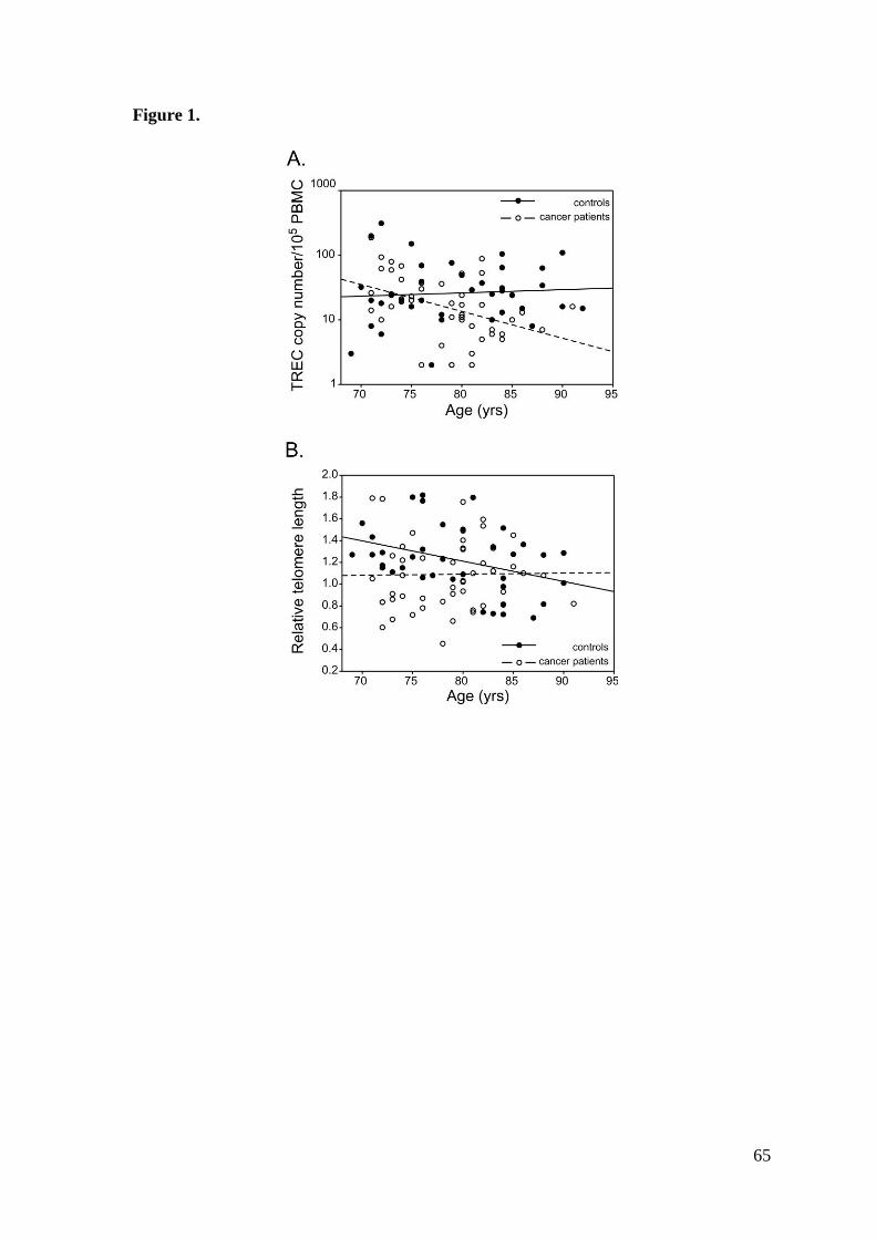
65
Figure 1.
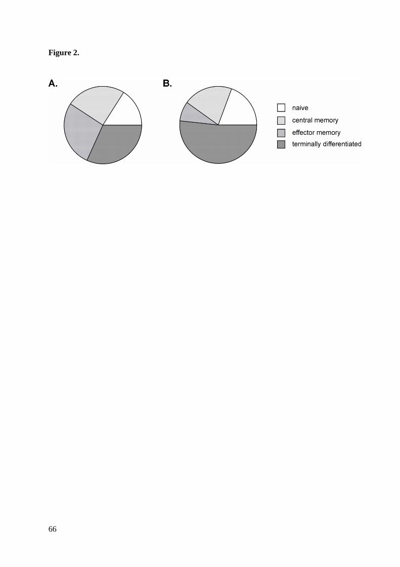
66
Figure 2.
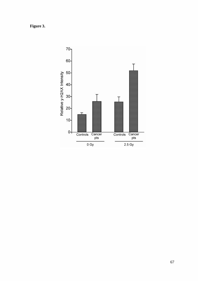
67
Figure 3.

68
Table 1S. Standard blood parameters: comparison between cancer patients and controls
Parameter All patients
Median [IQR] Cancer patients
Median [IQR] Controls
Median [IQR] p-value a
Leucocytes (x109/l)
Lymphocytes (x109/l)
Neutrophils (x109/l)
Hemoglobin (g/l)
Platelets (x109/l)
Total plasmatic proteins (g/l)
Albumin (%)
Gamma globulins (%)
Triglycerides (mmol/l)
Total cholesterol (mmol/l)
LDL-cholesterol (mmol/l)
HDL-cholesterol (mmol/l)
Creatinine (umol/l)
IgG (g/l)
IgA (g/l)
IgM (g/l)
IL-6 (ng/l)
CRP (mmol/l)
CMV b
n positive/n tested (%)
IgG (UI/ml)
6.6 [5.3-7.9]
1.6 [1.3-1.9]
3.8 [3.0-4.9]
127.0 [114.0-140.0]
236.0 [204.0-291.0]
66.8 [63.5-71.1]
57.0 [54.0-59.6]
15.2 [13.2-17.3]
1.1 [0.8-1.4]
4.9 [3.9-5.8]
2.8 [2.0-3.7]
1.4 [1.1-1.9]
76.0 [68.0-93.0]
10.5 [8.5-12.2]
2.3 [1.8-2.9]
0.9 [0.5-1.3]
3.0 [1.9-5.3]
3.5 [2.9-6.6]
88/91 (96.7%)
15000 [8200-21000]
6.7 [5.5-7.9]
1.7 [1.3-1.9]
3.9 [3.2-4.9]
128.0 [111.0-142.0]
253.5 [212.5-317.0]
68.4 [66.1-74.3]
57.9 [54.7-60.6]
14.9 [13.3-17.1]
1.2 [0.8-1.7]
5.1 [4.3-5.7]
3.3 [2.7-3.8]
1.4 [1.1-1.7]
77.5 [68.2-95.0]
10.3 [8.9-11.6]
2.6 [1.8-3.4]
0.8 [0.5-1.3]
2.6 [1.9-4.3]
3.5 [2.9-5.5]
49/52 (94.2%)
15000 [8125-19750]
5.9 [5.0-7.5]
1.5 [1.2-1.9]
3.6 [2.7-4.9]
126.0 [116.0-138.0]
225.0 [183.0-260.0]
64.0 [60.7-68.7]
55.9 [53.9-58.2]
15.8 [13.0-18.0]
0.9 [0.7-1.2]
4.5 [3.7-5.8]
2.4 [1.7-3.6]
1.6 [1.1-1.9]
75.0 [68.0-93.0]
11.0 [7.7-12.6]
2.2 [1.6-2.7]
0. 9 [0.5-1.4]
3.7 [2.1-6.3]
3.5 [3.4-10.8]
100/100 (100%)
15000 [9300-24000]
0.110
0.165
0.141
0.665
0.011
<0.001
0.530
0.549
0.009
0.229
0.006
0.194
0.642
0.934
0.104
0.853
0.033
0.044
0.498
a Mann-Whitney U test;
b CMV serology: Serum CMV antibody (IgG) determined by ELISA; antibodies titers>230 were considered to be positive.

69
Table 2S. Relationship between markers of immune senescence and geriatric scores in cancer patients and controls
TREC level Telomere length Telomerase activity
All patients Cancer
patients Controls
All patients Cancer
patients Controls
All patients Cancer
patients Controls
MPI -0.03, 0.77 -0.04, 0.78 -0.24, 0.16 -0.04, 0.74 0.01, 0.95 -0.26, 0.13 -0.03, 0.81 0.16, 0.25 0.00, 0.98
MMSE -0.1, 0.35 -0.28, 0.05* -0.14, 0.42 0.06, 0.61 0.15, 0.28 -0.31, 0.06 -0.14, 0.19 -0.17, 0.22 0.19, 0.25
CIRS-CI -0.04, 0.71 0.04, 0.79 -0.31, 0.07 0.04, 0.70 0.05, 0.72 -0.05, 0.76 -0.12, 0.26 0.01, 0.94 -0.18, 0.27
CIRS-SI 0.26, 0.02* -0.07, 0.61 0.45, 0.01** 0.16, 0.13 -0.11, 0.42 0.35, 0.03* -0.06, 0.60 0.01, 0.95 0.18, 0.27
GDS -0.03, 0.79 -0.19, 0.19 0.22, 0.18 -0.10, 0.34 -0.09, 0.54 -0.05, 0.75 -0.24, 0.02* -0.20, 0.15 -0.26, 0.12
BMI -0.03, 0.79 0.01, 0.96 -0.10, 0.57 0.02, 0.83 0.14, 0.31 -0.11, 0.53 0.27, 0.01** 0.29, 0.04* 0.14, 0.39
Concomitant
drugs -0.01, 0.91 0.11, 0.43 -0.39, 0.02* -0.13, 0.24 -0.18, 0.21 -0.24,0.15 -0.02, 0.89 0.13, 0.36 0.08, 0.65
Data are expressed as Spearman's rho coefficient, p-value. *
Correlation is significant at the level 0.05 (two-tailed) **
Correlation is significant at the level 0.01 (two-tailed)
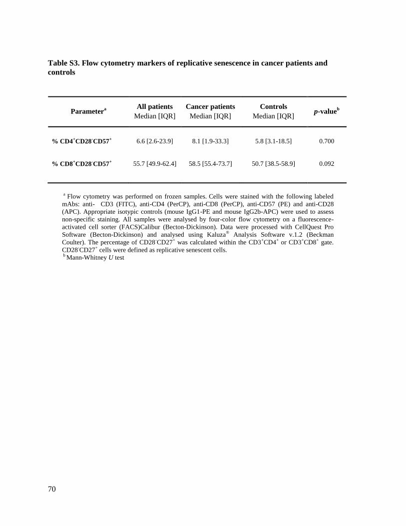
70
Table S3. Flow cytometry markers of replicative senescence in cancer patients and
controls
Parametera
All patients
Median [IQR]
Cancer patients
Median [IQR]
Controls
Median [IQR]
p-valueb
% CD4+CD28
-CD57
+
6.6 [2.6-23.9]
8.1 [1.9-33.3]
5.8 [3.1-18.5]
0.700
% CD8+CD28
-CD57
+
55.7 [49.9-62.4]
58.5 [55.4-73.7]
50.7 [38.5-58.9]
0.092
a Flow cytometry was performed on frozen samples.
Cells were stained with the following labeled
mAbs: anti- CD3 (FITC), anti-CD4 (PerCP), anti-CD8 (PerCP), anti-CD57 (PE) and anti-CD28
(APC). Appropriate isotypic controls (mouse IgG1-PE and mouse IgG2b-APC) were used to assess
non-specific staining. All samples were analysed by four-color flow cytometry on a fluorescence-
activated cell sorter (FACS)Calibur (Becton-Dickinson). Data were processed with CellQuest Pro
Software (Becton-Dickinson) and analysed using Kaluza® Analysis Software v.1.2 (Beckman
Coulter). The percentage of CD28-CD27
+ was calculated within the CD3
+CD4
+ or CD3
+CD8
+ gate.
CD28-CD27
+ cells were defined as replicative senescent cells.
b Mann-Whitney U test

71
Chapter 3

72
73
Premature Aging and Immune Senescence in HIV-Infected Children
Ketty Gianesin1, Antoni Noguera-Julian
2, Marisa Zanchetta
3, Maria Raffaella Petrara
1,
Osvalda Rampon4, Clàudia Fortuny
2, Mireia Camós
5, Carlo Giaquinto
4, Anita De Rossi
1,3
1 Section of Oncology and Immunology, Unit of Viral Oncology and AIDS Reference
Center, Department of Surgery, Oncology and Gastroenterology-DiSCOG, University of
Padova, Padova, Italy
2 Unitat d’Infectologia, Servei de Pediatria; Hospital Sant Joan de Déu-Universitat de
Barcelona, Barcelona, Spain
3 Istituto Oncologico Veneto (IOV)-IRCCS, Padova, Italy
4 Department of Mother and Child Health, University of Padova, Padova, Italy
5 Servei d’Hematologia; Hospital Sant Joan de Déu-Universitat de Barcelona, Barcelona,
Spain
Manuscript in preparation

74
ABSTRACT
Background. Several data indicate that HIV-infected adults undergo premature aging and
immune senescence; chronic immune activation may play a critical role in these dysfunctions
which, besides immune depression, may constitute important risk factors for the onset of
AIDS-associated and non-AIDS associated malignancies in HIV-infected individuals.
Limited data are available for HIV-infected children, in whom immune activation and
senescence are likely to be more deleterious, since their immune system co-evolves from
birth with HIV.
Patients and Methods. 71 HIV-infected (HIV+) children, aged from 0-5 years, 65 HIV-
exposed-uninfected (HEU) and 56 HIV-1-unexposed-uninfected (HUU) age-matched
children were studied. 42% of the HIV+ children were not on antiretroviral therapy (ART).
Telomere length (TL) and T-cell receptor rearrangement excision circle (TREC) levels were
quantified in peripheral blood cells by real-time PCR. Subgroups of 24 HIV+, 21 HEU and
18 HUU children were studied for CD4+ and CD8
+ cell differentiation (CD45RA, CD27,
CD31), senescence (CD28, CD57) and activation/exhaustion (CD38, HLA-DR, PD-1)
markers by flow cytometry. Statistical analyses were performed with SPSS v22.
Results. TL were significantly shorter in HIV+ than in HEU and HUU children (overall
p<0.001, adjusted for age); moreover, HIV+ ART-naïve children had shorter TL compared
with children on ART (median 2.11 [interquartile range (IQR) 1.75-2.37] vs 2.46 [2.07-2.68];
p=0.0029 adjusted for age). CD8+ recent thymic emigrant cells (CD45RA
+CD31
+) and TREC
levels were significantly lower in HIV+ than in HEU and HUU groups (overall, p=0.005 and

75
p=0.0249, respectively), while percentages of CD8 effector memory (CD45RA-CD27
-) and
terminally differentiated cells (CD45RA+CD27
-) were higher in the former (overall, p=0.033,
and p<0.001, respectively). CD8 senescent cells (CD28-CD57
+) were higher in HIV+ than in
HEU and HUU children (25.8% [12.4-43.2] vs 8.5% [6.8-16.7] vs 9.7% [3.3-27.3]; p=0.004),
as were CD8+ activated cells (CD38
+HLA-DR
+) (7.0% [5.2-12.2] vs 3.5% [3.9-7.6] vs 3.7%
[2.4-6.7]; p<0.001) and CD8+PD-1
+ cells (7.1% [5.0-12.4] vs 3.5% [2.1-5.9] vs 3.7% [2.4-
5.3]; p<0.001). Within CD4+ cell subset, percentages of senescent cells did not differ
between HIV+ and controls, although PD-1+ expression tended to be up-regulated in HIV+
children (overall, p=0.050).
Conclusions. HIV-infected children exhibit a premature biological aging with accelerated
immune senescence which affects in particular the CD8+ cell subset. HIV infection per se
seems to influence the aging process, rather than exposure to ART for prophylaxis or
treatment.

76
1. INTRODUCTION
The introduction of Antiretroviral Therapy (ART) has changed the natural history of
pediatric HIV infection; indeed, ART-based prophylaxis regimens have reduced mother-to-
child transmission (MTCT) of HIV from 15-20% to under 2% in high-income countries and
have also given rise to substantial improvements in terms of survival and quality of life in
HIV-infected children [Gibb DM et al, BMJ 2003; Judd A et al, Clin Infect Dis 2007]. In
view of this increased survival, HIV infection is now considered a chronic disease that
persists for many decades [Deeks SG et al, Lancet 2013]. However, despite its improvement
of the immune function and reduction of AIDS-related complications, including AIDS-
associated malignancies (ADM), ART does not restore full health. Indeed, many studies have
evidenced that ART-treated HIV-infected adults have a higher risk of non-AIDS-related
overall morbidity and mortality, compared to age-matched HIV-uninfected individuals. This
increased risk is mainly due to a range of non-AIDS-defining illnesses that are associated
with aging, including malignancies [Guaraldi G et al, Clin Infect Dis 2011], and it has been
advanced that the increase of non-AIDS-defining diseases, including non-AIDS-defining
malignancies (non-ADM) among HIV-infected patients is due to a premature aging [Effros
RB, Clin Infect Dis 2008]. The pathogenic mechanism underlying this increased risk is still
poorly understood. Chronic immune activation due to the persistence of circulating HIV
virions may play a key role for the senescent pathway. Activated cells undergo clonal
expansion in response to viral persistence, resulting in differentiation and accumulation of
nonfunctional senescent cells [Desai S & Landay A, Curr HIV/AIDS Rep 2010]. It has been
also advanced that premature and accelerated aging in HIV patients can be caused by adverse
effects of antiretroviral drugs. Nucleoside reverse transcriptase inhibitors (NRTIs) have been

77
shown to inhibit telomerase activity in replicating cell lines in vitro, leading to accelerated
shortening of telomere length, a marker of biological aging [Liu X et al, Nucleic Acids Res
2007; Hukezalie KR et al, PloS One 2012].
Clinical complications of HIV infection and ART treatment in children are more serious than
in adults. The course of HIV infection in the former is characterized by faster disease
progression and shorter time to AIDS, compared to adults. After infection, HIV plasma RNA
levels are higher in vertically infected children than in adults. In addition they persist at high
levels and decline slowly with age [De Rossi A et al, J Clin Invest 1996; McIntosh K et al,
Pediatr Infect Dis J 1996], whereas in adults a control of viral load is reached within few
weeks after infection [Henrard DR et al, JAMA 1995]. This gradual control of viral
replication has been associated with increasing maturation and development of the immune
system. The innate immunity, in children, is of particular relevance and plays a critical role in
HIV pathogenesis [Ricci E et al, J Acquir Immune Defic Syndr 2009; Freguja R et al, New
Microbiol 2012; Gianesin K et al, Plos One 2012] given that the adaptive immune system is
still under development [Prendergast AJ et al, Nat Rev Immunol 2012]. Since in children the
immune system co-evolves from birth with HIV and ART is required for decades, the effects
of chronic immune activation and immune senescence together with ART toxicity in the
course of their immune system’s maturation might be more deleterious.
To date only little data are available on premature aging in HIV-infected children [Mansoor
N et al, Clin Immunol 2009; Côté HC et al, PLoS One 2012; Diaz L et al, J Acquir Immune
Defic Syndr 2012; Méndez-Lagares G et al, Clin Microbiol Infect 2013]. In this study we
evaluated the status of biological aging and the degree of immune senescence in vertically
HIV-infected children.
78
2. PATIENTS & METHODS
2.1 Study Population
A total of 71 perinatally HIV-infected (HIV+) children, aged 0-5 years, 65 exposed-
uninfected (HEU) and 56 unexposed-uninfected (HUU) age-matched children were included
in the study. All HIV+ and HEU children attended to the Department of Mother and Child
Health, University of Padova (Padova, Italy) or to the Infectious Diseases Unit, Pediatrics
Department, Hospital Saint Joan de Déu, Universitat de Barcelona, (Barcelona, Spain). For
each HIV+ and HEU child, the first cryopreserved available sample after birth has been
chosen for the study. No HIV+ or HEU child was breastfed. HUU children were recruited at
the Pediatric Emergency Department of Azienda Ospedaliera Padova or Hospital Saint Joan
de Déu, Universitat de Barcelona. Exclusion criteria were: malignancies, chronic infections,
sarcoidosis, diabetes mellitus type-1, rheumatoid arthritis and systemic lupus erythematosus.
The study was approved by the Ethics Committee of Azienda Ospedaliera Padova (Prot.
n.#2921P); informed consent was obtained for patients and controls from their
parents/guardians.
2.2 Sample preparation
Peripheral blood mononuclear cells (PBMC) were isolated from EDTA-treated peripheral
blood by centrifugation on a Ficoll-Paque gradient (Pharmacia Biotech, Uppsala, Sweden).
PBMC and plasma samples were cryopreserved and stored in liquid nitrogen until use. Two
million PBMC were lysed as previously described [Righetti E et al, AIDS 2002].

79
2.3 Telomere length measurement by quantitative real-time PCR
Relative telomere length (TL) was determined by monochrome quantitative multiplex PCR
assay [Cawthon RM, Nucleic Acids Res 2009] with minor modifications. Each PCR reaction
was performed in a final volume of 25 µl containing 5 µl sample (~2 ng DNA/µl) and 20 µl
reaction mix containing 0.75 x SYBR Green I (Invitrogen), 10 mM Tris-HCl pH 8.3, 50 mM
KCl, 3 mM MgCl2, 0.2 mM each dNTP (Applied Biosystems), 1 mM DTT, 0.625 U
AmpliTaq Gold DNA polymerase, 1% DMSO (Sigma) and 900 nM of each of the primers.
The primer pair employed for telomere amplification were TELG 5’-
ACACTAAGGTTTGGGTTTGGGTTTGGGTTTGGGTTAGTGT-3’, and TELC 5’-
TGTTAGGTATCCCTATCCCTATCCCTATCCCTATCCCTAACA-3’, and the primer pair
for the amplification of the single-copy gene albumin were ALBU 5’-
CGGCGGCGGGCGGCGCGGGCTGGGCGGAAATGCTGCACAGAATCCTTG-3’ and
ALBD 5’-GCCCGGCCCGCCGCGCCCGTCCCGCCGGAAAAGCATGGTCGCCTGTT-
3’. The single-copy gene albumin was amplified simultaneously with the telomere template
in the same well and was used as a reference to adjust for different amounts of DNA in
different samples. The PCR reactions were performed on a LightCycler®
480 real-time PCR
detection system (Roche Applied Science). The thermal cycling profile was 15 min at 95ºC,
2 cycles of 15 s at 94ºC, 15 s at 49ºC, followed by 40 cycles of 15 s at 94ºC, 10 s at 62ºC, 15
s at 74ºC, 10 s at 84ºC, 15 s at 88ºC, with signal acquisition at the end of both the 74°C and
88°C steps. A standard curve was generated at each PCR run, consisting of DNA from the
RAJI cell line serially diluted from 100 to 0.41 ng/µl. All DNA samples and reference
samples were run in triplicate. LightCycler raw text files were converted to grid format using
LC480Conversion free software developed by the Heart Failure Research Center (HFRC),

80
Amsterdam,Netherlands(http://www.hartfaalcentrum.nl/index.php?main=files&fileName=L
C480Conversion.zip&description=LC480%20Conversion&sub=LC480Conversion). The
converted data were analyzed using LinRegPCR free software developed by Ruitjer et al
[Ruitjer JM et al, Nucleic Acids Res 2009]. Mean Ct values were used to calculate the
relative telomere length (TL) using the telomere/single-copy gene ratio according to the
formula: ΔCtsample= Cttelomere - Ctalbumin, ΔΔCt = ΔCtsample - ΔCtreference curve (where ΔCtreference
curve = Cttelomere_RAJI - Ctcontrol RAJI) and then T/S = 2-ΔΔCt
[Cawthon RM, Nucleic Acids Res
2009]. Intra and inter-assay reproducibility of both telomere and albumin PCR results was
evaluated using dilutions of the reference curve. The standard deviation of Ct values was
≤0.19 (% coefficient of variation ≤0.94) in six replicates of samples amplified in the same
PCR run, and ≤0.28 (% coefficient of variation ≤1.31) among mean values of triplicates in
different PCR runs.
2.4 T-cell receptor rearrangement excision circle (TREC) levels quantification
Thymic output in PBMC was studied by measuring TREC levels by real-time PCR, as
described previously [Ometto L et al, AIDS 2002]. TREC levels were expressed as the
number of TREC copies per 105 PBMC.
2.5 Viral load quantification
Plasma HIV RNA levels were determined in all HIV-infected children using the COBAS
Taqman HIV-1 test (Roche, Branchburg, NJ, USA). The lower limit of detection was 50 HIV
RNA copies/ml. HIV DNA levels in PBMC were measured by real-time PCR, as described
previously [Ometto L et al, AIDS 2002]. The HIV copy number was normalized against the
81
TERT copy number, and the final results were expressed as HIV DNA copies/106 cells.
Samples for HIV DNA quantification were available for 44 children.
2.6 Flow cytometry analysis
T-cell phenotyping was performed on cryopreserved PBMC. Cells were thawed, washed,
stained for 20 min in the dark with Live/Dead Fixable Near-IR Dead Cell Stain Kit (Life
Technologies), and with fluorescent-conjugated mononuclear antibodies: CD3-FITC, CD31-
PE, CD38-PE, CD57-PE, CD45RA-APC and PD-1-PECy7 (Becton-Dickinson), CD27-
PECy7 (Beckman Coulter) and CD4-VioBlue, CD8-VioGreen, HLA-DR-APC and CD28-
APC (Miltenyi Biotec). Appropriate isotypic controls (mouse IgG1-PE, mouse IgG2b-APC
and mouse IgG1k-PECy7) were used to evaluate non-specific staining. Cells were then
washed with Automacs Buffer (Miltenyi BioteC) and resuspended in PBS supplemented with
1% paraformaldehyde. All samples were analysed using LSRII Flow cytometer (Becton-
Dickinson Biosciences). A total of 100000 events were collected in the lymphocyte gate
using morphological parameters (forward- and side-scatter). Data were processed using
FACSDiva™ Software (Becton-Dickinson) and analysed using Kaluza® Analyzing
Software v1.2 (Beckman Coulter). Figure 1 showed the gating strategies in identifying the
CD4+ and CD8
+ T-cell subsets. Samples for flow-cytometry analysis were available for 24
HIV+ (15 with undetectable plasmaviremia (<50 copies/ml) and 9 with detectable
plasmaviremia (median 3.64 [2.75-5.16] log10 copies/ml)), 21 HEU and 18 HUU children.
Two HIV+ children were in therapy interruption at sample collection.

82
2.7 Statistical analysis
Comparisons between groups were made using nonparametric tests, the Mann-Whitney U-
test or Kruskal-Wallis test, where appropriate. Correlations were explored using Spearman’s
rho test. Linear regression was used to model the relationship between TL and age. Samples
with undetectable plasma HIV RNA were assigned a value of 20 copies/ml to include them
in the statistical analyses. All statistical analyses were performed using SPSS software
version 22 (SPSS, Inc., Chicago, IL, USA). All P-values were two-tailed, and were
considered significant when lower than 0·05.
3. RESULTS
3.1 Characteristics of the study population
Characteristics of the 71 HIV+, 65 HEU and 56 HUU children are reported in Table 1. The
median age of HIV+ children was 3.11 [interquartile range (IQR) 1.41-4.48 years], 1.73
[0.99-3.20] years for HEU and 1.85 [0.85-3.46] years for HUU children. 30/71 (42%) HIV+
children were ART-naïve, while the remaining were in ART (median time of 18 [11.0-36.5]
months). ART-naïve HIV+ children were younger than those under therapy (1.70 [0.69-3.59]
and 3.62 [2.17-4.81] years, respectively; p=0.005). 61/65 (93.8%) HEU children were
exposed to prophylaxis during gestational period (median 34 [17-38] weeks) and received
postnatal prophylaxis (6 weeks of zidovudine (AZT) monotherapy). Regarding HUU
children, 38 of them were hospitalized for acute infections (pneumonia, influence,
gastroenteritis) and 18 were healthy and referred for minor surgery.
83
3.2 Telomere length is shorter in HIV-infected children
The median TL in PBMC was significantly lower in HIV+ children than in HEU and HUU
children, being the median TL of 2.21 [1.94-2.58], 2.63 [2.25-3.21], 2.88 [2.49-3.1],
respectively (overall, p<0.0001 adjusted for age). TL inversely correlated with age in HEU
and HUU groups (regression coefficient (β)=-0.01024, p=0.0079 and β=-0.01001, p=0.0112,
respectively), but not in HIV+ children (β=-0.00175, p=0.5870) (Figure 2A). Of note, among
HIV+ group, ART-naïve children had shorter TL compared with children on ART (2.11
[1.75-2.37] versus 2.46 [2.07-2.68]; p=0.0029 adjusted for age). Neither for ART-naïve
children nor for those on ART, TL was associated with age (β=-0.00540, p=0.2269; β=-
0.00455, p=0.2579, respectively) (Figure 2B).
3.3 Thymic output is lower in HIV-infected children
Given that an effective immune response depends on thymic function, thymic output was
quantified by measuring TREC levels. HEU and HUU children had higher levels of TREC
than HIV+ children (5409 [3411-6712], 5370 [2380-8101], 3498 [2051-6780] TREC
copies/105 PBMC; overall p=0.0249 adjusted for age). TREC levels decreased significantly
with increasing age in HEU and HUU groups (β=-61, p=0.0088 and β=-86, p=0.0003,
respectively), but not in HIV+ children (β=-17, p=0.3526) (Figure 2C). No significant
differences in TREC levels/105 PBMC were found between ART-treated children and ART-
naive children (p=0.2995) (Figure 2D).

84
3.4 Phenotypic T-cell alterations occur early in HIV-infected children
There were no differences in the frequencies of CD3+ T cells between the three groups
(overall, p=0.590). Percentage of total CD4+ T cells were lower in the HIV+ group than in
the other groups (overall, p=0.001). Within CD4+ T cells, HIV+ children and control groups
did not significantly differ in percentages of naïve (CD45RA+CD27
+), central memory
(CD45RA-CD27
+), effector memory (CD45RA
-CD27
-) and terminally differentiated
(CD45RA+CD27-) cell subsets (Table 2). However, when central and effector memory cell
subsets, the major cellular reservoirs for HIV [Chomont N et al, Nat Med 2009] where
considered together, they tended to be lower in HIV+ children than in HEU and HUU
children [21.9% [15.3-38.3] versus 29.8% [18.6-39.4] and 34.3% [24.1-42.6]; p=0.158).
Percentage of senescent cells (CD4+CD28
-CD57
+) were similar between the three groups
(p=0.568), while activated CD38+HLA-DR
+ and exhausted PD-1
+ were more expanded in
HIV+ children than in controls. In particular, CD4+CD38
+HLA-DR
+ and CD4
+PD-1
+ were
higher in children with detectable viral load than in aviremic children (p=0.056 and p=0.037,
respectively).
The median of CD8+ cell percentage tended to be higher in HIV+ children than in control
groups (overall, p=0.063). Notably, significant differences emerged among CD8+ cell
subsets. HIV+ children showed a lower percentage of naïve cells (CD45RA+CD27
+) than
HEU and HUU children (46.2% [37.6-75.1], 77.1% [55.9-84.7], 71.0% [46.7-86.1]; overall
p=0.019). In particular, HIV+ children had a lower frequency of CD8+ recent thymic
emigrant (RTE) (overall, p=0.005) and a higher percentage of peripheral expanded cells
(PEC; CD45RA+CD31
-) than control groups (overall, p=0.040), suggesting a strong
peripheral proliferation. Moreover, the amount of CD8+ RTE cells decreases with age in

85
HEU and in HUU children, but not in HIV+ children (Figure 3A-C). Interestingly, CD8+
RTE cells were lower in children with detectable viral load than in aviremic children (41.8%
[22.6-64.4] versus 55.9% [53.6-76.7], p=0.039), and tended to inversely correlate with HIV
RNA (r=-0.363, p=0.080) (Figure 3D). Taken together, these findings suggest that, likely
because the pressure exerted by HIV, RTE cells are induced to rapidly proliferate in order to
mount an effective response against viral antigens.
The alteration of thymic function in HIV+ children is also evidenced by the absence of
correlation between CD4+ RTE and CD8
+ RTE, as shown in Figure 4A, conversely from
what is observed in HEU and HUU children (Figure 4B and C).
Among memory cell subsets, frequency of CD8+ central memory did not differ between
HIV+, HEU and HUU children (overall p=0.278); however in the HIV+ group, this subset
was more expanded in children with detectable viremia than in those with undetectable HIV
RNA (p=0.027) (not shown). Both effector memory (CD45RA-CD27
-) and terminally
differentiated cells (CD45RA+CD27
-) were more expanded in HIV+ children than in control
groups (Table 2). Furthermore, in HIV+ children the proportion of CD8+ cells with a
senescent phenotype (CD28-CD57
+) was higher than in HEU and HUU groups (25.8% [12.4-
43.2] versus 8.5% [6.8-16.7] and 9.7% [3.3-27.3]; p=0.004). This expansion was observed
only in children with detectable plasmaviremia, suggesting that the presence of viral load
stimulates the production of a senescent phenotype (41.6% [18.5-45.8] versus 4.4% [2.1-
13.4]; p=0.002). Additionally, the activation of CD8+ (CD38
+HLA-DR
+) cells was
significantly higher in HIV+ children than controls, being the median 6.9% [4.4-12.2] versus
4.5% [3.7-7.6] in HEU and 3.5% [2.9-7.6] in HUU children (overall p=0.015), and increased
significantly with HIV RNA levels (r=0.672; p<0.001). In all groups, the percentage of

86
activated CD8+ cells correlated with the percentage of senescent cells (HIV+: r=0.505,
p=0.016; HEU: r=0.731, p<0.001; HUU: r=0.564, p=0.027). Surprisingly, the frequency of
CD8+ activated cells tended to be higher in HEU children than in HUU (4.7% [3.9-7.6]
versus 3.4% [2.8-6.7] cells/ul; p=0.083). The percentage of PD-1 expressing CD8+ cells were
significantly higher in HIV+ children than in HEU and HUU groups (overall p=0.003 and
p<0.001, respectively). In particular, PD-1 expression in viremic subjects was significantly
higher than in those with undetectable plasmaviremia (p=0.002), and correlated with HIV
RNA levels (r=0.471, p=0.021) and immune activated cells (CD8+CD38
+HLA-DR
+) markers
(r=0.528, p=009).
As shown in Figure 5, telomere length was inversely correlated with percentages of CD8+
senescent (Figure 5A), activated (Figure 5D) and exhausted cells (Figure 5G) in HIV+
children, but not in HEU (Figure 5B, E, H) and HUU children (Figure 5C, F, I).
4. DISCUSSION
This is the first study describing biological aging and immune senescence in HIV-infected
children (aged 0-5 years), compared with age-matched HIV-exposed-uninfected and
unexposed-uninfected children.
Overall, the results demonstrated that HIV-infected children exhibit a premature biological
aging with an accelerated immune senescence that affects in particular the CD8+ T-cell
subset. Conversely, from the CARMA cohort study of Côté et al. [Côté HC et al, PLoS One
2012], in which no difference in telomere length was found between HIV+ children and
HIV- controls, in our study telomere length was significantly shorter in HIV-infected
children than in HEU and HUU children. This discordant result could be due to the different
87
age of the two cohorts: children enrolled in our study are younger (aged 0-5 years, median
3.1) compared to those of Côté’s study (aged 0-19 years, median 13.3). As telomere
shortening in PBMC is very rapid during the first 3 years of life [Zeichner SL et al, Blood
1999], it is possible that in our cohort the difference between HIV+ and controls has better
emerged. Moreover, the two cohorts differed for the duration of ART exposure. The longer
exposure among children of CARMA cohort (median 338 weeks), resulting in a better
control of plasmaviremia and in a reduction of immune activation, may lead to a loss of
association that conversely was found in our study population, consisting of ART-naïve or
recently under ART (median 48 weeks) children. Among HIV+ group, the shorter telomere
length in PBMC of ART-naïve compared to ART-treated children suggests that HIV
infection per se, rather than exposure to therapy, influences the aging process. The
mechanism behind shorter telomere length in HIV-infected individuals is unknown. Short
telomere lengths could be due to an excessive cellular replication consequent to chronic
immune activation, but virus in itself may play an active role. Interestingly, our results could
be partially explained by the evidence that telomerase activity is severely impaired in
uninfected CD34+ haematopoietic progenitor cells isolated from HIV-infected patients
[Vignoli M et al, AIDS 1998]. Moreover, HIV-infection and HIV-Tat protein have been
shown to down-modulate telomerase expression and activity in lymphoblastoid cells and in
peripheral blood lymphocytes [Ballon G et al, J Infect Dis 2001; Reynoso R et al, AIDS Res
Hum Retroviruses 2006; Comandini A et al, Mol Immunol. 2013].
Despite CD4+ T-cell subset is the target of HIV-infection, analysis of T-cell phenotype
showed that also CD8+ T-cell compartment is largely impaired in the HIV-infected children.
HIV+ children had a lower frequency of CD8+ naïve cells, whose reduction does not
88
correlate with age as it occurs in the control groups. In particular, the production of new
CD8+ cells, identified by the expression of CD31 marker, decreases with parallel increasing
level of HIV plasmaviremia, suggesting that HIV induces CD8+ peripheral expansion and
differentiation into effector cells, which play a central role in immunity against viruses by
producing cytokines and by killing infected target cells [Northfield JW et al, J Virol 2007].
As already described in cohorts of adults and older children [Brenchley JM et al, Blood 2003;
Montesano C, New Microbiol 2010; Diaz L, J Acquir Immune Defic Syndr 2012], the
decrease of naïve cell subset is associated with a skewed maturation of CD8+ T-cell towards
an effector phenotype that, without an adequate replenishment of new CD8+ naïve cells, that
induces an accumulation of cells with a senescent phenotype. The evidence that immune
senescence is more serious in children with detectable HIV viremia focuses the need to
control HIV replication and chronic immune activation. In particular, this aspect must be
taken into consideration when a treatment interruption has been planned [Klein N et al, PLoS
One 2013]. Notably, the importance to control the levels of immune activation is also shown
by studies during infancy and in older HEU children that revealed immunological alterations
from HUU [Clerici M et al, Blood 2000; Vigano A et al, AIDS 2007; Afran L et al, Clin Exp
Immunol 2014].
In conclusion, HIV-infected children exhibit a premature biological aging with an accelerated
immune senescence that affects in particular the CD8+ T-cell subset. The lower telomere
length and the higher percentages of senescent cells in HIV+ children, compared to children
on ART or HEU, suggest that HIV infection in itself, rather than exposure to ART for
prophylaxis or treatment, influences the aging process. These data support the importance of
maintaining undetectable viral load to avoid premature immune senescence and dysfunction

89
of CD8+ cells, compromising their tumor immune surveillance function and increasing the
risk of malignancies. Although ART suppresses viral load and partially restores immune
functions, immunological alterations persist in ART-treated children making them more
vulnerable to cancer development compared to controls.
90
References
Gibb DM, Duong T, Tookey PA, Sharland M, Tudor-Williams G, Novelli V, Butler K, Riordan A,
Farrelly L, Masters J, Peckham CS, Dunn DT; National Study of HIV in Pregnancy and Childhood
Collaborative HIV Paediatric Study. Decline in mortality, AIDS, and hospital admissions in
perinatally HIV-1 infected children in the United Kingdom and Ireland. BMJ 2003; 327:1019
Judd A, Doerholt K, Tookey PA, Sharland M, Riordan A, Menson E, Novelli V, Lyall EG, Masters J,
Tudor-Williams G, Duong T, Gibb DM; Collaborative HIV Paediatric Study (CHIPS); National
Study of HIV in Pregnancy and Childhood (NSHPC). Morbidity, mortality, and response to treatment
by children in the United Kingdom and Ireland with perinatally acquired HIV infection during 1996-
2006: planning for teenage and adult care. Clin Infect Dis 2007; 45:918-24
Deeks SG, Lewin RS, Havlir DV. The end of AIDS: HIV infection as a chronic disease. Lancet 2013;
382:1525-33
Guaraldi G, Orlando G, Zona S, Menozzi M, Carli F, Garlassi E, Berti A, Rossi E, Roverato A,
Palella F. Premature age-related comorbidities among HIV-infected persons compared with the
general population. Clin Infect Dis 2011; 53:1120-6
Effros RB, Fletcher CV, Gebo K, Halter JB, Hazzard WR, Horne FM, Huebner RE, Janoff EN,
Justice AC, Kuritzkes D, Nayfield SG, Plaeger SF, Schmader KE, Ashworth JR, Campanelli C,
Clayton CP, Rada B, Woolard NF, High KP. Aging and infectious diseases: workshop on HIV
infection and aging: what is known and future research directions. Clin Infect Dis 2008; 47:542-53
Desai S and Landay A. Early Immune Senescence in HIV Disease. Curr HIV/AIDS Rep 2010; 7:4-10
Liu X, Takahashi H, Harada Y, Ogawara T, Ogimura Y, Mizushina Y, Saneyoshi M, Yamaguchi T.
3'-Azido-2',3'-dideoxynucleoside 5'-triphosphates inhibit telomerase activity in vitro, and the
corresponding nucleosides cause telomere shortening in human HL60 cells. Nucleic Acids Res 2007;
35:7140-9
Hukezalie KR, Thumati NR, Côté HC, Wong JM. In vitro and ex vivo inhibition of human telomerase
by anti-HIV nucleoside reverse transcriptase inhibitors (NRTIs) but not by non-NRTIs. PLoS One
2012; 7:e47505
De Rossi A, Masiero S, Giaquinto C, Ruga E, Comar M, Giacca M, Chieco-Bianchi L. Dynamics of
viral replication in infants with vertically acquired human immunodeficiency virus type 1 infection. J
Clin Invest 1996; 97:323-30
McIntosh K, Shevitz A, Zaknun D, Kornegay J, Chatis P, Karthas N, Burchett SK. Age- and time-
related changes in extracellular viral load in children vertically infected by human immunodeficiency
virus. Pediatr Infect Dis J 1996; 15:1087-91
Henrard DR, Phillips JF, Muenz LR, Blattner WA, Wiesner D, Eyster ME, Goedert JJ. Natural
history of HIV-1 cell-free viremia. JAMA 1995; 274:554-8
91
Ricci E, Malacrida S, Zanchetta M, Montagna M, Giaquinto C, De Rossi A. Role of beta-defensin-1
polymorphisms in mother-to-child transmission of HIV-1. J Acquir Immune Defic Syndr 2009;
51:13-9
Freguja R, Gianesin K, Zanchetta M, De Rossi A. Cross-talk between virus and host innate immunity
in pediatric HIV-1 infection and disease progression. New Microbiol 2012; 35:249-57
Gianesin K, Freguja R, Carmona F, Zanchetta M, Del Bianco P, Malacrida S, Montagna M, Rampon
O, Giaquinto C, De Rossi A. The role of genetic variants of Stromal cell-Derived Factor 1 in pediatric
HIV-1 infection and disease progression. PLoS One 2012; 7:e44460
Prendergast AJ, Klenerman P and Goulder PJR. The impact of differential antiviral immunity in
children and adults. Nat Rev Immunol 2012; 12:636-48
Mansoor N, Abel B, Scriba TJ, Hughes J, de Kock M, Tameris M, Mlenjeni S, Denation L, Little F,
Gelderbloem S, Hawkridge A, Boom WH, Kaplan G, Hussey GD, Hanekom WA. Significantly
skewed memory CD8+ T cell subsets in HIV-1 infected infants during the first year of life. Clin
Immunol 2009; 130:280-9
Côté HC, Soudeyns H, Thorne A, Alimenti A, Lamarre V, Maan EJ, Sattha B, Singer J, Lapointe N,
Money DM, Forbes J; CIHR Emerging Team in HIV therapy, aging (CARMA), Wong J, Bitnun A,
Samson L, Brophy J, Burdge D, Pick N, van Schalkwyk J, Montaner J, Harris M, Janssen P.
Leukocyte telomere length in HIV-infected and HIV-exposed uninfected children: shorter telomeres
with uncontrolled HIV viremia. PLoS One 2012; 7:e39266
Díaz L, Méndez-Lagares G, Correa-Rocha R, Pacheco YM, Ferrando-Martínez S, Ruiz-Mateos E, del
Mar del Pozo-Balado M, León JA, Gurbindo MD, Isabel de José M, Leal M, Muñoz-Fernández MÁ.
Detectable viral load aggravates immunosenescence features of CD8 T-cell subsets in vertically HIV-
infected children. J Acquir Immune Defic Syndr 2012; 60:447-54
Méndez-Lagares G, Díaz L, Correa-Rocha R, León Leal JA, Ferrando-Martínez S, Ruiz-Mateos E,
Pozo-Balado MM, Gurbindo MD, de José MI, Muñoz-Fernández MA, Leal M, Pacheco YM. Specific
patterns of CD4-associated immunosenescence in vertically HIV-infected subjects. Clin Microbiol
Infect 2013;19:558-65
Righetti E, Ballon G, Ometto L, Cattelan AM, Menin C, Zanchetta M, Chieco-Bianchi L, De Rossi A.
Dynamics of Epstein-Barr virus in HIV-1-infected subjects on highly active antiretroviral therapy.
AIDS 2002;16:63-73
Cawthon RM. Telomere length measurement by a novel monochrome multiplex quantitative PCR
method. Nucleic Acids Res 2009; 37:e21
Ruijter JM, Ramakers C, Hoogars WMH, Karlen Y, Bakker O, van den Hoff MJB, Moorman AFM.
Amplification efficiency: linking baseline and bias in the analysis of quantitative PCR data. Nucleic
Acids Res 2009; 37:e45

92
Ometto L, De Forni D, Patiri F, Trouplin V, Mammano F, Giacomet V, Giaquinto C, Douek D, Koup
R, De Rossi A. Immune reconstitution in HIV-1-infected children on antiretroviral therapy: role of
thymic output and viral fitness. AIDS 2002; 16:839-49
Chomont N, El-Far M, Ancuta P, Trautmann L, Procopio FA, Yassine-Diab B, Boucher G, Boulassel
MR, Ghattas G, Brenchley JM, Schacker TW, Hill BJ, Douek DC, Routy JP, Haddad EK, Sékaly RP.
HIV reservoir size and persistence are driven by T cell survival and homeostatic proliferation. Nat
Med 2009;15:893-900
Zeichner SL, Palumbo P, Feng Y, Xiao X, Gee D, Sleasman J, Goodenow M, Biggar R, Dimitrov D.
Rapid telomere shortening in children. Blood. 1999 May 1;93(9):2824-30.
Vignoli M, Stecca B, Furlini G, Re MC, Mantovani V, Zauli G, Visani G, Colangeli V, La Placa M.
Impaired telomerase activity in uninfected haematopoietic progenitors in HIV-1-infected patients.
AIDS 1998; 12:999-1005
Ballon G, Ometto L, Righetti E, et al. Human immunodeficiency virus type 1 modulates telomerase
activity in peripheral blood lymphocytes. J Infect Dis 2001; 183:417-24
Reynoso R, Minces L, Salomon H, Quarleri J. HIV-1 infection downregulates nuclear telomerase
activity on lymphoblastoic cells without affecting the enzymatic components at the transcriptional
level. AIDS Res Hum Retrov 2006; 22:425-9
Comandini A, Naro C, Adamo R, Akbar AN, Lanna A, Bonmassar E, Franzese O. Molecular
mechanisms involved in HIV-1-Tat mediated inhibition of telomerase activity in human CD4 (+) T
lymphocytes. Mol Immunol 2013; 54:181-92
Northfield JW, Loo CP, Barbour JD, Spotts G, Hecht FM, Klenerman P, Nixon DF, Michaëlsson J.
Human immunodeficiency virus type 1 (HIV-1)-specific CD8+ T(EMRA) cells in early infection are
linked to control of HIV-1 viremia and predict the subsequent viral load set point. J Virol 2007;
81:5759-65
Brenchley JM, Karandikar NJ, Betts MR, Ambrozak DR, Hill BJ, Crotty LE, Casazza JP, Kuruppu J,
Migueles SA, Connors M, Roederer M, Douek DC, Koup RA. Expression of CD57 defines
replicative senescence and antigen-induced apoptotic death of CD8+ T cells. Blood 2003; 101:2711-
20
Montesano C, Anselmi A, Palma P, Bernardi S, Cicconi R, Mattei M, Castelli-Gattinara G, Ciccozzi
M, Colizzi V, Amicosante M. HIV replication leads to skewed maturation of CD8-positive T-cell
responses in infected children. New Microbiol 2010; 33:303-9
Klein N, Sefe D, Mosconi I, Zanchetta M, Castro H, Jacobsen M, Jones H, Bernardi S, Pillay D,
Giaquinto C, Walker AS, Gibb DM, De Rossi A; Paediatric European Network for Treatment of
AIDS 11 Trial Team. The immunological and virological consequences of planned treatment
interruptions in children with HIV infection. PLoS One 2013; 8:e76582

93
Clerici M, Saresella M, Colombo F, Fossati S, Sala N, Bricalli D, Villa ML, Ferrante P, Dally L,
Vigano' A. T-lymphocyte maturation abnormalities in uninfected newborns and children with vertical
exposure to HIV. Blood 2000; 96:3866-71
Viganò A, Saresella M, Schenal M, Erba P, Piacentini L, Tornaghi R, Naddeo V, Giacomet V, Borelli
M, Trabattoni D, Clerici M. Immune activation and normal levels of endogenous antivirals are seen in
healthy adolescents born of HIV-infected mothers. AIDS 2007; 21:245-8
Afran L, Garcia Knight M, Nduati E, Urban BC, Heyderman RS, Rowland-Jones SL. HIV-exposed
uninfected children: a growing population with a vulnerable immune system? Clin Exp Immunol
2014; 176:11-22

94
Table 1. Demographic and clinical characteristics of HIV+, HEU and HUU children
HIV+ (n=71) HEU (n=65) HUU (n=56)
Age, median [IQR] yrs 3.11 [1.40-4.48] 1.74 [0.99-3.31] 1.85 [0.84-3.46]
Gender, n (%)
M/F 39 (55%)/32 (45%)
34 (52%)/31
(48%) 29 (52%)/27 (48%)
Exposed to prophylaxis, n (%) 5 (7%) 61 (93.8%) -
Exposed to ART, n (%) 41 (58%) - -
Duration of ART exposure,
median [IQR] weeks 48 [72-144] - -
Percentage of lifetime on ART 57.5 [42.6-84.5] - -
Detectable plasmaviremia, n (%) 55 (77.5%) - -
Plasmaviremia at sample collection
(log10 copies/ml)
- -
ART-naive 5.25 [4.75-5.60] - -
on ART 1.70 [2.54-4.28] - -

95
Table 2. Frequencies of CD4+ and CD8
+ subsets
HIV+ (n=24) HEU (n=21) HUU (n=18) overall
p-value
CD3+
% 64.4 [58.8-69.2] 61.8 [49.8-68.4] 60.2 [54.6-66.4] 0.590
CD4+
% 39.6 [35.1-45.3] 53.4 [43.5-64.7] 52.5 [38.0-60.6] 0.001
naive
% 76.8 [59.9-84.4] 70.1 [59.9-81.0] 64.7 [56.9-75.3] 0.423
central memory
% 18.1 [13.2-31.1] 27.3 [17.4-32.6] 27.9 [20.7-35.1] 0.206
effector memory
% 3.4 [1.2-5.8] 2.5 [1.8-6.3] 4.4 [2.9-7.8] 0.220
t. differentiated
% 0.5 [0.2-1.0] 0.3 [0.1-0.6] 0.4 [0.3-0.1] 0.330
RTE
% 63.6 [54.9-72.4] 58.3 [46.0-68.2] 54.2 [49.3-61.7] 0.181
PEC
% 12.4 [5.3-16.4] 11.7 [8.2-16.1] 10.4 [8.1-15.2] 0.738
senescent
% 0.4 [0.1-0.7] 0.2 [0.1-0.5] 0.2 [0.1-1.3] 0.568
activated
% 3.3 [2.2-5.9] 2.1 [1.3-3.5] 2.8 [1.6-3.8] 0.041
exhausted
% 4.1 [3.4-6.6] 3.5 [1.9-5.2] 3.2 [2.7-5.1] 0.050
CD8+
% 31.2 [25.8-39.6] 28.9 [21.2-35.0] 25.9 [22.8-29.0] 0.063
naive
% 46.2 [37.6-75.1] 77.1 [55.9-84.7] 71.0 [46.7-86.1] 0.019
central memory
% 11.0 [6.9-23.2] 16.3 [10.9-26.5] 12.7 [9.8-18.9] 0.278
effector memory
% 7.1 [2.3-13.1] 2.1 [1.1-4.9] 4.3 [1.3-11.4] 0.033
t. differentiated
% 16.3 [4.5-36.4] 2.5 [1.0-8.6] 4.2 [2.1-16.2] 0.001
RTE
% 55.3 [41.4-71.8] 69.8 [60.4-80.1] 68.1 [59.5-79.3] 0.005
PEC
% 17.1 [6.5-29.2] 5.9 [3.8-15.3] 9.8 [4.6-15.2] 0.040
senescent
% 25.8 [12.4-43.2] 8.5 [6.8-16.7] 9.7 [3.3-27.3] 0.004
activated
% 7.0 [5.2-12.2] 4.7 [3.9-7.6] 3.4 [2.8-6.7] <0.001
exhausted
% 7.1 [5.0-12.4] 3.5 [2.1-5.9] 3.7 [2.4-5.3] <0.001
Ratio CD4+/CD8
+
1.23 [0.97-1.87] 1.78 [1.20-2.43] 1.81 [1.59-2.11] 0.026

96
FIGURE LEGENDS
Figure 1. Example of flow cytometry gating strategy to identify CD4+ and CD8
+ cell
subsets. Lymphocyte population was identified using FSC versus SSC plot (A), to select live
cells (B). Sequential gating was used to select the total CD3+ cell population (C) and the
CD3+CD4
+ and CD3
+CD8
+ cell subsets (D). Within CD3
+CD4
+ or CD3+CD8+ gate,
CD45RA and CD27 markers were used to identify naïve (CD45RA+CD27
+), central memory
(CD45RA-CD27
+), effector memory (CD45RA
-CD27
-) and terminally differentiated cells
(CD45RA+CD27
-) (E); CD45RA and CD31 markers were used to identify recent thymic
emigrant (RTE, CD45RA+CD31
+) and peripherally expanded cells (PEC, CD45RA
+CD31
-)
(F); CD57 and CD28 markers were used to select senescent cells (CD28-CD57
+) (G); CD38
and HLA-DR were used to identify activated cells (CD38+HLA-DR
+) (H); finally, exhausted
cells were identify by the expression of PD-1 (I).
Figure 2. Correlation between age and telomere length (A-B) and TREC levels (C-D).
(A) Telomere length as function of age in HIV+ (in black), in HEU (in grey) and HUU (in
white) children. (B) Telomere length as function of age in HIV+, subdivided in ART-naïve
(in grey) and ART-treated children (in black). (C) TREC levels as function of age in HIV+
(in black), in HEU (in grey) and HUU (in white) children. (D) TREC levels as function of
age in HIV+, subdivided in ART-naïve (in black) and in ART-treated children (in grey).
Figure 3. Correlation between frequency of CD8+ recent thymic emigrants cells and age
(A-C) and viral load (D). Scatter plots showing correlation between age and %CD8+ RTE
cells in HUU (A), HEU (B) and HIV+ (C) children. (D) Relationship between HIV-1 viral
load and %CD8+ RTE cells in HIV+ children.
Figure 4. Correlation between CD4+ and CD8
+ recent thymic emigrants cells. Scatter
plots showing absence of correlation between frequencies of naïve CD4+ and CD8
+ RTE
cells in HIV+ children (A); conversely, a strong relationship was found in HEU (B) and
HUU (C) children.
Figure 5. Correlation between telomere length and percentages of senescent (A-C),
activated (D-F) and exhausted cells (G-I). Telomere length correlates with senescent cells
in HIV+ (A) children but not in HEU (B) and HUU (C) children. In addition, in HIV+
children, telomere length was inversely correlated with activated (D) and exhausted (G) cells;
conversely, these relationships were not found in HEU (E, H) and HUU (F, I) children.

97
Figure 1.

98
Figure 2.

99
Figure 3.

100
Figure 4.

101
Figure 5.

102

103
Chapter 4

104

105
4. GENERAL CONCLUSIONS
This research PhD program has been conducted in the frame of exploratory studies, aimed at
evaluating the immune senescence and biological aging in two populations at higher risk of
cancer.
1. The world's population is aging and, since cancer incidence increases exponentially with
advancing age, diagnosis of new cancer cases among elderly people is expected to increase
significantly in the coming years, leading to important issues for their treatment. Aging is a
multidimensional process difficult to define well; elderly cancer patients differ each other in
their biologic age, metabolism, comorbid conditions, life expectancy, performance status,
social status, and in their treatment preferences (side-effects profile and convenience). In this
light, the need to better define this more vulnerable population is growing. Our study
represents the first attempt to investigate cellular parameters associated with immune aging
and senescence in elderly cancer patients, alongside a thorough cognitive and frailty
assessment. We found that in elderly cancer patients immune senescence is significantly
worse compared to age-matched controls. Indeed, cancer patients have lower thymic output,
lower percentages of naïve CD8+ and recent thymic emigrant CD8
+ cells, but an expanded
CD8+ memory pool. Moreover, they have lower telomere length in peripheral blood cells.
The fact that control patients present a more severe burden of associated diseases enforces
the magnitude of the difference observed for these immune senescence and biological aging
markers between the two groups, and it makes the possibility of a pre-existing condition that
would facilitate the onset of malignancies. In addition, preliminary data available from the
follow-up substudy seem to indicate that adjuvant chemotherapy aggravates the aging
process. These findings are in agreement with other studies [Unryn BM et al, Clin Cancer

106
Res 2006; Sanoff HK et al, J Natl Cancer Inst 2014] conducted in younger cohorts. These
observations could have important implications for the clinical treatment in elderly cancer
patients. Indeed, the chemotherapy-related DNA-damage may have a synergistic effect on
relapse risk, since it occurs in a setting of age-related reduction of DNA-damage repair
ability [Scarpaci S et al, Ageing Dev 2003]. Our study highlights the value of aging
biomarkers in addressing adjuvant chemotherapy to those patients who may better benefit
without late side-effects risk. However, the little number of subjects included in the study
undoubtedly represents a limitation to the study. In order to consolidate these findings, the
enrolment of other patients is in progress.
2. With the advent of ART and the consequent substantial improvement in life expectancy,
the proportion of perinatally HIV-infected children that become adolescents and young adults
is expanding. The aim of our study was to assess parameters of biological aging and immune
senescence in the context of HIV infection, given the increasing data about the excessive risk
of age-related morbidity, particularly in malignancies. Our study demonstrated that HIV-
infected children had a lower telomere length compared to age- and gender- matched
controls, suggesting an accelerated biological aging. The phenotypic analysis of peripheral
blood cells revealed that immune senescence is worse in CD8+ cell compartment than in
CD4+, and it is mainly due to the accelerated replication of CD8
+ cells in their attempt to
control the viral infection. Indeed, in HIV-infected children but not in control groups,
telomere length was inversely correlated with frequency of senescent, activated and
exhausted cells. Moreover, age-adjusted telomere length is shorter in ART-untreated than in
ART-treated children, thus suggesting that HIV itself, rather than exposure to antiretroviral
drugs, influences the aging process. It is known that NRTIs are able to inhibit telomerase

107
activity in vitro, leading to accelerated shortening of telomere length [Liu X et al, Nucleic
Acids Res 2007; Hukezalie KR et al, PLoS One 2012], but no study has proved that NRTIs
inhibited telomerase activity in vivo. The inverse association between telomere length and
duration of NRTI-containing ART has been recently demonstrated in a cohort of adults
[Leeansyah E et al, J Infect Dis 2013], but the small sample size and lack of an optimal
control of ART-naïve patients preclude definitive conclusions. The shortening of telomeres is
a slow process, and it is possible that, in this study, we were not able to find any association
between telomere length and ART, because the exposure was not long enough (median time
of ART exposure 48 [72-144] weeks).
Overall, these findings, together with the evidence that a worse immune senescence may
predispose to cancer onset, highlight the importance to control immune activation and the
need to maintain a persistent viral suppression, in order to delay the aging process that seems
already compromised at birth. In addition to current assays, i.e. HIV plasmaviremia and
CD4+ cells profile, our study indicates the relevance to analyze telomere length and CD8
+
cell subsets to assess the status of premature aging and immune senescence in this population
at high risk of cancer.

108

109
REFERENCES
United Nations, Department of Economic and Social Affairs, Population Division. World Population
Ageing 2013
Prince MJ, Wu F, Guo Y, Gutierrez Robledo LM, O'Donnell M, Sullivan R, Yusuf S. The burden of
disease in older people and implications for health policy and practice. Lancet 2014; [Epub ahead of
print]
López-Otín C, Blasco MA, Partridge L, Serrano M, Kroemer G. The hallmarks of aging. Cell 2013;
153:1194-217
Moskalev AA, Shaposhnikov MV, Plyusnina EN, Zhavoronkov A, Budovsky A, Yanai H, Fraifeld
VE. The role of DNA damage and repair in aging through the prism of Koch-like criteria. Ageing Res
Rev 2013; 12:661-84
Neri S, Gardini A, Facchini A, Olivieri F, Franceschi C, Ravaglia G, Mariani E. Mismatch repair
system and aging: microsatellite instability in peripheral blood cells from differently aged
participants. J Gerontol A Biol Sci Med Sci 2005; 60:285-92
Engels WR, Johnson-Schlitz D, Flores C, White L, Preston CR. A third link connecting aging with
double strand break repair. Cell Cycle 2007; 6:131-5
St Laurent G 3rd, Hammell N, McCaffrey TA. A LINE-1 component to human aging: do LINE
elements exact a longevity cost for evolutionary advantage? Mech Ageing Dev 2010; 131:299-305
Fraga MF, Ballestar E, Paz MF, Ropero S, Setien F, Ballestar ML, Heine-Suñer D, Cigudosa JC,
Urioste M, Benitez J, Boix-Chornet M, Sanchez-Aguilera A, Ling C, Carlsson E, Poulsen P, Vaag A,
Stephan Z, Spector TD, Wu YZ, Plass C, Esteller M. Epigenetic differences arise during the lifetime
of monozygotic twins. Proc Natl Acad Sci U S A 2005; 102:10604-9
Calvanese V, Lara E, Kahn A, Fraga MF. The role of epigenetics in aging and age-related diseases.
Ageing Res Rev 2009; 8:268-76
Esteller M. Cancer epigenomics: DNA methylomes and histone-modification maps. Nat Rev Genet
2007; 8: 286-98
Blasco MA. Telomeres and human disease: ageing, cancer and beyond. Nat Rev Genet2005; 6:611-22
Kujoth GC, Hiona A, Pugh TD, Someya S, Panzer K, Wohlgemuth SE, Hofer T, Seo AY, Sullivan R,
Jobling WA, Morrow JD, Van Remmen H, Sedivy JM, Yamasoba T, Tanokura M, Weindruch R,
Leeuwenburgh C, Prolla TA. Mitochondrial DNA mutations, oxidative stress, and apoptosis in
mammalian aging. Science 2005; 309:481-4
Kenney MC, Hertzog D, Chak G, Atilano SR, Khatibi N, Soe K, Nobe A, Yang E, Chwa M, Zhu F,
Memarzadeh M, King J, Langberg J, Small K, Nesburn AB, Boyer DS, Udar N. Mitochondrial DNA
haplogroups confer differences in risk for age-related macular degeneration: a case control study.
BMC Med Genet 2013; 14:4
Lezi E, Swerdlow RH. Mitochondria in neurodegeneration. Adv Exp Med Biol 2013; 942:269-86
110
Ashar FN, Moes A, Moore AZ, Grove ML, Chaves PH, Coresh J, Newman AB, Matteini AM,
Bandeen-Roche K, Boerwinkle E, Walston JD, Arking DE. Association of mitochondrial DNA levels
with frailty and all-cause mortality. J Mol Med 2014; [Epub ahead of print]
Hayflick L and Moorhead PS. The serial cultivation of human diploid cell strains. Exp Cell Res
1961;25:585-621
Campisi J and d'Adda di Fagagna F. Cellular senescence: when bad things happen to good cells. Nat
Rev Mol Cell Biol 2007; 8: 729-40
Dimri GP, Lee X, Basile G, Acosta M, Scott G, Roskelley C, Medrano EE, Linskens M, Rubelj I,
Pereira-Smith O, et al. A biomarker that identifies senescent human cells in culture and in aging skin
in vivo. Proc Natl Acad Sci U S A 1995; 92:9363-7
Price JS, Waters JG, Darrah C, Pennington C, Edwards DR, Donell ST, Clark IM. The role of
chondrocyte senescence in osteoarthritis. Aging Cell 2002; 1:57-65
Sis B, Tasanarong A, Khoshjou F, Dadras F, Solez K, Halloran PF.Accelerated expression of
senescence associated cell cycle inhibitor p16INK4A in kidneys with glomerular disease. Kidney Int
2007; 71:218-26
Takayama K, Kawakami Y, Lee S, Greco N, Lavasani M, Mifune Y, Cummins JH, Yurube T,
Kuroda R, Kurosaka M, Fu FH, Huard J. Involvement of ERCC1 in the pathogenesis of osteoarthritis
through the modulation of apoptosis and cellular senescence. J Orthop Res 2014; 32:1326-32
Gruber R, Koch H, Doll BA, Tegtmeier F, Einhorn TA, Hollinger JO. Fracture healing in the elderly
patient. Exp Gerontol 2006;41:1080-93
Linton PJ, Dorshkind K. Age-related changes in lymphocyte development and function. Nat Immunol
2004;5:133-39
Miller JP, Allman D. Linking age-related defects in B lymphopoiesis to the aging of hematopoietic
stem cells. Semin Immunol 2005;17:321-9
Wang J, Geiger H, Rudolph KL. Immunoaging induced by hematopoietic stem cell aging. Curr Opin
Immunol 2011; 23:532-6
Stenderup K, Justesen J, Clausen C, Kassem M. Aging is associated with decreased maximal life span
and accelerated senescence of bone marrow stromal cells. Bone 2003; 33:919-26
Wagner W, Bork S, Horn P, Krunic D, Walenda T, Diehlmann A, Benes V, Blake J, Huber FX,
Eckstein V, et al. Aging and replicative senescence have related effects on human stem and
progenitor cells. PLoS One 2009; 4:e5846
Mansilla E, Diaz Aquino V, Zambon D, Marin GH, Martire K, Roque G, Ichim T, Riordan NH, Patel
A, Sturla F, et al. Could metabolic syndrome, lipodystrophy, and aging be mesenchymal stem cell
exhaustion syndromes? Stem Cells Int 2011; 2011:943216
Chakkalakal JV, Jones KM, Basson MA, Brack AS. The aged niche disrupts muscle stem cell
quiescence. Nature 2012;490:355-60
Parrinello S, Coppe JP, Krtolica A, Campisi J. Stromal-epithelial interactions in aging and cancer:
senescent fibroblasts alter epithelial cell differentiation. J Cell Sci 2005; 118:485-96

111
Bavik C, Coleman I, Dean JP, Knudsen B, Plymate S, Nelson PS. The gene expression program of
prostate fibroblast senescence modulates neoplastic epithelial cell proliferation through paracrine
mechanisms. Cancer Res 2006; 66:794-802
Franceschi C and Campisi J. Chronic inflammation (inflammaging) and its potential contribution to
age-associated diseases. J Gerontol A Biol Sci Med Sci 2014;69 Suppl 1:S4-9
Erusalimsky JD, Kurz DJ. Cellular senescence in vivo: its relevance in ageing and cardiovascular
disease. Exp Gerontol 2005;40:634-42
Acosta JC, O'Loghlen A, Banito A, Guijarro MV, Augert A, Raguz S, Fumagalli M, Da Costa M,
Brown C, Popov N, Takatsu Y, Melamed J, d'Adda di Fagagna F, Bernard D, Hernando E, Gil J.
Chemokine signaling via the CXCR2 receptor reinforces senescence. Cell 2008; 133:1006-18
Coppé JP, Patil CK, Rodier F, Krtolica A, Beauséjour CM, Parrinello S, Hodgson JG, Chin K,
Desprez PY, Campisi J. A human-like senescence-associated secretory phenotype is conserved in
mouse cells dependent on physiological oxygen. PLoS One 2010; 5:e9188
Salminen A, Ojala J, Kaarniranta K, Haapasalo A, Hiltunen M, Soininen H. Astrocytes in the aging
brain express characteristics of senescence-associated secretory phenotype. Eur J Neurosci 2011;
34:3-11
Tsai KK, Chuang EY, Little JB, Yuan ZM. Cellular mechanisms for low-dose ionizing radiation
induced perturbation of the breast tissue microenvironment. Cancer Res 2005; 65:6734-44
Gorenne I, Kavurma M, Scott S, Bennett M. Vascular smooth muscle cell senescence in
atherosclerosis. Cardiovasc Res 2006; 72:9-17
Burton DG, Matsubara H, Ikeda K. Pathophysiology of vascular calcification: Pivotal role of cellular
senescence in vascular smooth muscle cells. Exp Gerontol 2010; 45:819-24
Kumar M, Seeger W, Voswinckel R. Senescence-associated secretory phenotype and its possible role
in chronic obstructive pulmonary disease. Am J Respir Cell Mol Biol 2014; 51:323-33
Hipp MS, Park SH, Hartl FU. Proteostasis impairment in protein-misfolding and -aggregation
diseases. Trends Cell Biol 2014; 24:506-14
Fontana L, Meyer T, Klein S, Holloszy J. Long-term calorie restriction is highly effective in reducing
the risk for atherosclerosis in humans. Pro Natl Acad Sci USA 2004; 101:6659-63
Colman RJ, Beasley TM, Kemnitz JW, Johnson SC, Weindruch R, Anderson RM. Caloric restriction
reduces age-related and all-cause mortality in rhesus monkeys. Nat Commun 2014;5:3557
Blackburn EH, Greider CW, Szostak JW. Telomeres and telomerase: the path from maize,
Tetrahymena and yeast to human cancer and aging. Nat Med 2006;12:1133-8
d'Adda di Fagagna F, Reaper PM, Clay-Farrace L, Fiegler H, Carr P, Von Zglinicki T, Saretzki G,
Carter NP, Jackson SP. A DNA damage checkpoint response in telomere-initiated senescence. Nature
2003; 426:194-8

112
Fumagalli M, Rossiello F, Clerici M, Barozzi S, Cittaro D, Kaplunov JM, Bucci G, Dobreva M, Matti
V, Beausejour CM, Herbig U, Longhese MP, d'Adda di Fagagna F. Telomeric DNA damage is
irreparable and causes persistent DNA-damage-response activation. Nat Cell Biol 2012; 14:355-65
Fumagalli M, Rossiello F, Mondello C, d'Adda di Fagagna F. Stable cellular senescence is associated
with persistent DDR activation. PLoS One 2014; 9:e110969
Popov N, Gil J. Epigenetic regulation of the INK4b-ARF-INK4a locus: in sickness and in health.
Epigenetics 2010; 5:685-90
Muller PA, Vousden KH. p53 mutations in cancer. Nat Cell Biol 2013;15:2-8
Coppé JP, Patil CK, Rodier F, Sun Y, Muñoz DP, Goldstein J, Nelson PS, Desprez PY, Campisi J.
Senescence-associated secretory phenotypes reveal cell-nonautonomous functions of oncogenic RAS
and the p53 tumor suppressor. PLoS Biol 2008; 6:2853-68
Rodier F, Coppé JP, Patil CK, Hoeijmakers WA, Muñoz DP, Raza SR, Freund A, Campeau E,
Davalos AR, Campisi J. Persistent DNA damage signalling triggers senescence-associated
inflammatory cytokine secretion. Nat Cell Biol 2009; 11:973-9
UNAIDS. Report on the Global AIDS epidemic 2013.
UNAIDS. 2013 progress report on the global plan towards the elimination of new HIV infections
among children by 2015 and keeping their mothers alive (accessed Jan 5, 2015)
WHO, UNAIDS, UNICEF 2013. Toward universal access: scaling up priority HIV/AIDS
interventions in the health sector-progress report 2010 (accessed Jan 2, 2015)
The Working Group on Mother-To-Child Transmission of HIV. Rates of mother-to-child
transmission of HIV-1 in Africa, America, and Europe: results from 13 perinatal studies. J Acquir
Immune Defic Syndr Hum Retrovirol 1995; 8:506-10
The European Collaborative Study, Maternal viral load and vertical transmission of HIV-1: an
important factor but not the only one. AIDS 1999; 13:1377-85
Ometto L, Bertorelle R, Mainardi M, Zanchetta M, Tognazzo S, Rampon O, Ruga E, Chieco-Bianchi
L, De Rossi A. Polymorphisms in the CCR5 promoter region influence disease progression in
perinatally human immunodeficiency virus type 1-infected children. J Infect Dis 2001; 183:814-8
Ricci E, Malacrida S, Zanchetta M, Montagna M, Giaqionto C, De Rossi A. Role of beta-defensin-1
polymorphism in mother-to-child transmission of HIV-1. J Acquir Immune Defic Syndr 2009; 51:
13-9
Ricci E, Malacrida S, Zanchetta M, Mosconi I, Montagna M, Giaquinto C, De Rossi A. Toll-like
receptor 9 polymorphisms influence mother-to-child transmission of human immunodeficiency virus
type 1. J Transl Med 2010; 8:49
Freguja R, Gianesin K, Zanchetta M, De Rossi A. Cross-talk between virus and host innate immunity
in pediatric HIV-1 infection and disease progression. New Microbiol 2012; 35:249-57
113
Gianesin K, Freguja R, Carmona F, Zanchetta M, Del Bianco P, Malacrida S, Montagna M, Rampon
O, Giaquinto C, De Rossi A. The role of genetic variants of Stromal cell-Derived Factor 1 in pediatric
HIV-1 infection and disease progression. PLoS One 2012; 7:e44460
Collaborative Group on AIDS Incubation and HIV Survival including the CASCADE EU Concerted
Action. Concerted Action on SeroConversion to AIDS and Death in Europe. Time from HIV-1
seroconversion to AIDS and death before widespread use of highly-active antiretroviral therapy: a
collaborative re-analysis. Lancet 2000; 355:1131-7
Blanche S, Mayaux MJ, Rouzioux C, Teglas JP, Firtion G, Monpoux F, Ciraru-Vigneron N, Meier F,
Tricoire J, Courpotin C, et al. Relation of the course of HIV infection in children to the severity of the
disease in their mothers at delivery. N Engl J Med 1994; 330:308-12
Barnhart HX, Caldwell MB, Thomas P, Mascola L, Ortiz I, Hsu HW, Schulte J, Parrott R, Maldonado
Y, Byers R. Natural history of human immunodeficiency virus disease in perinatally infected
children: an analysis from the Pediatric Spectrum of Disease Project. Pediatrics 1996; 97:710-6
Blanche S, Tardieu M, Duliege A, Rouzioux C, Le Deist F, Fukunaga K, Caniglia M, Jacomet C,
Messiah A, Griscelli C. Longitudinal study of 94 symptomatic infants with perinatally acquired
human immunodeficiency virus infection. Evidence for a bimodal expression of clinical and
biological symptoms. Am J Dis Child 1990; 144:1210-5
Becquet R, Marston M, Dabis F, Moulton LH, Gray G, Coovadia HM, Essex M, Ekouevi DK,
Jackson D, Coutsoudis A, Kilewo C, Leroy V, Wiktor SZ, Nduati R, Msellati P, Zaba B, Ghys PD,
Newell ML; UNAIDS Child Survival Group. Children who acquire HIV infection perinatally are at
higher risk of early death than those acquiring infection through breastmilk: a meta-analysis. PLoS
One 2012; 7:e28510
Marinda E, Humphrey JH, Iliff PJ, Mutasa K, Nathoo KJ, Piwoz EG, Moulton LH, Salama P, Ward
BJ; ZVITAMBO Study Group. Child mortality according to maternal and infant HIV status in
Zimbabwe. Pediatr Infect Dis J 2007; 26:519-26
Chakraborty R. HIV-1 infection in children: a clinical and immunologic overview. Curr HIV Res
2005; 3: 31-41
De Rossi A, Masiero S, Giaquinto C, Ruga E, Comar M, Giacca M, Chieco-Bianchi L. Dynamics of
viral replication in infants with vertically acquired human immunodeficiency virus type 1 infection. J
Clin Invest 1996; 97:323-30
McIntosh K, Shevitz A, Zaknun D, Kornegay J, Chatis P, Karthas N, Burchett SK. Age- and time-
related changes in extracellular viral load in children vertically infected by human immunodeficiency
virus. Pediatr Infect Dis J 1996; 15:1087-91
Mofenson LM, Korelitz J, Meyer WA 3rd, Bethel J, Rich K, Pahwa S, Moye J Jr, Nugent R, Read J.
The relationship between serum human immunodeficiency virus type 1 (HIV-1) RNA level, CD4
lymphocyte percent, and long-term mortality risk in HIV-1-infected children. National Institute of
Child Health and Human Development Intravenous Immunoglobulin Clinical Trial Study Group. J
Infect Dis 1997; 175:1029-38

114
Paediatric European Network for Treatment of AIDS (PENTA). HIV-1 viral load and CD4 cell count
in untreated children with vertically acquired asymptomatic or mild disease. AIDS 1998; 12:F1-8
Tobin NH and Aldrovandi GM. Immunology of Pediatric HIV Infection. Immunol Rev 2013;
254:143-69
Cooper ER, Charurat ER, Mofenson L, Hanson IC, Pitt J, Diaz C et al. Combination antiretroviral
strategies for the treatment of pregnant HIV-1-infected women and prevention of perinatal HIV-1
transmission. J Acquir Immune Defic Syndr 2002; 29: 484-94
Gibb DM, Duong T, Tookey PA, Sharland M, Tudor-Williams G, Novelli V, Butler K, Riordan A,
Farrelly L, Masters J, Peckham CS, Dunn DT; National Study of HIV in Pregnancy and Childhood
Collaborative HIV Paediatric Study. Decline in mortality, AIDS, and hospital admissions in
perinatally HIV-1 infected children in the United Kingdom and Ireland. BMJ 2003; 327:1019
Judd A, Doerholt K, Tookey PA, Sharland M, Riordan A, Menson E, Novelli V, Lyall EG, Masters J,
Tudor-Williams G, Duong T, Gibb DM; Collaborative HIV Paediatric Study (CHIPS); National
Study of HIV in Pregnancy and Childhood (NSHPC). Morbidity, mortality, and response to treatment
by children in the United Kingdom and Ireland with perinatally acquired HIV infection during 1996-
2006: planning for teenage and adult care. Clin Infect Dis 2007; 45:918-24
de Martino M, Tovo PA, Balducci M, Galli L, Gabiano C, Rezza G, Pezzotti P.Reduction in mortality
with availability of antiretroviral therapy for children with perinatal HIV-1 infection. Italian Register
for HIV Infection in Children and the Italian National AIDS Registry. JAMA 2000; 284: 190-7
Chiappini E, Galli L, Tovo P-A, Gabiano C, Lisi C, Castelli Gattinara G et al; Italian Register for
HIV Infection in Children. Changing patterns of clinical events in perinatally HIV-1-infected children
during the era of HAART. AIDS 2007; 21:1607-1615
Deeks SG, Lewin RS, Havlir DV. The end of AIDS: HIV infection as a chronic disease. Lancet 2013;
382:1525-33
Luzuriaga K, McManus M, Catalina M, Mayack S, Sharkey M, Stevenson M, Sullivan JL. Early
therapy of vertical human immunodeficiency virus type 1 (HIV-1) infection: control of viral
replication and absence of persistent HIV-1-specific immune responses. J Virol 2000; 74:6984-91
Chiappini E, Galli L, Tovo PA, Gabiano C, Gattinara GC, Guarino A, Badolato R, Giaquinto C, Lisi
C, de Martino M; Italian Register for HIV Infection in Children. Virologic, immunologic, and clinical
benefits from early combined antiretroviral therapy in infants with perinatal HIV-1 infection. AIDS
2006; 20:207-15
Zanchetta M, Anselmi A, Vendrame D, Rampon O, Giaquinto C, Mazza A, Accapezzato D, Barnaba
V, De Rossi A. Early therapy in HIV-1-infected children: effect on HIV-1 dynamics and HIV-1-
specific immune response. Antivir Ther 2008; 13:47-55
Goetghebuer T, Haelterman E, Le Chenadec J, Dollfus C, Gibb D, Judd A, Green H, Galli L, Ramos
JT, Giaquinto C, Warszawski J, Levy J; European Infant Collaboration group. Effect of early
antiretroviral therapy on the risk of AIDS/death in HIV-infected infants. AIDS 2009;23:597-604

115
Centers for Disease Control . 1993 revised classification system for HIV infection and expanded
surveillance case definition for AIDS among adolescents and adults. MMWR Morb Mortal Wkly Rep
1992; 41:1-19
Grulich AE, van Leeuwen MT, Falster MO, Vajdic CM. Incidence of cancers in people with
HIV/AIDS compared with immunosuppressed transplant recipients: a meta-analysis. Lancet 2007;
370:59-67
Engels EA, Pfeiffer RM, Goedert JJ, Virgo P, McNeel TS, Scoppa SM, Biggar RJ; HIV/AIDS Cancer
Match Study. Trends in cancer risk among people with AIDS in the United States 1980-2002. AIDS
2006;20:1645-54
Biggar RJ, Chaturvedi AK, Goedert JJ, Engels EA; HIV/AIDS Cancer Match Study. AIDS-related
cancer and severity of immunosuppression in persons with AIDS. J Natl Cancer Inst 2007; 99:962-72
Crum-Cianflone N, Hullsiek KH, Marconi V, Weintrob A, Ganesan A, Barthel RV, Fraser S, Agan
BK, Wegner S. Trends in the incidence of cancers among HIV-infected persons and the impact of
antiretroviral therapy: a 20-year cohort study. AIDS 2009; 23:41-50
Simard EP, Pfeiffer RM, Engels EA. Cumulative incidence of cancer among individuals with
acquired immunodeficiency syndrome in the United States. Cancer 2011; 117:1089-96
Kest H, Brogly S, McSherry G, Dashefsky B, Oleske J, Seage GR 3rd. Malignancy in perinatally
human immunodeficiency virus-infected children in the United States. Pediatr Infect Dis J
2005;24:237-42
Chiappini E, Galli L, Tovo PA, Gabiano C, Lisi C, Giaquinto C, Rampon O, Gattinara GC, De Marco
G, Osimani P, Manzionna M, Miniaci A, Pintor C, Rosso R, Esposito S, Viganò A, Dodi I,
Maccabruni A, Fundarò C, de Martino M; Italian Register for HIV Infection in Children. Cancer rates
after year 2000 significantly decrease in children with perinatal HIV infection: a study by the Italian
Register for HIV Infection in Children. J Clin Oncol 2007; 25:97-101
Alvaro-Meca A, Micheloud D, Jensen J, Díaz A, García-Alvarez M, Resino S. Epidemiologic trends
of cancer diagnoses among HIV-infected children in Spain from 1997 to 2008. Pediatr Infect Dis J
2011; 30:764-8
Simard EP, Shiels MS, Bhatia K, Engels EA. Long-term cancer risk among people diagnosed with
AIDS during childhood. Cancer Epidemiol Biomarkers Prev 2012; 21:148-54
Deeks SG, Verdin E, McCune JM. Immunosenescence and HIV. Curr Opin Immunol 2012; 24:501-6
Phillips AN, Neaton J, Lundgren JD. The role of HIV in serious diseases other than AIDS. AIDS
2008; 22:2409-18
Deeks SG.HIV Infection, Inflammation, Immunosenescence and Aging. Annu Rev Med 2011;
62:141-55
Chiappini E, Berti E, Gianesin K, Petrara MR, Galli L, Giaquinto C, de Martino M, De Rossi A.
Pediatric human immunodeficiency virus infection and cancer in the highly active antiretroviral
treatment (HAART) era. Cancer Lett 2014; 347:38-45
116
Desai S and Landay A. Early Immune Senescence in HIV Disease. Curr HIV/AIDS Rep 2010; 7:4-10
Brenchley JM, Price DA, Schacker TW, Asher TE, Silvestri G, Rao S, Kazzaz Z, Bornstein E,
Lambotte O, Altmann D, Blazar BR, Rodriguez B, Teixeira-Johnson L, Landay A, Martin JN, Hecht
FM, Picker LJ, Lederman MM, Deeks SG, Douek DC. Microbial translocation is a cause of systemic
immune activation in chronic HIV infection. Nat Med 2006; 12:1365-71
Khaitan A and, Unutmaz D. Revisiting immune exhaustion during HIV infection. Curr HIV/AIDS
Rep 2011;8:4-11
Ovadya Y and Krizhanovsky V. Senescent cells:SASPected drivers of age-related pathologies.
Biogerontology 2014; 15:627-42
Petrara MR, Freguja R, Gianesin K, Zanchetta M, De Rossi A. Epstein-Barr virus-driven
lymphomagenesis in the context of human immunodeficiency virus type 1 infection. Front Microbiol
2013; 4:311
Bhatia K, Shiels MS, Berg A, Engels EA. Sarcomas other than Kaposi sarcoma occurring in
immunodeficiency: interpretations from a systematic literature review. Curr Opin Oncol 2012; 24537-
46
Daniali L, Benetos A, Susser E, Kark JD, Labat C, Kimura M, Desai K, Granick M, Aviv A.
Telomeres shorten at equivalent rates in somatic tissues of adults. Nat Commun 2013;4:1597
Barrett EL, Richardson DS (2011) Sex differences in telomeres and lifespan. Aging Cell 10: 913-21
Brouilette SW, Moore JS, McMahon AD, Thompson JR, Ford I, Shepherd J, Packard CJ, Samani NJ;
West of Scotland Coronary Prevention Study Group. Telomere length, risk of coronary heart disease,
and statin treatment in the West of Scotland Primary Prevention Study: a nested case-control study.
Lancet 2007; 369:107-14
Cawthon RM, Smith KR, O'Brien E, Sivatchenko A, Kerber RA. Association between telomere
length in blood and mortality in people aged 60 years or older. Lancet 2003; 361:393-5
Heidinger BJ, Blount JD, Boner W, Griffiths K, Metcalfe NB, Monaghan P. Telomere length in early
life predicts lifespan. Proc Natl Acad Sci U S A 2012; 10:1743-8
Côté HC, Soudeyns H, Thorne A, Alimenti A, Lamarre V, Maan EJ, Sattha B, Singer J, Lapointe N,
Money DM, Forbes J; CIHR Emerging Team in HIV therapy, aging (CARMA), Wong J, Bitnun A,
Samson L, Brophy J, Burdge D, Pick N, van Schalkwyk J, Montaner J, Harris M, Janssen P.
Leukocyte telomere length in HIV-infected and HIV-exposed uninfected children: shorter telomeres
with uncontrolled HIV viremia. PLoS One 2012; 7:e39266
Pathai S, Lawn SD, Gilbert CE, McGuinness D, McGlynn L, Weiss HA, Port J, Christ T, Barclay K,
Wood R, Bekker LG, Shiels PG. Accelerated biological ageing in HIV-infected individuals in South
Africa: a case-control study. AIDS 2013; 27:2375-84
Liu X, Takahashi H, Harada Y, Ogawara T, Ogimura Y, Mizushina Y, Saneyoshi M, Yamaguchi T.
3'-Azido-2',3'-dideoxynucleoside 5'-triphosphates inhibit telomerase activity in vitro, and the
corresponding nucleosides cause telomere shortening in human HL60 cells. Nucleic Acids Res 2007;
35:7140-9

117
Hukezalie KR, Thumati NR, Côté HC, Wong JM. In vitro and ex vivo inhibition of human telomerase
by anti-HIV nucleoside reverse transcriptase inhibitors (NRTIs) but not by non-NRTIs. PLoS One
2012; 7:e47505
Leeansyah E, Cameron PU, Solomon A, Tennakoon S, Velayudham P, Gouillou M, Spelman T,
Hearps A, Fairley C, Smit de V, Pierce AB, Armishaw J, Crowe SM, Cooper DA, Koelsch KK, Liu
JP, Chuah J, Lewin SR. Inhibition of telomerase activity by human immunodeficiency virus (HIV)
nucleos(t)ide reverse transcriptase inhibitors: a potential factor contributing to HIV-associated
accelerated aging. J Infect Dis 2013; 207:1157-65
Unryn BM, Hao D, Glück S, Riabowol KT. Acceleration of telomere loss by chemotherapy is greater
in older patients with locally advanced head and neck cancer. Clin Cancer Res.2006; 12:6345-50
Sanoff HK, Deal AM, Krishnamurthy J, Torrice C, Dillon P, Sorrentino J, Ibrahim JG, Jolly TA,
Williams G, Carey LA, Drobish A, Gordon BB, Alston S, Hurria A, Kleinhans K, Rudolph KL,
Sharpless NE, Muss HB. Effect of cytotoxic chemotherapy on markers of molecular age in patients
with breast cancer. J Natl Cancer Inst 2014; [Epub ahead of print]
Scarpaci S, Frasca D, Barattini P, Guidi L, Doria G. DNA damage recognition and repair capacities in
human naïve and memory T cells from peripheral blood of young and elderly subjects. Mech Ageing
Dev 2003;124:517-24

118

119
Publications
Giunco S, Celeghin A, Gianesin K, Dolcetti R, Indraccolo S, De Rossi A. Cross Talk
Between EBV and Telomerase: the Role of TERT and NOTCH2 in the Switch of
Latent/Lytic Cycle of the Virus. Submitted to Cell Death & Disease
Menin C, Bojnik E, Del Bianco P, Elefanti L, Gianesin K, Keppel S, Stagni C, Vecchiato A,
De Rossi A. Opposite effect of telomere length on melanoma risk according to familial or
sporadic cancers. Submitted to Journal of Medical Genetics
Petrara MR, Penazzato M, Massavon W, Nabachwa S, Nannyonga M, Mazza A, Gianesin K,
Del Bianco P, Lundin R, Sumpter C, Zanchetta M, Giaquinto C, De Rossi A. Epstein-Barr
virus load in children infected with human immunodeficiency virus type 1 in Uganda. J
Infect Dis 2014; 210:392-9
Chiappini E, Berti E, Gianesin K, Petrara MR, Galli L, Giaquinto C, de Martino M, De Rossi
A. Pediatric human immunodeficiency virus infection and cancer in the highly active
antiretroviral treatment (HAART) era. Cancer Lett 2014; 347:38-45
Falci C, Gianesin K, Sergi G, Giunco S, De Ronch I, Valpione S, Soldà C, Fiduccia P,
Lonardi S, Zanchetta M, Keppel S, Brunello A, Zafferri V, Manzato E, De Rossi A, Zagonel
V. Immune senescence and cancer in elderly patients: results from an exploratory study. Exp
Gerontol 2013; 48:1436-42
Gianesin K, Freguja R, Carmona F, Zanchetta M, Del Bianco P, Malacrida S, Montagna M,
Rampon O, Giaquinto C, De Rossi A. The role of genetic variants of Stromal cell-Derived
Factor 1 in pediatric HIV-1 infection and disease progression. PLoS One 2012; 7:e44460
Freguja R, Gianesin K, Zanchetta M, De Rossi A. Cross-talk between virus and host innate
immunity in pediatric HIV-1 infection and disease progression. New Microbiol 2012; 35:249-
57
Freguja R, Gianesin K, Del Bianco P, Malacrida S, Rampon O, Zanchetta M, Giaquinto C,
De Rossi A. Polymorphisms of innate immunity genes influence disease progression in HIV-
1-infected children. AIDS 2012; 26:765-8