Embed Size (px)
Citation preview
1
UNIVERSITA’ DEGLI STUDI DI MILANO
Facoltà di Medicina e Chirurgia
Scuola di Dottorato in Metodologia Clinica
Dipartimento di Scienze della Salute
Venticinquesimo Corso di Dottorato
Settore Scientifico Disciplinare MED 09
Prognostic Value of Multidetector Computed Tomography Coronary
Angiography in Diabetes: Excellent Long-term Prognosis in Patients with
Normal Coronary Arteries
Tesi di Dottorato di Ricerca di:
Dr. Daniele Andreini
Matr. n. R08612
Tutor:
Chiar.mo Prof. Cesare Fiorentini
Coordinatore del Dottorato:
Chiar.mo Prof. Marco Cattaneo
Anno Accademico 2011-2012

2
INDEX
Introduction pag. 3
Materials and methods pag. 4
Results pag. 8
Discussion pag. 10
Limitations of the study pag.14
References pag. 15
Tables pag. 21
Figures pag. 26
3
INTRODUCTION
Diabetes mellitus (DM) is associated with premature atherosclerosis (1) and increased risk for
coronary artery disease (CAD) and CAD is the most common cause of death in diabetic adults (2).
However, CAD diagnosis may be missed or delayed in diabetic patients since the typical symptoms
are often masked despite diffuse multi-vessel coronary atherosclerosis is frequently present.
Therefore, there is a clear clinical need to detect CAD at an early stage in DM patients who are at
risk of both fatal and non fatal cardiac events before the onset of symptoms. Unfortunately, cardiac
stress imaging test have a limited negative predictive value in DM patients (3). Multidetector
computed tomography coronary angiography (MDCT-CA) is a reliable diagnostic modality for
evaluating patients with suspected CAD with high diagnostic performance for the detection of
obstructive coronary lesions, particularly in selected subgroups (4,5). Moreover, recent but robust
data support the prognostic role of MDCT-CA in patients with suspected CAD, demonstrating that
the detection of CAD by MDCT-CA predicts cardiac events (6-9) and the CONFIRM registry, a
large, international multicenter study, stated a strong role of MDCT-CA in the prediction of all-
cause mortality, with risk profiles differing for age and sex (10,11). However, data supporting the
prognostic value of MDCT-CA in diabetics are limited. Two early studies, characterized by a mid-
term follow-up (20 and 33 months, respectively) demonstrated that MDCT-CA is a predictor of
major adverse cardiac events (12,13). The aim of the present study was the evaluation of the long-
term prognostic role of MDCT-CA in a population of DM patients without known cardiac disease
and in whom CAD was suspected.
4
MATERIALS AND METHODS
Patients and study protocol.
Study population consisted of 539 consecutive DM patients who presented to our outpatient clinic or
were admitted to our hospital for cardiac evaluation (exercise electrocardiogram, stress
echocardiography or invasive coronary angiography [ICA]) between January 2006 and January
2007 because of suspected CAD (new onset chest pain, abnormal stress test, multiple cardiovascular
risk factors including DM). In all, MDCT-CA was performed in addition to standard clinical work-
up. A total of 90 patients were excluded because of known CAD (42 patients, of whom 27 for
previous myocardial infarction and 15 for previous coronary revascularization) or other known
cardiovascular diseases (48 patients, of whom 10 for heart failure, 4 for congenital heart disease, 20
for significant valvular disease, 7 for cardiomyopathy, 7 for ascending aorta aneurysm). Other
exclusion criteria were contraindications to contrast agents (5 patients), impaired renal function
(creatinine clearance <60 ml/min) (8 patients), inability to sustain a 15-second breath hold (2
patients) and cardiac arrhythmias (5 patients). Thus, the analytic study population consisted of 429
subjects. The study was approved by our institution’s scientific and ethical committees and all
patients gave written informed consent. A structured interview and clinical history were acquired,
and the following cardiac risk factors were assessed before MDCT-CA: diabetes mellitus (glucose
level of ≥7 mmol/l or the need for medications) (14), hypercholesterolemia (total cholesterol level
≥5 mmol/l or treatment with lipid-lowering drugs) (15), hypertension (blood pressure ≥140/90
mmHg or use of antihypertensive medications) (16), positive family history of CAD (presence of
CAD in first-degree relatives younger than 55 years [male] or 65 years [female] ) (17) and current
smoking. Pre-test probability of CAD was determined using the Diamond and Forrester method (18).
5
MDCT-CA scan protocol, image reconstruction and patient preparation.
Metoprolol was intravenously administered before MDCT-CA with a titration dose up to 20 mg in
patients with heart rate >65 bpm. In all patients, MDCT-CA was performed using a 64-slice scanner
(VCT, GE Medical System, Milwaukee, WI, 64x0.625 mm collimation, 330 msec gantry rotation
time). Dose modulation was attained with ―electrocardiographic gating‖ for a maximum gantry
delivery between 40% and 80% during the R-R interval. A bolus of 80 ml of high concentration
contrast (Iomeron 400 mg/ml, Bracco, Milan, Italy) was administered intravenously at 5 ml/sec,
followed by 50 ml of saline injected at the same infusion rate. The scan was initiated according to
bolus-tracking technique. Image data sets were analyzed using multi-planar reconstruction on post-
processing workstations (CardioQ3 package, Advantage Workstation version 4.2, GE Healthcare,
Milwaukee, WI).
MDCT-CA data analysis.
All MDCT-CA examinations were evaluated by two expert readers unaware of patient clinical data.
In case of disagreement, a joint reading was performed and a consensus decision was reached.
Coronary arteries were divided into 16 segments according to the AHA classification (19). Each
segment was classified as interpretable or not. Patients were excluded when proximal or mid
segment or more than 3 segments were uninterpretable (6). Then, the interpretable segments were
evaluated for presence of atherosclerotic plaques. Coronary plaques were defined as structures >1
mm2 within and/or adjacent to artery lumen, clearly distinguishable from vessel lumen and
surrounding pericardial tissue (6). One coronary plaque was assigned per coronary segment even in
presence of multiple plaques. Plaque type was determined using the following classification: 1) non-
calcified (plaques having lower density compared with the contrast-enhanced vessel lumen); 2)
calcified (high-density plaques); and 3) mixed (non-calcified and calcified components within a
single plaque). In case a segment contained calcified and non-calcified plaques, we classified the
6
plaque as calcified. The number of segments with non-calcified, calcified, and mixed plaques was
recorded. Vessel segments were graded on the basis of visually estimated obstruction of coronary
lumen as normal, non-obstructive lesions (<50%) and obstructive lesions (≥50%). The number of
segments with any obstructive plaque was also recorded. Patients were divided into 3 groups:
normal (no coronary plaques), non-obstructive CAD (lesions <50%) and obstructive CAD (lesions
≥50%). Moreover, coronary arteries were analyzed using 2 evaluation methods: presence of
obstructive lesions in major epicardial vessels and coronary plaque scores (6). To this purpose,
MDCT-CA scans were firstly analyzed and the number of major epicardial vessels exhibiting ≥50%
stenosis was recorded. Patients with obstructive CAD in diagonal or obtuse marginal branches were
included in the left anterior descending and left circumflex artery obstructive CAD groups,
respectively. In case of obstructive stenosis of the left main coronary artery (LMCA), patients were
assigned to this category even if they had other diseased vessels. Second, we analyzed the extent of
atherosclerotic burden using 2 coronary artery plaque scores (6): 1) the segment-involvement score
(SIS), i.e. the number of segments (minimum=0; maximum=16) with at least one plaque irrespective
of the degree of stenosis; and 2) the segment-stenosis score (SSS), i.e. the overall coronary artery
plaque extent. With the latter score, each coronary segment was graded as having no to severe
plaque (i.e., score from 0 to 3) based on the extent of obstruction of coronary lumen diameter. The
SSS of the 16 coronary segments were summed to yield a total score ranging from 0 to 48. For both
scores, a cut off of 5 was used to differentiate patients with low or high probability of cardiac events
(6).
Follow-up.
Follow-up, either clinical visit or telephone interview, was performed by researchers blinded to
MDCT-CA data. Hospital records were screened for clinical events to confirm the obtained
information. Outcome measures were a composite of hard cardiac events (cardiac death, non-fatal
7
myocardial infarction and unstable angina requiring hospitalization) and all cardiac events (cardiac
death, non-fatal myocardial infarction, unstable angina requiring hospitalization and
revascularization). All deaths were reviewed and classified as cardiac (death caused by acute
myocardial infarction, ventricular arrhythmias, or refractory heart failure) or non-cardiac. All
revascularizations were classified in early (patients underwent an early elective revascularization
within 60 days after MDCT-CA) and late revascularizations. Only late revascularizations were
considered as cardiac events, whereas patients with elective early revascularization were excluded
from the analysis. The diagnosis of non-fatal myocardial infarction was based on the presence of
typical chest pain, elevated cardiac enzymes, and typical ECG changes (20). Unstable angina was
defined as acute chest pain with or without the presence of ECG abnormalities and no cardiac
enzyme elevation (21).
Statistical analysis.
Statistical analysis was performed using SAS (version 9.1.3, SAS Institute Inc., Cary, North
Carolina) and SPSS 13.0 software (SPSS Inc, Chicago, IL). Statistical significance was defined as
P<0.05. Continuous variables are presented as mean SD, and discrete variables as absolute numbers
and percentages. To compare patient characteristics and MDCT-CA data, Chi-square or Fisher exact
tests were used for categorical variables and Student t-test for continuous variables. When not
normally distributed, continuous variables were expressed as median (25th
to 75th
percentile range)
and compared using nonparametric Mann-Whitney test. To identify the association between MDCT-
CA variables and outcomes, Cox regression analysis was used. First, univariate analysis of clinical
characteristics and MDCT-CA variables was performed to identify potential predictors. Hazard
ratios (HR) were calculated with 95% confidence intervals as an estimate of the risk associated with
a particular variable. To determine independent predictors of the composite end points, multivariate
analysis of MDCT-CA variables with P≤0.05 in univariate analysis was performed, which was
8
corrected for baseline characteristics (male gender, age, cardiovascular risk factors). Cumulative
event-free survival rates as a function over time were obtained by Kaplan-Meier method. Hard and
all cardiac event-free survival curves were compared using the log-rank test.
RESULTS
Patient’s characteristics.
Of the 429 patients prospectively enrolled, 24 were excluded because MDCT-CA images were
uninterpretable. Of the remaining 405 patients, 15 were lost to follow-up, whereas 390 (98%) had a
complete follow-up (mean 62±9 months, up to 72 months). In 40 patients, blood glucose levels were
controlled by diet, 281 patients took oral antidiabetic medication and 69 patients were using insulin.
Patients lost to follow-up had no significant differences in clinical characteristics and MDCT-CA
results. 279 events were recorded, whose 117 hard events (9 cardiac death, 66 non-fatal myocardial
infarction and 42 unstable angina) and 142 late revascularizations. Twenty patients with early
elective revascularizations were excluded from survival analysis. Indications for MDCT-CA were
chest pain (35%), multiple cardiac risk factors including DM (38%), and equivocal or abnormal
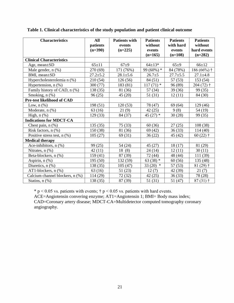
stress test (27%). Mean pre-test probability of CAD was 46±28%. Prevalence of male gender and
hypertension was significantly higher in patients with events than in those without events (Table 1).
MDCT-CA results.
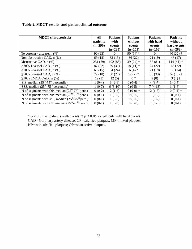
Table 2 shows MDCT-CA results and patient outcomes. SIS, SSS, number of segments with
obstructive plaques and prevalence of obstructive CAD, 3-vessel disease (VD) and LMCA disease
were significantly higher in patients with events than in patients without events.
9
Univariate predictors of events.
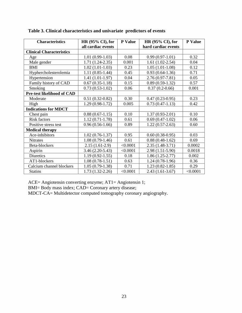
Univariate clinical predictors of events were male gender, hypertension, high pre-test likelihood of
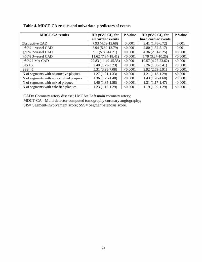
CAD and medical therapy with beta-blockers, aspirin and statins (Table 3). Univariate MDCT-CA
predictors of events are reported in Table 4. Regarding obstructive CAD, HR was 3.41 for hard
events and 7.93 for all events. HRs were particularly increased in patients with 3-VD (5.79 for hard
events and 11.62 for all events) and LMCA disease (10.57 for hard events and 22.83 for all events).
Multivariate predictors of events.
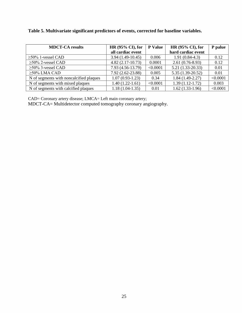
Significant independent predictors of hard events were 3-VD, LMCA disease and number of
segments with noncalcified, mixed and calcified plaques. Significant independent predictors of all
events were multi-VD, LMCA disease and number of segments with mixed and calcified plaques.
The HRs were particularly high in patients with 3-VD (5.21 for hard events and 7.93 for all events)
and LMCA disease (5.35 for hard events and 7.92 for all events) (Table 5).
Survival analysis.
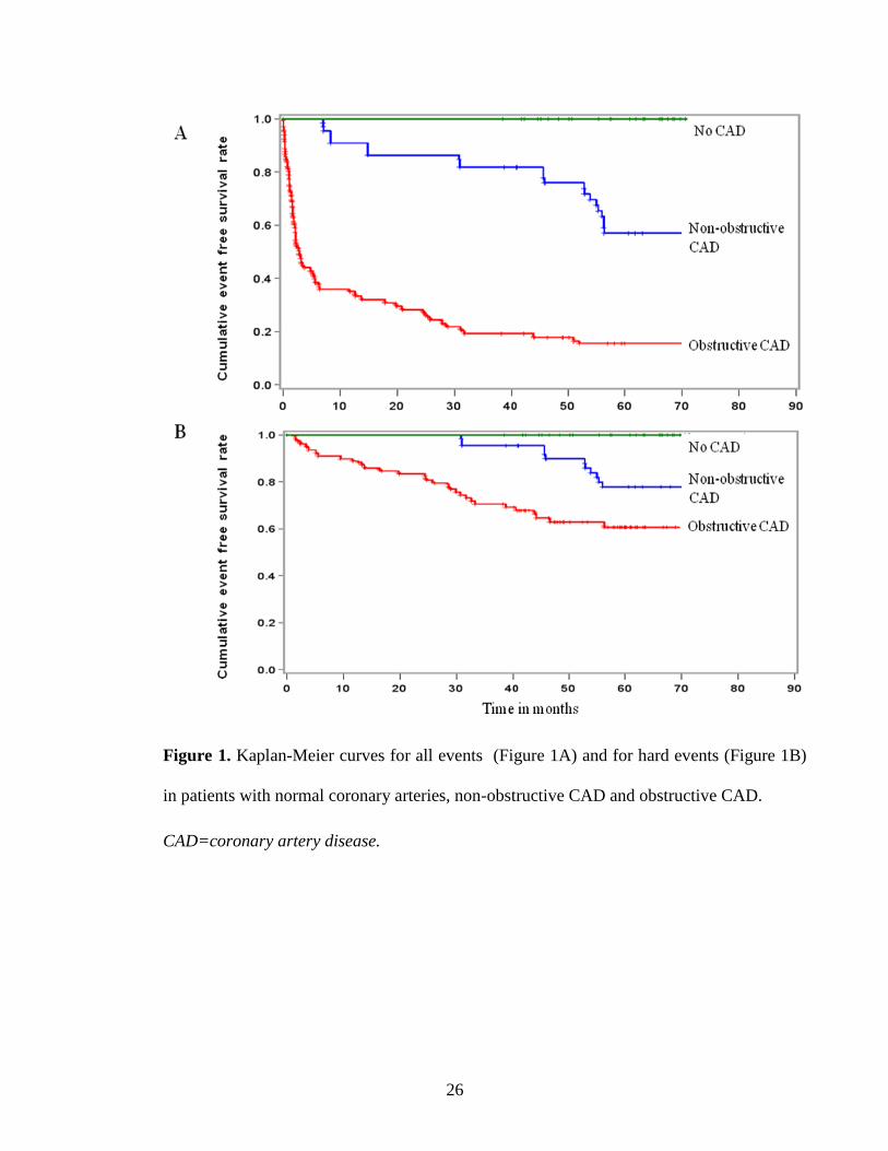
Kaplan-Meier survival curves are provided in Figures 1 to 3. No events occurred in patients with
normal coronary arteries. On the contrary, the 62-month cumulative hard and all event-free survival
rates were 78% and 56% in patients with non-obstructive CAD and 60% and 16% in those with
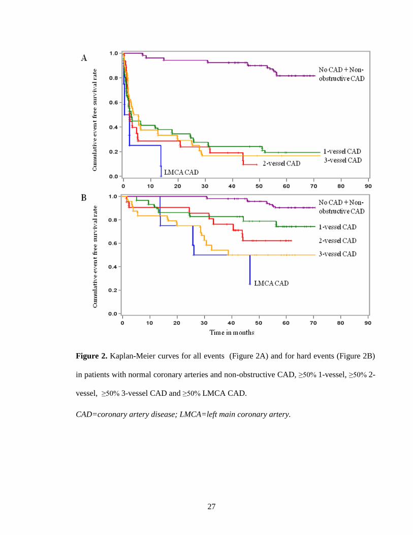
obstructive CAD, respectively (log-rank P value=0.0001) (Figure 1). Figure 2 shows the relationship
between CAD extension, expressed as number of major epicardial vessels exhibiting ≥50% stenosis,
and event-free survival rate. Regarding all events, cumulative event-free survival was 20% with 1-
VD, 12% with 2-VD, 18% with 3-VD and 0% with LMCA disease (log-rank P value=0.0001).
Excluding revascularization procedures, cumulative event-free survival was 73% with 1-VD, 62%
with 2-VD, 50% with 3-VD and 25% with LMCA disease (log-rank P value=0.0001). The
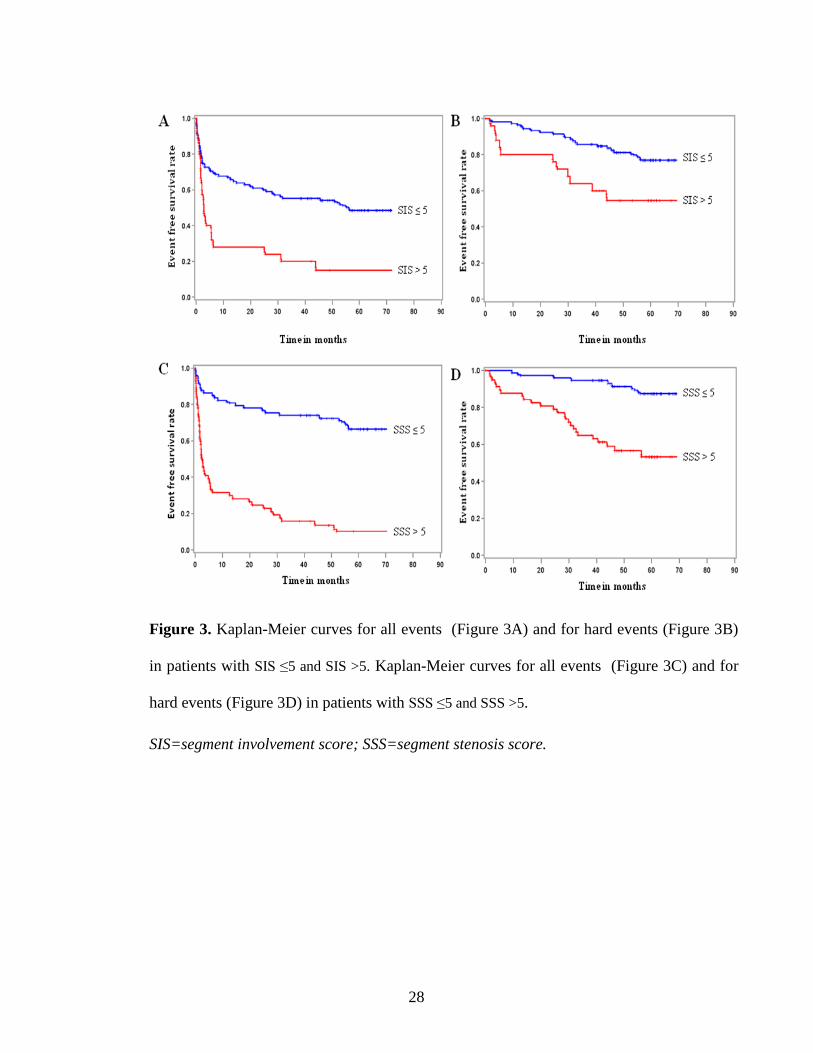
relationship between atherosclerotic burden, expressed as SIS and SSS, and event-free survival rate
is reported in Figure 3. Regarding all events, cumulative event-free survival was 50% with SIS≤5,
10
16% with SIS>5, 67% with SSS≤5 and 11% with SSS>5 (log-rank P value=0.0001). Regarding hard
events, cumulative event-free survival was 77% with SIS≤5, 54% with SIS>5, 88% with SSS≤5 and
54% with SSS>5 (log-rank P value=0.0001).
DISCUSSION
Management guidelines in Europe and the U.S. consider type 2 diabetes to be a cardiovascular
disease equivalent (22) and CAD is the major cause of morbidity, mortality and medical cost of DM
(22). Therefore, the early diagnosis of CAD to prevent progression and clinical events has intuitive
appeal. Recently, the American Diabetes Association (ADA) convened an expert panel that revisited
the issue of screening for CAD in DM patients, motivated by the goal of identifying patients with
high risk whose outcomes might be improved through more aggressive risk factor modification,
medical surveillance or revascularization of their CAD (22). Unfortunately, the sensitivity of clinical
risk assessment is limited in diabetics, mainly because typical symptoms of ischemia are often
absent and cardiac stress tests also have limited negative predictive value in these patients (3).
Particularly, diagnostic accuracy of exercise ECG is lower in diabetics in comparison with
nondiabetic patients (23) but also specificity of stress echocardiography and nuclear perfusion
imaging is markedly lower in diabetic patients (24,25). MDCT-CA is currently considered a reliable
diagnostic method for the evaluation of patients with suspected CAD, thanks to its high diagnostic
performance in the rule-out but also in the detection of obstructive coronary stenosis (4). Many
recent studies demonstrated an increased prevalence of obstructive and nonobstructive CAD and
fewer normal coronary arteries in diabetic patients in comparison with nondiabetic population
(22,26,27). However, only two mid-term follow-up (20 and 33 months, respectively) studies
supported the prognostic value of MDCT-CA in DM patients (12,13). As compared to previous
11
investigations, our study has longer follow-up in a large group of very selected diabetic patients who
underwent MDCT-CA for suspected CAD. Indeed, patients with any type of known cardiac disease
were excluded. Then, to the best of our knowledge, our study population is the most homogeneous
among the body of literature on the prognostic value of MDCT-CA in DM patients. The main
finding of our study is that MDCT-CA is able to provide long-term prognostic information in
diabetic patients with suspected CAD and may predict hard cardiac events. However, we found that
diabetic patients without evidence of CAD at MDCT-CA evaluation had an excellent prognosis at
62 months follow-up, without cardiac events recorded.
Specifically, detection of obstructive CAD at MDCT-CA was a strong predictor of cardiac events in
univariate analysis (HR 3.41 and 7.93 for hard and all events, respectively). Kaplan-Meier survival
curves confirmed this finding, showing an event-free survival of 60% for hard events and 16% for
all events in these patients. Moreover, MDCT-CA allowed a strong prognostic grading based on the
classification in 1-VD, 2-VD, 3-VD and LMCA disease. In both univariate and multivariate analysis,
HR for hard and all events were significantly increased in patients with 3-vessel CAD and LMCA
disease. Accordingly, survival curves free of hard events were progressively reduced, from 73%
with 1-VD to 25% with LMCA disease. Another major result of this study with important clinical
implications is that found in DM patients with non-obstructive CAD at MDCT-CA. In these patients,
all stress tests are usually negative because rarely this type of lesions triggers myocardial ischemia.
Although traditional non-invasive testing in this subset of patients suggests a prognosis similar to
that found in patients with normal coronary arteries, our data indicate a worse long-term outcome
instead. Indeed, Kaplan-Meier survival curves showed an event-free survival of 78% for hard
events and of 56% for all events in patients with non-obstructive CAD at 62-month follow-up. Thus,
these patients have a cardiac event probability intermediate between that found in patients with
normal coronary arteries and obstructive CAD. This finding requires some comments. First of all, a
12
recent study of van Velzen et al. demonstrated that MDCT-CA has an accuracy of 100% in
comparison with intravascular ultrasound (IVUS) in the detection of non-obstructive CAD and
coronary atherosclerosis (defined on IVUS as a plaque burden of ≥40% cross-sectional area) (28).
On the other hand, previous studies indicated that also plaque composition may be a predictor of
adverse events, demonstrated that vulnerable plaques may occur across the full spectrum stenosis
severity, suggesting that also non-obstructive lesions may contribute to coronary events (29) and
showed that lipid core size and minimal cap thickness, two major determinants of plaque
vulnerability, are not related to absolute plaque size or degree of stenosis (30). In our study, the
number of non-calcified plaques had HR by univariate and multivariate analysis for hard events
higher than the number of calcified and mixed plaques. Because non-obstructive plaques are more
frequent than obstructive plaques, it is conceivable that coronary occlusion and myocardial
infarction are due more frequently to moderate stenosis (31). Another possible explanation of the
high prognostic value of non-obstructive CAD in our study may reside in the long-term follow-up.
In fact, we cannot rule out that some moderate stenosis at the time of MDCT-CA examination could
have become obstructive over time. Nevertheless, the early identification of non-obstructive CAD
with MDCT-CA in diabetics is clinically important because may lead to a more aggressive strategy
of cardiovascular risk factor control and modification of clinical follow-up, accordingly with ADA
expert panel that recommended a more aggressive risk factor modification and medical surveillance
in high cardiac risk diabetic patients (22). Moreover, MDCT-CA also showed the ability to stratify
the prognosis of diabetics by means of the atherosclerotic burden evaluation. Indeed, in 2010,
Hadamitzky et al. demonstrated that atherosclerotic burden assessed with MDCT-CA was able to
stratify cardiac events in diabetics who were followed for 33 months (12). Our study confirms that
MDCT-CA is able to predict events on the basis of atherosclerotic burden evaluated with coronary
artery plaque scores. Indeed, event-free survival significantly decreased at 62-month follow-up from
13
67% for SSS ≤5 to 11% for SSS >5 considering revascularizations and from 88% for SSS ≤5 to 54%
for SSS >5 excluding revascularizations. Another remarkable finding is that atherosclerotic burden
maintains similar prognostic value and event-free survival rate using SIS.
Certainly, one of the main finding of our study regards the clinical scenario represented by diabetic
patients with normal coronary arteries at MDCT-CA evaluation. Indeed, in agreement with a
previous study that enrolled a smaller number of less homogeneous DM patients with shorter
follow-up (13), our study confirmed that the absence of CAD at MDCT-CA (found in 90 patients in
our study) is associated with an event-free survival of 100% for both hard and all cardiac events at
62-month follow-up. The excellent outcome in diabetic patients with completely absent CAD is
clinically relevant, because it suggests that MDCT-CA, in contrast to other imaging modalities, can
help to identify the truly low-risk for cardiovascular events diabetic patients. Indeed, although
nuclear stress imaging and stress echocardiography, the 2 most widely used stress imaging test in
the clinical practice, demonstrated a good prognostic value in the general population with suspected
CAD, as shown by very large meta-analysis of 31 studies and 69.655 patients and 13 studies and
32.739 patients, respectively (32,33), studies dedicated to diabetics not confirmed these results. In
fact, 7 studies with >100 patients each specifically addressed the prognostic value of SPECT
imaging in diabetics, although confirmed the higher events rate in the presence of an abnormal scan
compared with a normal scan, similar to nondiabetic population, demonstrated that the event rate in
the presence of a normal scan also appeared higher in comparison with the general population (34).
Similarly, five studies >100 patients that studied the prognostic value of stress echocardiography in
diabetics using either exercise or pharmacological stress, although confirmed the higher events rate
in the presence of an abnormal study compared with a normal study, similar to nondiabetic
population, shown that the event rate in the presence of a normal echocardiography also appeared
higher in comparison with the general population (35). Specifically, Kamalesh et al., who performed
14
a follow-up study in 233 patients (144 diabetics) with a negative stress echocardiography,
demonstrated that DM patients had a significantly higher incidence of nonfatal infarctions and a
higher annual hard event rate in comparison with nondiabetics (36). This issue has been confirmed
by Elhendy et al. that evaluated 563 DM patients with normal exercise echocardiography with
follow-up of up to 5 years, showing that although the 1-year event rate was 0%, there was a gradual
increase up to 7.6% at 5-year follow-up (37). On the contrary, in our study, we recorded an event
rate of 0% in patients with normal coronary arteries at 5-years follow-up (mean 62±9 months, up to
72 months). Therefore, this diagnostic modality can be used to reassure regarding their outcome
diabetic patients with suspected CAD, with a warranty period of 5 years in the presence of a MDCT-
CA completely normal.
Study limitations.
In interpreting these data, some limitations should be considered. First, this is a relatively small,
single-center study evaluating mainly caucasian patients and its results may not necessarily reflect
the patient population of other centers or countries. Second, we recognize that incomplete follow-up
may result in underreporting of cardiac events. However, the percentage of patients with complete
follow-up was remarkably high (98%). Third, MDCT-CA allowed the identification of patients with
obstructive CAD, likely resulting in an increased revascularization rate, which constituted a large
proportion of the composite all cardiac event end point. However, in our study MDCT-CA was
performed in addition to the standard diagnostic work-up. Moreover, all decisions regarding
revascularization were based on symptoms, presence of ischemia on non-invasive testing and ICA
results and patients with early elective revascularizations were excluded from the survival analysis.
Finally, the presence of obstructive CAD at MDCT-CA was strongly associated with hard cardiac
events not including revascularization procedures.
15
REFERENCES
1) McGill HC, Mcmahan CA, Malcom GT, Oalmann MC, Strong JP. Relation of
glycohemoglobin and adiposity to atherosclerosis in youth: Pathobiological Determinants of
Atherosclerosis in Youth (PDAY) Research Group. Arterioscler Thromb Vasc Biol
1995;15:431-440.
2) Rydén L, Standl E, Bartnik M, Van den Berghe G, Betteridge J, de Boer MJ, Cosentino F,
Jönsson B, Laakso M, Malmberg K, Priori S, Ostergren J, Tuomilehto J, Thrainsdottir I,
Vanhorebeek I, Stramba-Badiale M, Lindgren P, Qiao Q, Priori SG, Blanc JJ, Budaj A,
Camm J, Dean V, Deckers J, Dickstein K, Lekakis J, McGregor K, Metra M, Morais J,
Osterspey A, Tamargo J, Zamorano JL, Deckers JW, Bertrand M, Charbonnel B, Erdmann
E, Ferrannini E, Flyvbjerg A, Gohlke H, Juanatey JR, Graham I, Monteiro PF, Parhofer K,
Pyörälä K, Raz I, Schernthaner G, Volpe M, Wood D; Task Force on Diabetes and
Cardiovascular Diseases of the European Society of Cardiology (ESC); European
Association for the Study of Diabetes (EASD). Guidelines on diabetes, pre-diabetes, and
cardiovascular diseases: executive summary. The Task Force on Diabetes and
Cardiovascular Diseases of the European Society of Cardiology (ESC) and of the European
Association for the Study of Diabetes (EASD). Eur Heart J. 2007;28:88-136
3) Kamalesh M, Feigenbaum H, Sawada S. Assessing prognosis in patients with diabetes
mellitus--the Achilles' heel of cardiac stress imaging tests? Am J Cardiol. 2007;99:1016-9.
Review.
4) D’Othèe BJ, Siebert U, Cury R, Jadvar H, Dunn EJ, Hoffmann U. A systematic review on
diagnostic accuracy of CT-based detection of significant coronary artery disease. European
Journal of Radiology 2008;65: 449-61.
16
5) Andreini D, Pontone G, Pepi M, et al. Diagnostic accuracy of multidetector computed
tomography coronary angiography in patients with dilated cardiomyopathy. J Am Coll
Cardiol. 2007;49:2044 –2050.
6) Min JK, Shaw LJ, Devereux RB, Okin PM, Weinsaft JW, Russo DJ, Lippolis NJ, Berman
DS, Callister TQ. Prognostic value of multidetector coronary computed tomographic
angiography for prediction of all-cause mortality. J Am Coll Cardiol. 2007;50:1161-70.
7) Carrigan TP, Nair D, Schoenhagen P, Curtin RJ, Popovic ZB, Halliburton S, Kuzmiak S,
White RD, Flamm SD, Desai MY. Prognostic utility of 64-slice computed tomography in
patients with suspected but no documented coronary artery disease. Eur Heart J.
2009;30:362-71.
8) Aldrovandi A, Maffei E, Palumbo A, Seitun S, Martini C, Brambilla V, Zuccarelli A,
Tarantini G, Weustink AC, Mollet NR, Ruffini L, Crisi G, Ardissino D, de Feyter PJ, Krestin
GP, Cademartiri F. Prognostic value of computed tomography coronary angiography in
patients with suspected coronary artery disease: a 24-month follow-up study. Eur Radiol.
2009 Jul;19(7):1653-60. Epub 2009 Feb 18.
9) Chow BJ, Wells GA, Chen L, Yam Y, Galiwango P, Abraham A, Sheth T, Dennie
C, Beanlands RS, Ruddy TD. Prognostic value of 64-slice cardiac computed tomography
severity of coronary artery disease, coronary atherosclerosis, and left ventricular ejection
fraction. J Am Coll Cardiol. 2010;55:1017-28.
10) Min JK, Dunning A, Lin FY, et al. Age- and sex-related differences in all-cause mortality
risk based on coronary computed tomography angiography findings results from the
International Multicenter CONFIRM (Coronary CT Angiography Evaluation for Clinical
Outcomes: An International Multicenter Registry) of 23,854 patients without known
coronary artery disease. J Am Coll Cardiol. 2011;58: 849-60.
17
11) Chow BJ, Small G, Yam Y, et al. Incremental prognostic value of cardiac computed
tomography in coronary artery disease using CONFIRM: COroNary computed tomography
angiography evaluation for clinical outcomes: an InteRnational Multicenter registry. Circ
Cardiovasc Imaging. 2011;4: 463-72.
12) Hadamitzky M, Hein F, Meyer T, Bischoff B, Martinoff S, Schömig A, Hausleiter J.
Prognostic value of coronary computed tomographic angiography in diabetic patients
without known coronary artery disease. Diabetes Care. 2010;33:1358-63.
13) Van Werkhoven JM, Cademartiri F, Seitun S, Maffei E, Palumbo A, Martini C, Tarantini G,
Kroft LJ, de Roos A, Weustink AC, Jukema JW, Ardissino D, Mollet NR, Schuijf JD, Bax JJ.
Diabetes: prognostic value of CT coronary angiography--comparison with a nondiabetic
population. Radiology. 2010;256:83-92.
14) Report of the Expert Committee on the Diagnosis and Classification of Diabetes Mellitus.
Diabetes Care 1997;20: 1183–97.
15) Executive Summary of the Third Report of The National Cholesterol Education Program
(NCEP) Expert Panel on Detection, Evaluation, and Treatment of High Blood Cholesterol in
Adults (Adult Treatment Panel III). JAMA 2001;285: 2486 –97.
16) 2003 European Society of Hypertension-European Society of Cardiology guidelines for the
management of arterial hypertension. J Hypertens 2003;21: 1011–53.
17) Taylor AJ, Bindeman J, Feuerstein I, Cao F, Brazaitis M, O’Malley PG. Coronary calcium
independently predicts incident premature coronary heart disease over measured
cardiovascular risk factors: mean three-year outcomes in the Prospective Army Coronary
Calcium (PACC) project. J Am Coll Cardiol 2005;46: 807–14.

18
18) Diamond GA, Forrester JS. Analysis of probability as an aid in the clinical diagnosis of
coronary-artery disease. N Engl J Med 1979;300: 1350–58.
19) Austen WG, Edwards JE, Frye RL, Gensini GG, Gott VL, Griffith LS, McGoon DC,
Murphy ML, Roe BB. A reporting system on patients evaluated for coronary artery disease.
Report of the Ad Hoc Committee for Grading of Coronary Artery Disease, Council on
Cardiovascular Surgery, American Heart Association. Circulation. 1975;51:5-40.
20) Myocardial infarction redefined—a consensus document of The Joint European Society of
Cardiology/American College of Cardiology Committee for the redefinition of myocardial
infarction. Eur Heart J 2000;21: 1502–13.
21) Task Force for Diagnosis and Treatment of Non-ST-Segment Elevation Acute Coronary
Syndromes of European Society of Cardiology, Bassand JP, Hamm CW, Ardissino
D, Boersma E, Budaj A, Fernández-Avilés F, Fox KA, Hasdai D, Ohman EM, Wallentin
L, Wijns W. Guidelines for the diagnosis and treatment of non-ST-segment elevation acute
coronary syndromes. Eur Heart J. 2007;28:1598-660.
22) Bax JJ, Inzucchi SE, Bonow RO, Schuijf JD, Freeman MR, Barrett EJ; Global Dialogue
Group for the Evaluation of Cardiovascular Risk in Patients with Diabetes. Cardiac imaging
for risk stratification in diabetes. Diabetes Care. 2007;30:1295-304. Review.
23) Wackers FJ. Diabetes and coronary artery disease: the role of stress myocardial perfusion
imaging. Cleve Clin J Med. 2005;72:21-33.
24) Kang X, Berman DS, Lewin H, Miranda R, Erel J, Friedman JD, Amanullah AM.
Comparative ability of myocardial perfusion single-photon emission computed tomography
to detect coronary artery disease in patients with and without diabetes mellitus. Am Heart
J. 1999;137:949-57.
19
25) Hennessy TG, Codd MB, Kane G, McCarthy C, McCann HA, Sugrue DD. Evaluation of
patients with diabetes mellitus for coronary artery disease using dobutamine stress
echocardiography. Coron Artery Dis. 1997;8:171-4.
26) Maffei E, Seitun S, Nieman K, Martini C, Guaricci AI, Tedeschi C, Weustink AC, Mollet
NR, Berti E, Grilli R, Messalli G, Cademartiri F. Assessment of coronary artery disease and
calcified coronary plaque burden by computed tomography in patients with and without
diabetes mellitus. Eur Radiol. 2011;21:944-53.
27) Pundziute G, Schuijf JD, Jukema JW, Boersma E, Scholte AJ, Kroft LJ, van der Wall
EE, Bax JJ. Noninvasive assessment of plaque characteristics with multislice computed
tomography coronary angiography in symptomatic diabetic patients. Diabetes
Care. 2007;30:1113-9.
28) Van Velzen JE, Schuijf JD, de Graaf FR, Boersma E, Pundziute G, Spanó F, Boogers MJ,
Schalij MJ, Kroft LJ, de Roos A, Jukema JW, van der Wall EE, Bax JJ. Diagnostic
performance of non-invasive multidetector computed tomography coronary angiography to
detect coronary artery disease using different endpoints: detection of significant stenosis vs.
detection of atherosclerosis. Eur Heart J 2010. [Epub ahead of print].
29) Davies MJ, Thomas AC. Plaque fissuring: the cause of acute myocardial infarction, sudden
ischaemic death, and crescendo angina. Br Heart J 1985;53:363-373. Review
30) Mann JM, Davies MJ. Vulnerable plaque. Relation of characteristics to degree of stenosis in
human coronary arteries. Circulation 1996;94:928-931.
31) Falk E, Shah PK, Fuster V. Coronary plaque disruption. Circulation. 1995;92:657-671.
Review.
32) Shaw LJ, Iskandrian AE. Prognostic value of gated myocardial perfusion SPECT. J Nucl
Cardiol. 2004;11:171-85. Review.
20
33) Schuijf JD, Poldermans D, Shaw LJ, Jukema JW, Lamb HJ, de Roos A, Wijns W, van der
Wall EE, Bax JJ. Diagnostic and prognostic value of non-invasive imaging in known or
suspected coronary artery disease. Eur J Nucl Med Mol Imaging. 2006;33:93-104. Review.
34) Scholte AJ, Bax JJ, Wackers FJ. Screening of asymptomatic patients with type 2 diabetes
mellitus for silent coronary artery disease: combined use of stress myocardial perfusion
imaging and coronary calcium scoring. J Nucl Cardiol. 2006;13:11-8.
35) Anand DV, Lim E, Lahiri A, Bax JJ. The role of non-invasive imaging in the risk
stratification of asymptomatic diabetic subjects. Eur Heart J. 2006 ;27:905-12. Review.
36) Kamalesh M, Matorin R, Sawada S. Prognostic value of a negative stress echocardiographic
study in diabetic patients. Am Heart J. 2002;143:163-8.
37) Elhendy A, Arruda AM, Mahoney DW, Pellikka PA. Prognostic stratification of diabetic
patients by exercise echocardiography. J Am Coll Cardiol. 2001;37:1551-7.
21
Table 1. Clinical characteristics of the study population and patient clinical outcome
Characteristics All
patients
(n=390)
Patients with
events
(n=225)
Patients
without
events
(n=165)
Patients
with hard
events
(n=108)
Patients
without
hard events
(n=282)
Clinical Characteristics
Age, mean±SD 65±11 67±9 64±13* 65±9 66±12
Male gender, n (%) 270 (69) 171 (76%) 99 (60%) * 84 (78%) 186 (66%) †
BMI, mean±SD 27.2±5.2 28.1±5.6 26.7±5 27.7±5.5 27.1±4.8
Hypercholesterolemia n (%) 210 (54) 126 (56) 84 (51) 57 (53) 153 (54)
Hypertension, n (%) 300 (77) 183 (81) 117 (71) * 96 (89) 204 (72) †
Family history of CAD, n (%) 138 (35) 81 (36) 57 (34) 39 (36) 99 (35)
Smoking, n (%) 96 (25) 45 (20) 51 (31) 12 (11) 84 (30)
Pre-test likelihood of CAD
Low, n (%) 198 (51) 120 (53) 78 (47) 69 (64) 129 (46)
Moderate, n (%) 63 (16) 21 (9) 42 (25) 9 (8) 54 (19)
High, n (%) 129 (33) 84 (37) 45 (27) * 30 (28) 99 (35)
Indications for MDCT-CA
Chest pain, n (%) 135 (35) 75 (33) 60 (36) 27 (25) 108 (38)
Risk factors, n (%) 150 (38) 81 (36) 69 (42) 36 (33) 114 (40)
Positive stress test, n (%) 105 (27) 69 (31) 36 (22) 45 (42) 60 (22) †
Medical therapy
Ace-inhibitors, n (%) 99 (25) 54 (24) 45 (27) 18 (17) 81 (29)
Nitrates, n (%) 42 (11) 18 (8) 24 (14) 12 (11) 30 (11)
Beta-blockers, n (%) 159 (41) 87 (39) 72 (44) 48 (44) 111 (39)
Aspirin, n (%) 195 (50) 132 (59) 63 (38) * 60 (56) 135 (48)
Diuretics, n (%) 138 (35) 105 (47) 33 (20) * 57 (53) 81 (29) †
AT1-blockers, n (%) 63 (16) 51 (23) 12 (7) 42 (39) 21 (7)
Calcium channel blockers, n (%) 114 (29) 72 (32) 42 (25) 36 (33) 78 (28)
Statins, n (%) 138 (35) 87 (39) 51 (31) 51 (47) 87 (31) †
* p < 0.05 vs. patients with events; † p < 0.05 vs. patients with hard events.
ACE=Angiotensin convering enzyme; AT1=Angiotensin 1; BMI= Body mass index;
CAD=Coronary artery disease; MDCT-CA=Multidetector computed tomography coronary
angiography.
22
Table 2. MDCT results and patient clinical outcome
MDCT characteristics All
patients
(n=390)
Patients
with
events
(n=225)
Patients
without
events
(n=165)
Patients
with hard
events
(n=108)
Patients
without
hard events
(n=282)
No coronary disease, n (%) 90 (23) 0 90 (54) * 0 90 (32) †
Non-obstructive CAD, n (%) 69 (18) 33 (15) 36 (22) 21 (19) 48 (17)
Obstructive CAD, n (%) 231 (59) 192 (85) 39 (24) * 87 (81) 144 (51) †
≥50% 1-vessel CAD , n (%) 87 (22) 69 (31) 18 (11) * 24 (22) 63 (22)
≥50% 2-vessel CAD , n (%) 60 (15) 54 (24) 6 (4) * 21 (19) 39 (14)
≥50% 3-vessel CAD, n (%) 72 (18) 60 (27) 12 (7) * 36 (33) 36 (13) †
≥50% LMCA CAD, n (%) 12 (3) 12 (5) 0 * 9 (8) 3 (1) †
SIS, median (25th-75
th percentile) 1 (0-4) 3 (2-6) 0 (0-4) * 4 (3-7) 1 (0-5) †
SSS, median (25th-75
th percentile) 1 (0-7) 6 (3-10) 0 (0-5) * 7 (4-13) 1 (1-6) †
N of segments with OP, median (25th-75
th perc.) 0 (0-2) 2 (1-3) 0 (0-0) * 2 (1-3) 0 (0-1) †
N of segments with NP, median (25th-75
th perc.) 0 (0-1) 1 (0-2) 0 (0-0) 1 (0-2) 0 (0-1)
N of segments with MP, median (25th-75
th perc.) 0 (0-1) 1 (0-2) 0 (0-0) 1 (0-2) 0 (0-1)
N of segments with CP, median (25th-75
th perc.) 0 (0-1) 1 (0-3) 0 (0-0) 1 (0-3) 0 (0-1)
* p < 0.05 vs. patients with events; † p < 0.05 vs. patients with hard events.
CAD= Coronary artery disease; CP=calcified plaques; MP=mixed plaques;
NP= noncalcified plaques; OP=obstructive plaques.
23
Table 3. Clinical characteristics and univariate predictors of events
Characteristics HR (95% CI), for
all cardiac events
P Value HR (95% CI), for
hard cardiac events
P Value
Clinical Characteristics
Age 1.01 (0.99-1.03) 0.08 0.99 (0.97-1.01) 0.32
Male gender 1.71 (1.24-2.35) 0.001 1.61 (1.02-2.54) 0.04
BMI 1.02 (1.01-1.03) 0.23 1.05 (1.01-1.08) 0.12
Hyphercholesterolemia 1.11 (0.85-1.44) 0.45 0.93 (0.64-1.36) 0.71
Hypertension 1.41 (1.01-1.97) 0.04 2.76 (0.97-7.81) 0.05
Family history of CAD 0.67 (0.35-1.18) 0.15 0.89 (0.59-1.32) 0.57
Smoking 0.73 (0.53-1.02) 0.06 0.37 (0.2-0.66) 0.001
Pre-test likelihood of CAD
Moderate 0.51 (0.32-0.82) 0.30 0.47 (0.23-0.95) 0.23
High 1.29 (0.98-1.72) 0.005 0.73 (0.47-1.13) 0.42
Indications for MDCT
Chest pain 0.88 (0.67-1.15) 0.10 1.37 (0.93-2.01) 0.10
Risk factors 1.12 (0.71-1.78) 0.61 0.69 (0.47-1.02) 0.06
Positive stress test 0.96 (0.56-1.66) 0.89 1.22 (0.57-2.63) 0.60
Medical therapy
Ace-inhibitors 1.02 (0.76-1.37) 0.95 0.60 (0.38-0.95) 0.03
Nitrates 1.08 (0.79-1.46) 0.61 0.88 (0.48-1.62) 0.69
Beta-blockers 2.15 (1.61-2.9) <0.0001 2.35 (1.48-3.71) 0.0002
Aspirin 3.46 (2.20-5.43) <0.0001 2.98 (1.51-5.90) 0.0018
Diuretics 1.19 (0.92-1.55) 0.18 1.86 (1.25-2.77) 0.002
AT1-blockers 1.08 (0.78-1.51) 0.63 1.24 (0.78-1.96) 0.36
Calcium channel blockers 1.05 (0.79-1.38) 0.71 1.23 (0.82-1.85) 0.29
Statins 1.73 (1.32-2.26) <0.0001 2.43 (1.61-3.67) <0.0001
ACE= Angiotensin converting enzyme; AT1= Angiotensin 1;
BMI= Body mass index; CAD= Coronary artery disease;
MDCT-CA= Multidetector computed tomography coronary angiography.
24
Table 4. MDCT-CA results and univariate predictors of events
MDCT-CA results HR (95% CI), for
all cardiac events
P Value HR (95% CI), for
hard cardiac events
P Value
Obstructive CAD 7.93 (4.59-13.68) 0.0001 3.41 (1.78-6.72) 0.001
≥50% 1-vessel CAD 8.94 (5.80-13.79) <0.0001 2.80 (1.52-5.17) 0.001
≥50% 2-vessel CAD 9.1 (5.83-14.21) <0.0001 4.36 (2.31-8.25) <0.0001
≥50% 3-vessel CAD 11.62 (7.34-18.41) <0.0001 5.79 (3.27-10.25) <0.0001
≥50% LMA CAD 22.83 (11.49-45.35) <0.0001 10.57 (4,27-23.62) <0.0001
SIS >5 2.40 (1.79-3.23) <0.0001 2.26 (1.50-3.41) <0.0001
SSS >5 5.31 (3.98-7.08) <0.0001 3.92 (2.59-5.91) <0.0001
N of segments with obstructive plaques 1.27 (1.21-1.33) <0.0001 1.21 (1.13-1.29) <0.0001
N of segments with noncalcified plaques 1.36 (1.25-1.48) <0.0001 1.43 (1.28-1.60) <0.0001
N of segments with mixed plaques 1.46 (1.35-1.58) <0.0001 1.31 (1.17-1.47) <0.0001
N of segments with calcified plaques 1.23 (1.15-1.29) <0.0001 1.19 (1.09-1.29) <0.0001
CAD= Coronary artery disease; LMCA= Left main coronary artery;
MDCT-CA= Multi detector computed tomography coronary angiography;
SIS= Segment-involvement score; SSS= Segment-stenosis score.
25
Table 5. Multivariate significant predictors of events, corrected for baseline variables.
MDCT-CA results HR (95% CI), for
all cardiac event
P Value HR (95% CI), for
hard cardiac event
P palue
≥50% 1-vessel CAD 3.94 (1.49-10.45) 0.006 1.91 (0.84-4.3) 0.12
≥50% 2-vessel CAD 4.82 (2.17-10.73) 0.0001 2.61 (0.76-8.93) 0.12
≥50% 3-vessel CAD 7.93 (4.56-13.79) <0.0001 5.21 (1.33-20.33) 0.01
≥50% LMA CAD 7.92 (2.62-23.88) 0.005 5.35 (1.39-20.52) 0.01
N of segments with noncalcified plaques 1.07 (0.93-1.23) 0.34 1.84 (1.49-2.27) <0.0001
N of segments with mixed plaques 1.40 (1.22-1.61) <0.0001 1.39 (1.12-1.72) 0.003
N of segments with calcified plaques 1.18 (1.04-1.35) 0.01 1.62 (1.33-1.96) <0.0001
CAD= Coronary artery disease; LMCA= Left main coronary artery;
MDCT-CA= Multidetector computed tomography coronary angiography.
26
Figure 1. Kaplan-Meier curves for all events (Figure 1A) and for hard events (Figure 1B)
in patients with normal coronary arteries, non-obstructive CAD and obstructive CAD.
CAD=coronary artery disease.
27
Figure 2. Kaplan-Meier curves for all events (Figure 2A) and for hard events (Figure 2B)
in patients with normal coronary arteries and non-obstructive CAD, ≥50% 1-vessel, ≥50% 2-
vessel, ≥50% 3-vessel CAD and ≥50% LMCA CAD.
CAD=coronary artery disease; LMCA=left main coronary artery.
28
Figure 3. Kaplan-Meier curves for all events (Figure 3A) and for hard events (Figure 3B)
in patients with SIS ≤5 and SIS >5. Kaplan-Meier curves for all events (Figure 3C) and for
hard events (Figure 3D) in patients with SSS ≤5 and SSS >5.
SIS=segment involvement score; SSS=segment stenosis score.