Embed Size (px)
Citation preview

Università degli Studi di Ferrara
DOTTORATO DI RICERCA IN
"FARMACOLOGIA E ONCOLOGIA MOLECOLARE"
CICLO XXVI
COORDINATORE Prof. ANTONIO CUNEO
GENETIC POLYMORPHISMS OF THE FOLATE METABOLIC PATHWAY IN CHILDHOOD
ACUTE LYMPHOBLASTIC LEUKEMIA. A MOLECULAR STUDY AND A PROPOSAL FOR AN
INTERPRETATIVE MODEL.
Settore Scientifico Disciplinare MED/15
Dottorando Tutore
Dott. ORIOLI ELISA Dott. GEMMATI DONATO
Anni 2011/2013

1
Index
1 INTRODUCTION 3
1.1 ACUTE LYMPHOBLASTIC LEUKEMIA 4
1.2 MOLECULAR MECHANISMS OF ACUTE
LYMPHOBLASTIC LEUKEMIA 6
1.3 FOLATE 8
1.3.1 Folate distribution 13
1.3.2 Folate and cancer 15
1.3.3 Folate deficiency 17
1.3.4 Folate status and DNA methylation 19
1.3.5 Genetic variants and possible effects on cancer 20
1.4 METHOTREXATE 28
1.5 AIM AND SCOPE 31
2 MATERIALS AND METHODS 33
2.1 SELECTIONS OF CASES AND CONTROLS 34
2.2 GENOTYPIC ANALYSES 35
2.2.1 Estraction protocol 35
2.2.2 Polymerase Chain Reaction (PCR) 36
2.2.3 Genotype protocol 37
2.2.4 Cell preparation and culture 41
2.3 STATISTICAL ANALYSIS 42

2
3 RESULTS 43
3.1 GENOTYPIC ANALYSIS OF MTHFR, DHFR,
BCL-2 POLYMORPHISMS 44
3.1.1 Clinical-pathological characteristics of cases 44
3.1.2 Evaluation of childhood ALL risk: single analysis 45
3.1.3 Evaluation of childhood ALL risk (MTHFR C677T, DHFR):
combined analysis 47
3.1.4 Disease onset and different polymorphisms 48
3.1.5 Therapy duration and different polymorphisms 50
3.1.6 Kaplan-Meier analysis. Correlation with MTHFR A1298C polymorphism 53
3.1.7 Kaplan-Meier analysis. Correlation with Bcl-2 -938 C>A polymorphism 55
3.1.8 Folate and anti-folate: the in vitro model 58
4 DISCUSSION 63
4.1 DISCUSSION 64
4.2 CONCLUSION 66
5 BIBLIOGRAPHY 67
5.1 BIBLIOGRAPHY 68

3
1
INTRODUCTION

4
1.1 ACUTE LYMPHOBLASTIC LEUKAEMIA
Acute lymphoblastic leukemia (ALL) is defined as a clonal proliferative disorder originating from
lymphoid precursors in the hematopoietic bone marrow. As a result of this transformation, a single
B or T clone will stop the cell maturation process and will reveal an enhanced proliferation activity
and as a consequence there will be a large infiltration of malignant lymphoblasts in the
hematopoietic bone marrow and in the peripheral blood.
Althought ALL is considered a rare disease, it is the most frequent neoplasia in the pediatric
population accounting for 80% of leukemia with a peak incidence in those aged 2-5 years (Barry et
al., 2008). About 80% of ALL are malignant proliferations of precursors-B cells while less common
(about 20%) are ALL that involve precursors-T cells. The causes of ALL remains still unknown in
the majority of cases nevertheless, during the last 20 years survival rates have strongly improved
thanks to current treatment regimens. There are several conditions strongly linked to the pathology
and able to upgrade the risk of developing ALL: ionizing radiations and some congenital disorders
such as Fanconi‟s anemia, Bloom‟s Syndrome and Down‟s Syndrome (Kaatsch et al., 2010; Belson
et al., 2007; Mathew et al., 2006; Zwaan et al., 2010). In 1976, thanks to Franco-American-
Britannic Classification (FAB) it was possible to classify ALL in 3 main groups: L1, L2, L3,
according to the nucleocitoplasmatic characteristics of leukemic elements.
L1 includes small blast cells with scant cytoplasm. L1 is the most common form of ALL in young
under 15 years old.
L2 includes greater blast cells, with variable dimensions and irregular nuclei containing evident
nucleoli.
L3 includes big dimensions blast cells with strongly basophilic cytoplasm.
In 2008, the World Health Organization (WHO) revised the FAB classification of ALL and
classified ALL as follows:
Acute Lymphoid B Leukemia divided into:
Acute Lymphoid B Leukemia Not Otherwise Specified
Acute Lymphoid B Leukemia with recurrent genetic abnormalities
ALL with t(9;22)/BCR-ABL
ALL with abnormalities of MLL gene
ALL with t(12;21)/ETV6-RUNX1
ALL with hyperdiploidia
ALL with hypodiploidia
5
ALL with t(1;19)/TCF3-PBX1
ALL with t(5;14)/IL3-IGH
Acute Lymphoid T Leukemia divided into 3 categories according to signs and symptoms:
Signs and Symptoms arising from hematopoiesis suppression;
Signs and Symptoms arising from the presence of leukemic elements in peripheral blood and
releasing of inflammation mediators;
Signs and Symptoms arising from tissue infiltration.
Of particular relevance is the t(9;22) that brings to a disruption in the bcr region of BCR gene
(breakpoint cluster region) quite similar to Chronic Myeloid Leukemia (CML) in about 50% of
cases. In the remaining 50% of cases the breaking point is located more proximal (5‟) than in CML.
The fusion gene bcr-abl leads to the constitution of a protein of 190kD, different from the classical
fusion protein of 210kD produced in CML. This 190kD protein allows a differentiation on
molecular basis of ALL Ph+ from classical CML.
In the prognosis of ALL different factors assume a relevant importance: age, gender, blasts
morphology, leukocytosis, immuno-phenotype and cytogenetic factor. The complete remissions are
around 95% in children but considering adult patients (50-60 years old) the complete remissions
diminish to less than 60%. Furthermore, male patients have a lower tendency to the complete
remission than female patients (McCredie et al., 1999). Considering the blast morphology, the
remission percentage and the length of the disease are lower and statistically significant in L3
patients than L1 and L2 patients. Prognosis could be strongly influenced by particular
immunophenotypes. In the therapeutic phase three principal and consecutive phases there exist: the
induction therapy, the consolidation therapy and the maintenance therapy.
The remission induction therapy consists of a combination of pharmacological treatments with
vincristine, daunorubicin, prednisone and asparaginase that have distinct mechanisms for their
antileukemic effects and act synergistically (Kawedia et al., 2012).
The consolidation therapy allows the control of the minimal residual disease (MRD) and the
prevention of relapses.
During the maintenance therapy, that is essential for all ALL patients, the treatment is prolonged for
2-3 years using pharmacological treatment such as 6-mercaptopurin and Methotrexate (MTX) even
though there is no consensus on the best regimens and their duration (Pui et al., 2012). The marrow
transplantation is today the therapeutic strategy which aims to cut out MRD once the complete
remission is obtained.

6
1.2 MOLECULAR MECHANISMS OF ACUTE
LYMPHOBLASTIC LEUKAEMIA
The molecular analysis of common genetic alterations in lymphoblastic cells has strongly
contributed to the comprehension of ALL pathogenesis. There are different genetic sub-type
frequencies between adult and young ALL patients but the general molecular mechanisms that lead
to ALL are quite similar. They include: anomalous expression of pro-oncogenes, chromosomal
translocations that produce fusion genes coding for active kinases and altered transcriptional
factors, hyperploidy that includes more than 50 chromosomes (Gale et al., 1997; Wiemels et al.,
2002; Mori et al., 2002; Greaves et al., 2006). All the above alterations contribute to the leukemic
transformation of hematopoietic stem cells modifying their cellular function. The cells regulatory
processes are therefore altered leading to an unlimited capacity of self-renewal. Accordingly, this
mechanism removes the normal control of the cell proliferation, blocking the differentiation and
promoting the resistance to apoptosis (Pui et al., 2004). Some genetic lesions influence only one
pathway whereas other alterations can interfere with more pathways. The ABL proto-oncogene
codes for a tyrosin-kinase with a closely controlled activity. The fusion protein BCR-ABL (the
“Philadelphia chromosome”) is a kinase constitutively activate, instead. It could modify the signal
pathway that controls the cell proliferation, the survival and the self-renewal capacity of
hematopoietic stem cells (Pane et al., 2002). However, the oncogenic events that lead to
chromosomal rearrangements are probably insufficient to singly cause ALL: probably, a
combination of several factors such as gene-gene and gene-environment interactions appears to be
necessary. Recently, Eden summarized the major putative factors for causation of childhood
leukemia, principally treating the genetic predisposition, the genetic susceptibility and the
environmental factors (Eden et al., 2010). Childhood leukemia like other cancers derived from two
or more molecular changes in stem-like cells that can divide and at the same time, maintain an
immature state. Several common genetic mutations in leukemia such as TEL-AML1 and AML1-
ETO have been evaluated for their formation during the foetal development. Studies revealed that
these translocations occur at a rate of 1% or more in the normal population so mutations associated
with leukemia cannot cause by themselves the disease (Zuna et al., 2011; Mori et al., 2002). Some
studies in the early 20th
century proposed that infection was the cause of leukemia, now
hypothesized as a reaction to infection (McCarthy et al., 1992). Greaves and Kinlen proposed a link
with infection through population mixing and a key role of the infection in the promotion of a pre-
leukemic clone‟s transformation into leukemia (Greaves et al., 1988; Kinlen et al., 1990). Graves

7
met the hypothesis that infection through an immune response would lead to the postnatal genetic
errors seen in childhood ALL especially, strongly close to the “hygene” hypothesis. The absence of
an early immune challenge and priming, with delayed exposure to infections may predispose the
individual to pathological responses to infection that would create the second „genetic‟ hit through
proliferation or apoptotic stress considering that the first hit that is pre-leukemic clone, was present
from fetal life (Wiemels et al., 2002; Greaves et al., 2003). The infection hypothesis is supported by
several studies that emphasize the importance also of the maternal infection and influenza during
pregnancy concluding that a link there exists between maternal infection and increased risk of
leukemia (Lehtinen et al., 2003; Naumburg et al., 2002). Considering the environmental factor, it is
well known a remarkably high rate of cellular kinetics in fetal hematopoiesis and a high degree of
cellular proliferation that could set up a situation of perturbation via environmental insults,
including chemicals, inducing mutations (Wiemels et al., 2012). However, the only confirmed
chemical cause is ionizing radiation caused by atomic bomb exposure or diagnostic imaging during
pregnancy (Little et al., 2008). Other potential contributors to leukemogenesis are diet of the mother
and child, parental smoking, pesticides and traffic fumes (Metayer et al., 2008; Chang et al., 2009).
Several candidate gene studies have also implicated DNA repair, carcinogen detoxification and
folic acid metabolism pathways (Vijayakrishnan et al., 2010). It has been estimated that in
industrialized countries at least one third of cancer death can be attributed to inappropriate diet,
malnutrition and lifestyle (World Cancer Research Found, American Institute of Cancer Research
2007).

8
1.3 FOLATE
Nutrients and vitamins may influence cell metabolism, immune and inflammatory processes,
hormone pathways, response to carcinogens and stimulate detoxification and antioxidant
mechanisms, altering gene transcription and translation, DNA repair and apoptosis, modifying in
turn cell growth, differentiation, proliferation and death. All these stages are considered noteworthy
in tumorogenesis-related processes (Barta et al., 2006; Lampe et al., 2003; Mutch et al., 2005). The
gene-nutrient interactions are other important factors that could influence the establishment of
tumors: the genes encoding for metabolic enzymes contain a lot of polymorphic variations and
several of these have functional consequences on the expressed protein under a particular nutritional
condition (Mutch et al., 2005; Afman et al., 2006).
Folate, essential nutrition component in the human diet, not synthesized by mammalian cells is
involved in several metabolic pathways such as carbon transfer reactions in purine, pyrimidine
biosynthesis and amino acid interconversion (Iyer et al., 2009). The daily recommended intake
(DRI) is 400 ug/d for adults and 600 ug/d for pregnant women and the principal sources of folate
are green leafy vegetables, crucifers, lettuce, legumes, tomatoes, asparagus, potatoes, whole-grain
cereals, beans, citruses, strawberries, kiwi, fermented dairy products and milk which is well-known
as a source of both free and bound folate (Lin et al., 2000). Folate is a generic term for naturally
occurring food vitamin while the synthetic folic acid form is used in food fortification and
supplements (Iyer et al., 2009). The proton-coupled folate transporter allows the absorbtion of folic
acid across the intestinal epithelium and then reduces the synthetic molecule to Tetrahydrofolate
(THF). Serum folate circulates as monoglutamate derivatives and to be absorbed by the intestine,
needs an enzymatic hydrolysis by the glutamate cerboxypeptidase II (GCPII), an intestinal brush-
border enzyme (McNulty et al., 2004) which catalyzes the substitution of C1 at the N-5 or N-10
positions (Zhao et al., 2009). This reaction occurs at pH 6-7, therefore the acidification due to
ingested foods could change folate absorption by the hindrance of GCPII activity. GCPII is a Zn-
dependent exopeptidase, thus, zinc deficiency can reduce the efficiency of folate utilization (Wei et
al., 1998). Significant folate losses (>50%) can result from a variety of thermal and refining
processes. High time of boiling of green vegetables proving that folate bioavailability can be
affected by the method, the time of cooking and the type of food (McKillop et al., 2002). There are
many counteracting studies about the folate bioavailability, some of which indicate a higher
bioavailability of the synthetic folic acid than the natural folates and some others, indicate nearly

9
equivalent bioavailability of monoglutamyl and polyglutamyl vitamins forms (Hannon-Fletcher et
al., 2004; Melse-Boonstra et al., 2004; McKillop et al., 2006). It has been observed that the folic
acid absorption on an empty stomach is doubled as available as food folate while the folic acid
absorption on a full stomach is 1.7 folds as available as food folate (Hendler et al., 2001).
Folate is a water-soluble B vitamin essential for cell growth and development, involved as a
cofactor in several single-carbon transfer reactions. Different isoforms of folate (Figure 1) are
distinguishable by their oxidation-state but the term “folate” refers to a family of substances
composed of Para Amino Benzoic Acid (PABA), a pterin residue and a ɣ-linked glutamate moiety
(Iyer et al., 2009). The most oxidised and stable form of folate is folic acid (Figure 2), also called
pteroylmonoglutamic acid (PGA). The most abundant form of folate in serum and red cells is 5-
methyl-Tetrahydrofolate (Bailey et al., 1999) but each form of folate has specific roles in the
metabolic pathway, thus, from a medical point of view the two main one-carbon isoforms are 5,10-
methylene-Tetrahydrofolate (5,10-methylene-THF) and 5-methyl-Tetrahydrofolate (5-methyl-
THF), involved in amino acid metabolism, methylation processes and in DNA and RNA
biosynthesis (Shane et al., 2010). Folic acid is necessary for the synthesis of thymidylate (dTMP)
from deoxyuridine monophosphate (dUMP). Generally, when a situation of folate deficiency is
established, dUMP accumulates and uracil is incorporated into DNA, instead of thymine (Eto et al.,
1986). The clinical manifestation of folate deficiency is called Megaloblastic Anaemia,
characterised by red cell precursors (megaloblasts) larger than normal cells in the bone marrow,
presence of macrocytes in the peripheral blood and giantism of the proliferating cells (Chanarin et
al., 1985). Furthermore, from the time it is known that an imbalance of substrates and cofactors
involved in one-carbon metabolism and an altered enzymatic activity can lead to a backlog of
Homocysteine (Hcy) and S-adenosyl-homocysteine (SAH) responsible for specific pathological
conditions such as Hyperhomocysteinemia (Engbersen et al., 1995). The Homocysteine
accumulates in the cell and subsequently is exported into the blood causing toxic effects against
endothelial cells and circulating proteins (Abahji et al., 2007, Perla-Kajan et al., 2007).
Additionally, several studies linked low folate status to heterogeneous types of pathological
conditions, like anemia, Neural Tube Defects (NTDs) in offspring, depression, cardiovascular
disease (CVD) and cancer. Red blood cells have a very high turnover rate and low folate
availability can cause a reduction in the de-novo nucleic acid biosynthesis leading to prolongation
of the cell division phase. Without enough folate, the body cannot maintan new blood cells
proliferation, causing in turn, anemia. Even a vitamin increased requirement can create a secondary
deficitary status, like during pregnancy when folate needing doubles and the acquired low maternal
folate status can associate with premature birth and increased risk for NTDs (van der Put et al.,

10
2001). A chronic severe folate deficiency is instead associated with several mental problems such as
Alzheimer‟s disease, schizophrenia, depression, behavioural disorders and cognitive decline
(Luchsinger et al., 2007). Furthermore, many studies highlight a correlation between low folate
intake and elevated plasma Hcy levels which is a strong risk factor for venous and arterial
thrombotic disease, suggesting the hypothesis that lowering Hcy levels in the blood with an
appropriate consumption of folate could be a potential method to prevent CVD (Verhaar et al.,
2002; Klerk et al., 2002). Last but not least is the correlation between low folate level and elevated
cancer risk. Several studies underline the possibility to reduce the link thanks to optimization of
folate intake (Divisi et al., 2006, Aune et al., 2011) without forget that folate has been recognized a
potential risk factor for cancer as well as other supplementations do to other complex disease (Mok
et al., 2011).
Before the absorption in the small intestine, natural food folates are hydrolysed to
pteroylmonoglutamate forms and the bioavailability of ingested monoglutamates is significantly
higher than that of folate polyglutamates likely because of the requirement for hydrolysis of the
latter (Fitzpatrick et al., 2003). The polyglutamates folates have greater affinity for folate-dependant
enzymes (Table 1) than polyglutamate forms and the tetrahydrofolate that is produced together with
its derivatives are involved in a series of reactions as acceptor or donor of one carbon units. The two
main biochemical reactions within this metabolic pathway are:
i) Nucleotide synthesis, that is DNA synthsis
ii) DNA methylation
The 5,10-methenyl-THF, produced by 5,10-Methenyl-THF-cyclohydrolase enzyme (MTHFCH)
can be hydrolized to 10-formyl-THF. The latter is used as donor of one-carbon units by 5‟ribosil-5-
aminoimidazole-4-N-Succin-carboxamide-formyl-transferase (AICAR-FT) during purine and
formyl-methionine synthesis, with a consequent production of Tetrahydrofolate (THF). The
starting point of this metabolic pathway is represented by THF which can be used as acceptor of
one-carbon units in many reactions. THF can be linked to formimino, generating 5-formimino-
THF for the conversion to glutamate and to glycine. Furthermore, it can intervene in the splitting of
glycine into CO2 and NH4+ with creation of 5,10-methylene-THF. The methylation of THF to
5,10-methylene-THF, as methyl donor, can be catalysed also by Serine-Hydroxymethyl-Transferase
(SHMT) enzyme activity. 5,10-methylene-THF can be transformed into THF also by other two
pathways: as cofactor of Thymidylate Synthtase (TS) enzyme during the conversion of dUMP into
dTMP or by Dihydrofolate Reductase (DHFR) enzyme activity that generates THF by methylation

11
reaction of Dihydrofolate (DHF). MTHFR enzyme than catalyzes the reduction of 5,10-methylene-
THF to 5-methyl-THF, allowing the transfer of a methyl unit to cobalamin-I, cofactor of
Methionine Synthase (MS). Cobalamin-I is activated as methylcobalamine providing the methyl
unit for the remethylation of Hcy to methionine. Another enzyme implicated in the methyl unit
transfer reaction to Hcy methylation is the Bethaine-Hcy-Methyltransferase (BHMT). The reduced
activity of BHMT could lead theoretically, to an enhancement of Hcy level (Heil et al., 2000).
Moreover, folate and B12 vitamin as methyl-cobalamin are respectively substrate and cofactor for
the remethylation of Hcy to methionine. The latter is an essential amino acid and after their
conversion to S-adenosylmethionine (SAM) is the most important donor of methyl groups within
the cell. Therefore, a methionine shortage can bring to hypomethylation of lipids, proteins and
DNA. Also a low availability of 5-methyl-THF can diminish methionine synthesis and
consequently, SAM levels that is necessary to the cytosine methylation process (Friso et al., 2002).
Thus, it is clean that anoumalous levels of folate can have significant consequences for several
pathway primarly involved in the synthesis and methylation processes perturbing the biochemical
status within the cell. DNA methylation is directly dependant on the reduced folate pool finely
tuned MTHFR enzyme (Friso et al., 2002). Generally, the gene methylation degree correlates with
the transcription activity and an alterated methylation status, referring to oncogenes and onco-
suppressor genes has been identified as fundamental for the pathogenesis of tumours and
haematological malignancies (Kuppers et al., 1999, Vanesse et al., 1999).

12
Fig.1 Principal folate isoforms involved in the one-carbon metabolism.
—CH2
| ||
CH—CH2—N—C
| | |
—N H
|
H
Tetrahydrofolate
[H4-folate]
—CH2
| ||
CH—CH2—N—C
| | |
—N H
|
CH3
5-methyl-H4-folate
—CH2
| ||
CH—CH2—N—C
| | |
—N CH
| ||
H O
10-formyl-H4-folate
—CH2
| ||
CH—CH2—N—C
| |
—N
CH2
5,10-methylene-H4-folate
—CH2
| ||
CH—CH2—N—C
| |
—N
CH
5,10-methenyl-H4-folate
—CH2
| ||
CH—CH2—N—C
| | |
—N H
|
CH
||
NH2
5-formimino-H4-folate

13
Table 1 Main enzymes involved in the folate pathway
Enzima/Recettore Acronimo
Human Folate Receptor hFR
Reduced Folate Carrier RFC
Dihydrofolate Reductase DHFR
Methylenetetrahydrofolate Reductase MTHFR
Methionine Synthase MS
Methionine synthase reductase MTRR
Serine hydroxymethyltransferase SHMT
Methylenetetrahydrofolate dehydrogenase MTHFD
Thymidylate synthetase TS
Cystathionine-β-synthase CBS
1.3.1. Folate distribution
Folate, essential component in the human diet cannot be synthesized by mammalian cells so it must
be integrated by the diet in order to prevent nutritional deficiency that can occur by malnutrition or
by malabsorption. The latest is determined by several conditions such as Celiac disease, Chron‟s
disease, liver failure, tropical sprue and gluten sensitive enteropathy (Bemeur et al., 2010). Serum
and red blood cell folate concentrations are finely dependent on folate status changing with a faster
response of serum folate (Herbert et al., 1987). The plasmatic physiological concentrations vary in
the range 3 ng/ml-30 ng/ml. The circulating form 5-methyl-THF is used by the liver that finely
modulates this omeostasis as it allows the activation of specific biochemical reactions. There is in
fact a balance between THF isoforms used by tissues and isoforms synthesized and hydrolysed by
the cell in order to maintain an optimal bioavailability of 5-methyl-THF isoform. In case of high
consumption, malabsorption, underutilization or poor introduction by the liver, the standard level of
folate can be temporarily maintained by the vitaminic pool stored within the cell.
Red blood cells achieve all their amount of intracellular folate during erythropoiesis in the bone
marrow. The progenitor cells keep folate through membrane-associated folate receptors that in any
case are not significantly present on mature red blood cells (Antony et al., 1992). As a consequence,
mature red blood cells gain a neglectable amount of folate. This is the reason why the intracellular

14
folate will shift relatively slowly after an altered intake and may not reach steady-state levels until
after 35 weeks. Conversely, serum folate is used to provide informations about the recent dietary
intake: studies based on reduction of folate intake to inadequate levels showed that serum folate
decreased over 7 weeks and stabilized after 8 weeks (Gregory et al., 2010). Generally, serum and
intracellular folate results may provide different informations about the patient‟s folate status
mostly because the different isoforms of folate within the cell can be easily interconverted. In recent
years, studies have considered different approaches evaluating serum and intracellular folate as
markers of deficiency, but at the moment, is not present a gold standard to diagnose a folate
deficiency. Several studies consider an elevated Hcy levels as a sensitive marker of folate
deficiency: under condition of low dietary folate, the remethylation and the transsulfuration
pathways, responsible for maintaining adequate level of Hcy in blood and cells, are altered and as a
consequence it increases, being a potential cytotoxic sulphur amino acid (Shinawi et al., 2007). It is
evident that, the assessment of a patient‟s folate status is of clinical importance. From previous
studies it seems that serum folate is a superior marker in patients with deficit of B12 vitamin and
that serum folate appears to better discriminate between different levels of folate intake (Flynn et
al., 2003). It is important to consider also the genetic implication in the distribution of folate forms.
From the time it is known that the C677T polymorphism of MTHFR gene may alter folate
concentrations in populations with low folate status. This polymorphism also alters the distribution
of folate isoforms in red cells causing analytical variation in the measurement of folate
concentrations. At the same time the individual folate status appears to mediate the interaction
between MTHFR genotype and circulating folate concentrations. In the study of Shelnutt et al. after
a restriction folate diet for 7 weeks, those subjects with homozygote polymorphic genotype (677TT)
had significantly lower concentrations in serum folate, while red cell folate was unable to respond
as rapidly to the changes in folate intake (Shelnutt et al., 2004). However, some other studies have
shown no difference (Esfahani et al., 2003, Caudill et al., 1997). It is noteworthy that the difference
in serum and red blood cell folate results in the presence of specific genotypes seems to reflect a
real change in folate metabolism. To the diminished levels of folate in 677TT genotype is
associated an increment of plasma Hcy (Bowron et al., 2005, Crider et al., 2011). Other
observations suggest that the Hcy variation between different genotypes exhibits interaction with
folate status. Subjects 677TT present higher Hcy levels when folate intake is reduced but there is no
difference when folate status is high (Kluijtmans et al., 2003).

15
Fig.2 Acid folic molecule
1.3.2. Folate and cancer
Different nutrients have a role in genomic stability and integrity maintenance. Folate is an important
molecule used by the cell both for DNA synthesis and methylation, being the main source of metyl
groups (Oommen et al., 2005, Crider et al., 2012, Friso et al., 2002) (Fig. 3). Not only inadequate
intake/absorption, but also nutritional interactions (Navarro-Peran et al., 2005, Navarro Peran et al.,
2007) or genetic polymorphisms in the one-carbon transfer enzymes (Gemmati et al., 2009, Han et
al., 2012, Lee et al., 2011) can limit folate and the other related isoforms bioavailability. Recent
reviews on folic acid food fortification effectiveness suggest that high vitamin levels might either
prevent or promote some cancer types (Sauer et al., 2009; Kim et al., 2004; Crider et al., 2011)
hypothesizing that an early exposure to folic acid may prevent tumour supplying enough methyl
groups to properly maintain the methylation pattern but, after tumour growth, a higher intake of
folic acid may even promote its expansion. Till now, direct evidence that folate deficiency produces
hypermethylation of tumour suppressor gene promoters is lacking. Suboptimal folate level instead,
could be considered an accelerator of carcinogenesis by altering both DNA repair process and the
balance of purine and pyrimidine DNA precursors. Uracil is not normally present in DNA and when
misincorporated due to thymidine paucity, it activates risky removal processes, responsible for
DNA strand breakage, chromosomal damage and possible malignant transformation (Beetstra et al.,
2005, Narayanan et al., 2004). Genetic polymorphisms can reverse the situation, rebalancing the
cell folate isoforms modifying the cancer risk in carrier subjects (Gemmati et al., 2009, Han et al.,
2012; Lee et al., 2011). Nowadays, there is a growing body of interest about the linkage between
the key enzyme MTHFR and childhood leukemia (Yan et al., 2012) where MTHFR polymorphisms

16
play a crucial role in the intracellular folate balancing. Among the most investigated folate genes,
MTHFR is indeed, of particular interest because of its implication in the conversion of 5,10-
methylene-THF to 5-methyl-THF, the main form of circulating folate and the primary methyl donor
for the remethylation of Hcy to methionine. The most common identified and widely investigated
gene variant associated to MTHFR gene is the C677T. It is mainly studied in cardiovascular
disease, venous thrombosis and neoplasia (Gemmati et al., 1999; Klerk et al., 2002; Bolufer et al.,
2006). The SNP is associated with a reduced enzyme activity leading in the case of 677TT-
homozygous condition, to a reduction of about 65% (Frosst et al., 1995). Several studies found the
SNP protective against cancer, reducing in turn cancer susceptibility (Chen et al., 1996, Franco et
al., 2001, Gemmati et al., 2004). However, at the same time, recent papers ascribed to the same
677T-allele increased negative side effects, such as increased drug toxicity and low survival rates in
cancer patients (Gemmati et al., 2007). This dual-faced behaviour has been defined as the Judas-
allele characteristic (Gemmati et al., 2008) now better called false-friend allele (Gemmati et al.,
2009). Common gene variants within genes of enzymes considered pivotal for cancerous cell cycle,
acting as modifying the susceptibility, survival and response to treatment of patients, may influence
global disease outcome, thought the results obtained are still controversial (Wu et al., 2007,
Habuchi et al., 2006, Sinnet et al., 2006; Bolufer et al., 2006; Loktionov et al., 2003). Several
polymorphic genes involved in folate metabolism are recognized to work in concert, in order to
generate the different substrates necessary for the optimal folate cell cycling. A paradox there exists
when such polymorphisms are investigated among cancer populations: cancer patients carrying
such gene variants may complain the polymorphism‟s effect in dual and oppostite fashion. They
may have firstly reduced cancer susceptibility but a contextual increased drug-related toxicity,
particularly when they are treated with folate antagonists such as Methotrexate (MTX) (Gemmati et
al., 2008, Ongaro et al., 2009, Gemmati et al., 2007). Also a reduced survival rate is associated to
the presence of this polymorphisms, or combination of them, in carrier patients. The same allele
could be considered on one hand a protective factor against the establishment of the disease and on
the other hand, a risk factor for poor prognosis. No clear and definite mechanisms have been
suggested to explain this opposite behaviour but folate balancing and other acquired or transient
conditions together with a complex genetic background may contribute to the clinical phenotype in
patients. This could in part explain why such gene variants should be considered false-friend alleles
acting friendly in healthy subjects but unfriendly when the same individuals get sick.

17
Fig.3 Folate metabolism [Gemmati et al.,2008]
1.3.3 Folate deficiency: DNA damage and repair
Folate deficiency has been implicated in the development of several cancer types such as breast,
ovary, pancreas, colon and cervix but an entirely protective role for folate against carcinogenesis
has been questioned. In effect, recent papers suggest that an excessive intake of folate or synthetic
folic acid may enhance cancer risk by accelerating growth of precancerous lesions (Kim et al.,
2007). Folate plays a crucial role in maintaining genomic stability through DNA biosynthesis,
repair and methylation processes and its deficiency has been associated to the perturbation of each
of these pathways. If folate is limited, the balance of DNA precursor molecules is altered and
normal DNA repair is inhibited: low 5,10-methylene-THF levels hampers conversion of dUMP to
FOLATE
HOMOCYSTEINE
METHIONINE
MS
MTRR
B12
DNA SYNTHESIS
DNA METHYLATION
DNMT
CBS
B6
2
1
DHF THF 5-CH3-THF
MTHFR 5,10-CH2-THF
RFC
SHMT MTHFD
DHFR
10-CHO-THF
Purines
18
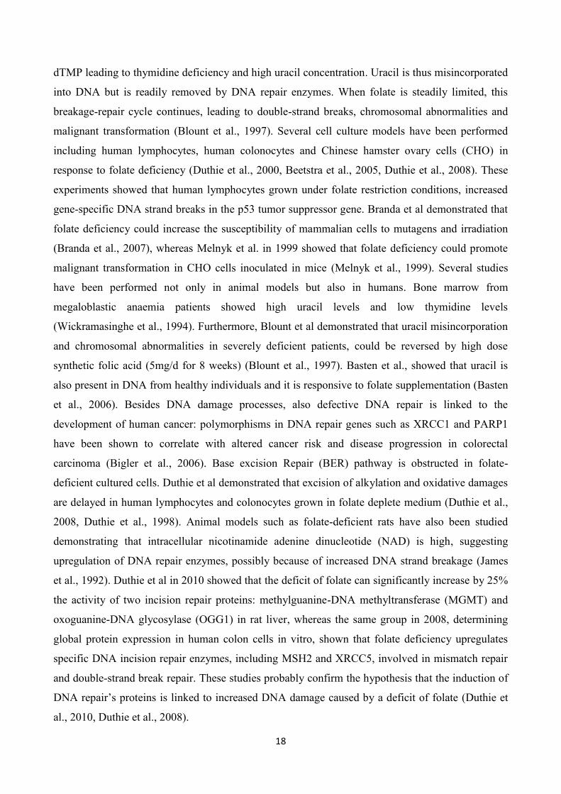
dTMP leading to thymidine deficiency and high uracil concentration. Uracil is thus misincorporated
into DNA but is readily removed by DNA repair enzymes. When folate is steadily limited, this
breakage-repair cycle continues, leading to double-strand breaks, chromosomal abnormalities and
malignant transformation (Blount et al., 1997). Several cell culture models have been performed
including human lymphocytes, human colonocytes and Chinese hamster ovary cells (CHO) in
response to folate deficiency (Duthie et al., 2000, Beetstra et al., 2005, Duthie et al., 2008). These
experiments showed that human lymphocytes grown under folate restriction conditions, increased
gene-specific DNA strand breaks in the p53 tumor suppressor gene. Branda et al demonstrated that
folate deficiency could increase the susceptibility of mammalian cells to mutagens and irradiation
(Branda et al., 2007), whereas Melnyk et al. in 1999 showed that folate deficiency could promote
malignant transformation in CHO cells inoculated in mice (Melnyk et al., 1999). Several studies
have been performed not only in animal models but also in humans. Bone marrow from
megaloblastic anaemia patients showed high uracil levels and low thymidine levels
(Wickramasinghe et al., 1994). Furthermore, Blount et al demonstrated that uracil misincorporation
and chromosomal abnormalities in severely deficient patients, could be reversed by high dose
synthetic folic acid (5mg/d for 8 weeks) (Blount et al., 1997). Basten et al., showed that uracil is
also present in DNA from healthy individuals and it is responsive to folate supplementation (Basten
et al., 2006). Besides DNA damage processes, also defective DNA repair is linked to the
development of human cancer: polymorphisms in DNA repair genes such as XRCC1 and PARP1
have been shown to correlate with altered cancer risk and disease progression in colorectal
carcinoma (Bigler et al., 2006). Base excision Repair (BER) pathway is obstructed in folate-
deficient cultured cells. Duthie et al demonstrated that excision of alkylation and oxidative damages
are delayed in human lymphocytes and colonocytes grown in folate deplete medium (Duthie et al.,
2008, Duthie et al., 1998). Animal models such as folate-deficient rats have also been studied
demonstrating that intracellular nicotinamide adenine dinucleotide (NAD) is high, suggesting
upregulation of DNA repair enzymes, possibly because of increased DNA strand breakage (James
et al., 1992). Duthie et al in 2010 showed that the deficit of folate can significantly increase by 25%
the activity of two incision repair proteins: methylguanine-DNA methyltransferase (MGMT) and
oxoguanine-DNA glycosylase (OGG1) in rat liver, whereas the same group in 2008, determining
global protein expression in human colon cells in vitro, shown that folate deficiency upregulates
specific DNA incision repair enzymes, including MSH2 and XRCC5, involved in mismatch repair
and double-strand break repair. These studies probably confirm the hypothesis that the induction of
DNA repair‟s proteins is linked to increased DNA damage caused by a deficit of folate (Duthie et
al., 2010, Duthie et al., 2008).

19
1.3.4 Folate status and DNA methylation
DNA methylation is the most widely studied mechanism of epigenetic gene regulation. Epigenetic
modifications are dynamic and can be strongly influenced by external factors, thus, these factors
may alter gene expression, potentially inducing disease formation (Li et al., 2002, Reik et al., 2001).
Evidences suggest DNA methylation is fleeting towards nutritional and environmental influences.
Methionine is converted into S-adenosylmethionine (SAM), the primary methyl donor for the DNA
methylation process and it derives from a cyclical cellular pathway called one-carbon metabolism.
During this cycle, DNA methyltransferases (DNMTs) covalently link methyl unit from SAM to
cytosine bases, composing 5-methylcytosine, thus methylating DNA. In mammals, DNA
methylation process is dynamic and this regulatory event can occur in a global or gene specific
manner (Maunakea et al., 2010). MTHFR is a key enzyme of the one-carbon metabolism and it is
involved in the conversion of Hcy to methionine in order to generate 5-methyl-THF. This is the
reason why MTHFR activity has been widely investigated in DNA methylation process. Shelnutt et
al in 2004 studied the relationship between the MTHFR C677T polymorphism in young women
under controlled folate intake and DNA methylation, showing that reducted dietary folate intake
was associated with decreased global methylation of leukocyte DNA. Furthermore, after a re-
supplementation with folate, there was a DNA re-methylation on the global scale (Shelnutt et a.,
2004). Folate is reduced to DHF and then to THF by DHFR enzyme. THF is necessary as one
carbon donor to form 5-methyl-THF that donates its methyl group to Hcy in order to convert it to
methionine. Under low dietary folate intake, SAM is reduced and SAH is elevated: folate depleting
is thus, generally associated with decreased DNA methylation and, potentially, increased proto-
oncogene expression (Kim et al., 2007). Many studies on animal models, have evaluated the
relationship between global methylation and maternal folate status. In a study conducted by McKay
et al., murine offspring under low folate diet during pregnancy showed a reduction of global
methylation in small intestinal tissue (McKay et al., 2011). Conversely, Ly et al., showed an
association between the measurement of global methylation following in utero folate
supplementation and the CpG hypomethylation of mammary tissue among rat offspring of 28 weeks
(Ly et al., 2011). Another study conducted on murine colorectal tissue, demonstrated that post
weaning folate supplementation equivalent to recommended intake levels for women of child-
bearing age linked to a decrease of global methylation levels (Sie et al., 2011). Several human
epidemiological studies have also taken into account the global DNA methylation, looking at the
link between folate intake and DNA methylation in colorectal cancer (CRC) patients and in
postmenopausal women. One study observed, in patients CRC negative, an association between

20
colonic mucosa and lymphocyte DNA methylation and serum and dietary folate concentrations,
whilst, in patients CRC positive the group observed a correlation between folate supplementation
and increasing in global DNA methylation (Pufulete et al., 2005). Another group observed that
among postmenopausal women, dietary and serum folate decrease, were associated with decreased
DNA methylation in leukocytes, in a dose-dependent manner (Rampersaud et al., 2000). However,
the positive impact of the folate supplementation is strongly dependent on dose and timing of
intervention and on health status (Pufulete et al., 2005).
1.3.5 Genetic variants and possible effects on cancer
The folate pathway seems to play a crucial role in the development of childhood ALL because of its
involvement in the DNA synthesis and repair processes and in the DNA methylation pathway.
Recently, especially in childhood acute lymphoblastic leukemia the influence of polymorphisms in
genes of the folate pathway has been studied, indeed. Malignant cells are rapidly dividing cells and
require a high amount of folate. Thus, these cells are vulnerable to folate deficiency. There are
strong evidences that suggest that maternal folate supplementation during pregnancy can reduce the
risk of childhood ALL (Milne et al., 2010). Furthermore, several studies suggest that some
functional polymorphisms of the MTHFR gene could alter cancer susceptibility by the regulation of
folate metabolism (Alcasabas et al., 2008, Damnjanovic et al., 2010). For this reason it possible to
speculate that individuals with reduced MTHFR enzyme activity may have reduced risk of
developing ALL because of an enhancement of folate bioavailability. From recent meta-analysis it
was found that MTHFR polymorphisms have a significant impact on childhood ALL risk
particularly when considering the variant T allele of MTHFR C677T that was found protective
against ALL risk (Yan et al., 2012). Nevertheless, in the past decade, studies on the implication of
MTHFR polymorphisms in the development of ALL reported conflicting results, frequently
showing no associations of the two common variants MTHFR C677T and A1298C with childhood
ALL risk (Chan et al., 2011, Kamel et al., 2007). A study conducted by Semsei et al. showed high
protection in boys carrying 677CT and 1298AA genotypes and less protection against ALL in girls
with 677CC and 1298AC genotypes (Semsei et al., 2010). The results are still inconclusive and
some authors suggest the hypothesis that C677T SNP could be in linkage with some unknown
functional SNPs that can modulate ALL risk. Another important factor to consider is ethnicity that
may influence SNP-cancer risk association through the different allele frequencies, the different
genetic backgrounds and the possible gene-environment interactions, due to different lifestyles (Yan

21
et al., 2012). Many authors considered also the association between MTHFR polymorphisms and
toxicity during chemotherapy, especially with methotrexate: some studies did not find significant
associations between the 677T allele and toxicity (Imanishi et al., 2007, Pakakasama et al., 2007),
whilst some others showed that individuals with 677T allele had to stop the treatment frequently,
suggesting the 677T allele as a toxicity predictor during the chemotherapy maintenance (Shimasaki
et al., 2008, Huang et al., 2008). All the studies conducted till now are not sufficient but necessary
at the same time, in order to get an appropriate treatment for individuals with unfavourable
polymorphisms towards leukemia treatment.
The MTHFR gene:
Methylenetetrahydrofolate reductase (MTHFR) catalyzes the conversion of 5, 10-
methylenetetrahydrofolate to 5-methyltetrahydrofolate, necessary as a co-substrate for the
remethylation of Homocysteine to methionine. MTHFR gene lies on chromosome 1, locus 1p36.3
and contains 11 exons, coding for a protein of 656 amino acid with a predicted molecular weight of
74.5 kD (Goyette et al., 1995)
In the MTHFR gene are present two well-described and widely studied common polymorphisms:
C677T and A1298C. Other less common polymorphisms have been reported at 1059 bp, 1289 bp,
1317 bp and 1793 bp (Trembath et al., 1999, Weisberg et al., 1998, Rady et al., 2002). The MTHFR
C677T polymorphism occurs in exon 4 and consists of a C-to-T nucleotide transition at nucleotide
677 leading to an alanine to valine change at codon 223 (A223V) in the catalytic domain of the
protein (Frosst et al., 1995). This substitution renders the enzyme thermolabile, affecting the
associated activity in vitro (Goyette et al., 2000, Yamada et al., 2001). Subjects carrying the CT
heterozygous condition have been shown to have 65 percent of their in vitro enzyme activity,
compared with homozygotes for the common variant CC whereas those with the homozygous
variant condition TT have been found to have 30 percent compared with the wild type (Frosst et al.,
1995). Moreover, Molloy et al. reported that CT heterozygous carriers have 10 percent lower red
cell folate levels, when compared with the wild type CC, whilst the TT homozygotes have 18
percent lower red cell folate (Molloy et al., 1997). Other observations showed that subjects carrying
the TT variant also present lowered circulating folate, B12 vitamin levels and increased plasma Hcy
levels (Ma et al., 1999, Jacques et al., 1996). Gemmati et al. in 1999 conducted a study to evaluate
the different correlation degree between plasma Hcy and folate levels among the three genotype
conditions of MTHFR C677T polymorphism (Gemmati et al., 1999). Homozygotes TT showed a
22
significant increasing of Hcy as the plasma folate level decreased. This correlation decreased among
heterozygotes CT and disappeared among wild-type CC. This observation is in line with the study
of Malinow who reported that depending on the number of polymorphic 677T allele in MTHFR
gene, the response to folate supplementation is different (Malinow et al., 1997). The same paper
reported a significant overrepresentation of TT-carriers among subjects with
hyperhomocysteinemia. In 1998 Bagley and Selhub hypothesized that MTHFR C677T
polymorphism could be associated with an altered distribution of folate within the red blood cells:
the polyglutamated 5-methyl-THF isoforms were the unique folate isoforms rediscovered within red
blood cells of subjects with 677CC genotype. The exclusive presence of formil folate isoforms
within red blood cells of subjects with 677TT genotype is in line with the hypothesis that there
exists, in vivo, a displacement of the activity of MTHFR thermolabile variant and that this
displacement will lead to an altered folate distribution within erythrocytes. The thermolabile variant
of the enzyme present in 677TT subjects catalyzes less efficiently the synthesis of 5-methyl-THF
respect to 677CC subjects (Bagley et al., 1998). Skibola et al in 1999 hypothesized a protection
against leukaemia explicable with an increment of methylene-THF cytosolic levels available for
thymidylate synthesis, an essential precursor for the DNA de-novo synthesis. Regarding MTHFR
polymorphisms and DNA methylation, it is generally known that DNA from subjects with the
thermolabile variant of MTHFR (TT) is hypomethylated respect to DNA from subjects with the
wild-type variant (CC). Nevertheless, this finding is not confirmed in a study of Narayanan
(Narayanan et al., 2004). Recent data, suggest the hypothesis that DNA methylation status could be
influenced by genotype only among those subjects presenting low plasma folate levels. Friso et al
thus, have demonstrated that subjects with MTHFR 677TT genotype and lower plasma folate
concentration had the lower methylation level compared to all the other groups (Friso et al., 2005).
A second MTHFR polymorphism lies in the S-adenosyl-methionine-regulatory domain of the
enzyme and it consists of an A-to-C transversion at nucleotide 1298, causing a glutamate to alanine
amino acid change at codon 429 (A1298C/E429A) (Matthews et al., 1984, Wiemels et al., 2001).
The binding of SAM causes conformational changes that inhibit MTHFR‟s activity (Matthews et
al., 1984). Studies performed on lymphocytes from individuals in homozygous variant 1298CC
demonstrate to have approximately 60 percent specific wild-type in vitro MTHFR activity, whereas
individuals heterozygotes 677CT and heterozygotes 1298AC were found to have 50-60 percent
wild-type MTHFR activity (Weisberg et al., 1998; van der Put et al., 1998). The C677T
polymorphism has been most extensively studied than A1298C and researches on the relationship
between plasma folate and Hcy levels and MTHFR A1298C are less consistent than those
performed on C677T variant, probably because of the lower effect of A1298C variant on the
23
enzyme properties. Some in vitro studies such as site-directed mutagenesis and in vitro expression
experiments have been performed to create a recombinant protein containing both gene variations
(Goyette et al., 2000, Yamada et al., 2001). Mutant enzymes with the two separate variants 1298 or
677 showed respectively about 68 percent and 45 percent of the control activity whereas the
recombinant enzyme containing both variations had 41 percent of the control activity. The two
polymorphisms sites are 2,1 kb apart and several groups have found a strong linkage disequilibrium
between 677 and 1298 gene variants (Stegmann et al., 1999, Chen et al., 2002).The two
polymorphisms very rarely exist on the same allele and double homozygous carriers
677TT/1298CC are extremely uncommon in general population. Furthermore, Zetterberg et al
showed in a study performed in 2002 an increased frequency of combined polymorphic alleles
C677T and A1298C in spontaneously aborted embryos (Zetterberg et al., 2002).
Generally, it was demonstrated that there are significant associations between specific
polymorphisms in folate-related genes and susceptibility to develop childhood ALL. MTHFR is the
most widely investigated gene linked to one-carbon metabolism and C677T and A1298C are the
two most common polymorphisms considered. Recent reviews have summarized the results of
several studies that focussed on the link between MTHFR polymorphisms and risk of ALL.
Interesting observations indicate the possibility of a different impact of polymorphisms on
susceptibility to ALL, depending on the type of populations (Koppen et al., 2010). Some studies
found an increased risk for ALL with the MTHFR A1298C in Filippino and non-white Brazilian
populations (Zanrosso et al., 2006, Alcasabas et al., 2008) whilst some others found a decreased risk
with the same polymorphisms within British and Egyptian populations (Skibola et al., 1999, Kamel
et al., 2007). Robien and Ulrich in 2003 showed that for MTHFR A1298C, the polymorphic allele
C frequency ranges from 0.27-0.36 in Western Europe to 0.17-0.19 among Asian populations
(Robien et al., 2003) and this finding could support the theory described above.
The DHFR gene:
Dihydrofolate reductase (DHFR) is ubiquitously expressed in all organisms and it is controlled by a
TATA-less promoter that, in turn is governed by several transcription factors, including Sp1 and
E2F (Chen et al., 1984, Jensen et al., 1997). The DHFR gene family includes the functional DHFR
gene that lies on chromosome 5q11.2 and three intronless pseudogenes: DHFRP1 on chromosome
18, DHFRP2 on chromosome 6, DHFRP4 on chromosome 3 (Anagnou et al., 1984). The functional
gene is expressed in three mRNA isoforms with alternatively spliced 3‟ UTR ends (Morandi et al.,

24
1982). DHFR catalyzes the reduction of dihydrofolate (DFH) to tetrahydrofolate (THF) with the
presence of NADPH as cofactor. DHFR is also necessary for the intracellular conversion of
synthetic folic acid into the THF forms needed for the folate/Hcy metabolism. Since THF is the
principal one-carbon donor within the folate cycle in order to synthesize purines and thymidylate,
absence of DHFR activity leads to fast breakdown of THF and consequent stop of de-novo DNA
synthesis and cell proliferation (Skibola et al., 1999). This is the reason why DHFR is a common
drug target in several pathologies (McGuire et al., 2003) and functional polymorphisms within
DHFR can potentially lead to changes in its expression or activity, thus influencing the risk of
folate-dependent diseases or therapeutic responses to antifolates, leading to higher adverse drug
event frequency. In 1989 Detera-Wadleigh firstly reported a polymorphism within the first intron of
DHFR as a restriction fragment leght polymorphism (RFLP) and in 2004 Johnson et al determined
the location and size of the polymorphism identifying a 19bp insertion to deletion and suggesting its
functional role, since Sp1 transcription factor binding-site is located within the deleted sequence
and the deletion allele could affect gene expression (Detera-Wadleigh et al., 1989; Johnson et al.,
2004). In fact, intron-1 has important roles in the transcription of DHFR and the amount of protein
produced by translation (Noè et al., 2003). In the past years has been observed a dose-dependent
relation between DHFR expression and the deletion. Subjects 19bp DD (deleted allele)
homozygotes had higher mRNA levels than subjects carrying the counterpart genotype WW (wild-
type allele): the DD genotype has been associated with up to 4.8-fold increase in mRNA levels
compared with the WW genotype (Xu et al., 2007). This could be responsible for a filling of THF
and other isoforms within the cell, supporting as a consequence DNA synthesis as reported for
MTHFR C677T (Frosst et al., 1995, Bagley et al., 1998, Kluijtmans et al., 1996). During the last
years several studies have been performed on DHFR but results are still inconclusive and often
conflicting: some studies provided evidences that the risk of having a child with spina bifida and the
risk of pre-term delivery is higher for women with the DHFR DD genotype (Johnson et al., 2004,
Johnson et al., 2005). Parle-McDermott otherwise, reported a lower risk of having a child with
Neural Tube Disease (NTD) in women with the deleted allele (Parle-McDermott et al., 2007). A
study reported a non-significant increase in mRNA levels for homozygous deleted individuals
whereas another study observed a significant increase in DHFR expression with the number of
deleted allele, suggesting the possible maternal protective role of 19 bp D allele in NTD by raising
the quantity of DHFR available for the reduction of DHF (Parle-McDermott et al., 2007; Xu et al.,
2007). Other authors have analysed the relation between the DHFR 19 bp ins/del polymorphism and
the risk of breast cancer reporting non-significant association with overall breast cancer risk (Cam
et al., 2009). However, an important consideration should be done when stratification according to

25
vitamin use is performed: patients that used the multivitamin supplements with the DHFR-DD
genotype had a 50 percent increase in breast cancer risk compared with women with DHFR-WW
genotype (Xu et al., 2007). It is possible that polymorphisms associated with a higher expression of
DHFR may protect against cancer because of the high amount of 5,10-methylene-THF available for
thymidilate synthesis, but at the same time a variation in the pool of 5-methyl-THF may influence
methylation processes and consequently, affect the cancer risk. Gemmati et al, reported a protective
role of DHFR 19 bp D allele in adult acute lymphoblastic leukemia particularly when the D-allele
was associated with MTHFR 677TT genotype (Gemmati et al., 2009). It has also been reported a
correlation between lower Hcy levels, increased serum and red blood cell folate level and DHFR-
DD genotype. These studies suggest the hypothesis that the 19 bp deletion could increment DHFR
expression facilitating the remethylation of Hcy (GelleKink et al., 2007, Stanislawska-Sachadin et
al., 2008). Nevertheless, the effects of DHFR 19 bp ins/del polymorphism have not yet been
clarified in detail in the context of solid tumours and data about adult or childhood haematological
neoplasia are not yet present, considering the single polymorphism or its combination with other
metabolic pathways.
DHFR is one of the key-enzyme of the one-carbon metabolic pathway and it is considered a target
molecule for clinically important drugs such as chemotherapeutic agents. Methotrexate (MTX) is a
potent inhibitor of DHFR, resulting in the depletion of reduced folates, cofactors for thymidylate
synthetase. The same gene variations that define THF accumulation within the cell may protect
against cancer risk as shown in the study of Gemmati et al. but contextually, they may affect
antifolate treatment as shown in the paper of Ongaro et al. In the latest, authors showed that MTX-
related hepatic toxicity increased 2.07-4.57-fold in adult acute lymphoblastic leukaemia patients
with the 19 bp deletion allele (Ongaro et al., 2009). This dual and opposite paradoxical effect can be
summarized as follow: cancer patients carrying such gene variants may have firstly reduced cancer
susceptibility but a contextual increased drug-related toxicity, particularly when they are treated
with folate antagonists such as Methotrexate (MTX) (Gemmati et al., 2008).
B cell lymphoma gene-2 (Bcl-2):
Bcl-2 is an inner mithocondrial membrane protein codified by the bcl-2 gene that lies on
chromosome 18q21.3 and consists of 3 exons and 2 promoters with different functional properties
(Hockenbery et al., 1990). P1 promoter drives the transcription activity while P2 promoter is
located 1400 bp upstream of the translation initiation site and diminishes P1 promoter‟s activity,

26
acting as a negative regulatory element (Young et al., 1993, Seto et al., 1988). Preliminary studies
have been effectuated in 1985 as a result of the involvement of the bcl-2 gene in the most common
t(14;18) translocation found in follicular B lymphomas (Bakhshi et al., 1985; Tsujimoto et al., 1985,
Bakhshi et al., 1987). The t(14;18) translocation leads to a chimeric transcript because of the fusion
of the 3‟ nontranslated region of bcl-2 with the joining (JH) segment of Ig heavy chain gene,
causing a deregulation of bcl-2 transcription (Clearly et al., 1986). The bcl-2 gene encodes a major
nonglycosilated protein of 26 Kd that resides in mithocondria and that promotes the survival of
several hematolymphoid cells (Hockenberry et al., 1990, Vaux et al., 1988, Nunez et al., 1990),
inhibiting apoptosis (programmed cell death). An increased survival of a cell population defines the
accumulation of genomic instability within the cells thus inducing leukemogenesis. Apoptosis is a
natural process regulated by several pathways that comprise genes that both promote or inhibit
apoptosis. Bcl-2 possesses anti-apoptotic activity and several studies have showed that bcl-2 gene
was upregulated in high proportion of acute myeloid leukemia (AML) patients (Karakas et al.,
1998, Bensi et al., 1995). Bcl-2 thus, may play a key role in the survival of hematolymphoid cells,
inhibiting apoptosis through the prevention of the release of cytochrome c from mithocondria,
inducing the block of caspase activation (Reed et al., 1994). It can be hypothesized that
polymorphisms in the bcl-2 gene, which modify protein function and/or expression could play an
important role in the balancing of apoptosis regulation mechanisms. A single-nucleotide
polymorphism (SNP) in the promoter region locates at nucleotide position -938 and consists of a C-
to-A change. The SNP -938C>A has been reported to decrease the prostate cancer risk but the study
considered a relative small sample size (Kidd et al., 2006). Other papers did not reveal any
significant association between the functional SNP -938 C>A and the risk of different cancer types
such as squamous cell carcinoma of the head and neck (SCCHN) or B-cell chronic lymphocytic
leukemia (B-CLL) (Chen et al., 2007, Nuckel et al., 2007). Nevertheless, the same authors showed
that the -938 C>A SNP had a statistically significant impact on transcriptional activity of a region
of the bcl-2 gene that inhibits promoter activity. The functional -938 C>A promoter polymorphism
is located in the inhibitory P2 promoter region and an increased activity of the inhibitory P2
promoter seems to be associated with the C allele (Nuckel et al., 2007), suggesting a potentially
different binding of transcription factors to the C and A alleles. Several others studies performed on
breast cancer, oropharyngeal squamous cell carcinoma, chronic lymphocytic leukemia,
glioblastoma multiforme and prostate cancer have showed an association between the A allele and
an higher Bcl-2 expression level. Furthermore, the -938 C>A genotype has been linked to survival
of patients suffering from those cancers (Bachmann et al., 2007; Lehnerdt et al., 2009, Nuckel et al.,
2007). Recently, Kunkele et al., have evaluated the possible correlation between -938 C>A

27
functional polymorphism and the risk group classification in childhood ALL, considering that this
SNP is known to influence the balance of apoptosis and survival in hematolymphoid cells. Data
suggest that genotyping ALL patients for -938 C>A polymorphism could be of benefit for therapy
response prediction: authors indeed, have showed that childhood ALL patients with bcl-2 -938CC
genotype have about 3-fold higher risk of belonging to a high-risk group (Kunkele et al., 2013).
Chauhan et al., suggested that bcl-2 could be used as possible marker for therapy response during
induction chemotherapy treatment of adult ALL patients (Chauhan et al., 2012), whilst Singh et al.
showed that Bcl-2 inhibition with concomitant use of methadone increased the induction of
apoptosis in childhood ALL cells (Singh et al., 2011). Until now, there are few reports on the
association between bcl-2 polymorphism and cancer risk or disease progression or therapy response
and survival. More intensive treatment regimens can leads to severe side effects and/or long-term
adverse effect so additional markers to ameliorate the specificity of treatment could be considered a
great goal, reducing short- and long-term adverse effects.

28
1.4 METHOTREXATE
During the last 20 years advances in specific chemotherapy allowed an improving of the survival
rates for ALL, with expected cure rates higher than 80 percent (Pui et al., 2008). Methotrexate
(MTX) (Fig. 4) was the first drug considered curative for patients with a solid tumour,
choriocarcinoma and it is an important component of the therapy for childhood ALL. Often, the
treatment with MTX causes severe and different toxicities that require the cessation or the reduction
of the therapy then, an appropriate predictor for the adverse outcome of MTX treatment would be
strong useful (Imanishi et al., 2007). MTX enters the cell by two mechanisms: with the first
mechanism the uptake of MTX occurs by passive diffusion, dependent on the amount of
extracellular concentrations of the drug; with the second mechanism MTX enters via active
transport mediated by the reduced folate carrier (RFC1) (Gorlick et al., 1996). Once inside the cell,
MTX is polyglutamylated by the enzyme folylpolyglutamate synthetase and it acts by inhibiting
two key enzyme of the folate metabolic pathway: DHFR and thymidilate synthase (TS). The block
of these enzymes affects DNA synthesis and cell replication. DHFR is necessary for the conversion
of DHF to THF and its inhibition therefore gets reduced THF isoforms necessary for folate
homeostasis out. TS catalyses the methylation of dUMP to dUTP, a precursor for DNA synthesis:
the inhibition of TS leads therefore to the depletion of thymidine and DNA damages (Salazar et al.,
2012). High-dose MTX is frequently used during the consolidation phase of ALL treatment.
Hypothesizing that MTX toxicity can be related to polymorphic variants in genes of the folate/MTX
metabolic pathway, the identification of these genetic markers could be considered a high contribute
to the individualization of chemotherapy in order to reduce related toxicities (Cheok et al., 2006).
MTHFR is a key enzyme of the folate metabolic pathway. It catalyzes the conversion of 5,10-
methylen-THF to 5-methyl-THF which is required for protein synthesis and nucleic acid
methylation. Alterations of folate isoforms as a consequence of impaired MTHFR activity may have
an important effect on the responsiveness of malignant and non-malignant cells to MTX. Several
data stress the pharmacogenetic link of MTHFR genotype in chemotherapeutic treatments including
MTX: Salazar et al found that ALL children with favorable MTHFR 677CC genotype treated with
high doses MTX (5g m-2
) had significantly better event-free survival (EFS) at 48 months than
patients with MTHFR 677TT genotype treated with the classical MTX dose 3g m-2
in a 24-h
infusion (Salazar et al., 2012). Other studies showed that patients with MTHFR 677T/1298A
haplotype had a poorer EFS, nevertheless different ALL protocols were used (Krajinovic et al.,
2004); Aplenc et al showed that the MTHFR C677T polymorphism was associated with relapse

29
whilst, MTHFR A1298C polymorphism was not (Aplenc et al., 2005). One of the major adverse
effects of MTX is nephrotoxicity. MTX and its metabolite 7-hydroxy-MTX precipitates in the renal
tubules causing renal dysfunction (Yarlagadda et al., 2008). As a consequence, MTX excretion may
be delayed, maintaining high plasma MTX levels, increasing in turn, toxicity. Haematological
toxicities such as thrombocytopenia may also occur, due to high serum MTX level. Kantar et al in a
recent study showed that childhood ALL patients with MTHFR A1298C polymorphism treated
with high dose MTX, had significantly higher MTX levels at 48 h and haematological toxicities
such as thrombocytopenia (Kantar et al., 2009). Recently, a sistematic review by Lopez-Lopez et al
concluded that today there is no evidence to support the use of either MTHFR C677T or the
A1298C polymorphisms as MTX toxicity markers (Lopez-Lopez et al., 2013). It is clear that other
polymorphisms involved in the folate pathway and other genetic and epigenetic mechanisms may
contribute to the variability in treatment responses. DHFR plays a crucial role in the conversion of
DHF to THF necessary for nucleic acid synthesis and methylation processes. It is considered a
major MTX target and plays an important role in the development of MTX resistance in ALL.
Several experimental and clinical settings were made, founding altered levels of DHFR and/or
DHFR gene amplification in relapsed leukemia patients and in leukemia and colon cell lines with
MTX-resistant phenotypes (Morales et al., 2009, Matherly et al., 1995, Serra et al., 2004, Goker et
al., 1995). Moreover, Bertino et al showed that DHFR levels increase within hours in both normal
leucocytes and acute leukemia cells after MTX treatment. MTX slowly dissociates from DHFR and
survival cells continue to synthesize THF and proliferate (Bertino et al., 2009). Chu et al have
demonstrated that DHFR translation is inhibited by binding of DHFR to its mRNA and this
inhibition is relieved by MTX which binds to DHFR and leads to a conformational change in the
protein (Chu et al., 1993). This mechanism thus, limits MTX effectiveness (Guo et al., 1999).
Several years ago, a polymorphism in the 3‟ untranslated region (UTR) of DHFR was found. This
SNP, a cytosine to thymine substitution at position 829 was associated with higher expression of
DHFR transcript (Goto et al., 2001). Also Mishra et al showed that the presence of the SNP
increased levels of DHFR mRNA, the amount of protein and resistance to MTX (Mishra et al.,
2007). Alterations in DHFR expression levels and thus, in sensitivity to MTX, can be also caused
by genetic polymorphisms in the promoter (Dulucq et al., 2008, Al-Shakfa et al., 2009). Dulucq et
al, showed an association of individual polymorphisms and resulting haplotypes with ALL
outcome, revealing a reduction in relapse-free survival with the presence of A and C alleles located
at position 317 and 1610 upstream from a minor transcription initiation site, respectively (Dulucq et
al., 2008). Thus, results are still scarce and inconclusive. However, the application of a tailored
therapy would represent a huge advance in the treatment of childhood ALL and the identification of

30
pharmacogenetic markers in treatments will provide an improvement in risk classifications allowing
an optimal and personalized therapy selection.
Fig.4 Methotrexate molecule.

31
1.5 AIM AND SCOPE
Acute lymphoblastic leukemia (ALL) is the most common pediatric cancer accounting for 80% of
childhood leukemia. Different gene polymorphisms (most of them SNPs) play an important role in
the susceptibility to childhood ALL which probably derives from a combination and relation of
genetic and environmental factors. Folates are bioactive nutrients involved in several processes
including DNA synthesis and methylation and purines synthesis. Polymorphisms in genes coding
for enzymes of the folate metabolic pathway can alter the intracellular folate status or distribution.
Several studies have investigated on the role of the genetic variants of the one carbon pathway and
on the susceptibility to develop childhood ALL. It is to note that current treatment regimens achieve
levels about 80% in overall survival (OS). Unfortunately, the side effects derived from the
chemotherapeutic agents used can be severe, especially for high-risk patients. Therefore, the
identification of additional markers which can improve risk stratification and individually tailored
therapy regimens would be a great goal, in order to avoid over-treatment which can increase long-
term adverse side effects.
The aim of the present study was, therefore, to investigate whether common polymorphisms (i.e.
MTHFR C677T and A1298C in addition to DHFR 19 bp INS/DEL and Bcl-2 -938 C>A) might
influence the risk of childhood ALL. For this purpose we genotyped by PCR 197 childhood ALL
patients and 201 matched healthy controls for the DHFR and MTHFR gene polymorphisms
hypothesizing their single/combined effect on cancer risk by means of intracellular folate isoforms
unbalancing. Similarly, we genotyped the same samples for the -938 C>A polymorphism in the
anti-apoptotic Bcl-2 gene, hypothesizing its effects on cancer risk by itself or in combination with
the previous ones polymorphisms. Finally, other parameters such as disease onset, therapy duration
and risk scoring, were stratified by the polymorphisms investigated.
In an exploratory way and to validate the proposed model we evaluated possible differences in
cellular viability under MTX treatment, between healthy PBL cells harvested from subjects with
opposite genotype condition considering DFHR and MTHFR genes. One of the most interesting
facets of the relationship between folate status and cancer risk is the modulation of the one-carbon
metabolism by key folate-metabolizing enzymes. The amount of intracellular THF is due to the
relative balancing between DHFR and MTHFR activities: the first by production and filling
activity, the second one by consumption and depletion activity. Changes in their activity resulting

32
from polymorphisms in DHFR and MTHFR genes could modify the susceptibility to cancer and
cancer treatment. MTX is indeed an antifolate agent necessary in the maintenance therapy of ALL.
It interferes with folate metabolism leading to depletion of reduced forms of THF (including 5,10-
methylene-THF) and to inhibition of nucleic acid synthesis, favouring cell death. Therefore a
paradox there exists when some polymorphisms are investigated among cancer populations:
patients carrying specific gene variants may have reduced cancer risk but increased drug-related
toxicity when they are treated with folate antagonists. The same allele could be considered,
therefore a protective factor against cancer establishment and a risk factor for prognosis of the same
disease. Genotyping of folate and apoptotic pathways‟ polymorphisms might be useful to assess the
effects on childhood ALL susceptibility and to assess the effects of anti folate drug therapy on
toxicity. Considering several parameters such as onset, therapy duration and risk stratification, the
latter one could provide important information to clinicians to optimize MTX therapy, reducing the
associated toxicity with possible effects on survival.
The proposed in vitro model could be considered a starting point for further investigations that
could in part explain the reason why some gene conditions result protective towards the onset of
cancer and at the same time risk factors for increased toxicity towards anti-folate therapy.

33
2
MATERIALS AND METHODS

34
2.1 SELECTION OF CASES AND CONTROLS
Recruitment of childhood ALL cases consisted of 197 Italian and Egyptian individuals with newly
diagnosed ALL according to the French-American-British classification. They were from the files
of patients who received hematological diagnosis by the Unit of Pediatry of University-Hospital of
Ferrara (n= 3), by the Unit of Pediatry of University-Hospital of Tanta, Egypt (n=20) and by the
Unit of Pediatry of Bari-Policlinico (n=174) in the period between January 2011 and January 2014.
The whole group of ALL cases consisted of patients with an age range of 0.5 – 17.33 years with a
mean age of 10.76 ± 5.68 years. 38.6% were male. Among ALL cases, 86% were ALL-B and 14%
were ALL-T. The control group (n= 201) consisted of Italian healthy individuals, who were not
relatives of the cases and were randomly selected from the local blood donor lists in the period
between January 2011 and January 2014. The age range was 1 – 21.5 years with a mean age of
15.83 ± 6.41 and 42% were male. Peripheral blood samples for all cases were collected at the date
of diagnosis before any pharmacological treatment and for controls at the moment of blood
donation by venipuncture. Informed consent was obtained from donors and patients‟ parents.
Within the control group genotyped for MTHFR C677T and DHFR -19bp INS/DEL (W/D), 4
volunteer homozygotes 677TT/-19bp DD and 4 volunteer homozygotes 677CC/-19bp WW were
selected for the cell culture protocol described in detail in the next chapter.
35
2.2 GENOTYPIC ANALYSIS
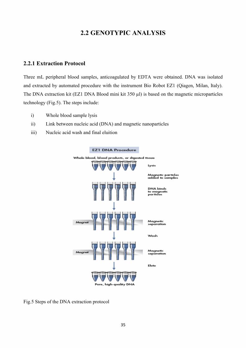
2.2.1 Extraction Protocol
Three mL peripheral blood samples, anticoagulated by EDTA were obtained. DNA was isolated
and extracted by automated procedure with the instrument Bio Robot EZ1 (Qiagen, Milan, Italy).
The DNA extraction kit (EZ1 DNA Blood mini kit 350 µl) is based on the magnetic microparticles
technology (Fig.5). The steps include:
i) Whole blood sample lysis
ii) Link between nucleic acid (DNA) and magnetic nanoparticles
iii) Nucleic acid wash and final eluition
Fig.5 Steps of the DNA extraction protocol

36
2.2.2 Polymerase Chain Reaction (PCR)
PCR technique is a useful tool for the exponential amplification of a DNA fragment, and its
principle is based on the mechanism of DNA replication in vivo: the double strand dsDNA is
denatured to a single strand ssDNA, duplicated, and this process is repeated along the reaction
according to the following formula: C = C0(1+E) n where,
C: final amount of DNA
C0: initial amount of DNA
E: efficiency
n: number of cycles
s: slope of the exponential phase.
During the denaturation step, the dsDNA melts opening up to ssDNA, and all enzymatic reactions
stop. To DNA denaturation, the temperature is usually raised up to 93-96 °C, breaking the H-bonds
and thus increasing the number of non-paired bases. The temperature at which half of the dsDNA is
single-stranded is known as the melting temperature, Tm. The second phase, i.e. annealing of
primers to ssDNA, takes place at temperatures closer to their Tm (usually 55-65 °C) and is named
as temperature of annealing (Ta). The oligonucleotides used as primers typically consist of
relatively short sequences (15-25 nt) complementary to recognition sites, flanking the segment of
target DNA to be amplified. Once the temperature is reduced, the two complementary ssDNA
chains tend to rehybridise into a dsDNA molecule. If primers adequately anneal to the template, the
ionic bond is strong enough between the template and the primer to stabilize the nascent double
stranded structure and allow the polymerase to attach and begin copying the template. The
extension phase is carried out across the target sequence by using a heat-stable DNA polymerase in
the presence of dNTPs and MgCl2, resulting in a duplication of the starting target material. This
enzyme has 5' --> 3' DNA polymerase activity, i.e. it adds dNTPs from 5' to 3' , reading the
template from 3' to 5' . After each cycle, the newly synthesized DNA strands can serve as template
in the next cycle. Potentially, after 30 PCR cycles there will be around 230-fold amplification,
assuming 100 % efficiency during each cycle. The efficiency of a PCR will vary from template to
template and according to the degree of optimization that has been carried out.

37
2.2.3 Genotype protocols
MTHFR C677T
PCR amplification was performed using specific Forward and Reverse primers (Table??) and the
PCR conditions were as follows: initial denaturation at 95°C for 5 minutes, followed by 30 cycles
of 45 seconds at 94°C, 60 seconds at 65°C, 90 seconds at 72°C and a last elongation step for 10
minutes at 72°C, adapted from Frosst et al (Frosst et al., 1995).
The amplicon was digested over night at a temperature of 65°C with restriction enzyme Taq I (New
England BioLabs, Beverly, MA), according to the manufacturer‟s instructions.
The amplified, digested product was separated on 10% acrylamide gel at 120V for 2 hours and half
and stained with Ethidium Bromide. The obtained band sizes are shown in Table 2.
MTHFR A1298C
PCR amplification was performed using specific Forward and Reverse primers (Table??) and the
PCR conditions were as follows: initial denaturation at 95°C for 5 minutes, followed by 30 cycles
of 30 seconds at 94°C, 13 seconds at 59°C, 17 seconds at 72°C and a last elongation step for 10
minutes at 72°C, adapted from Weisberg et al (Weisberg et al., 1998).
The amplicon was digested over night at a temperature of 37°C with restriction enzyme Fnu4HI
(New England BioLabs, Beverly, MA), according to the manufacturer‟s instructions.
The amplified, digested product was separated on 10% acrylamide gel at 120V for 2 hours and half
and stained with Ethidium Bromide. The obtained band sizes are shown in Table 2.

38
DHFR 19 bp INS/DEL
The genotyping protocol to detect the DHFR 19 bp deletion polymorphism was adapted from
Johnson et al. (Johnson et al., 2004). The deleted and non-deleted alleles were genotyped by using a
system of three specific primers: two different allele-specific forward primers, respectively for the
non-deleted and deleted polymorphic alleles and one common reverse primer. The PCR conditions
were as follows: : initial denaturation at 95°C for 5 minutes, followed by 30 cycles of 50 seconds at
94°C, 50 seconds at 62°C, 50 seconds at 72°C and a last elongation step for 10 minutes at 72°C.
PCR products were analyzed by 11% PAGE Ethidium Bromide stained, to distinguish the two
different alleles (non-deleted allele, 113 bp and deleted allele 92 bp) (Table 2).
Bcl-2 -938 C/A
PCR amplification was performed using specific Forward and Reverse primers (Table 2) and the
PCR conditions were as follows: initial denaturation at 96°C for 5 minutes, followed by 35 cycles
of 45 seconds at 96°C, 40 seconds at 56°C, 30 seconds at 72°C and a last elongation step for 10
minutes at 72°C, adapted from Chen et al (Chen et al., 2007).
The amplicon was digested over night at a temperature of 37°C with restriction enzyme Bcc I (New
England BioLabs, Beverly, MA), according to the manufacturer‟s instructions.
The amplified, digested product was separated on 10% acrylamide gel at 120V for 2 hours and half
and stained with Ethidium Bromide. The obtained band sizes are shown in Table 2.
All PCR cycles were performed in an Agilent Thermal Cycler apparatus (Sure Cycler 8800, Agilent
Technologies). Confirmation of genotypes was carried out by randomly regenotyping cases and
controls for all the polymorphisms investigated and DNA samples known for all the genotype
conditions were used as internal controls. There were no discrepancies between genotypes
determined in duplicate and compared with controls.

39
Table 2 Primer Sequences
PRIMER
SEQUENZA AMPLICON
SIZE
BAND
SIZE
MTHFR C677T F
MTHFR C677T R
5‟-TGA AGG AGA AGG TGT CTG CGG GA-3‟
5‟-AGG ACG GTG CGG TGA GAG TG-3‟
233 bp (C)
175bp (T)
58bp (T)
MTHFR A1298C F
MTHFR A1298C R
5‟-CTT TGG GGA GCT GAA GGA CTA CTA
C-3‟
5‟-CAC TTT GTG ACC ATT CCG GTT TG-3‟
138 bp(A) 119bp (C)
19bp (C)
DHFR-19bp INS/DEL F1
DHFR-19bp INS/DEL F2
DHFR-19bp INS/DEL R
5‟–CCA CGG TCG GGG TAC CTG GG-3‟
5‟-ACG GTC GGG GTG GCC GAC TC-3‟
5‟-AAA AGG GGA ATC CAG TCG G-3‟
113bp(W)
92bp (D)
Bcl-2 -938 F
Bcl-2 -938 R
5‟- CTGCCTTCATTTATCCAGCA-3‟
5‟-GGCGGCAGATGAATTACAA -3‟
300bp(C) 189,111 (A)
300 (C)
Table 3 Components for MTHFR C677T PCR
Genomic DNA 100ng
10X Buffer PCR 2,5 µl
MgCl2 1,5 mM
Primer 5' 0,25 mM
Primer 3' 0,25 mM
DNTPS 1,66 mM
Taq Roche [5u/μl] 2U/A
H2O -

40
Table 4 Components for MTHFR A1298C PCR
Genomic DNA 100 ng
10X Buffer PCR 2,5 µl
MgCl2 1,5 mM
Primer 5' 0,25 mM
Primer 3' 0,25 mM
DNTPS 1,66 mM
Taq Roche [5u/μl] 2U/A
H2O -
Table 5 Components for DHFR 19 bp INS/DEL
Genomic DNA 100 ng
10X Buffer PCR 2,5 µl
MgCl2 1,5 mM
Primer Forward1 0,25 mM
Primer Forward2 0,25 mM
Primer Reverse 0,25 mM
DNTPS 1,66 mM
DMSO 2%
Taq Roche [5u/μl] 2U/A
H2O -
Table 6 Components for Bcl-2 -938 C>A
Genomic DNA 100 ng
10X Buffer PCR 2,5 µl
MgCl2 1,5 mM
Primer 5' 0,25 mM
Primer 3' 0,25 mM
DNTPS 1,66 mM
Taq Roche [5u/μl] 2U/A
H2O -

41
2.2.4 Cell preparation and culture
Within the control group genotyped for MTHFR C677T and DHFR -19bp INS/DEL (W/D), 4
volunteers homozygotes 677TT/-19bp DD and 4 volunteers homozygotes 677CC/-19bp WW were
selected. Peripheral Blood was collected from healthy donors in the presence of sodium citrate by
venipuncture. Peripheral Blood Lymphocytes (PBL) were isolated by centrifugation on a layer of
Histopaque ® (Sigma Chemical Co.). PBL were resuspended in Rosewell Park Memorial Institute
culture medium (RPMI 1640; Sigma Chemical Co.), supplemented with 10% FCS, 2 mM L-
glutamine and antibiotics (penicillin 100 U/ml, streptomycin 100 mg/ml). Cultures were maintained
at 37°C in a humid atmosphere containing 5% CO2 and PBL were activated by PHA (5µg/ml) for
48 hours and then dead cells were removed by centrifugation on a layer of Histopaque ® (Sigma
Chemical Co.), whilst PHA-activated PBL were harvested, washed in PBS, and then resuspended in
Rosewell Park Memorial Institute culture medium (RPMI 1640; Sigma Chemical Co.) and
incubated in 96-well micro-plates (7,5x10⁵/ml) (Costar, Cambridge, MA) with MTX 100 µM
together with folic acid (1mg/L) for 5/6 days. Cells were cultured in duplicate and every 24 hours,
cell count and MTT viability assay were performed.
MTT Viability Assay (Roche Diagnostic GmbH, Germany):
The assay is based on the cleavage of the yellow tetrazolium salt MTT to purple formazan crystals
by metabolic active cells (Vistica et al., 1991). The formazan crystals formed are solubilized and the
resulting colored solution is quantified using a scanning multiwell spectrophotometer (ELISA
reader) with a wavelength to measure absorbance of the formazan product of 550nm.
Stage 1: cells grown in a 96 well culture plate are incubated with the yellow MTT solution for
approximately 4 hours;
Stage 2: after the incubation period, purple formazan salt is formed and it can be solubilized by
adding the solubilization solution (10% SDS, 0.01 M HCl) and incubating the plate overnight in
humidified atmosphere;
Stage 3: the solubilized formazan product is spectrophotometrically quantified using an ELISA
reader. An increase in number of living cells results in an increase in the total metabolic activity in
the sample.

42
2.3 STATISTICAL ANALYSIS
Statistical differencies between case and control populations were tested using χ2
and Student‟s
tests. Where appropriate, Yates‟ correction or Fisher‟s exact test were applied. P ≤ 0.05 was
considered statistically significant. Odds ratios (OR) and 95% confidence intervals (95% CI) were
used to estimate the risk of developing childhood ALL. Adjusted ORs for single or combined
comparisons were calculated with logistic regression models, using the homozygous wild-type
genotypes as the reference group. The expected frequency of control genotypes were checked by
the Hardy-Weinberg equilibrium test.
In a subgroup of cases coming from the Unit of Pediatry of Bari University (n=141) Kaplan-Meier
survival analyses and the log-rank test were used to evaluate the relationship between onset,
duration therapy and MTHFR A1298C and Bcl-2 genotypes corroborated also by Hazard Risk (HR)
calculation. The duration therapy was calculated from the date of first diagnosis until the date off
therapy. All analyses were performed by Systat V.5.0 (Systat Inc., Evanston, IL) and SPSS
Statistical Package (SPSS Inc., Chicago, IL).

43
3
RESULTS

44
3.1 GENOTYPIC ANALYSIS OF MTHFR, DHFR, BCL-2
POLYMORPHISMS
3.1.1 Clinical-pathological characteristics of cases
Table 7
BARI ALL CASES
Total number 141
Mean age (yy) ± SD
10.76yy ± 5.68
Range age (yy)
(0.5 – 17.33)
Gender (male/female)
76/68
Mean Onset (yy) ± SD
6.55yy ± 4.3
Range Onset (yy)
(0.5 – 17.33)
ALL Phenotype (%)
B 121 (83.5%)
T 16 (11.4%)
Translocation (%)
YES 30 (21%)
NO 95 (66%)
NP 19 (13%)
Risk Therapy (%)
HR 19 (13%)
MR 88 (62%)
SR 36 (25%)
Mean Therapy Duration
(dd)± SD
634.836 dd ± 263.16
Range Therapy Duration
(dd)
(180 – 1855)
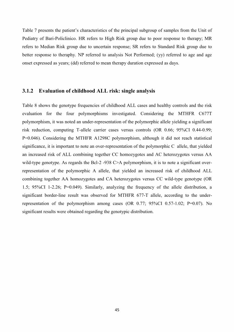
45
Table 7 presents the patient‟s characteristics of the principal subgroup of samples from the Unit of
Pediatry of Bari-Policlinico. HR refers to High Risk group due to poor response to therapy; MR
refers to Median Risk group due to uncertain response; SR refers to Standard Risk group due to
better response to theraphy. NP referred to analysis Not Performed; (yy) referred to age and age
onset expressed as years; (dd) referred to mean therapy duration expressed as days.
3.1.2 Evaluation of childhood ALL risk: single analysis
Table 8 shows the genotype frequencies of childhood ALL cases and healthy controls and the risk
evaluation for the four polymorphisms investigated. Considering the MTHFR C677T
polymorphism, it was noted an under-representation of the polymorphic allele yielding a significant
risk reduction, computing T-allele carrier cases versus controls (OR 0.66; 95%CI 0.44-0.99;
P=0.046). Considering the MTHFR A1298C polymorphism, although it did not reach statistical
significance, it is important to note an over-representation of the polymorphic C allele, that yielded
an increased risk of ALL combining together CC homozygotes and AC heterozygotes versus AA
wild-type genotype. As regards the Bcl-2 -938 C>A polymorphism, it is to note a significant over-
representation of the polymorphic A allele, that yielded an increased risk of childhood ALL
combining together AA homozygotes and CA heterozygotes versus CC wild-type genotype (OR
1.5; 95%CI 1-2.26; P=0.049). Similarly, analyzing the frequency of the allele distribution, a
significant border-line result was observed for MTHFR 677-T allele, according to the under-
representation of the polymorphism among cases (OR 0.77; 95%CI 0.57-1.02; P=0.07). No
significant results were obtained regarding the genotypic distribution.

46
Table 8
DHFR -19bp INS/DEL
(totcases=195)(totContr=201)
MTHFR C677T
(totcases=195)(totContr=201)
MTHFR A1298C
(totcases=197)(totContr=201)
Bcl-2 -938 C>A
(totcases=197)(totContr=197)
WW(%) WD (%) DD (%) CC (%) CT (%) TT (%) AA (%) AC (%) CC (%) CC (%) CA (%) AA(%)
CASES 86(44%) 84(43%) 25(13%) 86(44%) 81(41%) 28(14%) 84(42%) 82(41%) 31(15%) 66(33%) 87(44%) 44(22%)
CONTROLS 84(42%) 84(42%) 33(16%) 69(34%) 98(48%) 34(16%) 101(50%) 70(34%) 30(15%) 85(43%) 72(36%) 40 (20%)
P-value
(genotypic
distribution)
NS NS NS NS
OR(++/+- vs --) 0.9 (0.6-1.35) 0.66 (0.44-0.99) 1.35 (0.91-2.01) 1.5 (1-2.26)
P-value NS 0.046 NS 0.049
OR(++ vs other
genotypes) 0.7 (0.42-1.31) 0.8 (0.47-1.41) 1.06 (0.61-1.836) 1.12 (0.69-1.82)
P-value NS NS NS NS
ALLELS W D C T A C C A
CASES 256 (65%) 134 (33%) 253 (64%) 137 (35%) 250 (63%) 144 (36%) 219 (55%) 175 (44%)
CONTROLS 252 (62%) 150 (37%) 236 (58%) 166 (41%) 272 (67%) 230 (32%) 242 (61%) 152 (38%)
OR (+vs-) 0.87 (0.65-1.17) 0.77 (0.57-1.02) 1.2 (0.89-1.61) 1.27 0.96-1.69)
P-value NS 0.07 NS NS

47
3.1.3 Evaluation of childhood ALL risk (MTHFR C677T, DHFR): combined
analysis
Table 9 shows the combined results for DHFR and MTHFR C677T polymorphisms. All the
possible genotype combinations were considered: only the appreciable results were showed.
Interestingly, the DHFR-WW/MTHFR-TT genotype combination yielded a significant 3.3-fold risk
reduction (OR 0.3; 95%CI 0.11-0.8; P=0.01). Every combination of alleles was computed
considering subjects with DHFR WW- and MTHFR CC-genotypes as the reference. The evaluation
of the combined risk for double homozygotes (i.e. DHFR DD/MTHFR TT), however, did not show
statistical significance, probably for the lower number of subjects computed in the analysis.
Genotype
DHFR MTHFR
Cases n = 195 (%) Controls n = 201 (%) OR (95% CI) P
WW CC 47 (24) 34 (17) Reference
WD CT 39 (20) 48 (24) 0.58 (0.3-1.08) 0.08 DD CT 10 (5.1) 17 (8.5) 0.42 (0.17-1.04) 0.06
WW TT 7 (3.6) 17 (8.5) 0.3 (0.11-0.8) 0.01
Table 9

48
3.1.4 Disease onset and different polymorphisms.
Tables 10-13 show the stratification of the mean age disease onset by the three different genotype
conditions, among the four polymorphisms examined. For all the tables 10-13, the mean age disease
onset is expressed as years (yy).
We found significant results only for MTHFR A1298C polymorphism (Table12). Homozygotes CC
patients had a significant higher age disease onset when compared to homozygotes AA and
heterozygotes AC. An appreciable result, even though not significant, was obtained comparing
homozygotes CC patients towards homozygotes AA patients (P=0.07). Interestingly, no
homozygotes CC patients developed disease before 37 months, respect to the earlier age disease
onset observed among the remaining genotypes (respectively, 6 months for homozygotes AA and
12.6 months for heterozygotes AC).
Genotype DHFR -19bp INS/DEL (W/D)
WW (n=55) WD (n=66) DD (n=20)
Mean Onset ± SD (yy)
7.12 ± 4.58 5.57 ± 3.37 7.38 ± 4.5
Range (yy)
(1.05 – 16.58) (1.41 – 17.16) (0.5 – 17.33)
P DDVSWW
0.4
0.17
0.19
P DDvsWD+WW
P WWvsWD+DD
Table 10

49
Genotype MTHFR C677T
CC (n=58) CT (n=60) TT (n=23)
Mean Onset ± SD
6.51 ± 4.37 6.46 ± 4.4 6.9 ± 3.9
Range (yy)
(0.5 – 17.16) (1.05 – 17.33) (2 – 15.66)
P CCvsTT
0.33
0.45
0.32
P CCvsCT+TT
P TTvsCT+CC
Table 11
Genotype MTHFR A1298C
AA (n=58) AC (n=63) CC (n=20)
Mean Onset ± SD (yy)
6.31 ± 4.4 6.3 ± 3.9 8.0 ± 4.9
Range (yy)
(0.5 – 17.33) (1.05 – 17.16) (2.25 – 15.75)
P AAvsCC
0.07
0.05
0.28
P CCvsAC+AA
P AAvsAC+CC
Table 12

50
Genotype Bcl-2 -938 C>A
CC (n=46) CA (n=59) AA (n=36)
Mean Onset ± SD (yy)
6.9 ± 4.5 6.6 ± 3.9 5.9 ± 4.6
Range (yy)
(1.41 – 16.91) (2 – 17.16) (0.5 – 17.33)
P CCvsAA
0.16
0.24
0.15
P CCvsCA+AA
P AAvsCA+CC
Table 13
3.1.5 Therapy duration and different polymorphisms
Tables 14-17 show the stratification of the mean therapy duration by the three different genotypes
conditions, among the four polymorphisms examined. For all tables 14-17, the mean therapy
duration is expressed as days (dd).
We found a significant association for MTHFR A1298C polymorphism (Table 16) and Bcl-2 -938
C>A polymorphism (Table 17). As regard to the MTHFR A1298C polymorphism, CC
homozygotes showed a slight higher mean therapy duration (651.75dd ± 204.16dd) when compared
with the mean therapy duration of the heterozygotes AC computed together with homozygotes AA
(P=0.05, respectively 589.5dd ± 255.07dd and 651.75dd ± 204.16dd).
As regard to Bcl-2 -938 C>A polymorphism, interesting results were obtained comparing the two
homozygous conditions: -938-CC versus -938-AA (P=0.03). Comparing the AA homozygotes with
heterozygotes AC computed together with homozygotes CC we noted interesting results (P=0.056),
whereas a borderline result was obtained from the comparison of CC homozygotes with AC
heterozygotes computed together with AA homozygotes (P=0.061) (Table 17).
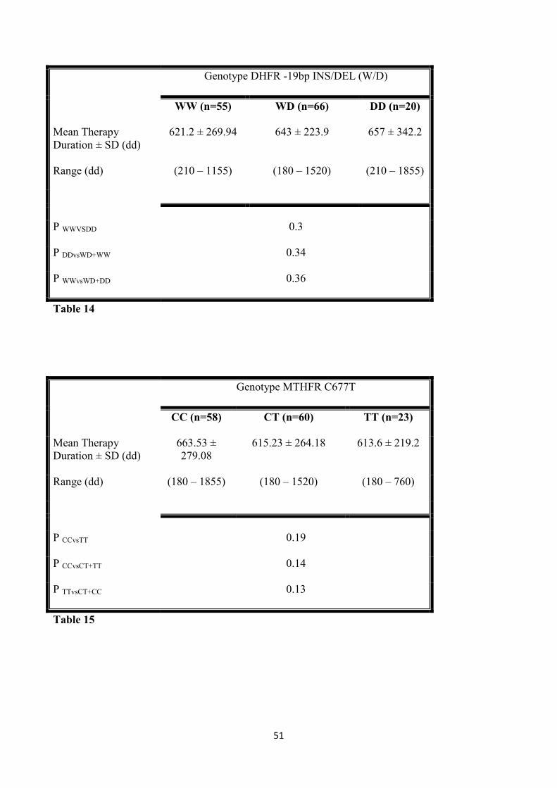
51
Genotype DHFR -19bp INS/DEL (W/D)
WW (n=55) WD (n=66) DD (n=20)
Mean Therapy
Duration ± SD (dd)
621.2 ± 269.94 643 ± 223.9 657 ± 342.2
Range (dd)
(210 – 1155) (180 – 1520) (210 – 1855)
P WWVSDD
0.3
0.34
0.36
P DDvsWD+WW
P WWvsWD+DD
Table 14
Genotype MTHFR C677T
CC (n=58) CT (n=60) TT (n=23)
Mean Therapy
Duration ± SD (dd)
663.53 ±
279.08
615.23 ± 264.18 613.6 ± 219.2
Range (dd)
(180 – 1855) (180 – 1520) (180 – 760)
P CCvsTT
0.19
0.14
0.13
P CCvsCT+TT
P TTvsCT+CC
Table 15

52
Genotype MTHFR A1298C
AA (n=58) AC (n=63) CC (n=20)
Mean Therapy
Duration ± SD (dd)
678.24 ±
284.86
589.5 ± 255.07 651.75 ±
204.16
Range (dd)
(180 – 1855) (180 – 1305) (210 – 850)
P AAvsCC
0.32
0.35
< 0.05
P CCvsAC+AA
P AAvsAC+CC
Table 16
Genotype Bcl-2 -938 C>A
CC (n=46) CA (n=59) AA (n=36)
Mean Therapy
Duration ± SD (dd)
588 ± 229.07 634.3 ± 268.35 694.72 ± 289.34
Range (dd)
(210 – 760) (180 – 1520) (180 – 1855)
% Genotype <760 dd 46 (100%) 55 (93.2%) 32 (88.8%)
% Genotype <730 dd 45 (97%) 51 (86%) 31 (86%)
P AAvsCC
0.033
0.061
0.056
P CCvsCA+AA
P AAvsCA+CC
Table 17

53
3.1.6 Kaplan-Meier analysis. Correlation with MTHFR A1298C polymorphism
Figure 6 shows the stratification of the age disease onset of single patients among the three
genotype conditions of MTHFR A1298C polymorphism. For all figures 6-8, the X-axis expresses
the onset age as years (yy); the Y-axis expresses the survival probability as percentage (%). It is to
note that the CC homozygotes had an appreciable different behavior in the establishment of the
disease when compared with the survival curves of AC heterozygotes and AA homozygotes, which
completely overlapped (HR 1.2868; P=0.5175). This is the reason why we combined together AA
homozygotes and AC heterozygotes and compared them with CC homozygotes patients (Fig.7),
showing an appreciable improvement in the significance (HR 1.3; P=0.2).
In addition, we divided the whole onset period (0.5yy – 17.33yy) by terziles: the first terzile in the
range 0.5yy – 7.25yy; the second terzile in the range 7.26yy – 12.83yy; the third terzile >12.83yy.
Interestingly, the sub-analysis by terziles, ascribed to MTHFR 1298CC homozygotes a significant
later onset compared with AA homozygotes together with AC heterozygotes, when the third terzile
is considered (HR 1.6; 95%CI 1.0206-2.5395) (Fig.8), which correspond to a contextual risk
reduction.
Fig.6
Onset MTHFR A1298C
0 5 10 15 20
0
20
40
60
80
100
Time (yy)
Su
rviv
al p
rob
ab
ility (
%)
MTHFR A1298CAAACCC
P=0,5175
HR=1.2868 (0.8038 - 2.0601)

54
Fig.7
Fig.8
Onset MTHFR A1298C
0 5 10 15 20
0
20
40
60
80
100
Time (yy)
Su
rviv
al p
rob
ab
ility (
%)
MTHFR A1298CAA/ACCC
P=0,2
HR=1.30 (0.8484 - 2.0124)
Onset MTHFR A1298C (>12.83 yy)
0 2 4 6 8 10 12 14 16 18 20
0
20
40
60
80
100
Time (yy)
Su
rviv
al p
rob
ab
ility (
%)
MTHFR A1298CAA/ACCC
P=0,07
HR=1,6 (1,0206-2,5395)

55
3.1.7 Kaplan-Meier analysis. Correlation with Bcl-2 -938 C>A polymorphism
Figure 9 shows the stratification of the therapy duration time of single patients among the three
genotype conditions of Bcl-2 -938 C>A polymorphism. Figure 10 shows an allargement of follow-
up period less than 2 years.
Interestingly, in a sub-analysis of the follow-up period less than 2 years, we noted an overlapping of
the CC homozygotes and CA heterozygotes curves, whereas the AA homozygotes curve strongly
diverged. This is the reason why we merged together the CC homozygotes and the CA
heterozygotes (Fig. 11). Considering those patients who stopped therapy within two years, the AA
patients were over-represented among those with longer therapy (HR 1.3; P=0.07) (Fig. 12).
Finally, comparing the opposite homozygotes Bcl-2 -938CC versus -938AA, the risk of longer
therapy duration ascribed to the -938AA genotype further increases, becoming significant (HR 1.4;
P=0.03) (Fig.13-14, respectively full follw-up period and 2-years follow-up period).
Fig.9
End therapy Bcl-2 -938 C>A (1855 dd)
0 400 800 1200 1600 2000
0
20
40
60
80
100
Time (dd)
Pa
tie
nts
on
th
era
py (
%)
Bcl-2 -938 C>ACCCAAA
P=0.14
HR=1.3438 (0.8721-2.0707)

56
Fig.10
Fig.11
End Therapy Bcl-2 -938 C>A (1855 dd)
0 100 200 300 400 500 600 700
50
60
70
80
90
100
Time (dd)
Pa
tie
nts
on
Th
era
py (
%)
Bcl-2 -938 C>ACCCAAA
P=0.14
HR=1.3438 (0.8721-2.0707)
End therapy Bcl-2 -938 C>A (1855 dd)
0 400 800 1200 1600 2000
0
20
40
60
80
100
Time (dd)
Patients
on thera
py (
%)
Bcl-2 -938 C>A
CC/CA
AA
P=0.10
HR=1.24 (0.8674-1.784)
0 200 400 600
Time (dd)

57
Fig.12
Fig.13
End Therapy Bcl-2 -938 C>A (760dd)
0 100 200 300 400 500 600 700
50
60
70
80
90
100
Time
Patients
on thera
py (
%)
Bcl-2 -938 C>A
CC/CA
AA
P=0.07
HR=1.3 (0.8751-1.8544)
End Therapy Bcl-2 -938 C>A (1855 dd)
0 300 600 900 120015001800
0
20
40
60
80
100
Time (dd)
Patients
on thera
py (
%)
Bcl-2 -938 C>A
CC
AA
P=0.03
HR=1.34 (0.871-2.0707)
0 200 400 600
Time (dd)

58
Fig.14
3.1.8 Folate and anti-folate: the in vitro cellular model
Figures 15 and 16 show the kinetics of cellular viability in the two opposite conditions (wild-type
homozygotes DHFR WW/MTHFR CC and polymorphic homozygotes DHFR DD/MTHFR TT)
when the respective PBL are treated or not with 100µM MTX. After an activation with PHA for 48
h, having the same basic culture conditions, the same pharmacological treatment and the same
starting number of cells (1.5 x10⁵cells/ml) for both genotypic conditions, the number of cells
monitored for 5 days, showed that WW/CC were mainly higher (see Tables 18 and 19). It is to note
that this gap increased over the five days since to reach a 1.4-fold higher at t5, ascribable to
WW/CC genotype. Considering the cellular viability after the MTX treatment we can observe that
the absolute number of residual viability was higher among WW/CC respect to viable DD/TT cells
for each day considered (Fig.17). This result was confirmed also considering the global treated
End therapy Bcl-2 -938 C>A (760dd)
0 100 200 300 400 500 600 700
50
60
70
80
90
100
Time (dd)
Patients
on thera
py (
%)
Bcl-2 -938 C>A
CC
AA
P=0.03
HR=1.4 (0.8769-2.1311)
0 200 400 600
Time (dd)

59
cells. The observation that MTX treated cells (global and viable) diverged between the two
genotypes leads us to analyze in detail cells number and viability respectively by cell counting and
MTT test. A sub-analysis revealed that the two different genotypes significantly diverged at t1
(1.22E+06 vs 8.45E+05; P=0.07) and t5 (9.5E+05 vs 4.61E+05; P=0.04) in favour of the WW/CC
genotype (Fig. 17). MTT test confirmed the results obtained (Tables 20, 21).
WW/CC
t0 t1 t2 t3 t4 t5
Viable cells num/ml –MTX
7,50E+05 1,46E+06 ±2.4E+05
1,78E+06 ±5.88E+05
2,46E+06 ±8.16E+05
4,02E+06 ±1.38E06
4,67E+06 ±1.5E+05
Viable cells num/ml
MTX100 µM
7,50E+05 1,22E+06
±4.62E+05 7,65E+05
±3.78E+05 6,81E+05
±3.12E+05 8,20E+05
±3.74E+05 9,50E+05 ±0.9E+05
Global cells num/ml
-MTX
7,50E+05 1,92E+06
±1.28E+05 2,42E+06
±6.48E+05 3,32E+06
±1.04E+06 5,0E+06
±1.76E+06 6,20E+06 ±3.5E+05
Global cells num/ml
MTX100 µM
7,50E+05 1,64E+06
±5.64E+05 1,56E+06
±4.04E+05 1,78E+06
±6.43E+05 2,12E+06
±7.49E+05 2,30E+06 ±1.5E+05
Table 18. t0-t5 indicate the days of cell culture with counting and MTT test every 24 h for WW/CC
genotype; Viable cells num/ml –MTX and Viable cells num/ml MTX100 µM indicate the count of the viable
cells as control and treated with 100 µM MTX, respectively; Global cells num/ml -MTX and Global cells
num/ml MTX 100 µM indicate the count of the total cells as control and treated with 100 µM MTX.
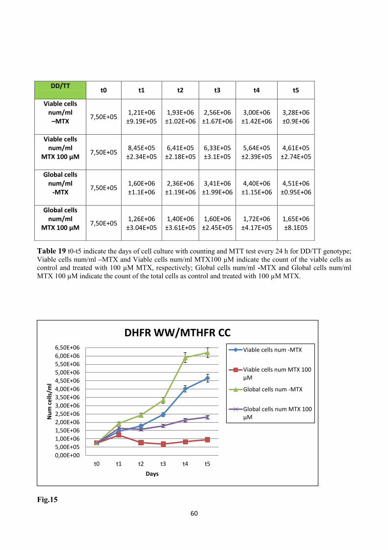
60
Table 19 t0-t5 indicate the days of cell culture with counting and MTT test every 24 h for DD/TT genotype;
Viable cells num/ml –MTX and Viable cells num/ml MTX100 µM indicate the count of the viable cells as
control and treated with 100 µM MTX, respectively; Global cells num/ml -MTX and Global cells num/ml
MTX 100 µM indicate the count of the total cells as control and treated with 100 µM MTX.
Fig.15
0,00E+00
5,00E+05
1,00E+06
1,50E+06
2,00E+06
2,50E+06
3,00E+06
3,50E+06
4,00E+06
4,50E+06
5,00E+06
5,50E+06
6,00E+06
6,50E+06
t0 t1 t2 t3 t4 t5
Nu
m c
ells
/ml
Days
DHFR WW/MTHFR CC
Viable cells num -MTX
Viable cells num MTX 100µM
Global cells num -MTX
Global cells num MTX 100µM
DD/TT
t0 t1 t2 t3 t4 t5
Viable cells num/ml –MTX
7,50E+05 1,21E+06
±9.19E+05 1,93E+06
±1.02E+06 2,56E+06
±1.67E+06 3,00E+06
±1.42E+06 3,28E+06 ±0.9E+06
Viable cells num/ml
MTX 100 µM
7,50E+05 8,45E+05
±2.34E+05 6,41E+05
±2.18E+05 6,33E+05 ±3.1E+05
5,64E+05 ±2.39E+05
4,61E+05 ±2.74E+05
Global cells num/ml
-MTX
7,50E+05 1,60E+06 ±1.1E+06
2,36E+06 ±1.19E+06
3,41E+06 ±1.99E+06
4,40E+06 ±1.15E+06
4,51E+06 ±0.95E+06
Global cells num/ml
MTX 100 µM
7,50E+05 1,26E+06
±3.04E+05 1,40E+06
±3.61E+05 1,60E+06
±2.45E+05 1,72E+06
±4.17E+05 1,65E+06 ±8.1E05

61
Fig. 16
Fig.17
0,00E+00
5,00E+05
1,00E+06
1,50E+06
2,00E+06
2,50E+06
3,00E+06
3,50E+06
4,00E+06
4,50E+06
5,00E+06
5,50E+06
6,00E+06
6,50E+06
t0 t1 t2 t3 t4 t5
Nu
m c
ells
/ml
Days
DHFR DD/MTHFR TT
Viable cells num -MTX
Viable cells num MTX 100µM
Global cells num -MTX
Global cells num MTX 100µM
0,00E+00
1,00E+05
2,00E+05
3,00E+05
4,00E+05
5,00E+05
6,00E+05
7,00E+05
8,00E+05
9,00E+05
1,00E+06
1,10E+06
1,20E+06
1,30E+06
1,40E+06
1,50E+06
t0 t1 t2 t3 t4 t5
Via
ble
ce
lls/m
l
Days
Viable cells treated with MTX WW/CC vs DD/TT
Viable cells MTX 100 µM WW/CC
Viable cells MTX 100 µM DD/TT
P=0.04
P= 0.07

62
DHFR DD/MTHFR TT MTT test
DAYS
OD (A550nm)
t0 t1 t2 t3 t4 t5
Viable cells -MTX
0,355 0,563 0,645 0,700 0,735 0,782
Viable cells MTX 100 µM
0,350 0,435 0,388 0,216 0,133 0,145
Blank 0.051 0.046 0.048 0.047 0.047
0.048
Table 20
DHFR WW/MTHFR CC MTT test
DAYS
OD (A550nm)
t0 t1 t2 t3 t4 t5
Viable cells - MTX
0,415 0,581 0,660 0,691 0,681 0,682
Viable cells MTX 100 µM
0,414 0,548 0,412 0,341 0,330 0,31
Blank 0.051 0.046 0.048 0.047 0.047
0.048
Table 21

63
4
DISCUSSION

64
4.1 DISCUSSION
Several molecular rearrangements are involved in the etio-pathogenesis of cancer disease and
similarly, numerous polymorphisms affect its susceptibility. Folate balancing has a critical role in
normal and malignant cell cycle by influencing fundamental processes such as DNA synthesis and
methylation (Blount et al., 1997). Sub-optimal/anomalous folate levels/distributions are correlated
to an elevated risk of developing several forms of neoplasia including ALL. From another point of
view, one of the most important processes related to the development of cancer is the balancing of
apoptosis regulation mechanisms: an increased survival of a cell population, indeed, defines the
accumulation of genomic instability within the cells (Karakas et al., 1998). Apoptosis is a natural
process regulated by several pathways that comprise genes that both promote or inhibit it. It can be
hypothesized that polymorphisms of genes involved in the regulation of apoptosis, which modify
protein function and/or expression could play an important role in the balancing of this cell
regulation mechanism (Kidd et al., 2006).
With the present thesis, we aimed to investigate the effect of three polymorphisms within two genes
of the folate metabolic pathway (MTHFR and DHFR) on the risk of developing childhood ALL,
and similarly, we investigated the effect of one polymorphism within the anti-apoptotic Bcl-2
regulatory gene on childhood ALL susceptibility. From this study we can observe that the
homozygous DHFR-DD genotype and the homozygous MTHFR 677-TT genotype are under-
represented among childhoods ALL cases (Tab.8). Nevertheless, the analysis of DHFR
polymorphism did not show significant results. Considering the MTHFR C677T polymorphism, it
was noted an under-representation of the polymorphic T-allele yielding a significant ALL risk
reduction of 1.5-fold (P=0.046). At the same time, considering the MTHFR A1298C
polymorphism, althought it did not reach statistical significance, it is important to note an over-
representation of the polymorphic C allele, that yielded an increased risk of ALL combining
together CC homozygotes and AC heterozygotes versus AA wild-type genotype. The two enzymes
considered, are involved in cyclization of folate isoforms. DHFR is responsible for the conversion
of dihydrofolate to tetrahydrofolate, favouring the synthesis of 5,10-methylene-THF (Skibola et al.,
1999). The elevated expression levels associated to the D-allele could therefore facilitate the filling
of the cell with 5,10-methylene-THF, supporting consequently the DNA synthesis process (Xu et
al., 2007). MTHFR catalyzes the reduction of 5,10-methylene-THF to 5-methyl-THF (Goyette et
al., 1995). A lower enzyme activity associated to the polymorphic T-allele leads to an

65
underutilization of methylene-THF within the cell, with consequent accumulation of partially
reduced folate isoforms. This statement is in agreement with studies demonstrating an accumulation
of THF formilated isoforms within red blood cells (Bagley et al., 1998). As a consequence, a
balance within the cell there exists between 5,10-methylene-THF isoforms and formilated THF
isoforms so the synthesis of purine and pyrimidine is positively influenced. If the rate of
intracellular THF isoforms is determined by the relative balancing between DHFR (by
production/filling) and MTHFR (by use/emptying) activities, we aimed to investigate if the
coexistence in the same individual, of the two polymorphic conditions could ascribe a synergistic or
additive effect on the risk of developing childhood ALL. Interestingly, from the combined analysis
of the two polymorphisms (DHFR -19bp INS/DEL and MTHFR C677T) the DHFR-WW/MTHFR-
TT genotype combination yielded a significant 3.3-fold childhood ALL risk reduction (P=0.01),
whilst border-line results are obtained from the combination of double heterozygotes DHFR-
WD/MTHFR-CT and from the combination of homozygotes DHFR-DD/homozygotes MTHFR-CT
(Tab.9). The evaluation of the combined risk for double homozygotes (i.e. DHFR DD/MTHFR TT),
in which a maximal protective effect is hypothesized however, did not show statistical significance,
probably for the lower number of subjects computed in the analysis. As regards the Bcl-2 -938 C>A
polymorphism, it is to note a significant over-representation of the polymorphic A allele among
childhood ALL cases, that yielded an increased risk of 1.5-fold of childhood ALL combining
together AA homozygotes and CA heterozygotes versus CC wild-type genotype (P=0.049). Bcl-2
seems to play a crucial role in the survival of hematolymphoid cells, by inhibiting apoptosis through
the prevention of cytochrome c release from mithocondria, blocking caspase activation (Reed et al.,
1994). Our results are in line with the data that ascribe to the A-allele a higher Bcl-2 transcription,
responsible for an increased Bcl-2 level (Nuckel et al., 2007). Since high Bcl-2 expression
correlates with unfavorable prognosis and disease stage this could lead to poor prognosis among A-
carrier patients. The polymorphic -938 A-allele could therefore represents a risk factor for the
susceptibility to childhood ALL.
These are the reasons why, the second phase of this study focusses on the relationship between
polymorphisms, disease onset and therapy duration. From the results obtained considering the
relationship between MTHFR A1298C and disease onset, it is to note that the CC homozygotes had
an appreciable different mean age onset when compared with the remaining genotypes (età e
P=0.05). We can ascribe to the MTHFR 1298CC homozygotes a significant later onset also
confirmed by survival analysis (P=0.05). Other significant results were obtained analyzing therapy
duration. MTHFR 1298CC had a significant higher mean therapy duration compared with the

66
remaining genotypes (P=0.05). Conversely, similar results were not obtained considering the
C677T polymorphism, although 677T-allele affects MTHFR enzyme activity at higher extent than
MTHFR A1298C polymorphism, maybe due to the stronger partial link diseq between the two gene
variants. The same analytical approach computing Bcl-2 -938 C>A polymorphism ascribed to the -
938 AA genotype a significant longer theraphy duration (P=0.03). Also -938 C-carriers had
appreciable longer therapy duration and this result completely strengthened the above mentioned
hypothesis that ascribes to the A-allele a higher Bcl-2 transcription, responsible for an increased
Bcl-2 level (Nuckel et al., 2007), correlating with unfavorable prognosis and disease stage.
In order to verify effective actions of the folate cycle polymorphisms investigated on base-line cells
viability and on residual viability of anti-folate treated cells, we selected two goups of opposite
genotypes for DHFR and MTHFR to improve possible differences. Accordingly, under the same in
vitro culture condition we observed appreciable effects in cell number, in favor of the WW/CC
genotype. The difference became even higher when the same two groups were treated with anti-
folate drug. On the contrary, the initial rate in cells growth was completely comparable between
DD/TT and WW/CC genotypes, being about + 60% for DD/TT genotype and about +58.3% for
WW/CC (data were referred to the first 48 hours of culture for the viable not treated cells).
The pharmacological induced restriction in folate availability (MTX) yields to different responses in
the two opposite genotypes, in favor of WW/CC, though they have the same initial growth rate.
This is in line with the hypothesis that the combination of DD/TT genotype helps the storage of
folate isoforms (useful for the cell viability) by production and filling activity, compared to the
WW/CC responsible for a lower filling and consuming activity. As for the hypothesized different
genotype-dependent intracellular folate distribution, also MTX being itself a synthetic analogue of
folate should follow the same handling process. Now, a higher MTX level inside the cell should
result in an elevated toxicity level. In fact, both global and residual viable treated cells significantly
increase among the WW/CC genotype.

67
Conclusion
It has been well established since the last 15 years that the role of some folate pathway gene
polymorphisms have effects on risk establishment, prognosis, survival and toxicity in several
hematological malignancies. The dual and opposite role often ascribed to this kind of
polymorphisms could be in part explained by the fact that the same mechanism (e.g. folate
storage/consuming) could be responsible for different outputs when an healthy cell or a cancer cell
is considered.
Theoretically, a cell genetically prone to accumulate folate isoforms (useful for the cell viability)
carries an advantage in proliferation. However, when a toxic folate analogue is furnished togheter
with folate, the cell is not able to discriminate the wright from wrong molecule and the positive
mechanism above mentioned became lethal.

68
5
BIBLIOGRAPHY

69
5.1 BIBLIOGRAPHY
Abahji TN, Nill L, Ide N, Keller C, Hoffmann U, Weiss N. Acute hyperhomocysteinemia induces
microvascular and macrovascular endothelial dysfunction. Arch Med Res. 2007 May;38(4):411-6.
Afman L, Müller M. Nutrigenomics: from molecular nutrition to prevention of disease. J Am Diet Assoc.
2006 Apr;106(4):569-76. Review.
Alcasabas P, Ravindranath Y, Goyette G, Haller A, Del Rosario L, Lesaca-Medina MY, Darga L, Ostrea EM
Jr, Taub JW, Everson RB. 5,10-methylenetetrahydrofolate reductase (MTHFR) polymorphisms and the risk
of acute lymphoblastic leukemia (ALL) in Filipino children. Pediatr Blood Cancer. 2008 Aug;51(2):178-82.
Al-Shakfa F, Dulucq S, Brukner I, Milacic I, Ansari M, Beaulieu P, Moghrabi A, Laverdière C, Sallan SE,
Silverman LB, Neuberg D, Kutok JL, Sinnett D, Krajinovic M. DNA variants in region for noncoding
interfering transcript of dihydrofolate reductase gene and outcome in childhood acute lymphoblastic
leukemia. Clin Cancer Res. 2009 Nov 15;15(22):6931-8.
Anagnou NP, O'Brien SJ, Shimada T, Nash WG, Chen MJ, Nienhuis AW. Chromosomal organization of the
human dihydrofolate reductase genes: dispersion, selective amplification, and a novel form of
polymorphism. Proc Natl Acad Sci U S A. 1984 Aug;81(16):5170-4.
Anderson OS, Sant KE, Dolinoy DC. Nutrition and epigenetics: an interplay of dietary methyl donors, one-
carbon metabolism and DNA methylation. J Nutr Biochem. 2012 Aug;23(8):853-9. doi:
10.1016/j.jnutbio.2012.03.003. Epub 2012 Jun 27. Review.
Ansari M, Krajinovic M. Pharmacogenomics in cancer treatment defining genetic bases for inter-individual
differences in responses to chemotherapy. Curr Opin Pediatr. 2007 Feb;19(1):15-22. Review.
Ansari M, Krajinovic M. Pharmacogenomics of acute leukemia. Pharmacogenomics. 2007 Jul;8(7):817-34.
doi: 10.2217/14622416.8.7.817. Review.
Antony AC. The biological chemistry of folate receptors. Blood. 1992 Jun 1;79(11):2807-20. Review
Aplenc R, Thompson J, Han P, La M, Zhao H, Lange B, Rebbeck T. Methylenetetrahydrofolate reductase
polymorphisms and therapy response in pediatric acute lymphoblastic leukemia. Cancer Res. 2005 Mar
15;65(6):2482-7.

70
Askari BS, Krajinovic M. Dihydrofolate reductase gene variations in susceptibility to disease and treatment
outcomes. Curr Genomics. 2010 Dec;11(8):578-83.
Assaraf YG. Molecular basis of antifolate resistance. Cancer Metastasis Rev. 2007 Mar;26(1):153-81.
Review.
Aune D, Deneo-Pellegrini H, Ronco AL, Boffetta P, Acosta G, Mendilaharsu M, De Stefani E. Dietary folate
intake and the risk of 11 types of cancer: a case-control study in Uruguay. Ann Oncol. 2011 Feb;22(2):444-
51.
Bagley PJ, Selhub J. A common mutation in the methylenetetrahydrofolate reductase gene is associated with
an accumulation of formylated tetrahydrofolates in red blood cells. Proc Natl Acad Sci U S A. 1998 Oct
27;95(22):13217-20.
Bailey LB, Gregory JF 3rd. Polymorphisms of methylenetetrahydrofolate reductase and other enzymes:
metabolic significance, risks and impact on folate requirement. J Nutr. 1999 May;129(5):919-22. Review
Bakhshi A, Jensen JP, Goldman P, Wright JJ, McBride OW, Epstein AL, Korsmeyer SJ. Cloning the
chromosomal breakpoint of t(14;18) human lymphomas: clustering around JH on chromosome 14 and near a
transcriptional unit on 18. Cell. 1985 Jul;41(3):899-906
Bakhshi A, Wright JJ, Graninger W, Seto M, Owens J, Cossman J, Jensen JP, Goldman P, Korsmeyer SJ.
Mechanism of the t(14;18) chromosomal translocation: structural analysis of both derivative 14 and 18
reciprocal partners. Proc Natl Acad Sci U S A. 1987 Apr;84(8):2396-400.
Barry EV, Silverman LB. Acute lymphoblastic leukemia in adolescents and young adults. Curr Hematol
Malig Rep. 2008 Jul;3(3):161-6.
Bárta I, Smerák P, Polívková Z, Sestáková H, Langová M, Turek B, Bártová J. Current trends and
perspectives in nutrition and cancer prevention. Neoplasma.2006;53(1):19-25.
Basten GP, Duthie SJ, Pirie L, Vaughan N, Hill MH, Powers HJ. Sensitivity of markers of DNA stability and
DNA repair activity to folate supplementation in healthy volunteers. Br J Cancer. 2006 Jun 19;94(12):1942-
7.
Beetstra S, Thomas P, Salisbury C, Turner J, Fenech M. Folic acid deficiency increases chromosomal
instability, chromosome 21 aneuploidy and sensitivity to radiation-induced micronuclei. Mutat Res. 2005
Oct 15;578(1-2):317-26.

71
Belson M, Kingsley B, Holmes A. Risk factors for acute leukemia in children: a review. Environ Health
Perspect. 2007 Jan;115(1):138-45. Review
Bémeur C, Desjardins P, Butterworth RF. Role of nutrition in the management of hepatic encephalopathy in
end-stage liver failure. J Nutr Metab. 2010
Bensi L, Longo R, Vecchi A, Messora C, Garagnani L, Bernardi S, Tamassia MG, Sacchi S. Bcl-2
oncoprotein expression in acute myeloid leukemia. Haematologica. 1995 Mar-Apr;80(2):98-102
Bertino JR. Cancer research: from folate antagonism to molecular targets. Best Pract Res Clin Haematol.
2009 Dec;22(4):577-82.
Bigler J, Ulrich CM, Kawashima T, Whitton J, Potter JD. DNA repair polymorphisms and risk of colorectal
adenomatous or hyperplastic polyps. Cancer Epidemiol Biomarkers Prev. 2005 Nov;14(11 Pt 1):2501-8.
Blount BC, Mack MM, Wehr CM, MacGregor JT, Hiatt RA, Wang G, Wickramasinghe SN, Everson RB,
Ames BN. Folate deficiency causes uracil misincorporation into human DNA and chromosome breakage:
implications for cancer and neuronal damage. Proc Natl Acad Sci U S A. 1997 Apr 1;94(7):3290-5.
Bolufer P, Barragan E, Collado M, Cervera J, López JA, Sanz MA. Influence of genetic polymorphisms on
the risk of developing leukemia and on disease progression. Leuk Res. 2006 Dec;30(12):1471-91. Epub 2006
Oct 4. Review.
Bowron A, Scott J, Stansbie D. The influence of genetic and environmental factors on plasma homocysteine
concentrations in a population at high risk for coronary artery disease. Ann Clin Biochem. 2005 Nov;42(Pt
6):459-62.
Branda RF, O'Neill JP, Brooks EM, Powden C, Naud SJ, Nicklas JA. The effect of dietary folic acid
deficiency on the cytotoxic and mutagenic responses to methyl methanesulfonate in wild-type and in 3-
methyladenine DNA glycosylase-deficient Aag null mice. Mutat Res. 2007 Feb 3;615(1-2):12-7.
Buckley JD, Buckley CM, Ruccione K, Sather HN, Waskerwitz MJ, Woods WG, Robison LL.
Epidemiological characteristics of childhood acute lymphocyticleukemia. Analysis by immunophenotype.
The Childrens Cancer Group. Leukemia. 1994 May;8(5):856-64.
Cam R, Eroglu A, Egin Y, Akar N. Dihydrofolate reductase (DHRF) 19-bp intron-1 deletion and
methylenetetrahydrofolate reductase (MTHFR) C677T polymorphisms in breast cancer. Breast Cancer Res
Treat. 2009 May;115(2):431-2

72
Caudill MA, Cruz AC, Gregory JF 3rd, Hutson AD, Bailey LB. Folate status response to controlled folate
intake in pregnant women. J Nutr. 1997 Dec;127(12):2363-70
Chaber R, Fiszer-Maliszewska L, Noworolska-Sauren D, Kwasnicka J, Wrobel G, Chybicka A. The BCL-2
protein in precursor B acute lymphoblastic leukemia in children. J Pediatr Hematol Oncol. 2013
Apr;35(3):180-7.
Chan JY, Ugrasena DG, Lum DW, Lu Y, Yeoh AE. Xenobiotic and folate pathway gene polymorphisms and
risk of childhood acute lymphoblastic leukaemia in Javanese children. Hematol Oncol. 2011 Sep;29(3):116-
23.
Chanarin I. Folate and cobalamin. Clin Haematol. 1985 Oct;14(3):629-41. Review.
Chang JS. Parental smoking and childhood leukemia. Methods Mol Biol. 2009;472:103-37
Chen MJ, Shimada T, Moulton AD, Cline A, Humphries RK, Maizel J, Nienhuis AW. The functional
human dihydrofolate reductase gene. J Biol Chem. 1984 Mar 25;259(6):3933-43.
Chen J, Giovannucci E, Kelsey K, Rimm EB, Stampfer MJ, Colditz GA, Spiegelman D, Willett WC, Hunter
DJ. A methylenetetrahydrofolate reductase polymorphism and the risk of colorectal cancer. Cancer Res.
1996 Nov 1;56(21):4862-4.
Chen J, Ma J, Stampfer MJ, Palomeque C, Selhub J, Hunter DJ. Linkage disequilibrium between the
677C>T and 1298A>C polymorphisms in human methylenetetrahydrofolate reductase gene and their
contributions to risk of colorectal cancer. Pharmacogenetics. 2002 Jun;12(4):339-42.
Chen K, Hu Z, Wang LE, Sturgis EM, El-Naggar AK, Zhang W, Wei Q. Single-nucleotide polymorphisms
at the TP53-binding or responsive promoter regions of BAX and BCL2 genes and risk of squamous cell
carcinoma of the head and neck. Carcinogenesis. 2007 Sep;28(9):2008-12. Epub 2007 Aug 11.
Cheok MH, Evans WE. Acute lymphoblastic leukaemia: a model for the pharmacogenomics of cancer
therapy. Nat Rev Cancer. 2006 Feb;6(2):117-29. Review.Erratum in: Nat Rev Cancer. 2006 Mar;6(3):249.
Chiusolo P, Giammarco S, Bellesi S, Metafuni E, Piccirillo N, De Ritis D, Marietti S, Federica S, Laurenti L,
Fianchi L, Hohaus S, Giuseppe L, Sica S. The role of MTHFR and RFC1 polymorphisms on toxicity and
73
outcome of adult patients with hematological malignancies treated with high-dose methotrexate followed by
leucovorin rescue. Cancer Chemother Pharmacol. 2012 Mar;69(3):691-6.
Chu E, Takimoto CH, Voeller D, Grem JL, Allegra CJ. Specific binding of human dihydrofolate reductase
protein to dihydrofolate reductase messenger RNA in vitro. Biochemistry. 1993 May 11;32(18):4756-60.
Costea I, Moghrabi A, Laverdiere C, Graziani A, Krajinovic M. Folate cycle gene variants and
chemotherapy toxicity in pediatric patients with acute lymphoblastic leukemia. Haematologica. 2006
Aug;91(8):1113-6.
Courtemanche C, Huang AC, Elson-Schwab I, Kerry N, Ng BY, Ames BN. Folate deficiency and ionizing
radiation cause DNA breaks in primary human lymphocytes: a comparison. FASEB J. 2004 Jan;18(1):209-
11.
Courtemanche C, Elson-Schwab I, Mashiyama ST, Kerry N, Ames BN. Folate deficiency inhibits the
proliferation of primary human CD8+ T lymphocytes in vitro. J Immunol. 2004 Sep 1;173(5):3186-92.
Crider KS, Bailey LB, Berry RJ. Folic acid food fortification-its history, effect, concerns, and future
directions. Nutrients. 2011 Mar;3(3):370-84. Epub 2011 Mar 15. Review
Crott JW, Mashiyama ST, Ames BN, Fenech M. The effect of folic acid deficiency and MTHFR C677T
polymorphism on chromosome damage in human lymphocytes in vitro.Cancer Epidemiol Biomarkers Prev.
2001 Oct;10(10):1089-96.
Crott JW, Mashiyama ST, Ames BN, Fenech MF. Methylenetetrahydrofolate reductase C677T
polymorphism does not alter folic acid deficiency-induced uracil incorporation into primary human
lymphocyte DNA in vitro. Carcinogenesis. 2001 Jul;22(7):1019-25.
Crott J, Fenech M. Preliminary study of the genotoxic potential of homocysteine in human lymphocytes in
vitro. Mutagenesis. 2001 May;16(3):213-7.
Curtin K, Slattery ML, Ulrich CM, Bigler J, Levin TR, Wolff RK, Albertsen H, Potter JD, Samowitz WS.
Genetic polymorphisms in one-carbon metabolism: associations with CpG island methylator phenotype
(CIMP) in colon cancer and the modifying effects of diet. Carcinogenesis. 2007 Aug;28(8):1672-9.
Damnjanovic T, Milicevic R, Novkovic T, Jovicic O, Bunjevacki V, Jekic B, Lukovic L, Novakovic I,
Redzic D, Milasin J. Association between the methylenetetrahydrofolate reductase polymorphisms and risk
of acute lymphoblastic leukemia in Serbian children. J Pediatr Hematol Oncol. 2010 May;32(4):e148-50.

74
de Deus DM, de Lima EL, Seabra Silva RM, Leite EP, Cartaxo Muniz MT. Influence of
Methylenetetrahydrofolate Reductase C677T, A1298C, and G80A Polymorphisms on the Survival of
Pediatric Patients with Acute Lymphoblastic Leukemia. Leuk Res Treatment. 2012;2012:292043. doi:
10.1155/2012/292043. Epub 2012 Oct 17.
de Jonge R, Hooijberg JH, van Zelst BD, Jansen G, van Zantwijk CH, Kaspers GJ, Peters GJ, Ravindranath
Y, Pieters R, Lindemans J. Effect of polymorphisms in folate-related genes on in vitro methotrexate
sensitivity in pediatric acute lymphoblastic leukemia. Blood. 2005 Jul 15;106(2):717-20. Epub 2005 Mar 29.
Erratum in: Blood. 2005 Aug 15;106(4):1198.
de Jonge R, Tissing WJ, Hooijberg JH, Jansen G, Kaspers GJ, Lindemans J, Peters GJ, Pieters R.
Polymorphisms in folate-related genes and risk of pediatric acute lymphoblastic leukemia. Blood. 2009 Mar
5;113(10):2284-9.
de Klerk N, Milne E. Overview of recent studies on childhood leukaemia, intra-uterine growth and diet.
Radiat Prot Dosimetry. 2008;132(2):255-8 Review.
De Mattia E, Toffoli G. C677T and A1298C MTHFR polymorphisms, a challenge for antifolate and
fluoropyrimidine-based therapy personalisation. Eur J Cancer. 2009 May;45(8):1333-51.
de Vogel S, Wouters KA, Gottschalk RW, van Schooten FJ, de Goeij AF, de Bruïne AP, Goldbohm RA, van
den Brandt PA, van Engeland M, Weijenberg MP. Dietary methyl donors, methyl metabolizing enzymes,
and epigenetic regulators: diet-gene interactions and promoter CpG island hypermethylation in colorectal
cancer. Cancer Causes Control. 2011 Jan;22(1):1-12.
Detera-Wadleigh SD, Coffman D, Shimada T. A frequent DHFR polymorphism detected by an intron
fragment. Nucleic Acids Res. 1989 Aug 11;17(15):6432.
Dianov GL, Timchenko TV, Sinitsina OI, Kuzminov AV, Medvedev OA, Salganik RI. Repair of uracil
residues closely spaced on the opposite strands of plasmid DNA results in double-strand break and deletion
formation. Mol Gen Genet. 1991 Mar;225(3):448-52.
Dickinson HO. The causes of childhood leukaemia. BMJ. 2005 Jun 4;330(7503):1279-80.
Divisi D, Di Tommaso S, Salvemini S, Garramone M, Crisci R. Diet and cancer. Acta Biomed. 2006
Aug;77(2):118-23. Review.
Dulucq S, St-Onge G, Gagné V, Ansari M, Sinnett D, Labuda D, Moghrabi A, Krajinovic M. DNA variants
in the dihydrofolate reductase gene and outcome in childhood ALL. Blood. 2008 Apr 1;111(7):3692-700.

75
Duthie SJ, Hawdon A. DNA instability (strand breakage, uracil misincorporation, and defective repair) is
increased by folic acid depletion in human lymphocytes in vitro. FASEB J. 1998 Nov;12(14):1491-7.
Duthie SJ. Folic acid deficiency and cancer: mechanisms of DNA instability. Br Med Bull. 1999;55(3):578-
92. Review.
Duthie SJ, Grant G, Narayanan S. Increased uracil misincorporation in lymphocytes from folate-deficient
rats Br J Cancer. 2000 Dec;83(11):1532-7.
Duthie SJ, Narayanan S, Blum S, Pirie L, Brand GM. Folate deficiency in vitro induces uracil
misincorporation and DNA hypomethylation and inhibits DNA excision repair in immortalized normal
human colon epithelial cells. Nutr Cancer. 2000;37(2):245-51.
Duthie SJ, Narayanan S, Brand GM, Pirie L, Grant G. Impact of folate deficiency on DNA stability. J Nutr.
2002 Aug;132(8 Suppl):2444S-2449S.
Duthie SJ, Mavrommatis Y, Rucklidge G, Reid M, Duncan G, Moyer MP, Pirie LP, Bestwick CS. The
response of human colonocytes to folate deficiency in vitro: functional and proteomic analyses. J Proteome
Res. 2008 Aug;7(8):3254-66
Duthie SJ. Folate and cancer: how DNA damage, repair and methylation impact on colon carcinogenesis. J
Inherit Metab Dis. 2011 Feb;34(1):101-9. Epub 2010 Jun 11. Review.
Duthie SJ. Folate and cancer: how DNA damage, repair and methylation impact on colon carcinogenesis. J
Inherit Metab Dis. 2011 Feb;34(1):101-9. Review.
Eden T. Aetiology of childhood leukaemia. Cancer Treat Rev. 2010 Jun;36(4):286-97. Review.
Engbersen AM, Franken DG, Boers GH, Stevens EM, Trijbels FJ, Blom HJ. Thermolabile 5,10-
methylenetetrahydrofolate reductase as a cause of mild hyperhomocysteinemia. Am J Hum Genet. 1995
Jan;56(1):142-50
Ercikan-Abali EA, Banerjee D, Waltham MC, Skacel N, Scotto KW, Bertino JR. Dihydrofolate reductase
protein inhibits its own translation by binding to dihydrofolate reductase mRNA sequences within the coding
region. Biochemistry. 1997 Oct 7;36(40):12317-22.
Eto I, Krumdieck CL. Role of vitamin B12 and folate deficiencies in carcinogenesis. Adv Exp Med Biol.
1986;206:313-30.

76
Farnham PJ, Means AL. Sequences downstream of the transcription initiation site modulate the activity of
the murine dihydrofolate reductase promoter. Mol Cell Biol. 1990 Apr;10(4):1390-8.
Fenech M, Aitken C, Rinaldi J. Folate, vitamin B12, homocysteine status and DNA damage in young
Australian adults. Carcinogenesis. 1998 Jul;19(7):1163-71.
Fenech M. The role of folic acid and Vitamin B12 in genomic stability of human cells. Mutat Res. 2001 Apr
18;475(1-2):57-67. Review.
Franco RF, Simões BP, Tone LG, Gabellini SM, Zago MA, Falcão RP. The methylenetetrahydrofolate
reductase C677T gene polymorphism decreases the risk of childhood acute lymphocytic leukaemia. Br J
Haematol. 2001 Dec;115(3):616-8.
Friso S, Choi SW. Gene-nutrient interactions and DNA methylation. J Nutr. 2002 Aug;132(8 Suppl):2382S-
2387S. Review
Friso S, Choi SW, Girelli D, Mason JB, Dolnikowski GG, Bagley PJ, Olivieri O, Jacques PF, Rosenberg IH,
Corrocher R, Selhub J. A common mutation in the 5,10-methylenetetrahydrofolate reductase gene affects
genomic DNA methylation through an interaction with folate status. Proc Natl Acad Sci U S A. 2002 Apr
16;99(8):5606-11.
Frosst P, Blom HJ, Milos R, Goyette P, Sheppard CA, Matthews RG, Boers GJ, den Heijer M, Kluijtmans
LA, van den Heuvel LP, et al. A candidate genetic risk factor for vascular disease: a common mutation in
methylenetetrahydrofolate reductase. Nat Genet. 1995 May;10(1):111-3.
Gale KB, Ford AM, Repp R, Borkhardt A, Keller C, Eden OB, Greaves MF. Backtracking leukemia to birth:
identification of clonotypic gene fusion sequences in neonatal blood spots. Proc Natl Acad Sci U S A. 1997
Dec 9;94(25):13950-4.
Gellekink H, Blom HJ, van der Linden IJ, den Heijer M. Molecular genetic analysis of the human
dihydrofolate reductase gene: relation with plasma total homocysteine, serum and red blood cell folate
levels. Eur J Hum Genet. 2007 Jan;15(1):103-9.
Gemmati D, Previati M, Serino ML, Moratelli S, Guerra S, Capitani S, Forini E, Ballerini G, Scapoli GL.
Low folate levels and thermolabile methylenetetrahydrofolate reductase as primary determinant of mild
hyperhomocystinemia in normal and thromboembolic subjects. Arterioscler Thromb Vasc Biol. 1999
Jul;19(7):1761-7.
Gemmati D, Ongaro A, Scapoli GL, Della Porta M, Tognazzo S, Serino ML, Di Bona E, Rodeghiero F, Gilli
G, Reverberi R, Caruso A, Pasello M, Pellati A, De Mattei M. Common gene polymorphisms in the
77
metabolic folate and methylation pathway and the risk of acute lymphoblastic leukemia and non-Hodgkin's
lymphoma in adults.Cancer Epidemiol Biomarkers Prev. 2004 May;13(5):787-94.
Gemmati D, Ongaro A, Tognazzo S, Catozzi L, Federici F, Mauro E, Della Porta M, Campioni D, Bardi A,
Gilli G, Pellati A, Caruso A, Scapoli GL, De Mattei M. Methylenetetrahydrofolate reductase C677T and
A1298C gene variants in adult non-Hodgkin's lymphoma patients: association with toxicity and survival.
Haematologica. 2007 Apr;92(4):478-85.
Gemmati D. Folate-Pathway gene variants in cancer: hematological malignancies 2008
Gemmati D, De Mattei M, Catozzi L, Della Porta M, Serino ML, Ambrosio C, Cuneo A, Friso S, Krampera
M, Orioli E, Zeri G, Ongaro A. DHFR 19-bp insertion/deletion polymorphism and MTHFR C677T in adult
acute lymphoblastic leukaemia: is the risk reduction due to intracellular folate unbalancing? Am J Hematol.
2009 Aug;84(8):526-9.
Glynn SA, Albanes D. Folate and cancer: a review of the literature. Nutr Cancer. 1994;22(2):101-19.
Review.
Göker E, Waltham M, Kheradpour A, Trippett T, Mazumdar M, Elisseyeff Y, Schnieders B, Steinherz P,
Tan C, Berman E, et al. Amplification of the dihydrofolate reductase gene is a mechanism of acquired
resistance to methotrexate in patients with acute lymphoblastic leukemia and is correlated with p53 gene
mutations. Blood. 1995 Jul 15;86(2):677-84.
Gorlick R, Goker E, Trippett T, Waltham M, Banerjee D, Bertino JR. Intrinsic and acquired resistance to
methotrexate in acute leukemia. N Engl J Med. 1996 Oct 3;335(14):1041-8. Review.
Goto Y, Yue L, Yokoi A, Nishimura R, Uehara T, Koizumi S, Saikawa Y. A novel single-nucleotide
polymorphism in the 3'-untranslated region of the human dihydrofolate reductase gene with enhanced
expression. Clin Cancer Res. 2001 Jul;7(7):1952-6.
Goyette P, Frosst P, Rosenblatt DS, Rozen R. Seven novel mutations in the methylenetetrahydrofolate
reductase gene and genotype/phenotype correlations in severe methylenetetrahydrofolate reductase
deficiency. Am J Hum Genet. 1995 May;56(5):1052-9.
Goyette P, Rozen R. The thermolabile variant 677C-->T can further reduce activity when expressed in cis
with severe mutations for human methylenetetrahydrofolate reductase. Hum Mutat. 2000;
Greaves MF. Speculations on the cause of childhood acute lymphoblastic leukemia. Leukemia. 1988
Feb;2(2):120-5. Review.

78
Greaves MF. Aetiology of acute leukaemia. Lancet. 1997 Feb 1;349(9048):344-9. Review.
Greaves M. Childhood leukaemia. BMJ. 2002 Feb 2;324(7332):283-7. Review.
Greaves MF, Maia AT, Wiemels JL, Ford AM. Leukemia in twins: lessons in natural history. Blood. 2003
Oct 1;102(7):2321-33.
Greaves M. Infection, immune responses and the aetiology of childhood leukaemia. Nat Rev Cancer. 2006
Mar;6(3):193-203. Review.
Gu Y, Pan Y, Meng B, Guan B, Fu K, Sun B, Zheng F. High levels of bcl-2 protein expression do not
correlate with genetic abnormalities but predict worse prognosis in patients with lymphoblastic lymphoma.
Tumour Biol. 2013 Jun;34(3):1441-50.
Guo W, Healey JH, Meyers PA, Ladanyi M, Huvos AG, Bertino JR, Gorlick R. Mechanisms of methotrexate
resistance in osteosarcoma. Clin Cancer Res. 1999 Mar;5(3):621-7.
Habuchi T. Common genetic polymorphisms and prognosis of sporadic cancers: prostate cancer as a model.
Future Oncol. 2006 Apr;2(2):233-45. Review
Han SS, Sue LY, Berndt SI, Selhub J, Burdette LA, Rosenberg PS, Ziegler RG. Associations between genes
in the one-carbon metabolism pathway and advanced colorectal adenoma risk in individuals with low folate
intake. Cancer Epidemiol Biomarkers Prev. 2012 Mar;21(3):417-27.
Hannon-Fletcher MP, Armstrong NC, Scott JM, Pentieva K, Bradbury I, Ward M, Strain JJ, Dunn AA,
Molloy AM, Kerr MA, McNulty H. Determining bioavailability of food folates in a controlled intervention
study. Am J Clin Nutr. 2004 Oct;80(4):911-8
Hayashi H, Horino M, Morishita M, Tazoe Y, Tsuboi S, Matsuyama T, Kosuge K, Yamada H, Tsuji D,
Inoue K, Itoh K. Dihydrofolate reductase gene intronic 19-bp deletion polymorphisms in a Japanese
population. Drug Metab Pharmacokinet.2010;25(5):516-8.
Herbert V. Making sense of laboratory tests of folate status: folate requirements to sustain normality. Am J
Hematol. 1987 Oct;26(2):199-207. Review.Erratum in: Am J Hematol 1988 Jun;28(2):134.
Hockenbery D, Nuñez G, Milliman C, Schreiber RD, Korsmeyer SJ. Bcl-2 is an inner mitochondrial
membrane protein that blocks programmed cell death. Nature.1990 Nov 22;348(6299):334-6

79
Huang L, Tissing WJ, de Jonge R, van Zelst BD, Pieters R. Polymorphisms in folate-related genes:
association with side effects of high-dose methotrexate in childhood acute lymphoblastic leukemia.
Leukemia. 2008 Sep;22(9):1798-800.Epub 2008 Mar 27.
Imanishi H, Okamura N, Yagi M, Noro Y, Moriya Y, Nakamura T, Hayakawa A, Takeshima Y, Sakaeda T,
Matsuo M, Okumura K. Genetic polymorphisms associated with adverse events and elimination of
methotrexate in childhood acute lymphoblastic leukemia and malignant lymphoma. J Hum Genet.
2007;52(2):166-71.
Iyer R, Tomar SK. Folate: a functional food constituent. J Food Sci. 2009 Nov-Dec;74(9):R114-22.
Jacques PF, Bostom AG, Williams RR, Ellison RC, Eckfeldt JH, Rosenberg IH, Selhub J, Rozen R. Relation
between folate status, a common mutation in methylenetetrahydrofolate reductase, and plasma homocysteine
concentrations.Circulation. 1996 Jan 1;93(1):7-9.
James SJ, Pogribny IP, Pogribna M, Miller BJ, Jernigan S, Melnyk S. Mechanisms of DNA damage, DNA
hypomethylation, and tumor progression in the folate/methyl-deficient rat model of hepatocarcinogenesis. J
Nutr. 2003 Nov;133(11 Suppl 1):3740S-3747S. Review.
Jencks DA, Mathews RG. Allosteric inhibition of methylenetetrahydrofolate reductase by
adenosylmethionine. Effects of adenosylmethionine and NADPH on the equilibrium between active and
inactive forms of the enzyme and on the kinetics of approach to equilibrium. J Biol Chem. 1987 Feb
25;262(6):2485-93.
Jensen DE, Black AR, Swick AG, Azizkhan JC. Distinct roles for Sp1 and E2F sites in the growth/cell cycle
regulation of the DHFR promoter. J Cell Biochem.1997 Oct 1;67(1):24-31.
Jensen CD, Block G, Buffler P, Ma X, Selvin S, Month S. Maternal dietary risk factors in childhood acute
lymphoblastic leukemia (United States). Cancer Causes Control. 2004 Aug;15(6):559-70.
Johnson WG, Stenroos ES, Spychala JR, Chatkupt S, Ming SX, Buyske S. New 19 bp deletion
polymorphism in intron-1 of dihydrofolate reductase (DHFR): a risk factor for spina bifida acting in mothers
during pregnancy? Am J Med Genet A. 2004 Feb 1;124A(4):339-45.
Johnson WG, Scholl TO, Spychala JR, Buyske S, Stenroos ES, Chen X. Common dihydrofolate reductase
19-base pair deletion allele: a novel risk factor for preterm delivery. Am J Clin Nutr. 2005 Mar;81(3):664-8.
Kaatsch P. Epidemiology of childhood cancer. Cancer Treat Rev. 2010 Jun;36(4):277-85.

80
Kalmbach RD, Choumenkovitch SF, Troen AP, Jacques PF, D'Agostino R, Selhub J. A 19-base pair
deletion polymorphism in dihydrofolate reductase is associated with increased unmetabolized folic acid in
plasma and decreased red blood cell folate. J Nutr. 2008 Dec;138(12):2323-7.
Kamel AM, Moussa HS, Ebid GT, Bu RR, Bhatia KG. Synergistic effect of methyltetrahydrofolate reductase
(MTHFR) C677T and A1298C polymorphism as risk modifiers of pediatric acute lymphoblastic leukemia. J
Egypt Natl Canc Inst. 2007 Jun;19(2):96-105.
Kantar M, Kosova B, Cetingul N, Gumus S, Toroslu E, Zafer N, Topcuoglu N, Aksoylar S, Cinar M, Tetik
A, Eroglu Z. Methylenetetrahydrofolate reductase C677T and A1298C gene polymorphisms and therapy-
related toxicity in children treated for acute lymphoblastic leukemia and non-Hodgkin lymphoma. Leuk
Lymphoma. 2009 Jun;50(6):912-7.
Karakas T, Maurer U, Weidmann E, Miething CC, Hoelzer D, Bergmann L. High expression of bcl-2 mRNA
as a determinant of poor prognosis in acute myeloid leukemia. Ann Oncol. 1998 Feb;9(2):159-65.
Karathanasis NV, Choumerianou DM, Kalmanti M. Gene polymorphisms in childhood ALL. Pediatr Blood
Cancer. 2009 Mar;52(3):318-23. Review.
Kawedia JD, Liu C, Pei D, Cheng C, Fernandez CA, Howard SC, Campana D, Panetta JC, Bowman WP,
Evans WE, Pui CH, Relling MV. Dexamethasone exposure and asparaginase antibodies affect relapse risk in
acute lymphoblastic leukemia. Blood. 2012 Feb 16;119(7):1658-64.
Kidd LR, Coulibaly A, Templeton TM, Chen W, Long LO, Mason T, Bonilla C, Akereyeni F, Freeman V,
Isaacs W, Ahaghotu C, Kittles RA. Germline BCL-2 sequence variants and inherited predisposition to
prostate cancer. Prostate Cancer Prostatic Dis. 2006;9(3):284-92.
Kim YI. Will mandatory folic acid fortification prevent or promote cancer? Am J Clin Nutr. 2004
Nov;80(5):1123-8. Review
Kim YI. Nutritional epigenetics: impact of folate deficiency on DNA methylation and colon cancer
susceptibility. J Nutr. 2005 Nov;135(11):2703-9.
Kim HN, Kim YK, Lee IK, Lee JJ, Yang DH, Park KS, Choi JS, Park MR, Jo DY, Kim HJ. Polymorphisms
involved in the folate metabolizing pathway and risk of multiple myeloma. Am J Hematol. 2007
Sep;82(9):798-801.
Kimura M, Umegaki K, Higuchi M, Thomas P, Fenech M. Methylenetetrahydrofolate reductase C677T
polymorphism, folic acid and riboflavin are important determinants of genome stability in cultured human
lymphocytes. J Nutr. 2004 Jan;134(1):48-56.

81
Kinlen LJ, Clarke K, Hudson C. Evidence from population mixing in British New Towns 1946-85 of an
infective basis for childhood leukaemia. Lancet. 1990 Sep 8;336(8715):577-82.
Kinlen LJ. Epidemiological evidence for an infective basis in childhood leukaemia. Br J Cancer. 1995
Jan;71(1):1-5.
Klerk M, Verhoef P, Clarke R, Blom HJ, Kok FJ, Schouten EG; MTHFR Studies Collaboration Group.
MTHFR 677C-->T polymorphism and risk of coronary heart disease: a meta-analysis. JAMA. 2002 Oct 23-
30;288(16):2023-31
Kluijtmans LA, van den Heuvel LP, Boers GH, Frosst P, Stevens EM, van Oost BA, den Heijer M, Trijbels
FJ, Rozen R, Blom HJ. Molecular genetic analysis in mild hyperhomocysteinemia: a common mutation in
the methylenetetrahydrofolate reductase gene is a genetic risk factor for cardiovascular disease. Am J Hum
Genet. 1996 Jan;58(1):35-41.
Kluijtmans LA, Young IS, Boreham CA, Murray L, McMaster D, McNulty H, Strain JJ, McPartlin J, Scott
JM, Whitehead AS. Genetic and nutritional factors contributing to hyperhomocysteinemia in young adults.
Blood. 2003 Apr 1;101(7):2483-8.
Koppen IJ, Hermans FJ, Kaspers GJ. Folate related gene polymorphisms and susceptibility to develop
childhood acute lymphoblastic leukaemia. Br J Haematol. 2010 Jan;148(1):3-14.
Krajinovic M, Lamothe S, Labuda D, Lemieux-Blanchard E, Theoret Y, Moghrabi A, Sinnett D. Role of
MTHFR genetic polymorphisms in the susceptibility to childhood acute lymphoblastic leukemia. Blood.
2004 Jan 1;103(1):252-7.
Krajinovic M, Lemieux-Blanchard E, Chiasson S, Primeau M, Costea I, Moghrabi A. Role of
polymorphisms in MTHFR and MTHFD1 genes in the outcome of childhood acute lymphoblastic leukemia.
Pharmacogenomics J. 2004;4(1):66-72.
Krajinovic M, Moghrabi A. Pharmacogenetics of methotrexate. Pharmacogenomics.2004 Oct;5(7):819-34.
Review.
Künkele A, Grosse-Lordemann A, Schramm A, Eggert A, Schulte JH, Bachmann HS. The BCL2-938 C > A
promoter polymorphism is associated with risk group classification in children with acute lymphoblastic
leukemia. BMC Cancer. 2013 Oct 2;13:452.
Küppers R, Klein U, Hansmann ML, Rajewsky K. Cellular origin of human B-cell lymphomas. N Engl J
Med. 1999 Nov 11;341(20):1520-9. Review

82
Lampe JW. Spicing up a vegetarian diet: chemopreventive effects of phytochemicals. Am J Clin Nutr. 2003
Sep;78(3 Suppl):579S-583S. Review.
Lee E, Nichols P, Groshen S, Spicer D, Lee AS. GRP78 as potential predictor for breast cancer response to
adjuvant taxane therapy. Int J Cancer. 2011 Feb 1;128(3):726-31.
Lehnerdt GF, Franz P, Bankfalvi A, Grehl S, Kelava A, Nückel H, Lang S, Schmid KW, Siffert W,
Bachmann HS The regulatory BCL2 promoter polymorphism (-938C>A) is associated with relapse and
survival of patients with oropharyngeal squamous cell carcinoma. Ann Oncol. 2009 Jun;20(6):1094-9.
Lehtinen M, Koskela P, Ogmundsdottir HM, Bloigu A, Dillner J, Gudnadottir M, Hakulinen T,
Kjartansdottir A, Kvarnung M, Pukkala E, Tulinius H, Lehtinen T.Maternal herpesvirus infections and risk
of acute lymphoblastic leukemia in the offspring. Am J Epidemiol. 2003 Aug 1;158(3):207-13.
Li E. Chromatin modification and epigenetic reprogramming in mammalian development. Nat Rev Genet.
2002 Sep;3(9):662-73. Review.
Little MP. Leukaemia following childhood radiation exposure in the Japanese atomic bomb survivors and in
medically exposed groups. Radiat Prot Dosimetry. 2008;132(2):156-65.
Loktionov A. Common gene polymorphisms and nutrition: emerging links with pathogenesis of
multifactorial chronic diseases (review). J Nutr Biochem. 2003 Aug;14(8):426-51. Review
Lopez-Lopez E, Martin-Guerrero I, Ballesteros J, Garcia-Orad A. A systematic review and meta-analysis of
MTHFR polymorphisms in methotrexate toxicity prediction in pediatric acute lymphoblastic leukemia.
Pharmacogenomics J. 2013 Dec;13(6):498-506.
Luchsinger JA, Tang MX, Miller J, Green R, Mayeux R. Relation of higher folate intake to lower risk of
Alzheimer disease in the elderly. Arch Neurol. 2007 Jan;64(1):86-92.
Ly A, Lee H, Chen J, Sie KK, Renlund R, Medline A, Sohn KJ, Croxford R, Thompson LU, Kim YI. Effect
of maternal and postweaning folic acid supplementation on mammary tumor risk in the offspring. Cancer
Res. 2011 Feb 1;71(3):988-97.
Ma J, Stampfer MJ, Christensen B, Giovannucci E, Hunter DJ, Chen J, Willett WC, Selhub J, Hennekens
CH, Gravel R, Rozen R. A polymorphism of the methionine synthase gene: association with plasma folate,
vitamin B12, homocyst(e)ine, and colorectal cancer risk. Cancer Epidemiol Biomarkers Prev. 1999
Sep;8(9):825-9.

83
Malinow MR, Nieto FJ, Kruger WD, Duell PB, Hess DL, Gluckman RA, Block PC, Holzgang CR,
Anderson PH, Seltzer D, Upson B, Lin QR. The effects of folic acid supplementation on plasma total
homocysteine are modulated by multivitamin use and methylenetetrahydrofolate reductase genotypes.
Arterioscler Thromb Vasc Biol. 1997 Jun;17(6):1157-62.
Masago K, Togashi Y, Fujita S, Nagai H, Sakamori Y, Okuda C, Kim YH, Mishima M. Effect of the BCL2
gene polymorphism on survival in advanced-stage non-small cell lung cancer patients who received
chemotherapy. Oncology. 2013;84(4):214-8.
Mason JB, Choi SW. Effects of alcohol on folate metabolism: implications for carcinogenesis. Alcohol. 2005
Apr;35(3):235-41. Review.
Matherly LH, Taub JW, Ravindranath Y, Proefke SA, Wong SC, Gimotty P, Buck S,Wright JE, Rosowsky
A. Elevated dihydrofolate reductase and impaired methotrexate transport as elements in methotrexate
resistance in childhood acute lymphoblastic leukemia. Blood. 1995 Jan 15;85(2):500-9.
Mathew CG. Fanconi anaemia genes and susceptibility to cancer. Oncogene. 2006 Sep 25;25(43):5875-84.
Review.
Matsuo K, Suzuki R, Hamajima N, Ogura M, Kagami Y, Taji H, Kondoh E, Maeda S, Asakura S, Kaba S,
Nakamura S, Seto M, Morishima Y, Tajima K. Association between polymorphisms of folate- and
methionine-metabolizing enzymes and susceptibility to malignant lymphoma. Blood. 2001 May
15;97(10):3205-9.
Matthews RG, Vanoni MA, Hainfeld JF, Wall J. Methylenetetrahydrofolate reductase. Evidence for spatially
distinct subunit domains obtained by scanning transmission electron microscopy and limited proteolysis. J
Biol Chem. 1984 Oct 10;259(19):11647-50.
Maunakea AK, Nagarajan RP, Bilenky M, Ballinger TJ, D'Souza C, Fouse SD, Johnson BE, Hong C,
Nielsen C, Zhao Y, Turecki G, Delaney A, Varhol R, Thiessen N, Shchors K, Heine VM, Rowitch DH, Xing
X, Fiore C, Schillebeeckx M, Jones SJ, Haussler D, Marra MA, Hirst M, Wang T, Costello JF. Conserved
role of intragenic DNA methylation in regulating alternative promoters. Nature. 2010 Jul 8;466(7303):253-7.
McCarthy D, Poynton C, Barrett J. Bone marrow transplantation for high-risk childhood lymphoblastic
leukaemia. Lancet. 1992 Oct 24;340(8826):1045.
McCredie M, Little J, Cotton S, Mueller B, Peris-Bonet R, Choi NW, Cordier S, Filippini G, Holly EA,
Modan B, Arslan A, Preston-Martin S. SEARCH international case-control study of childhood brain
tumours: role of index pregnancy and birth, and mother's reproductive history. Paediatr Perinat Epidemiol.
1999 Jul;13(3):325-41

84
McGuire JJ. Anticancer antifolates: current status and future directions. Curr Pharm Des. 2003;9(31):2593-
613. Review.
McKay JA, Waltham KJ, Williams EA, Mathers JC. Folate depletion during pregnancy and lactation reduces
genomic DNA methylation in murine adult offspring. Genes Nutr. 2011 May;6(2):189-96.
McKillop DJ, Pentieva K, Daly D, McPartlin JM, Hughes J, Strain JJ, Scott JM, McNulty H. The effect of
different cooking methods on folate retention in various foods that are amongst the major contributors to
folate intake in the UK diet. Br J Nutr. 2002 Dec;88(6):681-8.
McKillop DJ, McNulty H, Scott JM, McPartlin JM, Strain JJ, Bradbury I, Girvan J, Hoey L, McCreedy R,
Alexander J, Patterson BK, Hannon-Fletcher M, Pentieva K. The rate of intestinal absorption of natural food
folates is not related to the extent of folate conjugation. Am J Clin Nutr. 2006 Jul;84(1):167-73.
McNulty H, Pentieva K. Folate bioavailability. Proc Nutr Soc. 2004 Nov;63(4):529-36. Review.
Melnyk S, Pogribna M, Miller BJ, Basnakian AG, Pogribny IP, James SJ. Uracil misincorporation, DNA
strand breaks, and gene amplification are associated with tumorigenic cell transformation in folate
deficient/repleted Chinese hamster ovary cells. Cancer Lett. 1999 Nov 1;146(1):35-44.
Melse-Boonstra A, Lievers KJ, Blom HJ, Verhoef P. Bioavailability of polyglutamyl folic acid relative to
that of monoglutamyl folic acid in subjects with different genotypes of the glutamate carboxypeptidase II
gene. Am J Clin Nutr. 2004 Sep;80(3):700-4.
Mendes CC, Biselli JM, Zampieri BL, Goloni-Bertollo EM, Eberlin MN, Haddad R, Riccio MF, Vannucchi
H, Carvalho VM, Pavarino-Bertelli EC. 19-base pair deletion polymorphism of the dihydrofolate reductase
(DHFR) gene: maternal risk of Down syndrome and folate metabolism. Sao Paulo Med J. 2010
Jul;128(4):215-8.
Metayer C, Buffler PA.Residential exposures to pesticides and childhood leukaemia. Radiat Prot Dosimetry.
2008;132(2):212-9.
Metayer C, Scélo G, Chokkalingam AP, Barcellos LF, Aldrich MC, Chang JS, Guha N, Urayama KY,
Hansen HM, Block G, Kiley V, Wiencke JK, Wiemels JL, Buffler PA.Genetic variants in the folate pathway
and risk of childhood acute lymphoblastic leukemia. Cancer Causes Control. 2011 Sep;22(9):1243-58.
Milne E, de Klerk NH, van Bockxmeer F, Kees UR, Thompson JR, Baker D, Armstrong BK. Is there a
folate-related gene-environment interaction in the etiology of childhood acute lymphoblastic leukemia? Int J
Cancer. 2006 Jul 1;119(1):229-32.

85
Milne E, Laurvick CL, Blair E, Bower C, de Klerk N. Fetal growth and acute childhood leukemia: looking
beyond birth weight. Am J Epidemiol. 2007 Jul 15;166(2):151-9.
Milne E, Royle JA, Miller M, Bower C, de Klerk NH, Bailey HD, van Bockxmeer F, Attia J, Scott RJ,
Norris MD, Haber M, Thompson JR, Fritschi L, Marshall GM, Armstrong BK. Maternal folate and other
vitamin supplementation during pregnancy and risk of acute lymphoblastic leukemia in the offspring. Int J
Cancer. 2010 Jun 1;126(11):2690-9.
Mishra PJ, Humeniuk R, Mishra PJ, Longo-Sorbello GS, Banerjee D, Bertino JR. A miR-24 microRNA
binding-site polymorphism in dihydrofolate reductase gene leads to methotrexate resistance. Proc Natl Acad
Sci U S A. 2007 Aug 14;104(33):13513-8.
Mok E, Hankard R. Glutamine supplementation in sick children: is it beneficial? J Nutr Metab. 2011;.
Molloy AM, Daly S, Mills JL, Kirke PN, Whitehead AS, Ramsbottom D, Conley MR, Weir DG, Scott JM.
Thermolabile variant of 5,10-methylenetetrahydrofolate reductase associated with low red-cell folates:
implications for folate intake recommendations. Lancet. 1997 May 31;349(9065):1591-3.
Moon JH, Sohn SK, Lee MH, Jang JH, Kim K, Jung CW, Kim. BCL2 gene polymorphism could predict the
treatment outcomes in acute myeloid leukemia patients. Leuk Res. 2010 Feb;34(2):166-72
Morales C, García MJ, Ribas M, Miró R, Muñoz M, Caldas C, Peinado MA. Dihydrofolate reductase
amplification and sensitization to methotrexate of methotrexate-resistant colon cancer cells. Mol Cancer
Ther. 2009 Feb;8(2):424-32.
Morandi C, Masters JN, Mottes M, Attardi G. Multiple forms of human dihydrofolate reductase messenger
RNA. Cloning and expression in Escherichia coli of their DNA coding sequence. J Mol Biol. 1982 Apr
15;156(3):583-607.
Mori H, Colman SM, Xiao Z, Ford AM, Healy LE, Donaldson C, Hows JM, Navarrete C, Greaves M.
Chromosome translocations and covert leukemic clones are generated during normal fetal development. Proc
Natl Acad Sci U S A. 2002 Jun 11;99(12):8242-7.
Mutch DM, Wahli W, Williamson G. Nutrigenomics and nutrigenetics: the emerging faces of nutrition.
FASEB J. 2005 Oct;19(12):1602-16. Review
Narayanan S, McConnell J, Little J, Sharp L, Piyathilake CJ, Powers H, Basten G, Duthie SJ. Associations
between two common variants C677T and A1298C in the methylenetetrahydrofolate reductase gene and
measures of folate metabolism and DNA stability (strand breaks, misincorporated uracil, and DNA

86
methylation status) in human lymphocytes in vivo. Cancer Epidemiol Biomarkers Prev. 2004
Sep;13(9):1436-43.
Naumburg E, Bellocco R, Cnattingius S, Jonzon A, Ekbom A. Perinatal exposure to infection and risk of
childhood leukemia. Med Pediatr Oncol. 2002 Jun;38(6):391-7.
Navarro-Perán E, Cabezas-Herrera J, García-Cánovas F, Durrant MC, Thorneley RN, Rodríguez-López JN.
The antifolate activity of tea catechins. Cancer Res. 2005 Mar 15;65(6):2059-64.
Navarro-Perán E, Cabezas-Herrera J, Campo LS, Rodríguez-López JN. Effects of folate cycle disruption by
the green tea polyphenol epigallocatechin-3-gallate. Int J Biochem Cell Biol. 2007;39(12):2215-25.
Noé V, MacKenzie S, Ciudad CJ. An intron is required for dihydrofolate reductase protein stability. J Biol
Chem. 2003 Oct 3;278(40):38292-300.
Nückel H, Frey UH, Bau M, Sellmann L, Stanelle J, Dürig J, Jöckel KH, Dührsen U, Siffert W. Association
of a novel regulatory polymorphism (-938C>A) in the BCL2 gene promoter with disease progression and
survival in chronic lymphocytic leukemia. Blood. 2007 Jan 1;109(1):290-7. Epub 2006 Sep 7.
Nuñez G, London L, Hockenbery D, Alexander M, McKearn JP, Korsmeyer SJ. Deregulated Bcl-2 gene
expression selectively prolongs survival of growth factor-deprived hemopoietic cell lines. J Immunol. 1990
May 1;144(9):3602-10.
Ongaro A, De Mattei M, Della Porta MG, Rigolin G, Ambrosio C, Di Raimondo F, Pellati A, Masieri FF,
Caruso A, Catozzi L, Gemmati D. Gene polymorphisms in folate metabolizing enzymes in adult acute
lymphoblastic leukemia: effects on methotrexate-related toxicity and survival. Haematologica. 2009
Oct;94(10):1391-8.
Oommen AM, Griffin JB, Sarath G, Zempleni J. Roles for nutrients in epigenetic events. J Nutr Biochem.
2005 Feb;16(2):74-7. Review
Pakakasama S, Kanchanakamhaeng K, Kajanachumpol S, Udomsubpayakul U, Sirachainan N, Thithapandha
A, Hongeng S. Genetic polymorphisms of folate metabolic enzymes and toxicities of high dose methotrexate
in children with acute lymphoblastic leukemia. Ann Hematol. 2007 Aug;86(8):609-11.
Pane F, Intrieri M, Quintarelli C, Izzo B, Muccioli GC, Salvatore F. BCR/ABL genes and leukemic
phenotype: from molecular mechanisms to clinical correlations.Oncogene. 2002 Dec 9;21(56):8652-67.
Review.

87
Parle-McDermott A, Pangilinan F, Mills JL, Kirke PN, Gibney ER, Troendle J, O'Leary VB, Molloy AM,
Conley M, Scott JM, Brody LC. The 19-bp deletion polymorphism in intron-1 of dihydrofolate reductase
(DHFR) may decrease rather than increase risk for spina bifida in the Irish population. Am J Med Genet A.
2007 Jun 1;143A(11):1174-80.
Pereira TV, Rudnicki M, Pereira AC, Pombo-de-Oliveira MS, Franco RF. Do polymorphisms of 5,10-
methylenetetrahydrofolate reductase (MTHFR) gene affect the risk of childhood acute lymphoblastic
leukemia? Eur J Epidemiol. 2006;21(12):885-6.
Perła-Kaján J, Twardowski T, Jakubowski H. Mechanisms of homocysteine toxicity in humans. Amino
Acids. 2007;32(4):561-72.
Petra BG, Janez J, Vita D. Gene-gene interactions in the folate metabolic pathway influence the risk for
acute lymphoblastic leukemia in children. Leuk Lymphoma. 2007 Apr;48(4):786-92
Pietrzyk JJ, Bik-Multanowski M, Balwierz W, Skoczen S, Wojcik D, Chybicka A, Sikorska-Fic B, Matysiak
M, Szczepanski T, Sonta-Jakimczyk D, Ploszynska A, Balcerska A, Mycko K, Bodalski J, Krawczuk-Rybak
M, Kowalczyk J, Koltan A, Sobol G, Derwich K, Kwinta P. Additional genetic risk factor for death in
children with acute lymphoblastic leukemia: a common polymorphism of the MTHFR gene. Pediatr Blood
Cancer. 2009 Mar;52(3):364-8.
Pogribny IP, Muskhelishvili L, Miller BJ, James SJ. Presence and consequence of uracil in preneoplastic
DNA from folate/methyl-deficient rats. Carcinogenesis. 1997 Nov;18(11):2071-6.
Pogribny IP, Rusyn I. Environmental toxicants, epigenetics, and cancer. Adv Exp Med Biol. 2013;754:215-
32. Review.
Pufulete M, Al-Ghnaniem R, Rennie JA, Appleby P, Harris N, Gout S, Emery PW, Sanders TA. Influence of
folate status on genomic DNA methylation in colonic mucosa of subjects without colorectal adenoma or
cancer. Br J Cancer. 2005 Mar 14;92(5):838-42.
Pui CH, Relling MV, Downing JR. Acute lymphoblastic leukemia. N Engl J Med. 2004 Apr 8;350(15):1535-
48. Review.
Pui CH, Robison LL, Look AT. Acute lymphoblastic leukaemia. Lancet. 2008 Mar 22;371(9617):1030-43.
Pui CH, Mullighan CG, Evans WE, Relling MV. Pediatric acute lymphoblastic leukemia: where are we
going and how do we get there? Blood. 2012 Aug 9;120(6):1165-74

88
Radtke S, Zolk O, Renner B, Paulides M, Zimmermann M, Möricke A, Stanulla M, Schrappe M, Langer T.
Germline genetic variations in methotrexate candidate genes are associated with pharmacokinetics, toxicity,
and outcome in childhood acute lymphoblastic leukemia. Blood. 2013 Jun 27;121(26):5145-53.
Rady PL, Szucs S, Grady J, Hudnall SD, Kellner LH, Nitowsky H, Tyring SK, Matalon RK. Genetic
polymorphisms of methylenetetrahydrofolate reductase (MTHFR) and methionine synthase reductase
(MTRR) in ethnic populations in Texas; a report of a novel MTHFR polymorphic site, G1793A. Am J Med
Genet. 2002 Jan 15;107(2):162-8.
Rampersaud GC, Kauwell GP, Hutson AD, Cerda JJ, Bailey LB. Genomic DNA methylation decreases in
response to moderate folate depletion in elderly women. Am J Clin Nutr. 2000 Oct;72(4):998-1003.
Rampersaud GC, Kauwell GP, Bailey LB. Folate: a key to optimizing health and reducing disease risk in the
elderly. J Am Coll Nutr. 2003 Feb;22(1):1-8. Review.
Reed JC. Bcl-2 and the regulation of programmed cell death. J Cell Biol. 1994 Jan;124(1-2):1-6. Review
Reik W, Dean W, Walter J. Epigenetic reprogramming in mammalian development. Science. 2001 Aug
10;293(5532):1089-93. Review.
Robien K, Ulrich CM. 5,10-Methylenetetrahydrofolate reductase polymorphisms and leukemia risk: a HuGE
minireview. Am J Epidemiol. 2003 Apr 1;157(7):571-82. Review.
Roman E, Ansell P, Bull D. Leukaemia and non-Hodgkin's lymphoma in children and young adults: are
prenatal and neonatal factors important determinants of disease? Br J Cancer. 1997;76(3):406-15.
Salazar J, Altés A, del Río E, Estella J, Rives S, Tasso M, Navajas A, Molina J, Villa M, Vivanco JL,
Torrent M, Baiget M, Badell I. Methotrexate consolidation treatment according to pharmacogenetics of
MTHFR ameliorates event-free survival in childhood acute lymphoblastic leukaemia. Pharmacogenomics J.
2012 Oct;12(5):379-85.
Sauer J, Mason JB, Choi SW. Too much folate: a risk factor for cancer and cardiovascular disease? Curr
Opin Clin Nutr Metab Care. 2009 Jan;12(1):30-6.
Schüz J, Weihkopf T, Kaatsch P. Medication use during pregnancy and the risk of childhood cancer in the
offspring. Eur J Pediatr. 2007 May;166(5):433-41.

89
Schwahn B, Rozen R. Polymorphisms in the methylenetetrahydrofolate reductase gene: clinical
consequences. Am J Pharmacogenomics. 2001;1(3):189-201. Review.
Semsei AF, Antal P, Szalai C. Strengths and weaknesses of gene association studies in childhood acute
lymphoblastic leukemia. Leuk Res. 2010 Mar;34(3):269-71.
Serra M, Reverter-Branchat G, Maurici D, Benini S, Shen JN, Chano T, Hattinger CM, Manara MC, Pasello
M, Scotlandi K, Picci P. Analysis of dihydrofolate reductase and reduced folate carrier gene status in relation
to methotrexate resistance in osteosarcoma cells. Ann Oncol. 2004 Jan;15(1):151-60.
Serra M, Reverter-Branchat G, Maurici D, Benini S, Shen JN, Chano T, Hattinger CM, Manara MC, Pasello
M, Scotlandi K, Picci P. Analysis of dihydrofolate reductase and reduced folate carrier gene status in relation
to methotrexate resistance in osteosarcoma cells. Ann Oncol. 2004 Jan;15(1):151-60.
Shelnutt KP, Kauwell GP, Gregory JF 3rd, Maneval DR, Quinlivan EP, Theriaque DW, Henderson GN,
Bailey LB. Methylenetetrahydrofolate reductase 677C-->T polymorphism affects DNA methylation in
response to controlled folate intake in young women. J Nutr Biochem. 2004 Sep;15(9):554-60.
Shimasaki N, Mori T, Torii C, Sato R, Shimada H, Tanigawara Y, Kosaki K, Takahashi T. Influence of
MTHFR and RFC1 polymorphisms on toxicities during maintenance chemotherapy for childhood acute
lymphoblastic leukemia or lymphoma. J Pediatr Hematol Oncol. 2008 May;30(5):347-52
Shinawi M. Hyperhomocysteinemia and cobalamin disorders. Mol Genet Metab. 2007 Feb;90(2):113-21.
Review.
Sie KK, Medline A, van Weel J, Sohn KJ, Choi SW, Croxford R, Kim YI. Effect of maternal and
postweaning folic acid supplementation on colorectal cancer risk in the offspring. Gut. 2011
Dec;60(12):1687-94.
Singh A, Jayanthan A, Farran A, Elwi AN, Kim SW, Farran P, Narendran A. Induction of apoptosis in
pediatric acute lymphoblastic leukemia (ALL) cells by the therapeutic opioid methadone and effective
synergy with Bcl-2 inhibition. Leuk Res. 2011 Dec;35(12):1649-57.
Sinnett D, Labuda D, Krajinovic M. Challenges identifying genetic determinants of pediatric cancers--the
childhood leukemia experience. Fam Cancer. 2006;5(1):35-47. Review.
Skibola CF, Smith MT, Kane E, Roman E, Rollinson S, Cartwright RA, Morgan G. Polymorphisms in the
methylenetetrahydrofolate reductase gene are associated with susceptibility to acute leukemia in adults. Proc
Natl Acad Sci U S A. 1999 Oct 26;96(22):12810-5.

90
Sohn KJ, Jang H, Campan M, Weisenberger DJ, Dickhout J, Wang YC, Cho RC, Yates Z, Lucock M,
Chiang EP, Austin RC, Choi SW, Laird PW, Kim YI. The methylenetetrahydrofolate reductase C677T
mutation induces cell-specific changes in genomic DNA methylation and uracil misincorporation: a possible
molecular basis for the site-specific cancer risk modification. Int J Cancer. 2009 May 1;124(9):1999-2005.
Stanisławska-Sachadyn A, Brown KS, Mitchell LE, Woodside JV, Young IS, Scott JM, Murray L, Boreham
CA, McNulty H, Strain JJ, Whitehead AS. An insertion/deletion polymorphism of the dihydrofolate
reductase (DHFR) gene is associated with serum and red blood cell folate concentrations in women. Hum
Genet. 2008 Apr;123(3):289-95.
Stegmann K, Ziegler A, Ngo ET, Kohlschmidt N, Schröter B, Ermert A, Koch MC. Linkage disequilibrium
of MTHFR genotypes 677C/T-1298A/C in the German population and association studies in probands with
neural tube defects(NTD). Am J Med Genet. 1999 Nov 5;87(1):23-9.
Stern LL, Mason JB, Selhub J, Choi SW. Genomic DNA hypomethylation, a characteristic of most cancers,
is present in peripheral leukocytes of individuals who are homozygous for the C677T polymorphism in the
methylenetetrahydrofolate reductase gene. Cancer Epidemiol Biomarkers Prev. 2000 Aug;9(8):849-53.
Stolzenberg-Solomon RZ, Chang SC, Leitzmann MF, Johnson KA, Johnson C, Buys SS, Hoover RN,
Ziegler RG. Folate intake, alcohol use, and postmenopausal breast cancer risk in the Prostate, Lung,
Colorectal, and Ovarian Cancer Screening Trial. Am J Clin Nutr. 2006 Apr;83(4):895-904.
Stover PJ. Polymorphisms in 1-carbon metabolism, epigenetics and folate-related pathologies. J Nutrigenet
Nutrigenomics. Review
Swerts K, De Moerloose B, Dhooge C, Laureys G, Benoit Y, Philippé J. Prognostic significance of
multidrug resistance-related proteins in childhood acute lymphoblastic leukaemia. Eur J Cancer. 2006
Feb;42(3):295-309.
Thirumaran RK, Gast A, Flohr T, Burwinkel B, Bartram C, Hemminki K, Kumar R. MTHFR genetic
polymorphisms and susceptibility to childhood acute lymphoblastic leukemia. Blood. 2005 Oct
1;106(7):2590-1
Thompson JR, Gerald PF, Willoughby ML, Armstrong BK. Maternal folate supplementation in pregnancy
and protection against acute lymphoblastic leukaemia in childhood: a case-control study. Lancet. 2001 Dec
8;358(9297):1935-40.
Trembath D, Sherbondy AL, Vandyke DC, Shaw GM, Todoroff K, Lammer EJ, Finnell RH, Marker S,
Lerner G, Murray JC. Analysis of select folate pathway genes, PAX3, and human T in a Midwestern neural
tube defect population. Teratology. 1999 May;59(5):331-41.

91
Tsujimoto Y, Gorham J, Cossman J, Jaffe E, Croce CM. The t(14;18) chromosome translocations involved
in B-cell neoplasms result from mistakes in VDJ joining. Science. 1985 Sep 27;229(4720):1390-3.
Ulrich CM, Robien K, Sparks R. Pharmacogenetics and folate metabolism -- a promising direction.
Pharmacogenomics. 2002 May;3(3):299-313.
van der Linden IJ, Nguyen U, Heil SG, Franke B, Vloet S, Gellekink H, den Heijer M, Blom HJ. Variation
and expression of dihydrofolate reductase (DHFR) in relation to spina bifida. Mol Genet Metab. 2007
May;91(1):98-103.
van der Put NM, Steegers-Theunissen RP, Frosst P, Trijbels FJ, Eskes TK, van den Heuvel LP, Mariman EC,
den Heyer M, Rozen R, Blom HJ. Mutated methylenetetrahydrofolate reductase as a risk factor for spina
bifida. Lancet.1995 Oct 21;346(8982):1070-1.
van der Put NM, Gabreëls F, Stevens EM, Smeitink JA, Trijbels FJ, Eskes TK, van den Heuvel LP, Blom
HJ. A second common mutation in the methylenetetrahydrofolate reductase gene: an additional risk factor
for neural-tube defects? Am J Hum Genet. 1998 May;62(5):1044-51.
Vaux DL, Cory S, Adams JM. Bcl-2 gene promotes haemopoietic cell survival and cooperates with c-myc to
immortalize pre-B cells. Nature. 1988 Sep 29;335(6189):440-2.
Verhaar MC, Stroes E, Rabelink TJ. Folates and cardiovascular disease. Arterioscler Thromb Vasc Biol.
2002 Jan;22(1):6-13. Review.
Vijayakrishnan J, Houlston RS. Candidate gene association studies and risk of childhood acute
lymphoblastic leukemia: a systematic review and meta-analysis. Haematologica. 2010 Aug;95(8):1405-14.
Wei MM, Gregory JF 3rd. Organic Acids in Selected Foods Inhibit Intestinal Brush Border
Pteroylpolyglutamate Hydrolase in Vitro: Potential Mechanism Affecting the Bioavailability of Dietary
Polyglutamyl Folate. J Agric Food Chem.1998 Jan 19;46(1):211-219
Wei Q, Shen H, Wang LE, Duphorne CM, Pillow PC, Guo Z, Qiao Y, Spitz MR. Association between low
dietary folate intake and suboptimal cellular DNA repair capacity. Cancer Epidemiol Biomarkers Prev. 2003
Oct;12(10):963-9.
Weisberg I, Tran P, Christensen B, Sibani S, Rozen R. A second genetic polymorphism in
methylenetetrahydrofolate reductase (MTHFR) associated with decreased enzyme activity. Mol Genet
Metab. 1998 Jul;64(3):169-72.

92
Wickramasinghe SN, Fida S. Bone marrow cells from vitamin B12- and folate-deficient patients
misincorporate uracil into DNA. Blood. 1994 Mar 15;83(6):1656-61.
Wiemels JL, Smith RN, Taylor GM, Eden OB, Alexander FE, Greaves MF; United Kingdom Childhood
Cancer Study investigators. Methylenetetrahydrofolate reductase (MTHFR) polymorphisms and risk of
molecularly defined subtypes of childhood acute leukemia. Proc Natl Acad Sci U S A. 2001 Mar
27;98(7):4004-9.
Wiemels JL, Xiao Z, Buffler PA, Maia AT, Ma X, Dicks BM, Smith MT, Zhang L, Feusner J, Wiencke J,
Pritchard-Jones K, Kempski H, Greaves M. In utero origin of t(8;21) AML1-ETO translocations in
childhood acute myeloid leukemia. Blood. 2002 May 15;99(10):3801-5.
Wiemels J. Perspectives on the causes of childhood leukemia. Chem Biol Interact. 2012 Apr 5;196(3):59-67.
Xu X, Gammon MD, Wetmur JG, Rao M, Gaudet MM, Teitelbaum SL, Britton JA, Neugut AI, Santella RM,
Chen J. A functional 19-base pair deletion polymorphism of dihydrofolate reductase (DHFR) and risk of
breast cancer in multivitamin users. Am J Clin Nutr. 2007 Apr;85(4):1098-102.
Yamada K, Chen Z, Rozen R, Matthews RG. Effects of common polymorphisms on the properties of
recombinant human methylenetetrahydrofolate reductase. Proc Natl Acad Sci U S A. 2001 Dec
18;98(26):14853-8. Epub 2001 Dec 11.
Yan J, Yin M, Dreyer ZE, Scheurer ME, Kamdar K, Wei Q, Okcu MF. A meta-analysis of MTHFR C677T
and A1298C polymorphisms and risk of acute lymphoblastic leukemia in children. Pediatr Blood Cancer.
2012 Apr;58(4):513-8.
Yarlagadda SG, Perazella MA. Drug-induced crystal nephropathy: an update. Expert Opin Drug Saf. 2008
Mar;7(2):147-58. doi: 10.1517/14740338.7.2.147 . Review.
Young RL, Korsmeyer SJ. A negative regulatory element in the bcl-2 5'-untranslated region inhibits
expression from an upstream promoter. Mol Cell Biol. 1993 Jun;13(6):3686-97.
Zanrosso CW, Hatagima A, Emerenciano M, Ramos F, Figueiredo A, Félix TM, Segal SL, Giugliani R,
Muniz MT, Pombo-de-Oliveira MS. The role of methylenetetrahydrofolate reductase in acute lymphoblastic
leukemia in a Brazilian mixed population. Leuk Res. 2006 Apr;30(4):477-81. Epub 2005 Sep 22. Erratum in:
Leuk Res. 2009. doi: 10.1016/j.leukres.2009.01.019. Guigliani, Roberto [corrected to Giugliani, Roberto].
Leuk Res. 2009 Jul;33(7):1009.

93
Zetterberg H, Regland B, Palmér M, Ricksten A, Palmqvist L, Rymo L, Arvanitis DA, Spandidos DA,
Blennow K. Increased frequency of combined methylenetetrahydrofolate reductase C677T and A1298C
mutated alleles in spontaneously aborted embryos. Eur J Hum Genet. 2002 Feb;10(2):113-8.
Zhao R, Matherly LH, Goldman ID. Membrane transporters and folate homeostasis: intestinal absorption and
transport into systemic compartments and tissues. Expert Rev Mol Med. 2009 Jan 28;11:e4.
Zingg JM, Jones PA. Genetic and epigenetic aspects of DNA methylation on genome expression, evolution,
mutation and carcinogenesis. Carcinogenesis. 1997 May;18(5):869-82. Review.
Zintzaras E, Koufakis T, Ziakas PD, Rodopoulou P, Giannouli S, Voulgarelis M. A meta-analysis of
genotypes and haplotypes of methylenetetrahydrofolate reductase gene polymorphisms in acute
lymphoblastic leukemia. Eur J Epidemiol. 2006;21(7):501-10.
Zuna J, Madzo J, Krejci O, Zemanova Z, Kalinova M, Muzikova K, Zapotocky M, Starkova J, Hrusak O,
Horak J, Trka J. ETV6/RUNX1 (TEL/AML1) is a frequent prenatal first hit in childhood leukemia. Blood.
2011 Jan 6;117(1):368-9;
Zwaan CM, Reinhardt D, Hitzler J, Vyas P. Acute leukemias in children with Down syndrome. Hematol
Oncol Clin North Am. 2010 Feb;24(1):19-34.Review.