Embed Size (px)
Citation preview

Unit 3B
Human Form & Function
Body systems
The skeleton

Study Guide
Read:• Our Human Species (3rd edtn)
Chapter 4, sections 1-8, 13-14Complete:• Human Biological Science Workbook
Topic 13 – The Skeleton

Skeletal system• The skeletal system consists of the bones,
joints, ligaments and cartilages in the body.
• The functions of the skeletal system are to: – protect and support the internal organs, – provide anchor points for the muscles to
allow movement,– produce blood cells.

The microscopic structure of bone
Prof Alan Boyde, Wellcome Images

Bone• Bone is classified as a connective tissue.• It has a brittle, calcified matrix with many
collagen fibres, giving bones a degree of pliability.
• Several types of cells occur in bone. These include osteoblasts, which are young, bone-forming cells, and osteocytes, which are mature cells contained in cavities (lacunae).
• Bone has a rich blood supply.

There are two types of bone – compact bone, which is very hard and dense, and spongy bone, which is porous, consisting of a network of small bony plates.
Medical Art Services, Munich, Wellcome Images
Compact bone
Spongy bone

Compact bone• Compact bone consists of Haversian
systems. A Haversian system comprises circular layers of bone (lamellae) surrounding a central Haversian canal, which carries blood and lymph vessels.

Haversian systems
M I Walker, Wellcome Images
Haversian systemHaversian canal
Concentric lamellae

Haversian systems
Central canal
Lacunae housing mature osteocytes
Wellcome Photo Library
Concentric lamellae

The skeleton

Bone classificationBones can be classified according to their shape and their structure.Classification by shape• Long bone – e.g. Thigh bone (femur)• Short bone – e.g. wrist bones (carpals)• Flat bone – e.g. shoulder blade (scapula)• Irregular bone – e.g. vertebrae• Sesamoid bones develop in tendons – e.g. kneecap (patella)

Medical Art Services, Munich, Wellcome Images
Long bone
Short bones
Irregular bone
Flat bones

Bones classified by structure
Epiphysis
Diaphysis
Spongy or cancellous
bone
Compact or dense
boneM I Walker, Wellcome Images
D Gregory & D Marsgall, Wellcome Images
The Miles Kelly Art library, Wellcome Images

Structure of a long bone
Epiphysis
Diaphysis
Spongy or cancellous bone
Compact or dense bone
Articulating cartilageGrowth cartilage
Marrow cavity Periosteum
The Miles Kelly Art library, Wellcome Images

I*70*/*%R90S%T0((*77$*(%
97)[email protected]#0+8%K4.$4H.+4@8+K#08490+=)8+
T#H0+F4L)*H+.%K4.$4H.+4
S48K#3
()*4.)3!8"+T+1#)3
@+.=+83%/%*4$+0+.=+83S6+8+"F43
,4*).
,#H)8+5#H#+
@+.=+83%/%*4$+0+.=+83S6+8+"F43
9$4.")*
564%6)*+"%37484$-"

Axial & appendicular skeleton
Axial skeleton– Skull– Vertebral column
(backbone)– Ribcage
Appendicular skeleton– Pectoral girdle– Upper limbs– Pelvic girdle– Lower limbs

PU#+8%/%+==4"1#0)8+.%37484$-"
PU#+8%37484$-"P==4"1#0)8+.%37484$-"I*70*/*%R90S%T0((*77$*(%

97)88
I*70*/*%R90S%T0((*77$*(%

Vertebral column
Wellcome Photo Library
7 cervicalvertebrae
12 thoracicvertebrae
5 lumbarvertebrae
5 fused sacral vertebrae
Coccyx3-5 vertebrae
Cervical vertebrae
Thoracic vertebrae
Lumbar vertebrae
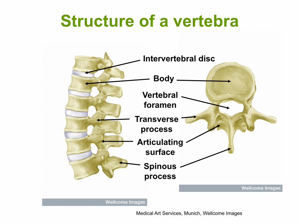
Structure of a vertebra
Medical Art Services, Munich, Wellcome Images
Body
Transverse process
Spinous process
Articulating surface
Vertebral foramen
Intervertebral disc

Ribcage
Gray’s Anatomy
Anterior Posterior
Sternum
Costal cartilage

Pelvic girdle
Male FemaleIlium
Pubis
IschiumIschium< 90° >100°
Wellcome Photo Library

Pectoral girdle
Clavicle
Scapula
Wellcome Photo Library

564%)==4.%+"1%8-Q4.%8#*H3
I*70*/*%R90S%T0((*77$*(
5+.3)3R4$+$+.3)3S6+8+"F43

564%6+"1
I*70*/*%R90S%T0((*77$*(

Medical Art Services, Munich, Wellcome Images
Joints

Joint classification• Joints can be classified by their structure and the
amount of movement they allow.
Structure Joint cavity MovementFibrous None NoneCartilaginous None None or slightSynovial Present Freely movable

Types of joints
Wellcome Photo Library
Immovable fibrous joint
Slightly movable cartilaginous joint
Movable synovial joint

The structure of a synovial joint
Wellcome Photo Library
Articulating cartilage
Joint cavity
Bursa
Synovial membrane
Fibrous capsule

Can you name these ligaments of the knee joint?

Synovial joints
Synovial joints have a joint cavity and are classified according to the type of movement they allow.

Flexion Extension
Rotation
Rotation
AdductionAdduction
Abduction
Hinge joint
Ball & socket joint
Pivot joint

(#"F4%]-#"$
I*70*/*%R90S%T0((*77$*(%

'+88%+"1%3-074$%]-#"$
I*70*/*%R90S%T0((*77$*(%

S#K-$%]-#"$
I*70*/*%R90S%T0((*77$*(%

9+1184%]-#"$
I*70*/*%R90S%T0((*77$*(%