Embed Size (px)
Citation preview
Unit 3B:Biological Bases of Behavior:
The Brain
Unit Overview
• The Tools of Discovery: Having Our Head Examined
• Older Brain Structures
• The Cerebral Cortex
• Our Divided Brain
• Right-Left Differences in the Intact Brain
• The Brain and Consciousness
Click on the any of the above hyperlinks to go to that section in the presentation.

The Tools of Discovery: Having Our Head Examined
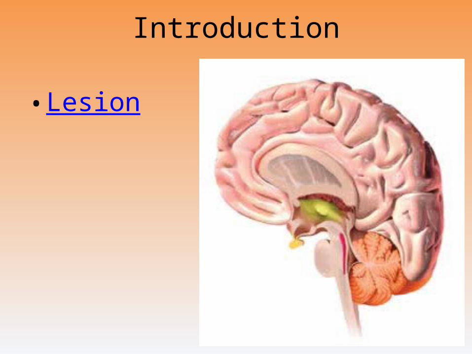
Introduction
• Lesion
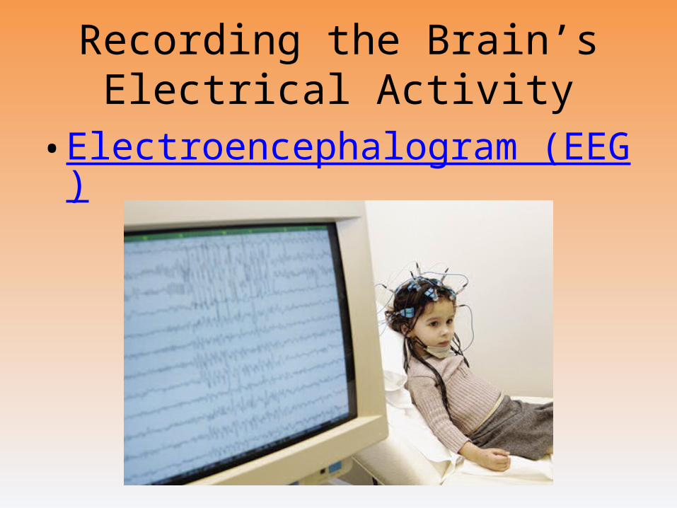
Recording the Brain’s Electrical Activity
• Electroencephalogram (EEG)
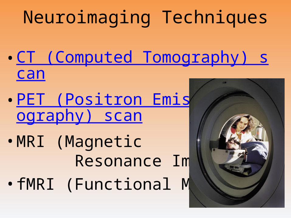
Neuroimaging Techniques
• CT (Computed Tomography) scan
• PET (Positron Emission Tomography) scan
• MRI (Magnetic Resonance Imaging)
• fMRI (Functional MRI)
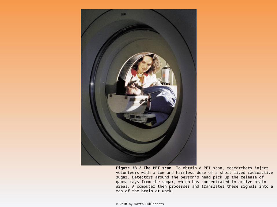
Figure 3B.2 The PET scan To obtain a PET scan, researchers inject volunteers with a low and harmless dose of a short-lived radioactive sugar. Detectors around the person’s head pick up the release of gamma rays from the sugar, which has concentrated in active brain areas. A com puter then processes and translates these signals into a map of the brain at work.
© 2010 by Worth Publishers
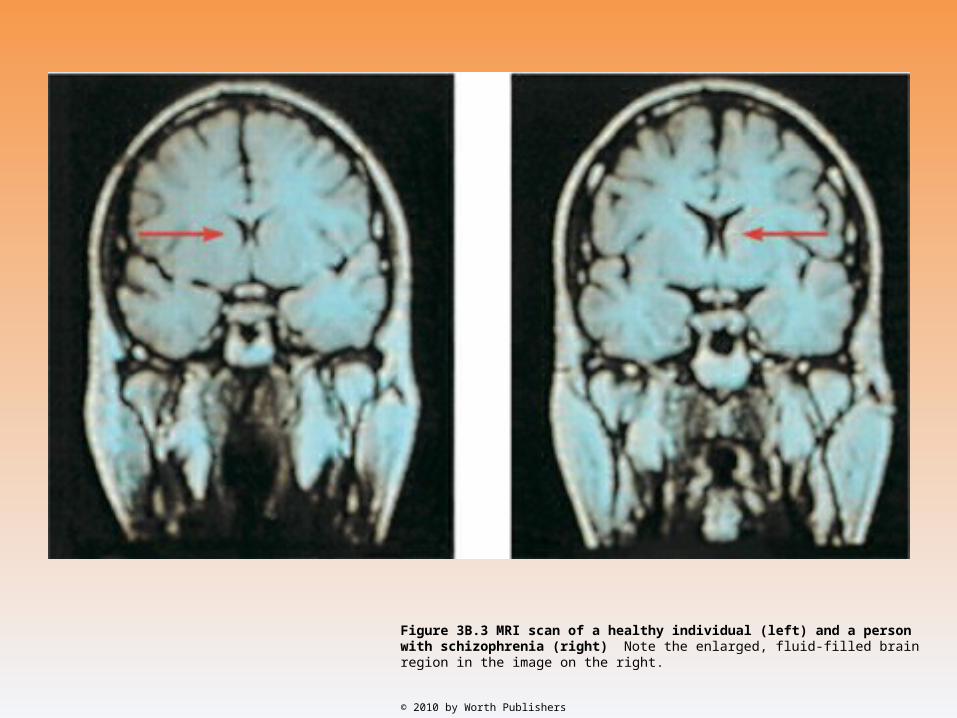
Figure 3B.3 MRI scan of a healthy individual (left) and a person with schizophrenia (right) Note the enlarged, fluid-filled brain region in the image on the right.
© 2010 by Worth Publishers
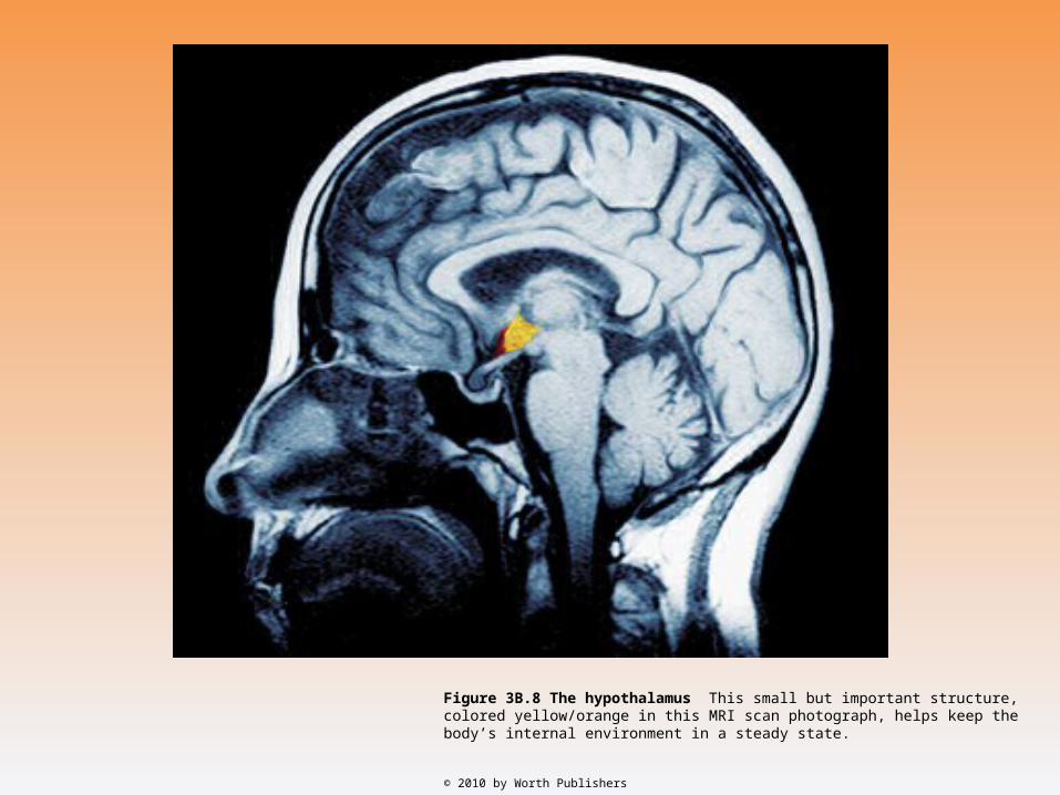
Figure 3B.8 The hypothalamus This small but important structure, colored yellow/orange in this MRI scan photograph, helps keep the body’s internal environment in a steady state.
© 2010 by Worth Publishers
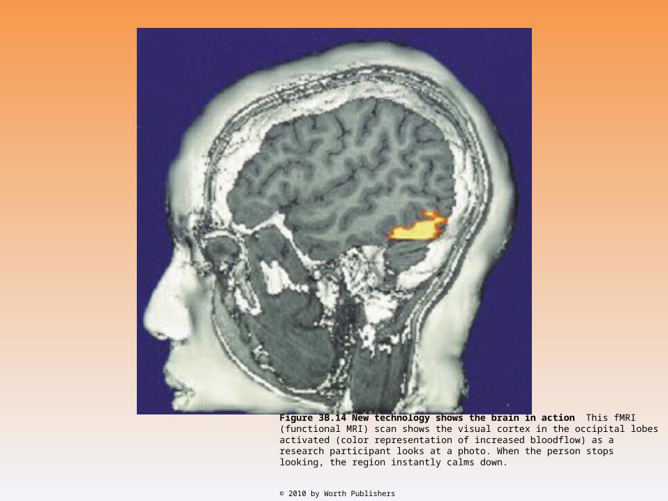
Figure 3B.14 New technology shows the brain in action This fMRI (functional MRI) scan shows the visual cortex in the occipital lobes activated (color representation of increased bloodflow) as a research participant looks at a photo. When the person stops looking, the region instantly calms down.
© 2010 by Worth Publishers
Older Brain Structures
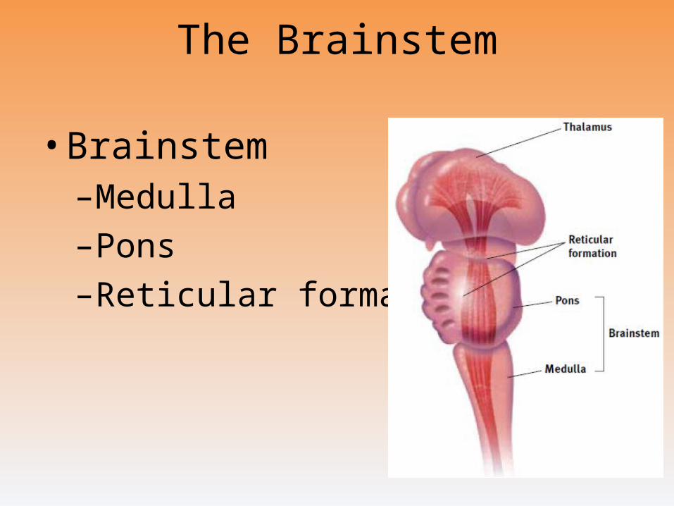
The Brainstem
• Brainstem–Medulla
–Pons
–Reticular formation
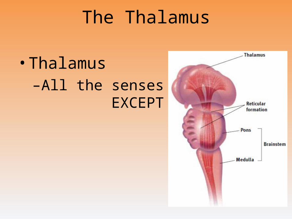
The Thalamus
• Thalamus–All the senses
EXCEPT smell
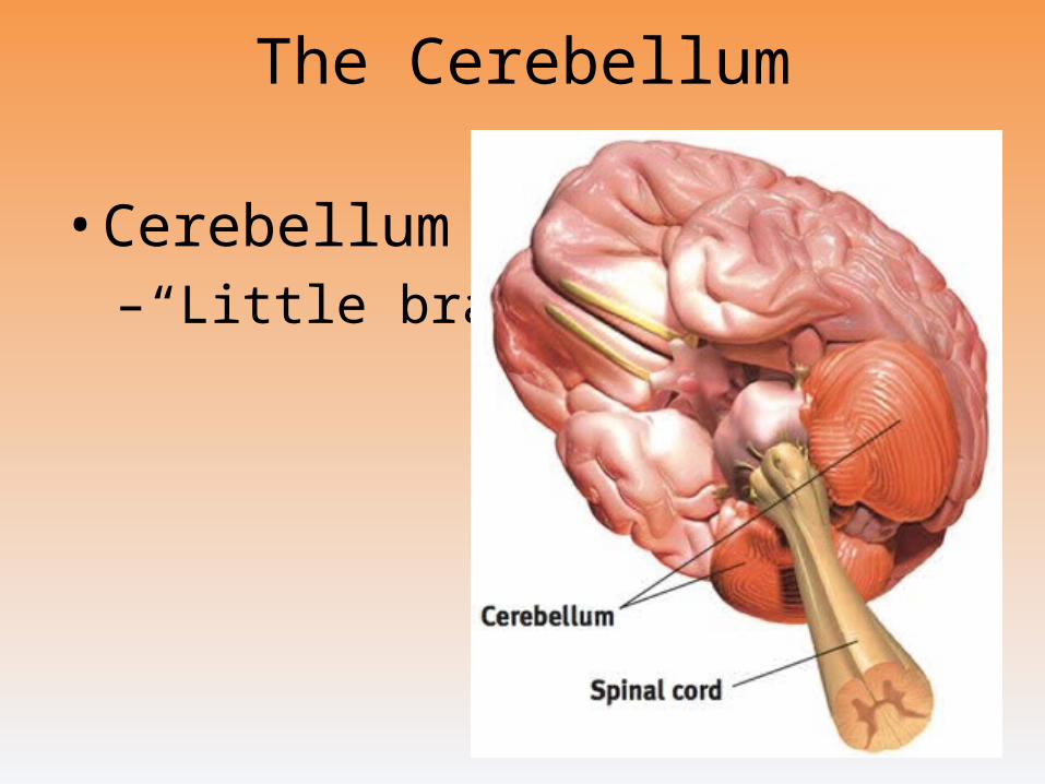
The Cerebellum
• Cerebellum–“Little brain”
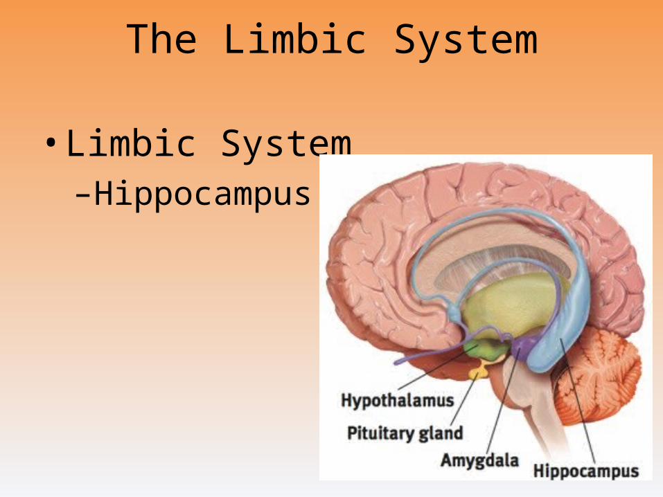
The Limbic System
• Limbic System–Hippocampus
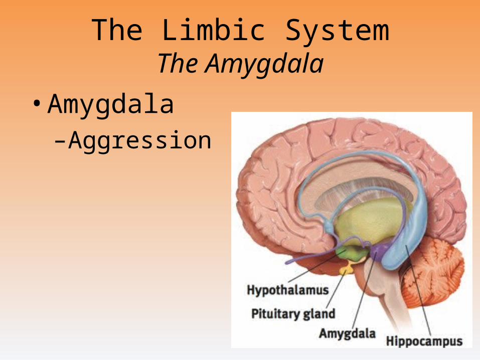
The Limbic SystemThe Amygdala
• Amygdala–Aggression
and fear
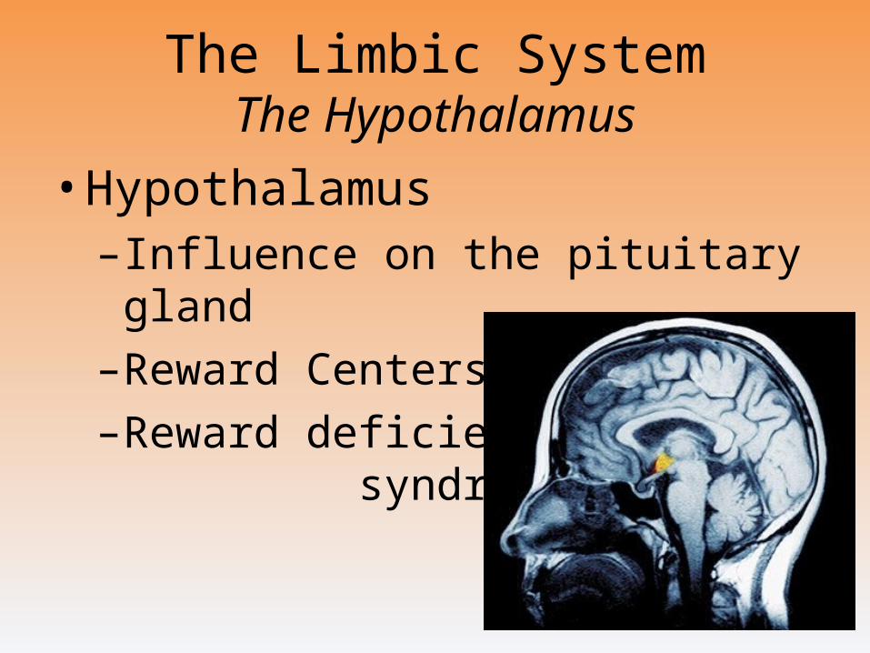
The Limbic SystemThe Hypothalamus
• Hypothalamus–Influence on the pituitary gland
–Reward Centers
–Reward deficiency syndrome
The Cerebral Cortex
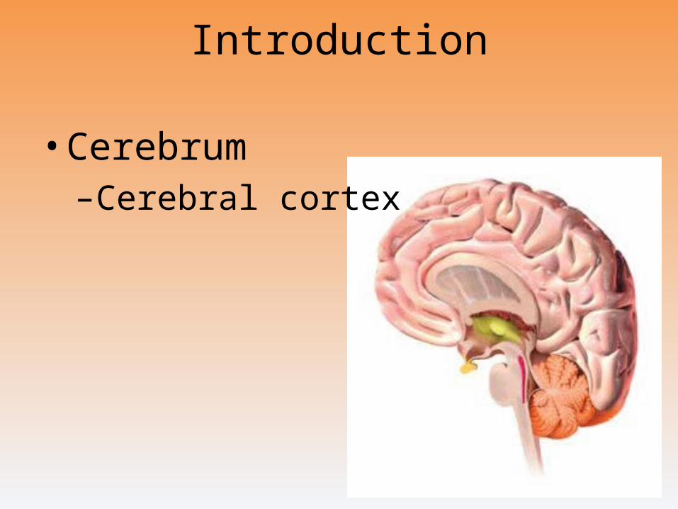
Introduction
• Cerebrum–Cerebral cortex
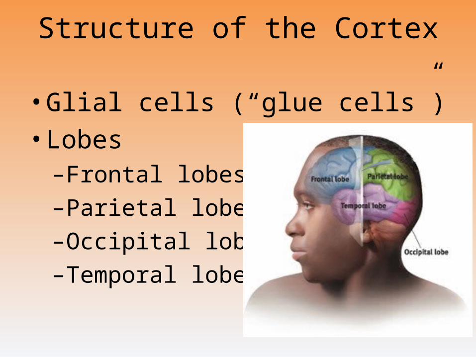
Structure of the Cortex
• Glial cells (“glue cells”)
• Lobes–Frontal lobes
–Parietal lobes
–Occipital lobes
–Temporal lobes
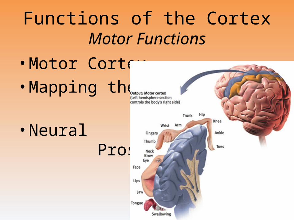
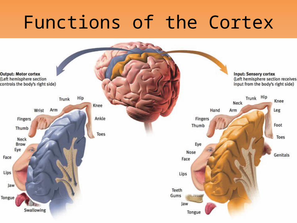
Functions of the CortexMotor Functions
• Motor Cortex
• Mapping the Motor Cortex
• Neural Prosthetics
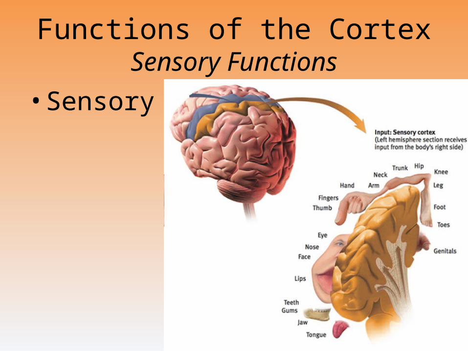
Functions of the CortexSensory Functions
• Sensory cortex
Functions of the Cortex
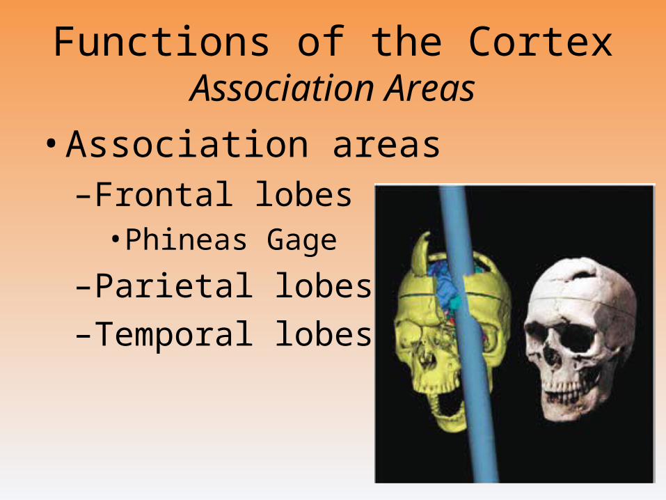
Functions of the CortexAssociation Areas
• Association areas–Frontal lobes
• Phineas Gage
–Parietal lobes
–Temporal lobes
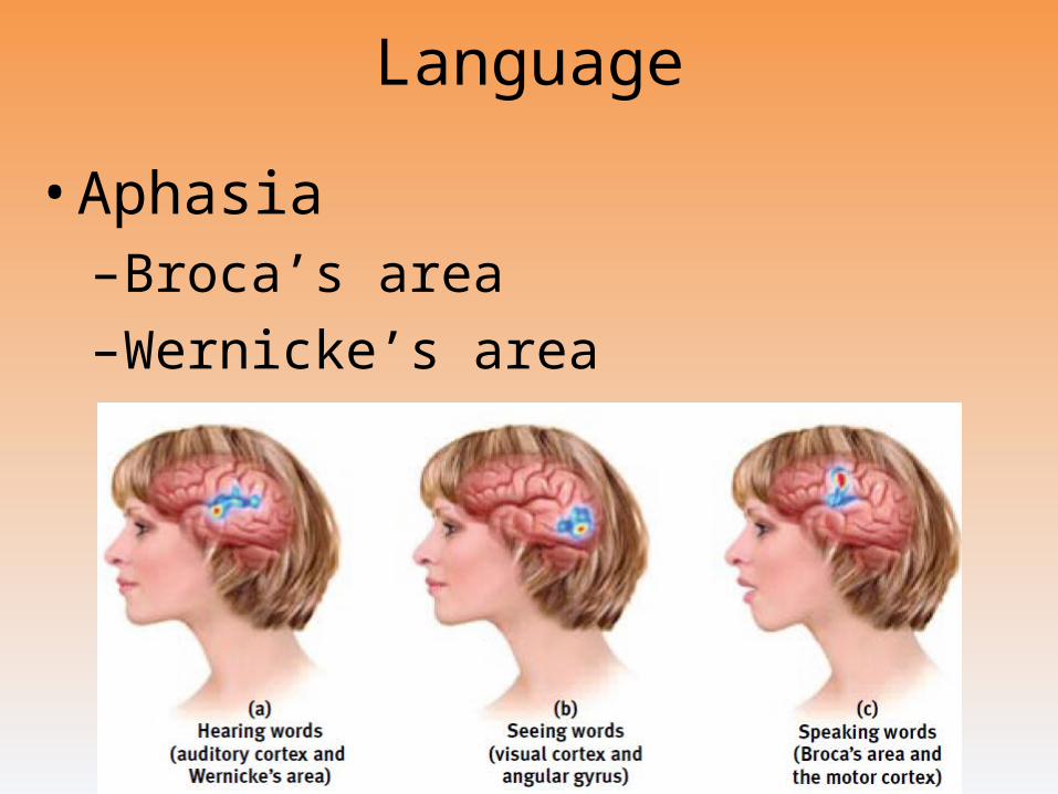
Language
• Aphasia–Broca’s area
–Wernicke’s area
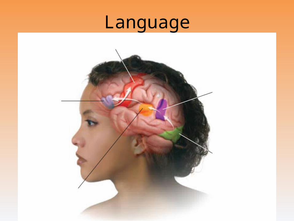
Language
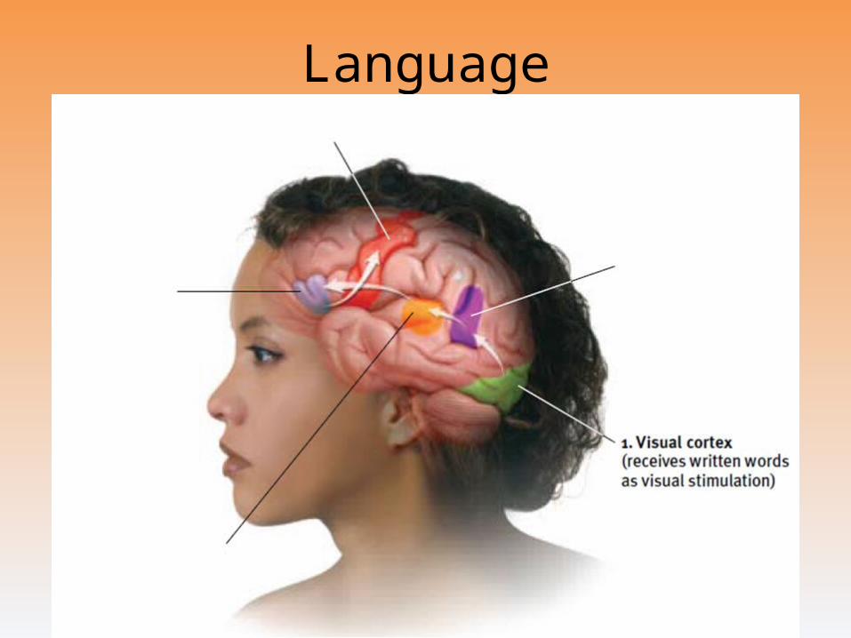
Language
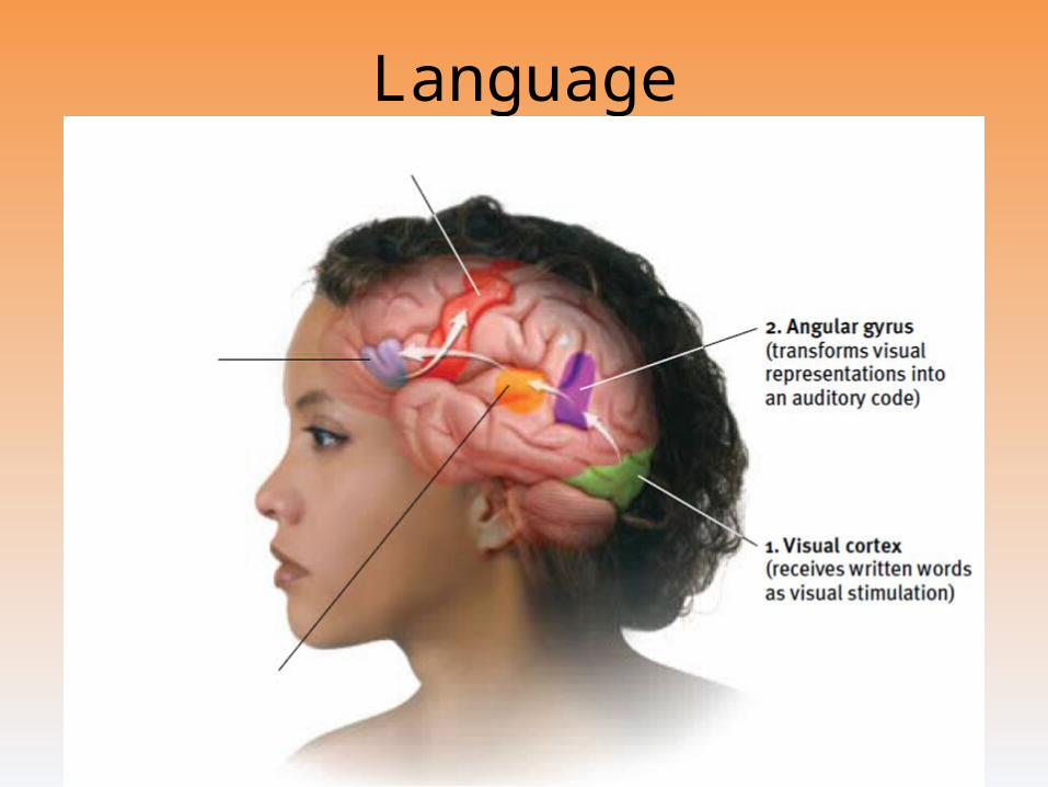
Language
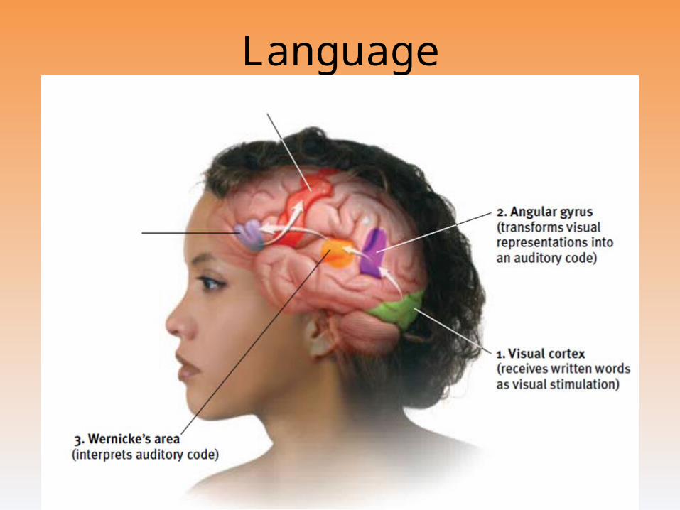
Language
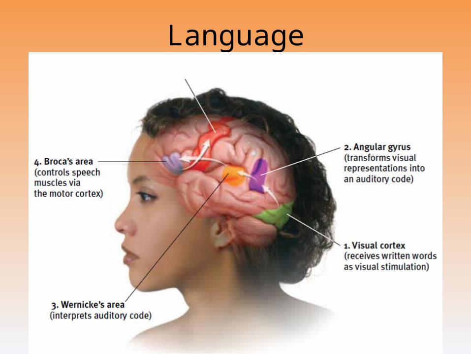
Language
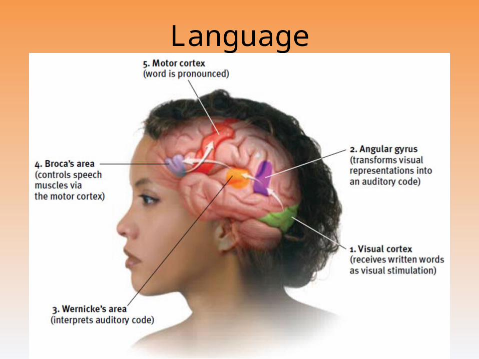
Language
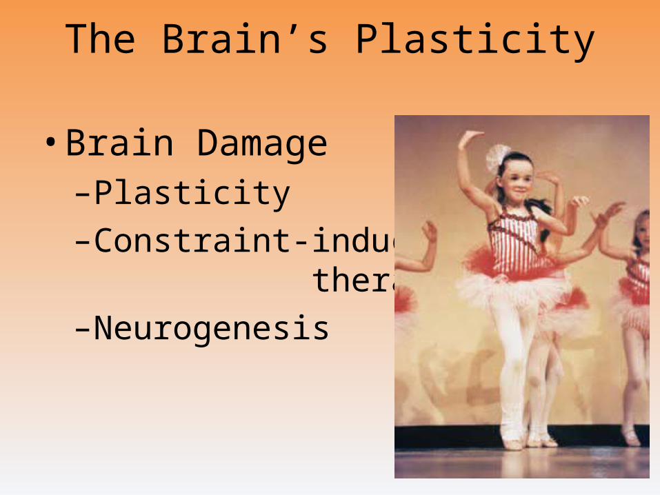
The Brain’s Plasticity
• Brain Damage–Plasticity
–Constraint-induced therapy
–Neurogenesis
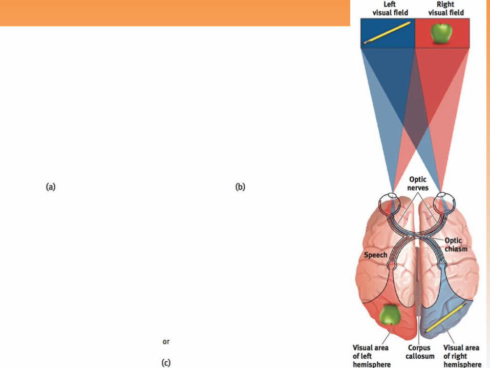
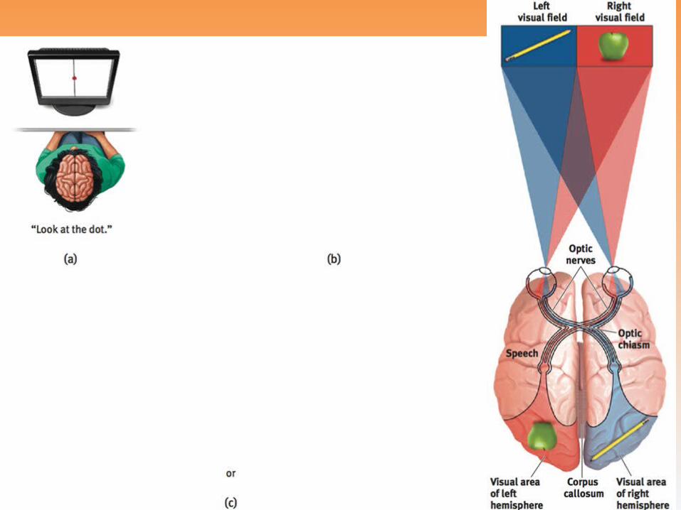
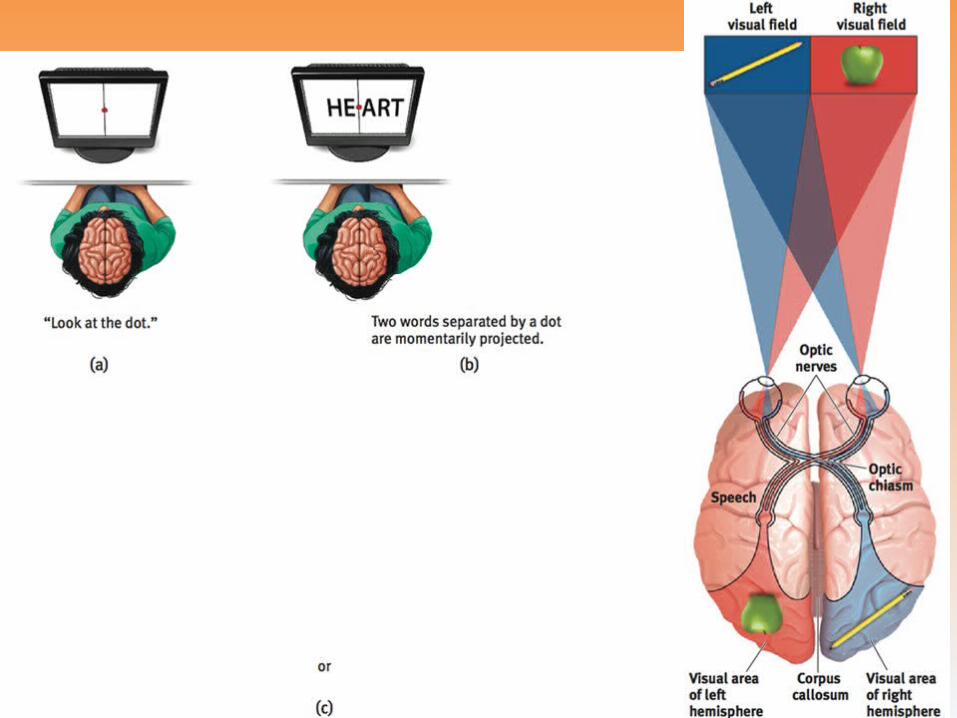
Our Divided Brain
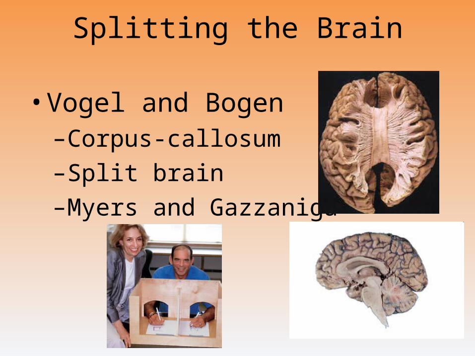
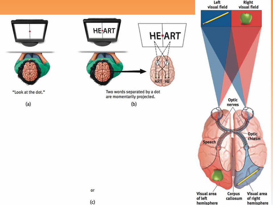
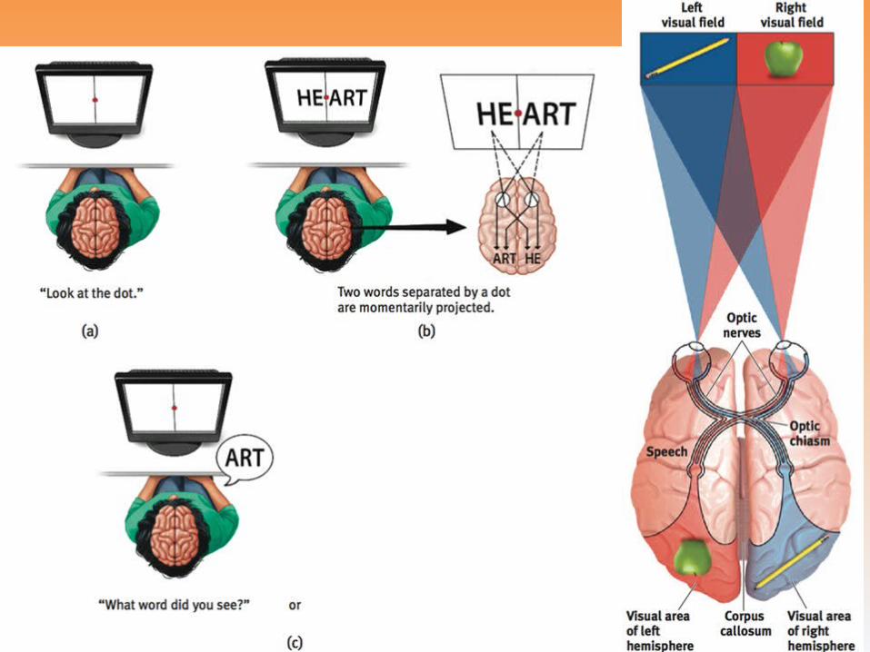
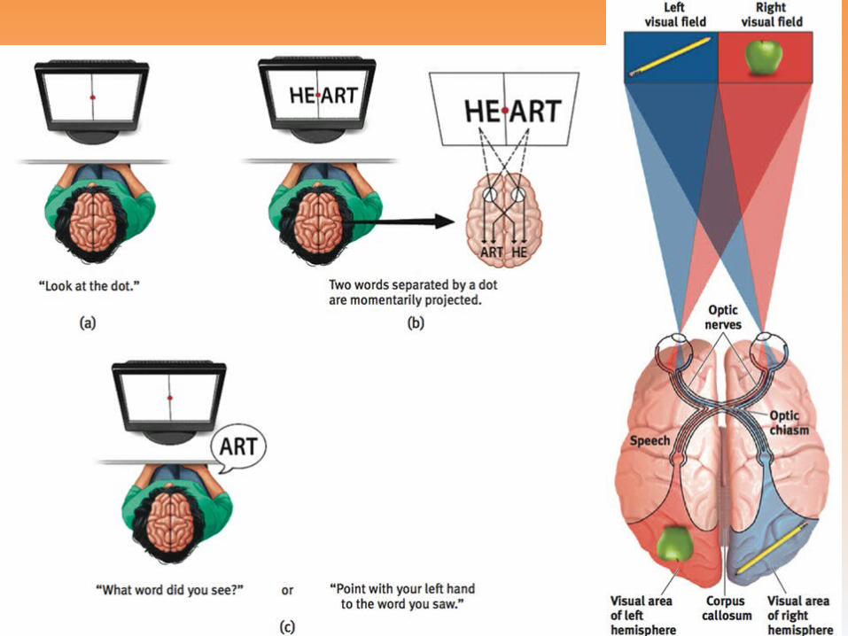
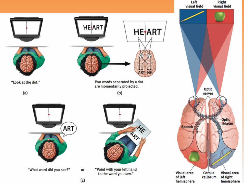
Splitting the Brain
• Vogel and Bogen–Corpus-callosum
–Split brain
–Myers and Gazzaniga
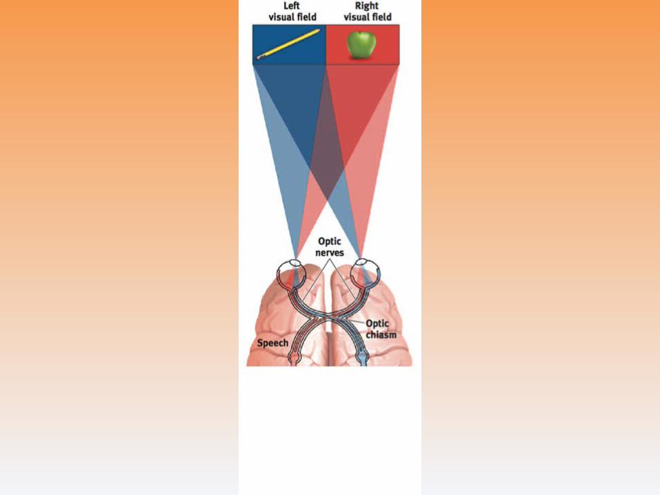
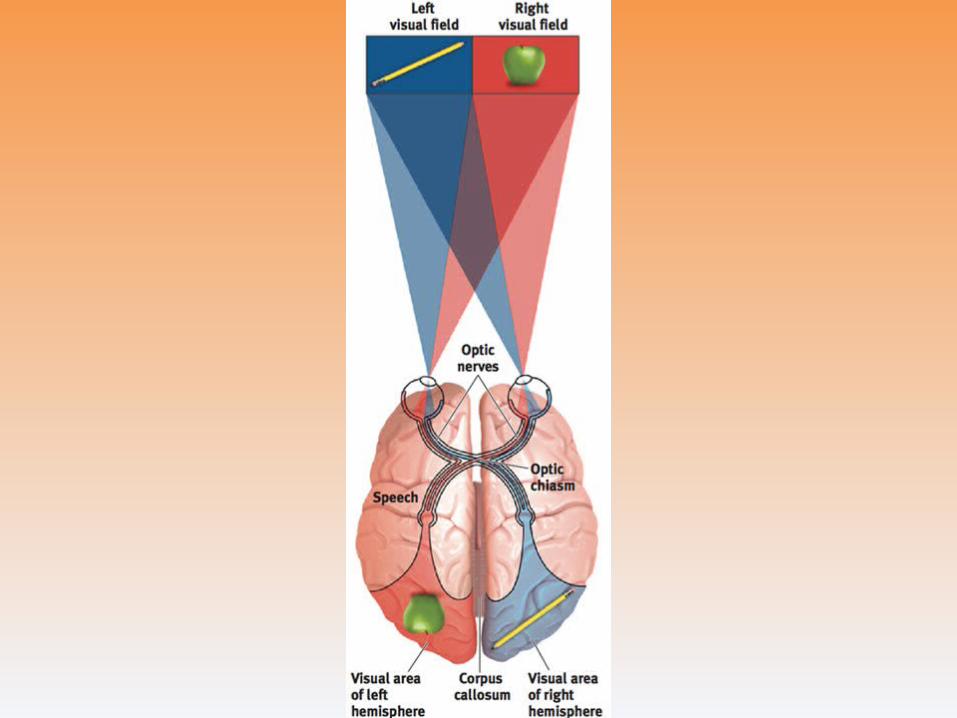

Right-Left Differences in the Intact Brain
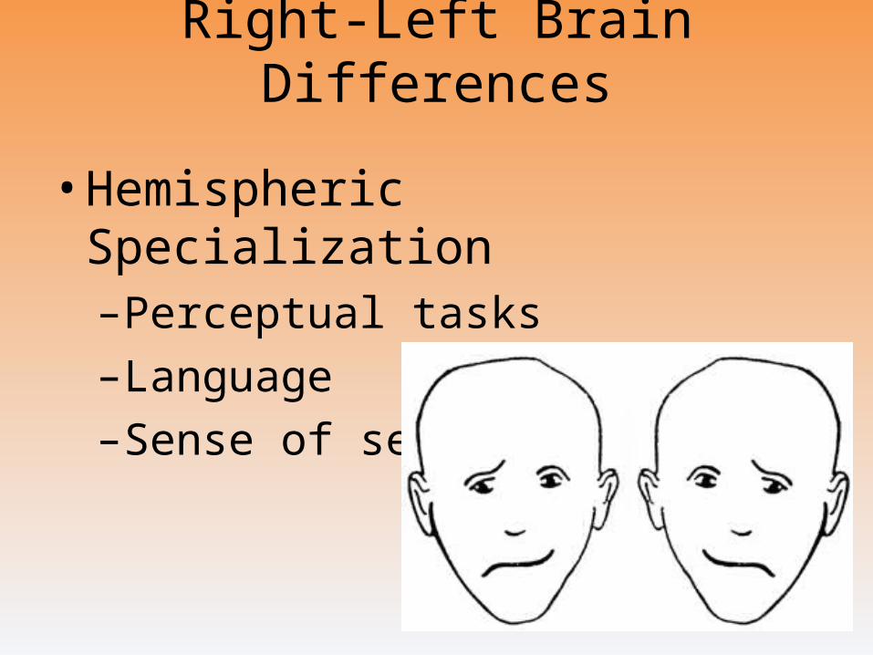
Right-Left Brain Differences
• Hemispheric Specialization–Perceptual tasks
–Language
–Sense of self
The Brain and Consciousness
Introduction
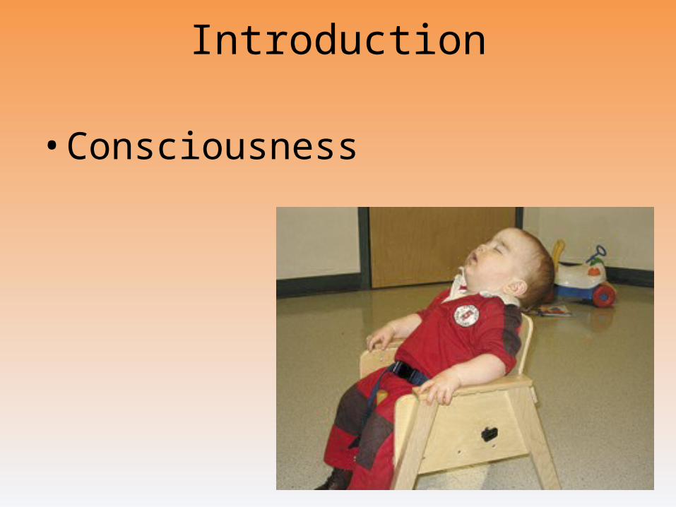
• Consciousness
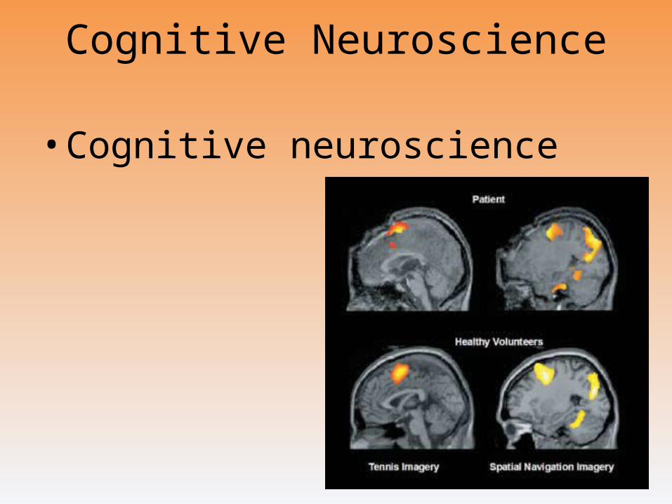
Cognitive Neuroscience
• Cognitive neuroscience
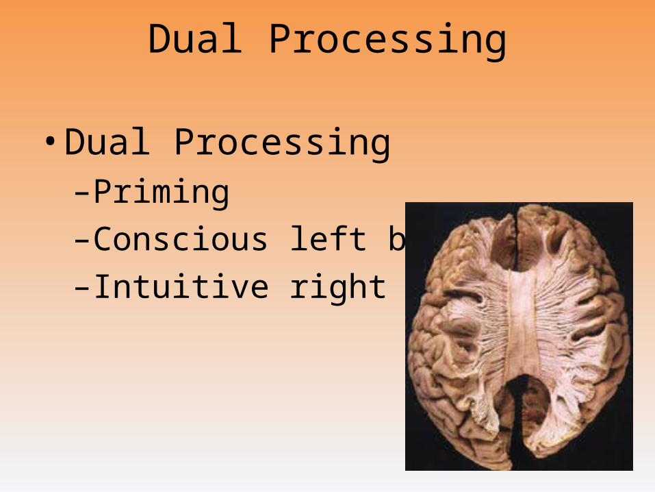
Dual Processing
• Dual Processing–Priming
–Conscious left brain
–Intuitive right brain
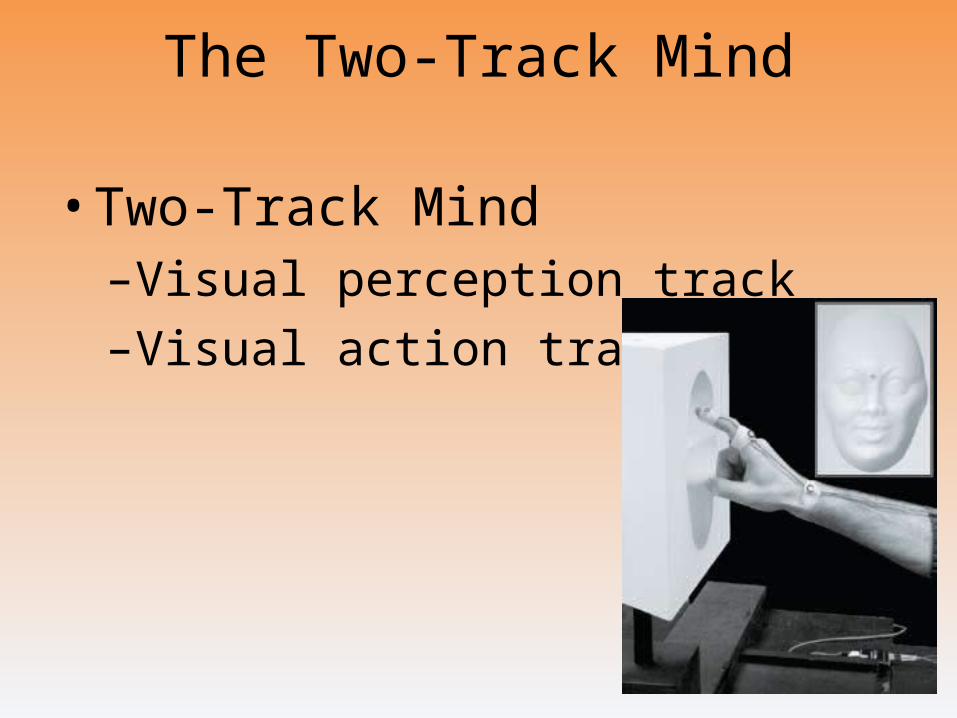
The Two-Track Mind
• Two-Track Mind–Visual perception track
–Visual action track
The End
Definition Slides

Lesion
= tissue destruction; a brain lesion is a naturally or experimentally caused destruction of brain tissue.
Electroencephalogram (EEG)
= an amplified recording of the waves of electrical activity that sweep across the brain’s surface. These waves are measured by electrodes placed on the scalp.
CT (computed tomography) Scan
= a series of X-ray photographs taken from different angles and combined by computer into a composite representation of a slice through the body.
• Also called CAT scan.
PET (positron emission tomography) Scan
= a visual display of brain activity that detects where a radioactive form of glucose goes while the brain performs a given task.
MRI (magnetic resonance imaging)
= a technique that uses magnetic fields and radio waves to produce computer-generated images of soft tissue. MRI scans show brain anatomy.
fMRI (functional MRI)
= a technique for revealing bloodflow and, therefore, brain activity by comparing successive MRI scans. fMRI scans show brain function.
Brainstem
= the oldest part of the central core of the brain, beginning where the spinal cord swells as it enters the skull; the brainstem is responsible for automatic survival functions.
Medulla
= the base of the brainstem; controls heartbeat and breathing.
Reticular Formation
= a nerve network in the brainstem that plays an important role in controlling arousal.
Thalamus
= the brain’s sensory switchboard, located on top of the brainstem; it directs messages to the sensory receiving areas in the cortex and transmits replies to the cerebellum and medulla.
Cerebellum
= the “little brain” at the rear of the brainstem; functions include processing sensory input and coordinating movement output and balance.
Limbic System
= doughnut-shaped neural system (including the hippocampus, amygdala, and hypothalamus) located below the cerebral hemispheres; associated with emotions and drives.
Amygdala
= two lima bean-sized neural clusters in the limbic system; linked to emotion.
Hypothalamus
= a neural structure lying below (hypo) the thalamus; it directs several maintenance activities (eating, drinking, body temperature), helps govern the endocrine system via the pituitary gland, and is linked to emotion and reward.
Cerebral Cortex
= the intricate fabric of interconnected neural cells covering the cerebral hemispheres; the body’s ultimate control and information-processing center.
Glial Cells
= cells in the nervous system that support, nourish, and protect neurons.
Frontal Lobes
= portion of the cerebral cortex lying just behind the forehead; involved in speaking and muscle movements and in making plans and judgments.
Parietal Lobes
= portion of the cerebral cortex lying at the top of the head and toward the rear; receives sensory input for touch and body position.
Occipital Lobes
= portion of the cerebral cortex lying at the back of the head; includes areas that receive information from the visual fields.
Temporal Lobes
= portion of the cerebral cortex lying roughly above the ears; includes the auditory areas, each receiving information primarily from the opposite ear.
Motor Cortex
= an area at the rear of the frontal lobes that controls voluntary movements.
Sensory Cortex
= area at the front of the parietal lobes that registers and processes body touch and movement sensations.
Association Areas
= areas of the cerebral cortex that are not involved in primary motor or sensory functions; rather, they are involved in higher mental functions such as learning, remembering, thinking, and speaking.
Aphasia
= impairment of language, usually caused by left hemisphere damage either to Broca’s area (impairing speaking) or to Wernicke’s area (impairing understanding).
Broca’s Area
= controls language expression that directs the muscle movements involved in speech.
Wernicke’s Area
= controls language reception – a brain area involved in language comprehension and expression; usually in the left temporal lobe.
Plasticity
= the brain’s ability to change, especially during childhood, by reorganizing after damage or by building new pathways based on experience.
Neurogenesis
= the formation of new neurons.
Corpus Callosum
= the large band of neural fibers connecting the two brain hemispheres and carrying messages between them.
Split Brain
= a condition resulting from surgery that isolates the brain’s two hemispheres by cutting the fibers (mainly those of the corpus callosum) connecting them.
Consciousness
= our awareness of ourselves and our environment.
Cognitive Neuroscience
= the interdisciplinary study of the brain activity linked with cognition (including perception, thinking, memory and language).
Dual Processing
=the principle that information is often simultaneously processed on separate conscious and unconscious tracks.