Embed Size (px)
Citation preview
Unit 11 Circulatory System The Heart
1. The Heart
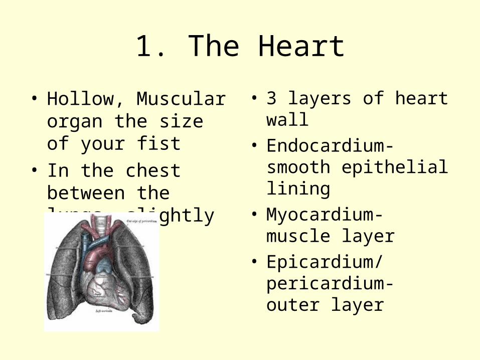
• Hollow, Muscular organ the size of your fist
• In the chest between the lungs, slightly left
• 3 layers of heart wall• Endocardium- smooth
epithelial lining• Myocardium- muscle
layer• Epicardium/
pericardium- outer layer
2. The 4 chambers of the Heart and the 2 Major Valves
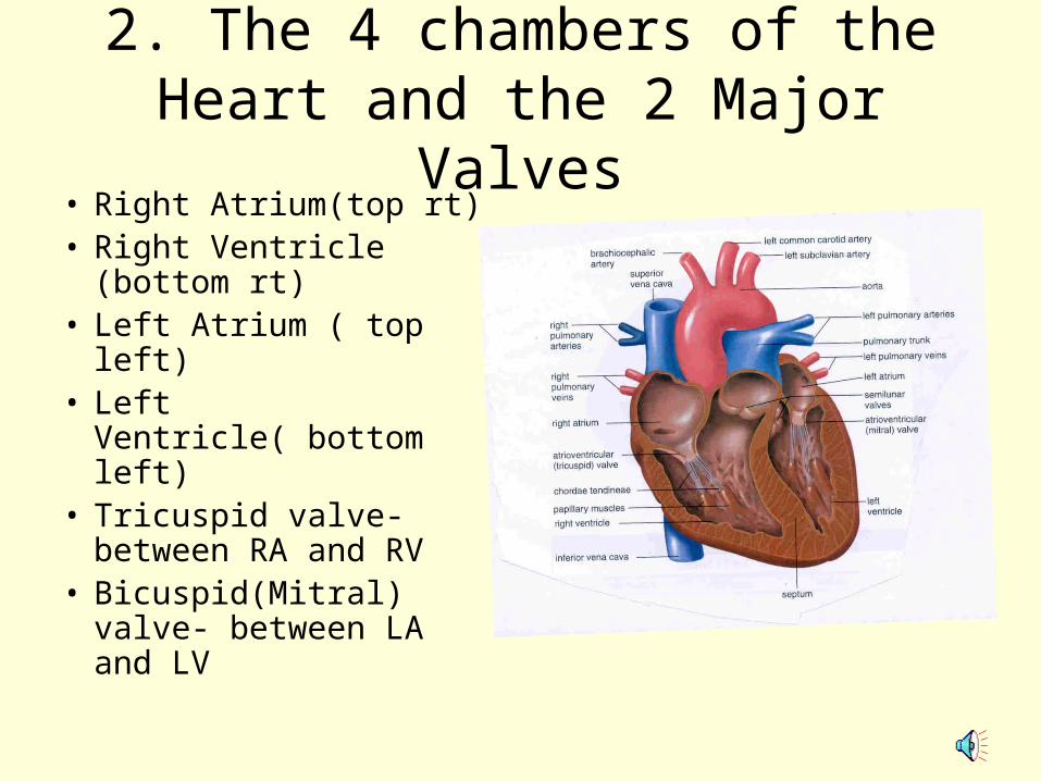
• Right Atrium(top rt)• Right Ventricle
(bottom rt)• Left Atrium ( top left)• Left Ventricle( bottom
left)• Tricuspid valve-
between RA and RV• Bicuspid(Mitral) valve-
between LA and LV
BICUSPID/MITRAL VALVE
• This valve between the LA and LV is important in dentistry because if you have ever had a severe strept infection that turns into Rheumatic Fever- this valve may be damaged. The cells of this valve are shaped similar to the strept bacteria cells. When your body produces ANTIBODIES to fight this invader, the valve cells are also attacked. That is why a health history asks if you have ever had Rheumatic fever, and if you have- you will need to be on an antibiotic BEFORE any dental treatment is done.
VALVES
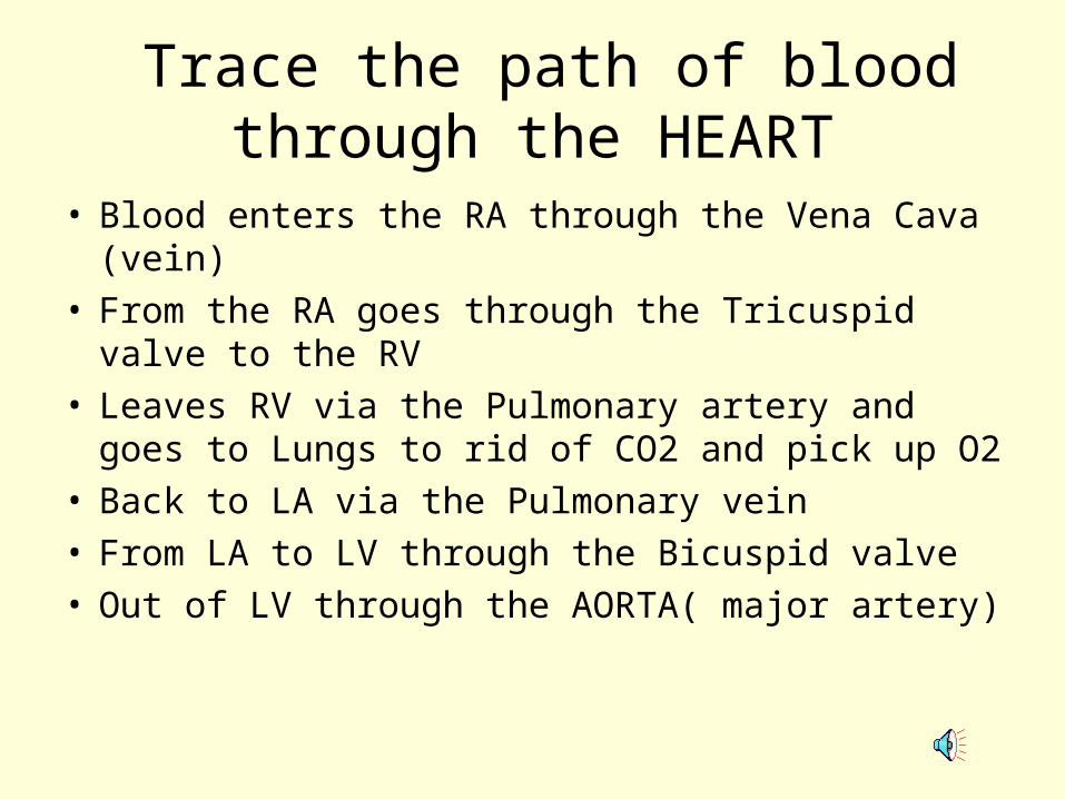
Trace the path of blood through the HEART
• Blood enters the RA through the Vena Cava (vein)
• From the RA goes through the Tricuspid valve to the RV
• Leaves RV via the Pulmonary artery and goes to Lungs to rid of CO2 and pick up O2
• Back to LA via the Pulmonary vein• From LA to LV through the Bicuspid valve• Out of LV through the AORTA( major artery)
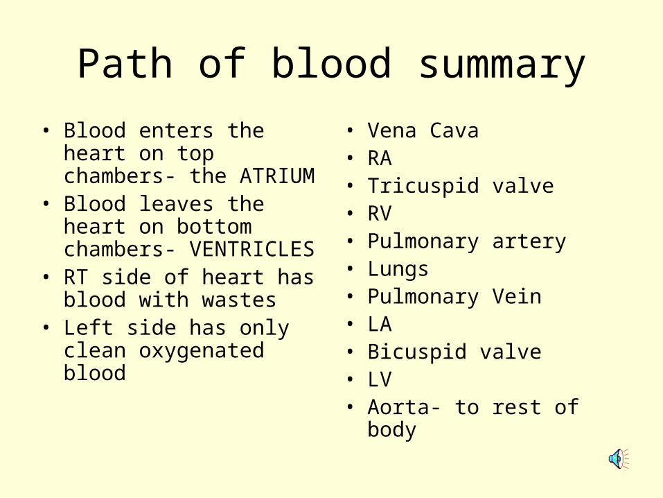
Path of blood summary
• Blood enters the heart on top chambers- the ATRIUM
• Blood leaves the heart on bottom chambers- VENTRICLES
• RT side of heart has blood with wastes
• Left side has only clean oxygenated blood
• Vena Cava• RA• Tricuspid valve• RV• Pulmonary artery• Lungs• Pulmonary Vein• LA• Bicuspid valve• LV• Aorta- to rest of body
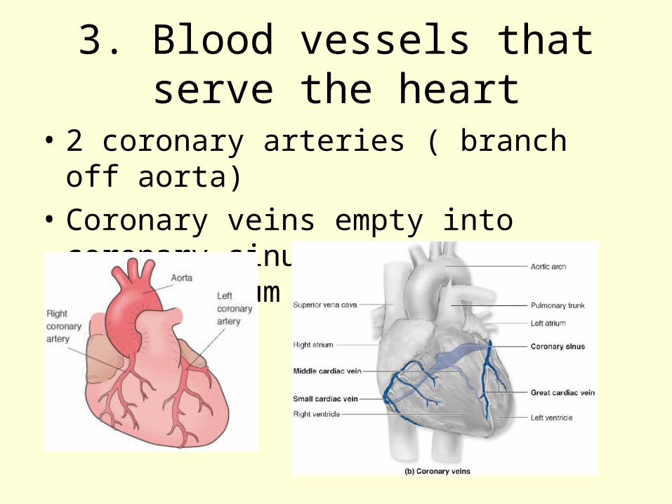
3. Blood vessels that serve the heart
• 2 coronary arteries ( branch off aorta)
• Coronary veins empty into coronary sinus that goes to right atrium
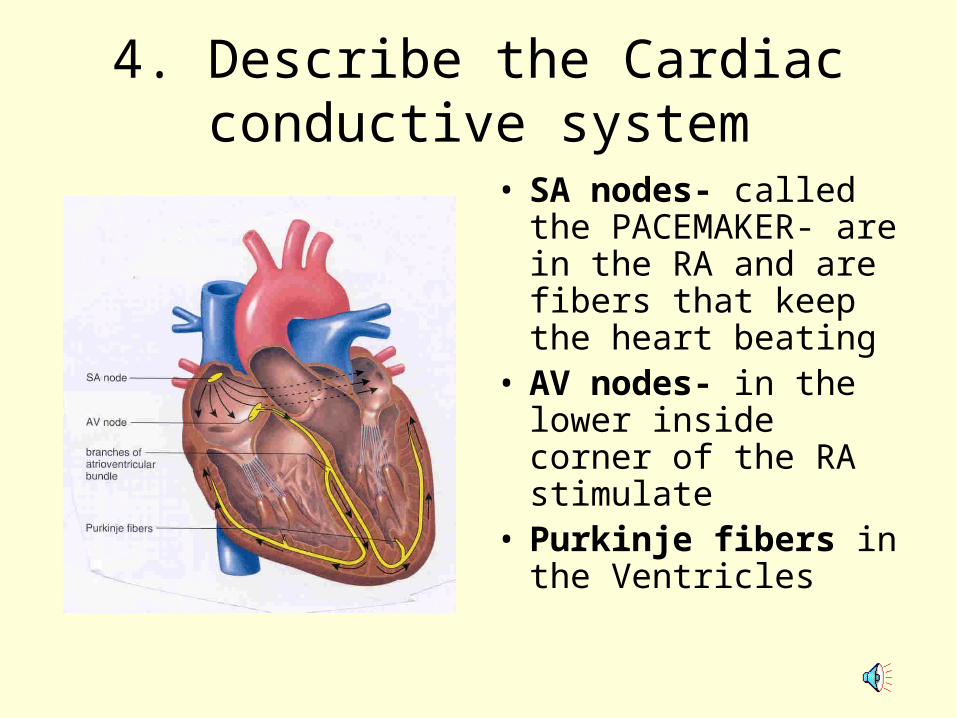
4. Describe the Cardiac conductive system
• SA nodes- called the PACEMAKER- are in the RA and are fibers that keep the heart beating
• AV nodes- in the lower inside corner of the RA stimulate
• Purkinje fibers in the Ventricles
5. Cardiac Cycle- 1 complete heartbeat
• 72 / min• Contraction- blood
forced out =SYSTOLIC
• Relaxation- heart fills with blood= DIASTOLIC
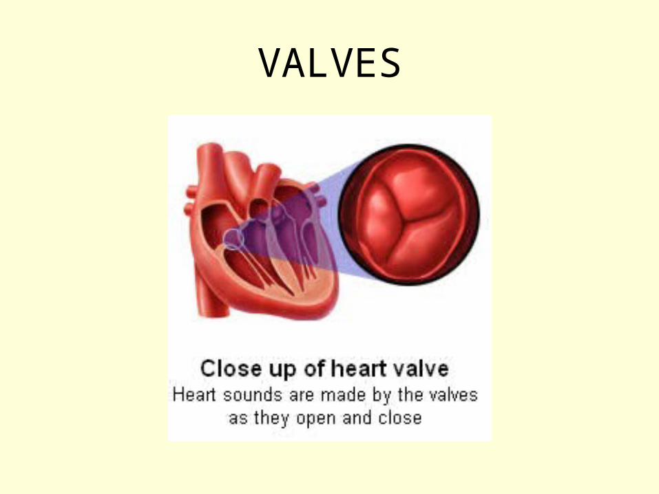
• Lub Dub sound is the valves opening and closing
6. Cardiac Output
• Cardiac output is the volume of blood pumped by the left ventrical into the aorta in 1 minute
• Depends on needs of the body
• Ave 5 liters/minute at rest
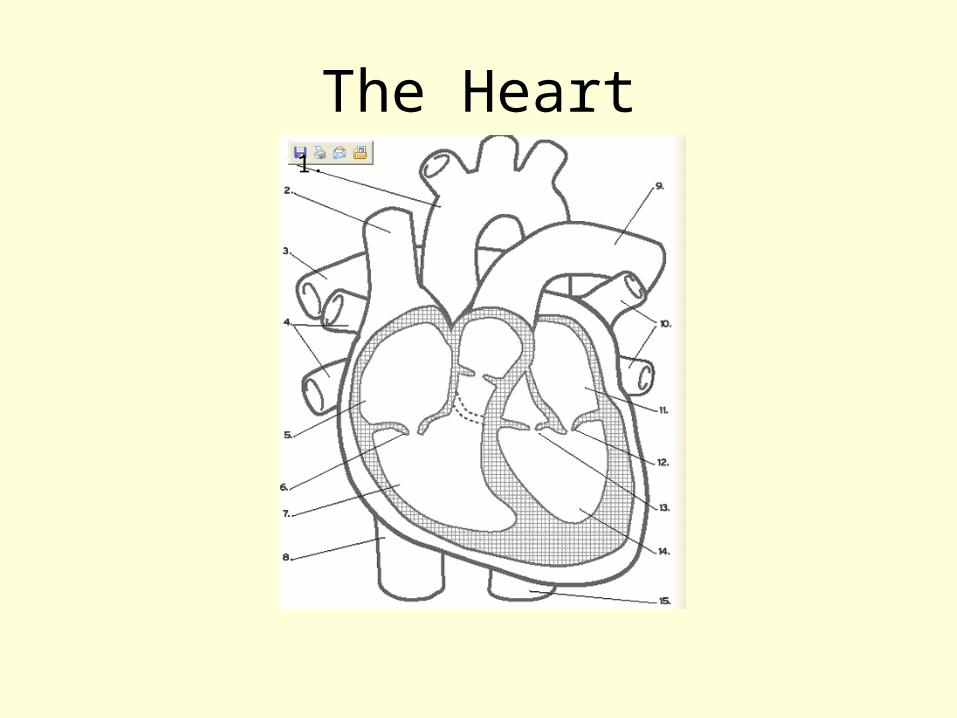
The Heart1.
THE END