Embed Size (px)
Citation preview
1
Unexpected function of the glucanosyltransferase Gas1 in the DNA damage
response linked to histone H3 acetyltransferases in Saccharomyces cerevisiae
Moriah Eustice* and Lorraine Pillus*,§
* Section of Molecular Biology, Division of Biological Sciences,
University of California, San Diego
§ UC San Diego Moores Cancer Center, La Jolla, California 92093-0347
Genetics: Early Online, published on February 13, 2014 as 10.1534/genetics.113.158824
Copyright 2014.

2
Running title: Gas1 and Sas3 function in DDR
Keywords: chromatin, acetyltransferase, DNA damage, cell cycle checkpoint,
glucan
Corresponding author:
Lorraine Pillus
Address: 9500 Gilman Drive, La Jolla, CA 92093-0347
E-mail address: [email protected]
Phone: 858 822-2442
3
ABSTRACT
Chromatin organization and structure are crucial for transcriptional
regulation, DNA replication and damage repair. Although initially characterized in
remodeling cell wall glucans, the β-1,3-glucanosyltransferase Gas1 was recently
discovered to regulate transcriptional silencing in a manner separable from its
activity at the cell wall. However, the function of Gas1 in modulating chromatin
remains largely unexplored. Our genetic characterization revealed that GAS1
had critical interactions with genes encoding the histone H3 lysine
acetyltransferases Gcn5 and Sas3. Specifically, whereas the gas1 gcn5 double
mutant was synthetically lethal, deletion of both GAS1 and SAS3 restored
silencing in Saccharomyces cerevisiae. The loss of GAS1 also led to broad DNA
damage sensitivity with reduced Rad53 phosphorylation and defective cell cycle
checkpoint activation following exposure to select genotoxins. Deletion of SAS3
in the gas1 background restored both Rad53 phosphorylation and checkpoint
activation following exposure to genotoxins that trigger the DNA replication
checkpoint. Our analysis thus uncovers previously unsuspected functions for
both Gas1 and Sas3 in DNA damage response and cell cycle regulation.
4
INTRODUCTION
Chromatin packages DNA in the nucleus and regulates accessibility to the
underlying genome. Tightly compacted chromatin, or heterochromatin, impedes
nuclear processes including transcription, DNA replication and DNA damage
repair (reviewed in Li and Reinberg 2011; Papamichos-Chronakis and Peterson
2013). Thus, genes found within heterochromatic regions are repressed, or
silenced (reviewed in Rusche et al. 2003). However, the degree of chromatin
compaction is highly dynamic, as cells must continuously alter transcriptional
programs in response to environmental or metabolic demands while promoting
replication and repair processes.
The basic unit of chromatin is the nucleosome, consisting of DNA wrapped
around an octamer of conserved core histone proteins (Kornberg and Lorch
1999). Post-translational modification (PTM) of histones is a prime means for
altering chromatin structure. These modifications are dynamic and tightly
controlled as they regulate higher order chromatin structure and DNA
accessibility by altering the interaction between DNA and histones in addition to
recruiting chromatin-modifying enzymes (reviewed in Kouzarides 2007; Campos
and Reinberg 2009). The localization of chromatin within the nucleus also plays
a fundamental role in chromatin dynamics, such that localization to the nuclear
periphery regulates processes including silencing and the DNA damage
response (DDR) (reviewed in Taddei and Gasser 2012; Bermejo et al. 2012).
5
The β-1,3-glucanosyltransferase Gas1, a member of the Gas family of
proteins, was initially characterized at the cell wall where it remodels chains of β-
1,3-glucan (Ragni et al. 2007). However, a pool of Gas1 also localizes to the
nuclear periphery (Huh et al. 2003) and genome-wide studies have identified
genetic and physical interactions between Gas1 and diverse components of the
chromatin modifying machinery (www.thebiogrid.org). Reflecting these findings,
deletion of GAS1 was recently discovered to lead to a unique constellation of
silencing defects in yeast. Specifically, loss of Gas1 catalytic activity increases
rDNA silencing and decreases telomeric silencing, yet has no observable change
at the HM cryptic mating-type loci. These alterations in silencing are not
remediated by the osmoregulator sorbitol (Koch and Pillus 2009), which rescues
the cell wall-associated defects of gas1 and other cell wall mutants (Turchini et al.
2000; Levin 2005). Combined, these data support a function for Gas1 in
chromatin-mediated processes that is separable from its role at the cell wall.
A genome-wide screen reported that GAS1 has a negative genetic
interaction with GCN5 (Costanzo et al. 2010), which encodes a prominent lysine
acetyltransferase (KAT). Gcn5-catalyzed acetylation of histone and non-histone
substrates affects numerous chromatin-dependent processes (reviewed in Lee
and Workman 2007; Koutelou et al. 2010). Gcn5 functions in several important
complexes including SAGA, ADA and SLIK/SALSA (Grant et al. 1997; Pray-
Grant et al. 2002) to acetylate nucleosomal substrates on histone H3, with lysine
14 (K14) as a predominant target (Kuo and Andrews 2013). Gcn5 acts as a co-
activator, with H3K14 acetylation correlating with active transcription (Pokholok et

6
al. 2005) and Gcn5 is enriched at the promoters of active genes (Robert et al.
2004).
Gcn5 functionally overlaps with another KAT, Sas3. Gcn5 and Sas3 share
nucleosomal H3 targets (reviewed in Lafon et al. 2007) and deletion of both
GCN5 and SAS3 is synthetically lethal (Howe et al. 2001). Further, both Gcn5
and Sas3 are recruited to similar genomic regions (Rosaleny et al. 2007).
Whereas Gcn5 has been studied extensively, less is known about Sas3, due in
part to the functional overlaps with Gcn5 as well as the limited independent
phenotypes defined for SAS3 mutants. Deletion of SAS3 leads to a modest
increase in silencing at the HM loci (Reifsnyder et al. 1996) and Sas3 localizes at
the boundary of the HM loci, blocking the spread of silent chromatin (Tackett et al.
2005). Sas3 physically associates with the N-terminus of Spt16, a subunit of the
FACT elongation complex (John et al. 2000), which is essential for recovery from
replication stress (O’Donnell et al. 2004) and boundary formation (Tackett et al.
2005).
In addition to functions in transcriptional regulation and silencing, Gcn5
and other histone modifying enzymes also have crucial roles in the DDR. One of
the earliest marks associated with DDR activation in yeast is the phosphorylation
of H2A at serine 129 (S129), which serves as a scaffold that amplifies the DNA
damage signal in part by recruiting the repair machinery (reviewed in Rossetto et
al. 2010). Subsequently, phosphorylation of other mediators, prominently
including the Rad53 kinase, triggers a cascade that leads to changes in
7
transcription and activation of cell cycle checkpoints, which foster the repair of
damaged DNA (reviewed in Branzei and Foiani 2006; Sirbu and Cortez 2013).
Deletion of GCN5 renders cells sensitive to DNA damaging agents such
as the topoisomerase I inhibitor camptothecin (CPT), the radiomimietic drug
methyl methanesulfonate (MMS) and the replication inhibitor hydroxyurea (HU)
(Choy and Kron 2002; Burgess et al. 2010). Indeed, Gcn5-catalzyed acetylation
of both histone and non-histone substrates features prominently at numerous
stages of the DNA damage response (Burgess et al. 2010; Lee et al. 2010;
Charles et al. 2011)
There is also some evidence that Sas3 may play a role in the DDR. For
example, Sas3 has a reported physical interaction with the DNA damage
checkpoint effector kinase Chk1 (Liu et al. 2000), although the functional
significance of this interaction has not been established. Further, mutants of
H3K14 and H3K23, nucleosomal substrates of Gcn5 and Sas3, are sensitive to
DNA-damaging agents (Qin and Parthun 2002; Tamburini and Taylor 2005).
However, what role, if any, Sas3 may play in DNA damage has not been defined.
Here we report that GAS1 has strong genetic interactions with the histone
H3 lysine acetyltransferases encoded by both GCN5 and SAS3. The gas1 gcn5
combination was synthetically lethal. In contrast, the gas1 sas3 double mutant
was viable and, moreover, displayed selective mutual suppression of each
individual mutant’s phenotypes. We also discovered that gas1 has broad DNA
damage sensitivity following exposure to the genotoxins MMS, HU and CPT.
Sensing and initial activation of the DNA damage response was intact in gas1

8
strains, as evidenced by phosphorylation of histone H2A. However, the MMS and
HU sensitivity of gas1 reflects failure to trigger the DNA damage cell cycle
checkpoint as demonstrated by loss of both the cell cycle delay and Rad53
phosphorylation. The deletion of SAS3 in the gas1 background specifically
suppressed both MMS and HU sensitivity, leading to restoration of cell cycle
delay and Rad53 phosphorylation. These findings define a role for Gas1 in the
DNA damage response that is separable from its cell wall function. We have also
identified a specific role for Sas3 in antagonizing the replication checkpoint,
which is unique and opposite to the role previously identified for Gcn5.
MATERIALS AND METHODS
Yeast strains and plasmids: Strains are listed in Supporting Information
Table S1, plasmids in Table S2 and oligonucleotides in Table S3. All mutations
are deletions, unless otherwise noted, and were constructed using standard
techniques (Amberg et al. 2005).
Growth, silencing and DNA damage assays: Plate assays are five-fold
serial dilutions adjusted to an A600 of 1.0 after growth to saturation in synthetic
complete (SC) medium. Dilution assays were incubated at 30°, except where
noted. Telomeric silencing assays were performed with the TELVR::URA3
reporter strain grown in SC medium and plated on SC as growth control or SC
supplemented with 0.1% 5-Fluoroorotic Acid (5-FOA) to assay silencing (Renauld
et al. 1993; van Leeuwen and Gottschling 2002). Silent mating type analysis was
9
performed with the hml::TRP1 reporter (Le et al. 1997). Silencing of the rDNA
was assayed using the RDN::Ty-1-mURA3 construct (Smith and Boeke 1997).
Strains were plated on SC as a growth control and SC-Ura for rDNA silencing.
HU sensitivity was analyzed with 0.2M HU. MMS sensitivity was analyzed with
0.015% MMS. CPT sensitivity was analyzed using 20μg/mL CPT dissolved in
DMSO added to plates buffered with 100mM potassium phosphate (pH 7.5) to
maintain CPT activity (Nitiss and Wang 1988) with growth control plates at the
same concentration of DMSO. DMSO is shown as a control with all CPT images
as gas1 is mildly sensitive to DMSO. For ultraviolet light (UV) sensitivity, strains
were plated at A600 of 1.0 and exposed to 60 J/m2. Where indicated plates were
supplemented with 1M sorbitol.
Protein immunoblots: Strains for analysis of H2AS129 and Rad53
phosphorylation following genotoxin exposure were incubated at 30° to an A600 of
0.4. Cultures were then treated with either indicated genotoxin or untreated as a
control. The concentrations of HU, MMS and CPT were the same as in dilution
assays. Cells were incubated with genotoxin for two hours at 30° with shaking.
Cell extracts were prepared by bead beating (Clarke et al. 1999). Proteins were
separated on 18% (H2A) or 8% (Rad53) SDS-polyacrylamide gels and
transferred to nitrocellulose. H2AS129 phosphorylation levels were analyzed with
the primary antiserum anti-H2A phospho S129 (1:5000, Abcam) and blots were
imaged using ECL Plus (GE Healthcare Amersham) with anti-H2A (1:5000,
Abcam) used as a probe for protein loading. For analysis of Rad53
10
phosphorylation the primary antiserum was anti-Rad53 (1:5000 dilution; Pike et
al. 2003, a gift from J. Heierhorst). Anti-tubulin (1:10000; Bond et al. 1986) used
as a probe for protein loading.
Flow cytometry: Cells were grown in SC with genotoxin conditions as
used for immunoblots, fixed with ethanol overnight, then treated with RNase A
(Clarke et al. 1999). Cells were stained with propidium iodide for two days at 4°,
sonicated and analyzed with Accuri (BD).
RESULTS
The synthetic lethality of GAS1 with GCN5 is separable from cell wall
functions: The function of Gas1 at the cell wall has been studied extensively
(reviewed in Popolo and Vai 1999; Orlean 2012), but less is known about the
pool of Gas1 that is contiguous with the nuclear periphery (Huh et al. 2003).
Genome-wide studies report over 50 interactions of GAS1 with genes encoding
nuclear proteins, many of which are active in chromatin dynamics and/or the
DDR (www.thebiogrid.org). However, few of these interactions have been
independently validated. Based on the silencing defects of gas1 and its reported
interactions, we chose to further define the chromatin-based functions of Gas1 by
analyzing interactions with genes encoding nuclear factors. We selected these
based on previous genome-wide analysis of synthetic interactions, such as the
synthetic lethality for gas1 and orc2-1 (Suter et al. 2004) or based on
independent observations from our laboratory. The initial analysis included genes
11
encoding the Orc2 subunit of the DNA replication origin recognition complex, the
histone lysine deacetylase Rpd3 and the ATPase Swr1. The double mutants
gas1 rpd3 and gas1 orc2-1 were synthetically lethal, however, these interactions
were at least partially rescued by the osmoregulator sorbitol (Figure S1A, S1B),
which rescues phenotypes of cell wall-defective mutants, including gas1 (Turchini
et al. 2000; Levin 2005). Conversely, deletion of SWR1 rescued both gas1
temperature and Calcofluor White (CFW) sensitivity (Figure S1C), which disrupts
the cell wall by inhibiting chitin synthesis (Roncero and Duran 1985). Although
these results do not eliminate the possibility that the proteins encoded by these
genes may also be significant for Gas1-related chromatin functions, we directed
our focus to other chromatin modifying enzymes as a means to define the roles
of Gas1 in chromatin dynamics that are separable from its cell wall function.
A recent genome-wide study indicated that GAS1 and GCN5 have a
negative genetic interaction (Costanzo et al. 2010). We found that the gas1 gcn5
heterozygous double mutant failed to sporulate unless covered by a plasmid
encoding either GAS1 or GCN5. When dissected, the resulting haploid double
mutants were not viable without the covering plasmid as demonstrated in two
ways: first by the inferred genotype of dead spores and second by inability to
grow on 5-FOA, which selects against the URA3-marked covering plasmids. The
catalytic activity of both Gas1 and Gcn5 is required for viability, as neither of the
previously defined catalytically inactive mutants, gcn5-KQL (gcn5*; Wang et al.
1998) or gas1-E161Q, E262Q (gas1**; Carotti et al. 2004), rescued the lethality
of the double mutant in plasmid-shuffle tests. Additionally, the osomoregulator

12
sorbitol did not rescue the synthetic lethality of gas1 gcn5 (Figure 1A). Thus, the
synthetic lethality of gas1 gcn5 is due to loss of the catalytic activities of Gas1
and Gcn5 and is separable from cell wall-associated functions.
The substrate specificity of Gcn5 is largely defined by the macromolecular
complexes in which it is found, including SAGA, ADA and SLIK/SALSA (Grant et
al. 1999; Lee et al. 2011; Figure 1B). To determine whether the synthetic lethality
observed for gas1 gcn5 was specifically mediated through one complex or
functional module, double mutants were generated with gas1 to include genes
encoding components of the SAGA modules and unique subunits for both
SLIK/SALSA and ADA. These included genes encoding a central component of
the HAT module (ADA2), key structural or functional components of other SAGA
modules including DUB (SGF73) and SPT (SPT20), in addition to genes
encoding unique components of SLIK/SALSA (RTG2) and ADA (AHC1 and
AHC2). The TAF module subunits are essential and shared with TFIID (Grant et
al. 1998) and thus were not analyzed.
Deletion of ADA2, which is required for Gcn5 association with all
complexes and nucleosome acetylation (Candau et al. 1997; Balasubramanian et
al. 2002), did not have a synthetic interaction with gas1, however modest
interactions were observed with distinct subunits of each Gcn5 complex (Figure
1C). As deletion of no single subunit defining modules or complexes
recapitulated the synthetic lethality of gas1 gcn5 at 30°, it is likely that Gcn5
catalytic activity itself is the critical factor in the interaction with Gas1, as is
observed for the gcn5Δ sas3Δ synthetic lethality (Howe et al. 2001).
13
The gas1 sas3 double mutant mutually suppresses select
phenotypes: In addition to the synthetic lethality, Gcn5 and Sas3 have
overlapping sites of genomic localization (Rosaleny et al. 2007) and share
nucleosomal substrates (Howe et al. 2001). Based on the similarities between
Gcn5 and Sas3, we chose to analyze the gas1 sas3 double mutant to determine
if the synthetic lethality observed with gas1 gcn5 was gene-specific.
In sharp contrast to gas1 gcn5, not only was gas1 sas3 viable but the
double mutant also displayed mutual suppression of select phenotypes (Figure
2A). Deletion of SAS3 suppressed phenotypes of gas1, including temperature
sensitivity and telomeric and rDNA silencing defects. In turn, deletion of GAS1
restored normal levels of cryptic mating type silencing in sas3. Deletion of SAS3
did not suppress the sensitivity of gas1 to CFW. This suggests that, like the gas1
gcn5 mutant, the interaction between GAS1 and SAS3 is separable from cell wall
functions of Gas1.
Sas3 is targeted to specific chromatin regions by the NuA3 complex
(Howe et al. 2002; Figure 2B), which includes the subunit Yng1, a PHD-finger
protein that recognizes methylated H3K4 (Martin et al. 2006). To determine
whether the NuA3 complex plays a role in suppression of gas1 phenotypes, we
generated the double mutant gas1 yng1. This mutant did not display synthetic
interactions and phenocopied gas1 (Figure 2C). Thus the interaction observed
between GAS1 and SAS3 depends on Sas3 activity but is independent of
specific substrate targeting properties of NuA3.

14
Based on the mutual suppression observed in the gas1 sas3 double
mutant, we next tested whether deletion of SAS3 suppressed the gas1 gcn5
synthetic lethality. The triple mutant gas1 gcn5 sas3 was not viable (Figure 2D).
These results suggest that the interactions between GAS1 and GCN5 or SAS3
are of distinct and opposite outcomes.
Due to the strength of the genetic interactions with H3 KATs, we analyzed
H3 acetylation (H3Ac) levels under suppressing conditions. As previously
reported, deletion of GAS1 did not alter levels of H3K9Ac, H3K14Ac at 30° (Koch
and Pillus 2009), which are targets of both Gcn5 and Sas3 (reviewed in Lafon et
al. 2007). At 37°, a condition under which deletion of SAS3 suppresses gas1
temperature sensitivity, neither the gas1 strain nor gas1 sas3 had altered global
levels of H3K9Ac, K14Ac (Figure S2). This suggests that the suppression
phenotypes of gas1 sas3 are not mediated through changes in global H3K9Ac,
H3K14Ac levels, which are largely intact in sas3 strains due to Gcn5.
Deletion of GAS1 leads to broad DNA damage sensitivity with
specific suppression in the absence of SAS3: Several studies have
demonstrated a role for Gcn5-based acetylation of histone and non-histone
substrates in the DDR (Choy and Kron 2002; Qin and Parthun 2002; Tamburini
and Tyler 2005; Liang et al. 2007; Burgess et al. 2010; Wang et al. 2012). GAS1,
SAS3 and GCN5 also all have numerous genetic and physical interactions with
key components of the DDR, as defined from previous genome-wide screens
15
(www.thebiogrid.org). Based on these connections, we asked whether the
chromatin functions of GAS1 may also influence DDR.
We evaluated the sensitivity of gas1 to a spectrum of DNA damaging
agents including MMS, HU, CPT and UV irradiation, which generates bulky DNA
adducts (Sertic et al. 2012). Deletion of GAS1 led to sensitivity to all chemical
agents tested, but not to UV. The genotoxin sensitivity was due to loss of the β-
1,3-glucanosyltransferase activity of Gas1 and was not rescued by sorbitol
(Figure 3A). DNA damage sensitivity was not shared with other members of the
GAS family, nor other components of the cell wall machinery tested (Figure S3),
demonstrating that the sensitivity was not a general phenotype of mutants with
cell wall defects.
As deletion of SAS3 suppressed specific phenotypes of gas1, we
analyzed the gas1 sas3 double mutant upon DNA damage. Deletion of SAS3
suppressed both the HU and MMS sensitivity of gas1 but did not rescue the CPT
sensitivity (Figure 3B). These results indicated that whereas Gas1 has a broad
role in the DDR, Sas3 has a more specific, and antagonistic, function.
Based on the DNA damage phenotypes we performed genetic analysis of
nucleosomal targets of Sas3 that have been implicated in DDR. The residues,
H3K14 and H3K23, have increased sensitivity to DNA damaging agents when
mutated to arginine (Qin and Parthun 2002; Tamburini and Taylor 2005). Lysine
to arginine mutations both block acetylation and maintain a positive charge, thus
mimicking the non-acetylated form. Conversely, mutation of lysine to alanine,
16
which neutralizes lysine’s positive charge, leads to disruption of DNA-
nucleosome contacts, as occurs with acetylation.
The H3K23A mutant suppressed all gas1 phenotypes, including
temperature- and genotoxin sensitivity, whereas the H3K14A, H3K23A double
mutant suppressed temperature sensitivity alone (Figure S4A). Conversely, the
H3K23R mutant had no obvious phenotype compared to the single gas1 mutant
and the double H3K14R, H3K23R was synthetically sick at elevated temperature
and with DNA damage (Figure S4B). The H3K14A mutant was also synthetically
sick with gas1 (Figure S4A) as were wild type and sas3 (S4C). The increased
sensitivity of the H3K14R, H3K23R mutant is consistent with reports that this
double mutant has increased sensitivity to genotoxic stress (Tamburini and
Taylor 2005) and was also observed in both the wild type and sas3 histone
mutant background (Figure S4D).
These findings indicate that changes in the acetylation status of Sas3
histone substrates can influence gas1 phenotypes. However, analysis of whether
this is mediated by Sas3 is complicated in the histone mutant background. Here,
gas1 growth is improved and suppression by deletion of SAS3 is less apparent
(compare growth in Figures 2A and 3B to S4A and S4B). It is possible that
HHT1-HHF1 may modulate suppression of gas1 phenotypes, as this histone
locus is deleted in the histone mutant background. Indeed, we found that adding
a centromeric plasmid containing wild type HHT1-HHF1 restored suppression of
gas1 phenotypes by deletion of SAS3 (Figure S5A), although global histone
levels remained comparable to the histone mutant strain (Figure S5B).
17
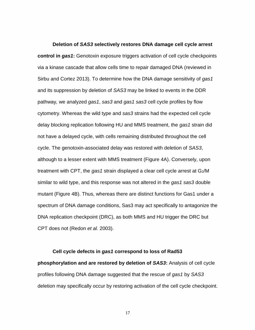
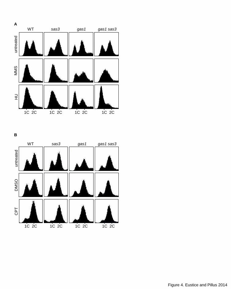
Deletion of SAS3 selectively restores DNA damage cell cycle arrest
control in gas1: Genotoxin exposure triggers activation of cell cycle checkpoints
via a kinase cascade that allow cells time to repair damaged DNA (reviewed in
Sirbu and Cortez 2013). To determine how the DNA damage sensitivity of gas1
and its suppression by deletion of SAS3 may be linked to events in the DDR
pathway, we analyzed gas1, sas3 and gas1 sas3 cell cycle profiles by flow
cytometry. Whereas the wild type and sas3 strains had the expected cell cycle
delay blocking replication following HU and MMS treatment, the gas1 strain did
not have a delayed cycle, with cells remaining distributed throughout the cell
cycle. The genotoxin-associated delay was restored with deletion of SAS3,
although to a lesser extent with MMS treatment (Figure 4A). Conversely, upon
treatment with CPT, the gas1 strain displayed a clear cell cycle arrest at G2/M
similar to wild type, and this response was not altered in the gas1 sas3 double
mutant (Figure 4B). Thus, whereas there are distinct functions for Gas1 under a
spectrum of DNA damage conditions, Sas3 may act specifically to antagonize the
DNA replication checkpoint (DRC), as both MMS and HU trigger the DRC but
CPT does not (Redon et al. 2003).
Cell cycle defects in gas1 correspond to loss of Rad53
phosphorylation and are restored by deletion of SAS3: Analysis of cell cycle
profiles following DNA damage suggested that the rescue of gas1 by SAS3
deletion may specifically occur by restoring activation of the cell cycle checkpoint.
18
One of the initial events following DNA damage in yeast is the phosphorylation of
histone H2A at serine 129, which is indicative of sensing of DNA damage
(reviewed in Rossetto et al. 2010). Downstream of H2AS129 phosphorylation,
the effector kinase Rad53 is hyperphosphorylated, which is largely responsible
for triggering cell cycle delay or arrest (Branzei and Foiani 2006; Sirbu and
Cortez 2013).
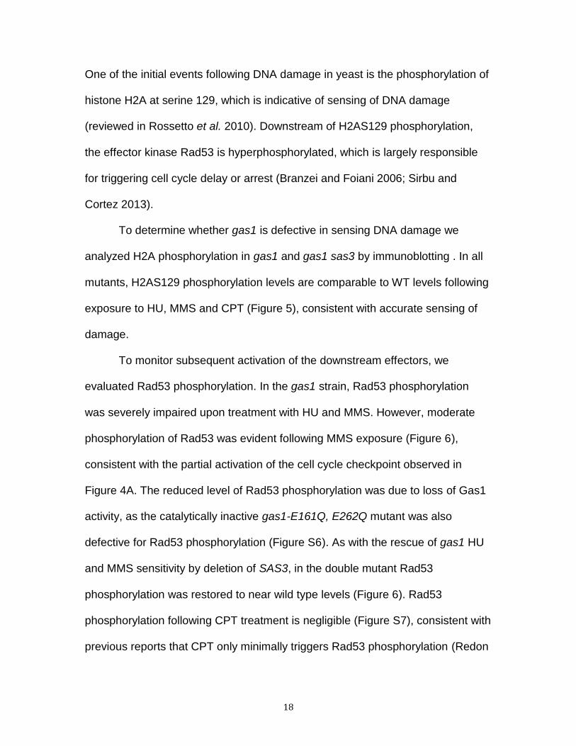
To determine whether gas1 is defective in sensing DNA damage we
analyzed H2A phosphorylation in gas1 and gas1 sas3 by immunoblotting . In all
mutants, H2AS129 phosphorylation levels are comparable to WT levels following
exposure to HU, MMS and CPT (Figure 5), consistent with accurate sensing of
damage.
To monitor subsequent activation of the downstream effectors, we
evaluated Rad53 phosphorylation. In the gas1 strain, Rad53 phosphorylation
was severely impaired upon treatment with HU and MMS. However, moderate
phosphorylation of Rad53 was evident following MMS exposure (Figure 6),
consistent with the partial activation of the cell cycle checkpoint observed in
Figure 4A. The reduced level of Rad53 phosphorylation was due to loss of Gas1
activity, as the catalytically inactive gas1-E161Q, E262Q mutant was also
defective for Rad53 phosphorylation (Figure S6). As with the rescue of gas1 HU
and MMS sensitivity by deletion of SAS3, in the double mutant Rad53
phosphorylation was restored to near wild type levels (Figure 6). Rad53
phosphorylation following CPT treatment is negligible (Figure S7), consistent with
previous reports that CPT only minimally triggers Rad53 phosphorylation (Redon
19
et al. 2003). Together, these data demonstrate that sas3 suppression of gas1 HU
and MMS sensitivity is linked to re-activation of the cell cycle delay via restoration
of Rad53 phosphorylation.
DISCUSSION
Our findings demonstrate that GAS1 has striking yet distinct genetic
interactions with genes encoding the lysine acetyltransferases Gcn5 and Sas3,
which themselves are synthetically lethal, have overlapping nucleosomal
substrates (Howe et al. 2001) and genome-wide localization patterns (Rosaleny
et al. 2007). Whereas the gas1 gcn5 double mutant is dead, there is mutual
suppression of specific phenotypes in the gas1 sas3 strain. The suppression
phenotypes include both silencing defects and specific relief of the newly
identified gas1 sensitivity to genotoxins. The strong genetic interactions with the
acetyltransferases and the DNA damage sensitivity of the gas1 mutant
demonstrate that Gas1 plays an important role in chromatin dynamics, which is
separable from its cell wall function. Further, whereas Gcn5 and Sas3 have often
been considered to be largely functionally overlapping, our results distinguish the
biological roles of Sas3 and Gcn5 in the important process of DNA repair.
Gas1 and Sas3 counter-balance silencing at all three silenced
regions: Previous research indicates that Gas1 and Sas3 contribute to
transcriptional silencing at distinct loci. Whereas loss of SAS3 leads to an
increase in silencing at the HM loci (Reifsnyder et al. 1996), gas1 mutants have

20
impaired silencing at telomeric loci and improved silencing within rDNA (Koch
and Pillus 2009). We demonstrate that deletion of both enzymes leads to
restoration of silencing to wild type levels at all loci analyzed (Figure 2A). Locus-
specific silencing relies on a balance of silencing proteins and other chromatin
factors, some of which are limiting (Smith et al. 1998; Benbow and DuBois 2008).
Altering the distribution of these factors can lead to changes in the strength of
silencing between loci (Lustig et al. 1996). As silencing is both strengthened
and/or disrupted at specific loci in the mutants under study, one potential
explanation for the mutual suppression observed in the gas1 sas3 strain is that
localization of limiting silencing factors is normalized. In this case, Sas3 and
Gas1 counteract the influence of each other, such that in the absence of both
enzymes balance is restored. This idea is in agreement with our previous
observation of a physical interaction between Gas1 and the deacetyltransferase
Sir2 (Koch and Pillus 2009), a limiting factor essential for establishment and
maintenance of silencing (Rusche et al. 2003).
Analysis of DNA damage sensitivity in gas1 cells reveals that Sas3
antagonizes the DNA replication checkpoint: In addition to previously defined
silencing defects (Koch and Pillus 2009) we found that deletion of GAS1 led to
DNA damage sensitivity. Strains lacking Gas1, or with defective catalytic activity,
were sensitive to the genotoxins MMS, HU and CPT but not UV (Figure 3A).
Thus, although Gas1 plays a broad role in DNA damage, there are distinctions
for particular types of damage or repair pathways.

21
Whereas H2AS129 phosphorylation, indicating sensing and initial DDR
activation, was intact in all strains analyzed, the levels of Rad53 phosphorylation
were significantly reduced in gas1 and restored by deletion of SAS3. Impairment
of the HU or MMS DNA damage-associated cell cycle delay and Rad53
phosphorylation levels in gas1 strains (Figures 4; 5) indicates that Gas1 may
function in triggering hyperphosphorylation of Rad53 and the subsequent cell
cycle checkpoint. Although GAS1 mutants failed to arrest in response to HU and
MMS, they did undergo CPT-induced G2/M arrest. These observations
strengthen the idea that Gas1 is broadly relevant to DDR, yet its contributions
appear to depend on the type of lesion.
Distinct mechanistic roles for Gas1 in DNA damage are further supported
by the suppression seen with deletion of SAS3, which rescued HU and MMS
sensitivity but not CPT sensitivity (Figure 3B). MMS and HU elicit a largely
overlapping transcriptional response, which is primarily dependent on Rad53
phosphorylation of substrates. By contrast, CPT leads to induction of a markedly
different set of genes (Travesa et al. 2012; Travesa and Wittenburg 2012). Both
MMS and HU trigger the replication checkpoint via fork arrest or by slowing fork
progression by reducing dNTP pools, respectively (reviewed in Branzei and
Foiani 2007). Conversely, CPT is considered to be “checkpoint blind” as
exposure leads to only modest induction of Rad53 phosphorylation and does not
trigger the replication checkpoint (Redon et al. 2003; Tourriere and Pasero 2007).
The primary checkpoints activated by DNA damage include delay of the
G1/S transition, block of the G2/M transition and the S-phase checkpoints.
22
Although there are overlaps in the proteins mediating these checkpoints there
are also distinctions that depend on the phase of the cell cycle, type of DNA
damage and repair pathway choice (reviewed in Warmerdam and Kanaar 2009;
Symington and Gautier 2011; Gobbini et al. 2013). Cell cycle checkpoints and
DNA damage repair require both positive and negative regulation to ensure
proper spatio-temporal dynamics and maintenance of genomic integrity
(reviewed in Panier and Durocher 2013). Thus, Sas3 may be particularly relevant
in antagonizing activation of the replication checkpoint pathway, specific to the
repression of the cell cycle delay prior to DNA replication mediated by Rad53
phosphorylation.
DNA damage occurs within the context of chromatin, yet the functions of
the chromatin-modifying enzymes and histone post-translational modifications in
the DNA damage response remain incompletely defined (reviewed in
Papamichos-Chronakis and Peterson 2013). Multiple chromatin factors, including
key silencing enzymes, are known to dynamically redistribute from telomeres to
sites of double-strand breaks (Martin et al. 1999; Mills et al. 1999). Further,
silencing at the HM loci was recently found to involve key factors of the
homologous recombination pathway (Kirkland and Kamakaka 2013). If Sas3 and
Gas1 act to balance chromatin-modifying enzymes, as proposed above, the
suppression of gas1 genotoxin sensitivity could relate to redistribution of the
same or similar factors that alter silencing phenotypes in the double mutant.
Indeed, localization of chromatin to the nuclear periphery is linked to both
maintenance of silencing (reviewed in Zimmer and Fabre 2011; Taddei and

23
Gasser 2012) and regulation of the DNA damage response (reviewed in Bermejo
et al. 2012). Thus the pool of Gas1 at the nuclear periphery may be optimally
localized at the interface of both silencing and DDR.
We found that the HHT1-HHF1 locus may, at least in part, mediate the
suppression observed by deletion of SAS3 in the gas1 background (Figure S5A).
The role of histones in the DNA damage response is complex, such that even
modest imbalances in histone levels can alter DNA damage sensitivity (see for
example: Gunjan and Verreault 2003; Sanders et al. 2004; Du et al. 2006).
Whereas the duplicate histone loci are believed to be largely redundant there are
distinctions both at the level of dosage (Cross and Smith 1988; Libuda and
Winston 2010) and in regulation of their expression (Zunder and Rine 2012). Our
findings here and previous work of others (Sanders et al. 2004; Du et al. 2006),
suggests that the HHT1-HHF1 locus may indeed have a unique function in DNA
damage. Histones are highly regulated at multiple levels including expression,
localization, PTM and degradation (reviewed in Kurat et al. 2013). Whether the
restoration of suppression by HHT1-HHF1 is relevant to precise histone levels or
some other aspect of this locus’s biology has yet to be determined.
Distinct functions for Gcn5 and Sas3: Although the function of Gcn5 in
both transcription and DNA damage has been analyzed extensively (Robert et al.
2004; Burgess et al. 2010; Lee et al. 2010), less is known about functions of
Sas3. Several lines of research indicate that Sas3 may have a role in cell cycle
regulation and DDR. Using a sas3 allele with diminished function, it was found
24
that both Gcn5 and Sas3 play a role in cell cycle regulation, with decreased Sas3
activity coupled with deletion of GCN5 leading to G2/M arrest (Howe et al. 2001).
Loss of SAS3 leads to a decrease of H3K14Ac, primarily at genes involved in cell
cycle regulation and cell division (Rosaleny et al. 2007). Sas3 physically interacts
with Chk1 (Liu et al. 2000), a Mec1 DNA damage pathway effector kinase and
Dpb4, which regulates DNA replication and telomere silencing (Tackett et al.
2005). As noted above, Sas3 physically associates with the FACT remodeling
complex via interaction with the N-terminus of Spt16 (John et al. 2000), which is
necessary for the DNA replication stress response (O’Donnell et al. 2004).
Several chromatin-remodeling complexes have been linked to the synthetic
lethality observed between SAS3 and GCN5, including RSC (Choi et al. 2008)
and ISWI (Lafon et al. 2012). Chromatin remodeling complexes have well-
established roles in the DDR, with Gcn5-based acetylation of Rsc4 identified as a
key factor in replication stress resistance (Charles et al. 2011).
Although Sas3 has often been considered to be largely functionally
redundant with Gcn5, previous research indicated that Sas3 can disrupt Gcn5-
based acetylation of H3K14 at distinct genomic loci (Rosaleyn et al. 2007). They
may also compete during other dynamic processes. Whereas Gcn5 has
primarily been implicated as a broad positive regulator of the DNA damage
response, our finding that Sas3 may function antagonistically in DDR further
demonstrates a unique, and opposing, function for Sas3. This possibility is
consistent with the strong yet opposing genetic interactions observed between
GAS1 and GCN5 and SAS3. Future studies should reveal how the protein
25
modifications controlled by these three enzymes are balanced to respond to
distinct forms of cellular and genotoxic stresses.
ACKNOWLEDGMENTS
We thank members of the Pillus lab, Douglass Forbes, Melissa Koch and Christie
Chang for helpful discussion and critical reading of the manuscript. We thank J.
Heierhorst and S. Elledge for anti-Rad53 reagents. M.E. is supported by the
GAANN Fellowship and the Eugene-Cota Robles Fellowship. This project was
initiated with support from NIH GM054778 and GM09177.
LITERATURE CITED
Amberg, D., D. Burke, and J. Strathern, 2005 Methods in yeast genetics: a Cold
Spring Harbor Laboratory course manual. Cold Spring Harbor Laboratory Press,
Cold Spring Harbor, NY.
Baker, S.P., and P.A. Grant, 2007 The SAGA continues: expanding the cellular
role of a transcriptional co-activator complex. Oncogene 26: 5329-5340.
Balasubramanian, R., M. G. Pray-Grant, W. Selleck, P. A. Grant, and S. Tan,
2002 Role of the Ada2 and Ada3 transcriptional coactivators in histone
acetylation. J. Biol. Chem. 277: 7989-7995.
26
Benbow, S. Z., and M. L. Dubois, 2008 The dosage of chromatin proteins affects
transcriptional silencing and DNA repair in Saccharomyces cerevisiae. FEBS
Letters 582: 497-502.
Bermejo, R., A. Kumar, and M. Foiani, 2012 Preserving the genome by
regulating chromatin association with the nuclear envelope. Trends Cell Biol. 22:
465-473.
Bond, J.F., J.L. Fridovich-Keil, L. Pillus, R.C. Mulligan, and F. Solomon, 1986 A
chicken-yeast chimeric β-tubulin protein incorporated into mouse microtubules in
vivo. Cell 44: 461-468.
Branzei, D., and M. Foiani, 2006 The Rad53 signal transduction pathway:
Replication fork stabilization, DNA repair, and adaptation. Exp. Cell Res. 312:
2654-2659.
Branzei, D., and M. Foiani, 2007 Interplay of replication checkpoints and repair
proteins at stalled replication forks. DNA Repair 6: 994-1003.
Burgess, R. J., H. Zhou, J. Han, and Z. Zhang, 2010 Gcn5 in replication-coupled
nucleosome assembly. Mol. Cell 37: 469-480.

27
Campos, E.I., and D. Reinberg, 2009 Histones: annotating chromatin. Annu. Rev.
Genet. 43: 559-599.
Candau, R., J. X. Zhou, C. D. Allis, and S. L. Berger, 1997 Histone
acetyltransferase activity and interaction with ADA2 are critical for GCN5 function
in vivo. EMBO J. 16: 555-565.
Carotti, C., E. Ragni, O. Palomares, T. Fontaine, G. Tedeschi et al., 2004
Characterization of recombinant forms of the yeast Gas1 protein and
identification of residues essential for glucanosyltransferase activity and folding.
Eur. J. Biochem. 271: 3635-3645.
Charles, G. M., C. Chen, S. C. Shih, S. R. Collins, P. Beltrao et al., 2011 Site-
specific acetylation mark on an essential chromatin-remodeling complex
promotes resistance to replication stress. Proc. Natl. Acad. Sci. U S A 108:
10620-10625.
Choi, J. K., D. E. Grimes, K. M. Rowe, and L. Howe, 2008 Acetylation of Rsc4p
by Gcn5p is essential in the absence of histone H3 acetylation. Mol. Cell Biol. 28:
6967-6972.
Choy, J.S., and S.J. Kron, 2002 NuA4 subunit Yng2 function in intra-S-phase
DNA damage response. Mol. Cell Biol. 22: 8215-8225.
28
Clarke, A. S., J. E. Lowell, S. J. Jacobson, and L. Pillus, 1999 Esa1p is an
essential histone acetyltransferase required for cell cycle progression. Mol. Cell
Biol. 19: 2515-2526.
Costanzo, M., A. Baryshnikova, J. Bellay, Y. Kim, E. D. Spear et al., 2010 The
genetic landscape of the cell. Science 327: 425-431.
Cross, S.L., and M.M. Smith, Comparison of the structure and cell cycle
expression of mRNAs encoding two histone H3-H4 loci in Saccharomyces
cerevisiae. Mol. Cell. Biol. 8: 945-954.
Du, L-L., T.M. Nakamura, and P. Russell, 2006 Histone modification-dependent
and independent pathways for recruitment of checkpoint protein Crb2 to double-
strand breaks. Genes & Dev. 20: 1583-1596.
Grant, P. A., L. Duggan, J. Côté, S. M. Roberts, J. E. Brownell et al., 1997 Yeast
Gcn5 functions in two multisubunit complexes to acetylate nucleosomal histones:
characterization of an Ada complex and the SAGA (Spt/Ada) complex. Genes
Dev. 11: 1640-1650.
29
Grant, P. A., D. Schieltz, M. G. Pray-Grant, D. J. Steger, J. C. Reese et al., 1998
A subset of TAF(II)s are integral components of the SAGA complex required for
nucleosome acetylation and transcriptional stimulation. Cell 94: 45-53.
Grant, P. A., A. Eberharter, S. John, R. G. Cook, B. M. Turner et al., 1999
Expanded lysine acetylation specificity of Gcn5 in native complexes. J. Biol.
Chem. 274: 5895-5900.
Gobbini, E., D. Cesena, A. Galbiati, A. Lockhart, and M.P. Longhese, 2013
Interplays between ATM/Tel1 and ATR/Mec1 in sensing and signaling DNA
double-strand breaks. DNA Repair 12: 791-799.
Gunjan, A., and A. Verreault, 2003 A Rad53 kinase-dependent surveillance
mechanism that regulates histone protein levels in S. cerevisiae. Cell 115: 537-
549.
Howe, L., D. Auston, P. Grant, S. John, R. G. Cook et al., 2001 Histone H3
specific acetyltransferases are essential for cell cycle progression. Genes Dev.
15: 3144-3154.
Howe, L., T. Kusch, N. Muster, R. Chaterji, J. R. Yates, 3rd et al., 2002 Yng1p
modulates the activity of Sas3p as a component of the yeast NuA3 histone
acetyltransferase complex. Mol. Cell Biol. 22: 5047-5053.

30
Huh, W. K., J. V. Falvo, L. C. Gerke, A. S. Carroll, R. W. Howson et al., 2003
Global analysis of protein localization in budding yeast. Nature 425: 686-691.
John, S., L. Howe, S. T. Tafrov, P. A. Grant, R. Sternglanz et al., 2000 The
something about silencing protein, Sas3, is the catalytic subunit of NuA3, a
yTAF(II)30-containing HAT complex that interacts with the Spt16 subunit of the
yeast CP (Cdc68/Pob3)-FACT complex. Genes Dev. 14: 1196-1208.
Kirkland, J.G., and R.T. Kamakaka, 2013 Long-range heterochromatin
association is mediated by silencing and double-strand DNA break repair
proteins. J. Cell Biol. 201: 809-826.
Koch, M. R., and L. Pillus, 2009 The glucanosyltransferase Gas1 functions in
transcriptional silencing. Proc. Natl. Acad. Sci. U S A 106: 11224-11229.
Kornberg, R.D., and Y. Lorch, 1999 Twenty-five years of the nucleosome,
fundamental particle of the eukaryote chromosome. Cell 98: 285-294.
Koutelou, E., C.L. Hirsch, and S.Y. Dent, 2010 Multiple faces of the SAGA
complex. Curr. Opin. Cell Biol. 22: 374-382.

31
Kouzarides, T., 2007 Chromatin modifications and their function. Cell 128: 693-
705.
Kuo, Y. M., and A. J. Andrews, 2013 Quantitating the specificity and selectivity of
Gcn5-mediated acetylation of histone H3. PLoS One 8: e54896.
Kurat, C.F., J. Recht, E. Radovani, T. Durbic, B. Andrews, and J. Fillingham,
2013 Regulation of histone gene transcription in yeast. Cell. Mol. Life Sci.
DOI:10.1007/s00018-013-1443-9.
Lafon, A., C. S. Chang, E. M. Scott, S. J. Jacobson, and L. Pillus, 2007 MYST
opportunities for growth control: yeast genes illuminate human cancer gene
functions. Oncogene 26: 5373-5384.
Lafon, A., E. Petty, and L. Pillus, 2012 Functional antagonism between Sas3 and
Gcn5 acetyltransferases and ISWI chromatin remodelers. PLoS Genet. 8:
e1002994.
Le, S., C. Davis, J. B. Konopka, and R. Sternglanz, 1997 Two new S-phase-
specific genes from Saccharomyces cerevisiae. Yeast 13: 1029-1042.

32
Lee, H. S., J. H. Park, S. J. Kim, S. J. Kwon, and J. Kwon, 2010 A cooperative
activation loop among SWI/SNF, gamma-H2AX and H3 acetylation for DNA
double-strand break repair. EMBO J. 29: 1434-1445.
Lee, K. K., M. E. Sardiu, S. K. Swanson, J. M. Gilmore, M. Torok et al., 2011
Combinatorial depletion analysis to assemble the network architecture of the
SAGA and ADA chromatin remodeling complexes. Mol. Syst. Biol. 7: 503.
Lee, K.K., and J. Workman, 2007 Histone acetyltransferase complexes: one size
doesn't fit all. Nat. Rev. Mol. Cell Biol. 8:284-95.
Levin, D. E., 2005 Cell wall integrity signaling in Saccharomyces cerevisiae.
Microbiol. Mol. Biol. Rev. 69: 262-291.
Li, G., and D. Reinberg, 2011 Chromatin higher-order structures and gene
regulation. Curr. Opin. Genet. Dev. 21: 175-186.
Liang, B., J. Qiu, K. Ratnakumar, and B. C. Laurent, 2007 RSC functions as an
early double-strand-break sensor in the cell's response to DNA damage. Curr.
Biol. 17: 1432-1437.
33
Libuda, D.E., and F. Winston, 2010 Alterations in DNA replication and histone
levels promote histone gene amplificaition in Saccharomyces cerevisiae.
Genetics 184: 985-997.
Liu, Y., G. Vidanes, Y. C. Lin, S. Mori, and W. Siede, 2000 Characterization of a
Saccharomyces cerevisiae homologue of Schizosaccharomyces pombe Chk1
involved in DNA-damage-induced M-phase arrest. Mol. Gen. Genet. 262: 1132-
1146.
Lustig, A. J., C. Liu, C. Zhang, and J. P. Hanish, 1996 Tethered Sir3p nucleates
silencing at telomeres and internal loci in Saccharomyces cerevisiae. Mol. Cell
Biol. 16: 2483-2495.
Martin, D. G., K. Baetz, X. Shi, K. L. Walter, V. E. MacDonald et al., 2006 The
Yng1p plant homeodomain finger is a methyl-histone binding module that
recognizes lysine 4-methylated histone H3. Mol. Cell Biol. 26: 7871-7879.
Martin, S. G., T. Laroche, N. Suka, M. Grunstein, and S. M. Gasser, 1999
Relocalization of telomeric Ku and SIR proteins in response to DNA strand
breaks in yeast. Cell 97: 621-633.
34
Mills, K. D., D. A. Sinclair, and L. Guarente, 1999 MEC1-dependent redistribution
of the Sir3 silencing protein from telomeres to DNA double-strand breaks. Cell
97: 609-620.
Nitiss, J., and J. C. Wang, 1988 DNA topoisomerase-targeting antitumor drugs
can be studied in yeast. Proc. Natl. Acad. Sci. U S A 85: 7501-7505.
O'Donnell, A. F., N. K. Brewster, J. Kurniawan, L. V. Minard, G. C. Johnston et al.,
2004. Domain organization of the yeast histone chaperone FACT: the conserved
N-terminal domain of FACT subunit Spt16 mediates recovery from replication
stress. Nucleic Acids Res. 32: 5894-5906.
Orlean, P., 2012 Architecture and biosynthesis of the Saccharomyces cerevisiae
cell wall. Genetics 192: 775-818.
Panier, S., and D. Durocher, 2013 Push back to respond better: regulatory
inhibition of the DNA double-strand break response. Nat. Rev. Cancer 13: 661-
672.
Papamichos-Chronakis, M., and C.L. Peterson, 2013 Chromatin and the genome
integrity network. Nat. Rev. Genet. 14: 62-75.
35
Pike, B.L., S. Yongkiettrakul, M.D. Tsai, and J. Heierhorst, 2003 Diverse but
overlapping functions of the two forkhead-associated (FHA) domains in Rad53
checkpoint kinase activation. J. Biol. Chem. 278: 30421-30424.
Pokholok, D. K., C. T. Harbison, S. Levine, M. Cole, N. M. Hannett et al., 2005
Genome-wide map of nucleosome acetylation and methylation in yeast. Cell 122:
517-527.
Popolo, L., and M. Vai, 1999 The Gas1 glycoprotein, a putative wall polymer
cross-linker. Biochim. Biophys. Acta. 1426: 385-400.
Pray-Grant, M. G., D. Schieltz, S. J. McMahon, J. M. Wood, E. L. Kennedy et al.,
2002 The novel SLIK histone acetyltransferase complex functions in the yeast
retrograde response pathway. Mol. Cell Biol. 22: 8774-8786.
Qin, S., and M. R. Parthun, 2002 Histone H3 and the histone acetyltransferase
Hat1p contribute to DNA double-strand break repair. Mol. Cell Biol. 22: 8353-
8365.
Ragni, E., T. Fontaine, C. Gissi, J. P. Latge, and L. Popolo, 2007 The Gas family
of proteins of Saccharomyces cerevisiae: characterization and evolutionary
analysis. Yeast 24: 297-308.
36
Redon, C., D. R. Pilch, E. P. Rogakou, A. H. Orr, N. F. Lowndes et al., 2003
Yeast histone 2A serine 129 is essential for the efficient repair of checkpoint-
blind DNA damage. EMBO Rep. 4: 678-684.
Reifsnyder, C., J. Lowell, A. Clarke, and L. Pillus, 1996 Yeast SAS silencing
genes and human genes associated with AML and HIV-1 Tat interactions are
homologous with acetyltransferases. Nat. Genet. 14: 42-49.
Renauld, H., O. M. Aparicio, P. D. Zierath, B. L. Billington, S. K. Chhablani et al.,
1993 Silent domains are assembled continuously from the telomere and are
defined by promoter distance and strength, and by SIR3 dosage. Genes Dev. 7:
1133-1145.
Robert, F., D. K. Pokholok, N. M. Hannett, N. J. Rinaldi, M. Chandy et al., 2004
Global position and recruitment of HATs and HDACs in the yeast genome. Mol.
Cell 16: 199-209.
Roncero, C., and A. Duran, 1985 Effect of Calcofluor white and Congo red on
fungal cell wall morphogenesis: in vivo activation of chitin polymerization. J.
Bacteriol. 163: 1180-1185.
37
Rosaleny, L. E., A. B. Ruiz-Garcia, J. Garcia-Martinez, J. E. Perez-Ortin, and V.
Tordera, 2007 The Sas3p and Gcn5p histone acetyltransferases are recruited to
similar genes. Genome Biol. 8: R119.
Rossetto, D., A.W. Truman, S.J. Kron, and J. Côté, 2010 Epigenetic
modifications in double-strand break DNA damage signaling and repair. Clin.
Cancer Res. 16:4543-4552.
Rusche, L.N., A.L. Kirchmaier, and J. Rine, 2003 The establishment, inheritance,
and funciton of silenced chromatin in Saccharomyces cerevisiae. Annu. Rev.
Biochem. 72: 481-516.
Sanders, S.L., M. Portoso, J. Mata, J. Bahler, R.C. Allshire, and T. Kouzarides
2004 Methylation of histone H4 lysine 20 controls recruitment of Crb2 to sites of
DNA damage. Cell 119:603-614.
Sertic, S., S. Pizzi, F. Lazzaro, P. Plevani, and M. Muzi-Falconi, 2012 NER and
DDR: classical music with new instruments. Cell Cycle 11: 668-674.
Sirbu, B. M., and D. Cortez, 2013 DNA damage response: three levels of DNA
repair regulation. Cold Spring Harb. Perspect. Biol. 5.
38
Smith, J. S., and J. D. Boeke, 1997 An unusual form of transcriptional silencing in
yeast ribosomal DNA. Genes Dev. 11: 241-254.
Smith, J. S., C. B. Brachmann, L. Pillus, and J. D. Boeke, 1998 Distribution of a
limited Sir2 protein pool regulates the strength of yeast rDNA silencing and is
modulated by Sir4p. Genetics 149: 1205-1219.
Suter, B., A. Tong, M. Chang, L. Yu, G.W. Brown, C. Boone, and J. Rine, 2004
The origin recognition complex links replication, sister chromatid cohesion and
transcriptional silencing in Saccharomyces cerevisiae. Genetics 167: 579-591.
Symington, L.S., and J. Gautier, 2011 Double-strand break end resection and
repair pathway choice. Annu. Rev. Genet. 45: 247-271.
Tackett, A. J., D. J. Dilworth, M. J. Davey, M. O'Donnell, J. D. Aitchison et al.,
2005 Proteomic and genomic characterization of chromatin complexes at a
boundary. J. Cell Biol. 169: 35-47.
Taddei, A., and S. M. Gasser, 2012 Structure and function in the budding yeast
nucleus. Genetics 192: 107-129.

39
Tamburini, B. A., and J. K. Tyler, 2005 Localized histone acetylation and
deacetylation triggered by the homologous recombination pathway of double-
strand DNA repair. Mol. Cell Biol. 25: 4903-4313.
Tourriere, H., and P. Pasero, 2007 Maintenance of fork integrity at damaged
DNA and natural pause sites. DNA Repair 6: 900-913.
Travesa, A., D. Kuo, R. A. de Bruin, T. I. Kalashnikova, M. Guaderrama et al.,
2012 DNA replication stress differentially regulates G1/S genes via Rad53-
dependent inactivation of Nrm1. EMBO J. 31: 1811-1822.
Travesa, A., and C. Wittenberg, 2012 Turned on by genotoxic stress. Cell Cycle
11: 3145-3146.
Turchini, A., L., Ferrario, and L. Popolo, 2000 Increase of external osmolarity
reduces morphogenetic defects and accumulaiton of chitin in a gas1 mutant of
Saccharomyces cerevisiae. J. Bacteriol. 182: 1167-71.
van Leeuwen, F., and D. E. Gottschling, 2002 Assays for gene silencing in yeast.
Methods Enzymol. 350: 165-186.
40
Wang, L., L. Liu, and S. L. Berger, 1998 Critical residues for histone acetylation
by Gcn5, functioning in Ada and SAGA complexes, are also required for
transcriptional function in vivo. Genes Dev. 12: 640-653.
Wang, Y., S. P. Kallgren, B. D. Reddy, K. Kuntz, L. Lopez-Maury et al., 2012
Histone H3 lysine 14 acetylation is required for activation of a DNA damage
checkpoint in fission yeast. J. Biol. Chem. 287: 4386-4393.
Warmerdam, D.O., and R. Kanaar, 2010 Dealing with DNA damage:
relationships between checkpoint and repair pathways. Mutat. Res. 704: 2-11.
Zimmer, C., and E. Fabre, 2011 Principles of chromosomal organization: lessons
from yeast. J. Cell Biol. 192: 723-733.
Zunder, R.M., and J. Rine, 2012 Direct interplay among histones, histone
chaperones, and a chromatin boundary protein in the control of histone gene
expression. Mol. Cell. Biol. 32: 4337-4349.
FIGURE LEGENDS
Figure 1. The gas1 gcn5 double mutant is synthetically lethal. (A) Synthetic lethality of gas1 gcn5
is due to loss of catalytic activity of both Gas1 and Gcn5 and is not rescued by the
osomoregulator sorbitol. Serial dilutions of wild type (LPY18050), gcn5 (LPY12264), gas1
(LPY18081), gas1 gcn5 (LPY16798) and gas1 gcn5 covered by plasmid-born p-gcn5-KQL
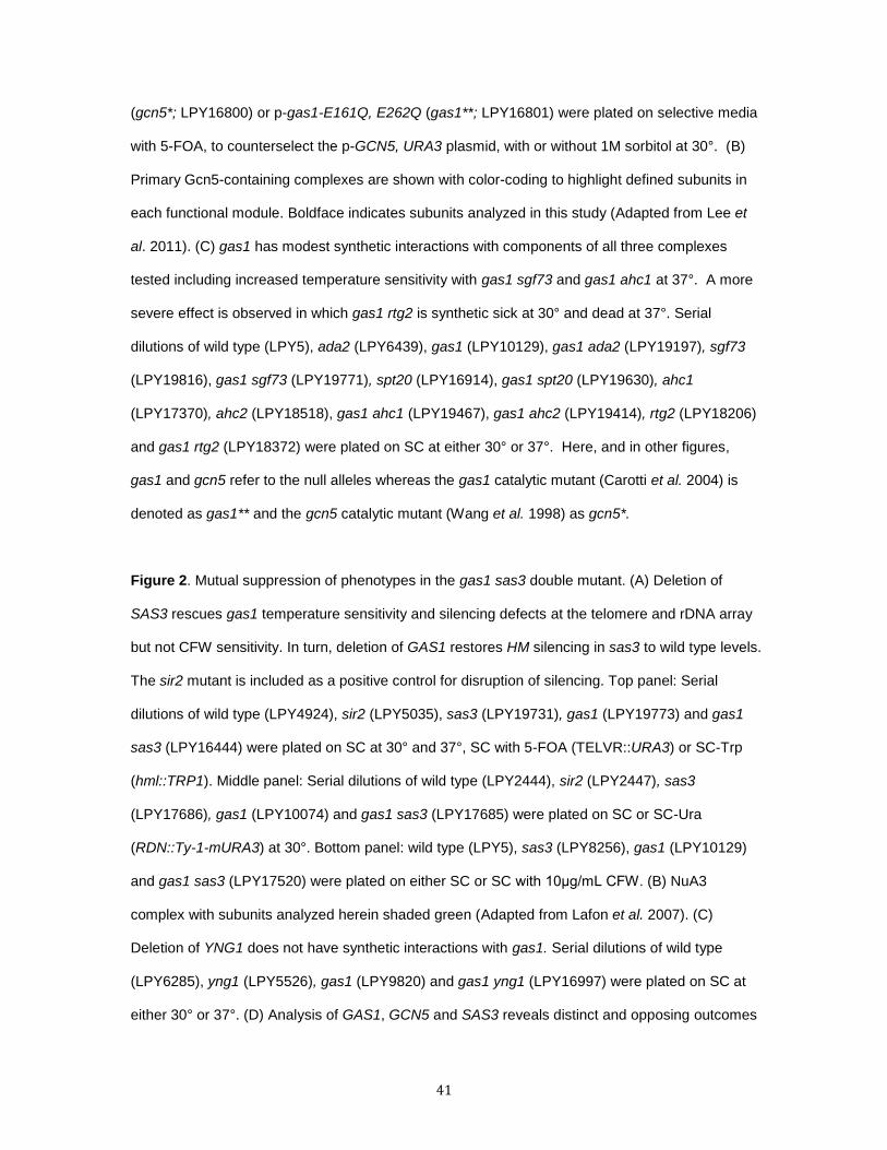
41
(gcn5*; LPY16800) or p-gas1-E161Q, E262Q (gas1**; LPY16801) were plated on selective media
with 5-FOA, to counterselect the p-GCN5, URA3 plasmid, with or without 1M sorbitol at 30°. (B)
Primary Gcn5-containing complexes are shown with color-coding to highlight defined subunits in
each functional module. Boldface indicates subunits analyzed in this study (Adapted from Lee et
al. 2011). (C) gas1 has modest synthetic interactions with components of all three complexes
tested including increased temperature sensitivity with gas1 sgf73 and gas1 ahc1 at 37°. A more
severe effect is observed in which gas1 rtg2 is synthetic sick at 30° and dead at 37°. Serial
dilutions of wild type (LPY5), ada2 (LPY6439), gas1 (LPY10129), gas1 ada2 (LPY19197), sgf73
(LPY19816), gas1 sgf73 (LPY19771), spt20 (LPY16914), gas1 spt20 (LPY19630), ahc1
(LPY17370), ahc2 (LPY18518), gas1 ahc1 (LPY19467), gas1 ahc2 (LPY19414), rtg2 (LPY18206)
and gas1 rtg2 (LPY18372) were plated on SC at either 30° or 37°. Here, and in other figures,
gas1 and gcn5 refer to the null alleles whereas the gas1 catalytic mutant (Carotti et al. 2004) is
denoted as gas1** and the gcn5 catalytic mutant (Wang et al. 1998) as gcn5*.
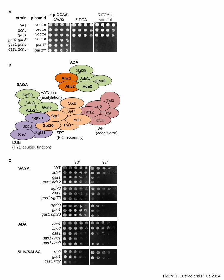
Figure 2. Mutual suppression of phenotypes in the gas1 sas3 double mutant. (A) Deletion of
SAS3 rescues gas1 temperature sensitivity and silencing defects at the telomere and rDNA array
but not CFW sensitivity. In turn, deletion of GAS1 restores HM silencing in sas3 to wild type levels.
The sir2 mutant is included as a positive control for disruption of silencing. Top panel: Serial
dilutions of wild type (LPY4924), sir2 (LPY5035), sas3 (LPY19731), gas1 (LPY19773) and gas1
sas3 (LPY16444) were plated on SC at 30° and 37°, SC with 5-FOA (TELVR::URA3) or SC-Trp
(hml::TRP1). Middle panel: Serial dilutions of wild type (LPY2444), sir2 (LPY2447), sas3
(LPY17686), gas1 (LPY10074) and gas1 sas3 (LPY17685) were plated on SC or SC-Ura
(RDN::Ty-1-mURA3) at 30°. Bottom panel: wild type (LPY5), sas3 (LPY8256), gas1 (LPY10129)
and gas1 sas3 (LPY17520) were plated on either SC or SC with 10μg/mL CFW. (B) NuA3
complex with subunits analyzed herein shaded green (Adapted from Lafon et al. 2007). (C)
Deletion of YNG1 does not have synthetic interactions with gas1. Serial dilutions of wild type
(LPY6285), yng1 (LPY5526), gas1 (LPY9820) and gas1 yng1 (LPY16997) were plated on SC at
either 30° or 37°. (D) Analysis of GAS1, GCN5 and SAS3 reveals distinct and opposing outcomes

42
for synthetic interactions. Serial dilutions of wild type (LPY5), gcn5 (LPY8242), sas3 (LPY16039),
gas1 (LPY10129), gas1 gcn5 + p-GCN5, URA3 (LPY16736), gas1 sas3 (LPY19823), gas1 gcn5
sas3 + p-GCN5, URA3 (LPY19101) were plated on SC or SC with 5-FOA, to select against p-
GCN5, URA3, at 30° with and without 1M sorbitol.
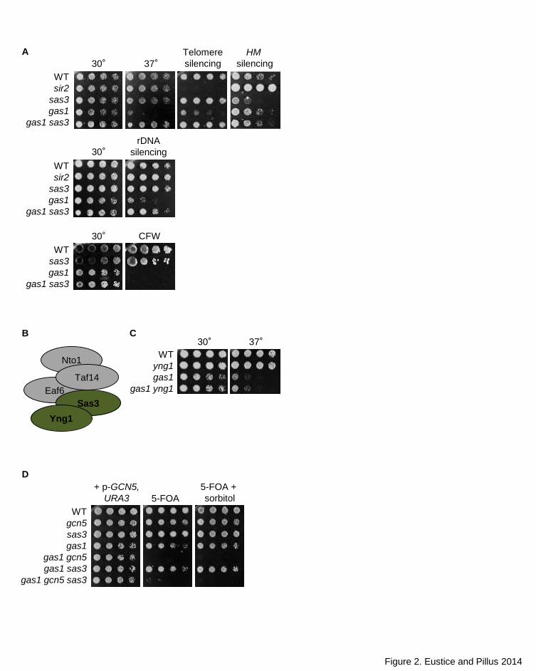
Figure 3. Loss of GAS1 leads to broad DNA damage sensitivity with phenotype-specific
suppression by deletion of SAS3. (A) gas1 mutants are sensitive to MMS, CPT and HU but not
exposure to UV. Sensitivity is due to loss of Gas1 catalytic activity and separable from cell wall
function as demonstrated by failure of sorbitol to rescue these phenotypes. Serial dilutions of wild
type (LPY18050), gas1 (LPY12247), gas1 + p-gas1-E161Q, E262Q (gas1**; LPY12251) and
gas1 + p-GAS1 (LPY12326) were plated on selective media with 0.015% MMS, 0.2M HU or
20μg/mL CPT in DMSO with or without 1M sorbitol or on SC buffered with phosphate and
supplemented with DMSO as a control. UV exposure was 60J/m2. (B) Deletion of SAS3
specifically suppressed the MMS and HU sensitivity of gas1, but not CPT sensitivity. Serial
dilutions of wild type (LPY5), sas3 (LPY8256), gas1 (LPY10129) and gas1 sas3 (LPY17520)
were plated on SC plates using the same concentration of genotoxins and plate conditions as in
3A.
Figure 4. Deletion of SAS3 rescues gas1 defects in cell cycle arrest. (A) Treatment of gas1 with
HU fails to trigger the cell cycle delay observed in wild type whereas the cell cycle delay following
treatment with MMS is severely impaired in gas1. Cycle delay is significantly restored in the
double mutant gas1 sas3. (B) CPT treatment triggers cell cycle arrest in all strains tested. Strains
and genotoxin concentrations are as in Figure 3A.
Figure 5. H2AS129 is phosphorylated following genotoxin exposure in all strains. Levels of
H2AS129 phosphorylation following exposure to HU (top), MMS (middle) and CPT (bottom) are
comparable to wild type in all strains analyzed. Strains and genotoxin concentrations are as in
Figure 3A.

43
Figure 6. Rad53 phosphorylation is significantly reduced in gas1 and restored in gas1 sas3
Following exposure to HU (top) and MMS (bottom). Note that overall levels of Rad53 are
diminished in gas1. Strains and genotoxin concentrations are as in Figure 3A.
Taf5
Taf9
Taf6
Taf10
Taf12
5-FOA
5-FOA +
sorbitol
vector
vector
vector
vector
gcn5*
gas1**
A
C 30° 37°
sgf73
gas1
gas1 sgf73
spt20
gas1
gas1 spt20
rtg2
gas1
gas1 rtg2
ahc1
ahc2
gas1
gas1 ahc1
gas1 ahc2
SAGA
SLIK/SALSA
ADA
Figure 1. Eustice and Pillus 2014
WT
ada2
gas1
gas1 ada2
strain plasmid
WT
gcn5
gas1
gas1 gcn5
gas1 gcn5
gas1 gcn5
+ p-GCN5,
URA3
B
Sgf29
Ada3
Ada2Gcn5
Ubp8 Tra1
Spt7
Sgf11Sus1
Spt20
Spt3Ada1
Spt8
HAT/core
(acetylation)
DUB
(H2B deubiquitination)
SPT
(PIC assembly)
SAGA
ADA
TAF
(coactivator)
Sgf73
Sgf29
Ada3Gcn5
Ahc1
Ahc2 Ada2
Eaf6
CFW
WT
sir2
sas3
gas1
gas1 sas3
rDNA
silencing
30° 37°Telomere
silencing
30°
WT
sir2
sas3
gas1
gas1 sas3
30°
WT
sas3
gas1
gas1 sas3
HM
silencing
A
30° 37°
WT
yng1
gas1
gas1 yng1
B C
Figure 2. Eustice and Pillus 2014
WT
gcn5
sas3
gas1
gas1 gcn5
gas1 sas3
gas1 gcn5 sas3
5-FOA
5-FOA +
sorbitol
+ p-GCN5,
URA3
D
Nto1
Taf14
Sas3
Yng1
Figure 3. Eustice and Pillus 2014
MMS30° HU
DMSO CPT
WT
sas3
gas1
gas1 sas3
WT
sas3
gas1
gas1 sas3
B
MMS
MMS +
sorbitol
UV
WT
gas1
gas1
gas1
HU
HU +
sorbitol
CPT
CPT +
sorbitolDMSO
30°
A
strain plasmid
vector
vector
gas1**
GAS1
WT
gas1
gas1
gas1
vector
vector
gas1**
GAS1
WT
gas1
gas1
gas1
vector
vector
gas1**
GAS1
WT
gas1
gas1
gas1
vector
vector
gas1**
GAS1
WT sas3 gas1 gas1 sas3
untr
eate
dH
UM
MS
A
untr
eate
dC
PT
DM
SO
B
WT sas3 gas1 gas1 sas3
Figure 4. Eustice and Pillus 2014
1C 2C 1C 2C 1C 2C 1C 2C
1C 2C 1C 2C 1C 2C 1C 2C
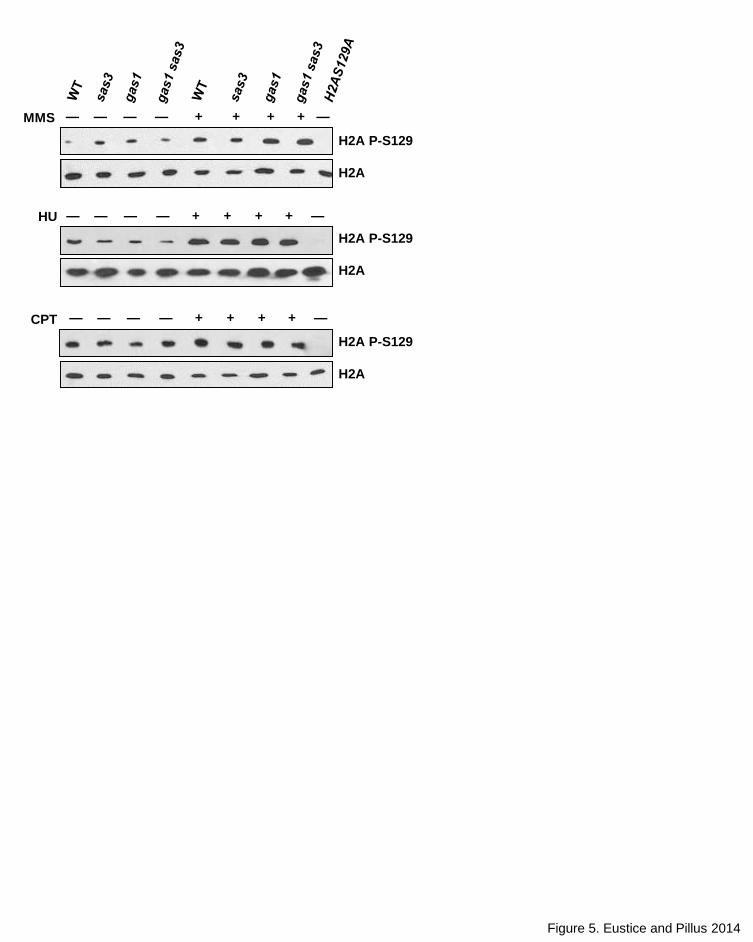
Figure 5. Eustice and Pillus 2014
— +— +— +— + —
H2A P-S129
H2A
MMS
HU — +— +— +— + —
CPT
H2A P-S129
H2A
— +— +— +— + —
H2A P-S129
H2A
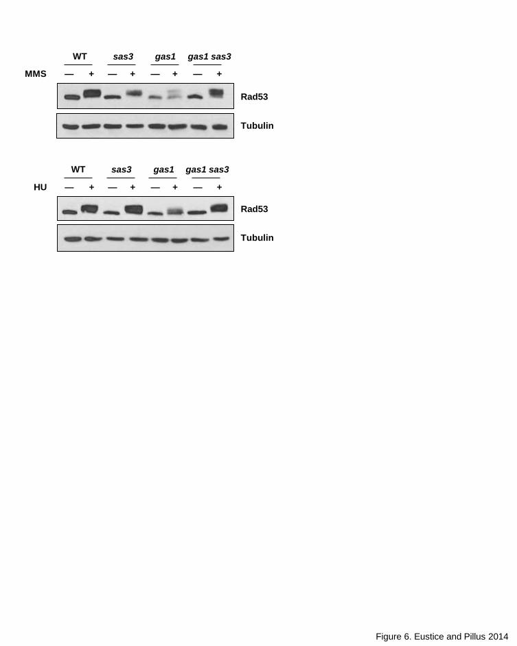
Figure 6. Eustice and Pillus 2014
— + — ++ +
Rad53
HU
Tubulin
Rad53
Tubulin
— —
gas1
— +
gas1 sas3
— ++ +MMS
WT sas3
— —
gas1 gas1 sas3WT sas3