Embed Size (px)
Citation preview
For the past 60 years, human genetic research has focused on DNA as the heritable molecule that carries information about phenotype from the parent to the offspring. Mutations in single genes or a small number of genes have been tightly linked to some phenotypes, but for most phenotypes the situation is more complex and, in many cases, environmental factors are involved. In these instances, genome-wide association studies (GWASs) have enabled the identification of SNPs that are weakly associated with increased disease risk, but the odds ratios are generally small, and it remains impossible to predict phenotype at an individual level.
In parallel, molecular biologists using animal models have realized that, in addition to DNA sequence, there are a number of other layers of information, termed epi-genetic marks (BOX 1), that influence transcription. These epigenetic marks are fairly stable over the lifetime of an individual and have a role in determining phenotype. At some loci, the epigenetic marks are not tightly linked to the DNA sequence of the genome; both probabilistic and environmental events can influence the establishment of epigenetic states at these loci1. Considering the epige-nome as well as the genome may allow us to develop bet-ter tools for predicting phenotype at an individual level.
Moreover, there is evidence that epigenetic marks can sometimes be transmitted from parent to offspring via the gametes, and studies have been published in the past couple of years that support this idea. In this Review, we describe the evidence for this form of inheritance, focusing on mammals but also looking at informative examples from other species. The molecular nature of
the epigenetic marks that are inherited is unknown in most cases, but the recent emergence of high-throughput sequencing technologies makes this problem tractable. An emerging theme in cases of transgenerational epige-netic inheritance via the gametes (BOX 1) is the involve-ment of repeats and transposable elements, and recent progress in our understanding of the establishment of heterochromatin at repeats reveals the importance of RNA; this raises the possibility that RNA may have a role in transgenerational epigenetic inheritance via the gametes.
Evidence in mammalsReprogramming of the epigenome. The epigenetic marks that are established in most tissues during an organism’s lifetime are irrelevant with respect to the next genera-tion. Only those of the mature gametes have the poten-tial to contribute to the phenotype of the offspring. Moreover, there is considerable reprogramming between generations — and, in particular, of the gametic epi-genome immediately after fertilization — to endow the cells of the early pre-implantation embryo with the capacity to differentiate into all cell types of a fully developed organism. Studies carried out in mice more than 30 years ago found that global DNA methylation levels, which were analysed using methylation-sensitive restriction enzymes, were much lower just after fertili-zation compared to those found in mature gametes and after implantation2. The idea that DNA methylation erasure and resetting is the basis of epigenetic repro-gramming emerged from this finding2. However, our understanding of the function of DNA methylation
Queensland Institute of Medical Research, Herston, Queensland 4006, Australia.Correspondence to E.W. e-mail: [email protected]:10.1038/nrg3188Published online 31 January 2012
HeterochromatinThe portion of the genome that stays highly condensed throughout the cell cycle. It contains a high proportion of repetitive sequences, is gene-poor overall and is enriched for histone marks, such as histone H3 lysine 9 trimethylation (H3K9me3) and H4K20me3, as well as DNA methylation. Heterochromatin is generally associated with gene silencing.
Understanding transgenerational epigenetic inheritance via the gametes in mammalsLucia Daxinger and Emma Whitelaw
Abstract | It is known that information that is not contained in the DNA sequence — epigenetic information — can be inherited from the parent to the offspring. However, many questions remain unanswered regarding the extent and mechanisms of such inheritance. In this Review, we consider the evidence for transgenerational epigenetic inheritance via the gametes, including cases of environmentally induced epigenetic changes. The molecular basis of this inheritance remains unclear, but recent evidence points towards diffusible factors, in particular RNA, rather than DNA methylation or chromatin. Interestingly, many cases of epigenetic inheritance seem to involve repeat sequences.
REVIEWS
NATURE REVIEWS | GENETICS VOLUME 13 | MARCH 2012 | 153
© 2012 Macmillan Publishers Limited. All rights reserved
Histone modificationsCovalent alterations of histone tail residues that can alter chromatin structure. Modifications include phosphorylation, methylation, acetylation, sumoylation and ubiquitylation.
remains poor, and the common assumption that the sole role of DNA methylation is to control transcrip-tion no longer stands up to critical review (BOX 2). We now know of many other epigenetic marks, such as histone modifications, that could have an equally or more important role in transcriptional control, but the techni-cal difficulty of analysing histone marks in small tissue samples, such as pre-implantation embryos, has limited progress in understanding their reprogramming. The crucial question in relation to transgenerational epige-netic inheritance via the gametes is whether or not there are parts of the epigenome, either DNA methylation or chromatin, that are instructive (that is, they drive tran-scription) and that at which the classic intergenerational reprogramming does not occur.
Parental imprinting. The first evidence for epigenetic marks that escape reprogramming in the early developing embryo came from the discovery of parental imprint-ing in mice3–5. A small group of genes was discovered with monoallelic expression that is dependent on the
parent-of-origin of the allele. The maternal and paternal alleles of these genes are in different transcriptional states in the cells of the adult. This implies that differ-ent epigenetic states are established in the germline of the parents, inherited via the gametes and remembered across millions of cell divisions. Recent evidence sug-gests that the number of genes that are subject to some form of imprinting, previously thought to be around 100, may be much greater, at least in the brain6,7.
At parentally imprinted genes, the alleles must undergo reprogramming each generation in the ger-mline, depending on the sex of the individual. In other words, the memory only lasts one generation. For this reason, parental imprinting is not normally consid-ered to be an example of transgenerational epigenetic inheritance via the gametes.
Non-Mendelian patterns of expression at transgenes. The original evidence that epigenetic information could be inherited through the gametes across more than one generation in mammals involved transgenes8–13. These studies reported that the transcriptional activity of some transgenes in mice was variable among inbred litter-mates and that the likelihood of activity was sometimes, and to some extent, inherited to the next generation (FIG. 1a). In other words, the offspring of mice in which the transgene was active were more likely to have an active transgene. Because these experiments were car-ried out in inbred backgrounds, an epigenetic mecha-nism was inferred. In some of these cases, transcriptional activity of the transgenes inversely correlated with DNA methylation levels at the transgene promoter9,12,14. Transgenes generally insert into the genome as multi-copy arrays, and the larger the array, the greater the probability that the transgene will be transcriptionally silent, methylated and packaged into heterochromatin15.
It is interesting that, in a number of instances, the transgenes that show transgenerational epigenetic inher-itance via the gametes also show a degree of imprint-ing8,9,11 — that is, expression status is influenced by the parent-of-origin of the allele as well as by the activity state of the locus in the parent. For example, the hepa-titis B surface antigen transgene stays unmethylated and active when it has been paternally inherited, but it is methylated and silenced when it has been mater-nally inherited. Importantly, this silenced state cannot be reversed even when it has subsequently been passed through the male germline8, and it is this that makes the behaviour of the hepatitis B surface antigen transgene different from classic parental imprinting. The fact that many of the transgenes that display transgenerational epigenetic inheritance also display some degree of gametic imprinting may help us to unravel the mecha-nisms that are involved in transgenerational epigenetic inheritance via the gametes.
Non-Mendelian patterns of expression at endogenous genes. Non-Mendelian patterns of expression have also been reported at endogenous genes, but only rarely. The best-characterized endogenous allele at which transgen-erational epigenetic inheritance via the gametes has
Box 1 | Working definitions regarding epigenetics and epigenetic inheritance
Here we provide the definitions of key terms that we use in this Review. For some of these terms, alternative definitions may have been proposed elsewhere.
EpigeneticsThis is the study of changes in gene expression that occur in the absence of changes in DNA sequence and that are fairly stable across the life of an individual. The term was first coined by Conrad Waddington to describe the fact that there must be something ‘over and above’ genetics that enables cells with the same genetic information to differentiate into various different cell types during development. At the time, there was no knowledge of the molecules involved. The problem with this definition is that it is based on what it is not: in this case, it is ‘not the primary DNA sequence’. As the molecular nature of epigenetic processes becomes clearer, the word is likely to be replaced by more specific descriptors.
Epigenetic marksThese are molecular modifications to the DNA, such as the methylation of cytosine residues, or modifications of proteins that are associated with DNA, such as methylation of histones. Epigenetic marks are often, but not always, associated with changes in transcriptional activity. Originally, these modifications were called ‘epigenetic marks’ because they were found to underlie epigenetic phenomena (see above). We now know that some of these modifications are not as stable as was previously thought. For example, the pattern of cytosine methylation in the coding region of some genes changes during the cell cycle115,116 (BOX 2); histone acetylation is extremely dynamic, and the acetylation state changes within the 2 hours of deposition of the acetyl group117. RNA is not usually considered to be an epigenetic mark, but it has been found to underlie some epigenetic processes.
Transgenerational (or intergenerational) epigenetic effectsThese are regarded as effects on the phenotype (or on patterns of gene expression) that are detected across more than one generation and that cannot be explained by Mendelian genetics (or changes to the primary DNA sequence). This includes the effects of environmental exposures on adults that alter the phenotype of the developing embryo via the placenta or the newborn via the milk.
Transgenerational epigenetic inheritance via the gametesThis term refers to effects on phenotype (or on patterns of gene expression) that are passed from one generation to the next by molecules in the germ cells and that cannot be explained by Mendelian genetics (or by changes to the primary DNA sequence). We do not restrict the molecules to those that are usually regarded as ‘epigenetic marks’ but include RNA and proteins. The clause ‘via the gametes’ emphasizes the difference between this phenomenon and the effects of environmental exposure that alters phenotype via the placenta, and so on (see above).
R E V I E W S
154 | MARCH 2012 | VOLUME 13 www.nature.com/reviews/genetics
© 2012 Macmillan Publishers Limited. All rights reserved
Intracisternal A particle(IAP). A long terminal repeat (LTR)-containing retrotransposon of mice that resembles a retrovirus but has a defective env gene.
GenisteinAn isoflavone found in plants that acts as an antioxidant and binds to the oestrogen receptor, hence its classification as a phytoestrogen.
been reported is agouti viable yellow (Avy)16. The Avy allele arose 50 years ago as a result of an intracisternal A particle (IAP) retrotransposon insertion upstream of the agouti locus. Although the Avy locus is unique in the mouse genome, the IAP element is present in thousands of copies. The agouti protein indirectly results in yellowness of the coat and, if it is overproduced, it has some other phenotypic consequences, including obesity. Inbred mice that carry this allele show variable coat col-ours, ranging from yellow to mottled (yellow and brown patches) to pseudoagouti (brown)17,18. At the Avy locus, transcriptional control of the agouti coding sequence is driven by promoter elements in the retrotransposon18. The DNA methylation state of the IAP promoter at Avy inversely correlates with transcriptional activity16. More recently, histone marks that are associated with tran-scriptional activity have been reported at the locus in yellow mice, and those that are associated with silencing have been reported at the locus in pseudoagouti mice19. The range of coat colours in offspring is unaffected by the coat colour of the sire following transmission of Avy through the male germline, suggesting that the epige-netic marks are cleared following passage through the male germline16 (FIG. 1b). Following transmission of Avy through the female, yellow dams produce a higher per-centage of yellow offspring than pseudoagouti dams do16 (FIG. 1b). This suggests that there is a failure to clear the epigenetic marks that are established at the Avy locus in the germline of the dam: that is, there is transgen-erational epigenetic inheritance via the female gamete.
Because transgenerational epigenetic inheritance was only seen following maternal transmission of the Avy allele, there was a need to rule out maternal effects that could occur after fertilization, so fertilized eggs were removed from yellow dams and transferred to
pseudopregnant pseudoagouti dams16. The higher percentage of yellow offspring was still seen. We can conclude that some factor in the egg of yellow moth-ers is different to that of pseudoagouti mothers and is responsible. In the past, we have assumed that this is the chromatin or the DNA methylation state at the Avy locus, but it could be some diffusible factor in the cytoplasm.
Another endogenous allele that has been reported to show transgenerational epigenetic inheritance via the gametes in the mouse is axin fused (AxinFu)20. Alleles such as Avy and AxinFu are called metastable epialleles: ‘metastable’ to emphasize the probabilistic nature of their expression and ‘epiallele’ to emphasize that the allelic forms differ with respect to epigenetic state, rather than DNA sequence.
A number of different strategies have been used to try to identify other metastable epialleles in the mouse, and a handful have been found21,22. All metastable epial-leles, at least in mice, are associated with the recent inser-tion of a repetitive element23. Until recently, there has not been evidence of metastable epialleles in humans. This may be, in part, because of the difficulties that are associated with studying epigenetics in an outbred pop-ulation. A recent study identified a handful of human genomic regions that exhibit inter-individual epigenetic variation that occurs systemically (that is, similarly in all tissues within an individual). The researchers showed a link between the establishment of epigenetic state at these loci and the season of conception and have called these regions ‘putative metastable epialleles’24.
There are two reports that abnormal epigenetic states induced by nuclear transfer can be passed on to the next generation in the mouse25,26. The researchers found that following the transfer of the maternal pronucleus from one strain to the enucleated egg of another strain, the expression of a handful of genes — including those encoding the major urinary proteins (MUPs) and the olfactory marker protein (OMP) — that are normally active in both strains was reduced. When male mice resulting from nuclear transfer were backcrossed to one of the original inbred strains, the offspring had reduced expression of MUPs and OMP: that is, the repression was paternally heritable. In the case of the MUP genes the repression correlated with increased DNA methylation, suggesting that transcriptional silencing was involved. Interestingly, MUPs are members of a recently evolved gene family and, like transgenes, are present as tandem copies of nearly identical sequence.
Environmental effectsEnviromentally induced epigenetic changes. One of the most exciting findings about the behaviour of metasta-ble epialleles is that the probability of expression can be influenced by the environment27. In the case of Avy, the decision about whether the locus will be ‘on’ or ‘off ’ is made in the early post-implantation embryo28. The percentage of yellow pups decreases as a result of expo-sure of the dam to a diet that is rich in methyl groups (for example, from folic acid, betaine or vitamin B12), genistein or ethanol27,29–32. Environmental epigenomics is becoming an area of great interest for those working
Box 2 | DNA methylation in eukaryotes is complex
DNA methylation involves the methylation of cytosine residues mainly at CpG dinucleotides in mammals. In the 1980s, it was found that in vitro DNA methylation of promoters using purified DNA methylases resulted in transcriptional silencing118. The simplest conclusion from these studies is that DNA methylation is always associated with gene silencing, but this has turned out not to be true. Below we note some key findings that have revealed more complexities about the function of DNA methylation.• Levels of C methylation vary considerably from species to species: in plants, it is up
to 50%119; in humans, ~1% of Cs are methylated120; in fungi, it varies from 5% to almost undetectable121, depending on the species; and it is ~0.1% in bees and Drosophila melanogaster122,123.
• DNA methylation patterns vary across genomes. In mammals, most genes that have CpG-rich promoters lack methylation in this CpG-rich region even when the gene is silent (the globin genes in non-erythroid cells are an example)124. In cell lines and tumours, DNA methylation at CpG-rich promoters is often associated with transcriptional silencing; by contrast, a recent report identified thousands of heavily methylated CpG islands in oocytes and pre-implantation embryos, and these were preferentially located within active transcription units125.
• In Neurospora crassa (filamentous fungi) mutation of the DNA methyltransferase dim‑2 eliminates DNA methylation but does not affect growth or sexual reproduction126. Furthermore, it was shown that DNA methylation depends on histone methylation127.
• In female mammals, the inactive X chromosome is hypermethylated at only a subset of gene-rich regions and, unexpectedly, overall it is hypomethylated relative to its active counterpart128.
R E V I E W S
NATURE REVIEWS | GENETICS VOLUME 13 | MARCH 2012 | 155
© 2012 Macmillan Publishers Limited. All rights reserved
Nature Reviews | Genetics
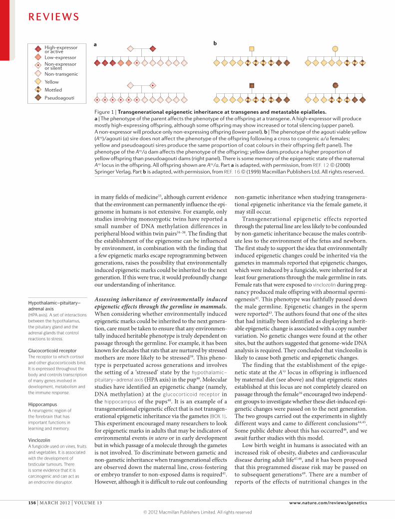
a b High-expressor or activeLow-expressorNon-expressoror silentNon-transgenic
Yellow
Mottled
Pseudoagouti
Figure 1 | Transgenerational epigenetic inheritance at transgenes and metastable epialleles. a | The phenotype of the parent affects the phenotype of the offspring at a transgene. A high-expressor will produce mostly high-expressing offspring, although some offspring may show increased or total silencing (upper panel). A non-expressor will produce only non-expressing offspring (lower panel). b | The phenotype of the agouti viable yellow (Avy)/agouti (a) sire does not affect the phenotype of the offspring following a cross to congenic a/a females; yellow and pseudoagouti sires produce the same proportion of coat colours in their offspring (left panel). The phenotype of the Avy/a dam affects the phenotype of the offspring; yellow dams produce a higher proportion of yellow offspring than pseudoagouti dams (right panel). There is some memory of the epigenetic state of the maternal Avy locus in the offspring. All offspring shown are Avy/a. Part a is adapted, with permission, from REF. 12 © (2000) Springer Verlag. Part b is adapted, with permission, from REF. 16 © (1999) Macmillan Publishers Ltd. All rights reserved.
Hypothalamic–pituitary–adrenal axis(HPA axis). A set of interactions between the hypothalamus, the pituitary gland and the adrenal glands that control reactions to stress.
Glucocorticoid receptorThe receptor to which cortisol and other glucocorticoids bind. It is expressed throughout the body and controls transcription of many genes involved in development, metabolism and the immune response.
HippocampusA neurogenic region of the forebrain that has important functions in learning and memory.
VinclozolinA fungicide used on vines, fruits and vegetables. It is associated with the development of testicular tumours. There is some evidence that it is carcinogenic and can act as an endrocrine disruptor.
in many fields of medicine33, although current evidence that the environment can permanently influence the epi-genome in humans is not extensive. For example, only studies involving monozygotic twins have reported a small number of DNA methylation differences in peripheral blood within twin pairs34–38. The finding that the establishment of the epigenome can be influenced by environment, in combination with the finding that a few epigenetic marks escape reprogramming between generations, raises the possibility that environmentally induced epigenetic marks could be inherited to the next generation. If this were true, it would profoundly change our understanding of inheritance.
Assessing inheritance of environmentally induced epigenetic effects through the germline in mammals. When considering whether environmentally induced epigenetic marks could be inherited to the next genera-tion, care must be taken to ensure that any environmen-tally induced heritable phenotype is truly dependent on passage through the germline. For example, it has been known for decades that rats that are nurtured by stressed mothers are more likely to be stressed39. This pheno-type is perpetuated across generations and involves the setting of a ‘stressed’ state by the hypothalamic– pituitary–adrenal axis (HPA axis) in the pup40. Molecular studies have identified an epigenetic change (namely, DNA methylation) at the glucocorticoid receptor in the hippocampus of the pups40. It is an example of a transgenerational epigenetic effect that is not transgen-erational epigenetic inheritance via the gametes (BOX 1). This experiment encouraged many researchers to look for epigenetic marks in adults that may be indicators of environmental events in utero or in early development but in which passage of a molecule through the gametes is not involved. To discriminate between gametic and non-gametic inheritance when transgenerational effects are observed down the maternal line, cross-fostering or embryo transfer to non-exposed dams is required41. However, although it is difficult to rule out confounding
non-gametic inheritance when studying transgenera-tional epigenetic inheritance via the female gamete, it may still occur.
Transgenerational epigenetic effects reported through the paternal line are less likely to be confounded by non-gametic inheritance because the males contrib-ute less to the environment of the fetus and newborn. The first study to support the idea that environmentally induced epigenetic changes could be inherited via the gametes in mammals reported that epigenetic changes, which were induced by a fungicide, were inherited for at least four generations through the male germline in rats. Female rats that were exposed to vinclozolin during preg-nancy produced male offspring with abnormal spermi-ogenesis42. This phenotype was faithfully passed down the male germline. Epigenetic changes in the sperm were reported43. The authors found that one of the sites that had initially been identified as displaying a herit-able epigenetic change is associated with a copy number variation. No genetic changes were found at the other sites, but the authors suggested that genome-wide DNA analysis is required. They concluded that vinclozolin is likely to cause both genetic and epigenetic changes.
The finding that the establishment of the epige-netic state at the Avy locus in offspring is influenced by maternal diet (see above) and that epigenetic states established at this locus are not completely cleared on passage through the female16 encouraged two independ-ent groups to investigate whether these diet-induced epi-genetic changes were passed on to the next generation. The two groups carried out the experiments in slightly different ways and came to different conclusions44,45. Some public debate about this has occurred46, and we await further studies with this model.
Low birth weight in humans is associated with an increased risk of obesity, diabetes and cardiovascular disease during adult life47,48, and it has been proposed that this programmed disease risk may be passed on to subsequent generations49. There are a number of reports of the effects of nutritional changes in the
R E V I E W S
156 | MARCH 2012 | VOLUME 13 www.nature.com/reviews/genetics
© 2012 Macmillan Publishers Limited. All rights reserved
Nature Reviews | Genetics
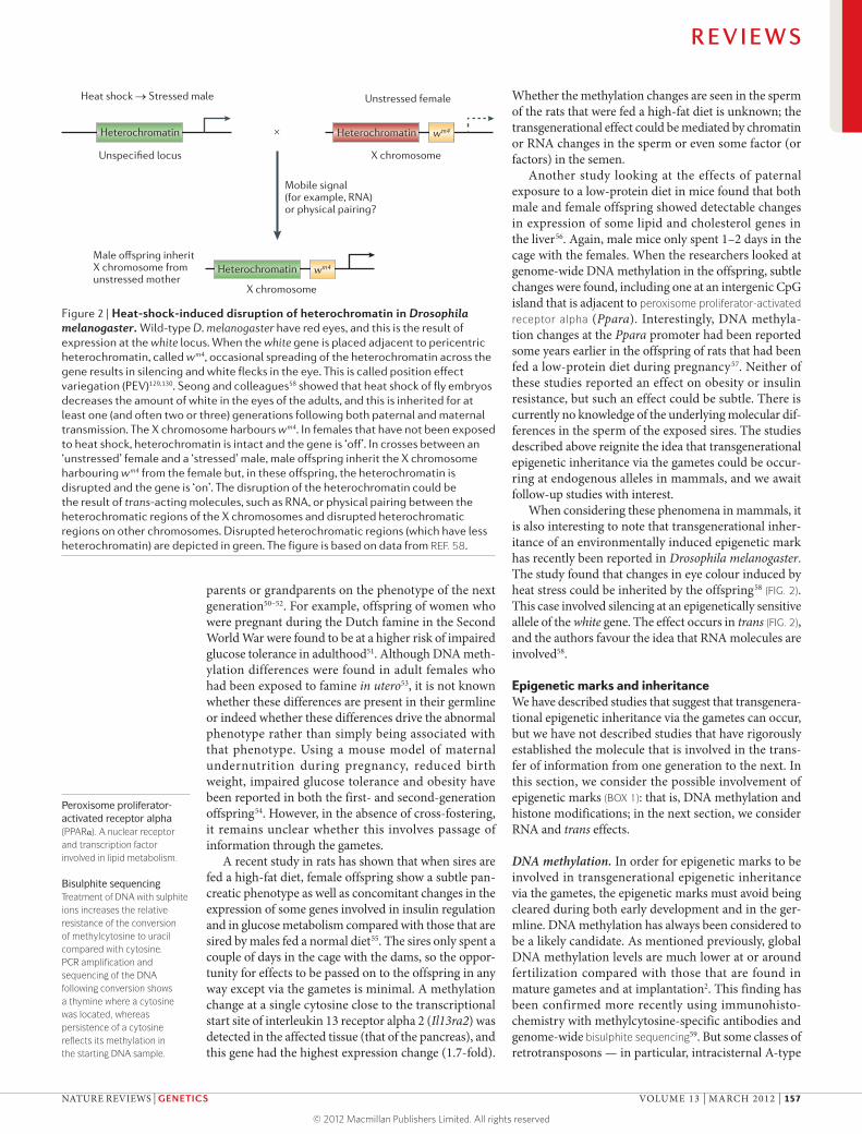
Heat shock → Stressed male Unstressed female
X chromosome
X chromosome
Male offspring inherit X chromosome fromunstressed mother
Mobile signal (for example, RNA) or physical pairing?
Unspecified locus
Heterochromatin Heterochromatin wm4
Heterochromatin wm4
×
Figure 2 | Heat-shock-induced disruption of heterochromatin in Drosophila melanogaster. Wild-type D. melanogaster have red eyes, and this is the result of expression at the white locus. When the white gene is placed adjacent to pericentric heterochromatin, called wm4, occasional spreading of the heterochromatin across the gene results in silencing and white flecks in the eye. This is called position effect variegation (PEV)129,130. Seong and colleagues58 showed that heat shock of fly embryos decreases the amount of white in the eyes of the adults, and this is inherited for at least one (and often two or three) generations following both paternal and maternal transmission. The X chromosome harbours wm4. In females that have not been exposed to heat shock, heterochromatin is intact and the gene is ‘off’. In crosses between an ‘unstressed’ female and a ‘stressed’ male, male offspring inherit the X chromosome harbouring wm4 from the female but, in these offspring, the heterochromatin is disrupted and the gene is ‘on’. The disruption of the heterochromatin could be the result of trans-acting molecules, such as RNA, or physical pairing between the heterochromatic regions of the X chromosomes and disrupted heterochromatic regions on other chromosomes. Disrupted heterochromatic regions (which have less heterochromatin) are depicted in green. The figure is based on data from REF. 58.
Peroxisome proliferator-activated receptor alpha(PPARα). A nuclear receptor and transcription factor involved in lipid metabolism.
Bisulphite sequencingTreatment of DNA with sulphite ions increases the relative resistance of the conversion of methylcytosine to uracil compared with cytosine. PCR amplification and sequencing of the DNA following conversion shows a thymine where a cytosine was located, whereas persistence of a cytosine reflects its methylation in the starting DNA sample.
parents or grandparents on the phenotype of the next generation50–52. For example, offspring of women who were pregnant during the Dutch famine in the Second World War were found to be at a higher risk of impaired glucose tolerance in adulthood51. Although DNA meth-ylation differences were found in adult females who had been exposed to famine in utero53, it is not known whether these differences are present in their germline or indeed whether these differences drive the abnormal phenotype rather than simply being associated with that phenotype. Using a mouse model of maternal undernutrition during pregnancy, reduced birth weight, impaired glucose tolerance and obesity have been reported in both the first- and second-generation offspring54. However, in the absence of cross-fostering, it remains unclear whether this involves passage of information through the gametes.
A recent study in rats has shown that when sires are fed a high-fat diet, female offspring show a subtle pan-creatic phenotype as well as concomitant changes in the expression of some genes involved in insulin regulation and in glucose metabolism compared with those that are sired by males fed a normal diet55. The sires only spent a couple of days in the cage with the dams, so the oppor-tunity for effects to be passed on to the offspring in any way except via the gametes is minimal. A methylation change at a single cytosine close to the transcriptional start site of interleukin 13 receptor alpha 2 (Il13ra2) was detected in the affected tissue (that of the pancreas), and this gene had the highest expression change (1.7-fold).
Whether the methylation changes are seen in the sperm of the rats that were fed a high-fat diet is unknown; the transgenerational effect could be mediated by chromatin or RNA changes in the sperm or even some factor (or factors) in the semen.
Another study looking at the effects of paternal exposure to a low-protein diet in mice found that both male and female offspring showed detectable changes in expression of some lipid and cholesterol genes in the liver56. Again, male mice only spent 1–2 days in the cage with the females. When the researchers looked at genome-wide DNA methylation in the offspring, subtle changes were found, including one at an intergenic CpG island that is adjacent to peroxisome proliferator-activated receptor alpha (Ppara). Interestingly, DNA methyla-tion changes at the Ppara promoter had been reported some years earlier in the offspring of rats that had been fed a low-protein diet during pregnancy57. Neither of these studies reported an effect on obesity or insulin resistance, but such an effect could be subtle. There is currently no knowledge of the underlying molecular dif-ferences in the sperm of the exposed sires. The studies described above reignite the idea that transgenerational epigenetic inheritance via the gametes could be occur-ring at endogenous alleles in mammals, and we await follow-up studies with interest.
When considering these phenomena in mammals, it is also interesting to note that transgenerational inher-itance of an environmentally induced epigenetic mark has recently been reported in Drosophila melanogaster. The study found that changes in eye colour induced by heat stress could be inherited by the offspring58 (FIG. 2). This case involved silencing at an epigenetically sensitive allele of the white gene. The effect occurs in trans (FIG. 2), and the authors favour the idea that RNA molecules are involved58.
Epigenetic marks and inheritanceWe have described studies that suggest that transgenera-tional epigenetic inheritance via the gametes can occur, but we have not described studies that have rigorously established the molecule that is involved in the trans-fer of information from one generation to the next. In this section, we consider the possible involvement of epigenetic marks (BOX 1): that is, DNA methylation and histone modifications; in the next section, we consider RNA and trans effects.
DNA methylation. In order for epigenetic marks to be involved in transgenerational epigenetic inheritance via the gametes, the epigenetic marks must avoid being cleared during both early development and in the ger-mline. DNA methylation has always been considered to be a likely candidate. As mentioned previously, global DNA methylation levels are much lower at or around fertilization compared with those that are found in mature gametes and at implantation2. This finding has been confirmed more recently using immunohisto-chemistry with methylcytosine-specific antibodies and genome-wide bisulphite sequencing59. But some classes of retrotransposons — in particular, intracisternal A-type
R E V I E W S
NATURE REVIEWS | GENETICS VOLUME 13 | MARCH 2012 | 157
© 2012 Macmillan Publishers Limited. All rights reserved
EpimutationsMitotically heritable changes in epigenetic state but not gene sequence. Epimutation usually takes place by an abnormal increase or decrease in the methylation status of a gene. The heritability of epimutations across generations is currently under debate.
MLH1MutL homologue 1, colon cancer nonpolyposis type 2 (Escherichia coli) is a human gene coding for a protein that has an important role in DNA repair.
MSH2MutS homologue 2, colon cancer nonpolyposis type 2 (Escherichia coli) is another human gene coding for a protein involved in DNA repair.
Polycomb repressive complex 1(PRC1). Silencing of the homeotic genes in development requires the Polycomb group proteins (PcGs). PcGs form two distinct multiprotein complexes, PRC1 and PRC2.
particles (IAPs) — have been found to remain meth-ylated in mature gametes and early pre-implantation embryos in mice60. Similar findings were reported in primordial germ cells61,62 and, more recently, using genome-wide analyses, these findings have been con-firmed63. Another study64 used methylated DNA immu-noprecipitation (meDIP) followed by hybridization to promoter arrays to look at DNA collected from a range of developmental stages — from mature gametes, the morula, the blastocyst and so forth. This study identi-fied ~100 non-imprinted, non-repetitive genes at which promoter DNA methylation does not change, which is consistent with an escape from post-fertilization DNA methylation reprogramming64. These studies demon-strate the principle that some loci escape DNA meth-ylation reprogramming and so suggest the potential for DNA methylation to be involved in the transmission of epigenetic information through the gametes.
Changes in DNA methylation have been associated with many cancers, and transgenerational epigenetic inheritance via the gametes has been suggested as the explanation for the inheritance of colorectal cancer in some cases of this disease that involve epimutations in MLH1 and MSH2. However, in the case of MSH2, the epigenetic changes turned out to be associated with mutations in cis65,66 and, in the case of MLH1, it has also been difficult to rule out genetic changes and to deter-mine whether methylation is transmitted in the gametes or is triggered post-fertilization67–72.
A study carried out to determine whether DNA methylation at the Avy locus could explain the transgen-erational memory of coat colour reported that the methylation at the promoter was completely absent in blastocysts from pseudoagouti dams28, making it an unlikely candidate. However, this analysis only investi-gated CpG methylation, and now we know that Cs can be methylated at non-CpG sites. For example, in human embryonic stem cells, 25% of the methylated C residues are found at CHG and CHH (where H is A, T or C)73. Most studies using bisulphite sequencing only report the methylation at Cs that are part of CpG dinucleotides. Presumably, researchers will now go back and reana-lyse data to look at these other sites. It has also recently been discovered that Cs can be hydroxymethylated, for-mylated and carboxylated74–77. Despite the fact that the levels of these modifications are low, they could turn out to play some part in transgenerational inheritance.
Chromatin proteins. For many decades, it was thought that all histones were cleared from the DNA in mature sperm and replaced by protamines. However, we now know that ~1–2% of the haploid genome in mice and ~4% of that in humans remains packaged into nucle-osomes in sperm78,79. Recent genome-wide analysis of the histone marks in human and mouse spermatozoa has revealed that some genes retain extensive histone H3 lysine 27 trimethylation (H3K27me3) at their pro-moters — a mark that is associated with silencing. These genes are enriched for ones that are not expressed dur-ing gametogenesis and/or early development78,79. Although the authors raise the possibility that this mark
is retained and could carry epigenetic information from one generation to the next79, an alternative explanation is also possible: the mark simply correlates with tran-scriptional silencing that is re-established so rapidly that the period in which the mark is ‘missing’ has not been detected. Studies in early mouse pre-implantation embryos have shown that Polycomb repressive complex 1 (PRC1) components derived from the maternal genome are transferred to the paternal genome after fertiliza-tion, providing an opportunity for the direct effects of maternal heterochromatin on silencing in the zygote80.
There is an increasing awareness of the dynamic nature of chromatin. For example, heterochromatin pro-tein 1 (HP1) binds in vivo with a residence time of only a few minutes81, and nucleosome turnover has been shown to be extremely rapid82, making the notion that histone marks could be a vehicle for transgenerational epigenetic inheritance via the gametes less attractive. Furthermore, not all histone modifications direct the transcriptional activity of the underlying gene83. For example, in organ-isms as diverse as yeast and humans, gene activity has been found to be associated with gradients of some histone modifications with a 5′ to 3′ polarity along the gene, suggesting a transcription-coupled generation of the histone marks, rather than vice versa.
RNA and other trans effectsMolecular biologists studying transgenerational epi-genetic inheritance via the gametes have generally focused on the nucleus. However, recent studies suggest that trans-acting and/or non-nuclear factors could be involved. For example, RNA, which occurs inside and outside the nucleus.
Evidence of trans effects. An important example that demonstrates the occurrence of trans effects is paramu-tation — a phenomenon in which homologous DNA sequences communicate in trans to establish meioti-cally heritable expression states. It has been looked at in most detail in some rare instances of non-Mendelian inheritance of phenotypes in a number of plant spe-cies84. The epigenetic state at an allele that is subject to paramutation is influenced by the epigenetic state of the homologous allele. After it has been established, this new epigenetic state is heritable and stable for many genera-tions in some cases85. Because the trans effect is between homologous chromosomes, it has been suggested that its basis could be the transitory physical interaction of non-homologous chromosomes86, sometimes known as ‘chromosome kissing’87. However, genetic screens have been carried out and reveal the involvement of the RNA interference (RNAi) machinery, implying a role for small RNAs88,89 (discussed further below). In the light of sev-eral examples discussed in this Review involving repeat sequences, it is interesting to note that the sequences involved in many cases of paramutation contain direct or inverted repeats90.
The focus on the nucleus has been particularly true among those scientists researching transgenerational epigenetic inheritance in mammals. However, reports of ‘paternal effect genes’ provided early evidence for
R E V I E W S
158 | MARCH 2012 | VOLUME 13 www.nature.com/reviews/genetics
© 2012 Macmillan Publishers Limited. All rights reserved
Nature Reviews | Genetics
DMR Chr9 Rasgrf1 DMR
Chr7 piRNA cluster
Promoter m m m
Direct repeats
piRNAs target pit-RNA at regions that are homologous to the DMRs
RMER4B retrotransposon
DMR m m m
Pol II
Nascent pit-RNA MILI1 or MIWI2
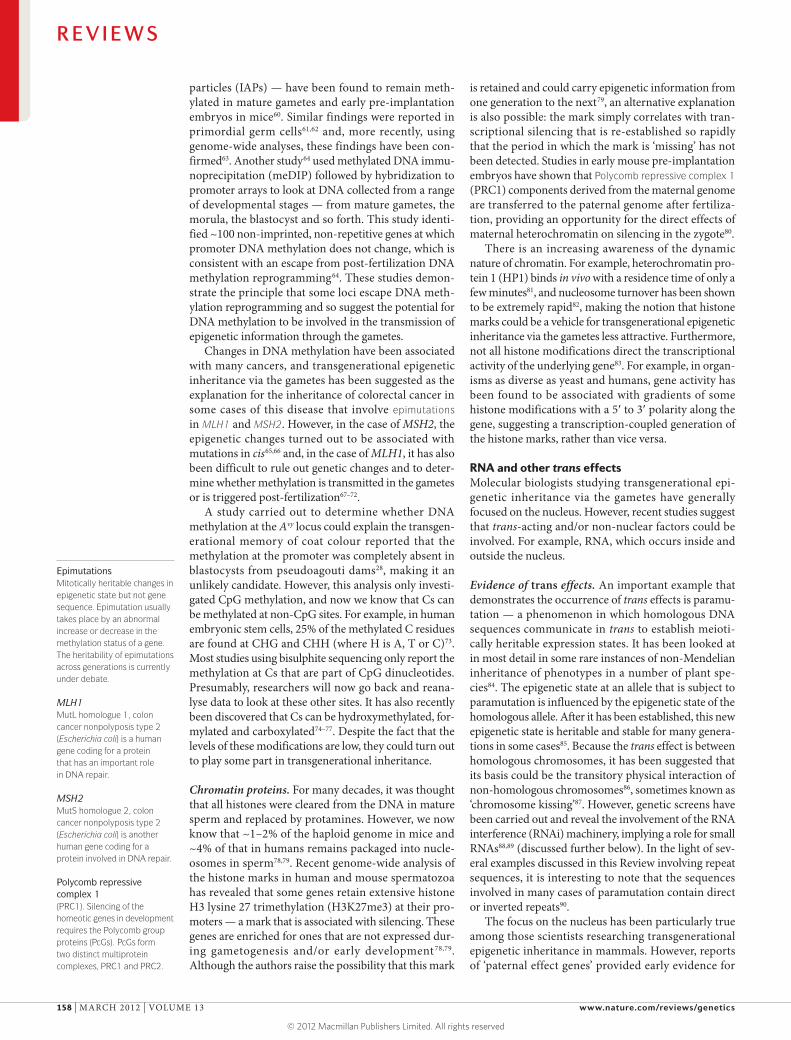
Figure 3 | Imprinting of the Rasgrf1 differentially methylated region via piRNA-directed DNA methylation. In mice, PIWI-interacting RNAs (piRNAs) are generated from the chromosome 7 (Chr7) piRNA cluster and bound by the PIWI family proteins MILI1 (also known as PIWIL2) and/or MIWI2 (also known as PIWIL4). The piRNA–MILI1 and/or piRNA–MIWI2 complexes then target the nascent antisense RNA that is transcribed through the RAS-protein-specific guanine nucleotide-releasing factor 1 (Rasgrf1) differentially methylated region (DMR). This RNA is designated here as piRNA-targeted non-coding RNA (pit-RNA). The DNA methyltransferases DNMT3A, DNMT3B and DNMT3L form a complex that is recruited in a process that involves the piRNA complex and is responsible for methylation of the Rasgrf1 DMR that spans the RMER4B retrotransposon. m, DNA methylation; Pol II, RNA polymerase II. The figure is based on data from REF. 104.
MicroRNAs(miRNAs). Evolutionarily conserved small non-coding RNAs (~22-nucleotides long) that silence gene expression by degrading or inhibiting translation of mRNA transcripts in a sequence-specific manner.
Endogenous small interfering RNAs(endo-siRNAs). Small RNAs that originate, in a Dicer- dependent manner, from long double-stranded (sense–antisense or hairpin) precursors. Initially mainly thought of as a mechanism of host defence against exogenous double-stranded RNA, endo-siRNAs are now known to also regulate endogenous mRNAs in mouse oocytes and Caenorhabditis elegans.
PIWI-interacting RNAs(piRNAs). Small (24–31 bp) RNAs that are associated with PIWI-clade proteins of the Argonaute family. They ensure genome stability in the germline of flies, mice and zebrafish by silencing transposable and repetitive elements.
trans effects in mice. In this particular context, pater-nal effects are phenotypic effects that are observed in wild-type offspring of sires that are heterozygous for a mutation: that is, the effects are seen despite the fact that the offspring did not themselves inherit the mutant allele91,92. For example, using coat colour as a readout for expression from the Avy locus, haploinsufficiency for modifiers of epigenetic reprogramming in sires was found to alter the coat colour of wild-type offspring that had inherited the Avy allele from the dam91. These findings suggest that some ‘trans-acting factor’ intro-duced into the zygote along with the paternal gamete influences the establishment of epigenetic state at a gene present on the maternal set of chromosomes (in this case, the Avy allele). There are two possible explanations for trans effects of this type: first, the involvement of diffusible factors such as RNA or protein, and second, chromosome kissing87.
RNA in gametes and roles in silencing transposons. The highly condensed sperm nucleus is transcriptionally and translationally inert and contains little cytoplasm, but RNA populations have been detected in mature sperm93, and sperm-borne RNA has been detected in the zygote94. In addition to mRNAs, long non-coding RNAs (lncRNAs) and small RNAs of various classes have been found. These include microRNAs (miRNAs), endogenous small interfering RNAs (endo-siRNAs) and PIWI-interacting RNAs (piRNAs), and these classes of small RNAs can be involved in gene silencing. Oocytes also contain large amounts of RNA of all classes95–97. Indeed, the egg has evolved special strategies to maintain a store of RNA that enables the zygote to function in the absence of transcription until the two-cell stage.
It is also interesting that it has been shown in plants and worms that small RNA molecules can travel between cells98. Transposable elements are the targets of siRNA-mediated silencing in animals, fungi and plants, and there is evidence that some small RNAs can be involved in silencing transposable elements in adjacent germline cells. For example, a recent study in Arabidopsis thaliana has found a population of siRNAs from Athila retrotrans-posons generated in the vegetative nucleus of pollen that may have a role in silencing the Athila retrotransposons in the adjacent sperm cells99.
Following from this, it is easy to envisage how small RNAs could act in trans at fertilization as mobile sig-nals that influence the silencing of genetic elements containing retrotransposons, such as Avy, introduced on the complementary haploid genome. In mouse oocytes, endo-siRNAs are also known to be required for retro-transposon silencing96,97,100. The large amounts of these endo-siRNAs in the oocyte could theoretically direct repeat silencing in the early embryo as well.
Roles of piRNAs in gametes. piRNAs have a role in the silencing of mobile genetic elements in many eukary-otic organisms101 and, in D. melanogaster, PIWI has been shown to influence the epigenetic phenomenon position effect variegation (PEV)102. Although some aspects of the mechanisms of piRNA biogenesis remain unclear, they are produced from piRNA clusters that, at least in D. melanogaster, lie at heterochromatin–euchromatin boundaries in the most repeat-rich regions of the genome. They are specifically expressed in reproduc-tive organs and are found in fairly high levels in sper-matocytes101,103. Interestingly, in mammals, piRNAs have recently been shown to play a part in the estab-lishment of parental imprints104. A study in mice found that piRNAs are required for the establishment of the DNA methylation at the RAS-protein-specific guanine nucleotide-releasing factor 1 (Rasgrf1) locus in the pater-nal germline104. A retrotransposon sequence within a non-coding RNA that spans the differentially methyl-ated region of this imprinted gene is targeted by piRNAs generated from a different locus (FIG. 3). These experi-ments reveal a clear role for piRNAs in the establishment of DNA methylation imprints during reprogramming in primordial germ cells (PGCs). One scenario in this case is that RNAs are involved in establishing the epigenetic mark, and then DNA methylation carries the information in the gametes; another scenario is that piRNAs are also carried in the gametes and influence DNA methylation in the offspring.
piRNAs have also been shown to be involved in the phenomenon of hybrid dysgenesis. Hybrid dysgenesis refers to the fact that when wild D. melanogaster are crossed to laboratory strains, an incompatibility is some-times observed: progeny from laboratory males mated to wild females develop normally, but those from the recip-rocal cross display abnormalities. The underlying cause has been traced to the mobilization of transposons that are present in the genome of the wild strain but that are absent in the laboratory strain105. The differential behav-iour of the reciprocal crosses implies the existence of a
R E V I E W S
NATURE REVIEWS | GENETICS VOLUME 13 | MARCH 2012 | 159
© 2012 Macmillan Publishers Limited. All rights reserved
Position effect variegation(PEV). This term describes a type of phenotypic variegation among cells of the same type that is the result of mosaic silencing of a particular gene. The variegation in these cases is due to the position of the gene adjacent to a heterochromatic region of the chromosome.
maternal factor that influences the ability of the progeny to silence inherited elements106. We now know that these factors are piRNAs107.
MicroRNAs. In general, miRNAs direct post-transcriptional repression by pairing to the mRNAs of protein-coding genes, but they have also been found in the mammalian nucleus, where their function remains unclear108,109. miRNAs that are present in sperm or oocytes could, by reducing the levels of their target mRNAs in the zygote, indirectly alter the epigenetic state of the developing embryo.
miRNAs have been implicated in transgenerational epigenetic inheritance at the Kit locus in mice110. When breeding mice to be heterozygous for a mutant allele designated Kittm1Alf, an under-representation of offspring with a wild-type phenotype was found. Significantly more offspring than expected showed white tail tips and white feet, a phenotype that is usually associated with heterozygosity for the mutant allele. In fact, genetically wild-type mice were born at the expected frequency, but a proportion of them had retained the phenotype of their heterozygous parents, suggesting that transgenerational epigenetic inheritance via the gametes was involved. In the wild-type offspring with the mutant phenotype, Kit mRNA levels were found to be reduced (to a level similar to that expected in heterozygotes), and Kit RNA molecules of abnormal size were detected in the testes of mutant sires. Additionally, Kit mRNA was found in the mature gametes of the heterozygous males but not the wild-type males110. Two miRNAs (miR-221 and miR-222) that had previously been identified as having the potential to target Kit mRNA111 were injected into wild-type zygotes, and this resulted in mice with the white-tail phenotype110. Similar paramutation-like phe-nomena have been reported by the same group at some other loci in the mouse112,113. To our knowledge, these are the only experiments that directly link a molecule of any kind to transgenerational epigenetic inheritance via the gametes. It is worth noting that the mutant allele Kittm1Alf, at which the observation of transgenerational epigenetic inheritance was made, carries a transgene insertion and that transgenerational epigenetic inheritance via the gametes is not observed at other null Kit alleles.
Conclusions and perspectivesThe number of alleles at which it has been shown that the epigenetic state, independent of the underlying geno-type, is inherited across generations remains small and, when it does occur, the process shows incomplete penetrance (only a proportion of offspring are affected). Indeed, transgenerational epigenetic inheritance via the gametes is likely to be rare. It seems counterintuitive to evolve a widespread system to inherit marks that are normally considered to have evolved to enable complex cell types to emerge from one genome. In the gam-etes, there is only one copy of a particular gene. If that is ‘marked’ in such a way that that is not cleared (that is, the mark is inherited), then this mark would affect the gene’s activity in all cell types of the developing embryo, affecting the organism’s ability to develop all of the cor-rect cell types. As such, the probability of a positive outcome (such as being better adapted to a changed environment) is outweighed by the probability of a negative effect (such as failure to develop). For exam-ple, a recent study in Caenorhabditis elegans highlights the importance of reprogramming between generations. Mutants that are null for the H3K4me2 demethylase spr‑5 (the mammalian orthologue is KDM1A (also known as LSD1)) exhibit progressive sterility over many generations114. This sterility correlates with the misregu-lation of genes in spermatogenesis and the transgen-erational accumulation of H3K4me2, suggesting that H3K4me2 needs to be cleared between generations.
Nevertheless, transgenerational epigenetic inherit-ance via the gametes has been shown to occur in some parts of the genome across eukaryotes. It seems to occur mainly at retrotransposons and other repeated elements, and it is true that these are the areas of the genome about which we know the least. Whereas DNA methylation is still the most popular candidate for the molecular basis of transgenerational epigenetic inheritance via the gam-etes, data collected in various situations provides mixed support for this view. Perhaps future studies should focus less on looking for an epigenetic mark that is retained across generations and more on looking for processes (or factors) that disrupt or enhance the re-establishment of silent heterochromatin between generations. RNA may be the best candidate.
1. Peaston, A. E. & Whitelaw, E. Epigenetics and phenotypic variation in mammals. Mamm. Genome 17, 365–374 (2006).
2. Monk, M., Boubelik, M. & Lehnert, S. Temporal and regional changes in DNA methylation in the embryonic, extraembryonic and germ cell lineages during mouse embryo development. Development 99, 371–382 (1987).
3. DeChiara, T. M., Robertson, E. J. & Efstratiadis, A. Parental imprinting of the mouse insulin-like growth factor II gene. Cell 64, 849–859 (1991).
4. Bartolomei, M. S., Webber, A. L., Brunkow, M. E. & Tilghman, S. M. Epigenetic mechanisms underlying the imprinting of the mouse H19 gene. Genes Dev. 7, 1663–1673 (1993).
5. Bartolomei, M. S., Zemel, S. & Tilghman, S. M. Parental imprinting of the mouse H19 gene. Nature 351, 153–155 (1991).
6. Gregg, C. et al. High-resolution analysis of parent-of-origin allelic expression in the mouse brain. Science 329, 643–648 (2010).
7. Gregg, C., Zhang, J., Butler, J. E., Haig, D. & Dulac, C. Sex-specific parent-of-origin allelic expression in the mouse brain. Science 329, 682–685 (2010).
8. Hadchouel, M., Farza, H., Simon, D., Tiollais, P. & Pourcel, C. Maternal inhibition of hepatitis B surface antigen gene expression in transgenic mice correlates with de novo methylation. Nature 329, 454–456 (1987).
9. Kearns, M., Preis, J., McDonald, M., Morris, C. & Whitelaw, E. Complex patterns of inheritance of an imprinted murine transgene suggest incomplete germline erasure. Nucleic Acids Res. 28, 3301–3309 (2000).
10. Allen, N. D., Norris, M. L. & Surani, M. A. Epigenetic control of transgene expression and imprinting by genotype-specific modifiers. Cell 61, 853–861 (1990).
11. Swain, J. L., Stewart, T. A. & Leder, P. Parental legacy determines methylation and expression of an autosomal transgene: a molecular mechanism for parental imprinting. Cell 50, 719–727 (1987).
12. Sutherland, H. G. et al. Reactivation of heritably silenced gene expression in mice. Mamm. Genome 11, 347–355 (2000).
13. Sapienza, C., Peterson, A. C., Rossant, J. & Balling, R. Degree of methylation of transgenes is dependent on gamete of origin. Nature 328, 251–254 (1987).
14. McGowan, R., Campbell, R., Peterson, A. & Sapienza, C. Cellular mosaicism in the methylation and expression of hemizygous loci in the mouse. Genes Dev. 3, 1669–1676 (1989).
15. Garrick, D., Fiering, S., Martin, D. I. & Whitelaw, E. Repeat-induced gene silencing in mammals. Nature Genet. 18, 56–59 (1998).
16. Morgan, H. D., Sutherland, H. G., Martin, D. I. & Whitelaw, E. Epigenetic inheritance at the agouti locus in the mouse. Nature Genet. 23, 314–318 (1999).
17. Wolff, G. L. Influence of maternal phenotype on metabolic differentiation of agouti locus mutants in the mouse. Genetics 88, 529–539 (1978).
R E V I E W S
160 | MARCH 2012 | VOLUME 13 www.nature.com/reviews/genetics
© 2012 Macmillan Publishers Limited. All rights reserved
18. Duhl, D. M., Vrieling, H., Miller, K. A., Wolff, G. L. & Barsh, G. S. Neomorphic agouti mutations in obese yellow mice. Nature Genet. 8, 59–65 (1994).
19. Dolinoy, D. C., Weinhouse, C., Jones, T. R., Rozek, L. S. & Jirtle, R. L. Variable histone modifications at the Avy metastable epiallele. Epigenetics 5, 637–644 (2010).
20. Rakyan, V. K. et al. Transgenerational inheritance of epigenetic states at the murine AxinFu allele occurs after maternal and paternal transmission. Proc. Natl Acad. Sci. USA 100, 2538–2543 (2003).
21. Druker, R., Bruxner, T. J., Lehrbach, N. J. & Whitelaw, E. Complex patterns of transcription at the insertion site of a retrotransposon in the mouse. Nucleic Acids Res. 32, 5800–5808 (2004).
22. Weinhouse, C. et al. An expression microarray approach for the identification of metastable epialleles in the mouse genome. Epigenetics 6, 1105–1113 (2011).
23. Rakyan, V. K., Blewitt, M. E., Druker, R., Preis, J. I. & Whitelaw, E. Metastable epialleles in mammals. Trends Genet. 18, 348–351 (2002).
24. Waterland, R. A. et al. Season of conception in rural Gambia affects DNA methylation at putative human metastable epialleles. PLoS Genet. 6, e1001252 (2010).This paper was the first to identify what are thought to be metastable epialleles in humans.
25. Roemer, I., Reik, W., Dean, W. & Klose, J. Epigenetic inheritance in the mouse. Curr. Biol. 7, 277–280 (1997).
26. Reik, W. et al. Adult phenotype in the mouse can be affected by epigenetic events in the early embryo. Development 119, 933–942 (1993).
27. Wolff, G. L., Kodell, R. L., Moore, S. R. & Cooney, C. A. Maternal epigenetics and methyl supplements affect agouti gene expression in Avy/a mice. FASEB J. 12, 949–957 (1998).
28. Blewitt, M. E., Vickaryous, N. K., Paldi, A., Koseki, H. & Whitelaw, E. Dynamic reprogramming of DNA methylation at an epigenetically sensitive allele in mice. PLoS Genet. 2, e49 (2006).
29. Kaminen-Ahola, N. et al. Maternal ethanol consumption alters the epigenotype and the phenotype of offspring in a mouse model. PLoS Genet. 6, e1000811 (2010).
30. Waterland, R. A. & Jirtle, R. L. Transposable elements: targets for early nutritional effects on epigenetic gene regulation. Mol. Cell. Biol. 23, 5293–5300 (2003).
31. Cooney, C. A., Dave, A. A. & Wolff, G. L. Maternal methyl supplements in mice affect epigenetic variation and DNA methylation of offspring. J. Nutr. 132, 2393S–2400S (2002).
32. Dolinoy, D. C., Weidman, J. R., Waterland, R. A. & Jirtle, R. L. Maternal genistein alters coat colour and protects Avy mouse offspring from obesity by modifying the fetal epigenome. Environ. Health Perspect. 114, 567–572 (2006).
33. Rakyan, V. K., Down, T. A., Balding, D. J. & Beck, S. Epigenome-wide association studies for common human diseases. Nature Rev. Genet. 12, 529–541 (2011).
34. Fraga, M. F. et al. Epigenetic differences arise during the lifetime of monozygotic twins. Proc. Natl Acad. Sci. USA 102, 10604–10609 (2005).
35. Oates, N. A. et al. Increased DNA methylation at the AXIN1 gene in a monozygotic twin from a pair discordant for a caudal duplication anomaly. Am. J. Hum. Genet. 79, 155–162 (2006).
36. Mill, J. et al. Evidence for monozygotic twin (MZ) discordance in methylation level at two CpG sites in the promoter region of the catechol-O-methyltransferase (COMT) gene. Am. J. Med. Genet. B 141, 421–425 (2006).
37. Baranzini, S. E. et al. Genome, epigenome and RNA sequences of monozygotic twins discordant for multiple sclerosis. Nature 464, 1351–1356 (2010).This is currently the most comprehensive study of the epigenome in monozygotic twins.
38. Kaminsky, Z. A. et al. DNA methylation profiles in monozygotic and dizygotic twins. Nature Genet. 41, 240–245 (2009).
39. Francis, D. D. & Meaney, M. J. Maternal care and the development of stress responses. Curr. Opin. Neurobiol. 9, 128–134 (1999).
40. Weaver, I. C. et al. Epigenetic programming by maternal behaviour. Nature Neurosci. 7, 847–854 (2004).
41. Youngson, N. A. & Whitelaw, E. Transgenerational epigenetic effects. Annu. Rev. Genomics Hum. Genet. 9, 233–257 (2008).
42. Anway, M. D., Cupp, A. S., Uzumcu, M. & Skinner, M. K. Epigenetic transgenerational actions of endocrine disruptors and male fertility. Science 308, 1466–1469 (2005).
43. Guerrero-Bosagna, C., Settles, M., Lucker, B. & Skinner, M. K. Epigenetic transgenerational actions of vinclozolin on promoter regions of the sperm epigenome. PLoS ONE 5, e13100 (2010).
44. Waterland, R. A., Travisano, M. & Tahiliani, K. G. Diet-induced hypermethylation at agouti viable yellow is not inherited transgenerationally through the female. FASEB J. 21, 3380–3385 (2007).
45. Cropley, J. E., Suter, C. M., Beckman, K. B. & Martin, D. I. Germ-line epigenetic modification of the murine Avy allele by nutritional supplementation. Proc. Natl Acad. Sci. USA 103, 17308–17312 (2006).
46. Cropley, J. E., Suter, C. M. & Martin, D. I. Methyl donors change the germline epigenetic state of the Avy allele. FASEB J. 21, 3021; author reply 3021–3022 (2007).
47. Hales, C. N. & Barker, D. J. Type 2 (non-insulin- dependent) diabetes mellitus: the thrifty phenotype hypothesis. Diabetologia 35, 595–601 (1992).
48. McMillen, I. C. & Robinson, J. S. Developmental origins of the metabolic syndrome: prediction, plasticity, and programming. Physiol. Rev. 85, 571–633 (2005).
49. Gluckman, P. D., Hanson, M. A. & Beedle, A. S. Non-genomic transgenerational inheritance of disease risk. Bioessays 29, 145–154 (2007).
50. Kaati, G., Bygren, L. O. & Edvinsson, S. Cardiovascular and diabetes mortality determined by nutrition during parents’ and grandparents’ slow growth period. Eur. J. Hum. Genet. 10, 682–688 (2002).
51. Lumey, L. H., Stein, A. D., Kahn, H. S. & Romijn, J. A. Lipid profiles in middle-aged men and women after famine exposure during gestation: the Dutch Hunger Winter Families Study. Am. J. Clin. Nutr. 89, 1737–1743 (2009).
52. Pembrey, M. E. et al. Sex-specific, male-line transgenerational responses in humans. Eur. J. Hum. Genet. 14, 159–166 (2006).
53. Heijmans, B. T. et al. Persistent epigenetic differences associated with prenatal exposure to famine in humans. Proc. Natl Acad. Sci. USA 105, 17046–17049 (2008).
54. Jimenez-Chillaron, J. C. et al. Intergenerational transmission of glucose intolerance and obesity by in utero undernutrition in mice. Diabetes 58, 460–468 (2009).
55. Ng, S. F. et al. Chronic high-fat diet in fathers programs β-cell dysfunction in female rat offspring. Nature 467, 963–966 (2010).This is a carefully executed study revealing transgenerational epigenetic inheritance of phenotype in rats. This is a clear case of inheritance of non-genetic information via the ejaculate.
56. Carone, B. R. et al. Paternally induced transgenerational environmental reprogramming of metabolic gene expression in mammals. Cell 143, 1084–1096 (2010).This is another study that reveals the inheritance of epigenetic information (in this case, DNA methylation) from sire to offspring.
57. Lillycrop, K. A. et al. Feeding pregnant rats a protein-restricted diet persistently alters the methylation of specific cytosines in the hepatic PPARα promoter of the offspring. Br. J. Nutr. 100, 278–282 (2008).
58. Seong, K. H., Li, D., Shimizu, H., Nakamura, R. & Ishii, S. Inheritance of stress-induced, ATF-2-dependent epigenetic change. Cell 145, 1049–1061 (2011).This elegant study uses genetics in D. melanogaster to show that transgenerational memory of an environmental event (heat stress) can occur in trans.
59. Santos, F., Hendrich, B., Reik, W. & Dean, W. Dynamic reprogramming of DNA methylation in the early mouse embryo. Dev. Biol. 241, 172–182 (2002).
60. Lane, N. et al. Resistance of IAPs to methylation reprogramming may provide a mechanism for epigenetic inheritance in the mouse. Genesis 35, 88–93 (2003).
61. Hajkova, P. et al. Epigenetic reprogramming in mouse primordial germ cells. Mech. Dev. 117, 15–23 (2002).
62. Hajkova, P. et al. Chromatin dynamics during epigenetic reprogramming in the mouse germ line. Nature 452, 877–881 (2008).
63. Popp, C. et al. Genome-wide erasure of DNA methylation in mouse primordial germ cells is affected by AID deficiency. Nature 463, 1101–1105 (2010).
64. Borgel, J. et al. Targets and dynamics of promoter DNA methylation during early mouse development. Nature Genet. 42, 1093–1100 (2010).
65. Chan, T. L. et al. Heritable germline epimutation of MSH2 in a family with hereditary nonpolyposis colorectal cancer. Nature Genet. 38, 1178–1183 (2006).
66. Ligtenberg, M. J. et al. Heritable somatic methylation and inactivation of MSH2 in families with Lynch syndrome due to deletion of the 3′ exons of TACSTD1. Nature Genet. 41, 112–117 (2009).
67. Suter, C. M., Martin, D. I. & Ward, R. L. Germline epimutation of MLH1 in individuals with multiple cancers. Nature Genet. 36, 497–501 (2004).
68. Hitchins, M. P. et al. Inheritance of a cancer-associated MLH1 germ-line epimutation. N. Engl. J. Med. 356, 697–705 (2007).
69. Chong, S., Youngson, N. A. & Whitelaw, E. Heritable germline epimutation is not the same as transgenerational epigenetic inheritance. Nature Genet. 39, 574–575; author reply 575–576 (2007).
70. Suter, C. M. & Martin, D. I. Inherited epimutation or a haplotypic basis for the propensity to silence? Nature Genet. 39, 573; author reply 576 (2007).
71. Hitchins, M. P. & Ward, R. L. Erasure of MLH1 methylation in spermatozoa—implications for epigenetic inheritance. Nature Genet. 39, 1289 (2007).
72. Goel, A. et al. De novo constitutional MLH1 epimutations confer early-onset colorectal cancer in two new sporadic Lynch syndrome cases, with derivation of the epimutation on the paternal allele in one. Int. J. Cancer 128, 869–878 (2011).
73. Lister, R. et al. Human DNA methylomes at base resolution show widespread epigenomic differences. Nature 462, 315–322 (2009).
74. Kriaucionis, S. & Heintz, N. The nuclear DNA base 5-hydroxymethylcytosine is present in Purkinje neurons and the brain. Science 324, 929–930 (2009).
75. Tahiliani, M. et al. Conversion of 5-methylcytosine to 5-hydroxymethylcytosine in mammalian DNA by MLL partner TET1. Science 324, 930–935 (2009).
76. Ito, S. et al. Tet proteins can convert 5-methylcytosine to 5-formylcytosine and 5-carboxylcytosine. Science 333, 1300–1303 (2011).
77. He, Y. F. et al. Tet-mediated formation of 5-carboxylcytosine and its excision by TDG in mammalian DNA. Science 333, 1303–1307 (2011).
78. Hammoud, S. S. et al. Distinctive chromatin in human sperm packages genes for embryo development. Nature 460, 473–478 (2009).This paper uses genome-wide methods to show that nucleosomes and histone marks retained in human sperm are enriched at loci important for development.
79. Brykczynska, U. et al. Repressive and active histone methylation mark distinct promoters in human and mouse spermatozoa. Nature Struct. Mol. Biol. 17, 679–687 (2010).This study shows that promoters with distinct gene function and developmental expression patterns are selectively marked by active and repressive histone methylation in human and mouse sperm. They propose a model in which sperm-inherited nucleosomes are retained during zygotic reprogramming.
80. Puschendorf, M. et al. PRC1 and Suv39h specify parental asymmetry at constitutive heterochromatin in early mouse embryos. Nature Genet. 40, 411–420 (2008).
81. Festenstein, R. et al. Modulation of heterochromatin protein 1 dynamics in primary Mammalian cells. Science 299, 719–721 (2003).
82. Deal, R. B., Henikoff, J. G. & Henikoff, S. Genome-wide kinetics of nucleosome turnover determined by metabolic labelling of histones. Science 328, 1161–1164 (2010).
83. Henikoff, S. & Shilatifard, A. Histone modification: cause or cog? Trends Genet. 27, 389–396 (2011).
84. Chandler, V. L. Paramutation’s properties and puzzles. Science 330, 628–629 (2010).
85. Arteaga-Vazquez, M. A. & Chandler, V. L. Paramutation in maize: RNA mediated trans-generational gene silencing. Curr. Opin. Genet. Dev. 20, 156–163 (2010).
86. Patterson, G. I., Thorpe, C. J. & Chandler, V. L. Paramutation, an allelic interaction, is associated with a stable and heritable reduction of transcription of the maize b regulatory gene. Genetics 135, 881–894 (1993).
87. Cavalli, G. Chromosome kissing. Curr. Opin. Genet. Dev. 17, 443–450 (2007).
88. Alleman, M. et al. An RNA-dependent RNA polymerase is required for paramutation in maize. Nature 442, 295–298 (2006).
89. Sidorenko, L. et al. A dominant mutation in mediator of paramutation2, one of three second-largest subunits of a plant-specific RNA polymerase, disrupts multiple siRNA silencing processes. PLoS Genet. 5, e1000725 (2009).
R E V I E W S
NATURE REVIEWS | GENETICS VOLUME 13 | MARCH 2012 | 161
© 2012 Macmillan Publishers Limited. All rights reserved
90. Stam, M. & Mittelsten Scheid, O. Paramutation: an encounter leaving a lasting impression. Trends Plant Sci. 10, 283–290 (2005).
91. Chong, S. et al. Modifiers of epigenetic reprogramming show paternal effects in the mouse. Nature Genet. 39, 614–622 (2007).
92. Yazbek, S. N., Spiezio, S. H., Nadeau, J. H. & Buchner, D. A. Ancestral paternal genotype controls body weight and food intake for multiple generations. Hum. Mol. Genet. 19, 4134–4144 (2010).
93. Krawetz, S. A. Paternal contribution: new insights and future challenges. Nature Rev. Genet. 6, 633–642 (2005).
94. Zhao, Y. et al. Characterization and quantification of mRNA transcripts in ejaculated spermatozoa of fertile men by serial analysis of gene expression. Hum. Reprod. 21, 1583–1590 (2006).
95. Watanabe, T. et al. Identification and characterization of two novel classes of small RNAs in the mouse germline: retrotransposon-derived siRNAs in oocytes and germline small RNAs in testes. Genes Dev. 20, 1732–1743 (2006).
96. Watanabe, T. et al. Endogenous siRNAs from naturally formed dsRNAs regulate transcripts in mouse oocytes. Nature 453, 539–543 (2008).
97. Tam, O. H. et al. Pseudogene-derived small interfering RNAs regulate gene expression in mouse oocytes. Nature 453, 534–538 (2008).
98. Melnyk, C. W., Molnar, A. & Baulcombe, D. C. Intercellular and systemic movement of RNA silencing signals. EMBO J. 30, 3553–3563 (2011).
99. Slotkin, R. K. et al. Epigenetic reprogramming and small RNA silencing of transposable elements in pollen. Cell 136, 461–472 (2009).
100. Murchison, E. P. et al. Critical roles for Dicer in the female germline. Genes Dev. 21, 682–693 (2007).
101. Saito, K. & Siomi, M. C. Small RNA-mediated quiescence of transposable elements in animals. Dev. Cell 19, 687–697 (2010).
102. Pal-Bhadra, M. et al. Heterochromatic silencing and HP1 localization in Drosophila are dependent on the RNAi machinery. Science 303, 669–672 (2004).
103. Gan, H. et al. piRNA profiling during specific stages of mouse spermatogenesis. RNA 17, 1191–1203 (2011).
104. Watanabe, T. et al. Role for piRNAs and noncoding RNA in de novo DNA methylation of the imprinted mouse Rasgrf1 locus. Science 332, 848–852 (2011).This study reveals a role for piRNAs in directing DNA methylation at the imprinted Rasgrf1 locus in the mouse.
105. Bucheton, A., Paro, R., Sang, H. M., Pelisson, A. & Finnegan, D. J. The molecular basis of I-R. hybrid dysgenesis in Drosophila melanogaster: identification, cloning, and properties of the I factor. Cell 38, 153–163 (1984).
106. Bregliano, J. C. et al. Hybrid dysgenesis in Drosophila melanogaster. Science 207, 606–611 (1980).
107. Brennecke, J. et al. An epigenetic role for maternally inherited piRNAs in transposon silencing. Science 322, 1387–1392 (2008).
108. Hwang, H. W., Wentzel, E. A. & Mendell, J. T. A hexanucleotide element directs microRNA nuclear import. Science 315, 97–100 (2007).
109. Taft, R. J. et al. Nuclear-localized tiny RNAs are associated with transcription initiation and splice sites in metazoans. Nature Struct. Mol. Biol. 17, 1030–1034 (2010).
110. Rassoulzadegan, M. et al. RNA-mediated non-Mendelian inheritance of an epigenetic change in the mouse. Nature 441, 469–474 (2006).
111. Lewis, B. P., Shih, I. H., Jones-Rhoades, M. W., Bartel, D. P. & Burge, C. B. Prediction of mammalian microRNA targets. Cell 115, 787–798 (2003).
112. Wagner, K. D. et al. RNA induction and inheritance of epigenetic cardiac hypertrophy in the mouse. Dev. Cell 14, 962–969 (2008).
113. Grandjean, V. et al. The miR-124–Sox9 paramutation: RNA-mediated epigenetic control of embryonic and adult growth. Development 136, 3647–3655 (2009).
114. Katz, D. J., Edwards, T. M., Reinke, V. & Kelly, W. G. A C. elegans LSD1 demethylase contributes to germline immortality by reprogramming epigenetic memory. Cell 137, 308–320 (2009).
115. Metivier, R. et al. Cyclical DNA methylation of a transcriptionally active promoter. Nature 452, 45–50 (2008).
116. Kangaspeska, S. et al. Transient cyclical methylation of promoter DNA. Nature 452, 112–115 (2008).
117. Scharf, A. N., Barth, T. K. & Imhof, A. Establishment of histone modifications after chromatin assembly. Nucleic Acids Res. 37, 5032–5040 (2009).
118. Yisraeli, J., Frank, D., Razin, A. & Cedar, H. Effect of in vitro DNA methylation on beta-globin gene expression. Proc. Natl Acad. Sci. USA 85, 4638–4642 (1988).
119. Montero, L. M. et al. The distribution of 5-methylcytosine in the nuclear genome of plants. Nucleic Acids Res. 20, 3207–3210 (1992).
120. Ehrlich, M. et al. Amount and distribution of 5-methylcytosine in human DNA from different types of tissues of cells. Nucleic Acids Res. 10, 2709–2721 (1982).
121. Antequera, F., Tamame, M., Villanueva, J. R. & Santos, T. DNA methylation in the fungi. J. Biol. Chem. 259, 8033–8036 (1984).
122. Lyko, F. et al. The honey bee epigenomes: differential methylation of brain DNA in queens and workers. PLoS Biol. 8, e1000506 (2010).
123. Lyko, F., Ramsahoye, B. H. & Jaenisch, R. DNA methylation in Drosophila melanogaster. Nature 408, 538–540 (2000).
124. Bird, A. P., Taggart, M. H., Nicholls, R. D. & Higgs, D. R. Non-methylated CpG-rich islands at the human alpha-globin locus: implications for evolution of the α-globin pseudogene. EMBO J. 6, 999–1004 (1987).
125. Smallwood, S. A. et al. Dynamic CpG island methylation landscape in oocytes and preimplantation embryos. Nature Genet. 43, 811–814 (2011).
126. Kouzminova, E. & Selker, E. U. dim-2 encodes a DNA methyltransferase responsible for all known cytosine methylation in Neurospora. EMBO J. 20, 4309–4323 (2001).
127. Tamaru, H. & Selker, E. U. A histone H3 methyltransferase controls DNA methylation in Neurospora crassa. Nature 414, 277–283 (2001).
128. Weber, M. et al. Chromosome-wide and promoter-specific analyses identify sites of differential DNA methylation in normal and transformed human cells. Nature Genet. 37, 853–862 (2005).
129. Henikoff, S. Position-effect variegation after 60 years. Trends Genet. 6, 422–426 (1990).
130. Ebert, A., Lein, S., Schotta, G. & Reuter, G. Histone modification and the control of heterochromatic gene silencing in Drosophila. Chromosome Res. 14, 377–392 (2006).
AcknowledgmentsE.W. is a National Health and Medical Research Council (NHMRC) Australia Fellow. We thank N. Youngson for helpful discussions and M. Blewitt for critical reading of the manuscript.
Competing interests statementThe authors declare no competing financial interests.
R E V I E W S
162 | MARCH 2012 | VOLUME 13 www.nature.com/reviews/genetics
© 2012 Macmillan Publishers Limited. All rights reserved