Embed Size (px)
Citation preview

Epigenetic Transgenerational Actions of Vinclozolin onPromoter Regions of the Sperm EpigenomeCarlos Guerrero-Bosagna, Matthew Settles, Ben Lucker, Michael K. Skinner*
Center for Reproductive Biology, School of Biological Sciences, Washington State University, Pullman, Washington, United States of America
Abstract
Previous observations have demonstrated that embryonic exposure to the endocrine disruptor vinclozolin during gonadalsex determination promotes transgenerational adult onset disease such as male infertility, kidney disease, prostate disease,immune abnormalities and tumor development. The current study investigates genome-wide promoter DNA methylationalterations in the sperm of F3 generation rats whose F0 generation mother was exposed to vinclozolin. A methylated DNAimmunoprecipitation with methyl-cytosine antibody followed by a promoter tilling microarray (MeDIP-Chip) procedure wasused to identify 52 different regions with statistically significant altered methylation in the sperm promoter epigenome.Mass spectrometry bisulfite analysis was used to map the CpG DNA methylation and 16 differential DNA methylationregions were confirmed, while the remainder could not be analyzed due to bisulfite technical limitations. Analysis of thesevalidated regions identified a consensus DNA sequence (motif) that associated with 75% of the promoters. Interestingly,only 16.8% of a random set of 125 promoters contained this motif. One candidate promoter (Fam111a) was found to be dueto a copy number variation (CNV) and not a methylation change, suggesting initial alterations in the germline epigenomemay promote genetic abnormalities such as induced CNV in later generations. This study identifies differential DNAmethylation sites in promoter regions three generations after the initial exposure and identifies common genome featurespresent in these regions. In addition to primary epimutations, a potential indirect genetic abnormality was identified, andboth are postulated to be involved in the epigenetic transgenerational inheritance observed. This study confirms that anenvironmental agent has the ability to induce epigenetic transgenerational changes in the sperm epigenome.
Citation: Guerrero-Bosagna C, Settles M, Lucker B, Skinner MK (2010) Epigenetic Transgenerational Actions of Vinclozolin on Promoter Regions of the SpermEpigenome. PLoS ONE 5(9): e13100. doi:10.1371/journal.pone.0013100
Editor: Carlo Gaetano, Istituto Dermopatico dell’Immacolata, Italy
Received July 8, 2010; Accepted August 31, 2010; Published September 30, 2010
Copyright: � 2010 Guerrero-Bosagna et al. This is an open-access article distributed under the terms of the Creative Commons Attribution License, whichpermits unrestricted use, distribution, and reproduction in any medium, provided the original author and source are credited.
Funding: This study was supported by a Department of Defense and a National Institutes of Health, National Institute of Environmental Health Sciences grant toMKS. The funders had no role in study design, data collection and analysis, decision to publish, or preparation of the manuscript.
Competing Interests: The authors have declared that no competing interests exist.
* E-mail: [email protected]
Introduction
Epigenetic changes derived from exposure to endocrine
disruptors have been described in several tissues and organisms
[1,2,3,4,5,6,7]. These endocrine disruptor induced epigenetic
changes may have a wide range of phenotypic consequences with
implications from disease etiology [8,9] to evolution [7,10,11].
Disease conditions affected include cancers [12,13], reproductive
defects [1,14,15,16] and obesity [6,17]. Recent studies have
suggested disease associated with exposure to either endocrine
disruptors [1,14,18,19,20,21] or nutrient restriction [6,22] can
become transgenerationally transmitted. In particular, exposures
during embryonic gonadal development and sex determination
are capable of inducing adult onset disease states that can be
perpetuated across multiple generations [1,9]. The first example
involved exposure to vinclozolin, a fungicide commonly used in
agriculture and known to be an anti-androgenic endocrine
disrupting compound [23]. In adult male rats younger than 120
days of age that are derived from vinclozolin-exposed ancestors,
the main disease phenotype observed is a spermatogenic cell defect
in the testis [1,19]. Additional transgenerational disease develops
as animals age (6–14 months), including increased frequencies of
tumors, prostate disease, kidney disease, immune abnormalities
and other defects in spermatogenesis [14]. Transgenerational
disease has also been seen in females as a consequence of
vinclozolin treatment. These diseases include uterine hemorrhage
and/or anemia late in pregnancy [18]. Changes in behavior and
learning capacity have also been observed following vinlozolin
exposure [10,24,25], including transgenerationally transmitted
changes in mate preference [10] and anxiety behavior [25]. These
transgenerational epigenetic phenotypes appear to be part of a
genome-wide effect of vinclozolin treatment during germ line
development. Evidence for this is that the embryonic testis
transcriptome is substantially altered in males from the F1 through
the F3 generations, after F0 generation maternal exposure to
vinclozolin [26].
A previous report has shown that these vinclozolin-induced
transgenerational effects correlate with DNA methylation [1].
DNA methylation refers to the addition of a methyl group to CG
dinucleotides (CpGs) [27], which through interactions with other
epigenetic systems [28] and environmental signals [7,29] can
regulate gene expression without changes in DNA sequences.
Importantly, these transgenerational epigenetic effects are trig-
gered during a window of exposure in which the germ line
epigenome is developing, between embryonic days 8 to 14 (E8–
E14 in the rat). The comparable period in the human is between 6
weeks and mid gestation. During this developmental period, the
germ line is undergoing major reprogramming in its global DNA
methylation status [29,30,31]. Prior to sex determination, while
primordial germ cells migrate down the genital ridge towards the
PLoS ONE | www.plosone.org 1 September 2010 | Volume 5 | Issue 9 | e13100

developing gonad, they undergo an important reduction in global
DNA methylation, becoming demethylated around the time of
entry into the gonads [32]. Allelic differences in DNA methylation,
which is characteristic of imprinted genes, are defined during this
developmental period of the germ line [33]. Therefore, external
agents capable of permanently altering the germ line epigenome
during this critical period of establishment of DNA methylation
marks and reprogramming can persist transgenerationally [1,9].
F3 generation epigenetic changes in DNA methylation induced
by vinclozolin were previously assessed with the use of methyla-
tion-sensitive restriction enzyme digestion and bisulfite sequencing
[1]. These techniques are reliable for assessing DNA methylation
status at an individual gene scale, but have limitations in terms of
assessing genome-wide methylation [34]. Methylated DNA
immuno-precipitation (MeDIP) followed by tilling array analysis
(MeDIP-Chip) is one of the tools that allows for a genome-wide
approach. The procedure is based on enriching methylated DNA
in a sample using immuno-precipitation with an antibody for
methylated cytosine, followed by tiling microarray chip hybrid-
izations [35]. This method has been used to map the methylome in
Arabidopsis thaliana [36], human breast cancer metastasis [37] and
the human Major Histocompatibility Complex [38]. Other studies
have compared genome-wide methylation changes derived from
two experimental conditions, for example assessing methylation in
cancer cells compared to controls [34,39]. However, few studies
use genome-wide methylation approaches to evaluate whole
organism exposures to environmental compounds. Examples exist
for exposure to cocaine and BPA [40,41].
The present study shows promoter genome-wide DNA
methylation changes in the germ line of F3 generation rats whose
F0 generation mothers were exposed to the endocrine disruptor
vinclozolin. This study confirms with more recent technology our
previous findings that an endocrine disrupting agent (vinclozolin)
has the ability to induce transgenerational epigenetic modifications
in the male germ line. This study identifies several promoter
regions that have altered DNA methylation status three genera-
tions after the initial exposure and identifies common genomic
features present in these regions. In addition to a potential
common consensus motif among the regions that presented a
transgenerational change in methylation, an alteration in copy
number variation (CNV) was identified.
Results
Transgenerational Genome-wide Promoter AlterationsThe vinclozolin induced F3 generation epigenetic alterations in
DNA methylation of promoter regions in sperm was evaluated in
the present study. The strategy used for analyzing genome-wide
promoter changes in DNA methylation was immuno-precipitation
of methylated fragments with methyl-cytosine antibody followed
by promoter tiling microarray chip hybridizations (MeDIP-Chip).
This analysis was performed in two pooled DNA samples from two
different experiments with F3 generation sperm obtained from
control generation animals and compared to two samples obtained
from vinclozolin generation animals. Comparison of F3 control
versus vinclozolin samples with a comparative hybridization
bioinformatics approach produced a list of 52 promoter regions
that had statistically significant altered methylation patterns in the
sperm (Fig. 1). These 52 differential methylation regions
(Supplementary Table S1) were present in 48 different promoters.
The gene promoter information, chromosomal location and
statistical p-value are listed in Supplementary Table S1.
Subsequent quantitative analysis of these regions methylation
status was performed using bisulfite treatment followed by mass
spectrometry. This method detects changes in methylation in
selected regions at an individual CpG resolution [42]. In 16 of
these regions changes in methylation patterns were confirmed and
measured (Table 1, Fig. 2, 3, 4, 5). In 21 of the regions change in
methylation could not be detected in small selected sites
(Supplementary Fig. S1), due to the inability to interrogate the
entire region because bisulfite primers could not be designed.
Therefore, the change could have occurred in adjacent CpG sites
not able to be assayed. In the remaining 15 differential
methylation regions, no bisulfite primers could be designed for
the region so they were not investigated with the bisulfite mass
spectrometry procedure. Therefore, 16/48 of the different
methylation promoters were confirmed, while the others could
not be investigated due to bisulfite analysis limitations. The main
limitation in the primer design for bisulfate treated DNA is that
primers have to account for the large number of C to T
conversions, which create long strings of T that are difficult to
design specific primers for every region of interest. Neither the
software programs available nor manual analysis for bisulfite
primers was successful. A complete discussion on the constraints
for design for bisulfite treated DNA has previously been discussed
[43]. The list of the 16 genes containing these confirmed
promoters, the p-values (paired students t-test, p,0.05), and the
direction and the magnitude of the changes are shown in Table 1.
The information on all the other 32 promoters is presented in
Supplementary Table S1. The changes in DNA methylation
observed in these confirmed regions were remarkably concordant
between the MeDIP-Chip and the Mass Spectrometry analyses.
The genomic locations of these transgenerational changes in DNA
methylation (analyzed by these two methods) are shown in
Figure 1. The magnitude of the differential methylation with the
tiling array and mass spectrometry for the confirmed 16 regions is
shown in Figures 2, 3, 4, 5, while the non-confirmed sites are
presented in Supplementary Figure S1. Combined data for the
two separate experiments of vinclozolin exposure versus controls
are depicted in these figures.
In order to further compare the methylation status of the pooled
DNA (in which the MeDIP-Chip was performed) with the DNA
from the individual animal rat sperm samples from which these
pools were formed, we measured DNA methylation with
pyrosequencing [44] for two of the genes presenting change in
methylation. The regions chosen were from the predicted gene
RGD1561412/Olf735, which had the largest increase in methyl-
ation observed among all the annotated confirmed genes (47.6%),
and KCNE2, which presented the largest decrease in methylation
among the annotated confirmed genes (25.9%). The pyrosequenc-
ing analysis confirmed the tiling array and mass spectrometry
bisulfite procedures, and replicated the observed alteration in
DNA methylation performed in the individual animals compared
to the pooled samples for KCNE2 and RGD1561412/Olf735
promoters (Fig. 5). Therefore, the individual animal sperm DNA
sample analysis confirmed the differential methylation observed in
the pools. DNA methylation levels in the individual animal was
statistically significant and consistent with the levels measured in
the pooled DNA (Fig. 5), suggesting pooling the sperm DNA did
not create artificial differences in DNA methylation compared
with the individuals that originated the pools.
Analysis of the Differential Methylation GenomicFeatures
Genomic features of the promoter differential DNA methylation
regions were analyzed to determine if common sequence features
could be identified. Common genomic features can be distin-
guished by analysis of nucleic acid sequence patterns or motifs.
Transgenerational Epigenome
PLoS ONE | www.plosone.org 2 September 2010 | Volume 5 | Issue 9 | e13100

Some of these motifs are known to serve as binding sites for
transcription factors [45]. It has been previously shown that motifs
derived from methylation-prone sequences are generally associat-
ed with CpG islands and are non-randomly distributed along the
genome [46]. There are two ways of finding shared motifs in sets
of sequences: (i) applying ab initio motif discovery algorithms which
search for recurring patterns in a set of DNA sequences, or (ii)
assessing whether previously characterized motifs present in
transcription factor binding site databases are statistically over-
represented in the sequences [47]. The ab initio tool used in this
study was the GLAM2 algorithm (Gapped Local Alignment of
Motifs, available online in the MEME suite), which aims to find
motifs while considering insertions or deletions, a variable that is
not incorporated by other algorithms [48]. GLAM2SCAN was
used to search matches of the GLAM2 built motif in specified
sequence databases.
The set of 16 differential methylation regions that had been
confirmed with mass spectrometry was used as input in GLAM2
and the best motif obtained was used in further comparisons. The
logo (e.g. representation of the motif characteristic sequence)
obtained for this motif is shown in Figure 6a and the probability
matrix provided in Supplementary Table S3. This motif identified
with GLAM2 was named EDM1 (Environmental Induced
Differential Methylation Consensus Sequence 1). Interestingly,
EDM1 contains few possibilities of formation of CpG dinucleo-
tides depending on the sequence, from 0 to 3 CpG. Using
GLAM2SCAN, EDM1 was tested for prevalence of matches
against four sets of sequences: (i) the 16 promoters containing the
regions positively confirmed to be changed, (ii) the 48 promoters
containing the 52 regions showing change in the array, (iii) a set of
125 random promoters, and (iv) a set of 75 imprinted promoter
regions from mouse and rat databases. With a GLAM2SCAN cut–
off score value .20, EDM1 was present in 75% of the positively
confirmed set of promoters and in 60.4% of the set of 48
promoters showing change in the tiling array. In contrast, the
presence of EDM1 is significantly (p = 0.0001) reduced to 16.8%
in the set of 125 random promoters tested (Fig. 6b). In addition,
the presence of EDM1 was tested in a set of 75 promoter regions
of known mouse and rat imprinted genes (Supplemental Table
S4). Interestingly, this analysis showed that EDM1 was present in
58.7% of this set of imprinted gene promoter regions, which is an
incidence significantly (p = 0.001) higher than in the set of random
promoters, but reduced in comparison with the confirmed
promoters, (Fig. 6b and Supplemental Table S4). When EDM1
was present in the promoter of the imprinted genes set, it was at an
average of 5.7 hits/promoter, which is a higher frequency than the
hits/promoter in the set of confirmed or random promoters
(Fig. 6b). Therefore a consensus sequence motif EDM1 was
identified and associated with the transgenerational differential
methylation.
CTCF binding plays an important role in the establishment of
methylation in imprinted genes [49]. Therefore, the possibility that
the regions with transgenerational epigenetic changes had an
altered prevalence of CTCF binding sites was examined. A
previously published consensus motif of CTCF binding sites [50]
was tested with the online MEME tool ‘‘Find Individual Motif
Occurrence’’ (FIMO) in these sequences. FIMO is an algorithm
that aligns a motif (or a set of motifs) to sequences in a database.
Figure 1. Chromosomal distribution of the transgenerational change in DNA methylation in promoter regions identified by MeDIPfollowed by comparative chip hybridizations. The red arrow indicates regions with confirmed methylation change through mass spectrometry.Blue arrows indicate regions in which change was not able to be confirmed, either because of insufficient CpG site measurement through massspectrometry or not changed. Grey arrows indicate regions where primers could not be designed to test them. Green arrow indicates atransgenerational copy number variation (CNV) event in Fam111a.doi:10.1371/journal.pone.0013100.g001
Transgenerational Epigenome
PLoS ONE | www.plosone.org 3 September 2010 | Volume 5 | Issue 9 | e13100
The occurrence of the CTCF motif was compared among the
promoter sets and not found to be different (Fig. 6b). The cut-off
value used in FIMO was p,1024. The EDM1 sequence was also
compared to the CTCF motif and to other known motifs within
the JASPAR database [51] using the STAMP web tool [52]. No
significant similarity with the CTCF motif was found (E-value of
0.997) (Fig. 6b). When testing EDM1 for similarities with
eukaryotic transcription factor binding sites, it was found that
the highest scores of similarity were obtained with the transcription
factor binding sites for AZF1 (a zinc-finger factor found in
Saccharomyces cerevisiaes), FOXP1 (M00987), HMG-IY, STE11
(M00274) and BR-C (M00092) (Figure 7).
Another genomic feature analyzed was repeat elements. An
important number of methylated DNA regions are associated with
repeat elements [53] and known to have altered DNA methylation
patterns in cancer cells [54]. Germ cells have been shown to have
repeat elements that are enriched in the DNA methylation
associated SNPs [C/T]G or C[G/A] [55]. The promoters with
transgenerational differential methylation were analyzed in the
online algorithm Repeat Masker in order to interrogate changes in
the representation of particular repetitive elements. An increased
representation of Long Terminal Repeat (LTR) elements was
observed in the confirmed promoter regions (13.01%) as
compared to the set of random promoters (6.37%) (Table 2). In
addition, variations were found in the representation of the
different classes of LTR elements. The most striking difference
among these LTR classes was a 10-fold increase in the number of
the endogenous retrovirus-like (ERV) class I elements in the
confirmed promoters (6%) compared to the random set of
promoters (0.64%). An important and statistically significant
increase in ERV class II elements (also known as IAPs) was also
observed (from 2.34% to 4.49%). Interestingly, in the set of 48
promoters changed in the array, which include confirmed and
non-confirmed changes in methylation, the statistically significant
difference observed was an increase of ERV class I elements
regarding the random set (from 0.64% to 2.47%) (Table 2).
Therefore, ERV elements (class I and II) are over represented in
the set of promoters that have transgenerational changes in DNA
methylation. None of the other features examined were different
between the confirmed promoter set and the random promoter
set.
Copy Number Variation AnalysisThe consistency of the DNA methylation measurements
observed between the MeDIP-Chip and bisulfite mass spectrom-
etry analyses was monitored to validate procedures. In the case of
the candidate promoter Fam111a the analysis did not compare.
This region presented a highly significant change in methylation in
the MeDIP-Chip promoter array between the F3 generation
control and vinclozolin generation sperm (Supplementary Table
S1), however, its methylation level was unchanged in the mass
spectrometry methylation analysis, which had approximately 90%
levels of DNA methylation (Fig. 8a). The hypothesis was
investigated that this inconsistent result could be attributed to a
gene copy number variation (CNV). Copy number variation can
be associated with particular genomic regions and the incidence of
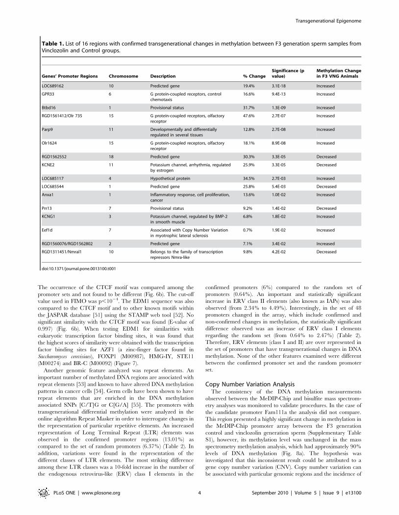
Table 1. List of 16 regions with confirmed transgenerational changes in methylation between F3 generation sperm samples fromVinclozolin and Control groups.
Genes’ Promoter Regions Chromosome Description % ChangeSignificance (pvalue)
Methylation Changein F3 VNG Animals
LOC689162 10 Predicted gene 19.4% 3.1E-18 Increased
GPR33 6 G protein-coupled receptors, controlchemotaxis
16.6% 9.4E-13 Increased
Btbd16 1 Provisional status 31.7% 1.3E-09 Increased
RGD1561412/Olr 735 15 G protein-coupled receptors, olfactoryreceptor
47.6% 2.7E-07 Increased
Parp9 11 Developmentally and differentiallyregulated in several tissues
12.8% 2.7E-08 Increased
Olr1624 15 G protein-coupled receptors, olfactoryreceptor
18.1% 8.9E-08 Increased
RGD1562552 18 Predicted gene 30.3% 3.3E-05 Decreased
KCNE2 11 Potassium channel, arrhythmia, regulatedby estrogen
25.9% 3.3E-05 Decreased
LOC685117 4 Hypothetical protein 34.5% 2.7E-03 Increased
LOC685544 1 Predicted gene 25.8% 5.4E-03 Decreased
Anxa1 1 Inflammatory response, cell proliferation,cancer
13.6% 1.0E-02 Increased
Prr13 7 Provisional status 9.2% 1.4E-02 Decreased
KCNG1 3 Potassium channel, regulated by BMP-2in smooth muscle
6.8% 1.8E-02 Increased
Eef1d 7 Associated with Copy Number Variationin myotrophic lateral sclerosis
0.7% 1.9E-02 Increased
RGD1560076/RGD1562802 2 Predicted gene 7.1% 3.4E-02 Increased
RGD1311451/Nmral1 10 Belongs to the family of transcriptionrepressors Nmra-like
9.8% 4.2E-02 Decreased
doi:10.1371/journal.pone.0013100.t001
Transgenerational Epigenome
PLoS ONE | www.plosone.org 4 September 2010 | Volume 5 | Issue 9 | e13100

Figure 2. Comparison of the methylation signal in regions where transgenerational methylation change is confirmed betweenvinclozolin and control. Analysis of methylation through MeDIP followed by comparative hybridization is graph on right with genome locationand log signal intensity presented with shaded area being the differential methylation region. Heavy red line indicates control and blue line indicates
Transgenerational Epigenome
PLoS ONE | www.plosone.org 5 September 2010 | Volume 5 | Issue 9 | e13100

disease [56], and can vary among different populations of humans
[57]. The potential presence of a CNV in the promoter region of
Fam111a was determined by comparing the signal of the tiling
array with genomic DNA inputs between vinclozolin and control
samples (i.e. comparative genomic hybridization), CGH. Interest-
ingly, it was found that the signal in the array hybridization versus
input was significantly higher in vinclozolin than in control F3
generation sperm (Fig. 8b). This indicates an increase in copy
number in the vinclozolin group for the Fam111a promoter. This
observation was not found in any other gene promoters in which
the difference between vinlcozolin and control was highly
significant, such as the example case for RGD1561412/Olr735
(Fig. 8c). No other promoter region in the genome was found to
contain a CNV (data not shown), but genome wide analysis for
regions outside promoters is now required to assess the specificity
of the CNV identified.
Discussion
An environmental exposure that can influence a critical
developmental period (e.g. embryonic development) for an organ
system can later in life promote an adult onset disease [7,9,58], in
part due to alterations in the epigenome. In the event a permanent
alteration in the germ line epigenome develops, a transgenera-
tional phenotype is possible [1,14]. Previous reports demonstrated
that the environmental endocrine disruptor vinclozolin can
promote a transgenerational disease phenotype and altered
DNA methylation in the germ line [9]. Vinclozolin is a commonly
used fungicide in the fruit (e.g. wine) industry [58] and its two
major metabolites (M1 and M2) are anti-androgenic compounds
[59]. These initial findings on the transgenerational epigenetic
effects of vinclozolin are expanded in the present study by
interrogating genome-wide methylation changes in the promoter
regions of F3 generation sperm. Observations identify a number of
genes that have their promoter DNA methylation patterns altered
in the F3 generation male germ line, following an early embryonic
(in utero) exposure of the F0 gestating female to the endocrine
disruptor vinclozolin. In the genome wide promoter analysis only
48 different promoters were found to have differential DNA
methylation in the F3 vinclozolin generation sperm. Some
promoters had multiple regions such that 52 total regions were
identified. The comparative hybridization MeDIP-Chip tilling
array procedure was reproducible and reliably identified the
differential methylation with an average region of 500–600 bp in
size. The MeDIP-Chip analysis does not map the alteration in
DNA methylation at the CpG level, but requires an alternate
procedure such as bisulfite conversion of cytosine residues followed
by mass spectrometry [42]. A limitation to this bisulfite mass
spectrometry procedure is that only limited sites can be
interrogated due to the inability to design bisulfite PCR primers
for all regions. This is due to the complexity of partially converting
a 4 base genome sequence to a 3 base sequence. Analysis
presented in Figures 2, 3, 4 and Supplementary Figure S1
demonstrates the sub-regions within each candidate that was
mapped and shows the sequence that could not be mapped. From
the 52 candidate regions identified 16 were confirmed with
bisulfite mass spectrometry, 21 were unconfirmed with the specific
site analyzed and 15 were not able to be analyzed. The 21
unconfirmed regions did not show changes in the CpG
methylation sites interrogated, but adjacent sequences in the same
region could not be interrogated due to limitations of primer
design for bisulfite treated DNA. These regions were categorized
as unconfirmed. Therefore, the 36 unconfirmed candidates are still
viable differential methylation sites, but will require more
advanced technology to map the entire region at the CpG level
resolution. Neither the computer software programs available nor
manual procedures used could generate the bisulfite promoters
needed for these 36 promoters. Therefore, further analysis of these
36 unconfirmed regions is needed. The current study focused on
the 16 confirmed sites for follow up analysis.
The differential DNA methylation regions identified were found
in the F3 generation sperm epigenome following vinclozolin
exposure of the F0 generation [1]. As previously described [7], the
presence of a phenotype and epigenetic modification in the F3
generation following exposure of a gestating F0 generation female
constitutes an epigenetic transgenerational phenotype and inher-
itance mechanism. Studies are now ongoing to investigate the
alterations in the corresponding F1 and F2 generation sperm
epigenome for comparison with the F3 sperm epigenome
alterations reported in the current study. Since the F1 and F2
generations are not directly defined as transgenerational [7], it will
be interesting to assess the similarities of sperm epigenome
alterations between those generations. In addition, a consistent
epigenetic alteration would be predicted for subsequent genera-
tions to the F3 (e.g. F4). All these studies are beyond the scope of
the current study, but will be useful to clarify that the differential
DNA methylation sites identified in the current study are
consistently transmitted transgenerationally through the sperm.
The current study documents a transgenerational epigenetic effect
from an environmental exposure on the sperm epigenome. This
indicates the epigenetic modification was not erased or eliminated
during early embryonic development nor during germline
programming at gonadal sex determination. This germ-line
mediated epigenetic transgenerational inheritance phenomena
now needs to be further investigated on a mechanistic level.
The 16 confirmed differential methylation regions were further
investigated and discussed below. The DNA methylation patterns
of the promoters of a number of genes were found to be
transgenerationally transmitted including the annotated genes
GPR33, KCNE2, ANXA1, Olr735, Olr1624, Parp-9, KCNG1,
Eef1d and Nmral1. GPR33 is an orphan chemo-attractant G
protein-coupled receptor that has been identified as an inactivated
pseudogene involved in leukocyte chemotaxis in humans, as well
as in several great ape and rodent species [60]. Interestingly, in
species where this inactivation occurred, no genetic variation in
the gene is observed [60]. Epigenetic inactivation of this gene
would be a plausible alternate hypothesis to consider in the
absence of genetic variation. KCNE2 encodes a single transmem-
brane domain protein that modulates a variety of K+channel
functions in several tissues. Alterations in KCNE2 associates with
human cardiac arrhythmogenesis and long QT syndrome [61], as
well as with down regulation of two major components of murine
cardiac action potential repolarization currents and changes in
gastric secretion [62]. In addition, KCNE2 expression is estrogen
dependent [63] and is down regulated in gastric cancer while its
over expression suppresses cell proliferation and tumorgenesis in a
gastric cancer cell line [64]. KCNG1 is another gene encoding a
potassium channel that is included in the confirmed regions.
vinclozolin and arrow indicates transcriptional start site and direction. The graph on left is the bisulfite mass spectrometry analysis of CpG sites withinthe bar in shaded area of right graph indicated with percent methylation data presented for each gene (a-n). Horizontal axis shows chromosomallocalizations. Inset legend presented in Figure 4.doi:10.1371/journal.pone.0013100.g002
Transgenerational Epigenome
PLoS ONE | www.plosone.org 6 September 2010 | Volume 5 | Issue 9 | e13100

Figure 3. Refer to Figure 2 Legend.doi:10.1371/journal.pone.0013100.g003
Transgenerational Epigenome
PLoS ONE | www.plosone.org 7 September 2010 | Volume 5 | Issue 9 | e13100

Figure 4. Refer to Figure 2 Legend.doi:10.1371/journal.pone.0013100.g004
Transgenerational Epigenome
PLoS ONE | www.plosone.org 8 September 2010 | Volume 5 | Issue 9 | e13100

Figure 5. Comparison of the transgenerational methylation change observed between vinclozolin and control in (a) KCNE2 and (b)RGD1561412/Olr735. Analysis of methylation through MeDIP followed by comparative hybridization is graph on top with genome location andlog signal intensity presented with shaded area being the differential methylation region. Heavy red line indicates control and blue line indicatesvinclozolin and arrow indicates transcriptional start site and direction. The graph on bottom left is the bisulfite mass spectrometry analysis of CpGsites within bar in shaded area of the top graph indicated with percent methylation data presented. Horizontal axis shows chromosomal localizations.In addition, comparison between methylation for these genes in individual animal samples of DNA is shown using pyrosequencing, bottom rightgraph with percent methylation presented for individual animals (n = 6) mean6 SEM for specific CpG in the differential methylation region. Horizontalaxis shows chromosomal localizations.doi:10.1371/journal.pone.0013100.g005
Transgenerational Epigenome
PLoS ONE | www.plosone.org 9 September 2010 | Volume 5 | Issue 9 | e13100

KCNG1 has been shown to be up-regulated when treating human
cells with the Bone Morphogenetic Protein-2 (BMP-2) factor [65].
The promoters of two olfactory receptor genes were found to be
transgenerationally altered in regards to their DNA methylation.
These were Olr1624 and the recently annotated gene Olr735.
This finding correlates with previous data showing that early
exposure to vinclozolin affects sexual selection (i.e. mate
preference) in rats [10] and also alters the expression of genes
related to olfactory transduction in the male amygdale, including
Camk2a, Camk2d and Prkg2 [25]. The ANXA1 gene was
identified and previously the Annexin V protein has been shown to
be increased in the sperm of mice exposed to vinclozolin during
gestation [66]. ANXA1 expression in prostate carcinogenesis
correlates with enhancing tumor aggressiveness through increasing
IL-6 expression and activity [67]. Interestingly, vinclozolin was
found to promote transgenerational prostate disease [68].
The confirmed gene Parp-9, in turn, is a macro-domain
containing poly (ADP-ribose) polymerases involved in transcrip-
tional regulation in response to immunoregulatory cytokines.
Parp-9 has been shown to be developmentally regulated in mouse
and to present differential expression in tissues, being expressed in
the thymus and specific regions of the brain and gut [69].
RGD1311451/Nmral1 is another of the confirmed genes that
would also have a role in regulation of transcription. Interestingly,
the RGD1311451/Nmral1 transcript belongs to a family of
transcriptional repressors (NmrA-like) that regulate expression
through discrimination between oxidized and reduced dinucleo-
tides and are known as ‘redox sensors’ [68].
In order to identify genomic elements associated with the
transgenerational differential DNA methylation sites, we investi-
gated if a consensus DNA sequence could be identified among the
regions with confirmed transgenerational epigenetic change. A
bioinformatics tool (GLAM2) was used to identify a consensus
sequence and an Environmental Induced Differential Methylation
Consensus Sequence 1 (EDM1) motif was identified. The EDM1
sequence was present in 75% of the positively confirmed
promoters, in 60.4% of the 48 promoters with differential
methylation, while being present in only 16.8% of a random set
of promoters. The prevalence of the EDM1 sequence in future
environmentally induced differential DNA methylation sites will
need to be confirmed and further investigated. In the event a
consensus sequence may be identified for environmental alter-
ations in the epigenome, the sequence may be used to cross species
and help select sensitive genomic sites. The identification of
EDM1 suggests a consensus sequence may be involved in
promoting the regions sensitivity to or maintenance of the
epigenetic transgenerational differential DNA methylation. Com-
parison of EDM1 with known eukaryotic transcription factors
revealed similarities with AZF1(a), FOXP1 (M00987), HMG-IY,
STE11 (M00274) and BR-C (M00092). AZF1 is a zinc-finger
factor found in Saccharomyces cerevisiaes known to bind to DNA
elements with the sequence AAAAGAAA [70]. In mammals, zinc
fingers proteins regulate normal cell proliferation and differenti-
ation through development, acting as tumor suppressors or
oncogenes [71]. FOXP1 is thought to have a role in hepatocar-
cinogenesis that would be mediated by epigenetic mechanisms.
FOXP1 is one of the targets of miR-1 micro RNA. Activation of
mir-1-1 gene occurs through demethylation, leading to reduced
expression of FOXP1, which is up-regulated in hepato-cellular
carcinogenesis [72]. HMG-IY is an important intermediate factor
in the pathway leading to proliferation in the human pancreatic
adenocarcinoma cell line [73]. STE11 is well characterized in
Saccharomyces cerevisiaes, where it is known to be part of the Hog1
MAP (mitogen-activated protein) kinase pathway, which is
Figure 6. Identification of a DNA sequence motif EDM1 (Environmental Induced Differential Methylation Consensus Sequence 1).(a) Logo representation of the motif EDM1. This motif was obtained with the GLAM2 tool of MEME suite from the set of 16 regions that had beenconfirmed with mass spectrometry to present transgenerational changes in methylation. Glam2 score value for this motif is 194.184 (b) Resultsobtained when scanning EDM1 with GLAM2SCAN for prevalence of matches against four sets of sequences: (i) the 16 promoters containing theregions positively confirmed to be changed, (ii) the 48 promoters containing the regions confirmed to show change in the array, (iii) a set of 125random promoters, and (iv) a set of 75 imprinted promoter regions from mouse and rat databases. Results shown are from matches in GLAM2SCANscoring equal or higher than the cut-off value of 20.doi:10.1371/journal.pone.0013100.g006
Transgenerational Epigenome
PLoS ONE | www.plosone.org 10 September 2010 | Volume 5 | Issue 9 | e13100

activated after exposure to high osmolarity [74]. BR-C (Broad
Complex) is important for metamorphosis in drosophila and
silkworms, and responds to hormonal signaling pathways [75,76].
Further investigation on the hormonal and epigenetic roles of
these transcription factors is needed in order to elucidate possible
pathways in which environmental stimuli produces epigenetic
transgenerational changes in the male germ line.
The transgenerational differential methylation consensus se-
quence EDM1 identified was present at relatively high frequency
(60.4%) in the regions found, and at a low (16.8%) frequency in
the random set of promoter regions compared. Interestingly,
analysis of mouse and rat imprinted gene promoters (Supplemen-
tal Table S4) also revealed a high frequency (58.7%) of the
presence of EDM1. The possibility that the EDM1 motif may be
involved in the transgenerational differential methylation and
DNA methylation in imprinted genes will need to be addressed in
the future. Mechanistically this may reveal why these sites may be
more sensitive to environmental changes in the epigenome
compared to the rest of the genome. Interestingly, EDM1 was
not similar to a CTCF consensus motif. This comparison was
performed due to the importance of CTCF in methylation of
imprinted genes [49]. The presence of the CTCF motif in the
promoters with differential methylation and the random set of
promoters was the same and suggests that CTCF binding is likely
not the mechanism involved in the transgenerational changes in
methylation. Recent findings also suggest that CTCF binding is
not the mechanism for establishment of methylation in the male
germ line but only in somatic cells and female germ line
[77,78,79]. Other features analyzed in the promoter regions that
presented transgenerational change in methylation were repeat
elements. An increase in ERV elements class I and class II (also
known as IAPs) was found in the regions with methylation change.
It is important to note that IAPs have been shown to have their
methylation patterns affected by nutrition in the Agouti mouse
model, leading to a concomitant change in coat color [80,81].
Recent evidence suggests that methylation in IAPs is more
Figure 7. Analysis of similarities between EDM1 and eukaryotic transcription factor binding sites with the STAMP tool. The five topchanges and respective significance values are shown with E value, sequence alignment and logo for each.doi:10.1371/journal.pone.0013100.g007
Transgenerational Epigenome
PLoS ONE | www.plosone.org 11 September 2010 | Volume 5 | Issue 9 | e13100

resistant to erasure than other genomic regions [31,82]. This
would lead to the speculation that if an environmental insult is
capable of altering methylation in IAPs, in the further generations
this methylation would be resistant to be changed, except by
exposure to another significant environmental insult.
The ability of an environmental factor to promote an epigenetic
transgenerational phenotype has been termed epigenetic transge-
nerational inheritance [1,9,83,84,85,86]. In the event the germ
line has a permanent epigenetic alteration, a transgenerational
epigenetic phenotype may be transmitted [1]. The possibility that
an initial epigenetic event may promote a secondary genetic
alteration has been proposed as a potential mechanism for
transgenerational inheritance [83,84]. Therefore, the degree
epigenetic versus genetic molecular events are involved needs to
be assessed [9,84]. The current study demonstrates transgenera-
tional epigenetic alterations (i.e. differential methylation) in the
promoters for a number of genes. Interestingly, an event of copy
number variation (CNV) was also identified in one promoter,
Fam111a. The possibility that the epigenetic changes that
developed in early F1 generations promoted an alteration in the
CNV of Fam111a that developed at the F3 generation suggests
secondary epimutations involving genetic alterations is possible
[83]. In the event an environmental factor promoted an epigenetic
alteration that subsequently promoted a genetic change, such as a
SNP polymorphism or CNV, a combination of epigenetic and
genetic processes will be involved in the transgenerational
inheritance. The current observations support this hypothesis
and further genome wide approaches are now needed to elucidate
the molecular mechanisms involved.
Materials and Methods
In vivo proceduresGestating outbred Harlan Sprague–Dawley mother rats from
timed pregnant colonies housed at the Washington State University
vivarium were given intra-peritoneal injections of vinclozolin
Table 2. Repeat Masker analysis and comparison between the set of 16 promoter regions confirmed to have transgenerationalchange in methylation, a set of random 125 promoters and the set of 48 promoter regions showing transgenerational methylationchange in the array. Asterisks (*) show significant change regarding the set of random promoters with Fisher exact test (p,0.05).
16 Promoter Regions PositivelyConfirmed
Random Background Sample ofPromoter Regions
48 Promoter Regions Changed in theArray
Number ofsequences:
16 1541 48
Total length: 110132 bp 9347759 bp 298409 bp
GC level: 45.28% 45.83% 44.41%
Bases masked: 38027 bp (34.53%) 2675013 bp (28.62%) 92875 bp (31.12%)
Number ofelement*
Length(bp)
Percent ofsequence (%)
Number ofelement*
Length(bp)
Percent ofsequence (%)
Number ofelement*
Length(bp)
Percent ofsequence (%)
SINEs: 107 13889 12.61 7139 895350 9.58 219 29458 9.87
Alu/B1 36 3935 3.57 2468 269240 2.88 69 7559 2.53
B2-B4 58 8880 8.06 3344 505688 5.41 115 18526 6.21
IDs 12 962 0.87 1013 87450 0.94 27 2284 0.77
MIRs 1 112 0.1 313 32857 0.35 8 1089 0.36
LINEs: 12 5674 5.15 1582 768699 8.22 61 29494 9.88
LINE1 11 5573 5.06 1431 749698 8.02 59 29230 9.8
LINE2 1 101 0.09 135 17727 0.19 2 264 0.09
L3/CR1 0 0 0 10 986 0.01 0 0 0
LTR elements: 29 14326 13.01* 1671 595669 6.37 57 22255 7.46
ERVL 4 1169 1.06 253 74435 0.8 6 1535 0.51
ERVL-MaLRs 6 1599 1.45 802 241450 2.58 23 6219 2.08
ERV_classI 6 6608 6* 155 60213 0.64 8 7360 2.47*
ERV_classII (IAPs) 13 4950 4.49* 457 218947 2.34 20 7141 2.39
DNA elements: 2 839 0.76 326 58302 0.62 9 2099 0.7
hAT-Charlie 2 839 0.76 222 37684 0.4 7 1596 0.53
TcMar-Tigger 0 0 0 28 4668 0.05 2 503 0.17
Unclassified: 1 108 0.1 56 30219 0.32 2 374 0.13
Total interspersedrepeats:
34836 31.63 2348239 25.12 83680 28.04
Small RNAs: 3 260 0.24 322 32732 0.35 6 591 0.2
Satellites: 0 0 0 70 11909 0.13 0 0 0
Simple repeats: 42 2561 2.33 3494 210711 2.25 105 7134 2.39
Low complexity 13 370 0.34 1568 71569 0.77 39 1470 0.49
doi:10.1371/journal.pone.0013100.t002
Transgenerational Epigenome
PLoS ONE | www.plosone.org 12 September 2010 | Volume 5 | Issue 9 | e13100

(100 mg/kg/day) from embryonic day 8 to 14 of gestation (i.e., F0
generation) as previously described [1,14]. The sperm-positive
vaginal smear date was taken as embryonic day 0. Gestating control
mothers (i.e., F0 generation) received vehicle alone (i.e., sesame oil
and DMSO). At least eight lines (individual F0 injected females)
were generated for controls and eight lines for vinclozolin
generations for these analyses. The F1–F3 generation animals
derived from a vinclozolin-exposed F0 mother are referred to as
vinclozolin-generation animals, while those from control F0
mothers are identified as control generation animals. Adult F1
vinclozolin-generation (offspring from F0 mothers) males were bred
to F1 vinclozolin-generation females to generate the F2 vinclozolin
generation. Adult F2 vinclozolin-generation males were bred to F2
vinclozolin-generation females to generate the F3 vinclozolin-
generation. Rats for the control (vehicle treated) groups (i.e.,
generations F1–F3) were bred in the same manner for all the
generations. No inbreeding or sibling crosses were generated. All
procedures were approved by the Washington State University
Animal Use and Care Committee (IACUC approval # 02568-014).
Sperm extraction, DNA isolation and methylated DNAimmunoprecipitation
Sperm were extracted from the cauda epididymus as previously
described [19], from a total of six F3 vinclozolin generation
animals and six F3 control generation animals. Each experimental
group contained three animals from each control and vinclozolin
exposures, which were one year apart. Therefore, animals were
divided into four groups: three vinclozolin generation animals and
three control-generation animals from the first exposure (V1 and
C1 respectively), and three vinclozolin generation animals and
three control generation animals from the second exposure (V2
and C2 respectively). Sperm samples were resuspended in 1 ml
buffer 0.5M tris-HCl, pH 8, 0.5M EDTA, 10% SDS and treated
with 100 ml proteinase K (20 mg/mL) and 100 ml DDT (0.1 M)
at 55 C for 5 hours. DNA was isolated by phenol:chloroform:i-
soamyl alcohol extraction method, washed with 70% ethanol and
resuspended. Sperm DNA was pooled for each of the four above
mentioned groups, adding equal amounts of sperm DNA from
each animal to the pools. The addition of equal amounts of DNA
Figure 8. Identification of differential DNA methylation site and associated copy number variation. (a) Comparison of thetransgenerational methylation change observed between vinclozolin F3 generation sperm (VNG) and control F3 generation sperm (CTR) in Fam111a.Analysis of methylation through MeDIP followed by comparative hybridizations of methylated DNA immuno-precipitations and mass spectrometryanalyses are shown. (b) Comparative genomic hybridization signals between F3 generation VNG and CTR sperm are shown for Fam111a.(c) Comparative genomic hybridization signals between VNG and CTR are shown for RGD1561412/Olr735.doi:10.1371/journal.pone.0013100.g008
Transgenerational Epigenome
PLoS ONE | www.plosone.org 13 September 2010 | Volume 5 | Issue 9 | e13100

from each individual animal to the pools was confirmed to
represent each animal equally. Therefore, a total of four pools of
sperm DNA (C1, C2, V1 and V2), each including DNA from 3
animals, were used as samples for further immunoprecipitation
(MeDIP) of methylated DNA fragments. This design that includes
pooled DNA for IP was chosen in order to include animal
variability in the experimental procedures. The MeDIP was
performed as follows: 6 ug of genomic DNA was subjected to
series of three 20 pulse sonications at 20% amplitude and the
fragment size verified through 2% agarose gels with approximately
a 500 kb size; the sonicated genomic DNA was resuspended in
350 ul TE and denaturated for 10 min at 95uC and then
immediately placed on ice for 5 min; 100 ul of 5X IP buffer
(50 mM Na-phosphate pH7, 700 mM NaCl, 0.25% Triton X-
100) was added to the sonicated and denatured DNA. An
overnight incubation of the DNA was performed with 5 ug of
antibody anti-5-methylCytidine monoclonal from Diagenode S.A
at 4uC on a rotating platform. Protein A/G beads from Santa
Cruz were prewashed on PBS-BSA 0.1% and resuspended in
40 ul 1X IP buffer. Beads were then added to the DNA-antibody
complex and incubated 2 h at 4uC on a rotating platform. Beads
bound to DNA-antibody complex were washed 3 times with 1 ml
1X IP buffer; washes included incubation for 5 min at 4uC on a
rotating platform and then centrifugation at 6000 rpm for 2 min.
Beads-DNA-antibody complex were then resuspended in 250 ul
digestion buffer (50 mM Tris HCl pH 8, 10 mM EDTA, 0.5%
SDS) and 3.5 ul of proteinase-k (20 mg/ml) was added to each
sample and then incubated overnight at 55uC on a rotating
platform. DNA purification was performed first with phenol and
then with chloroform:isoamyl alcohol. Two washes were then
performed with 70% ethanol, 1 M NaCl and glycogen. ChIP
selected DNA was then resuspended in 30 ul TE buffer. In order
to account for ChIP bias that would interfere with the comparative
hybridization, and to homogenize intra-sample variability, several
parallel IPs were performed for each sample and then 8 successful
IPs were pooled per sample; therefore one pool of IP material was
made per group of pooled sperm DNA (C1, C2, V1 and V2).
Tilling Array MeDIP-Chip AnalysisRoche Nimblegen’s Rat ChIP 385K Promoter 2 array set
(Catalog Number: 05224195001) was used, which contains at total
of 777,752 probes (388,901 on array 1 and 388,853 on array 2),
with probe sizes ranging from 50-75mer in length and a median
probe spacing of 105 bp. The array set tiles 15,911 regions
encompassing the promoters of 22,833 primary transcripts
(approximately 4,500 bp upstream and 1,125 bp downstream
from transcription start site). This represented approximately
97Mb and 3.7% coverage of the rat genome. Two different
comparative (ChIP vs ChIP) hybridizations experiments were
performed, each encompassing 4 samples (2 treatment and 2
control) and 4 array sets. The first was a dye balance design as
follows: C1 in the green channel against V1 in the red channel; V1
in the green channel against C2 in the red channel; C2 in the
green channel against V2 in the red channel; V2 in the green
channel against C1 in the red channel. The second one was a dye
swap design as follows: C1 in the green channel against V1 in the
red channel; V1 in the green channel against C1 in the red
channel; C2 in the green channel against V2 in the red channel;
V2 in the green channel against C2 in the red channel.
Bioinformatic and statistic analyses of chip dataFor each hybridization experiment, raw data from both the Cy3
can Cy5 channels were imported into R (R Development Core
Team (2010), R: A language for statistical computing, R Foun-
dation for Statistical Computing, Vienna, Austria. ISBN 3-900051-
07-0, URL http://www.R-project.org), checked for quality and
converted to MA values (M = Cy5-Cy3; A = (Cy5+Cy3)/2). Each
array exhibited quality issue where the Cy3 channel failed to
decline, relative to the Cy5 channel, in a linear manner with
intensity and GC. In order to combat this quality issue the following
normalization procedure was conducted. Within each array, probes
were separated into groups by GC content and each group was
separately normalized using the loess normalization procedure. This
allowed for groups with optimal GC content, which exhibited a
reduced quality issue, to receive a normalization curve specific to
that group. After each array was normalized within array, the arrays
were then normalized across arrays using the A-quantile normal-
ization procedure.
Following normalization each probe within each array was
subjected to a smoothing procedure, whereby the probe’s M values
were replaced with the median value of all probe M values within a
600 bp window is at least 4 probes were present in the window and
NA if less than 4 probes were present in the window. Each probe’s A
values were likewise smoothed using the same procedure. Following
normalization and smoothing each probe’s M value represents the
median intensity difference between Cy5 and Cy3 of a 600 bp
window. Significance was assigned to probe differences between
vinclozolin generation and control generation by calculated by the
median value of the intensity differences as compared to a normal
distribution scaled to the experimental mean and standard deviation
of M. Regions of interest were then determined by combining
consecutive probes with significance p-values less than 1e-7.
Significance was assigned to probe differences between vinclozolin
generation and control generation by calculating the median value
of the interesting differences as compared to a normal distribution
scaled to the experimental mean and standard deviation of the
mean. A Z-score and P-value were computed from that distribution
(i.e. the tails) with the use of R code analysis. The statistically
significant differential DNA methylations were identified and P-
value associated with each region presented. Each region of interest
was then annotated for gene and CpG content.
Regions chosen for validation were then described as overlap-
ping regions of interest between the 2 hybridization experiments.
This list was further reduced to those overlapping regions with an
average intensity value exceeding 9.5 (log scale), at least one
100 bp region with two CpGs or a CpG density.1.
Copy Number Variation was determined by comparing directly
the normalized genomic DNA of the same four pooled samples on
the same microarray platform, from a separate ChIP vs. Input
experiment (not discussed here). A consistent difference in
normalized intensity values across the entire promoter region
was determined to be evidence for a CNV.
Individual CpG Methylation AnalysesEach pool of sperm DNA (same as those used for the ChIPs) or
individual animal sperm DNA was bisulfite treated according to
the method described by Clark et al. [43]. Candidates were chosen
based on the promoter tilling array data and individual CpG
methylation was measured in the selected regions. In the pools,
mass spectrometry analysis was performed for the detection and
quantitative analysis of DNA methylation [42]. This system uses
homogeneous base specific cleavage (MassCLEAVE) and matrix-
assisted laser desorption/ionization time-of-flight mass spectrom-
etry [42]. Mass spectrometry analyses of methylation was
performed by Sequenom Inc. protocols by the University of
Arizona, Genetics Core Laboratory, Tucson Arizona. Amplicons
analyzed are described in the Supplementary Table S2. In the
individual animal sperm that generated the pools, pyrosequencing
Transgenerational Epigenome
PLoS ONE | www.plosone.org 14 September 2010 | Volume 5 | Issue 9 | e13100

analysis was also performed. Pyrosequencing is a sequencing-by-
synthesis method that quantitatively monitors the real-time
incorporation of nucleotides through the enzymatic conversion
of released pyrophosphate into a proportional light signal [44].
Quantitative determination of CpG DNA methylation in these
regions requires using PCR products from previously bisulfite
treated DNA. For RGD1561412/Olr 735 primers used were:
forward, 59 TGTTTAGTTTATTGGGGTTATAGAA 39; re-
verse, 59 ACCTCAAAATCATAAATAACCACC 39, biotinilated
at the 59 end; sequencing reverse: 59 TTGGGGTTATA-
GAAAGGTA 39. For KCNE2 primers used were: forward,
GTAATTTAGTTTTTAAGGAGGTGTTGA 39; reverse, 59
TTCCTCACAAAAATACTAATATCCC 39, tailed at the 59
end; sequencing forward, 59 TTTAGGAGGTGTTCATTATA
39. PCR reactions were performed in a final volume of 30 ml,
containing 3 ml 106 PCR Qiagen Hot Star buffer, dnase free
water adjusted to 30 ml final volume, 0.3 ml of the forward primer
(10 mM) plus 0.3 ml of the reverse primer for KCNE2 (10 mM) or
0.12 ml of the reverse tailed primer (2.5 mM) and 0.27 ml of
universal biotinilated (10 mM) primer for RGD1561412/Olr 735,
1.2 ml of dNTPs (10 mM), 0.4 ml (0.5 mg/ml) of ET SSB
(Biohelix), 0.15 ml Qiagen Hot Star taq polymerase, 1 ml of
bisulfite treated DNA template (20 ng/ml). PCR was programmed
as follows: 95uC/15 min61 cycle; 95uC/30 sec, annealing
temperature/30 sec, 72uC/30 sec, 645 cycles; 72uC/5 min61
cycle. Annealing temperatures was 60uC RGD1561412/Olr 735
and 63uC KCNE2. PCR products were then sent to EpigenDx
(Worchester MA) to perform the pyrosequencing analyses. Paired
t-test (2-tails) was performed with the Biostat 9.0 software
(Analystsoft, Inc.) in pyrosequencing and mass spectrometry
analyses, p,0.05 was considered as a significant difference.
Analysis of sequence featuresFor ab initio discovery of motifs in the regions (inside promoters)
that presented a transgenerational change in DNA methylation,
GLAM2 algorithm, available at MEME suite [87], were used.
GLAM2SCAN, another tool in MEME suite, was used to search
for matches of the GLAM2 built motif in our set of sequences.
STAMP [52] was used to detect similarities among motifs we have
obtained and also to test for similarities of these motifs with a
database of transcription factors (eukaryotic). The cut-off p-value
used was 10-4. Statistical comparison of the incidence of the motif
found with GLAM2 among the different sets of promoters was
performed with Fisher exact test. RepeatMasker (A.F.A. Smit, R.
Hubbey and P. Green, RepeatMasker at http://repeatmasker.org)
was used to detect for differences in the presence of repeat
elements among our set of sequences.
Supporting Information
Figure S1 Comparison of the methylation signal in regions
where transgenerational methylation change could not be
confirmed between vinclozolin and control. Analysis of methyla-
tion through MeDIP followed by comparative hybridization (right
graph) and through bisulfite mass spectrometry (left graph) is
shown for each gene (a-r). Horizontal axis shows chromosomal
localizations. For the (c) and (h) genes the probe density for
hybridization signal was insufficient to allow a tiling graph to be
generated in the shaded regions.
Found at: doi:10.1371/journal.pone.0013100.s001 (2.90 MB
PDF)
Table S1 List of 52 regions (belonging to 48 promoters) showing
transgenerational methylation change in the array and their
characteristics. Confirmation status, gene name (Rat Genome
Database, RGD), known description of function, RGD and Entrez
identification numbers, raw p values and chromosome localiza-
tions are listed.
Found at: doi:10.1371/journal.pone.0013100.s002 (0.09 MB
PDF)
Table S2 List of amplicons used for mass spectrometry
methylation analysis, with information on length, number of
CpG sites analyzed, location and strand amplified.
Found at: doi:10.1371/journal.pone.0013100.s003 (0.96 MB
PDF)
Table S3 Letter probability matrix for EDM1. Numbers
indicate the probability of occurrence of the nucleotide for each
position in the motif.
Found at: doi:10.1371/journal.pone.0013100.s004 (0.02 MB
PDF)
Table S4 List of promoters of known imprinted genes that were
tested against EDM1.The table includes location of the gene,
information regarding if the promoter was from Mus musculus or
Rattus norvegicus and also information regarding if EDM1
presented at least one hit inside the promoter region of that gene.
The list was compiled from the ‘Catalogue of Parent of Origin
Effects: Imprinted Genes and Related Effects’ from the University
of Otago, New Zealand. The 75 promoter used were selected
because there was information on their Transcription Start Site at
the NCBI Nucleotide tool and on the direction of their
transcription at the NCBI Gene tool. The promoter region of
each gene was calculated by adding 5000 bp upstream and 1200
downstream of the Transcription Start Site (ATG) obtained from
the NCBI Nucleotide tool.
Found at: doi:10.1371/journal.pone.0013100.s005 (0.08 MB
PDF)
Acknowledgments
We acknowledge the technical assistance of Ms. Sean Leonard and thank
Ms. Heather Johnson for assisting in preparation of the manuscript.
Author Contributions
Conceived and designed the experiments: CGB MKS. Performed the
experiments: CGB MS BL. Analyzed the data: CGB MS MKS.
Contributed reagents/materials/analysis tools: BL. Wrote the paper:
CGB MS BL MKS.
References
1. Anway MD, Cupp AS, Uzumcu M, Skinner MK (2005) Epigenetic
transgenerational actions of endocrine disruptors and male fertility. Science
308: 1466–1469.
2. Dolinoy DC, Weidman JR, Waterland RA, Jirtle RL (2006) Maternal genistein
alters coat color and protects Avy mouse offspring from obesity by modifying the
fetal epigenome. Environ Health Perspect 114: 567–572.
3. Ho SM, Tang WY, Belmonte de Frausto J, Prins GS (2006) Developmental
exposure to estradiol and bisphenol A increases susceptibility to prostate
carcinogenesis and epigenetically regulates phosphodiesterase type 4 variant 4.
Cancer Res 66: 5624–5632.
4. Dolinoy DC, Huang D, Jirtle RL (2007) Maternal nutrient supplementation
counteracts bisphenol A-induced DNA hypomethylation in early development.
Proc Natl Acad Sci U S A 104: 13056–13061.
5. Guerrero-Bosagna CM, Sabat P, Valdovinos FS, Valladares LE, Clark SJ (2008)
Epigenetic and phenotypic changes result from a continuous pre and post natal
dietary exposure to phytoestrogens in an experimental population of mice. BMC
Physiol 8: 17.
6. Waterland RA, Travisano M, Tahiliani KG, Rached MT, Mirza S (2008)
Methyl donor supplementation prevents transgenerational amplification of
obesity. Int J Obes (Lond) 32: 1373–1379.
Transgenerational Epigenome
PLoS ONE | www.plosone.org 15 September 2010 | Volume 5 | Issue 9 | e13100

7. Jirtle RL, Skinner MK (2007) Environmental epigenomics and disease
susceptibility. Nat Rev Genet 8: 253–262.
8. Edwards TM, Myers JP (2007) Environmental exposures and gene regulation in
disease etiology. Environ Health Perspect 115: 1264–1270.
9. Skinner MK, Manikkam M, Guerrero-Bosagna C (2010) Epigenetic transge-
nerational actions of environmental factors in disease etiology. Trends inEndocrinology and Metabolism.
10. Crews D, Gore AC, Hsu TS, Dangleben NL, Spinetta M, et al. (2007)
Transgenerational epigenetic imprints on mate preference. Proc Natl Acad
Sci U S A 104: 5942–5946.
11. Guerrero-Bosagna C, Sabat P, Valladares L (2005) Environmental signaling and
evolutionary change: can exposure of pregnant mammals to environmental
estrogens lead to epigenetically induced evolutionary changes in embryos? Evol
Dev 7: 341–350.
12. Cheng RY, Hockman T, Crawford E, Anderson LM, Shiao YH (2004)
Epigenetic and gene expression changes related to transgenerational carcino-
genesis. Mol Carcinog 40: 1–11.
13. Yamasaki H, Loktionov A, Tomatis L (1992) Perinatal and multigenerational
effect of carcinogens: possible contribution to determination of cancer
susceptibility. Environ Health Perspect 98: 39–43.
14. Anway MD, Leathers C, Skinner MK (2006) Endocrine disruptor vinclozolin
induced epigenetic transgenerational adult-onset disease. Endocrinology 147:
5515–5523.
15. Howdeshell KL, Hotchkiss AK, Thayer KA, Vandenbergh JG, vom Saal FS
(1999) Exposure to bisphenol A advances puberty. Nature 401: 763–764.
16. Newbold RR, Padilla-Banks E, Jefferson WN (2006) Adverse effects of the model
environmental estrogen diethylstilbestrol are transmitted to subsequent gener-
ations. Endocrinology 147: S11–17.
17. Newbold RR, Padilla-Banks E, Jefferson WN, Heindel JJ (2008) Effects of
endocrine disruptors on obesity. Int J Androl 31: 201–208.
18. Nilsson EE, Anway MD, Stanfield J, Skinner MK (2008) Transgenerational
epigenetic effects of the endocrine disruptor vinclozolin on pregnancies andfemale adult onset disease. Reproduction 135: 713–721.
19. Anway MD, Memon MA, Uzumcu M, Skinner MK (2006) Transgenerational
effect of the endocrine disruptor vinclozolin on male spermatogenesis. J Androl
27: 868–879.
20. Salian S, Doshi T, Vanage G (2009) Impairment in protein expression profile of
testicular steroid receptor coregulators in male rat offspring perinatally exposed
to Bisphenol A. Life Sci.
21. Stouder C, Paoloni-Giacobino A (2009) Transgenerational effects of theendocrine disruptor vinclozolin on the methylation pattern of imprinted genes
in the mouse sperm. Reproduction DOI.10.1530/REP-09-0340.
22. Bertram C, Khan O, Ohri S, Phillips DI, Matthews SG, et al. (2008)
Transgenerational effects of prenatal nutrient restriction on cardiovascular and
hypothalamic-pituitary-adrenal function. J Physiol 586: 2217–2229.
23. Wong C, Kelce WR, Sar M, Wilson EM (1995) Androgen receptor antagonist
versus agonist activities of the fungicide vinclozolin relative to hydroxyflutamide.
J Biol Chem 270: 19998–20003.
24. Ottinger MA, Lavoie E, Thompson N, Barton A, Whitehouse K, et al. (2008)Neuroendocrine and behavioral effects of embryonic exposure to endocrine
disrupting chemicals in birds. Brain Res Rev 57: 376–385.
25. Skinner MK, Anway MD, Savenkova MI, Gore AC, Crews D (2008)
Transgenerational epigenetic programming of the brain transcriptome and
anxiety behavior. PLoS ONE 3: e3745.
26. Anway MD, Rekow SS, Skinner MK (2008) Transgenerational epigenetic
programming of the embryonic testis transcriptome. Genomics 91: 30–40.
27. Laird PW, Jaenisch R (1996) The role of DNA methylation in cancer genetic
and epigenetics. Annu Rev Genet 30: 441–464.
28. Chen ZX, Riggs AD (2005) Maintenance and regulation of DNA methylation
patterns in mammals. Biochem Cell Biol 83: 438–448.
29. Surani MA (2001) Reprogramming of genome function through epigeneticinheritance. Nature 414: 122–128.
30. Reik W, Dean W, Walter J (2001) Epigenetic reprogramming in mammalian
development. Science 293: 1089–1093.
31. Popp C, Dean W, Feng S, Cokus SJ, Andrews S, et al. (2010) Genome-wideerasure of DNA methylation in mouse primordial germ cells is affected by AID
deficiency. Nature.
32. Hajkova P, Erhardt S, Lane N, Haaf T, El-Maarri O, et al. (2002) Epigenetic
reprogramming in mouse primordial germ cells. Mech Dev 117: 15–23.
33. Constancia M, Pickard B, Kelsey G, Reik W (1998) Imprinting mechanisms.
Genome Res 8: 881–900.
34. Irizarry RA, Ladd-Acosta C, Carvalho B, Wu H, Brandenburg SA, et al. (2008)
Comprehensive high-throughput arrays for relative methylation (CHARM).Genome Res 18: 780–790.
35. Weber M, Davies JJ, Wittig D, Oakeley EJ, Haase M, et al. (2005)
Chromosome-wide and promoter-specific analyses identify sites of differential
DNA methylation in normal and transformed human cells. Nat Genet 37:
853–862.
36. Zhang X, Shiu SH, Cal A, Borevitz JO (2008) Global analysis of genetic,
epigenetic and transcriptional polymorphisms in Arabidopsis thaliana using
whole genome tiling arrays. PLoS Genet 4: e1000032.
37. Rodenhiser DI, Andrews J, Kennette W, Sadikovic B, Mendlowitz A, et al.(2008) Epigenetic mapping and functional analysis in a breast cancer metastasis
model using whole-genome promoter tiling microarrays. Breast Cancer Res 10:
R62.
38. Tomazou EM, Rakyan VK, Lefebvre G, Andrews R, Ellis P, et al. (2008)
Generation of a genomic tiling array of the human major histocompatibility
complex (MHC) and its application for DNA methylation analysis. BMC Med
Genomics 1: 19.
39. Cheng AS, Culhane AC, Chan MW, Venkataramu CR, Ehrich M, et al. (2008)
Epithelial progeny of estrogen-exposed breast progenitor cells display a cancer-
like methylome. Cancer Res 68: 1786–1796.
40. Novikova SI, He F, Bai J, Cutrufello NJ, Lidow MS, et al. (2008) Maternal
cocaine administration in mice alters DNA methylation and gene expression in
hippocampal neurons of neonatal and prepubertal offspring. PLoS ONE 3:
e1919.
41. Yaoi T, Itoh K, Nakamura K, Ogi H, Fujiwara Y, et al. (2008) Genome-wide
analysis of epigenomic alterations in fetal mouse forebrain after exposure to low
doses of bisphenol A. Biochem Biophys Res Commun 376: 563–567.
42. Ehrich M, Nelson MR, Stanssens P, Zabeau M, Liloglou T, et al. (2005)
Quantitative high-throughput analysis of DNA methylation patterns by base-
specific cleavage and mass spectrometry. Proc Natl Acad Sci U S A 102:
15785–15790.
43. Clark SJ, Statham A, Stirzaker C, Molloy PL, Frommer M (2006) DNA
methylation: bisulphite modification and analysis. Nat Protoc 1: 2353–2364.
44. Tost J, Gut IG (2007) DNA methylation analysis by pyrosequencing. Nat Protoc
2: 2265–2275.
45. Das MK, Dai HK (2007) A survey of DNA motif finding algorithms. BMC
Bioinformatics 8 Suppl 7: S21.
46. Feltus FA, Lee EK, Costello JF, Plass C, Vertino PM (2006) DNA motifs
associated with aberrant CpG island methylation. Genomics 87: 572–579.
47. Frith MC, Fu Y, Yu L, Chen JF, Hansen U, et al. (2004) Detection of functional
DNA motifs via statistical over-representation. Nucleic Acids Res 32:
1372–1381.
48. Frith MC, Saunders NF, Kobe B, Bailey TL (2008) Discovering sequence motifs
with arbitrary insertions and deletions. PLoS Comput Biol 4: e1000071.
49. Engel N, Thorvaldsen JL, Bartolomei MS (2006) CTCF binding sites promote
transcription initiation and prevent DNA methylation on the maternal allele at
the imprinted H19/Igf2 locus. Hum Mol Genet 15: 2945–2954.
50. Kim TH, Abdullaev ZK, Smith AD, Ching KA, Loukinov DI, et al. (2007)
Analysis of the vertebrate insulator protein CTCF-binding sites in the human
genome. Cell 128: 1231–1245.
51. Sandelin A, Alkema W, Engstrom P, Wasserman WW, Lenhard B (2004)
JASPAR: an open-access database for eukaryotic transcription factor binding
profiles. Nucleic Acids Res 32: D91–94.
52. Mahony S, Benos PV (2007) STAMP: a web tool for exploring DNA-binding
motif similarities. Nucleic Acids Res 35: W253–258.
53. Dindot SV, Person R, Strivens M, Garcia R, Beaudet AL (2009) Epigenetic
profiling at mouse imprinted gene clusters reveals novel epigenetic and genetic
features at differentially methylated regions. Genome Res 19: 1374–1383.
54. Choi SH, Worswick S, Byun HM, Shear T, Soussa JC, et al. (2009) Changes in
DNA methylation of tandem DNA repeats are different from interspersed
repeats in cancer. Int J Cancer 125: 723–729.
55. Xie H, Wang M, Bischof J, Bonaldo Mde F, Soares MB (2009) SNP-based
prediction of the human germ cell methylation landscape. Genomics 93:
434–440.
56. Wain LV, Armour JA, Tobin MD (2009) Genomic copy number variation,
human health, and disease. Lancet 374: 340–350.
57. Lin CH, Lin YC, Wu JY, Pan WH, Chen YT, et al. (2009) A genome-wide
survey of copy number variations in Han Chinese residing in Taiwan. Genomics
94: 241–246.
58. Kelce WR, Monosson E, Gamcsik MP, Laws SC, Gray LE, Jr. (1994)
Environmental hormone disruptors: evidence that vinclozolin developmental
toxicity is mediated by antiandrogenic metabolites. Toxicol Appl Pharmacol
126: 276–285.
59. Pothuluri JV, Freeman JP, Heinze TM, Beger RD, Cerniglia CE (2000)
Biotransformation of vinclozolin by the fungus Cunninghamella elegans. J Agric
Food Chem 48: 6138–6148.
60. Rompler H, Schulz A, Pitra C, Coop G, Przeworski M, et al. (2005) The rise and
fall of the chemoattractant receptor GPR33. J Biol Chem 280: 31068–31075.
61. Abbott GW, Sesti F, Splawski I, Buck ME, Lehmann MH, et al. (1999) MiRP1
forms IKr potassium channels with HERG and is associated with cardiac
arrhythmia. Cell 97: 175–187.
62. Roepke TK, Anantharam A, Kirchhoff P, Busque SM, Young JB, et al. (2006)
The KCNE2 potassium channel ancillary subunit is essential for gastric acid
secretion. J Biol Chem 281: 23740–23747.
63. Kundu P, Ciobotaru A, Foroughi S, Toro L, Stefani E, et al. (2008) Hormonal
regulation of cardiac KCNE2 gene expression. Mol Cell Endocrinol 292: 50–62.
64. Yanglin P, Lina Z, Zhiguo L, Na L, Haifeng J, et al. (2007) KCNE2, a down-
regulated gene identified by in silico analysis, suppressed proliferation of gastric
cancer cells. Cancer Lett 246: 129–138.
65. Fantozzi I, Platoshyn O, Wong AH, Zhang S, Remillard CV, et al. (2006) Bone
morphogenetic protein-2 upregulates expression and function of voltage-gated
K+ channels in human pulmonary artery smooth muscle cells. Am J Physiol
Lung Cell Mol Physiol 291: L993–1004.
Transgenerational Epigenome
PLoS ONE | www.plosone.org 16 September 2010 | Volume 5 | Issue 9 | e13100

66. Elzeinova F, Novakova V, Buckiova D, Kubatova A, Peknicova J (2008) Effect of
low dose of vinclozolin on reproductive tract development and sperm
parameters in CD1 outbred mice. Reprod Toxicol 26: 231–238.
67. Inokuchi J, Lau A, Tyson DR, Ornstein DK (2009) Loss of annexin A1 disrupts
normal prostate glandular structure by inducing autocrine IL-6 signaling.
Carcinogenesis 30: 1082–1088.
68. Lamb HK, Stammers DK, Hawkins AR (2008) Dinucleotide-sensing proteins:
linking signaling networks and regulating transcription. Sci Signal 1: pe38.
69. Hakme A, Huber A, Dolle P, Schreiber V (2008) The macroPARP genes Parp-9
and Parp-14 are developmentally and differentially regulated in mouse tissues.
Dev Dyn 237: 209–215.
70. Slattery MG, Liko D, Heideman W (2006) The function and properties of the
Azf1 transcriptional regulator change with growth conditions in Saccharomyces
cerevisiae. Eukaryot Cell 5: 313–320.
71. Gebelein B, Mesa K, Urrutia R (1996) A novel profile of expressed sequence tags
for zinc finger encoding genes from the poorly differentiated exocrine pancreatic
cell line AR4IP. Cancer Lett 105: 225–231.
72. Datta J, Kutay H, Nasser MW, Nuovo GJ, Wang B, et al. (2008) Methylation
mediated silencing of MicroRNA-1 gene and its role in hepatocellular
carcinogenesis. Cancer Res 68: 5049–5058.
73. Burghardt B, Wenger C, Barabas K, Racz G, Olah A, et al. (2001) GRP-
receptor-mediated signal transduction, gene expression and DNA synthesis in
the human pancreatic adenocarcinoma cell line HPAF. Peptides 22: 1119–1128.
74. Hilder TL, Malone MH, Johnson GL (2007) Hyperosmotic induction of
mitogen-activated protein kinase scaffolding. Methods Enzymol 428: 297–312.
75. Reza AM, Kanamori Y, Shinoda T, Shimura S, Mita K, et al. (2004) Hormonal
control of a metamorphosis-specific transcriptional factor Broad-Complex in
silkworm. Comp Biochem Physiol B Biochem Mol Biol 139: 753–761.
76. Wilson TG, Yerushalmi Y, Donnell DM, Restifo LL (2006) Interaction between
hormonal signaling pathways in Drosophila melanogaster as revealed by genetic
interaction between methoprene-tolerant and broad-complex. Genetics 172:
253–264.77. Matsuzaki H, Okamura E, Fukamizu A, Tanimoto K (2010) CTCF binding is
not the epigenetic mark that establishes post-fertilization methylation imprinting
in the transgenic H19 ICR. Hum Mol Genet.78. Szabo PE, Tang SH, Silva FJ, Tsark WM, Mann JR (2004) Role of CTCF
binding sites in the Igf2/H19 imprinting control region. Mol Cell Biol 24:4791–4800.
79. Tanimoto K, Shimotsuma M, Matsuzaki H, Omori A, Bungert J, et al. (2005)
Genomic imprinting recapitulated in the human beta-globin locus. Proc NatlAcad Sci U S A 102: 10250–10255.
80. Cooney CA, Dave AA, Wolff GL (2002) Maternal methyl supplements in miceaffect epigenetic variation and DNA methylation of offspring. J Nutr 132:
2393S–2400S.81. Dolinoy DC, Huang D, Jirtle RL (2007) Maternal nutrient supplementation
counteracts bisphenol A-induced DNA hypomethylation in early development.
Proc Natl Acad Sci U S A 104: 13056–13061.82. Lane N, Dean W, Erhardt S, Hajkova P, Surani A, et al. (2003) Resistance of
IAPs to methylation reprogramming may provide a mechanism for epigeneticinheritance in the mouse. Genesis 35: 88–93.
83. Youngson NA, Whitelaw E (2008) Transgenerational epigenetic effects. Annu
Rev Genomics Hum Genet 9: 233–257.84. Whitelaw NC, Whitelaw E (2008) Transgenerational epigenetic inheritance in
health and disease. Curr Opin Genet Dev 18: 273–279.85. Gluckman PD, Hanson MA, Beedle AS (2007) Non-genomic transgenerational
inheritance of disease risk. Bioessays 29: 145–154.86. Franklin TB, Mansuy IM (2009) Epigenetic inheritance in mammals: Evidence
for the impact of adverse environmental effects. Neurobiol Dis.
87. Bailey TL, Boden M, Buske FA, Frith M, Grant CE, et al. (2009) MEMESUITE: tools for motif discovery and searching. Nucleic Acids Res 37:
W202–208.
Transgenerational Epigenome
PLoS ONE | www.plosone.org 17 September 2010 | Volume 5 | Issue 9 | e13100