Embed Size (px)
Citation preview

UNDERSTANDING COMPLEX MACROMOLECULAR SYSTEMS FROM SPARSE DATA
JOIN THE ASTBURY CONVERSATION
Monday 11 and Tuesday 12 April 2016

WELCOME TO THE ASTBURY CONVERSATION
I am delighted to welcome you to the first ever Astbury Conversation event.
Every two years the Astbury Conversation Symposium will enable the very best researchers at all career stages to discuss recent innovations, new techniques and technologies in the field of structural molecular biology.
To provide a focal point, while allowing for interaction across all areas of this field, the symposium will be focused around the work of the Astbury Conversationalist, and the topic of their plenary talk. For our first event, we are delighted to welcome as our Conversationalist Professor Michael Levitt FRS, who will discuss “Birth and future of multiscale modelling of macromolecules”.
The Astbury Conversation will become a biennial event for the field of structural molecular biology, promoting discussions of the latest inanovations, including the highlight plenary lecture, along with a public display of the emerging integrated technologies available for structural molecular biology research in the 21st century. We are honoured to welcome some of the world’s leading figures in this field.
I hope that you will enjoy your time with us and that you will make the most of this opportunity.
Professor Sheena Radford FRS
Tuesday 12 April
08:30 – 09:00 Arrival, coffee (Parkinson Court)
Session 3: 09:00 – 10:30 The Great Hall
09:00 – 09:30 Dr Sarah Harris (Astbury Centre, University of Leeds)
09:30 – 09:45 Miss Anais Cassaignau (University College London)
09:45 – 10:00 Dr Philip Robinson (University of Glasgow)
10:00 – 10:30 Professor Helen Saibil, FMedSci, FRS (Birkbeck, University of London)
10:30 – 11:15 Coffee break (Refectory – with exhibitors)
Session 4: 11:15 – 12:45
11:15 – 11:45 Professor David Barford (University of Cambridge)
11:45 – 12:00 Dr Neil Kad
12:00 – 12:30 Dr Tim Clausen (Institute of Molecular Pathology, Vienna)
12:30 – 12:45 Flash presentations
12:45 – 13:55 Lunch and posters (The Refectory – with exhibitors)
Session 5: 13:55 – 15:00
13:55 – 14:10 Professor Genji Kurisu (Osaka University)
14:10 – 14:25 Professor Peter Stockley (Astbury Centre, University of Leeds)
14:25 – 14:55 Lewis Kay (University of Toronto)
14:55 – 15:00 Closing remarks and prizes
15:00 Symposium closes
Exhibition opens in Parkinson Court. Refreshments served
17:00 Public Lecture: Understanding the Secret Life of Molecules (The Great Hall)
18:00 Reception in Parkinson Court
19:00 Close
PROGRAMME
Monday 11 April
13:00 – 14:00 Welcome and registration. Refreshments in Parkinson Court
Session 1: 14:00 – 15:30 The Great Hall
14:00 – 14:30 Professor Sheena Radford, FMedSci, FRS (Astbury Centre, University of Leeds)
14:30 – 14:45 Miss Erin Cutts (student Oxford)
14:45 – 15:00 Dr Argyris Politis (Kings College London)
15:00 – 15:30 Professor Dave Stuart FRS (University of Oxford)
15:30 – 15:45 Flash presentations
15:45 – 16:15 Coffee break
Session 2: 16:15 – 18:00
16:15 – 16:30 Flash presentations
16:30 – 17:00 Professor Martin Blackledge (Institut de Biologie Structurale, Grenoble)
17:00 – 17:15 Dr Saskia Bakker (University of Glasgow)
17:15 – 17:30 Professor Andy Baldwin (University of Oxford)
17:30 – 18:00 Professor Michael Levitt FRS (Stanford University)
18:00 – 18:15 Group photograph in Beech Grove Plaza (or Refectory if raining)
18:15 – 20:00 Drinks, posters, networking. Dinner. (The Refectory)
20:00 Close Go to accommodation
PROFESSOR SHEENA RADFORD, FMEDSCI, FRS (ASTBURY CENTRE, UNIVERSITY OF LEEDS)
Sheena Radford, FRS, is a British biophysicist who has been a Professor at the University of Leeds since 2000. She gained her first degree (in Biochemistry) at the University of Birmingham, undertook her PhD in Cambridge, held a post-doctoral post at the University of Oxford, and joined the University of Leeds in 1995. She is now Director of the Astbury Centre for Structural Molecular Biology at Leeds. Her research focus is protein folding, and the role that protein misfolding plays in disease. These topics are tackled using a broad range of techniques including protein chemistry, structural molecular biology and sophisticated biophysical methods.
Sheena currently supervises 13 PhD students and 11 postdoctoral research fellows, and is a prolific scientific author and frequent speaker at international conferences. She serves on many editorial and committee roles on a University, national and international basis.
RARE EVENTS AND RARE CONFORMATIONS: TOWARDS COMBATTING AMYLOID DISEASEUnderstanding how different proteins assemble into the ordered, insoluble aggregates associated with amyloid disease is a formidable challenge. Whilst it is generally accepted that protein misfolding is required for the formation of amyloid fibrils, the point at which the folding and aggregation free energy landscapes diverge, and the role of different amino acid residues in determining folding versus aggregation, remain obscure. Even more challenging is the identification of early aggregation-prone monomers and oligomeric species and their structural characterisation, since such species are aggregation-prone, short-lived and rapidly equilibrating. In this seminar I will describe how different biophysical methods are being used to reveal the mechanism by which normally soluble proteins convert into amyloidogenic conformations, how bimolecular collisions between protein variants can result in very different outcomes of assembly, and how we have used small molecules to modulate the aggregation process.
Sponsored by
MISS ERIN CUTTS (UNIVERSITY OF OXFORD)
BBS Prize Student talk
STRUCTURAL BASIS OF CYTOADHERENT CELL MEMBRANE PROTRUSIONS CONTRIBUTING TO SEVERE MALARIA PATHOLOGYCytoadherence of Plasmodium falciparum infected erythrocytes to the endothelial cells of the microvasculature causes the most severe and lethal pathology of malaria. Adherence occurs at protein dense protrusions on the surface of the infected erythrocyte, termed “knobs”. To date, three distinct knob localized proteins have been shown to influence cytoadherence; members of the P. falciparum erythrocyte membrane protein 1 (PfEMP1) family by direct interaction with endotheial receptors and members of the Plasmodium Helical Interspersed Sub-Telomeric (PHIST) family and Knob-associated Histidine Rich Protein (KAHRP), by unknown mechanisms. These proteins all have large regions of intrinsic disorder making structural studies very challenging, but by using a combination of NMR, crystallography, biophysics and computational methods, we have determined the structure of the complex of PfEMP1 and cytoskeletal spectrin. In addition, we have developed a novel method for computational docking of long, charged, disordered proteins, that we have applied to model KAHRP with spectrin. Putting this work together with previously published findings has enabled us to build a mechanistic model of the knob structure that sheds light on how these proteins influence cytoadhesion.
DR ARGYRIS POLITIS (KINGS COLLEGE LONDON)
INSIGHTS INTO THE STRUCTURE AND DYNAMICS OF TRANSIENT PROTEIN COMPLEXES FROM HYBRID MASS SPECTROMETRYThe combination of mass spectrometric approaches and computational methods is being used increasingly to study protein complex structures and their dynamical interactions with their surrounding environment and other biomolecules1,2. Here we utilise the synergistic power of complementary mass spectrometry (MS)-based approaches to study the structure and dynamics of large and heterogeneous protein assemblies. In particular we employed native, ion mobility and labelling MS to build architectural models and predict dynamic interactions in transient protein complexes. Our results allowed us to benchmark the combination of labelling MS – covalent labelling and chemical cross linking MS – with native MS approaches for their ability to predict near native model structures in a set of complexes with known structures and suggest a novel workflow of combined mass spectrometric and computational methods to study the dynamics of heterogeneous protein assemblies. By applying our method on the yeast Cop9 Signalosome (CSN)3, a flexible complex acting as a regulator of the ubiquitin proteasome system, we probed its conformational states, while adding a cullin ring ligase (CRL) enabled us to model the Cop9-CRL complex architecture and revealed regions of enhanced structural flexibility.
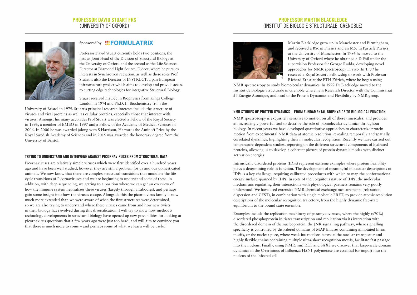
PROFESSOR DAVID STUART FRS (UNIVERSITY OF OXFORD)
Sponsored by
Professor David Stuart currently holds two positions; the first as Joint Head of the Division of Structural Biology at the University of Oxford and the second as the Life Sciences Director at Diamond Light Source, Didcot, where he pursues interests in Synchrotron radiation; as well as these roles Prof Stuart is also the Director of INSTRUCT, a pan-European infrastructure project which aims to develop and provide access to cutting edge technologies for integrative Structural Biology.
Stuart received his BSc in Biophysics from Kings College London in 1974 and Ph.D. In Biochemistry from the
University of Bristol in 1979. Stuart’s principal research interests include the structure of viruses and viral proteins as well as cellular proteins, especially those that interact with viruses. Amongst his many accolades Prof Stuart was elected a Fellow of the Royal Society in 1996, a member of EMBO in 1997 and a Fellow of the Academy of Medical Sciences in 2006. In 2006 he was awarded (along with S Harrison, Harvard) the Aminoff Prize by the Royal Swedish Academy of Sciences and in 2015 was awarded the honorary degree from the University of Bristol.
TRYING TO UNDERSTAND AND INTERVENE AGAINST PICORNAVIRUSES FROM STRUCTURAL DATAPicornaviruses are relatively simple viruses which were first identified over a hundred years ago and have been well studied, however they are still a problem for us and our domesticated animals. We now know that there are complex structural transitions that modulate the life cycle transitions of Picornaviruses and we are beginning to understand some of these, in addition, with deep sequencing, we getting to a position where we can get an overview of how the immune system neutralizes these viruses (largely through antibodies), and perhaps gain some insight into how the viruses escape. Alongside this the picornavirus family is now much more extended than we were aware of when the first structures were determined, so we are also trying to understand where these viruses came from and how new twists in their biology have evolved during this diversification. I will try to show how methods/technology developments in structural biology have opened up new possibilities for looking at picornavirus questions that a few years ago were just too hard, and will aim to convince you that there is much more to come – and perhaps some of what we learn will be useful!
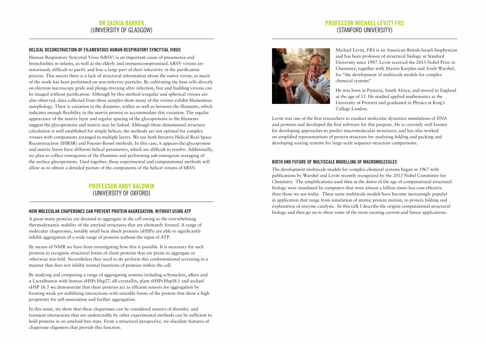
PROFESSOR MARTIN BLACKLEDGE (INSTITUT DE BIOLOGIE STRUCTURALE, GRENOBLE)
Martin Blackledge grew up in Manchester and Birmingham, and received a BSc in Physics and an MSc in Particle Physics at the University of Manchester. In 1984 he moved to the University of Oxford where he obtained a D.Phil under the supervision Professor Sir George Radda, developing novel approaches for NMR spectroscopy in vivo. In 1989 he received a Royal Society Fellowship to work with Professor Richard Ernst at the ETH Zürich, where he began using
NMR spectroscopy to study biomolecular dynamics. In 1992 Dr Blackledge moved to the Institut de Biologie Structurale in Grenoble where he is Research Director with the Commisariat à l’Energie Atomique, and head of the Protein Dynamics and Flexibility by NMR group.
NMR STUDIES OF PROTEIN DYNAMICS – FROM FUNDAMENTAL BIOPHYSICS TO BIOLOGICAL FUNCTION NMR spectroscopy is exquisitely sensitive to motion on all of these timescales, and provides an increasingly powerful tool to describe the role of biomolecular dynamics throughout biology. In recent years we have developed quantitative approaches to characterize protein motion from experimental NMR data at atomic resolution, revealing temporally and spatially correlated dynamics, highlighting their in molecular recognition. Recently we have carried out temperature-dependent studies, reporting on the different structural components of hydrated proteins, allowing us to develop a coherent picture of protein dynamic modes with distinct activation energies.
Intrinsically disordered proteins (IDPs) represent extreme examples where protein flexibility plays a determining role in function. The development of meaningful molecular descriptions of IDPs is a key challenge, requiring calibrated procedures with which to map the conformational energy surface spanned by IDPs. In spite of the ubiquitous nature of IDPs, the molecular mechanisms regulating their interactions with physiological partners remains very poorly understood. We have used extensive NMR chemical exchange measurements (relaxation dispersion and CEST), in combination with single molecule FRET, to provide atomic resolution descriptions of the molecular recognition trajectory, from the highly dynamic free-state equilibrium to the bound state ensemble.
Examples include the replication machinery of paramyxoviruses, where the highly (>70%) disordered phosphoprotein initiates transcription and replication via its interaction with the disordered domain of the nucleoprotein, the JNK signalling pathway, where signalling specificity is controlled by disordered domains of MAP kinases containing annotated linear motifs, or the nuclear pore, where weak interactions between the nuclear transporter and highly flexible chains containing multiple ultra-short recognition motifs, facilitate fast passage into the nucleus. Finally, using NMR, smFRET and SAXS we discover that large-scale domain dynamics in the C-terminus of Influenza H5N1 polymerase are essential for import into the nucleus of the infected cell.
DR SASKIA BAKKER (UNIVERSITY OF GLASGOW)
HELICAL RECONSTRUCTION OF FILAMENTOUS HUMAN RESPIRATORY SYNCYTIAL VIRUS Human Respiratory Syncytial Virus (hRSV) is an important cause of pneumonia and bronchiolitis in infants, as well as the elderly and immunocompromised. hRSV virions are notoriously difficult to purify and lose a large part of their infectivity in the purification process. This means there is a lack of structural information about the native virion, as much of the work has been performed on non-infective particles. By cultivating the host cells directly on electron microscopy grids and plunge-freezing after infection, free and budding virions can be imaged without purification. Although by this method irregular and spherical virions are also observed, data collected from these samples show many of the virions exhibit filamentous morphology. There is variation in the diameter, within as well as between the filaments, which indicates enough flexibility in the matrix protein to accommodate this variation. The regular appearance of the matrix layer and regular spacing of the glycoproteins in the filaments suggest the glycoproteins and matrix may be linked. Although three-dimensional structure calculation is well established for simple helices, the methods are not optimal for complex viruses with components arranged in multiple layers. We use both Iterative Helical Real-Space Reconstruction (IHRSR) and Fourier-Bessel methods. In this case, it appears the glycoprotein and matrix layers have different helical parameters, which are difficult to resolve. Additionally, we plan to collect tomograms of the filaments and performing sub-tomogram averaging of the surface glycoproteins. Used together, these experimental and computational methods will allow us to obtain a detailed picture of the components of the helical virions of hRSV.
PROFESSOR ANDY BALDWIN (UNIVERSITY OF OXFORD)
HOW MOLECULAR CHAPERONES CAN PREVENT PROTEIN AGGREGATION, WITHOUT USING ATPA great many proteins are doomed to aggregate in the cell owing to the overwhelming thermodynamic stability of the amyloid structures that are ultimately formed. A range of molecular chaperones, notably small heat shock proteins (sHSPs) are able to significantly inhibit aggregation of a wide range of proteins without the input of ATP.
By means of NMR we have been investigating how this is possible. It is necessary for such proteins to recognise structural forms of client proteins that are prone to aggregate or otherwise mis-fold. Nevertheless they need to do perform this conformational screening in a manner that does not inhibit normal functions of proteins within the cell.
By studying and comparing a range of aggregating systems including a-Synuclein, aBeta and a-Lactalbumin with human sHSPs Hsp27, aB-crystallin, plant sHSPs Hsp18.1 and archael sHSP 16.5 we demonstrate that these proteins act as efficient sensors for aggregation by forming weak yet stabilising interactions with unstable forms of the protein that show a high propensity for self-association and further aggregation.
In this sense, we show that these chaperones can be considered sensors of disorder, and transient interactions that are undetectable by other experimental methods can be sufficient to hold proteins in an amyloid-free state. From a structural perspective, we elucidate features of chaperone oligomers that provide this function.
PROFESSOR MICHAEL LEVITT FRS (STANFORD UNIVERSITY)
Michael Levitt, FRS is an American-British-Israeli biophysicist and has been professor of structural biology at Stanford University since 1987. Levitt received the 2013 Nobel Prize in Chemistry, together with Martin Karplus and Arieh Warshel, for “the development of multiscale models for complex chemical systems”
He was born in Pretoria, South Africa, and moved to England at the age of 15. He studied applied mathematics at the University of Pretoria and graduated in Physics at King’s College London.
Levitt was one of the first researchers to conduct molecular dynamics simulations of DNA and proteins and developed the first software for this purpose. He is currently well known for developing approaches to predict macromolecular structures, and has also worked on simplified representations of protein structure for analysing folding and packing and developing scoring systems for large-scale sequence-structure comparisons.
BIRTH AND FUTURE OF MULTISCALE MODELLING OF MACROMOLECULESThe development multiscale models for complex chemical systems began in 1967 with publications by Warshel and Levitt recently recognized by the 2013 Nobel Committee for Chemistry. The simplifications used then at the dawn of the age of computational structural biology were mandated by computers that were almost a billion times less cost-effective than those we use today. These same multiscale models have become increasingly popular in application that range from simulation of atomic protein motion, to protein folding and explanation of enzyme catalysis. In this talk I describe the origins computational structural biology and then go on to show some of the most exciting current and future applications.
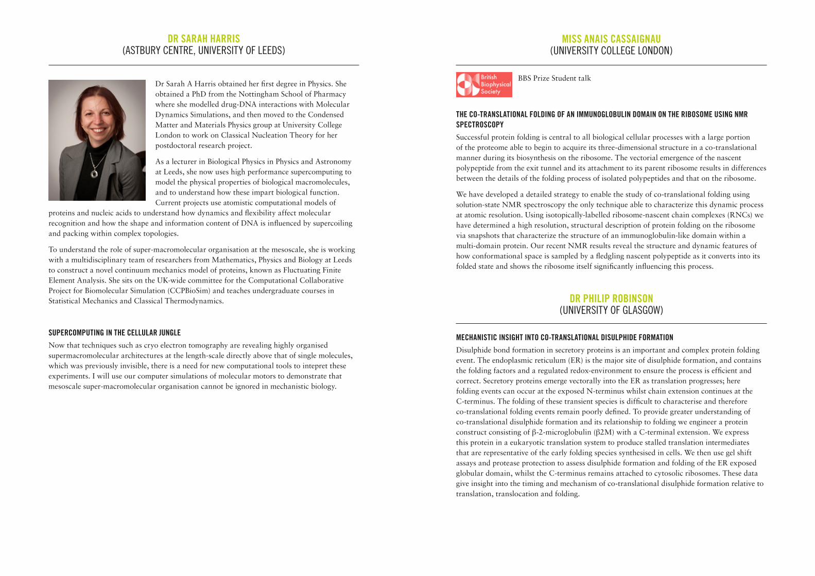
DR SARAH HARRIS (ASTBURY CENTRE, UNIVERSITY OF LEEDS)
Dr Sarah A Harris obtained her first degree in Physics. She obtained a PhD from the Nottingham School of Pharmacy where she modelled drug-DNA interactions with Molecular Dynamics Simulations, and then moved to the Condensed Matter and Materials Physics group at University College London to work on Classical Nucleation Theory for her postdoctoral research project.
As a lecturer in Biological Physics in Physics and Astronomy at Leeds, she now uses high performance supercomputing to model the physical properties of biological macromolecules, and to understand how these impart biological function. Current projects use atomistic computational models of
proteins and nucleic acids to understand how dynamics and flexibility affect molecular recognition and how the shape and information content of DNA is influenced by supercoiling and packing within complex topologies.
To understand the role of super-macromolecular organisation at the mesoscale, she is working with a multidisciplinary team of researchers from Mathematics, Physics and Biology at Leeds to construct a novel continuum mechanics model of proteins, known as Fluctuating Finite Element Analysis. She sits on the UK-wide committee for the Computational Collaborative Project for Biomolecular Simulation (CCPBioSim) and teaches undergraduate courses in Statistical Mechanics and Classical Thermodynamics.
SUPERCOMPUTING IN THE CELLULAR JUNGLENow that techniques such as cryo electron tomography are revealing highly organised supermacromolecular architectures at the length-scale directly above that of single molecules, which was previously invisible, there is a need for new computational tools to intepret these experiments. I will use our computer simulations of molecular motors to demonstrate that mesoscale super-macromolecular organisation cannot be ignored in mechanistic biology.
MISS ANAIS CASSAIGNAU (UNIVERSITY COLLEGE LONDON)
BBS Prize Student talk
THE CO-TRANSLATIONAL FOLDING OF AN IMMUNOGLOBULIN DOMAIN ON THE RIBOSOME USING NMR SPECTROSCOPYSuccessful protein folding is central to all biological cellular processes with a large portion of the proteome able to begin to acquire its three-dimensional structure in a co-translational manner during its biosynthesis on the ribosome. The vectorial emergence of the nascent polypeptide from the exit tunnel and its attachment to its parent ribosome results in differences between the details of the folding process of isolated polypeptides and that on the ribosome.
We have developed a detailed strategy to enable the study of co-translational folding using solution-state NMR spectroscopy the only technique able to characterize this dynamic process at atomic resolution. Using isotopically-labelled ribosome-nascent chain complexes (RNCs) we have determined a high resolution, structural description of protein folding on the ribosome via snapshots that characterize the structure of an immunoglobulin-like domain within a multi-domain protein. Our recent NMR results reveal the structure and dynamic features of how conformational space is sampled by a fledgling nascent polypeptide as it converts into its folded state and shows the ribosome itself significantly influencing this process.
DR PHILIP ROBINSON (UNIVERSITY OF GLASGOW)
MECHANISTIC INSIGHT INTO CO-TRANSLATIONAL DISULPHIDE FORMATIONDisulphide bond formation in secretory proteins is an important and complex protein folding event. The endoplasmic reticulum (ER) is the major site of disulphide formation, and contains the folding factors and a regulated redox-environment to ensure the process is efficient and correct. Secretory proteins emerge vectorally into the ER as translation progresses; here folding events can occur at the exposed N-terminus whilst chain extension continues at the C-terminus. The folding of these transient species is difficult to characterise and therefore co-translational folding events remain poorly defined. To provide greater understanding of co-translational disulphide formation and its relationship to folding we engineer a protein construct consisting of β-2-microglobulin (β2M) with a C-terminal extension. We express this protein in a eukaryotic translation system to produce stalled translation intermediates that are representative of the early folding species synthesised in cells. We then use gel shift assays and protease protection to assess disulphide formation and folding of the ER exposed globular domain, whilst the C-terminus remains attached to cytosolic ribosomes. These data give insight into the timing and mechanism of co-translational disulphide formation relative to translation, translocation and folding.
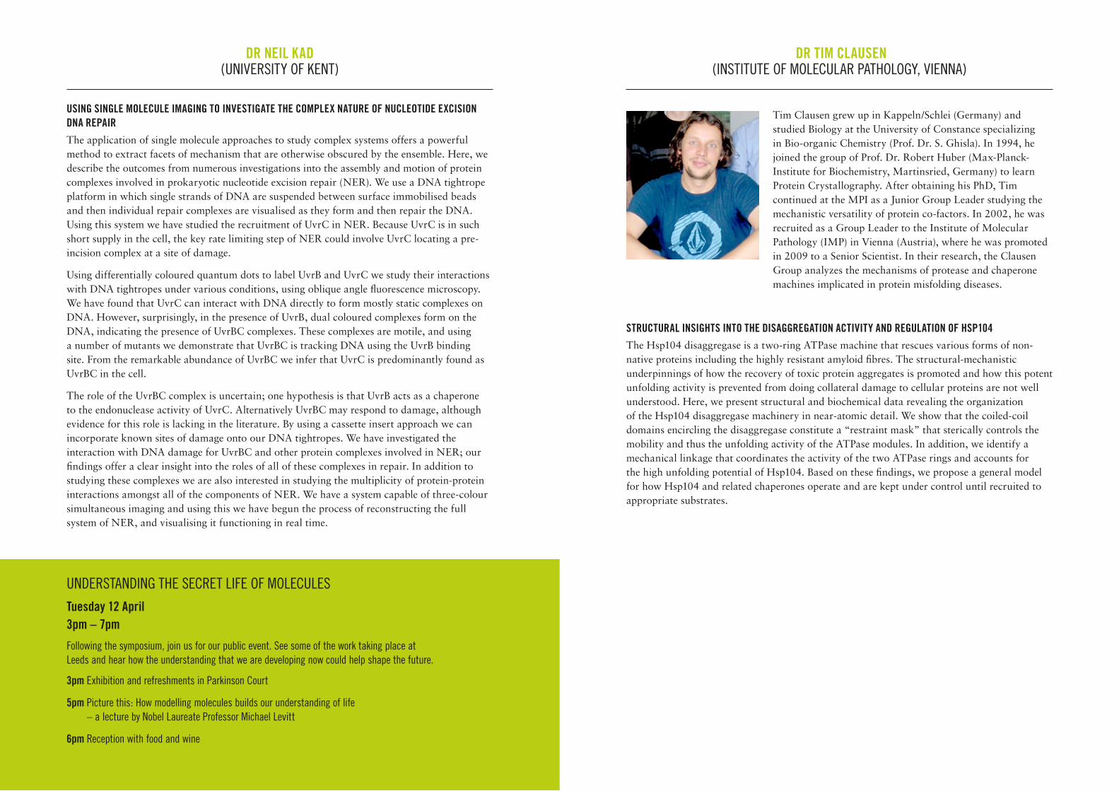
PROFESSOR HELEN SAIBIL, FMEDSCI, FRS (BIRKBECK, UNIVERSITY OF LONDON)
Helen Saibil is a Canadian-British biologist who is the Bernal Professor of Structural Biology at Birkbeck College, London. She obtained a BSc in Biophysics at McGill University in Montreal. Her PhD, on structural studies of retinal photoreceptors, was done at Kings College London, under the supervision of Maurice Wilkins. After a postdoc in the laboratory of Marc Chabre at the Centre d’Etudes Nucleaires, Grenoble, she did further work at Kings, then moved to Oxford University, and from there to Birkbeck, where she set up a cryo-electron microscopy lab to study macromolecular machines. Her current research involves molecular and cellular studies of molecular chaperones, protein folding
and misfolding, as well as protein refolding in membrane pore formation. She is a member of EMBO and a Fellow of the Royal Society and of the Academy of Medical Sciences, and a Member of the Academia Europaea.
MEMBRANE PORE-FORMING PROTEINS IN THE MOLECULAR ARMS RACE BETWEEN HOST AND PATHOGENPathogens have evolved weapons to invade and damage our cells, and our immune system has evolved defences against these attacks. Among the weaponry used by both sides in this continual war are proteins that punch holes in cell membranes. Membrane perforation enables pathogens to take over host cells and resources for their own replication, and also enables host immune systems to kill invading pathogens. The membrane attack complex-perforin (MACPF)/ cholesterol dependent cytolysin (CDC) superfamily of membrane pore-forming proteins is used by a wide range of pathogens as well as by host immune systems. This talk focuses on the mechanisms by which MACPF and CDC proteins convert from their soluble, monomeric forms into large arcs and rings that insert into membranes and perforate them. Studies of pore assembly on liposomes in vitro and examples of their action in vivo will be presented.
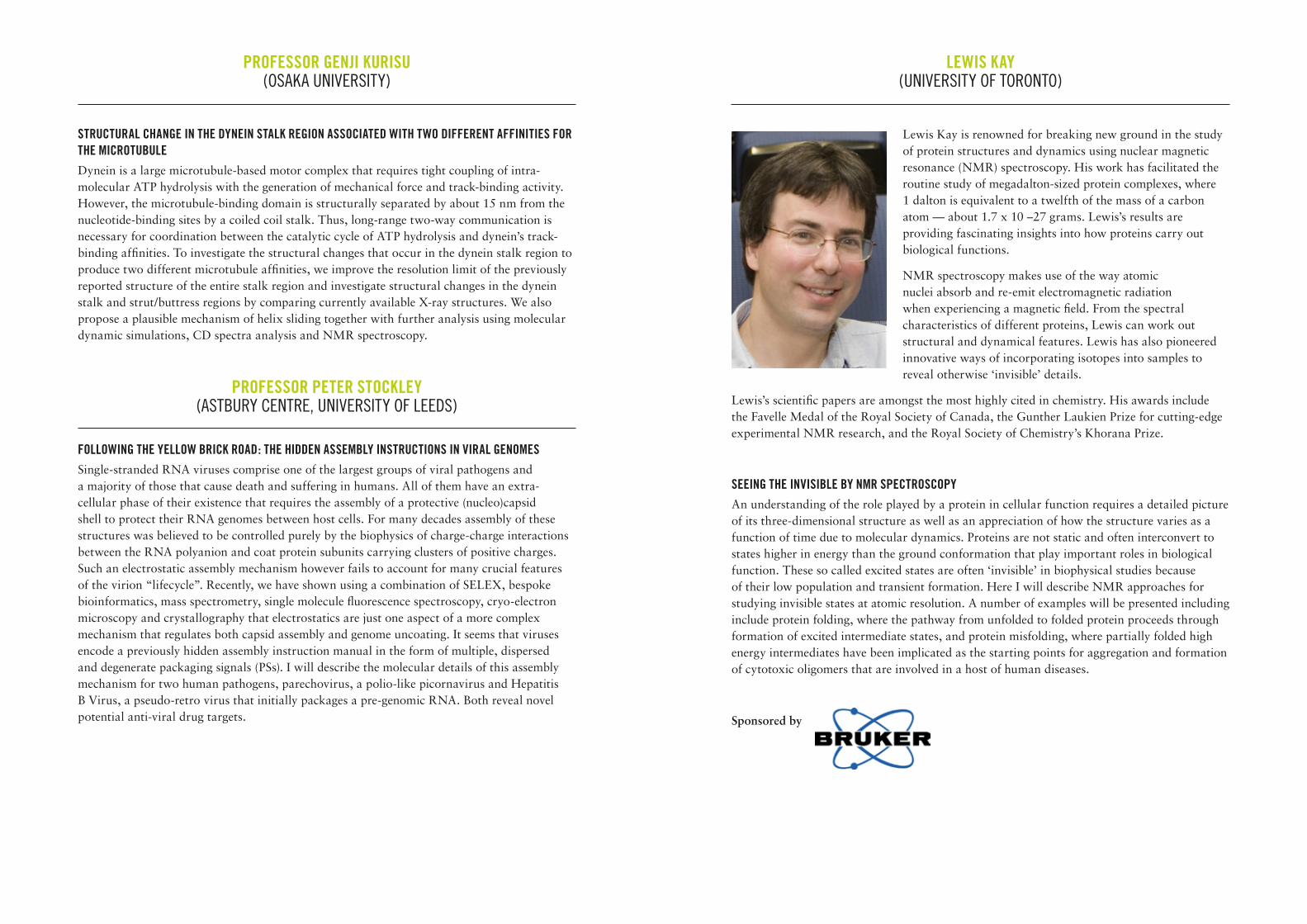
PROFESSOR DAVID BARFORD (UNIVERSITY OF CAMBRIDGE)
Dr. David Barford is the joint head of the Structural Studies division at the MRC Laboratory of Molecular Biology. Barford studied Biochemistry at the University of Bristol then went on to earn a DPhil from University of Oxford, supervised by Louise Johnson. Barford worked at the University of Dundee MRC Protein Phosphorylation Unit, with Philip Cohen and Tricia Cohen. He was a Cold Spring Harbor Laboratory Fellow (CSHL, NY) from 1991-1994. In 1994, he was appointed University Lecturer at the University of Oxford. From 1999 to 2013 Barford was co-Head of the
Division of Structural Biology at the Institute of Cancer Research in London. He moved to the MRC Laboratory of Molecular Biology in 2013.
MOLECULAR BASIS FOR REGULATION OF THE ANAPHASE-PROMOTING COMPLEX IN MITOSISThe anaphase-promoting complex/cyclosome (APC/C) is a large multimeric RING E3 ubiquitin ligase that controls chromosome segregation and mitotic exit. Its regulation by coactivator subunits, phosphorylation and the mitotic checkpoint complex ensures the correct order and timing of distinct cell cycle transitions. Two E2s, UbcH10 and Ube2S are responsible for assembly of polyubiquitin chains on APC/C substrates that include cyclins, mitotic kinases and spindle assembly factors.
We have used single particle cryo-electron microscopy to determine different functional states of the APC/C. I will discuss these structures and explain how the APC/C catalyses protein ubiquitination reactions and how the APC/C is regulated.
DR NEIL KAD(UNIVERSITY OF KENT)
USING SINGLE MOLECULE IMAGING TO INVESTIGATE THE COMPLEX NATURE OF NUCLEOTIDE EXCISION DNA REPAIRThe application of single molecule approaches to study complex systems offers a powerful method to extract facets of mechanism that are otherwise obscured by the ensemble. Here, we describe the outcomes from numerous investigations into the assembly and motion of protein complexes involved in prokaryotic nucleotide excision repair (NER). We use a DNA tightrope platform in which single strands of DNA are suspended between surface immobilised beads and then individual repair complexes are visualised as they form and then repair the DNA. Using this system we have studied the recruitment of UvrC in NER. Because UvrC is in such short supply in the cell, the key rate limiting step of NER could involve UvrC locating a pre-incision complex at a site of damage.
Using differentially coloured quantum dots to label UvrB and UvrC we study their interactions with DNA tightropes under various conditions, using oblique angle fluorescence microscopy. We have found that UvrC can interact with DNA directly to form mostly static complexes on DNA. However, surprisingly, in the presence of UvrB, dual coloured complexes form on the DNA, indicating the presence of UvrBC complexes. These complexes are motile, and using a number of mutants we demonstrate that UvrBC is tracking DNA using the UvrB binding site. From the remarkable abundance of UvrBC we infer that UvrC is predominantly found as UvrBC in the cell.
The role of the UvrBC complex is uncertain; one hypothesis is that UvrB acts as a chaperone to the endonuclease activity of UvrC. Alternatively UvrBC may respond to damage, although evidence for this role is lacking in the literature. By using a cassette insert approach we can incorporate known sites of damage onto our DNA tightropes. We have investigated the interaction with DNA damage for UvrBC and other protein complexes involved in NER; our findings offer a clear insight into the roles of all of these complexes in repair. In addition to studying these complexes we are also interested in studying the multiplicity of protein-protein interactions amongst all of the components of NER. We have a system capable of three-colour simultaneous imaging and using this we have begun the process of reconstructing the full system of NER, and visualising it functioning in real time.
DR TIM CLAUSEN (INSTITUTE OF MOLECULAR PATHOLOGY, VIENNA)
Tim Clausen grew up in Kappeln/Schlei (Germany) and studied Biology at the University of Constance specializing in Bio-organic Chemistry (Prof. Dr. S. Ghisla). In 1994, he joined the group of Prof. Dr. Robert Huber (Max-Planck-Institute for Biochemistry, Martinsried, Germany) to learn Protein Crystallography. After obtaining his PhD, Tim continued at the MPI as a Junior Group Leader studying the mechanistic versatility of protein co-factors. In 2002, he was recruited as a Group Leader to the Institute of Molecular Pathology (IMP) in Vienna (Austria), where he was promoted in 2009 to a Senior Scientist. In their research, the Clausen Group analyzes the mechanisms of protease and chaperone machines implicated in protein misfolding diseases.
STRUCTURAL INSIGHTS INTO THE DISAGGREGATION ACTIVITY AND REGULATION OF HSP104The Hsp104 disaggregase is a two-ring ATPase machine that rescues various forms of non-native proteins including the highly resistant amyloid fibres. The structural-mechanistic underpinnings of how the recovery of toxic protein aggregates is promoted and how this potent unfolding activity is prevented from doing collateral damage to cellular proteins are not well understood. Here, we present structural and biochemical data revealing the organization of the Hsp104 disaggregase machinery in near-atomic detail. We show that the coiled-coil domains encircling the disaggregase constitute a “restraint mask” that sterically controls the mobility and thus the unfolding activity of the ATPase modules. In addition, we identify a mechanical linkage that coordinates the activity of the two ATPase rings and accounts for the high unfolding potential of Hsp104. Based on these findings, we propose a general model for how Hsp104 and related chaperones operate and are kept under control until recruited to appropriate substrates.
UNDERSTANDING THE SECRET LIFE OF MOLECULES
Tuesday 12 April 3pm – 7pm
Following the symposium, join us for our public event. See some of the work taking place at Leeds and hear how the understanding that we are developing now could help shape the future.
3pm Exhibition and refreshments in Parkinson Court
5pm Picture this: How modelling molecules builds our understanding of life – a lecture by Nobel Laureate Professor Michael Levitt
6pm Reception with food and wine
PROFESSOR GENJI KURISU (OSAKA UNIVERSITY)
STRUCTURAL CHANGE IN THE DYNEIN STALK REGION ASSOCIATED WITH TWO DIFFERENT AFFINITIES FOR THE MICROTUBULEDynein is a large microtubule-based motor complex that requires tight coupling of intra-molecular ATP hydrolysis with the generation of mechanical force and track-binding activity. However, the microtubule-binding domain is structurally separated by about 15 nm from the nucleotide-binding sites by a coiled coil stalk. Thus, long-range two-way communication is necessary for coordination between the catalytic cycle of ATP hydrolysis and dynein’s track-binding affinities. To investigate the structural changes that occur in the dynein stalk region to produce two different microtubule affinities, we improve the resolution limit of the previously reported structure of the entire stalk region and investigate structural changes in the dynein stalk and strut/buttress regions by comparing currently available X-ray structures. We also propose a plausible mechanism of helix sliding together with further analysis using molecular dynamic simulations, CD spectra analysis and NMR spectroscopy.
PROFESSOR PETER STOCKLEY (ASTBURY CENTRE, UNIVERSITY OF LEEDS)
FOLLOWING THE YELLOW BRICK ROAD: THE HIDDEN ASSEMBLY INSTRUCTIONS IN VIRAL GENOMESSingle-stranded RNA viruses comprise one of the largest groups of viral pathogens and a majority of those that cause death and suffering in humans. All of them have an extra-cellular phase of their existence that requires the assembly of a protective (nucleo)capsid shell to protect their RNA genomes between host cells. For many decades assembly of these structures was believed to be controlled purely by the biophysics of charge-charge interactions between the RNA polyanion and coat protein subunits carrying clusters of positive charges. Such an electrostatic assembly mechanism however fails to account for many crucial features of the virion “lifecycle”. Recently, we have shown using a combination of SELEX, bespoke bioinformatics, mass spectrometry, single molecule fluorescence spectroscopy, cryo-electron microscopy and crystallography that electrostatics are just one aspect of a more complex mechanism that regulates both capsid assembly and genome uncoating. It seems that viruses encode a previously hidden assembly instruction manual in the form of multiple, dispersed and degenerate packaging signals (PSs). I will describe the molecular details of this assembly mechanism for two human pathogens, parechovirus, a polio-like picornavirus and Hepatitis B Virus, a pseudo-retro virus that initially packages a pre-genomic RNA. Both reveal novel potential anti-viral drug targets.
LEWIS KAY (UNIVERSITY OF TORONTO)
Lewis Kay is renowned for breaking new ground in the study of protein structures and dynamics using nuclear magnetic resonance (NMR) spectroscopy. His work has facilitated the routine study of megadalton-sized protein complexes, where 1 dalton is equivalent to a twelfth of the mass of a carbon atom — about 1.7 x 10 –27 grams. Lewis’s results are providing fascinating insights into how proteins carry out biological functions.
NMR spectroscopy makes use of the way atomic nuclei absorb and re-emit electromagnetic radiation when experiencing a magnetic field. From the spectral characteristics of different proteins, Lewis can work out structural and dynamical features. Lewis has also pioneered innovative ways of incorporating isotopes into samples to reveal otherwise ‘invisible’ details.
Lewis’s scientific papers are amongst the most highly cited in chemistry. His awards include the Favelle Medal of the Royal Society of Canada, the Gunther Laukien Prize for cutting-edge experimental NMR research, and the Royal Society of Chemistry’s Khorana Prize.
SEEING THE INVISIBLE BY NMR SPECTROSCOPYAn understanding of the role played by a protein in cellular function requires a detailed picture of its three-dimensional structure as well as an appreciation of how the structure varies as a function of time due to molecular dynamics. Proteins are not static and often interconvert to states higher in energy than the ground conformation that play important roles in biological function. These so called excited states are often ‘invisible’ in biophysical studies because of their low population and transient formation. Here I will describe NMR approaches for studying invisible states at atomic resolution. A number of examples will be presented including include protein folding, where the pathway from unfolded to folded protein proceeds through formation of excited intermediate states, and protein misfolding, where partially folded high energy intermediates have been implicated as the starting points for aggregation and formation of cytotoxic oligomers that are involved in a host of human diseases.
Sponsored by
Astex is a leader in innovative drug discovery and development, committed to the fight against cancer and diseases of the central nervous system. We are a leader in innovative small-molecule
therapeutics with particular expertise in fragment-based drug discovery, the most important advance in discovery chemistry in the last 20 years. Although our discovery platform has been successfully applied to therapeutic targets addressing a variety of disease areas, our primary clinical area of focus is currently cancer. In addition, we are expanding our capabilities and building expertise in the area of fragment-based drug discovery diseases of the CNS. For more information about Astex Pharmaceuticals, visit www.astx.com. For more information about our parent company, Otsuka Pharmaceutical, visit www.otsuka.com/en/
Bruker Corporation is a manufacturer of scientific instruments for molecular and materials research, as well as for industrial and applied analysis.
Bruker systems cover a broad spectrum of applications in all fields of research and development and are used in all industrial production processes for the purpose of ensuring quality and process reliability. Bruker continues to build upon its extensive range of products and solutions, its broad base of installed systems and a strong reputation among its customers. Being one of the world’s leading analytical instrumentation companies, Bruker is strongly committed to further fully meet its customers’ needs as well as to continue to develop state-of-the-art technologies and innovative solutions for today’s analytical questions.
With over 60 years of innovation and leadership, FEI enables customers to find meaningful answers to questions that accelerate breakthrough discoveries, increase productivity, and ultimately change the world. FEI designs, manufactures, and supports the broadest range of high-performance microscopy workflows that provide images and answers in the micro-, nano-, and picometer scales. Combining hardware and software expertise in electron, ion, and light microscopy with deep application knowledge in the materials science, life sciences, electronics, and natural resources markets, the worldwide FEI team of 2,700+ employees is dedicated to customers’ pursuit of discovery and resolution to global challenges.
ASTBURY CONVERSATION 2016 SPONSORS
Formulatrix was established in 2002 to provide protein crystallization automation solutions. Since then, we’ve started developed the next generation of liquid handlers using microfluidic technology. Headquartered in Bedford, Massachusetts, we supply software and robotic automation solutions to leading pharmaceutical companies and academic research institutions around the world. Our team works tirelessly to provide the best products in the industry with support that is second to none.
Lhasa Limited is a not-for-profit organisation and educational charity that facilitates collaborative data sharing projects in the pharmaceutical, cosmetics and chemistry-related industries. A pioneer in the production of knowledge-based systems for forward thinking scientists, Lhasa Limited
continues to draw on over thirty years of experience to create user-friendly, state of the art in silico prediction and database systems for use in metabolism, toxicology and related sciences.
MedImmune pioneers innovative research across key therapeutic areas including RIA, cardiovascular & metabolic
disease, oncology, neuroscience, and more. We develop medicines for unmet medical needs worldwide. We’re an industry leader in personalized medicine – developing new products that may allow doctors to prescribe a specific, customized treatment for each patient’s individual needs. We are the global biologics research and development arm of our parent company AstraZeneca, a recognized global leader in biopharmaceutical research and development. By combining AstraZeneca’s worldwide presence with our distinct and unique culture, processes, and standards, we continue to solve global healthcare challenges, redefine science, deliver better medicines, and help people live longer, healthier lives.
Life Sciences – From Cell Culture to Protein Purification and beyond. From cell culture and protein expression, through to protein purification, Pall Laboratory partners with you to provide a
wide range of sterile filtration, ultrafiltration and detection products.
Thermo Scientific help scientists meet the challenges they face in the lab or in the field every day. From routine analysis to new discoveries, our innovations help professionals do the science
they need to do, the way they want to do it. Our high-end analytical instruments, laboratory equipment, software, services, consumables and reagents help our customers solve complex analytical challenges, improve patient diagnostics and increase laboratory productivity.
We would like to express our appreciation to the following organisations for their sponsorship of the Astbury Conversation 2016
AstexBibby BioOutsourceBiotexBritish Biophysical Society Bruker
Dell ComputersFEIFormulatrixGEHiChromLhasa
Medimmune PALLThermoUCBVWRWellcome Trust
UCB aspires to be the patient-centric global biopharma leader, making a difference to the lives of people living with severe diseases. UCB is a global biopharmaceutical company, with a focus on neurology and immunology.
Rather than starting researching any new drug with the science alone, we want to better connect patients with science and science with patients. With no such thing as an “average patient”, we want to use all the tools, channels and scientific advances at our disposal to better understand the various expressions of a disease and embed the real needs of specific patient populations in our science and innovation process. This holistic approach to care will ultimately ensure the right drug and the right care reach the right patients to help them live the lives they choose
At VWR, we enable science by supplying critical products to the world’s top Pharmaceutical, Healthcare, Biotech, Industrial, Food and Beverage, Educational, and
Governmental organisations as well as specialist areas such as Microbiology, Chromatography, Cell Diagnostics and Life Sciences. We provide our customers with an expansive choice of premier products, such as Laboratory Chemicals, Glassware and Plastic ware, Equipment and Instruments, Furniture, Protective Safety Clothing and Safety Products, as well as other Life Science and Laboratory Products and Supplies. We offer over 1,500,000 products and represent in excess of 5,000 of the world’s finest manufacturers to meet our customers’ ever growing requirements.
Name: Peter Adams Title: Redesigning photosynthetic membranes: development of bio-inspired photonic
nanomaterials
Biological cell membranes rely upon hierarchical organization to elicit functional responses. In natural photosynthetic membranes, light harvesting (LH) membrane protein complexes act as a framework for coordination of chromophores which absorb solar energy and channel it to downstream bioenergetic processes. Supported lipid bilayers (SLBs) are established as simple model membranes, into which membrane proteins can be incorporated. As an alternative to lipids, certain diblock copolymers can from membrane-mimetic systems with potential advantages (increased robustness, functionality, responsivity). Here, we present research into two different bio-inspired LH systems.
Firstly, a modular, artificial LH system is described, where amphiphilic diblock polymers, poly(ethylene oxide)-block-poly(butadiene), act a matrix for noncovalent arrangement of BODIPY energy donor and bacteriochlorin energy acceptor chromophores. The polymer/ chromophore composites form nanoscale micelles in aqueous solution and defect-free monolayer and bilayer films on solid substrates. Donor-acceptor Forster resonance energy transfer is shown by steady state and time-resolved fluorescence spectroscopy and the system is modelled by theoretical calculations. Supported polymer bilayers demonstrated energy transfer efficiency up to 90%. Secondly, we present ongoing research into the redesign of protein/ lipid LH systems. Purified plant proteins and lipids are used as building blocks to form novel reconstructed protein/SLBs with defined compositions and 3-D organization using a combination of surface patterning and photolithography techniques. Atomic force and fluorescence microscopy and spectroscopy show protein arrangement and light harvesting functionality can be controlled. These new protein/chromophore and polymer/chromophore bio-inspired systems could act as a platforms to investigate membrane self-assembly and organization and could lead to applications in chip-based nanodevices.
Name: Zainab Ahdash Title: Native MS coupled with modelling provides novel structural insights into the
helicase-nuclease (HerA-NurA) system
The helicase–nuclease complex (HerA-NurA) is implicated in repairing double-stranded DNA breaks. In particular, the HerA ATPase cooperates with the NurA nuclease enabling the generation of a channel that transverses the HerA-NurA complex where the DNA is translocated and processed. Here, we used mass spectrometry (MS)-based approaches to study the assembly structure and dynamics of the HerA-NurA complex and in response to DNA and ATP binding. Using native MS, we established a 6:2 composition of the HerA-NurA complex consistent with previous findings (Byrne et al., 2014). Utilising tandem MS and in-solution disruption, we performed dissociation experiments revealing a possible mechanism of cooperatively between the two proteins consistent with the “opening” of the ATPase and nuclease subcomplexes, followed by the recruitment of dsDNA.
POSTER ABSTRACTS
Coupling native MS with ion mobility (IM-MS), a method that allows separation of ions based on their overall shape (Zhou, Politis et al, 2014), we calculated the orientationally averaged collision cross section (CCS) of the identified (sub)complexes enabling us to model the gas-phase HerA-NurA complex structure, in close agreement with a recently published electron microscopy map (Byrne et al., 2014). By mixing the complex with a 25 base pairs dsDNA and non-hydrolyzable ATP analogs, we unravelled a novel mechanism of synergistic binding initiated by the recruitment of dsDNA and followed by the simultaneous binding of six ATPs. In particular, the addition of DNA has a subtle effect on the overall structure as measured by IM-MS, consistent with the binding in the internal cavity formed by the two protein subunits. Overall, our findings suggest a novel mode of cooperation between the two proteins enabling nucleotide binding and DNA translocation in the helicase-nuclease system.
Name: Lizzie Allan Email: [email protected] Title: Probing allostery in IgE-Fc using anti-IgE antibodies
The interaction between immunoglobulin E (IgE) and its ‘low affinity’ receptor, CD23, regulates a number of important processes in allergic disorders. The Fc fragment of IgE, which binds CD23, has a high degree of conformational plasticity and can be allosterically regulated. We have used X-ray crystallography to determine the structures of transiently populated conformations of IgE-Fc trapped by anti-IgE Fabs. In combination with kinetic and thermodynamic molecular interaction studies,, these structures are helping us gain a greater understanding of the energy landscape of IgE-Fc and the importance of IgE-Fc dynamics in receptor engagement. We have identified a! nd characterised conformations of IgE-Fc that cannot bind CD23 as a result of allosterically-mediated changes in domain orientations, intra-domain re-organization and perturbations in dynamics. This research not only advances our knowledge of a central molecule in allergic disease, but it is also providing insights that may enable the development of therapeutic approaches that take advantage of IgE’s allosteric and dynamic properties.
Name: Teresa Almeida Email: [email protected] Title: Challenges of targeting dynamic protein binding sites with small molecules:
EB1 interaction with SxIP motifs
EB1 is a key element in macromolecular interactions at the microtubule plus ends, having a fundamental role in microtubule polymerisation. Several disease states have been associated with EB1, such as cancer and neuronal diseases. While the N-terminal domain binds to microtubules plus ends, the C-terminal domain (EB1c) recruits a vast range of other proteins that have been shown to exert different regulatory functions on microtubule behaviour – stabilising the polymerisation or creating instability that leads to depolymerisation. Diverse EB1c binding partners are recognised through a conserved SxIP motif within an intrinsically disordered region enriched in basic, serine, and proline, residues. Crystal structure of EB1c in complex with a peptide containing the SxIP motif demonstrates that the isoleucine-proline dipeptide is inserted into a well-defined cavity of EB1 that may be suitable for small molecule targeting. Based on crystal structure of the complex, we identified a molecular scaffold that acts as SxIP motif mimetic, by using a combination of ligand and structure-based virtual screening approaches and solved the NMR structure of the EB1 complex with small molecules
that include the scaffold. Further analysis of the previous published crystal structure, in addition to our NMR data, shows that part of the binding site is formed by the initially unstructured region at the C-terminus of EB1c that folds on complex formation. Despite fitting well into the binding site the small molecules interacted weakly and failed to induce the fold of the C-terminal region. By using a range of SxIP containing peptides we identified additional interactions of EB1c with ligands that are required for engaging the dynamic C-terminal region of EB1c and high binding affinity. We propose a sequential model of the EB1 interaction with targets where the partially formed SxIP binding pocket is initially reco! gnised, followed by the subsequent stabilisation of the unstructured region. We discuss the use of the model for the design of the next generation of small molecule inhibitors.
Name: Irene Arrata Title: Using Adhirons to identify high affinity peptide/helix mimetics interactions
The design of oligomeric folded molecules with 3D structural complexity approaching that of tertiary protein structure is a major challenge in supramolecular chemical biology.[1] Whilst some progress has been made with the de novo design of tertiary foldamers,[2] these approaches employ limited sequence diversity and result in highly symmetrical 3D structures. Alternatively, it may be possible to replace parts of bio-macromolecules sequences with non-natural building blocks.[3]
We have chosen to pursue the latter approach by studying the recognition between helix mimetics and proteins. In the current work, we focus on interactions with peptides and employ biological selection methods to accelerate the discovery of optimised amino acid sequences that bind to helix mimetics (Figure 1). This approach could ultimately be used 1) as a reverse screening method for discovering protein-protein interactions inhibitors by mining informatics databases for the selected sequences, 2) to build mimetic/peptide hybrids with well-defined tertiary folds.
Using orthogonal functionalisation,[4] we biotinylated N-alkylated aromatic oligoamides p53 mimetics and performed Adhiron display[5] to generate a randomised library of high affinity but selective binding proteins. The obtained hits were subcloned and expressed, and several attempts to confirm the binding were performed, using various methods. Ongoing work will establish whether the orthogonal group is involved in the binding, or if the non-biotinylated proteomimetic itself is sufficient.
Name: Saskia Bakker Email: [email protected] Title: Helical reconstruction of filamentous human Respiratory Syncytial Virus
Human Respiratory Syncytial Virus (hRSV) is an important cause of pneumonia and bronchiolitis in infants, as well as the elderly and immunocompromised. hRSV virions are notoriously difficult to purify and lose a large part of their infectivity in the purification process. This means there is a lack of structural information about the native virion, as much of the work has been performed on non-infective particles. By cultivating the host cells directly on electron microscopy grids and plunge-freezing after infection, free and budding virions can be imaged without purification. Although by this method irregular and spherical virions are also obs! erved, data collected from these samples show many of the virions exhibit filamentous morphology. There is variation in the diameter, within as well as between
the filaments, which indicates enough flexibility in the matrix protein to accommodate this variation. The regular appearance of the matrix layer and regular spacing of the glycoproteins in the filaments suggest the glycoproteins and matrix may be linked. Although three-dimensional structure calculation is well established for simple helices, the methods are not optimal for complex viruses with components arranged in multiple layers. We use both Iterative Helical Real-Space Reconstruction (IHRSR) and Fourier-Bessel methods. In this case, it appears the glycoprotein and matrix layers have different helical parameters, which are difficult to resolve. Additionally, we plan to collect tomograms of the filaments and performing sub-tomogram averaging of the surface glycoproteins. Used together, these experimental and computational methods will allow us to obtain a detailed picture of the components of the helical virions of hRSV.
Name: Banushan Balansethupathy Title: Evaluating the role of protein dynamics in dsRNA recognition
MicroRNAs (miRNAs) play a vital role in post-transcriptional gene regulation through RNA interference. The biogenesis of these small non-coding RNAs involves a series distinct enzymatic processing steps each of which requires different modular RNA binding proteins. Two multi domain double-stranded (ds) RNA-binding proteins (dsRBPs) – TAR element binding protein (TRBP) and the Protein Activator of PKR (PACT) – associate with Dicer and have been shown to facilitate the processing of miRNAs. TRBP and PACT are related dsRBPs; each contains three dsRNA-binding domains (dsRBDs) that are separated by unstructured linker regions. In both proteins the two N-terminal domains bind to dsRNA while the C-terminal third domain mediates protein-protein interactions. The two domains of TRBP that do bind dsRNA have been shown to interact with different binding affinities. The dsRNA binding properties of PACT have not been extensively studied. We are investigating the origins of the differential binding properties in these related multi-domain proteins and its significance for miRNA processing.
Name: Andy Baldwin Title: How molecular chaperones can prevent protein aggregation, without using ATP
A great many proteins are doomed to aggregate in the cell owing to the overwhelming thermodynamic stability of the amyloid structures that are ultimately formed. A range of molecular chaperones, notably small heat shock proteins (sHSPs) are able to significantly inhibit aggregation of a wide range of proteins without the input of ATP.
By means of NMR we have been investigating how this is possible. It is necessary for such proteins to recognise structural forms of client proteins that are prone to aggregate or otherwise mis-fold. Nevertheless they need to do perform this conformational screening in a manner that does not inhibit normal functions of proteins within the cell.
By studying and comparing a range of aggregating systems including a-Synuclein, aBeta and a-Lactalbumin with human sHSPs Hsp27, aB-crystallin, plant sHSPs Hsp18.1 and archael sHSP 16.5 we demonstrate that these proteins act as efficient sensors for aggregation by forming weak yet stabilising interactions with unstable forms of the protein that show a high propensity for self association and further aggregation.
In this sense, we show that these chaperones can be considered sensors of disorder, and
transient interactions that are undetectable by other experimental methods can be sufficient to hold proteins in an amyloid-free state. From a structural perspective, we elucidate features of chaperone oligomers that provide this function.
Name: Matt Batchelor Title: Structural dynamics in single α-helical domains
Using a combination of NMR, molecular modelling and other biophysical techniques we are investigating the properties of long, isolated single α-helices. These unusual structures are found in unconventional myosins as well as other proteins. We are interested in the interactions that govern helix stability, the dynamic behaviour of potential salt bridge pairings between the many charged side-chains, and the flexibility of the helix as a whole.
Name: Hester Beard Title: Chemical tools to identify the molecular target of a small molecule effective against
glioblastoma multiforme
Glioblastoma multiforme (GBM) is the most malignant form of brain cancer among adults, with recurrence following roughly 7 months after treatment.1 The development of novel therapies for GBM is challenging due to problems with resistance and damage to unaffected areas of the brain.2 We have identified a brain-penetrable small molecule which selectively induces the self-destruction of human glioblastoma cells through a new mechanism, which is also effective against a range of patient-derived GBM cell models.
A photo-reactive benzophenone probe was synthesised to identify the molecular target of the small molecule in GBM cells. The probe was found to retain its activity relative to the small molecule in a cell viability assay. Chemical proteomics was used to identify a new molecular target. Biophysical measurements will be used to validate the interaction between the small molecule and the target using a set of designed chemical probes
Name: Tharin Blumenschein Title: Decoding the Structure and function of WWP2 WW domains
The WWP2 E3 ubiquitin ligase has previously been shown to regulate TGFbeta/Smad signalling activity linked to cancer metastasis. The WWP2 gene also has the capacity to generate several different isoforms through alternative slicing, which each contain unique combinations of protein binding and active ubiquitin ligase domains. Substrate selection and subsequent ubiquitin-mediated degradation at the proteasome is enabled by the four WW domains located between the catalytic HECT domain at the C-terminal and the N-terminal C2 phospholipid binding domain. Three isoforms of WWP2 containing unique combinations of the four individual WW domains have been found to have different substrate preference in the TGFbeta pathway, either favouring recruitment of inhibitory I-Smad7 or activating R-Smad2/3. Here we use NMR spectroscopy to examine the structure of the fourth WW domain of WWP2 in fusion with the GB1 solubility enhancing tag and observe its binding characteristics with specific Smad substrate peptides that contain WW domain recognition motifs.
Name: Doryen Bubeck
The membrane attack complex (MAC) is a fundamental component of immune defence that drills holes in bacterial membranes and Kill pathogens. MAC lesions were first identified in 1964, yet half a century later details of its structure and assembly mechanism remain undiscovered. Here we use electron cryo-microscopy to visualize the human pore complex at subnanometer resolution. We determine the protein composition of the MAC and identify interaction interfaces that hold the assembly together. Unlike closely related pore-forming proteins, the MAC’s asymmetric pore and “split-washer” shape suggest a killing mechanism that involves not only membrane rupture, but also distortion.
Name: Samuel Bunce
Understanding the self-assembly of proteins/peptides into highly ordered supramolecular structures is of key importance; both as a fundamental biological process and to elucidate the underlying mechanisms of pathological disease states such as amyloidosis .1 Studying these highly complex systems, that involve many energetically different assembly pathways and intermediary structures, requires the use of a wide range of analytical and biophysical techniques. One of the most applicable is Photoinduced Crosslinking (PIC) in which transient and/or weak supramolecular connectivity is transformed into a stable covalent form, providing analytically tractable products under conditions that may ordinarily produce disassembly.2,3 Therefore this technique is particularly well suited to explore amyloidogenic peptide systems, such as fragments from the Alzheimer’s beta (Aβ) peptide, in which fleeting intermediary structures may play a key role in the pathogenesis of degenerative diseases. This presentation will describe the synthesis of a photoactivatable amino acid analogue (TFMD-Phe) and preliminary studies on A–16-22 fibril formation.
Name: Julie Busch
Centrioles are the microtubule organising centres of the cell and as such play an important role in may cellular functions. The duplication of centrioles occurs once per cell cycle in a highly regulated manner. Errors in the duplication process lead to severe conditions such as ciliopathies and primary microcephaly.
Here we study the interaction of the human proteins; SAS-6 – the well characterised structural core of the forming centriole- and STIL – essential for centriole duplication, but its function and structure yet to be determined.
In vivo localisation experiments and co-immunoprecipitation assays have shown the possible role of STIL as interaction partner of the extended coiled coil region of SAS-6 and hint at a phosphorylation dependence of this interaction.
We show the direct interaction of the intrinsically disordered STAN motif found in STIL with the SAS-6 coiled coil region by NMR.
In an effort to pinpoint the interaction epitopes we identify a 100 amino acid long region of the SAS-6 coiled coil to be sufficient for interaction.
Fluorescence polarisation experiments confirm our observation. Using STIL STAN constructs with phosphomimiking mutations suggested to have an effect in in vivo experiments, we follow up on the observed possible effect of phosphorylation on the interaction.
Currently we are working on using this technique to compare all posible phosphorylation sites in the STAN motif and their effect on the interaction. We hope to utilise a technique allowing us to express protein with poshophorylated serine residues to evaluate the use of phosphomimiking mutations in such experiments.
Name: Anais Cassaignau Title: The co-translational folding of an immunoglobulin domain on the ribosome using
NMR spectroscopy
Successful protein folding is central to all biological cellular processes with a large portion of the proteome able to begin to acquire its three-dimensional structure in a co-translational manner during its biosynthesis on the ribosome. The vectorial emergence of the nascent polypeptide from the exit tunnel and its attachment to its parent ribosome results in differences between the details of the folding process of isolated polypeptides and that on the ribosome.
We have developed a detailed strategy to enable the study of co-translational folding using solution-state NMR spectroscopy the only technique able to characterize this dynamic process at atomic resolution. Using isotopically-labelled ribosome-nascent chain complexes (RNCs) we have determined a high resolution, structural description of protein folding on the ribosome via snapshots that characterize the structure of an immunoglobulin-like domain within a multi-domain protein. Our recent NMR results reveal the structure and dynamic features of how conformational space is sampled by a fledgling nascent polypeptide as it converts into its folded state and shows the ribosome itself significantly influencing this process.
Name: Rebecca Chandler-Bostock Title: Enterovirus E empty capsids: Dead-end product or viral building block?
Enterovirus-E (formerly bovine enterovirus) is an enterovirus of the picornaviridae. It is a model virus within the genus that includes poliovirus, rhinovirus and enterovirus-71. This study aims to determine if RNA-free empty capsids produced during infection are functional intermediates in virion assembly or surplus dead-end products. During viral assembly, 5S protomers (VP0, VP1, VP3) form 14S pentamers (VP0, VP1, VP3)5. The provirion is formed from pentamers [(VP0, VP1, VP3)5]12, that rearrange to form mature virions through VP0 cleavage to VP2 and VP4. Pentamers also form 80S RNA-free empty capsids [(VP0, VP1, VP3)5]12. Li et al (2012) proposed that empty capsids assembled in vitro are too stable to be direct precursors of virions and are dead-end products. The ratio of empty capsids to mature viruses was increased by inhibiting RNA synthesis with guanidine hydrochloride (GuHCl). BHK cells were infected with BEV at MOI=10, GuHCl was added at 4 hours post-infection for maximum empty capsid production. Radiolabelled empty capsids and mature viruses were purified on sucrose density gradients, and their ratios determined by scintillation counting. Pulse-chase experiments showed that labelled empty particles chase into full virions once GuHCl inhibition was lifted. The stability of the 80S empty capsids was studied by TEM. At 37 oC empty capsids dissociate to pentamers within 24 hours, whereas mature BEV capsids are stable for more than 30 minutes at 60 oC. This study demonstrates that the empty capsids assembled in vivo can be recycled into mature virions and must therefore differ from those reassembled in vitro.
Name: Sylwia Czarnota Title: NMR STUDIES OF S-COMT
Catechol-O-methyltransferase (COMT) is a bisubstrate magnesium-dependent enzyme, catalyzing the transfer of a methyl group from S-adenosyl-L-methionine (SAM) to one of the hydroxyls in a catechol substrate, preferentially the 3-hydroxyl. The end products of this reaction are the corresponding ether (mono-O-methyl ether) of the catechol substrate and S-adenosyl-L-homocysteine (SAH).
The overall objective of this project is to investigate the structure and mechanism of COMT by nuclear magnetic resonance spectroscopy. Specifically, the aim is to test whether enzyme-catalysed methyl transfer by catechol-O-methyltransferase is facilitated by transient reaction barrier compression mediated by the protein and whether this can facilitate quantum mechanical tunnelling of the transferred methyl group. Conditions of protein purification and expression were established, 15N and 2H13C15N labelled NMR spectra were recorded. Backbone NMR assignment of the enzyme-SAM-dinitrocatechol ternary complex is done and another complex is in progress. Residual dipolar couplings in substrate analogues will be measured to probe protein dynamics, in particular carbon-hydrogen vector changes on compression with 13C labelled catechol. Relaxation measurements will be done to monitor side-chain motions and high pressure NMR will be used to probe pressure dependence of backbone resonances in the closed complex. Preliminary high pressure TROSY experiments have been recorded.
Name: Hannah Davies Title: Probing medin monomer structure and its amyloid nucleation using rapid
13C-detected NMR in combination with structural bioinformatics
Aortic medial amyloid (AMA) is the most prevalent amyloid disease discovered to date, however there is remarkably little known about this disease. AMA is characterized by aberrant deposition of a 5.4 KDa protein called medin within the medial layer of large arteries. Amyloid proteins are notoriously difficult to study in their soluble form due to their transient and heterogeneous nature, but these early stages provide key information and opportunities for therapeutic targeting. Here we demonstrate a combined experimental and computational approach to elucidate the early stages of medin nucleation. Together, Ab initio modeling and 13C-detected solution NMR were able to generate a model for soluble monomeric medin comprising a stable core of three β-strands and shorter more labile strands at the termini. Subsequent molecular dynamics measurements suggested that detachment of the short, C-terminal β-strand from the soluble fold exposes key amyloidogenic regions allowing for dimerisation and subsequent fibril formation. This information is critical for understanding the initiation and progression of AMA and enhances our understanding of protein aggregation in general.
Name: Simon P. Davies, Jonathan D. Lippiat, Stephen P. Muench Email: [email protected] Title: Structural Study of Kv1.1 Inhibition as a Target for Pain Relief
Inhibition of the Kv1.1 voltage-gated potassium channel by small molecules has been proposed as a strategy for treating pain. Our current understanding of the structure of Kv1.1 is limited, being based primarily on homology models. Moreover, despite a number of small molecule inhibitors being available, our knowledge of their modes of binding is poor and hinders their development. This project aims to determine the structure of the human Kv1.1 channel in complex with Kvβ2 and inhibitor molecules. This will be achieved through transgenic expression of the channel in human embryonic kidney (HEK) cells, using a variety of fusion tags to aid purification. The use of styrene maleic acid polymers (SMA) will permit the extraction of Kv1.1 surrounded by native lipids, removing the need for detergents and creating a more natural environment. Seven Kv1.1 expression constructs have been developed, along with two Kvβ2 constructs, with expression trials carried out in HEK293 TSA and HEK293S GnTI- cells. The inclusion of C-terminal enhanced yellow fluorescent protein (EYFP) tags has allowed protein expression to be monitored. Protein purification trials have delivered encouraging results thus far, with Western blotting of transfected cell lysate and purified fractions demonstrating the presence of Kv1.1 fusion proteins. Initial negative stain analysis of purified full-length Kv1.1 channels has revealed a mono-dispersed preparation, with CD spectroscopy indicating correctly folded protein. After optimising and scaling up the protein production pipeline, we will conduct state-of-the-art single particle cryo-EM, with the aim of producing high resolution structures of both the apo and inhibitor-bound Kv1.1.
Name: Sam Dawes Title: ER stress signalling pathways: Molecular details of IRE1α activation
IRE1α is the most conserved arm of the stress response pathway the unfolded protein response (UPR). IRE1α is a type I transmembrane protein with an N-terminal luminal domain (LD) that resides in the endoplasmic reticulum and acts as an upstream activator, and thus results in activation of a C-terminal cytoplasmic domain (CD) that signals numerous downstream pathways. While some progress has been made, little is known about the mechanisms of activation of IRE1. Through a variety of biophysical and structural techniques we demonstrate a delicate equilibrium between IRE1α LD and the Hsp70 molecular chaperone BiP.
Names: Charlotte A Dodson & Richard Bayliss Title: Activation of Aurora-A by TPX2 and phosphorylation are independent and synergistic
Protein kinases are regulated by phosphorylation and by the binding of partner proteins. We have quantitatively addressed the interplay of these two factors for the first time using the example of the mitotic kinase Aurora-A in vitro. Using these two activators individually and in combination, the activity of Aurora-A can be tuned across a dynamic range of around 500-fold. The energetic contribution to catalysis provided by the binding of the regulatory protein TPX2 to Aurora-A is independent of the phosphorylation state of the enzyme, and the contribution of phosphorylation to catalysis is twice that of TPX2 binding. There is no pre-defined order to the actions of TPX2 and phosphorylation which act independently: either activator can activate the enzyme, and the combined effect of the two is the exact sum of the individual components. Our analysis challenges the notion of on-off regulation in kinases,

shows that phosphorylation state is an inaccurate indicator of Aurora-A activity and provides a framework for quantitative modelling the whole network of interacting kinases. We extend our analysis to other kinases and outline an indirect test to detect those enzymes whose activation loop undergoes structural rearrangement upon activation. We expect this test to help drug discovery programmes which target an inactive kinase conformation select suitable targets in the absence of an X-ray structure.
Name: Ciaran Doherty Title: Pulling Apart Aggregation-Prone Proteins
Protein aggregation is linked with the onset of various neurodegenerative disorders including Parkinson’s disease, initiated by the aggregation of the alpha-synuclein (α-Syn). The pathway by which the protein aggregates however, is poorly understood. In this study, the critical initial step in the aggregation pathway is probed at the single molecule level via a technique utilising single molecule force spectroscopy (SMFS). The method uses a mechanically strong scaffold protein as a display system for a central aggregation-prone region of α-Syn. The α-Syn71-82 region has been shown to be necessary for the aggregation of the full length α-Syn protein, and also sufficient to form amyloid like fibrils similar to those formed by the full length protein. The results show that the interaction of α-Syn71-82 monomers immobilised on the AFM tip and substrate can be studied using this method. Additionally, by applying a dynamic force spectroscopy technique, information about the nature of the interaction was revealed, such as the lifetime of the dimer (in the range of seconds). The technique was also applied to the same central peptide region of a homologous protein: γ-Synuclein (γ-Syn). We show that while there is no obvious detectable homodimer interaction of γ-Syn peptide (γ-Syn71-82), a heterodimer interaction between α-Syn and γ-Syn was detected. Further in vitro analysis suggested that the α-Syn71-82 peptide inhibited the fibrillation process of α-Syn71-82. This may have important implications for the aggregation process of the full length disease linked α-Syn.
Name: Rachel Dods Title: Targeting the M2-1 protein of human respiratory syncytial virus (HRSV) for anti-
viral drug development
Human respiratory syncytial virus (HRSV) is the leading cause of lower respiratory tract illness in young children and the immunocompromised, with over 250,000 annual fatalities worldwide. HRSV-mediated diseases are especially prevalent in developing countries, where no financially viable treatment exists. The essential M2-1 protein of HRSV represents a potential anti-viral target for the treatment of HRSV-mediated diseases. M2-1 is a transcription anti-terminator with a vital role in viral gene expression. M2-1 binds both viral RNA and the polymerase co-factor phosphoprotein (P); these interactions are essential to M2-1’s anti-termination activity.
To identify potential anti-viral compounds we computationally docked a library of compounds to the RNA and P binding site of the crystal structure of M2-1. The highest scoring compounds were further analysed using in cellulo and biophysical techniques. This identified hit compounds that significantly inhibited the growth of HRSV. Synthetic chemistry was used to establish structure-activity relationships between M2-1 and the potential compounds by generating libraries of molecules based on hit compounds, as well as assessing their pharmacokinetic properties. In addition, resistance studies using live HRSV were performed
to characterize their mode of action, as well as to better understand the viral response to the selected inhibitors. The work presented here represents an effective strategy to rationally design anti-viral compounds for the M2-1 protein of HRSV with potential applications for other related viruses.
Name: Ieva Drulyte Title: Structural studies of the putative polyketide cyclase IdmH by nuclear magnetic
resonance spectroscopy
Polyketides represent a broad and stunningly diverse class of natural products which are known to possess a range of beneficial therapeutic properties. They are produced by multienzyme proteins known as polyketide synthases (PKS). Reprogramming these biosynthetic assembly lines presents a unique opportunity to synthesize engineered natural products with novel or improved pharmaceutical properties. The aim of ongoing work within our lab is to structurally characterise PKS and the associated modifying enzymes responsible for the biosynthesis of antibiotic indanomycin and gain a better understanding of the chemistry involved in the generation of mature polyketide. At least two post-PKS enzymatic modifications are needed to produce mature indanomycin. It is postulated that indane ring formation is mediated by a putative cyclase IdmH, a 36 kDa homo-dimeric protein encoded within the indanomycin gene cluster. A mechanism for this reaction is consistent with a Diels-Alder [4+2] cycloaddition, which, despite being widely used in synthetic organic chemistry, is an extremely rare reaction in nature. We successfully produced 15N,13C,2H-labelled IdmH, collected triple resonance spectra and assigned the majority of backbone resonances. This project aims to determine the structure of IdmH by NMR spectroscopy and validate the reaction mechanism of this novel enzyme.
Name: Christel Garcia-Petit Email: [email protected] Title: Structural study of the nuclear core complex in the Fanconi Anemia DNA repair
pathway
The Fanconi Anemia (FA) pathway is necessary for the repair of DNA interstrand crosslinks (ICLs), an extremely toxic form of DNA damage disrupting all processes that require strand separation. Homozygous mutations in the FA pathway lead to Fanconi Anemia, a devastating childhood genome instability disorder, typified by bone marrow failure and a high predisposition to cancers. The key event in this pathway is the monoubiquitination of two substrates, FANCD2 and FANCI, signaling downstream effectors to repair DNA. This monoubiquitination relies on a nine-protein core complex consisting of three functional modules. Approximatel! y 90% of FA patient mutations reside in the core complex, showing the importance of its integrity. Despite extensive research efforts, the roles of the individual proteins within the complex are still elusive. To gain insight into the core complex function and mechanisms, we are conducting structural studies on the proteins of the core complex. A method has been developed to produce and purify the three functional modules and their structural characterization is being carried out using structural biology tools. We aim to determine the 3D architecture of each module and ultimately of the entire core complex.
Name: Claire Grison Title: Selective Inhibition of Protein-Protein Interactions Using Oligoamide Proteomimetic
Scaffolds
The ability to understand and manipulate biological function using molecules programmed with specific and selective molecular recognition is a challenging but crucial objective in Chemical Biology.1 In terms of DNA recognition a well-defined “code” has been elaborated that makes of polyamides assembled from a limited set of monomer building blocks.2 For protein recognition however, non-natural scaffolds that are amenable to predictable sequence based optimization to reproduce the protein-binding specificity and selectivity of the natural-ligands upon which they are based are still needed.3In the context of protein-protein interactions (PPIs) we and others have been developing inhibitors of α-helix mediated PPIs.4 The current presentation will introduce an entirely new oligoamide proteomimetic scaffold (Fig. 1) assembled using robust solid-phase synthesis that is capable of reproducing the vectoral presentation of side chains made from the i, i + 4 and i + 7 positions of an α-helix. Our biophysical analyses reveal that inhibition of the p53/ hDM2 interaction is dependent upon the side chain composition and spacing between monomer units and that the preference for selective versus dual inhibition of p53/hDM2 and Mcl-1 is determined by the stererochemistry of the central monomer in the mimetic – a remarkable observation that illustrates for the first time the relevance of stereochemistry for α-helix mimetic foldamers.
Name: Kieran Hand
Amyloid sees the conversion of soluble protein into an insoluble biologically inactive form that bears little resemble to the native fold of the precursor protein, but instead exhibits a distinct ultrastructure, and possesses unique staining properties that are shared amongst the amyloidoses. Over 31 confirmed proteins are now implicated in 50 known human proteinopathies including Alzheimer’s, Parkinson’s, type 2 diabetes, Huntington’s and light chain associated diseases. In the case of amyloid-light chain (AL) amyloidosis, extracellular deposition of the precursor protein is systemic, causing irreversible damage to virtually all organs and tissues. The exact molecular mechanisms of amyloid formation remain unresolved, but what also remains part of an ongoing investigation is whether the initial site of fibrillation occurs intracellularly or extracellularly. In this study we place focus on 3 homologous Immunoglobulin light chain variable domains of the Kappa isotype and use a multi-disciplinary approach to investigate the ability of amyloidogenic and nonamyloidogenic light chains to internalise into a variety of cell types. Determining the subcellular compartmentalization of internalised proteins, the establishment of any post translational modifications and any changes in cellular chemistry that may occur in response to the uptake of exogenous protein are specific areas that are undergoing investigation. Using a computationally generated immunoglobulin light chain model, we also wish to further study the inter-residue relationships and their ability to dictate a canonical/non-canonical dimer interface. Following the completion of this model, in silico mutagenesis will be used to systematically identify stabilising and destabilising mutations which contribute to the stability of amyloidogenic and nonamyloidogenic light chains and their propensity to form amyloid fibrils.
Name: Benjamin Hanson
Fluctuating finite element analysis (FFEA) has been shown to be able to reproduce mesoscale dynamics obtained from molecular dynamics (MD) simulations, as well as have predictive capabilities for the dynamic properties of molecular motors [1]. We are now developing FFEA a step further; utilising the relatively large time step we can take to study the post-microsecond dynamics of molecular motors, such as conformational changes and binding events.
To study the ATP cycle and associated motion of cytoplasmic dynein, a dimeric motor responsible for carrying cargo along microtubules, we have integrated a kinetic model into FFEA, allowing us to switch between two different structures (pre / post powerstroke state for example) or bind to / unbind from a microtubule binding site with a given rate. These rates are coupled to the mechanical energy of the system as calculated by FFEA, therefore introducing the possibility of a mechanical gating mechanism within the motor.
Here we show the effect the priming stage of the ATP cycle on the diffusion of the dynein stalk head along the microtubule, as well as preliminary results into how, or if, the motor can walk with only mechanical communication between the two monomers.
Name: Jacob Hauser Title: LALDI tags: clickable label-assisted desorption/ionisation enhancers for MS-based
chemical proteomics
Label-assisted laser desorption/ionisation mass spectrometry (LALDI-MS) is a novel and powerful tool for the selective analysis of labelled species by mass spectrometry. LALDI-MS is carried out in a similar manner to matrix-assisted laser desorption/ionisation mass spectroscopy (MALDI-MS), however the external matrix component of MALDI has been replaced with a laser desorption/ionisation-enhancing label. Through this development, LALDI-MS has gained several benefits over MALDI-MS, eradicating concerns over optimum analyte–matrix pairings and allowing analysis to be performed directly against a complex background without the need for purification. As a result, LALDI-MS is able to provide cleaner spectra, increased sensitivity, and a broader range of detection, greatly facilitating the analysis of the resulting mass spectra.
Presented on this poster is the preliminary proof of concept studies for our LALDI-MS probes, as well as the design and synthesis of a laser desorption/ionisation enhancer that incorporates a clickable handle. The intention of this project being, to create a general purpose reagent that, in future work, could be used to study enzymatic activities in high throughput and against biological backgrounds
Name: Emma Hesketh Title: Mechanisms of assembly and genome packaging in an RNA virus revealed by high-
resolution cryo-EM
The Picornaviridae family of viruses contain many important pathogens such as; Poliovirus, hepatitis A virus and foot-and-mouth disease virus which all cause devastating diseases in mammals. Members of the Picornavirales are relatively simple and have a single stranded, positive sense RNA genome encapsidated in a non-enveloped capsid with pseudo T=3 icosahedral symmetry. To unravel the packaging mechanism of Picornavirales we are using cryo-EM to produce high resolution single particle reconstructions of Cowpea mosaic virus
(CPMV). CPMV is a plant-infecting virus of the order Picornavirales. It has a bipartite genome and therefore must encapsidate different sizes of RNA and exclude host DNA, consequently, CPMV is an amenable system to study genome encapsidation. In addition, CPMV has become a major focus of research in biotechnology and is being developed for many biotechnology applications, including biosensors, targeted nano-containers for drug delivery, nanomaterials, imaging agents and as a platform for novel vaccine development. Despite many years of study on the replication cycle of the Picornavirales, remarkably little is known about the process of RNA encapsidation. The true potential of CPMV in biotechnology may not be realised until we achieve a full understanding of the mechanisms that underlie capsid assembly and encapsidation of genomic RNA (or other cargo).
Name: Alex Heyam Title: The C-terminal domain of the Dicer partner protein PACT is a non-canonical
double-stranded RNA-binding domain that mediates homodimerisation
Abstract: Dicer processing of pre-miRNAs and other dsRNAs is a key step in the biogenesis of miRNAs and siRNAs. This process is assisted by the homologous proteins TRBP and PACT, which bind to the helicase domain of Dicer. The mechanism by which they assist miRNA biogenesis is poorly understood, but could include facilitating substrate positioning, assisting loading of duplex miRNA into Argonaute proteins, or discriminating between different classes of pre-miRNA.
PACT and TRBP each have three double-stranded RNA-binding domains (dsRBDs) connected by flexible linker regions. While the first two dsRBDs bind dsRNA as expected, the third domain (D3) lacks key residues involved in RNA recognition and instead is responsible for binding to Dicer. Both PACT and TRBP have been reported to form homodimers, an interaction that is dependent on the presence of D3. Self-association of either protein has the potential to affect their avidity for dsRNA or the stoichiometry of the Dicer or RISC-loading complexes.
We have recombinantly expressed and purified several constructs of PACT-D3, and confirmed homodimerisation using both SEC-MALLS and analytical ultracentrifugation. Our data demonstrate that PACT-D3 homodimerises via a different mechanism to dsRBD-5 of Staufen1, the only homodimeric dsRBD that has been structurally characterised. Our NMR data support a model in which the homodimerisation interface of PACT-D3 overlaps with the surface that binds to Dicer. These data suggest that PACT homodimerisation and formation of a Dicer-PACT complex could be mutually incompatible. The high level of sequence conservation between PACT and TRBP suggest a similar homodimerisation mechanism may also occur in TRBP.
Name: Anna Higgins Title: Determining the mechanism of BAM-assisted OMP folding
The bacterial outer membrane (OM) is a distinct feature of Gram negative bacteria and its impenetrability is a factor that renders these bacteria more resistant to antibiotics. In addition to a protective barrier, the outer envelope carries out vital functions of adhesion, secretion, signalling and transport, all of which are principally mediated by β-barrel OM proteins (OMPs). As both the membrane and OMPs are unique and essential in these bacteria, they may present a potential drug target. The biogenesis of OMPs is problematic owing to their hydrophobicity and distance from their site of origin. Consequently the β-barrel assembly machinery (BAM) complex is required for the biogenesis of β-strand OMPs, as while some OMPs are capable of spontaneous folding in vitro, the rates are too slow to be physiologically viable. BamA, the central component of the BAM complex, itself a β-barrel, is hypothesised to open via lateral gating between the first (β1) and last (β16) strands to allow substrate OMP attachment and folding. This project examines the overall mechanism of BAM-assisted OMP folding, beginning by probing the nature of lateral gating. We have done this by creating disulphide bonds which will link the β1 and β16 strands and may prevent barrel opening.
Name: James Horn Title: BAM! The β-barrel assembly machinery – understanding its interactions with model
outer-membrane proteins
β-barrel outer-membrane proteins (OMPs) form a large class of proteins which have a diverse range of functions in the outer membrane in Gram-negative bacteria, often acting as molecular gatekeepers controlling cell-to-cell interactions and the movement of substances into and out of the bacterium. The β-barrel assembly machine (BAM), a multi-component protein complex, plays a major role in inserting OMPs into the outer-membrane. Being essential and ubiquitous, the BAM complex is an attractive target for novel antibiotics, but many key questions regarding its interactions remain unresolved. This project will investigate how the BAM complex and its different subunits (BamA–E) facilitate the folding of different OMPs at a structural and mechanistic level. Förster Resonance Energy Transfer (FRET) is a powerful technique that gives structural information regarding the distance between two labelled amino acid residues. To monitor folding of the model E. coli OMP tOmpA into lipid bilayers we are developing a FRET-based assay. This assay will allow the kinetics of folding of tOmpA to be accurately measured in the presence and absence of the BAM complex. Intermolecular FRET between BAM subunits and tOmpA, and intramolecular FRET within tOmpA, using fluorescent donor and acceptor molecules will be used to gain information about the sites of interaction between the two proteins and the mechanism and kinetics of β-barrel folding. Together, these experiments will help guide further studies into the mechanisms of assisted and unassisted folding of OMPs.
Name: Daniel Hurdiss Title: New structural insights into the genome and minor capsid proteins of BK
polyomavirus using cryo-electron microscopy
BK polyomavirus is the causative agent of several devastating diseases in transplant patients and the immunosuppressed, including polyomavirus associated nephropathy and haemorrhagic cystitis in kidney and bone marrow recipients’ respectively. No effective antiviral therapies are available for treatment of BK infection, meaning that a better understanding of the virus structure and lifecycle is essential in order to identify novel targets for pharmacological intervention. We produced infectious virions and VP1 only virus-like particles in cell culture, and determined their three-dimensional structures using cryo-electron microscopy and single-particle image processing. The resulting 7.1 Å resolution structure of BK and 8.7 Å resolution of the VLP are the highest resolution cryo-EM structures of any polyomavirus to date. These structures reveal that the architecture of the major structural protein component of BK polyomavirus bears a striking similarity to previous structures from other non-human hosts, but give new insight into the location of the enigmatic minor structural proteins, VP2 and VP3. We also observe two shells of electron density, which we attribute to a structurally ordered part of the viral genome, and discrete contacts between this density and both VP1 and the minor capsid proteins, suggesting a role in recognition and packaging of the viral genome.
Name: Rivka Isaacson Title: Molecular Mechanisms of Cytoplasmic Protein Quality Control
In the crowded environment of the cell, quality control mechanisms are vital. Proteins that are obsolete or have strayed from their operative environments must be recycled or rehoused. When hydrophobic proteins are, for any reason, exposed to the cytosol they are rapidly captured by protective complexes which shield them from the aqueous surroundings and decide their fate (by either targeting them to their correct membrane homes or marking them for degradation by the ubiquitin/proteasome pathway). The BAG6 holdase is a heterotrimeric protein complex, comprising BAG6, UBL4a and TRC35, which works closely with the cochaperone SGTA to triage hydrophobic proteins and pass them along the appropriate pathway. SGTA also interacts with viral proteins and hormone receptors and is upregulated in numerous cancer types. These functions require further investigation to determine the scope of SGTA as a therapeutic target. Our lab has solved the solution structure of the N-terminal dimerization domain of SGTA (see figure) and characterised its interaction with two different ubiquitin-like (UBL) domains in the BAG6 holdase (one from UBL4A and the other from BAG6 itself) using NMR chemical shift perturbation data and other biophysical techniques including ITC and MST. Very recently we have solved the structure of an E3 ligase interacting with this system (currently under review) and I will present our latest results. We continue to structurally characterise further key players that participate in these processes, with the aim of clarifying the intricate network of molecular interactions that governs these processes in health and disease.
Name: Antreas C. Kalli Email: [email protected] Title: Interactions of Peripheral Membrane Proteins with Membranes: Insights from High
Throughput Molecular Dynamics Simulations
Many signaling events in cells are regulated by the association of peripheral membrane proteins with specific lipids (e.g. phosphatidylinositol phosphates; PIPs) in cell membranes. It remains challenging to investigate the molecular mechanisms by which peripheral proteins associate with membranes, in part due to the complexity of these procedures. Here we will discuss a high-throughput simulation approach for determining the favored mode of interaction of a peripheral membrane protein with lipid bilayers of differing phospholipid composition. This computational approach enables us to predict ! peripheral protein/membrane interactions across a number of peripheral protein domains of known structure. In particular, we have determined the mechanism of localization of 13 different pleckstrin homology (PH) domains to a PIP-containing bilayer and we have examined the extent of conservation/variation of their interactions with the lipids. The PH/PIP complexes obtained from our simulations are in good agreement with available structural/functional data. By applying a potential of mean force approach we can also calculate the free energy of interaction of a PH domain with a PIP-containing bilayer. Additionally, we have extended our studies to other peripheral proteins that contain PH and C2 lipid-binding domains. Our results suggest novel models for the interactions of these lipid-binding domains with membranes.
Name: Outi Kamarainen Email: [email protected] Title: Dynamics of protein ligand interactions – impact on drug discovery
Introducing a new drug to market is a lengthy and expensive process (typically 10-15 years and $1.2 billion). Better understanding of how and why a drug molecule binds to a target and what changes in the atomistic structure and chemistry could improve the binding affinity and shorten the process. In addition to structure-based approaches, the role of thermodynamics and molecular motions in binding selectivity and efficiency have attracted increasing attention. Whilst calorimetric methods can quantify total free energy and entropy change, it is difficult to estimate contributions from the different components of entropy, one of the largest unknow! ns being the magnitude of the configurational entropy. Whilst NMR and other structural techniques provide atomistic information they are labour intensive. Molecular dynamics simulations of drugs and target protein on the other hand provide more details of the atomistic movements with lower cost and time thus providing valuable clues for lead optimisation. The well characterised N-terminal domain of the Hsp90 chaperone protein is used as a model system to study the changes in conformational flexibility (configurational entropy) upon binding of small molecule inhibitors using simulations, NMR and ITC. The aim is to develop and validate in silico methdology to estimate overall binding entropies of different ligands.
Name: Kwan Ting Kan, Hui Lu Title: Characterization of the functional mechanism of mitochondrial i-AAA protease Yme1
The majority of mitochondrial proteins are synthesised in the cytosol and then have to be imported post translationally into mitochondria for their function. Within the mitochondrial intermembrane space (IMS), a family of small Tim (translocase of inner membrane) proteins plays an essential chaperone-like function during import of mitochondrial membrane proteins. In particular Tim9 and Tim10 are the most abundant and well-characterised members of the small Tim family. Like many other IMS proteins, correct oxidative protein folding in the organelle is fundamentally important for their function. Accumulation of misfolded or non-functional protein would lead to increased toxicity and dysfunctional organelle, thus mitochondrial protein quality control systems are essential to remove non-native proteins to maintain proteostasis. Recent studies have demonstrated that the small Tim proteins are substrates for the mitochondrial i-AAA protease Yme1 (yeast mitochondrial escape 1). Interestingly, experimental results had suggested that Yme1 have both chaperone and proteolytic activity towards small Tim proteins. However, the exact mechanism of this seemingly conflicting interaction between Yme1 and small Tim proteins is unknown. The overall aim of our study is to understand the functional mechanism of Yme1 towards the small Tim proteins. Yme1 is a mitochondrial inner membrane protein consisting of three domains with the chaperone and proteinase domains located in the IMS. Constructs with Yme1 individual (chaperone and proteinase) domains, as well as both domains together, will be expressed and purified from E. coli. Their effects on the folding and stability of the small Tim proteins will be analysed using biochemical and biophysical methods.
Name: Theo K. Karamanos Title: Increased flexibility of a sparsely-populated intermediate prevents amyloid
formation
The balance between protein folding and misfolding is a crucial determinant of amyloid assembly. Transient intermediates that are sparsely-populated during protein folding have been identified as key players in amyloid aggregation. However, due to their ephemeral nature structural characterization of these species remains challenging. Here, using the power of non-uniformly sampled NMR methods we investigate the folding pathway of amyloidogenic and non-amyloidogenic variants of β2-microglobulin (β2m) in atomic detail. Despite folding via common intermediate states, we show that the decreased population and increased flexibility of an intermediate that accumulates during folding oblates amyloid formation by increasing the energy barrier for amyloid assembly. The results show that subtle changes in its conformational dynamics can have a dramatic effect in determining whether a protein is amyloidogenic without perturbation of the mechanism of protein folding.
Name: Amit Kumar Title: Understanding protein aggregation during bioprocessing
The aggregation of biopharmaceuticals during their expression, purification and formulation presents a significant, often unsurmountable barrier to the translation of a promising candidate sequence to a blockbuster therapeutic. While the application of hydrodynamic force to proteins is widely accepted to trigger aggregation, the relationships between the type and magnitude of flow, protein structure and stability and aggregation propensity is poorly understood. To
address this timely issue we have designed and developed an instrument which mimics the extensional flow conditions found in bioprocess steps (e.g. nano-filtration) and analysed the effects of flow on the biophysical properties (structure and dispersity) of several test proteins (bovine serum albumin, β2 microglobulin and monoclonal antibodies). Our results suggest that the developed device can produce flow conditions analogues to those found in industry and induces amorphous protein aggregation (varying in size from 40 nm to over micron) at concentrations as low as 1 mg/ml within 5–10 minutes.
Name: André Matagne Title: Robotein®: A robotic platform dedicated to high-throughput protein production and
analysis
Robotein® is a technological platform built on competences and infrastructures available in the academic setting of two labs that offer a complete structural biology portfolio: the Center for Protein Engineering (CIP, Université de Liège, Belgium) and the Structural Biology and Bioinformatics Centre (SBBC, Université Libre de Bruxelles, Belgium).
Equipped with two Hamilton workstations (one EasyPick Microlab STARlet and one Microlab STAR), one system for biomolecular interactions analysis (Octet HTX by fortéBIO – Pall Life Sciences), one system for automated electrophoretic separation of RNA, DNA and proteins (LabChip GXII by Caliper Life Sciences – Perkin Elmer), one high-throughput protein arrayer (Marathon Classic Microarrayer by Arrayjet) combined with an infrared imager (Cary 620 FTIR imaging microscope by Agilent) and two microplate readers (Infinite M200 PRO by TECAN), we offer automated screening for optimal cloning and gene expression in both bacteria and yeast, protein purification and protein biochemical and biophysical characterization, on either a collaborative or service basis. (http://www.robotein.ulg.ac.be)
Name: Esther Martin Title: A mass spectrometry approach to investigate the structure and function of nascent
chain associated complex (NAC)
As newly synthesised polypeptides emerge from the ribosome, it is crucial for them to fold into their unique, biologically active structure. Aggregation and misfolding of proteins is associated with several neurodegenerative diseases such as Alzheimer’s and Parkinson’s. To prevent such misfolding events, nascent chains interact with chaperones at the exit tunnel of the ribosome which promote folding into their correct state. Nascent chain associated complex (NAC) is a ribosome-associated chaperone that is highly conserved among eukaryotes. The heterodimeric complex consists of two different subunits, alpha-NAC and beta-NAC, and is thought to interact with the ribosome in a 1:1 stoichiometry. Previous reports have shown that while both alpha-NAC and beta-NAC can form contacts with the nascent chain, it is only beta-NAC that mediates ribosome binding of the complex. Alpha-NAC also contains a ubiquitin-associated domain (UBA), however the function of this domain within the NAC complex is unclear. Recently, it has been shown that NAC depletion reduces the lifespan of C. elegans nematodes through impaired protein homeostasis.1
Native mass spectrometry was used to study C. elegans NAC and ion mobility-mass spectrometry (IM-MS) revealed that compact and extended forms of the heterodimer exist in the gas phase. Deletion of the UBA domain did not influence the conformers observed by IM-MS but did lower the stability of the dimer as analysed by collision induced unfolding (CIU).
Crosslinking experiments demonstrated that NAC interacts with disordered proteins and in-gel tryptic digests were performed to locate the substrate binding site. Hydroxyl radical labelling of the complex revealed that there are more surface accessible residues on alpha-NAC than beta-NAC and therefore this could be used as a tool to map interaction sites. Work is currently ongoing to investigate how NAC affects the aggregation of amyloidogenic proteins.
Name: Sophie McKenna Email: [email protected] Title: Evaluating the disorder of the hydrophilic acylated surface proteins from
Leishmania donovani
Protozoan parasites of the genus Leishmania cause a diverse range of tropical diseases referred to as Leishmaniasis that lack effective treatment or licensed vaccines. The hydrophilic acylated surface proteins (HASPs) are present in all human infective Leishmania species, are highly immunogenic and their expression is stage-regulated during human infection. HASPs share highly conserved N- and C-terminal domains, but a subset, the HASPBs, have a divergent central domain containing extensive hydrophilic amino acid repeats that vary in number and composition, both within and between Leishmania species. Biophysical characterisation of the HASPs from Leishmania donovani supported bioinformatic predicted intrinsic disorder. NMR analysis of HASPA was performed, leading to resonance assignment of 94% of the backbone nuclei. HN, N, Cα and Cβ chemical shift and 1H-15N heteronuclear NOE analysis indicated no propensity for local secondary structure. Paramagnetic relaxation enhancement (PRE) analysis revealed that HASPA does not contain extensive long-range contacts. These data indicate that HASPA is intrinsically disordered in solution. HASPs associate with the membrane via an N-terminal dual acylation motif that is myristoylated and palmitoylated. Myristoylation is catalysed by N-myristoyltransferase (NMT). Recombinant NMT was shown for the first time to catalyse recombinant HASP myristoylation in vitro and the kinetic parameters of this reaction were determined. Myristoylated [15N] labelled HASPA was characterised by NMR spectroscopy in the absence and presence of LDAO micelles. Myristoylation had little effect on the solution properties of HASPA with NMR data showing HASPA to remain largely unstructured. Analysis of the dynamics of membrane-associated HASPA was also consistent with the protein being extended and disordered. These data suggest that acylation serves only to tether HASPs to the membrane. A functional role for the HASPs has remained elusive since their initial identification 15 years ago. They are expressed at high concentration on the cell surface of Leishmania parasites yet no binding partner has been identified. Here, we have demonstrated that HASPs are extended, disordered, highly soluble membrane-associated proteins that resist aggregation, features that are indicative of the entropic bristle subclass of IDPs. Our biophysical analyses have therefore suggested a possible function for HASPs that can be evaluated in future work.
Name: Ona Miller Email: [email protected] Title: The Bacillus anthracis TIE protein
Gram-positive bacteria produce a number of surface proteins containing self-generating, intramolecular cross-links between amino acid side-chains. Evidence suggests these “TIE” (thioester, isopeptide, ester domain) proteins may mediate the covalent adhesion of bacteria to host receptors, facilitating their colonization and persistence. We present the first full-length
TIE protein structure, from Bacillus anthracis; this represents the first complete structure of a surface-anchored Gram-positive bacterial protein. BaTIE is composed of an adhesin domain containing an internal, reactive thioester bond presented on a linear array of isopeptide domains approximately 20nm away from the bacterial cell wall. The BaTIE structure also suggests a model for bacterial adhesion through thioester domains, and represents a novel class of thioester domain structure.
Name: Francesca Ester Morreale Email: [email protected] Title: A fragment-based approach to target protein-protein interactions in the Fanconi
anemia pathway
In recent years, fragment-based drug discovery has emerged as a valid approach for early stage drug development, particularly useful to identify druggable pockets on protein surfaces. Here we report the application of fragment screening to probe the druggability of an E2 ubiquitin-conjugating enzyme, Ube2T, with the aim to obtain fragments binding to defined pockets on the protein surface, which can be developed further into potent and selective protein-protein interaction (PPI) inhibitors. Ube2T specifically interacts with FANCL, a monomeric RING E3 ligase, for the strict monoubiquitination of FANCD2. Different druggable sites have been predicted on Ube2T surface, in particular one corresponds to the Ube2T-FANCL interface. This exclusive E2-E3 pair is the catalytic centre of the Fanconi anemia (FA) DNA repair pathway, which is activated in the presence of DNA interstrand cross-links. The identification of small molecules binding Ube2T and modulating its PPIs can be useful to further explore the events leading to the activation of the FA pathway, moreover anticancer activity of such compounds can be explored, as modulation of DNA repair is an emerging target for the development of inhibitors of tumour cell growth.
Name: Henrik Muller Email: [email protected] Title: Polydisperse high molecular weight αB-crystallin oligomers act as conformational
sensors
Small heat shock proteins (sHSPs) such as human αB-crystallin (αB) are a family of chaperones which stabilise misfolded proteins to prevent their aggregation. They exist as rapidly interconverting ensembles of polydisperse high molecular weight (MW) oligomers and do not undergo major conformational changes upon substrate binding. This renders the study of their detailed structural and dynamical architecture incredibly challenging. Here we demonstrate that the C-termini of αB-oligomers act as conformational sensors. The Ig26 domain of the giant muscle fibre protein titin is redox-sensitive and associated with cardiomyopathies. Fine tuning its unfolding propensity allows us to mimic oxidative stress and investigate αB’s response to it by solution-state NMR-spectroscopy. The presence of αB-oligomers counteracts oxidative stress and prevents the irreversible unfolding of Ig26. A quantitative comparison of Ig26’ RCI S2 order parameters with its cross peak intensities in absence and presence of αB indicates that the less ordered an Ig26 residue is, the stronger αB interacts with it. Chemical exchange NMR measurements indicate interactions on the ms time scale. This finding shelves the widespread belief that sHSPs bind only to specific hydrophobic epitopes on the surface of unfolding proteins. The C-terminal αB-peptide recapitulates the preference of αB-oligomers to interact preferably with dynamic substrate segments. As the C-termini
regulate the self-assembly kinetics of αB-oligomers, the equilibrium of low and high MW αB-species within its polydisperse distribution should be influenced by protein substrates. In fact, a combination of mass spectrometric measurements and pulsed field gradient spin echo (PGSE) NMR spectroscopy allows us to conclude that Ig26 binds preferably to the highest MW αB-oligomers. Taken together, our results shed light on the chaperone mechanism of polydisperse ensembles of high molecular weight αB-oligomers under conditions of oxidative stress. Moreover, they indicate how heat shock proteins in general may sense the folding state of potential substrates and recognise misfolded proteins.
Name: Katie O’Dwyer
The Ras-Raf-MEK-ERK (MAPK) pathway plays an important role in cell signalling and regulates cell growth, differentiation and survival. The pathway is dysregulated in a number of human cancers due to activating mutations of its key components. The most common cancer-causing mutation occurs in the activation segment of B-Raf, where a valine substituted for a glutamate induces an active conformation. Targeting of the B-RafV600E mutant with first generation inhibitors leads to a paradoxical activation of the MAPK pathway, driven by heterodimerisation of B-Raf with other Raf family members [1-3]. Recent studies have revealed a potential allosteric activation role for the closely related KSR family in the MAPK pathway, with an increase in KSR:Raf heterodimerisation observed following treatment with Raf inhibitors, coupled with an increase in phosphorylated MEK [4-5]. To further understand the role of KSR in the MAPK pathway, we are working towards the structure solution of the B-Raf:KSR:MEK complex, to gain insights on its heterodimerisation with B-Raf and the implications this may have for MEK activation. Here we show data from the complex co-expression, purification and characterisation.
Name: Daniel O’Brien and Patricija van Oosten-Hawle Email: [email protected] Title: Maintenance of organismal proteostasis by transcellular stress signalling
An emerging, but poorly understood theme in stress biology is that different tissues within an organism respond differently to local challenges of proteostasis and how this affects cell stress responses in distal tissues. For example, in C. elegans, we found that locally reduced expression of hsp90 in the intestine signals an induction of the protective HSR in the bodywall muscle. This transcellular stress signaling response ensures that a local imbalance of proteostasis is responded by a compensatory activation of chaperone expression in adjacent tissues to confer systemic protection. However, we currently! only have fragmentary knowledge of how this is regulated in an organism, nor has the molecular identity of such transcellular stress signal components been determined. To understand how a stress event in the sender tissue initiates a protective response in target tissues and to determine cellular components that mediate this response, we analyzed organism-wide transcriptional changes upon a tissue-specific imbalance of chaperone expression, using RNA-seq. We identified a set of candidate genes involved in the cellular secretory pathway and provide evidence for the requirement of secretory components to induce the transcellular HSR between tissues.
Name: Antony Oliver Email: [email protected] Title: Novel hinge-domain interfaces essential for Smc5/6 function
Novel hinge-domain interfaces essential for Smc5/6 function Aaron Alt, Hung Q. Dang, Grant A. McGregor, Owen S. Wells, Alan R. Lehmann, Laurence H. Pearl, Johanne M. Murray, Antony W. Oliver We have determined the X-ray crystal structure of the heterodimeric hinge-region of fission yeast Smc5/6 at 2.8 Å resolution. Our structural data reveals the presence of two novel interfaces, conserved across Smc5/6 proteins, but absent from both Condensin and Cohesin. Site-directed mutagenesis experiments generate substantial DNA repair defects in both yeast and human cells, consistent with roles of the interfaces in hinge opening / hinge dynamics. Small-angle X-ray scattering experiments support this working hypothesis.
Name: Shaun Rawson Email: [email protected] Title: Cryo-EM structures as a platform for structure based drug design
Structure based drug design has traditionally been underpinned by X-ray crystallography and NMR. This talk discusses the power of single particle electron microscopy in informing drug design pipelines. From low (~25Å) to high (<4Å) resolution EM structures, it is possible to further our understanding of inhibitor binding. At a modest resolution Streptavidin tags can be used to label bound inhibitors permitting their localization within large protein complexes in a relatively rapid (weeks) timeframe using negative stain microscopy. Modest resolution EM structures ~<8Å are becoming more routine through advances in technology and data processing algorithms. This ! permits the identification of secondary structural elements and provides a constraint for producing structures based on known crystal structures or homology models. Moreover, with regards to large protein complexes these models provide information on protein-protein interfaces which can be targeted to modulate function. Finally, a number of <4Å EM structures have recently been published whish permits not just the identification of secondary structural elements but the position of side chains. These atomic models provide a foundation for in silico design of small molecule inhibitors.
Name: Philip Robinson Email: [email protected] Title: Mechanistic insight into co-translational disulphide formation
Disulphide bond formation in secretory proteins is an important and complex protein folding event. The endoplasmic reticulum (ER) is the major site of disulphide formation, and contains the folding factors and a regulated redox-environment to ensure the process is efficient and correct. Secretory proteins emerge vectoraly into the ER as translation progresses; here folding events can occur at the exposed N-terminus whilst chain extension continues at the C-terminus. The folding of these transient species is difficult to characterise and therefore co-translational folding events remain poorly defined. To provide greater understanding of co-translati! onal disulphide formation and its relationship to folding we engineer a protein construct consisting of β-2-microglobulin (β2M) with a C-terminal extension. We express this protein in a eukaryotic translation system to produce stalled translation intermediates that are representative of the early folding species synthesised in cells. We then use gel shift assays and protease protection to assess disulphide formation and folding of the ER exposed
globular domain, whilst the C-terminus remains attached to cytosolic ribosomes. These data give insight into the timing and mechanism of co-translational disulphide formation relative to translation, translocation and folding.
Name: Bobby Romel Title: Bivalent Binding to BET Bromodomains
Bromodomain and extra-terminal (BET) family of proteins are one of the major readers of epigenetic marks and an important target class in oncology and other disease areas.
BET proteins contain two separate bromodomains and existing inhibitors bind monovalently. Here we describe the discovery of novel probe compounds capable of engaging both bromodomains bivalently (bi-BETs, Kd 100 pM), achieving exquisite cellular potency for BRD4 binding. The binding of the compounds was investigated by solution-state NMR and showed that both bromodomains were engaged by the ligands in solution. In order to elucidate the binding mode of the compounds, we have used an integrative structural biology approach where NMR, X-ray crystallography, SAXS and AUC were combined to investigate the molecular details of the BRD4-biBET interaction.
Using this approach, we have found evidence for a unique in cis binding mode. The two warheads of a single bi-BET can engage with both bromodomains in a single BRD4. These remarkable compounds will be of significant utility in further studies of BET / BRD4 biology. This approach may be particularly applicable to the epigenetics field where multiple weak molecular marks are recognised by interactions with multidomain proteins or protein complexes.
Name: Phil Rowell Email: [email protected] Title: Identifying molecular modulators of the pro-apoptotic protein Bax
Bax, a member of the Bcl-2 family, is a benign, cytosolic protein in healthy cells, but acts to induce programmed cell death (apoptosis) in cells that become diseased or are subjected to stress. The ability to avoid programmed cell death is a feature of all cancers, with Bax activity inhibited either in many cases. Conversely, increased Bax activity is implicated in a number of other human pathologies, including diabetes and some neurodegenerative disorders. The ability to modulate Bax activity in the cell therefore provides an opportunity for developing therapeutics against numerous diseases. A great deal of research has been undertaken to uncover the mechanism of Bax activation in the cell, with much of this concentrating on its interactions with other endogenous proteins, in particular other members of the Bcl-2 family. Our knowledge of how such interactions serve to activate or inhibit Bax activity has greatly expanded in recent years, but much remains to be discovered. This project aims to identify synthetic molecules that are able to bind Bax and to test the ability of such molecules to activate or inhibit Bax in cellulo. It will also seek to characterise the nature of any functional binding interactions using structural and biochemical techniques.
Name: David J. Scott Title: Disordered proteins in complexes
Many proteins are now know to be intrinsically disordered: they have no defined three dimensional structure. Despite this obvious handicap, intrinsically disordered proteins (IDPs) have turned up across all the domains of Life and play crucial roles in carrying out many fundamental processes in Biology. I will present some examples of how complexes of these proteins remain disordered, even when bound to their binding partners, and how this disorder appears to be important for the IDPs biological function.
Name: Hugh Smith
Beta 2-microglobulin (β2m) is a part of the major histocompatibility complex class I (MHC-I), and plays an important role in the human immune system. In patients with renal failure undergoing long term haemodialysis, β2m is not effectively cleared from the immune system, resulting in elevated plasma β2m levels and eventually deposition of β2m amyloid fibrils in the bones and joints, a condition known as dialysis related amyloidosis (DRA).
A naturally occurring β2m variant, D76N was discovered in a French family in 2012, who were heterozygous for the D76N mutation. The affected individuals, had normal plasma β2m levels and kidney function yet suffered from a hereditary, late onset, fatal systemic β2m amyloidosis disease. Interestingly, wild type (WT) β2m was not present in the D76N β2m amyloid fibrils.
The aim of this project is to investigate how the amino acid substitution D76N leads to faster rates of amyloid formation and to understand the observed lack of co-aggregation between WT β2m and D76N by examining the protein-protein interactions between D76N monomers and with other β2m variants.
Name: Leanna Smith Title: Architectural insights into CHFR structure and substrate recognition through
homology modelling of the central RING domain
Checkpoint with fork-head associated and RING domains (CHFR) is a key component of the mitotic antephase checkpoint, with down regulation exhibited in numerous cell lines and tumours via promoter hypermethylation or mutation. The N-terminal FHA and C-terminal cysteine rich domain (CRD) are well characterized, binding phosphopeptides and poly(ADP-ribose), respectively. Biochemical assays have also demonstrated the importance of the RING domain in mediating CHFR’s E3 ubiquitin ligase activity in vitro and in vivo. However, and in contrast to the N-terminal FHA and C-terminal CRD, no high-resolution structure of the RING domain is available and details underpinning substrate recognition are unknown. Here we have undertaken a detailed bioinformatical approach to gain structural insights into the RING domain and investigate CHFR’s interaction with E2 ubiquitin-conjugating enzymes. Analysis of amino acid sequences of full-length CHFR proteins as well as FHA and RING domain-containing homologues from numerous species were used to create a comparative model of the central RING domain in human CHFR. Furthermore, we hypothesise possible binding modes of known CHFR substrates, such as UbcH5A and Mms2-Ubc13. Our results could form the basis for future biochemical and cell-based assays investigating the structural basis of CHFR substrate recognition.
Name: Albert Solernou Email: [email protected] Title: A new chemically sensitive coarse-grained model to simulate the
fibrin clotting process
Understanding the process of fibrin clot formation remains as one of the challenges that could lead to rational drug design to prevent and treat a series of cardiovascular diseases. Despite of recent experimental advances, there still exist several limitations to understand and predict the resulting structure and the mechanical properties at the molecular level. Similarly, 50 years of increasing computational power according to Moore’s law have not been enough to fully simulate this process. Reflecting the limitation that all-atom molecular dynamics suffer, there has been a growing interest in developing biomolecular coarse grain (CG) methodologies. Fluctuating Finite Element Analysis (FFEA) is a recently proposed CG model that treat globular proteins as a viscoelastic continua subject to thermal noise, aiming to simulate mesoscale processes. In this work, we introduce a significant advance for the FFEA approach to model specific protein-protein interactions. Using a bottom-up strategy we used a variation of the \”force matching\” method to to map all-atom forces, onto potentials of mean force between different CG-site pairs, which were finally integrated into the FFEA model. After adjusting the Young’s modulus for the fibrin monomer using available experimental data, we computed the FFEA trajectories at a very low computational cost.
Name: Peter Stockley Title: Following The Yellow Brick Road: The Hidden Assembly Instructions in Viral
Genomes
Single-stranded RNA viruses comprise one of the largest groups of viral pathogens and a majority of those that cause death and suffering in humans. All of them have an extra-cellular phase of their existence that requires the assembly of a protective (nucleo)capsid shell to protect their RNA genomes between host cells. For many decades assembly of these structures was believed to be controlled purely by the biophysics of charge-charge interactions between the RNA polyanion and coat protein subunits carrying clusters of positive charges. Such an electrostatic assembly mechanism however fails to account for many crucial features of the virion “lifecycle”. Recently, we have shown using a combination of SELEX, bespoke bioinformatics, mass spectrometry, single molecule fluorescence spectroscopy, cryo-electron microscopy and crystallography that electrostatics are just one aspect of a more complex mechanism that regulates both capsid assembly and genome uncoating. It seems that viruses encode a previously hidden assembly instruction manual in the form of multiple, dispersed and degenerate packaging signals (PSs). I will describe the molecular details of this assembly mechanism for two human pathogens, parechovirus, a polio-like picornavirus and Hepatitis B Virus, a pseudo-retro virus that initially packages a pre-genomic RNA. Both reveal novel potential anti-viral drug targets.
Name: Rhys Thomas Email: [email protected] Title: Investigating the role of protein dynamics in the aggregation of biopharmaceuticals
Development of protein drugs is frequently hindered by aggregation; a phenomenon which can occur at all phases of drug production and is one of the most significant obstacles to biopharmaceutical development. Aggregation affects not only the efficacy and safety of protein drugs e.g. by eliciting immunogenic responses, but also the ability to process protein drugs during purification and formulation. The purpose of this project is to examine the folding, dynamics and aggregation of an existing protein drug, granulocyte colony-stimulating factor (G-CSF). Colleagues at MedImmune have produced a variant of G-CSF, called G-CSF C3, which shows vastly increased soluble expression in E. coli compared to wild-type. Comparison of the biophysical characteristics and the folding and aggregation of G-CSF wt and C3 will allow the relationship between protein dynamics and aggregation propensity to be delineated. In particular, this project will utilise NMR to investigate and characterise sparsely populated native-like conformations, which are suspected to promote aggregation of G-CSF. In turn, this could reveal a role for such sparsely populated states in the aggregation of other protein drugs and provide a way to identify and characterise these species during the protein drug development process.
Name: James Torpey
Alpha synuclein (aSyn) is a 140-residue intrinsically disordered protein localized at presynaptic terminals and is of cryptic physiological function. aSyn has been identified as the main protein component of Lewy bodies found in Parkinson’s disease (PD) and other synucleinopathies, and is consequently a lucrative therapeutic target. In this work we aim to probe the interactions of aSyn with novel peptide-based inhibitors, developed by Mason et al. at the University of Bath, targeting the region of the protein chain in which familial PD-causing mutations are located. We also wish to learn more about the role played by post-translational modifications (PTMs) made to aSyn in both physiological and disease processes, particularly with respect to their impact on the protein’s innate ability to associate with lipid membranes. PTMs appear to be of significant importance in the development of the diseased-state, yet given the level of contradictory information in the literature further study is imperative. We aim to use NMR spectroscopy and other biophysical techniques to probe the structural and dynamic changes induced in aSyn by inhibitor-binding or by PTMs; namely the phosphorylation or nitration of different residues.
Name: Reto Walser Title: Determining structures of biomacromolecular complexes from highly-ambiguous
NMR distance restraints and cryo-EM data
Solution NMR spectroscopy has greatly benefited from a number of technical advances in the last decades. Especially the development of transverse relaxation optimised spectroscopy (TROSY) [1, 2] in combination with uniform, high-level deuteration have pushed the molecular weight (MW) limit of systems accessible to solution state NMR well above 100 kDa [3, 4]. At the same time cryo-EM has experienced dramatic improvements in sensitivity, methodology and instrumentation. Recently this has enabled determination of high resolution structures [5].
While the accessible NMR observables at high MW are usually of local nature (chemical shift
perturbations (CSPs), paramagnetic relaxation enhancements (PREs) and in favourable cases intermolecular NOEs), cryo-EM is able to determine global information on the shape of the complex under study. Cryo-EM, in contrast to NMR, has a lower MW limit, which currently is somewhere near 100 kDa. In the boundary region in which NMR reaches its upper MW limit and cryo-EM its lower MW limit, synergies between the two techniques can be harnessed and the overall nature of the structural information delivered by both techniques can be used synergistically in an optimal manner.
Docking guided by local data such as CSPs generally results in a number of different structures which all fulfil the experimental restraints. Here we present a methodology wherein additional cryo-EM data of relatively low resolution (ca. 15 -20 Å) is sufficient to distinguish correct from incorrect CSP-based docking structures.
Name: Yiming Wang Title: Large-scale Molecular Dynamics Study of Protein Aggregation and Aggregation
with Inhibitors
Protein misfolding and aggregation are associated with fatal neurodegenerative diseases including Alzheimer’s and the prion diseases. We investigate the molecular-level mechanisms of early-stage protein aggregation by simulating multi-peptide systems at long time scales. The method that we use is discontinuous molecular dynamics (DMD) simulations coupled to PRIME20, a force field developed in house that models hydrogen bonding and hydrophobic interactions, the two driving forces for protein aggregation. DMD/PRIME20 simulations are fast enough to follow the steps in the aggregation process starting from an initial configuration of random coils to oligomers and then to proto-filaments with cross-β structures. Simulations of the impact of naturally-derived inhibitors (resveratrol, curcumin and vanillin) on the monomer-binding, oligomerization and fibrillation of Aβ(17-36) are presented. The U-shaped protofilament structure formed by Aβ(17-36) is similar to the corresponding part of the Aβ(1-42) fibril model based on solid state NMR data. We find that the effectiveness of inhibiting Aβ(17-36) fibril formation is in the rank order resveratrol > curcumin > vanillin, in agreement with experimental findings. We also investigate how species-specific amino acid substitutions (I138M, I139M, S143N) affect the nucleation-dependent aggregation (non-seeded, seeded and cross-seeded) of human (Hu), bank vole (BV) and Syrian hamster (SHa) prion protein fragments PrP(120-144). Protofilaments with various parallel in-register β-sheet alignments are spontaneously formed toward the end of the simulation. The aggregation propensities of the three PrP120-144s (Hu > BV > SHa) shown in our simulation are consistent with experimental finding.
Name: Anthony Watts Title: Biophysical Characterization Of G Protein-Coupled Receptors: The Importance Of The Lipid Environment
G protein-coupled receptors (GPCRs) are the largest class of cell surface receptors, and one of the largest families of proteins in mammals, accounting for ~1% of genome-encoded proteins. They are of significant interest to the pharmaceutical industry, with ~40% of drugs acting as modulators of GPCR function.
The field of GPCR structural biology has undergone rapid technological advances, culminating in 27 unique receptor structures published to date – yet the precise functional states these
structures represent is often unclear, especially since some constructs fail tocouple to G proteins. More generally, the concern remains that the determined structures may
be influenced by the crystal lattice and/or the membrane mimetic used. Therefore, studying GPCR structure and function with more native, active receptors in a membrane environment is of great interest.
In our lab, we carry out Surface Plasmon Resonance (SPR), Microscale Themophoresis (MST), Nuclear Magnetic Resonance (NMR) and Electron Paramagnetic Resonance (EPR) spectroscopy, as well as radioligand binding assays, to characterise the binding and signalling features of wild type GPCRs in functionally relevant membrane environments. These include liposomes and nanodiscs of “artificial” lipid compositions, as well as discs formed by native lipids, i.e., lipodisqsTM.
Name: Alice Webb Title: Molecular Mechanisms of a Genetic Disease; Protein-protein interactions and
conformations
There is increasing evidence for the role of mRNA splicing defects in human disease. Mutations in both core splicing components and regulatory proteins have been linked to a range of disease phenotypes. For example, mutations in EFTUD2, a spliceosomal GTPase, are associated with mandibulofacial dysostosis type Guion-Almeida and mutations in SF3B4, a component of the U2 spliceosome complex, are linked to development of Nager syndrome. More recently mutations in TXNL4A, a member of the U5 spliceosome complex, have been detected in Burn-McKeown syndrome patients.
We have detected mutations in PPIL1, a peptidylprolyl isomerase, which is recruited to the spliceosome during the B stage of assembly, prior to spliceosome activation. We are using over-expression of PPIL1 (wild-type and mutant alleles) and a binding partner, SKIP, for biochemical binding assays and structural analysis to elucidate the mechanism underlying the disease.
WT and most PPIL1 variants express to high level in E. coli and have been purified by affinity chromatography. However, some show poor solubility. Thermal stability and solubility was assessed using intrinsic fluorescence and light scattering over a temperature ramp, using our Optim machine. This revealed instability and increased aggregation of the mutant proteins over the wild-type allele which already suggests defects in protein function.
We have expressed and purified an interaction domain from binding partner, SKIP – a component of the spliceosome, for binding assays using Biacore and thermophoresis. NMR spectrometry is being used to assess PPIL1 proline cis-trans isomerase activity on model peptides, and to reveal conformational changes in PPIL1 structure due to the mutations.
Name: Miriam Weckener Title: Peptoclostridium difficile: Covalent attachment to the host through novel adhesins
Peptoclostridium difficile is a Gram-positive pathogen that mostly infects elderly and hospitalised patients who have been treated with antibiotics for a long time. Since 2000, highly virulent strains have emerged which cause more severe cases of colitis associated with increasing mortality rates. Due to the higher virulence and resistances to antibiotics, treatment
of P. difficile infections becomes increasingly difficult. The adhesion of pathogens to host tissues is one of the most important steps in establishing an infection, and has been identified as a new target for novel antimicrobial agents. Until recently, bacterial adhesion has been understood to involve multivalent, non-covalent interactions, which are difficult to prevent by small molecules. Recently, novel adhesins called Thioester-Isopeptide-Ester domain (TIE) proteins have been found to bind covalently to their targets. TIE protein domains contain intramolecular bonds which either increase the proteins’ stability, as in the isopeptide and ester domains, or convey the adhesion function, as in the thioester domain. P. difficile expresses at least three different TIE proteins which differ in their size and domain architecture. Structural studies of two TIE proteins are conducted to get an insight on the determinants of target specificity and epithelial cell binding experiments are used for target identification.
Name: Geoff Wells Email: [email protected] Title: Reversible Keap1 inhibitors as pharmacological modulators of mitochondrial
function and quality control
Mitophagy orchestrates the autophagic degradation of dysfunctional and superfluous mitochondria, preventing their accumulation and contributing to cellular homeostasis. Impaired mitophagy is implicated in the pathogenesis of cancer and several neurodegenerative disorders, particularly Parkinson’s disease. Despite its important role, few modulators of the process exist. Previously we demonstrated the involvement of the transcription factor Nrf2 (Nuclear factor E2-related factor-2) in the regulation of mitophagy using a small molecule activator (HB229/PMI). PMI directly inhibits the protein-protein interaction between Nrf2 and Keap1 (Kelch-like ECH-associated protein-1), a redox-sensitive protein that targets Nrf2 for ubiquitination. In contrast to PMI, most Nrf2 inducers (e.g. dimethyl fumarate, sulforaphane) are reactive electrophiles that covalently modify Keap1 to inhibit its activity. Here we demonstrate that reversible (e.g. PMI), but not irreversible Keap1 inhibitors stimulate mitophagy without apparent toxicity to the organelle. We also identified the recently reported Keap1 inhibitor [6] as a potent mitophagy inducer. Our initial modelling-guided SAR studies with [6] resulted in compounds which retained low-mid nanomolar Keap1 binding activity. De-symmetrisation of these molecules resulted in improved Keap1 binding affinity and promising cell-based activity. This study describes the unique features of reversible Keap1 inhibitors as pharmacological agents for the treatment of pathological conditions characterized by impaired mitophagy.
Name: Fiona Wheelan Title: Structural and biophysical characterisation of Staphylococcal biofilm forming
proteins: Aap and SasG
Biofilm formation by Staphylococci on implanted medical devices is a key clinical problem. Biofilm formation [1] also contributes to antibiotic resistance and persistent infections. Staphylococcus epidermidis and S. aureus are able to form protein-dependent biofilms mediated by the homologous proteins Aap and SasG, respectively. Both proteins comprise an N-terminal adhesin domain followed by a long series of tandem repeats, with a C-terminal cell-wall crosslinking motif. We have determined X-ray crystallographic structures of short repeats and used ensemble and single-molecule biophysical techniques to define the solution conformation of multiple contiguous repeats. A single protein chain with multiple
repeating units is able to form an elongated and relatively rigid structure (length ≈ 100 nm). Single-molecule mechanical unfolding studies show that the domains are strong despite the presence of a repeat region that is intrinsically disordered in isolation, suggesting an unusual cooperative folding mechanism [2]. We have redefined the boundaries of the adhesin domain that is proposed to be involved in host cell colonisation. This globular domain has a Ca2+-binding lectin fold that would be projected from the cell-surface by the rod-like repeat region. Functional studies of the role of SasG and Aap in bacterial aggregation and host cell binding are ongoing.
Name: Simon White Email: [email protected] Title: Hepatitis B: A DNA Virus That Assembles Using a Mechanism from an RNA Virus
Recent single molecule fluorescence correlation spectroscopy (smFCS) in vitro reassembly assays of (+)ssRNA viruses have revealed the presence of multiple cognate coat protein (CP) binding sites within viral genomes. These assays recreate observed in vivo RNA packaging selectivity that is obscured at higher protein concentrations in in vitro reassembly. The RNA sites bound by viral CPs, termed packaging signals (PSs), have been identified using RNA SELEX with viral CPs. PSs act cooperatively to promote the correct CP-CP interactions allowing efficient formation of capsids of the correct size and symmetry. We performed RNA SELEX against core protein of Hepa! titis B Virus (HBV) and identified aptamer sequences with homology to a number of regions in the HBV pre-genome. These sites are conserved across HBV strains, suggesting that the virus may use RNA PSs during assembly. The effects of three individual such sites on in vitro assembly of core protein were determined using smFCS assays. All three trigger formation of virus-like particles (VLPs), whilst sequence variants had no such effect. Asymmetric cryoEM reconstruction of the T=4 VLPs reveals a unique RNA-core protein complex, that likely acts as an assembly initiation site. The results suggest that HBV has evolved from an RNA virus precursor.
Name: Lukasz Wieteska Email: [email protected] Title: New insights into regulation model of BIP chaperone
Binding Immunoglobulin Protein (BIP) also known as GRP78 and HSPA5 is the only chaperone that can be found in the lumen of ER. Its main function is to bind newly synthesized proteins to promote their folding and prevent from aggregation. Failure of BIP function has been linked to cancer progression, anticancer drug resistance and neurodegenerative disease. The regulation of BIP inside ER, its unique features are not well understood and more research in this field is of great importance. BIP is a two domains protein – it consists of Nucleotide Binding and Substrate Binding Domains (NBD and SBD respectively). Oligomeric state of BIP, its activity and behavior vary with ! the ER environment fluctuations which include: change of Ca2+ level, redox state, ATP and substrate concentration. In our study we are focused on changes in biophysical parameters and structural alteration of BIP for ER conditions that correspond to normal and stress states. With the use of titration calorimetry, size exclusion chromatography, enzyme activity assay and NMR we demonstrate that redox potential influences NBD binding affinity, however does not change oligomeric state of the protein. Contrary to this, higher Ca2+ level modulates both affinity and deoligomerization upon ATP binding.
Name: Leon Willis
Protein-based therapeutics (biopharmaceuticals) represent an important new class of drugs in the fight against diseases such as cancer and chronic inflammation. One disadvantage of biopharmaceuticals, compared to small molecule drugs, is their tendency to form potentially harmful aggregates during their production and storage. Many bioprocessing steps, such as nanofiltration, pumping and vial-filling, involve hydrodynamic forces which theoretically may perturb protein structure. The precise type of hydrodynamic flow can hugely affect the type and magnitude of force applied onto a protein, which consequently can cause the protein to unfold and aggregate.
Using a custom-made device we have subjected a range of model proteins, including Bovine Serum Albumin, β2m and monoclonal antibodies, to stress under defined fluid fields which are analogous to those encountered during bioprocessing. Using a combination of biophysical techniques, including Dynamic Light Scattering, Nanoparticle Tracking Analysis, UV-Visible spectroscopy and Transmission Electron Microscopy, we have observed and quantified the aggregation of the test proteins. We have determined that the structure of the protein molecule is perhaps the most important factor in its susceptibility to aggregation, something which will be further investigated in the future.
Name: Wei-Feng Xue Title: The seeding and cross-seeding potential of amyloid particles
What are the molecular mechanisms that govern the amyloid fibrils’ potential to seed the formation of new amyloid and to damage cells in amyloid-associated diseases such as Alzheimer’s disease, Parkinson’s disease, type 2 diabetes, and systemic amyloidoses? This is a key biological question that is still unresolved. One crucial step in amyloid formation is that the addition of preformed amyloid particles, the seeds, can greatly accelerate amyloid growth, and this phenomenon is called seeding. In some cases these seed particles are considered as infectious entities, capable of transmitting the disease to neighbouring cells, tissues, or another individual of the same or a different species, as in the case of the TSEs and possibly in other amyloid diseases such as Alzheimer’s disease. Here, we study the fundamental process of amyloid seeding by a combination of test tube-based in vitro approaches as well as cell-based in vivo approaches using the baker’s yeast Saccharomyces cerevisiae as a safe and experimentally tractable model. We show a quantitative comparison of the seeding potency of well-characterised amyloid seed samples and the growth of the amyloid fibrils using natural seeds or seeds formed from other amyloid proteins through so called “cross-seeding”.
NOTES

NOTESNOTES

COME BACK AND JOIN THE ASTBURY CONVERSATION 2018The next Astbury Conversation is planned for Spring 2018 – watch out for more details and speaker list at www.astburyconversation.leeds.ac.uk
Astbury Centre for Structural Molecular Biology
University of Leeds Leeds
LS2 9JTW: www.astbury.leeds.ac.uk
The Astbury conversation is sponsored by