Embed Size (px)
Citation preview

UNCLASSIFIED
SECURITY CLASSIFICATION OF THIS PAGEAD A 4 89AD-A240 890 oroved
RE1OP0RPM!f ENTATION F 'H0704-0188It It' i I ,; ~la REPORT SECURITY CLASSIF CATION ". - lb
Unclassified " ""
2a SECURITY CLASSIFICATION T4 I"RITY 3 DISTRIBUTION/AVAILABILITY OF REPORT
2b. DECLASSIFICATION/DOWNGRADING SCHEDULE, Approved for public release; distribution
is unlimited.
4 PERFORMING ORGANIZATION REPORT NUMBER(S) S MONITORING ORGANIZATION REPORT NUMBER(S)
USAFSAM-JA-88-15
6a. NAME OF PERFORMING ORGANIZATION 6b OFFICE SYMBOL 7a NAME OF MONITORING ORGANIZATION
USAF School of Aerospace (If applicable)
Medicine USAFSAM/RZP
6c. ADDRESS (City, State, and ZIPCode) 7b ADDRESS (City, State, and ZIP Code)
Human Systems Division (AFSC)Brooks AFB, TX 78235-5301
8a. NAME OF FUNDING/SPONSORING " 8b OFFICE SYMBOL 9 PROCUREMENT INSTRUMENT IDENTIFICATION NUMBERORGANIZATION USAF School o (If applicable)
Aerospace Medicine USAFSAM/RZP
Bc. ADDRESS (City, State, and ZIP Code) 10 SOURCE OF FUNDING NUMBERS
Human Systems Division (AFSC) PROGRAM PROJECT TASK WORK UNITHBranSytesBT Division CELEMENT NO NO NO ACCESSON NOBrooks AFE, TX 78235-5301 62202F 7757 01 85
11. TITLE (Include Security Classification)
Thermoregulatory Responses of Rats Exposed to 2.45-GHz Radiofrequency Radiation: AComparison of E and H Orientation
12 PERSONAL AUTHOR(S)Frei, Melvin R., Jauchem, James R., Padilla, Jimmy M., and Merritt, James H.
13a. TYPE OF REPORT _113b TIME COVERED !4. DATE OF REPORT (Year, Month, Day) S PAGE COUNTInterim FROM TO 12
16 SUPPLEMENTARY NOTATION
Article submitted to Radiation Environmental Biophysics, 28: 235-246(1989).
17 COSATI CODES 18 SUBJECT TERMS (Continue on rever;: if necessary and identify by block number)
FIELD GROUP SUB-GROUP Radiofrequency Radiation(RFR), Whole Body Irradiation,0 Thermoregulatory Response, Body Temperature Regulation, Rats06 04 Physiological Effects, Specific Absorption Rate(SAR),Blood Presur e Heart RAte Re.nirtory Rip. Orientation
19 ABSTRACT (Continue on reverse if necessary and identify by block number)
Ketamine-anesthetized Sprague-Dawley rats were exposed in both E and H orientations of far-field 2.45-GHz continuous-wave radiofrequency radiation (RFR) at a power density of 60 mW/cm
2
(whole-body average specific absorption rate of 14 W/kg). Intermittent exposures were per-
formed in both orientations in the same animal to repeatedly increase colonic temperature
from 38.5 to 39.5 C. Tympanic, subcutaneous (sides toward and away from RFR source), and
colonic temperature, ECG, arterial blood pressure, and respiratory rate were continuously
recorded. Results suggest that, when interpreting results of RFR exposure, animal orientation
during irraoiation must be considered.
20 DiSrRiBUTiON/AVAILABILITY OF ABSTRACT 21 ABSTRACT SECURITY CLASSIFICATION
5d UNCLASSIFiEDIUNLIMI-ED C3 SAME AS RPT C DTIC USERS Unclassified
2La PJAME Oj -tSPON!IBLE NiDIVIDUAL 22b TE j,(:nc!ud,' 4raCo37de) _', OFFICE SYMBOL
Melvin R. Frei (512) 536-2249 USAFSAM/RZP
DD Form 1473, JUN 86 Previous editions are obsolete SECURITY CLASSIFICATION OF THIS PAGEUNCLIASSIFIED

Radiatim andRadiat Environ Biophys (1989) 28:235-246 Fj~bV#9V'fflrW
"- Spinger-Verlag 1989
Ac..loa ro.r. 4Thermal and physiological responses of rats "exposed to 2.45-GHz radiofrequency radiation: i,: T~b
a comparison of E and H orientation
M.R. Frei -2 . J.R. Jauchem'. J.M. Padilla', and J.H. Merritt'Radiation Physics Branch. US Air Force School of Aerospace Medicine.
Brooks Air Force Base, TX 78235. USA _It
Department of Biology, Trinity University. San Antonio. TX 78284. USA Availability ctdos
R-ceived September 5. 1988 / Accepted in revised form March 21. 1989 a"lo/r
Summary. Ketamine-anesthetized Sprague-Dawley rats were exposed in iV' t
both E and H orientations to far-field 2.45-GHz continuous-wave radio-frequency radiation (RFR) at a power density of 60 mW cm- (whole-body average specific absorption rate of - 14 Wikg). Intermittent expo-sures were performed in both oiientations in the same animal to r-pea-tedly increase colonic temperature from 38.5 to 39.5' C. Tympanic. sub-cutaneous (sides toward and away from RFR source). and colonic tem-perature. ECG, arterial blood pressure, and respiratory rate were contin-uously recorded. The pattern of heat distribution within the animal andthe physiological responses were significantly different between E- andH-orientation exposure. Irradiation in E orientation resulted in greaterperipheral and tympanic heating, while irradiation in H orientation re- .
suited in greater core heating. Heart rate and blood pressure increasedsignificantly during irradiation and returned to baseline levels when ex-posure was discontinued, the increases were significantly greater in Ethan in H orientation. Respiratory rate increased significantly duringirradiation in H, but not in E orientation. The physiological responsescould have been influenced by the different levels or rates of subcutane-ous and tympanic heating, or the differential between core and peripheralheating during E- and H-orientation irradiation. These results suggestthat, when interpreting results of RFR exposure. animal orientationduring irradiation must be considered.
Introduction -_Meaningful evaluation of the biological effects of radiofrequency radiation Mob
(RFR) depends on quantitative determination of radiation exposure levels,in order to establish accurate relationships between experimental results -'
and absorbed energy. Using whole-body average specific absorption rate(SAR) as a standard for expressing the amount of energy absorbed has __
provided some uniformity to experimentation in this field. However. average __
0.

236
SAR provides little information concerning absorbed energy distributionwithin irradiated animals. Since physiological and behavioral responses arehighly temperature dependent. the internal temperature distribution (thelocal SAR) should be known in order to properly evaluate the effects ofRFR exposure.
The local and whole-body average energy absorbed by biological tissuesvaries with a great number of factors, such as carrier frequency, powerdensity, physical dimensions of the organism. and orientation of the subjectwith respect to the electric and magnetic fields. With respect to the latterfactor, several studies (Gandhi 1974, Allen et al. 1976. Merritt et al. 1977:Durney et al. 1980; Lotz 1985: Chou et al. 1985: McRee and Davis 1987)have shown that at equivalent average power densities, E-orientation irradi-ation (long axis of body parallel to electric field) results in higher whole-bodyaverage SARs than aoes H-orientation exposure (long axis parallel to mag-netic field), at least under certain exposure conditions.
Investigations by Chou et al. (1985), D'Andrea et al. (1985). and McReeand Davis (1987) showed that orientation during RFR exposure influencedthe local SARs in rat carcasses. However, little is known concerning theeffects of orientation on patterns of heat distribution (local SARs) in livinganimals. or how patterns of heat distribution affect behavioral and physio-logical responses in irradiated animals.
The present study was designed to compare the effects of acute whole-body exposure to 2.45-GHz RFR in E and H orientation upon the patternsof heat distribution (local SARs), the overall heating and cooling responses.and changes in cardiovascular and respiratory function in ketamine-anesthe-tized rats. This is the first study to investigate possible orientation effectsupon cardiovascular and respiratory changes in a living animal exposedto RFR. The carrier frequency of 2.45 GHz was chosen for this study be-cause it is frequently used in RFR bioeffects studies. The anesthetized ratmodel was chosen for this study because this model allows convenient andreliable acquisition of physiological data, and because preliminary exposuresof rats at 2.45 GHz in E orientation produced extremely high subcutaneoustemperatures which would likely be painful to unanesthetized animals.
Methods
Animals
Twelve male Sprague-Dawley rats (Charles River Laboratories), weighingbetween 261 and 319 g (mean - SEM. 288+5 g) were used in this study.Animals were housed in polycarbonate cages with free access to food andwater, and maintained on a 12 h/12 h, light/dark cycle (lights on at 0600)in a climatically controlled environment (ambient temperature of 24 + 10 C).Before surgery, animals were fasted for 18 h (water ad libitum). An aorticcannula was installed via the left carotid artery to measure arterial pressure.Ketamine HCI (Vetalar), 150 mgjkg I.M.. was administered as the anesthet-ic, with supplemental doses provided as necessary during experimentation.
237
Administration of ketamine at approximately this dose has been shownto provide adequate prolonged anesthesia in Sprague-Dawley rats (Smithet al. 1980). and produces a stable animal preparation compatible with phys-iological monitoring (Jauchem et al. 1984a. b; Frei et al. 1988. 1989).
Immediately after surgery. the animal was placed on a holder in theRFR exposure chamber. The holder consisted of seven 0.5-cm (O.D.) Plexi-glas rods mounted in a semi-circular pattern on 4 x 6 cm Plexiglas plates.The animal was instrumented to continuously monitor and record the ECG.mean arterial blood pressure. and respiratory rate. as described previously(Jauchem et al. 1984a. b). Temperature was monitored at four sites: leftsubcutaneous (SC) (lateral. mid-thoracic. side facing the antenna): rightSC (lateral. mid-thoracic. side away from RFR source): right tympanic:and colonic (5-6 cm post-anus). The non-RFR-perturbing thermistor probeswere attached to a BSD-200 precision thermometry system (BSD MedicalCorporation) to obtain continuous (12-s sampling intervals) temperaturereadings.
RFR equipment
The continuous-wave RFR fields were generated bv a model 1325 RF powersource (Cober Electronics. Inc.. Stamford. CT. USA), and were transmittedby a model 644 standard-gain horn antenna (Narda Microwave Corpora-tion, Hauppawge. NY. USA). Irradiation was performed under far-fieldconditions (animal positioned on boresight 115 cm from antenna). and theincident power density of the field was determined with an electromagneticradiation monitor (model 8616 (Narda Microwave Corporation. Haup-pawge, NY. USA) employing a model 8623 probe). The field distributionwas measured over a 40 cm horizontal distance (20 cm both sides of bore-sight). The field was found to be virtually constant in E orientation ( < 1%variation) and varied < 10% in H orientation. During exposures. generatorpower output was moniturcd continuously with a model 432-B power meter(Hewlett-Packard). and was recorded on a Gould 2600S reo..rder. Irradia-tion was performed in an Eccosorb RF-shielded anechoic chamber (Rantec.Emerson Electric Co.. Calabasas. CA. USA) at Brooks Air Force Base.Texas. USA. The chamber temperature (27 + 0.50 C) and relative humidity(20 -± 5%) were monitored during experimentation.
Exposure conditions
Animals were exposed individually in both the E and H orientation (leftlateral exposure. long axis parallel to electric or magnetic field) to 2.45-GHzRFR at a boresight power density of 60 mW cm 2 (SARs of 14.5 + 2.2 and
:2 .a2.6 W/kg for E and H. respectively). Normalized SARs. determinedcalorimetrically according : the methods of Allen and Hurt (1979) andPadilla and Bixbv (1986) on eleven animal carcatses. wcre 0.24 and 0.21(W/kg)/(mW/cmy), for E and H. respectively. These values are not signifi-cantly different.
A stable regimen of colonic temperature (Tc) change was used for corn-

238
panng the effects of irradiation in E and H orientation. After initial exposurein E or H orientation had increased Tc to 39.5' C, irradiation was discontin-ued. When Tc returned to 38.5 ° C, irradiation was initiated until Tc againincreased to 39.50 C. This procedure was repeated for three cycles. Theantenna was then rotated, and three additional cycles were completed inthe second orientation. The orientation in which the animils were firstexposed was alternated daily. After irradiation in both orientations. theanimals were euthanized, and the carcasses were then exposed to RFPin E or H orientation for 12-15 min.
Irradiation of iive aaimals produces localized heating; however, localizedtemperature increases are not related solely to the absorption of RF energy.Heating or cooling related to blood flow, changes in metabolic activity,and heat transfer to the environment must also be considered. In this paper,live animal heating rates are termed SHRs (specific heating rates, W/kg),and carcass heating rates are termed SARs.
The term "local SAR ", as used in numerous recent investigations (Chouet al. 1985; D'Andrea et al. 1985, 1987. McRee and Davis 1987) refersto the calculated SAR based upon a temperature increase at a specific moni-toring site, such as the colon. Although the probe measures the temperatureof a very small area of tissue (essentially the point in contact with theprobe), the local SAR is expressed in terms of Wkg.
The following differential equations. which closely model the tempera-ture changes in the present experiments, rely on Newton's law of cooling(where the rate of heat loss is proportional to the temperature differencebetween the sample and its environment). Accordingly, as the sample's tem-perature begins to increase, heat loss begins almost immediately. Equations(1) and (2) were derived from similar equations presented by McRee (1974).Equation (3) is from Johnson (1975).
dH(t)/dt = K- n[H(t)- A] = Rate of heating, (1)dCt')!dt'= -n[C(')-A] = Rate of cooling, (2)SAR or SHR=4186(S)K, (3)
where
H(t) =the temperature of the sample during irradiation with respect to timet.
C(t') the temperature of the sample after irradiation with respect to timet'.4 = the temperature of the sample's environment, usually air, kept at aconstant temperature.K= the rate of heating due to RF energy or energy from othei sources,usually constant.n= the Newtonian cooling constant or. more appropriately, the heat lossconstant.S- the spe~cfic heat of the sample. 0.824 cal/gm° C for our study.
Time is in seconds, temperature is in degrees C, and the prefix d denotes,d.,Terctial. For determining the SARs or SIRs, points were chosen on

239
the heating curve and matched with points on the cooling curve that wereat the same temperature. Subtracting the cooling rate from the heatingrate at the selected points results in the cancellation of the heat loss rate.Given two points in time, tl and t2, where H(t1)=C(t2), then:
dH(tl)/dt-dC(t2)/dt' =K-n[H(tl )- A] +n[C(t2)- Al = K. (4)
Hence,
SAR or SHR =4186(O.824)[dH(t1)/dr -dC(t2)ldt'] in Wikg. (5)
Software, similar to that written by Lozano and Hurt (1984), has beenwritten for the BSD-200 computer, which calculates average localized SARsand SHRs after the temperature data has been recorded on disk.
Statistics
Data obtained from repeated cycles in the two exposure orientations wereaveraged for each animal and are expressed as group means+ SEM. Stu-dent's t-test for paired data (two-tailed) was used to determine if significantdifferences existed between values obtained in E and H orientation. Forheart rate (HR). mean arterial blood pressure (1BP). and respiratory rate(RR) data. analyses were performed on changes between Tc of 38.5 and39.50 C, rather than on absolute values. Student's t-test for unpaired datawas used to determine if significant differences existed in the localized SARsin the E- and H-orientation irradiated carcasses. P values of less than 0.05were considered to indicate significance in all tests.
Results
Thermal changes
Summarized in Table 1 are the respective times for Tc to increase from38.5 to 39.5' C in E and H orientations. and to return to 38.50 C. Table 2shows the right and left SC and tympanic temperature increases that accom-panied the 1V C Tc increase. The times to accomplish and to recover froma 1° C Tc increase were virtuaily equal for E- and H-orientation exposures.The SC temperature increase on the side facing the RFR source (left side)
Table 1. Colonic temperature (T) changes (mean -SEM) in rats (n=12) exposed in E and H orientation to 2.45-GHz CW radiofrequencyradiation
Exposure condition t, (min) [d (min) b
E orientation 8.6± 0.4 16.6- 1.3H orientation 8.0+0.2 15.2-- 1A
' t,=time to achieve a I C T increaseb t, = time to recover to initial temperature after irradiation
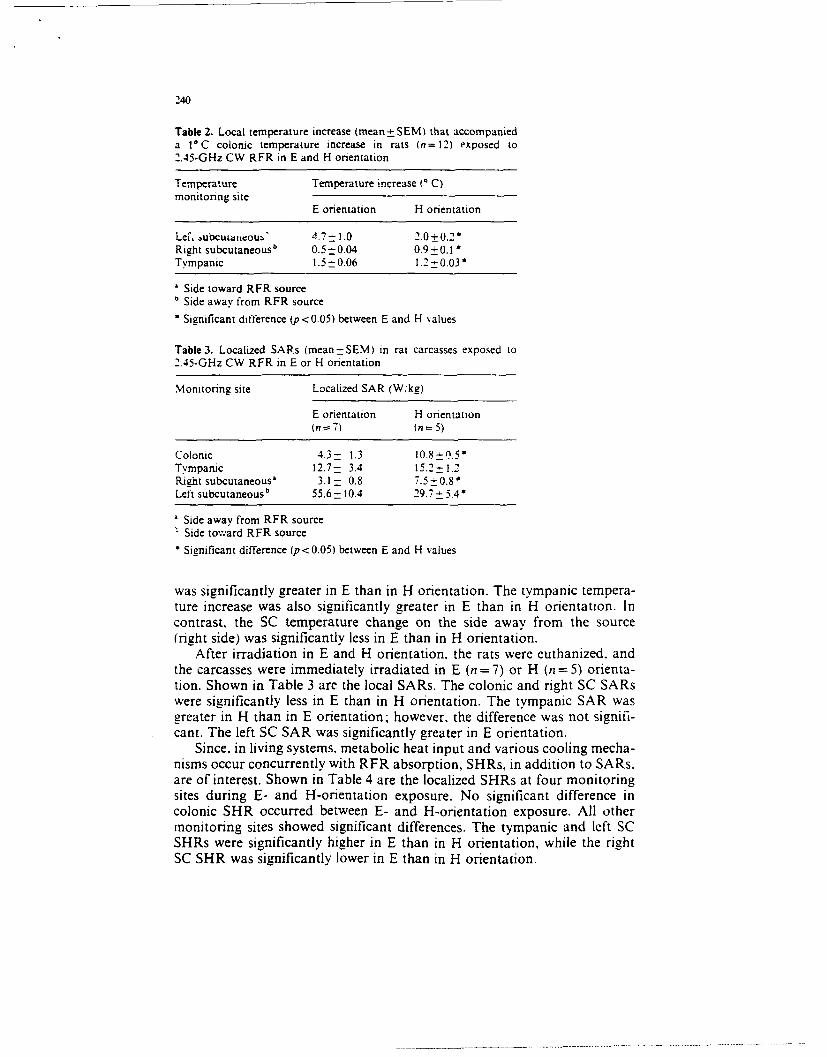
240
Table 2. Local temperature increase (mean± SEM) that accompanieda IVC colonic temperature increase in rats (n=12) exposed to2.45-GHz CW RFR in E and H orientation
Temperature Temperature increase ( C)monitoring site
E orientation H orientation
Lefi, ubcudiacouz" 4.7 - 1.0 2.0±0.2*Right subcutaneousb 0.5- 0.04 0.9_0.1 *Tympanic 1.5-0.06 1.2 -0.03
Side toward RFR sourceb Side away from RFR source
* Significant difference (p < 0.05) between E and H values
Table 3. Localized SARs (mean=_SEM) in rat carcasses exposed to
2.45-GHz CW RFR in E or H orientation
Monitoring site Localized SAR (Wikg)
E orientation H orientation(n = 7 ) (n=5)
Colonic 4.3- 1.3 10.8_-_Q.5*Tympanic 12.7- 3.4 15.2-- 1.2Right subcutaneous' 3.1 0.8 7.5 _ 0.8Left subcutaneous b 55.6-- 10.4 29.7 - 5.4 *
a Side away from RFR source
Side toward RFR source
* Significant difference (p<0.05) between E and H values
was significantly greater in E than in H orientation. The tympanic tempera-ture increase was also significantly greater in E than in H orientation. Incontrast, the SC temperature change on the side away from the source(right side) was significantly less in E than in H orientation.
After irradiation in E and H orientation, the rats were euthanized. andthe carcasses were immediately irradiated in E (n = 7) or H (n = 5) orienta-tion. Shown in Table 3 are the local SARs. The colonic and right SC SARswere significantly less in E than in H orientation. The tympanic SAR wasgreater in H than in E orientation, however, the difference was not signifi-cant. The left SC SAR was significantly greater in E orientation.
Since, in living systems, metabolic heat input and various cooling mecha-nisms occur concurrently with RFR absorption, SHRs, in addition to SARs.are of interest. Shown in Table 4 are the localized SHRs at four monitoringsites during E- and H-orientation exposure. No significant difference incolonic SHR occurred between E- and H-orientation exposure. All othermonitoring sites showed significant differences. The tympanic and left SCSHRs were significantly higher in E than in H orientation, while the rightSC SHR was significantly lower in E than in H orientation.

241
Table 4. Localized specific heating rates (mean -SEM) in rats 0n = 12)exposed to 2.45-GHz CW RFR (60 mW :m ) in E and H orienta-ion
Temperature Localized specific heating rates (W kg)monitoring site
E orientation H orientation
Colonic 12.3 + 0.5 11.8 -0.4Tympanic 15.1 -0.6 13.1 -0.3*Left subcutaneous' 56.6-9.6 23.7 - 3.1 *Right subcutaneous b 64 - 0.6 10,3 - 0.8 *
Side toward RFR sourceSicle away from RFR source
Significant difference ip<0.05) between E and H values
Initial HR0 Drie-3r on 295±11 T
*-H Orientation 295± 1
30
" !0
-10.
3'38.5 39.0 39.5 39.0
COLONIC TEMPERATURE C)Fig. I. Heart rate changes in rats dunng RFR-tnduced I°C temperature cvcles in E andH orientation in= 12). Irradiation was initiated at 38.50 C and discontinued at 39.5' C. Thedata are normalized to the values obtained at 38.5' C
Physiological responses
The HR changes that accompanied the I C Tc cycles are displayed inFig. 1. In both E and H orientations. HR significantly increased as Tcincreased from 38.5 to 39.50 C. and returned to near baseline levels duringthe recovery periods: the HR changes were significantly greater in E orienta-tion.
Shown in Fig. 2 are the BP changes that occurred during the 10 C Tccycles. During exposures in both E and H orientation. BP significantlyincreased during irradiation. and returned to baseline after cessation ofirradiation. BP changes during E-orientation exposure were significantlygreater than during H-orientation exposure.
The RR changes that occurred during the I C Tc cycles are shownin Fig. 3. During exposure in E orientation. RR did not change significantly:however, exposure in H orientation resulted in a significant RR increase.

242
20T
16
Z12
CL 4
00 E Orientation 92±3
z *-H Orientation 95 ±5
-4
38.5 39.0 39.5 390COLONIC TEMPERATURE (tj
Fig. 2. Mean arterial blood pressure changes in rats during RFR-induced 1* C temperaturecycles in E and H orientations in= 12). The data are normalized to the values obtained at38.5* C
10
Inta RR
- 2
0
38.5 39.0 39.5 39.0COLONIC TEMPERATURE (b)
Fig. 3. Respiratory rate changes in rats during RFR-induced 1* C temperature cycles in Eand H orientations (n= 12). The data are normalized to the values obtained at 38.5' C

243
Discussion
In studies of environmental heating, core-to-skin thermal gradients are usu-Aly assumed to be minimal (Simon et al. 1986). Dunng RFR exposures.however, particularly at higher frequencies, relatively large thermal gradientsmust be taken into account. Evidence suggests that during irradiation atequivalent power densities at certain frequencies. exposure in E orientationresults in higher SARs than H-orientation exposure. Results of the presentstudy indicate that even when E- and H-orientation exposures are madeat similar SARs. as opposed to equivalent power densities, drastic differ-ences in sites of energy deposition create large thermal gradients withinthe exposed animal.
The inequality of heat-distribution patterns in live animals exposed toRFR in E and H orientation is of considerable significance. Most RFR-induced bioeffects are regarded as being thermal in nature: however. duringthe last decade, numerous investigators have categorized noted effects asbeing " nonthermal" in nature. In some studies, the power levels used duringirradiation were extremely low, and were very unlikely to cause a tempera-ture increase. In other studies at higher power levels, effects were consideredto be nonthermally induced because the core temperature (usually colonicor rectal) did not appreciably change during irradiation. This latter lineof reasoning may be questioned because. depending on exposure conditions.localized areas of the body may experience large temperature changes wi'ilethe core temperature remains virtually unchanged. For example. Spiegelet al. (1980) found that in human models irradiated with 80-MHz RFR(SAR of 1.4 Wikg) the temperature increase in the thigh area was 3--4 Cgreater than in the colonic area. D'Andrea et al. (1985) noted. in irradiatedrat carcasses, localized hot spots that were significantly higher than whole-body average temperature changes. In an extension of their earlier work.D'Andrea et al. (1987) found that the higher than average local SARs notedin rat carcasses were reflected in localized hot spots in ketamine-anesthetizedrats. Frei et al. (1988) showed an unequal heating pattern in anesthetizedrats exposed to 2.8-GHz RFR (SAR of 8-20 W '-g) in H orientation: SCand tympanic heating was significantly greater than colonic heating.
In the present study, irradiation in H orientation produced a more uni-form pattern of heat distribution in the rat than did E-orientation exposure(Table 2). Comparison between live animal (Table 4) and carcass (Table 3)exposure suggests that the Tc increase during E-orientation irradiation oflive animals was due in great part to heat transfer from periphery to corevia the circulatory system. Conversely. the SC temperature increase seenin H orientation may have been partially due to heat transfer from coreto periphery. Results of this study indicate that animal orientation duringirradiation must be considered in interpretation of results of RFR exposure.It is possible that some previous reports of nonthermally-induced RFRbioeffects were influenced by undetected orientation-specific localized hots-pots. particularly in anesthetized or restrained animals.
The present study showed that several physiological parameters were

244
influenced by the heat-distribution pattern. The HR increase was significant-ly greater during irradiation in E than in H orientation (Fig. 1). Althoughthe exact mechanism responsible for the greater HR increase during E-orientation exposure is unclear, it is probable that the difference was primar-ily related to the level or rate of peripheral heating. This topic was discussedin a recent article (Frei et al. 1989) concerning exposure of rats to 9.3-GHzRFR (H orientation) in which levels of SC temperature increase closelyresembled the present 2.45-GHz E-orientatiun-induced increase. The higherSC temperature. or the faster rate of SC temperature increase during E-orientation exposure. may have influenced the level of stress hormone release(epinephrine. norepinephrine, corticosterone), which is known to enhancecardiovascular function. Lotz (1985) exnosed rhesus monkeys to 225-MHzRFR in E and H orientation and found that corticosterone release wassignificantly greater during E-orientation exposure.
Adair (1977) and Adair et al. ( )84) have shown that minor changes(<0.10 C) in hypothalamic temperature can influence thermoregulatory re-sponse in the whole animal. Although we did not directly measure hypotha-lamic temperature. tympanic temperature has been shown to be a reliabieindicator of hypothalamic temperature in humans (Benzinger and Taylor1963). and of brain temperature in rats (Robinson et al. 1967: Dunscombeet al. 1980). We obtained a small (<0.30 C), but consistently higher tvm-panic temperature change in E orientation than in H orientation. This differ-ence may have contributed significantly to the heightened cardiovascularresponse seen during exposure in E orientation.
Previous studies of human cardiovascular responses to hyperthermiahave revealed an increase in HR accompanied by a decrease in BP (Kimet al. 1979- Tonnesen et al. 1987). The BP decrease was attributed primarilyto cutaneous vasodilation and increased skin blood flow, an effect alsonoted by Phillips et al. (1975) and Adair and Adams (1980) during RFRexposure in which the Tc increased only slightly. In our experiments, how-ever. BP increased significantly (Fig. 2), and the increase in E orientationwas significantly greater than in H orientation. Pettigrew et al. (1974) alsoreported a rise in BP during hyperthermia in humans. and Cooper et al.(1962) noted hypertension in rats exposed to RFR. In these and the presentstudy, local skin vasodilation may have been counteracted bv other cardio-vascular adjustments. such as increased cardiac output due to increasedHR (Gorman and Proppe 1984).
In the present study, RR significantly increased during irradiation inthe H orientation, but not in the E orientation (Fig. 3). Irradiation of car-casses showed that the major site of energy deposition in H orientationoccurred in the animal core. Activation of deep receptors during irradiationin H orientation may have influenced respiratory rate to a greater degreethan peripheral receptor activation in E orientation. Simon (1974) has sug-gested that activation of deep thermal receptors can play an importantrole in stimulation of respiration.
In summary, our results showed significant differences between the ef-fects of E- and H-orientation exposure to RFR (2.45 GHz. 60 mWcm2.

245
CW) upon the pattern of heat distribution in ketamine-aiiesthetized ratsand rat carcasses. These differences were also reflected in the animals' HR,BP, and RR changes during irradiation. During RFR exposure that in-creased colonic temperature from 38.5 to 39.50 C, the SC temperature in-crease on the side nearest the RFR source, and the tympanic temperatureincrease, were both significantly greater in E than in H orientation: theopposite was true for the SC temperature away from the source. HR andBP increased significantly during irradiation, and returned to baseline levelswhen exposure was discontinued: the increases were significantly greaterin E than in H orientation. RR increased significantly during irradiationin H but not in E orientation. Although the physical and biological mecha-nisms are unclear at this time, at the commonly used frequency of 2.45 GHzthere is a tremendous difference in sites of energy deposition between Eand 11 orientation, this difference significantly affects the rat's responsesto RFR exposure. Additicnal studies are needed to determine if these re-sponses also occur at othe, frequencies and power levels.
.4cknowledzement This study was supported by the AFSC Universii, Resident ResearchProiessorshir) and Human Systems Di; sion Research Scholarship programs. The authors wishto acknowledge the RFR exposure suppor of the Radiation Pnysics Branch. Radiation Sci,:ncesDivision. USAF School of Aerospace Medicine. Brooks AFB. TX. and tihc technical supportof SSgt Raul Escarciga. of the Radiatin Physics Branch.
References
Adair 'R (19771 Skin. preoptic. and core temperatures influence behavioral thermoregulation.J App Physiol 42: 559-564
Adair ER. Adams BW (1980) Microwaves induce peripheral vasodilation in squirrel monkey.Science 207:1381-1383
Adair ER. Adams BW. Akel GM (1984) Minimal changes in hypothalamic temperature accom-pany microwave-induced alteration of thermoregulatorv behavior. Bioelectromagnetics5:13-30
Ailen SJ. Hurt WD. Krupp JH. Ratliff JA. Durney CH. Johnson CC (1976) Measurementof radiofrequency power absorption in monkeys, monkey phantoms. and human phantomsexposed to 10-50 MHz fields. Report USAFSAM-TR-76-5. US Air Force School of Acr-ospace Medicine. Brooks Air Force Base. Texas
Allen SJ. Hurz WD (1979) Calorimetric measurements of microwave energy absorption bymice after Nimultaneous expeo;ure of i8 animals. Radio Sci 14 (6 Suppl): 1-4
Benzinger TH. Taylor GW (1963) Cranial measurements of internal temperature in mar'.In: Herzfeld CM led) Temperature - its measurement and control in science and ndustr.vol 3, pt 3. Reinhold. New York. p I1 I
Chou CK. Guy AW. McDougall JA, Lai H (1985) Specific absorption rate in rats exposedto 2,450-MHz microwaves under seven exposure conditions. Bioelectromagnetics 6:73-88
Cooper T. Pinakatt T. Jellinek M, Richardson AV (1962) Effects of reserpine on circulationof the rat after microwave irradiation. Am J Physiol 202:1171-1174
D'Andrea JA. Emmerson RY. Bailey CM. Olsen RG. Gandhi OP (1985) Microwave r-diationabsorption in the rat: Freq u ncy-dependent SAR distribution in body and tail. Bioelectromagnetics 6:199-206
D'Andrea JA, Emmerson RY. DeWitt JR. Gandhi OP (1987) Absorption of microwave radia-tion by the anesthetized rat: Electromagnetic and thermal hotspots in body and tail. Bioelec-tromagnetics 8 : 385-396
Dunscombe PB. Rail DL. Steinert TJ, Ramsey NW. Rose FC (1980) A comparison between

246
whole-body and localized brain heating on some physiological responses of the rat. Cli,Phys Physiol Meas 1:211-219
Durney CH. Iskander MF, Massoudi H, Allen SJ. Mitchell JC (1980) Radiofrequency Radia-(ion Dosimetry Handbook, 3rd Edn. Report USAFSAM-TR-80-32. US Air Force Schoolof Aerospace Medicine, Brook- Air Force Base, Texas
Frei MR. Jauchem JR, Heinmets F (1988) Physiological effects of 2.8 GHz radio-frequencyradiation: A comparison of pulsed and continuous-wave radiation. J Microwave PowerEE 23:85-93
Frei MR, Jauchem JR, Heinmets F (1989) Thermoregulatory responses of rats exposed to9.3-GHz radiofrequency radiation. Radiat Environ Biophys 28:67-77
Gandhi OP (1974) Polarization and frequency effects on whole animal energy absorptionof RF eneriy. Proc IEEE 62:1171-1175
Gorman AJ, Proppe DW (1984) Mechanisms producing tachycardia in conscious baboonsduring environmental heat stress. J AppI Physiol 56:441-446
Jauchem JR, Frei MR, Heinmets F (1984a) Heart rate changes due to 5.6-GHz radiofrequencyradiation: Relation to average power density. Proc Soc Exp Biol Med 177:383-387
Jauchern JR. Frei MR. Heinmets F (1984b) Increased susceptibility to radiofrequency radiationdue to pharmacological agents. Aviat Space Environ Med 55:1036-1040
Johnson CC (1975) Recommendations for specifying EM wave irradiation conditions in bloef-fects research. J Microwave Power 10:249-250
Kim YD. Lake CR. Lees DE. Schuette JM. Bull JM. Weise V. Kopin IJ (1979) Hemodynamicand plasma catecholamine responses to hyperthermic cancer therapy in humans. Am JPhystol 237: H570-H574
Lotz WG (1985) Hyperthermia in radiofrequency-exposed rhesus monkeys: A comparisonof frequency and orientation effects. Radiat Res 102:59-70
Lozano LG. Hurt WD (1984) Computer program for qampling data and determining specificabsorption rates considering high heat loss. Report USAFSAM-TR-84-18. US Air ForceSchool of Aerospace Medicine. Brooks Air Force Base. Texas
McRee DI (1974) Determination of energy absorption of microwave radiation using the coolingcurve technique. J Microwave Power 9:263-270
McRee DI. Davis HG (1987) Effects on energy absorption of orientation and size of animalsexposed to 2.45-GHz microwave radiation. Health Phys 52:39-43
Memtt JH. Chamness AF. Hartzell RH, Allen SJ (1977) Orientation effects of microwave-induced hyperthermia and neurochemical correlates. J Microwave Power 12:167-172
Padilla JM, Bixby RR (1986) Using Dewar-flask calorimetry and rectal temperature to deter-mine the specific absorption rates of small rodents. Report USAFSAM-TR-86-3. US AirForce School of Aerospace Medicine, Brooks Air Force Base. Texas
Pettigrew RT, Gait JM. Ludgate CM. Horn DB. Smith AN (1974) Circulatory and biochemicaleffects of whole body hyperthermia. Br J Surg 61: 727-730
Phillips RD. Hunt EL. Castro RD, King NW (1975) Thermoregulatory, metabolic, and cardio-vascular response of rats to microwaves. J Appi Physiol 38: 630-635
Robinson SM. Hutchison VH. Blatt WF (1967) A note on the relationship of tympanic.intraperitoneal. and brain temperature in the rat. Can J Physiol Pharmacol 45:355-358
Simon E (1974) Temperature regulation: The spinal cord as a site of extrahypothalamic thermo-regulatory functions. Rev Physiol Biochem Pharmacol 71:1-76
Simon E. Pierau F. Taylor CM (1986) Central and peripheral thermal control of effectorsin homeothermic temperature regulation. Physiol Rev 66:235-298
Smith DI, Pekoe GM, Martin LL, Coalgate B (1980) The interaction of ketamine with theopiate receptor. Life Sci 26:789-795
Spiegel RJ. Deffenbaugh DM. Mann JE (1980) A thermal model of the human body exposedto an electromagnetic field. Bioelectromagnetics 1:253-270
Tonnesen AS. Marnock A. Bull JMC, Morgenweck CJ. Fallon KD (1987) Sweating. hemody-namic responses. and thermal equilibrium during hyperthermia in humans. J Appi Phystol62:1596-1602