Embed Size (px)
Citation preview

Mass SpectrometryDOI: 10.1002/anie.200900613
Ultraviolet Photodissociation: Developments towardsApplications for Mass-Spectrometry-Based ProteomicsTony Ly and Ryan R. Julian*
lasers · mass spectrometry · phosphorylation ·radicals · sequence determination
1. Introduction
Mass spectrometers are increasingly powerful devices.Improvements in resolution,[1] sensitivity,[2] dynamic range,[3]
and ionization sources[4, 5] have enabled dramatic contribu-tions to the study of proteins[6] and other biological mole-cules.[7] However, regardless of instrument performance, massspectrometers are fundamentally restricted to the measure-ment of mass/charge and relative abundance. Although themolecular weight of an intact molecule is frequently a veryuseful piece of data, modern mass spectrometers can typicallybe utilized to obtain much more information. For example,fragmentation is frequently employed to examine covalentbond structure in both large and small molecules.[8] Non-covalent interactions can also be explored to determinequaternary structure in proteins[9] or examine solution-phasetertiary structure.[10] Exchange reactions can be implementedto explore both gas- and solution-phase behavior.[11–13] Ionmobility (in various guises) can be used with mass spectrom-etry for rapid separations[14, 15] or for examining intact gas-phase structure.[16] Imaging experiments can be utilized todetermine spatial relationships between compounds in com-
plex samples, such as tissues.[17] There-fore, although only mass/charge andrelative abundance are ever measured,the knowledge that can be obtained bymass spectrometry is quite diverse.
Among the many technologiescoupled to mass spectrometers, it isnot surprising to find that lasers are
commonly employed. Lasers of varying sizes, powers, andwavelengths have been implemented in numerous ion sourcesand used for ionization, dissociation, and even spectrosco-py.[18] Although the combination of ultraviolet (UV) laserswith mass spectrometers has a substantial history,[19–21]
including several elegant experiments employing multiplelasers,[22,23] the method has become more common onlyrecently as a means for generating novel dissociation path-ways. Advances in both laser and MS technology are drivingthe union. This emerging technology complements the moreestablished methods that employ low-energy photons.[24]
One area in particular in which MS continues to play amajor role is the field of proteomics, as detailed in severalreviews.[25–27] The technical and chemical challenges associ-ated with this field are strongly connected to the fact thatproteins are highly diverse and complicated molecules, as aretheir surroundings. The relative abundances of proteins spanat least 10 orders of magnitude.[28] The 20 naturally occurringside chains introduce substantial chemical diversity, but thisdiversity is dwarfed by the challenges resulting from post-translational modifications (over 200 known modificationscannot be predicted from genomic information).[29] When allof these isoforms are included, the number of differentmolecules to be evaluated is staggering. The sheer numbersrequire methodology that can be scaled to high-throughputformats. When all of these considerations are taken intoaccount, no single technique or methodology is sufficient.
Unraveling of all of the information contained in proteomes poses atremendous chemical challenge, which is balanced by the promise ofpotentially transformational knowledge. Mass spectrometry offers anunprecedented arsenal of tools for diverse proteomic investigations.Recently, it was demonstrated that ultraviolet light can be utilized toinitiate unique and potentially useful fragmentations in peptides andproteins. Either nonspecific dissociation or highly specific dissociationat engineered chromophoric sites is possible following photonabsorption. The level of specificity and control over fragmentation inthese experiments is greater than with other fragmentation methods.Novel techniques made possible by this technology are poised to makesubstantial contributions to the field of proteomics.
[*] T. Ly, Prof. R. R. JulianDepartment of Chemistry, University of CaliforniaRiverside, CA 92521 (USA)E-mail: [email protected]: http://faculty.ucr.edu/~ ryanj/
R. R. Julian and T. LyMinireviews
7130 � 2009 Wiley-VCH Verlag GmbH & Co. KGaA, Weinheim Angew. Chem. Int. Ed. 2009, 48, 7130 – 7137
Rather, a multipronged approach is preferable. Recentdevelopments in high energy photodissociation mass spec-trometry have enabled new approaches to several challengesin proteomics, as detailed herein.
2. Implementation of Photodissociation
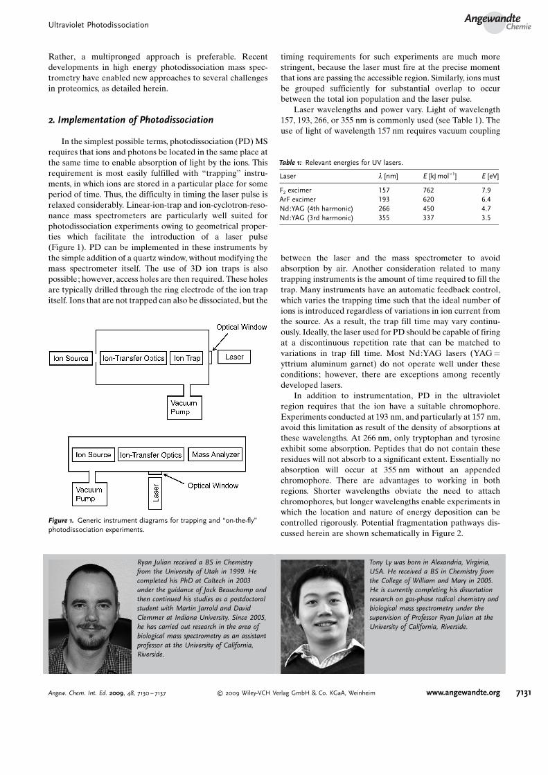
In the simplest possible terms, photodissociation (PD) MSrequires that ions and photons be located in the same place atthe same time to enable absorption of light by the ions. Thisrequirement is most easily fulfilled with “trapping” instru-ments, in which ions are stored in a particular place for someperiod of time. Thus, the difficulty in timing the laser pulse isrelaxed considerably. Linear-ion-trap and ion-cyclotron-reso-nance mass spectrometers are particularly well suited forphotodissociation experiments owing to geometrical proper-ties which facilitate the introduction of a laser pulse(Figure 1). PD can be implemented in these instruments bythe simple addition of a quartz window, without modifying themass spectrometer itself. The use of 3D ion traps is alsopossible; however, access holes are then required. These holesare typically drilled through the ring electrode of the ion trapitself. Ions that are not trapped can also be dissociated, but the
timing requirements for such experiments are much morestringent, because the laser must fire at the precise momentthat ions are passing the accessible region. Similarly, ions mustbe grouped sufficiently for substantial overlap to occurbetween the total ion population and the laser pulse.
Laser wavelengths and power vary. Light of wavelength157, 193, 266, or 355 nm is commonly used (see Table 1). Theuse of light of wavelength 157 nm requires vacuum coupling
between the laser and the mass spectrometer to avoidabsorption by air. Another consideration related to manytrapping instruments is the amount of time required to fill thetrap. Many instruments have an automatic feedback control,which varies the trapping time such that the ideal number ofions is introduced regardless of variations in ion current fromthe source. As a result, the trap fill time may vary continu-ously. Ideally, the laser used for PD should be capable of firingat a discontinuous repetition rate that can be matched tovariations in trap fill time. Most Nd:YAG lasers (YAG =
yttrium aluminum garnet) do not operate well under theseconditions; however, there are exceptions among recentlydeveloped lasers.
In addition to instrumentation, PD in the ultravioletregion requires that the ion have a suitable chromophore.Experiments conducted at 193 nm, and particularly at 157 nm,avoid this limitation as result of the density of absorptions atthese wavelengths. At 266 nm, only tryptophan and tyrosineexhibit some absorption. Peptides that do not contain theseresidues will not absorb to a significant extent. Essentially noabsorption will occur at 355 nm without an appendedchromophore. There are advantages to working in bothregions. Shorter wavelengths obviate the need to attachchromophores, but longer wavelengths enable experiments inwhich the location and nature of energy deposition can becontrolled rigorously. Potential fragmentation pathways dis-cussed herein are shown schematically in Figure 2.
Ryan Julian received a BS in Chemistryfrom the University of Utah in 1999. Hecompleted his PhD at Caltech in 2003under the guidance of Jack Beauchamp andthen continued his studies as a postdoctoralstudent with Martin Jarrold and DavidClemmer at Indiana University. Since 2005,he has carried out research in the area ofbiological mass spectrometry as an assistantprofessor at the University of California,Riverside.
Tony Ly was born in Alexandria, Virginia,USA. He received a BS in Chemistry fromthe College of William and Mary in 2005.He is currently completing his dissertationresearch on gas-phase radical chemistry andbiological mass spectrometry under thesupervision of Professor Ryan Julian at theUniversity of California, Riverside.
Figure 1. Generic instrument diagrams for trapping and “on-the-fly”photodissociation experiments.
Table 1: Relevant energies for UV lasers.
Laser l [nm] E [kJmol�1] E [eV]
F2 excimer 157 762 7.9ArF excimer 193 620 6.4Nd:YAG (4th harmonic) 266 450 4.7Nd:YAG (3rd harmonic) 355 337 3.5
Ultraviolet PhotodissociationAngewandte
Chemie
7131Angew. Chem. Int. Ed. 2009, 48, 7130 – 7137 � 2009 Wiley-VCH Verlag GmbH & Co. KGaA, Weinheim www.angewandte.org

3. Applications in Proteomics
As mentioned in the Introduction, proteomics poses manyexperimental challenges. UVPD offers several advantagesover other fragmentation techniques. First, energy depositionoccurs on a timescale that is orders of magnitude faster thanthat of other methods. Even low-energy PD methods, such asinfrared multiphoton dissociation (IRMPD), typically takeplace on the millisecond timescale.[24,30] UVPD can easilyoccur on the nanosecond to microsecond timescale. Directdissociations occur much faster. This speed is advantageousany time a method is scaled up for a high-throughputapplication, because more data will be acquired in less time.For example, on an ion-trap instrument, an additional 1800peaks could be examined during a 1 h liquid-chromatography(LC) separation. This speed is even more advantageous incertain instrumental configurations, such as TOF–TOF, inwhich the time required for fragmentation can be substantialrelative to the total time for a scan. Gains in efficiency in thiscase could be of an order of magnitude or more. A secondadvantage afforded by some UVPD experiments is controlover the introduction of energy. In other words, the specificityof photon/chromophore interactions can be used to dictatethe location and amount of energy introduced into amolecule. This control enables directed dissociation experi-ments that are not possible with other methods.
3.1. Alternative Sequencing Methods3.1.1. Direct Bond Absorption
Short-wavelength lasers are capable of exciting thepeptide backbone directly, which obviates the need forsuitable side chains or appended chromophores.[31–35] Frag-mentation can then take place by at least two possiblemechanisms. The first involves internal conversion of theenergy into vibrational modes, followed by fragmentation in
the ground electronic state. This process should lead tofragmentation resembling that observed with collision-in-duced dissociation (CID), IRMPD, or other methods ofdissociation under statistical control. Interestingly, PD withshort-wavelength lasers does not lead to such fragmentation,and many fragments that are not typically observed by CIDare generated. In particular, an extensive range of x- or a-series ions are produced, depending on the location of thecharge in the peptide. For relevant structures and standardnomenclature, see Scheme 1.[36, 37] Reilly and co-workersattributed the origin of these nonstatistical products to direct
or prompt dissociation, which occurs in the excited state.Initially, it was thought that this dissociation may occur only at157 nm,[38] but other experiments have demonstrated similarfragmentation at 193 nm.[34,19] The timescale for observation iscritical in these experiments. Unique fragmentation domi-nates at short observation times (microseconds), whereasfragmentation observed after several milliseconds moreclosely resembles CID. The v-, w-, and d-type ions observedin these experiments can also be used to extract side-chaininformation. PD can discriminate readily between leucine andisoleucine as a result of side-chain losses which are nottypically observed by CID or other statistical fragmentationmethods (Figure 3). To sum up, UVPD reveals usefulsequence information that is complementary to or additionalto that obtained by other methods. This approach wasrecently reviewed.[39]
3.1.2. Noncovalent Chromophore Attachment
Wavelengths longer than 193 nm are not absorbed well bypeptides and proteins owing to the paucity of chromophoresat these wavelengths. Although this issue can be circumventedby the covalent attachment of a suitable chromophore, such
Figure 2. Simplified diagram of energy absorption and fragmentationpathways in PD. Absorption (solid line) of photon energy leads toexcitation to a higher electronic state. Coupling to an antibondingstate (dotted line) leads to direct dissociation. Otherwise, statisticalredistribution of the energy into vibrational modes occurs; this processcan be followed by unimolecular dissociation in the ground electronicstate.
Scheme 1. Standard fragmentation nomenclature and correspondingstructures.[36, 37]
R. R. Julian and T. LyMinireviews
7132 www.angewandte.org � 2009 Wiley-VCH Verlag GmbH & Co. KGaA, Weinheim Angew. Chem. Int. Ed. 2009, 48, 7130 – 7137

an approach is undesirable because of the additional chemicaltransformations that must be carried out. An alternativesolution is to attach chromophores through noncovalentinteractions. In this case, it is only necessary to add therelevant molecules to the sample solution prior to analysis.Two examples of such molecules are shown in Figure 4. This
experiment is quite counterintuitive in that energy must beabsorbed by the chromophore, converted into vibrationalenergy, and then transferred to the biomolecule prior tofragmentation of the noncovalent union. Of course, theamount of energy transferred must then be sufficient to breakcovalent bonds! Nevertheless, this process has been shown tobe effective by two research groups with several differentmolecules.[40, 41] A sample spectrum showing abundant frag-mentation is shown in Figure 4c. One key appears to be amultidentate interaction at the noncovalent junction. Thisinteraction decreases the likelihood of the simultaneousexcitation of all noncovalent bonds prior to significant energytransfer.
3.1.3. Femtosecond Fragmentation
Recent experiments conducted at 800 nm with a femto-second titanium sapphire laser are also of interest for thisMinireview.[42] Although it is clear that 800 nm is not in theUV portion of the electromagnetic spectrum, the fragmenta-tion observed in these experiments is much more similar tothat of UVPD[23] than it is to that of IRMPD. Furthermore, asthere are no peptide chromophores in this region of thespectrum, nonlinear absorption is probably occurring. Also,the relevant energetics dictate that several photons must beabsorbed. These experiments generate a substantial amountof directly observable photoionized peptide and fragmentsderived from this species (Figure 5). The photoionizedpeptide is hydrogen-deficient and (by mass) identical to the
peptide radicals discussed in Section 3.3. Not surprisingly,many of the products observed in these experiments are alsosimilar to those described in Section 3.3. Other fragmentssimilar to those generated by short-wavelength UVPD, asdescribed in Section 3.1.1, are also observed (such as w- anda-series ions). The results suggest that several processes mayoccur in these experiments, but further results will be neededfor an understanding of the exact mechanisms involved.Nevertheless, it is clear that a high-intensity pulse of shortduration can be used to elicit fragmentation, as observed inprevious experiments with femtosecond pulses.[23]
3.2. Selective Fragmentation3.2.1. Appended-Chromophore Suicide
One advantage of UVPD is that a paucity of appropriatechromophores exists at certain wavelengths. For example,absorption by native peptides and proteins is minimal at355 nm. Wilson and Brodbelt cleverly used this property todirect fragmentation in modified peptides.[43] These experi-ments rely on derivatization of one end of the peptide with achromophore that absorbs at 355 nm. Internal conversion ofthe initial photoexcitation energy into vibrational energyleads to fragmentation of the peptide. The key to theseexperiments lies in the fact that fragments which do notcontain the chromophore cease to absorb further energy andare therefore detected readily. On the other hand, fragments
Figure 3. PD spectrum acquired at 157 nm demonstrating completesequence coverage in the x ion series. Leucine is differentiated fromthe isobaric amino acid isoleucine by the characteristic mass shiftbetween x3 and w3 (*). Adapted from reference [31] and reprinted withpermission.
Figure 4. The noncovalent attachment of a UV chromophore, forexample, with a) naphthoyl [18]crown-6 ether (NC) or b) [18]crown-6dipyrrolylquinoxaline, facilitates fast deposition of energy into thepeptide. c) The resulting fragmentation is identical to collision-induceddissociation; however, UVPD was shown to occur on a much shortertimescale. Adapted from references [40] and [41] with permission.
Figure 5. Femtosecond laser induced dissociation mass spectrometry(fs-LID MS/MS) of angiotensin II with 800 nm photons. The backbonefragmentation observed is consistent with dissociation of the [M+H]2+C
species and other high-energy fragmentation processes. Adapted fromreference [42] with permission.
Ultraviolet PhotodissociationAngewandte
Chemie
7133Angew. Chem. Int. Ed. 2009, 48, 7130 – 7137 � 2009 Wiley-VCH Verlag GmbH & Co. KGaA, Weinheim www.angewandte.org
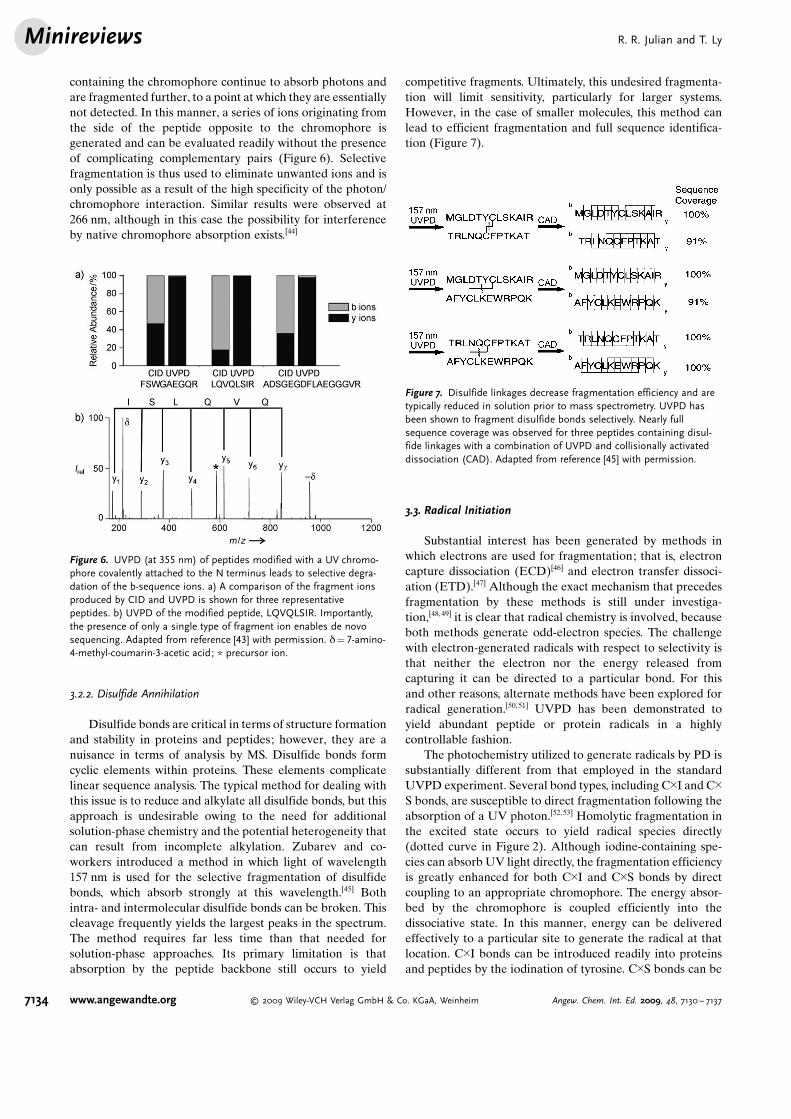
containing the chromophore continue to absorb photons andare fragmented further, to a point at which they are essentiallynot detected. In this manner, a series of ions originating fromthe side of the peptide opposite to the chromophore isgenerated and can be evaluated readily without the presenceof complicating complementary pairs (Figure 6). Selectivefragmentation is thus used to eliminate unwanted ions and isonly possible as a result of the high specificity of the photon/chromophore interaction. Similar results were observed at266 nm, although in this case the possibility for interferenceby native chromophore absorption exists.[44]
3.2.2. Disulfide Annihilation
Disulfide bonds are critical in terms of structure formationand stability in proteins and peptides; however, they are anuisance in terms of analysis by MS. Disulfide bonds formcyclic elements within proteins. These elements complicatelinear sequence analysis. The typical method for dealing withthis issue is to reduce and alkylate all disulfide bonds, but thisapproach is undesirable owing to the need for additionalsolution-phase chemistry and the potential heterogeneity thatcan result from incomplete alkylation. Zubarev and co-workers introduced a method in which light of wavelength157 nm is used for the selective fragmentation of disulfidebonds, which absorb strongly at this wavelength.[45] Bothintra- and intermolecular disulfide bonds can be broken. Thiscleavage frequently yields the largest peaks in the spectrum.The method requires far less time than that needed forsolution-phase approaches. Its primary limitation is thatabsorption by the peptide backbone still occurs to yield
competitive fragments. Ultimately, this undesired fragmenta-tion will limit sensitivity, particularly for larger systems.However, in the case of smaller molecules, this method canlead to efficient fragmentation and full sequence identifica-tion (Figure 7).
3.3. Radical Initiation
Substantial interest has been generated by methods inwhich electrons are used for fragmentation; that is, electroncapture dissociation (ECD)[46] and electron transfer dissoci-ation (ETD).[47] Although the exact mechanism that precedesfragmentation by these methods is still under investiga-tion,[48, 49] it is clear that radical chemistry is involved, becauseboth methods generate odd-electron species. The challengewith electron-generated radicals with respect to selectivity isthat neither the electron nor the energy released fromcapturing it can be directed to a particular bond. For thisand other reasons, alternate methods have been explored forradical generation.[50, 51] UVPD has been demonstrated toyield abundant peptide or protein radicals in a highlycontrollable fashion.
The photochemistry utilized to generate radicals by PD issubstantially different from that employed in the standardUVPD experiment. Several bond types, including C�I and C�S bonds, are susceptible to direct fragmentation following theabsorption of a UV photon.[52,53] Homolytic fragmentation inthe excited state occurs to yield radical species directly(dotted curve in Figure 2). Although iodine-containing spe-cies can absorb UV light directly, the fragmentation efficiencyis greatly enhanced for both C�I and C�S bonds by directcoupling to an appropriate chromophore. The energy absor-bed by the chromophore is coupled efficiently into thedissociative state. In this manner, energy can be deliveredeffectively to a particular site to generate the radical at thatlocation. C�I bonds can be introduced readily into proteinsand peptides by the iodination of tyrosine. C�S bonds can be
Figure 6. UVPD (at 355 nm) of peptides modified with a UV chromo-phore covalently attached to the N terminus leads to selective degra-dation of the b-sequence ions. a) A comparison of the fragment ionsproduced by CID and UVPD is shown for three representativepeptides. b) UVPD of the modified peptide, LQVQLSIR. Importantly,the presence of only a single type of fragment ion enables de novosequencing. Adapted from reference [43] with permission. d = 7-amino-4-methyl-coumarin-3-acetic acid; * precursor ion.
Figure 7. Disulfide linkages decrease fragmentation efficiency and aretypically reduced in solution prior to mass spectrometry. UVPD hasbeen shown to fragment disulfide bonds selectively. Nearly fullsequence coverage was observed for three peptides containing disul-fide linkages with a combination of UVPD and collisionally activateddissociation (CAD). Adapted from reference [45] with permission.
R. R. Julian and T. LyMinireviews
7134 www.angewandte.org � 2009 Wiley-VCH Verlag GmbH & Co. KGaA, Weinheim Angew. Chem. Int. Ed. 2009, 48, 7130 – 7137

introduced at cysteine or phosphorylated serine or threonineresidues. Both processes are described in greater detail below.
3.3.1. Whole Protein Radicals
The iodination of proteins and peptides in solution occurspreferentially at tyrosine residues, and is further selective inwhole proteins for the most exposed residues.[54] Therefore,careful control of the reaction can yield abundant monoiodo-substituted protein, in which the iodine substituent residesmainly at a single tyrosine residue. Subsequent photoactiva-tion with a 266 nm photon yields a radical at the tyrosineresidue in substantial yield (Figure 8a,b).[55] Collisional acti-vation of this radical product leads to fragmentation primarilyin the vicinity of the radical (Figure 8a,c). Fragmentation atthe tyrosine residue and proximate proline residues wasobserved to be particularly abundant. This level of control-lable selectivity in directing the fragmentation of a wholeprotein had not been demonstrated previously. It was furtherdemonstrated that the time required to analyze proteomicsdata can be decreased significantly (by several orders ofmagnitude) if fragmentation positions are controlled and thenumber of fragments limited in this manner. Efficiency isimproved, because the information content of the data isincreased when the results are predictable. Such an approach
remains hypothetical, as it has not been applied to a high-throughput experiment, but offers the promise of substantialadvantages over current methods.
3.3.2. Identification of Phosphorylation
The identification of posttranslational modifications(PTMs) remains an important challenge in all proteomicsexperiments.[29] Even if PTMs are not targeted specifically inan experiment, their presence can still complicate dataevaluation and must be accounted for. As demonstratedabove, the specificity of the photon/chromophore interactioncan provide a control over energy deposition that is notavailable with other fragmentation methods. With respect toPTMs, phosphorylation at threonine and serine residuesprovides an excellent and specific handle for derivatization.[56]
By eliminating the phosphate and introducing a chromophorecontaining a C�S bond, a photoinitiated radical can be placedin precisely the required location for the fragmentation of thepeptide backbone. In other words, photoactivation of themodified peptide leads to a single fragmentation at themodified site.[57] This experiment offers not only tremendoussimplicity for the interpretation of the results, but alsofundamental sensitivity enhancements due to the greatlydiminished number of peaks generated. For example, in a 20residue peptide, around 40 fragments would be expected fromECD/ETD, whereas the photodissociation experiment wouldyield only two or three. Sample spectra illustrating thisdifference are shown for a smaller peptide in Figure 9. Therelevant information in Figure 9b is contained in the largestpeak in the spectrum, and virtually all of the fragmentintensity identifies the site of phosphorylation. Therefore, thegas-phase chemistry of UVPD is at least an order ofmagnitude more sensitive than traditional approaches. It hasalso been demonstrated that the identification of phosphor-ylation sites by this method is much more reliable than by themore traditional CID methodology.
Figure 8. a) Illustration of the concept of selective fragmentation in awhole protein. b) UVPD of iodine-labeled proteins generates a radicalon the side chain of tyrosine residues site specifically. c) CID of theradical product leads to backbone fragmentation in proximity to themodified tyrosine residue (Tyr74). CytC =cytochrome c.
Figure 9. ECD (top) and radical directed dissociation (RDD; bottom)spectra of the phosphopeptide CTTSsFKK reveal that both techniquesidentify the phosphorylation site correctly. However, RDD is superior infragmentation efficiency (in terms of both yield and selectivity), whichbecomes important for phosphopeptides present at low concentra-tions. The bottom spectrum was adapted from reference [57] withpermission. * Loss of H3PO4.
Ultraviolet PhotodissociationAngewandte
Chemie
7135Angew. Chem. Int. Ed. 2009, 48, 7130 – 7137 � 2009 Wiley-VCH Verlag GmbH & Co. KGaA, Weinheim www.angewandte.org
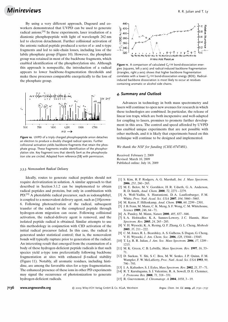
By using a very different approach, Dugourd and co-workers demonstrated that UVPD can be used to generateradical anions.[58] In these experiments, laser irradiation of adianionic phosphopeptide with light of wavelength 262 nmled to electron detachment. Further collisional activation ofthe anionic radical peptide produced a series of a- and x-typefragments and led to side-chain losses, including loss of thelabile phosphate group (Figure 10). However, the phosphategroup was retained in most of the backbone fragments, whichenabled identification of the phosphorylation site. Althoughthis approach is nonspecific, the introduction of a radicalappears to lower backbone-fragmentation thresholds andmake these processes comparable energetically to the loss ofthe phosphate group.
3.3.3. Noncovalent Radical Delivery
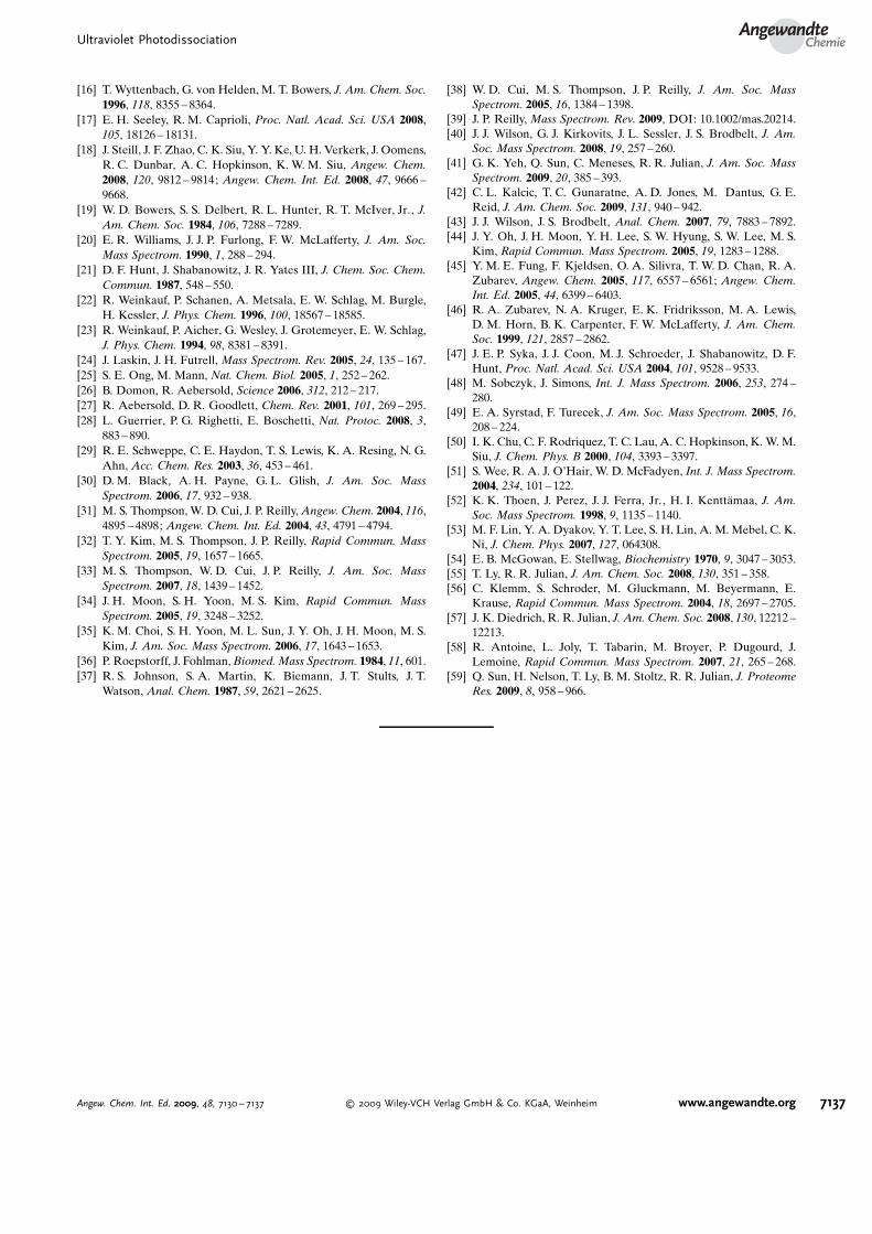
Ideally, routes to generate radical peptides should notrequire derivatization in solution. A similar approach to thatdescribed in Section 3.1.2 can be implemented to obtainradical peptides and proteins, but only in combination withPD.[59] A photolabile radical precursor, such as iodonaphthyl,is coupled to a noncovalent delivery agent, such as [18]crown-6. Following photoactivation of the radical, subsequenttransfer of the radical to the complexed peptide throughhydrogen-atom migration can occur. Following collisionalactivation, the radical-delivery agent is removed, and theisolated peptide radical is obtained. Similar attempts to usethis methodology in conjunction with CID activation of theinitial radical precursor failed. In this case, the radical isgenerated under statistical control; that is, the noncovalentbonds will typically rupture prior to generation of the radical.An interesting result that emerged from the examination of abody of these hydrogen-deficient peptide radicals is that suchspecies yield a-type ions preferentially following backbonefragmentation at sites with enhanced b-radical stability(Figure 11). Notably, all aromatic residues, including histi-dine, are among the favorable sites for a-type fragmentation.The enhanced presence of these ions in other PD experimentsmay signal the occurrence of photoionization to generatehydrogen-deficient radicals.
4. Summary and Outlook
Advances in technology in both mass spectrometry andlasers will continue to open new avenues for research in whichthese technologies are combined. In particular, the release oflinear ion traps, which are both inexpensive and well-adaptedfor coupling to lasers, promises to promote further develop-ment in this area. The control and speed afforded by UVPDhas enabled unique experiments that are not possible withother methods, and it is likely that experiments based on thistechnique will continue to be designed and implemented.
We thank the NSF for funding (CHE-0747481).
Received: February 2, 2009Revised: March 10, 2009Published online: July 16, 2009
[1] S. Kim, R. P. Rodgers, A. G. Marshall, Int. J. Mass Spectrom.2006, 251, 260 – 265.
[2] M. E. Belov, M. V. Gorshkov, H. R. Udseth, G. A. Anderson,R. D. Smith, Anal. Chem. 2000, 72, 2271 – 2279.
[3] A. Wolf-Yadlin, S. Hautaniemi, D. A. Lauffenburger, F. M.White, Proc. Natl. Acad. Sci. USA 2007, 104, 5860 – 5865.
[4] M. Karas, F. Hillenkamp, Anal. Chem. 1988, 60, 2299 – 2301.[5] J. B. Fenn, M. Mann, C. K. Meng, S. F. Wong, C. M. Whitehouse,
Science 1989, 246, 64 – 71.[6] A. Pandey, M. Mann, Nature 2000, 405, 837 – 846.[7] S. A. Hofstadler, K. A. Sannes-Lowery, J. C. Hannis, Mass
Spectrom. Rev. 2005, 24, 265 – 285.[8] V. H. Wysocki, K. A. Resing, Q. F. Zhang, G. L. Cheng, Methods
2005, 35, 211 – 222.[9] C. M. Jones, R. L. Beardsley, A. S. Galhena, S. Dagan, G. Cheng,
V. H. Wysocki, J. Am. Chem. Soc. 2006, 128, 15044 – 15045.[10] T. Ly, R. R. Julian, J. Am. Soc. Mass Spectrom. 2006, 17, 1209 –
1215.[11] M. K. Green, C. B. Lebrilla, Mass Spectrom. Rev. 1997, 16, 53–
71.[12] D. Suckau, Y. Shi, S. C. Beu, M. W. Senko, J. P. Quinn, F. M.
Wampler, F. W. McLafferty, Proc. Natl. Acad. Sci. USA 1993, 90,790 – 793.
[13] I. A. Kaltashov, S. J. Eyles, Mass Spectrom. Rev. 2002, 21, 37 – 71.[14] R. T. Kurulugama, S. J. Valentine, R. A. Sowell, D. E. Clemmer,
J. Proteome Res. 2008, 71, 318 – 331.[15] R. Guevremont, J. Chromatogr. A 2004, 1058, 3 – 19.
Figure 10. UVPD of a triply charged phosphopeptide anion detachesan electron to produce a doubly charged radical species. Furthercollisional activation yields backbone fragments that retain the phos-phate group. These fragments enable identification of the phosphor-ylation site. Key fragment ions that identify Ser4 as the phosphoryla-tion site are circled. Adapted from reference [58] with permission.
Figure 11. A comparison of calculated Cb�H bond-dissociation ener-gies (squares, left y axis) and radical-induced backbone fragmentation(triangles, right y axis) shows that higher backbone fragmentationcorrelates with a lower Cb�H bond-dissociation energy (BDE). Radical-induced backbone dissociation is most likely to occur at residuescontaining aromatic or alcohol side chains.
R. R. Julian and T. LyMinireviews
7136 www.angewandte.org � 2009 Wiley-VCH Verlag GmbH & Co. KGaA, Weinheim Angew. Chem. Int. Ed. 2009, 48, 7130 – 7137
[16] T. Wyttenbach, G. von Helden, M. T. Bowers, J. Am. Chem. Soc.1996, 118, 8355 – 8364.
[17] E. H. Seeley, R. M. Caprioli, Proc. Natl. Acad. Sci. USA 2008,105, 18126 – 18131.
[18] J. Steill, J. F. Zhao, C. K. Siu, Y. Y. Ke, U. H. Verkerk, J. Oomens,R. C. Dunbar, A. C. Hopkinson, K. W. M. Siu, Angew. Chem.2008, 120, 9812 – 9814; Angew. Chem. Int. Ed. 2008, 47, 9666 –9668.
[19] W. D. Bowers, S. S. Delbert, R. L. Hunter, R. T. McIver, Jr., J.Am. Chem. Soc. 1984, 106, 7288 – 7289.
[20] E. R. Williams, J. J. P. Furlong, F. W. McLafferty, J. Am. Soc.Mass Spectrom. 1990, 1, 288 – 294.
[21] D. F. Hunt, J. Shabanowitz, J. R. Yates III, J. Chem. Soc. Chem.Commun. 1987, 548 – 550.
[22] R. Weinkauf, P. Schanen, A. Metsala, E. W. Schlag, M. Burgle,H. Kessler, J. Phys. Chem. 1996, 100, 18567 – 18585.
[23] R. Weinkauf, P. Aicher, G. Wesley, J. Grotemeyer, E. W. Schlag,J. Phys. Chem. 1994, 98, 8381 – 8391.
[24] J. Laskin, J. H. Futrell, Mass Spectrom. Rev. 2005, 24, 135 – 167.[25] S. E. Ong, M. Mann, Nat. Chem. Biol. 2005, 1, 252 – 262.[26] B. Domon, R. Aebersold, Science 2006, 312, 212 – 217.[27] R. Aebersold, D. R. Goodlett, Chem. Rev. 2001, 101, 269 – 295.[28] L. Guerrier, P. G. Righetti, E. Boschetti, Nat. Protoc. 2008, 3,
883 – 890.[29] R. E. Schweppe, C. E. Haydon, T. S. Lewis, K. A. Resing, N. G.
Ahn, Acc. Chem. Res. 2003, 36, 453 – 461.[30] D. M. Black, A. H. Payne, G. L. Glish, J. Am. Soc. Mass
Spectrom. 2006, 17, 932 – 938.[31] M. S. Thompson, W. D. Cui, J. P. Reilly, Angew. Chem. 2004, 116,
4895 – 4898; Angew. Chem. Int. Ed. 2004, 43, 4791 – 4794.[32] T. Y. Kim, M. S. Thompson, J. P. Reilly, Rapid Commun. Mass
Spectrom. 2005, 19, 1657 – 1665.[33] M. S. Thompson, W. D. Cui, J. P. Reilly, J. Am. Soc. Mass
Spectrom. 2007, 18, 1439 – 1452.[34] J. H. Moon, S. H. Yoon, M. S. Kim, Rapid Commun. Mass
Spectrom. 2005, 19, 3248 – 3252.[35] K. M. Choi, S. H. Yoon, M. L. Sun, J. Y. Oh, J. H. Moon, M. S.
Kim, J. Am. Soc. Mass Spectrom. 2006, 17, 1643 – 1653.[36] P. Roepstorff, J. Fohlman, Biomed. Mass Spectrom. 1984, 11, 601.[37] R. S. Johnson, S. A. Martin, K. Biemann, J. T. Stults, J. T.
Watson, Anal. Chem. 1987, 59, 2621 – 2625.
[38] W. D. Cui, M. S. Thompson, J. P. Reilly, J. Am. Soc. MassSpectrom. 2005, 16, 1384 – 1398.
[39] J. P. Reilly, Mass Spectrom. Rev. 2009, DOI: 10.1002/mas.20214.[40] J. J. Wilson, G. J. Kirkovits, J. L. Sessler, J. S. Brodbelt, J. Am.
Soc. Mass Spectrom. 2008, 19, 257 – 260.[41] G. K. Yeh, Q. Sun, C. Meneses, R. R. Julian, J. Am. Soc. Mass
Spectrom. 2009, 20, 385 – 393.[42] C. L. Kalcic, T. C. Gunaratne, A. D. Jones, M. Dantus, G. E.
Reid, J. Am. Chem. Soc. 2009, 131, 940 – 942.[43] J. J. Wilson, J. S. Brodbelt, Anal. Chem. 2007, 79, 7883 – 7892.[44] J. Y. Oh, J. H. Moon, Y. H. Lee, S. W. Hyung, S. W. Lee, M. S.
Kim, Rapid Commun. Mass Spectrom. 2005, 19, 1283 – 1288.[45] Y. M. E. Fung, F. Kjeldsen, O. A. Silivra, T. W. D. Chan, R. A.
Zubarev, Angew. Chem. 2005, 117, 6557 – 6561; Angew. Chem.Int. Ed. 2005, 44, 6399 – 6403.
[46] R. A. Zubarev, N. A. Kruger, E. K. Fridriksson, M. A. Lewis,D. M. Horn, B. K. Carpenter, F. W. McLafferty, J. Am. Chem.Soc. 1999, 121, 2857 – 2862.
[47] J. E. P. Syka, J. J. Coon, M. J. Schroeder, J. Shabanowitz, D. F.Hunt, Proc. Natl. Acad. Sci. USA 2004, 101, 9528 – 9533.
[48] M. Sobczyk, J. Simons, Int. J. Mass Spectrom. 2006, 253, 274 –280.
[49] E. A. Syrstad, F. Turecek, J. Am. Soc. Mass Spectrom. 2005, 16,208 – 224.
[50] I. K. Chu, C. F. Rodriquez, T. C. Lau, A. C. Hopkinson, K. W. M.Siu, J. Chem. Phys. B 2000, 104, 3393 – 3397.
[51] S. Wee, R. A. J. O�Hair, W. D. McFadyen, Int. J. Mass Spectrom.2004, 234, 101 – 122.
[52] K. K. Thoen, J. Perez, J. J. Ferra, Jr., H. I. Kentt�maa, J. Am.Soc. Mass Spectrom. 1998, 9, 1135 – 1140.
[53] M. F. Lin, Y. A. Dyakov, Y. T. Lee, S. H. Lin, A. M. Mebel, C. K.Ni, J. Chem. Phys. 2007, 127, 064308.
[54] E. B. McGowan, E. Stellwag, Biochemistry 1970, 9, 3047 – 3053.[55] T. Ly, R. R. Julian, J. Am. Chem. Soc. 2008, 130, 351 – 358.[56] C. Klemm, S. Schroder, M. Gluckmann, M. Beyermann, E.
Krause, Rapid Commun. Mass Spectrom. 2004, 18, 2697 – 2705.[57] J. K. Diedrich, R. R. Julian, J. Am. Chem. Soc. 2008, 130, 12212 –
12213.[58] R. Antoine, L. Joly, T. Tabarin, M. Broyer, P. Dugourd, J.
Lemoine, Rapid Commun. Mass Spectrom. 2007, 21, 265 – 268.[59] Q. Sun, H. Nelson, T. Ly, B. M. Stoltz, R. R. Julian, J. Proteome
Res. 2009, 8, 958 – 966.
Ultraviolet PhotodissociationAngewandte
Chemie
7137Angew. Chem. Int. Ed. 2009, 48, 7130 – 7137 � 2009 Wiley-VCH Verlag GmbH & Co. KGaA, Weinheim www.angewandte.org