Embed Size (px)
Citation preview
Sonderdruck aus European Journal of Forest Pathology, Band 17 (1987), Heft 1, S. 1-11
VERLAG P A U L PAREY . SPITALERSTRASSE 12. H A M B U R G 1 Alle Rechte, auch die der Übersetzung, des Nachdrucks, der photomechanischen Wiedergabe und der Speicherung in
Datenverarbeitungsanlagen, vorbehalten. O 1987 Verlag Paul Parey, Hamburg und Berlin
y5 kJL-
Laboratoire de Phytopathologie, ORSTOM, Abidjan, Ivory Coast
Ultrastructural aspects of rubber tree root rot diseases
By M. NICOLE, J P. GEIGER and D. NANDRIS
Abstract
The aggression of Hevea brusiliensis roots by two root-rot fungi, Rigidoporus lignosus and Phellinus noxitls is studied at cellular level. Ultrastructural observations reveal the root penetration and tissues colonization by the fungal hy hae, showing clearly alteration in the host cell wall, leading to the root decay. This aper descri1es the different mechanisms used by these two fungi in degrading lignified and non {gnified tissues.
1 Introduction
Rigidiporus lignosus and Phellinus noxius are two of the most severe decay-causing fungi of living trees in West Africa (PICHEL 1956) especially in the Ivory Coast (NANDRIS et al. 1985). The root rot they cause is a lethal disease of Heveu brasiliensis (rubber tree). The fungi, living in the soil, contaminate the trees by developing rhizomorphs (R. lignosus) or mycelial sleeves (P. noxius) respectively. Light microscopical observations have shown that these organisms penetrate the tap root of rubber trees through lenticels or wounds and then destroy a large part of root tissues (NICOLE et al. 1982 a). Biochemical studies confirm that the major cell wall components are degraded by extracellular fungal enzymes (GEIGER et al. 1986a, c, d). But little information is available on the ultrastructure of white rots caused by R. lignosus and P. noxius.
This paper reports some accurate observations on H. brusiliensis infected with these fungi in an attempt to clarify the infective process of both parasites which was investigated separately in two parallel experimental protocols, to examine whether differences exist.
2 Material and methods
2.1 Pathogen material and inoculum
Isolates of R. lignosus (strain I) and of P. noxius (strain 4) were collected in 1978 on rubber trees naturally infected in plantations. Sticks of rubber tree wood (8 cm long by 2 cm diam.) were sterilized in Roux flasks and seeded with 8 mycelial implants collected from a 2 Yo malt-agar culture of each fungus. Because of the different degradative abilities of each parasite, the incubation before plant inoculation lasted 5 months for P. noxius and 11 months for R. lignosus. Such infected segments constitute the inoculum.
2.2 Artificial infection of rubber trees
The following methods were applied under controlled conditions (NANDRIS et al. 1983). Seed of H. brusiliensis (clone GT1) was collected in plantations of the rubber research
U. S. Copyright Clearance Center Code Statement: 0300-1237/S7/1701-000l/$ 02.50/0 Eur. J. For. Path. 17(1987) 1-11 O 1987Verlag Paul Parey, Hamburgund Berlin-. -- ---
ISSN 0300-1237
O 10005477
l I
2 M. Nicole, J. P. Geiger and D. Nandris .
institute of the Ivory Coast (IRCA). After germination in sandy tubs, the young seedlings were pricked in tubs (1 x 1 x 1 m) filled with forest soil in which a high humidity level was monitored by watering to saturation. The control of this level was realized with a neu- tronic moisture gauge (Solo 20). For each one-month-old plant, five inoculum segments were fixed to the tap root at 20 cm depth in the soil. Infected rubber trees were then col- lected at different stages of infection (NICOLE et al. 1983) for electron microscopy prepa- ration.
2.3 Transmission electron microscopy
Classical techniques of plant preparation for electron microscopy were modified and adapted to rubber root tissues as follows:
The infected and non infected plants were immediately fixed "in toto", after sampling, with 3 % glutaraldehyde (2 h, 4°C) buffered with O. 1 M sodium cacodylate (pH 7.2). The roots were then cut in small segments and fixed again (? h, 4°C). After several washings in buffer (4 x 15 min. and one night, 4"C), the samples were postfixed (2 h, 4°C) in 1 "LO osmium tetroxid. Following a further rinse in sodium cacodylate, the segments were de- hydrated in a gradual ethanol series from 5 to IO0 % and embedded in Spurr's resin (SPURR 1969) or in Epon 812 (LUFT 1961) preceded of an epoxy propane change (2 x 1 h dura- tion). Because of the woody nature of the roots, the pieces were infiltrated in resin for at least 5 days. Polymerisation was carried out for 3 days at 70°C. Sections of 60-50 nm were cut with glass or diamond knives on a LKB ultramicrotom, and mounted on 200 mesh grids. After staining with a saturated alcohol solution of uranyl acetate (5 min.), followed by lead citrate (REYNOLDS 1963), the sections were examined on a Siemens Elmiskop 102 electron microscope, operating at 80 kv.
2.4 Scanning electron microscopy
After the double fixation (glutaraldehyde-osmium tetroxid), samples of infected roots were dehydrated in a gradual aceton series and then subsequently dried in a critical point drying apparatus. Blocks were coated with gold and examined under a Stereoscan 180 microscope.
3 Results
3.1 Root penetration
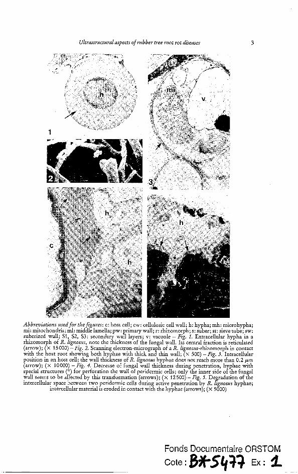
Microscopical observations of R. lignoszis rhizomorphs spreading along the root show differences in the hyphal wall thickness which ranges between 0.2 and 0.7 pm (Fig. 1, 2). The medium layer, which constitutes 60 to 70 Yo of the wall, is reticulated. When hyphae invade the root, the fungal wall thickness decreases to less than 0.2 pm (Fig. 3). Only the inner side of the fungal wall is affected by this modification (Fig. 4).
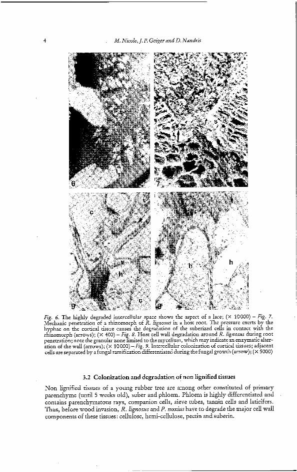
During active penetration, intercellular spaces between two peridermic cells (Fig. 5) can be highly degraded making the decay resemble a lace (Fig. 6). The dissolution of the middle lamella appears to facilitate the penetration of hyphae. Such observations were not conducted on roots infected with P. noxius because a crusty mycelial sleeve along the root, consisting of stones, sand and hyphae, prevents accurate observations.
For both fungi, penetration occurs through lenticels or wounds. R. lignoszts- rhizomorphs exert a pressure on the cortical tissues until they tear (Fig. 7). This mechani- cal action is relayed by a more active mechanism causing the loss of the cortical cells rigid- ity. Examination of Figure 8 reveals a granular zone in front of the apex of some hyphae, suggesting a cell wall degradation by fungal secretions.
Ultrastructural aspects of rubber tree root rot diseases 3
Abbreniutions used for thefigures: c: host cell; cw: cellulosic cell wall; h: hypha; mh: microhypha; mi: mitochondria; ml: middle lamella; pw: rimary wall; r: rhizomorph; s: suber; st: sieve tube; sw: suberized wall; S1, S2, S3: secondary Waf; layers; v: vacuole - Fig. 1. Extracellular hypha in a rhizomorph of R. lignosus, note the thickness of the fungal wall. Its central fraction is reticulated (arrow); ( x 15 000) - Fig. 2. Scanning electron-microgra h of a R. lignosus-rhizomorph in contact with the host root showing both hyphae with thick an8thin wall; (x 500) - Fig. 3. Intracellular position in an host cell; the wall thickness of R. lignosus hyphae does not reach more than 0.2 pm (arrow); ( x 10000) - Fig. 4. Decrease of fungal wall thickness during penetration, hyphae with special structures (‘:) for perforation the wall of peridermic cells; only the inner side of the fungal wall seems to be affected by this transformation (arrows); ( x 12500) - Fig. fi. Degradation of the intercellular space between two peridermic cells during active penetration by R. lignosus hyphae;
intercellular material is eroded in contact with the hyphae (arrows); ( x 5000)
Fonds Documentaire BRSTOM
4 M. Nicole, J. P. Geiger and D. Nandris
Fig. 6. The highly degraded intercellular space shows the aspect of a lace; (x 10000) - Fig. 7. Mechanic penetration of a rhizomorph of R. lignost~ in a host root. The pressure exerts by the hyphae on the cortical tissue causes the degradation of the suberized cells in contact with the rhizomorph (arrows); (x 400) -Fig. 8. Host cell wall degradation around R. lignoszrs during root penetration; note the granular zone limited to the mycelium, which may indicate an enzymatic alter- ation of the wall (arrows); (X 10000) - Fig. 9. Intercellular colonization of cortical tissues; adjacent cells are separated by a fungal ramification differentiated during the fungal growth (arrow); (x 5000)
3.2 Colonization and degradation of non lignified tissues
Non lignified tissues of a young rubber tree are among other constituted of primary parenchyme (until 5 weeks old), suber and phloem. Phloem is highly differentiated and contains parenchymatous rays, companion cells, sieve tubes, tannin cells and laticifers. Thus, before wood invasion, R. Zignosws and P. noxius have to degrade the major cell wall components of these tissues: cellulose, hemi-cellulose, pectin and suberin.
Ultrastructural aspects of rubber tree root rot diseases 5
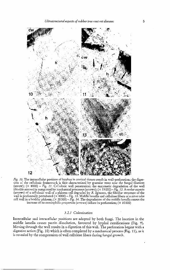
Fig. 10. The intracellular position of hyphae in cortical tissues result in wall-perforation; the diges- tion of the cellulosic framework is first characterized by granular zone near the fungal filament (arrow); (X 8000) - Fig. 11. Cellulosic wall penetration; the enzymatic degradation of the wall (double arrows) is completed by mechanical rocesses (arrows); (X 15000)-Fig. 12. Aveolar aspects (arrows) of a cellulosic wall of a phloem ce17 degraded by R. ligizosus, the fibrillar structure of the wall is profoundly pertubated ( x 8000) -Fig. 13. Middle lamella and cellulose fibers of a sieve tube cell wall in a healthy phloem; (X 20000) -Fig. 14. The degradation of the middle lamella causes the
increase of its osmiophilic properties (arrows) before its perforation; ( x ~0000)
3.2.1 Colonizdtion
Intercellular and intracellular positions are adopted by both fungi. The location in the middle lamella causes pectin dissolution, favoured by hyphal ramifications (Fig. 9). Moving through the wall results in a digestion of this wall. The perforation begins with a digestive action (Fig. 10) which is often completed by a mechanical process (Fig. Il), as it is revealed by the compression of wall cellulose fibers during fungal growth.
6 M. Nicole, J. P. Geiger and D. Nandris
1 - -
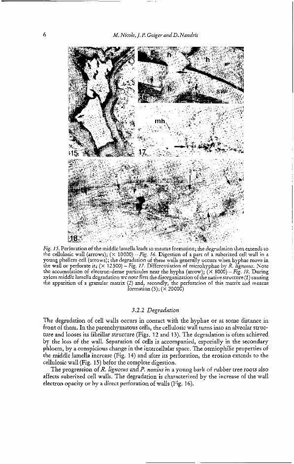
18 Fig. 15. Perforation of the middle lamella leads to meatus formation; the degradation then extends to the cellulosic wall (arrows); (X 10000) -Fig. 16. Digestion of a part of a suberized cell wall in a young hellem cell (arrows); the degradation of these walls generally occurs when hyphae move in the waIor perforate it; (x 12500) -Fig. 17. Differentiation of microhyphae by R. LignosrLs. Note the accumulation of electron-dense particules near the hypha (arrow); (X 8000) -Fig. 18. During xylem middle lamella degradation we note first the disorganization of the native structure (1) causing the apparition of a granular matrix (2) and, secondly, the perforation of this matrix and meatus
formation (3); (X 20000)
3.2.2 Degradation
The degradation of cell walls occurs in contact with the hyphae or at some distance in front of them. In the parenchymatous cells, the cellulosic wall turns into an alveolar struc- ture and looses its fibrillar structure (Figs. 12 and 13). The degradation is often achieved by the loss of the wall. Separation of cells is accompanied, especially in the secondary phloem, by a conspicious change in the intercellular space. The osmiophilic properties of the middle lamella increase (Fig. 14) and after its perforation, the erosion extends to the cellulosic wall (Fig. 15) befor the complete digestion.
The progression of R. Zignosta and P. noxius in a young bark of rubber tree roots also affects suberized cell walls. The degradation is characterized by the increase of the wall electron opacity or by a direct perforation of walls (Fig. 16).
Ultrastructural aspects of rubber tree root rot diseases 7
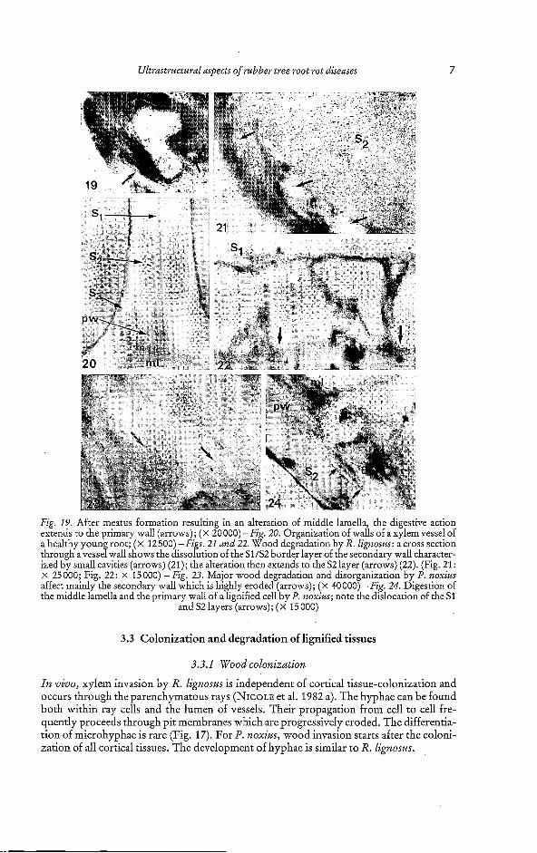
Fig. 19. After meatus formation resulting in an alteration of middle lamella, the digestive action extends to the primary wall (arrows); ( x 20000) -Fig. 20. Organization of walls of a xylem vessel of a healthy youn root; (x 12500) -Figs. 21 and 22. Wood degradation by R. ligizosus: a cross section through a vesse?wall shows the dissolution of the SUS2 border layer of the secondary wall character- ized by small cavities (arrows) (21); the alteration then extends to the S2 layer (arrows) (22). (Fig. 21: x 25000; Fig. 22: x 15000) -Fig. 23. Major wood degradation and disorganization by P. norius affect mainly the secondary wall which is hi hly eroded (arrows); ( x 40 000) -Fig. 24. Digestion of the middle lamella and the primary wall of alignified cell by P. norius; note the dislocation of the S1
and S2 layers (arrows); (x 15000)
3.3 Colonization and degradation of lignified tissues
3.3.1 Wood colonization
In vivo, xylem invasion by R. ligizosus is independent of cortical tissue-colonization and occurs through the parenchymatous rays (NICOLE et al. 1982 a). The hyphae can be found both within ray cells and the lumen of vessels. Their propagation from cell to cell fre- quently proceeds through pit membranes which are progressively eroded. The differentia- tion of microhyphae is rare (Fig. 17). For l'. noxiiu, wood invasion starts after the coloni- zation of all cortical tissues. The development of hyphae is similar to R. lignosits.
8 M. Nicole, J. P. Geiger and D. Nandris
3.3.2 Wood degradation by R. lignosus
The cell wall degradation is progressive and proceeds in contact with or in front of hyphae. The alteration occurs inwards, from the middle lamella towards the lumen. Two stages characterize the middle lamella and the primary wall degradation: a) an apparition of a granular matrix, after the disorganization of the native structure (Fig. 18); b) a progres- sive perforation of this matrix, causing meatus formation (Fig. 19). Then, the alteration reaches the less lignified secondary wall. The junction of S1 and S, layer is particularly susceptible to the fungal action. Indeed, small cavities appear at this level (Figs. 20 and 21), amalgamate and then extend inwards until causing the disparition of the half S2 layer (Fig. 22), showing a strong digestion of the wall framework. S3 layer is saved up by this mechanism. When the progression of decay occurs outwards, the S3 layer is degraded; on this case only S2 and S1 layers are less altered.
3.3.3 Wood degradation by P. noxius
Unlike R. lignosus, wall degradation by P. noxius occurs as well inwards as outwards, in contact or not with the mycelium. However, the secondary wall is more susceptible to the enzymatic action (Fig. 23). This degradation often begins by the apparition of a strong osmiophilic reaction of the wall. The middle lamella and the primary wall generally show less alteration and complete digestion was rare (Fig. 24).
4 Discussion
The decrease of the wall thickness of R. lignosus hyphae is a major fact occurring in root penetration; it corresponds an important stage of the biological cycle of this fungus. Indeed, two types of mycelium have been identified with fundamental differences in mor- phological characteristics (BOISSON 1973) and in enzymatic secretions (GEIGER 1975). One type of mycelium (called B) is specified in propagation by rhizomorph formation, while the other (A) possesses the infectious potential.
So, the decrease of wall thickness during penetration can be related to the transforma- tion of mycelium B into mycelium A. Cytochemical tests have shown that Polysaccharid components of the wall are involved in this modification (unpublished data). The chitin level being constant in the wall of both A and B mycelium (NICOLE 1982), other polymers may probably be hydrolysed during the wall transformation, for example a! and ß D- glucan, major polysaccharides of Basidiomycetae (BURNETT and TRINCI 1978; CHET and H~TTERMANN 1980). This coincides with important modifications in the glucose pathway during B to A transformation, mainly under anaerobic environment (BAREYRE and BOISSON 1969). Such anoxic conditions are fundamental for the initiation or R. lignosus pathogenesis (NANDRIS et al. 1983).
Root penetration and colonization of non-lignified tissues by both fungi result in wall perforation caused by hyphal growth or (and) a chemical digestion (PERIES and IRUGAL- BANDARA 1973; NICOLE et al. 1982 a). Nevertheless, several features indicate that wall degradation is predominantly enzymatic: a) irrespective of the contact with the hyphae the cell wall is more electron-opaque than the rest of the wall; b) the fibrillar structure of cellulose and the structure of suberin are disorganized; c) the disintegration of tissues. Cellulases, xylanases, glucosidases, glycosidases and galactosidases were identified as causing cellulose and hemi-cellulose degradation, and pectic enzymes - polygalac- turonase, pectin methyl-esterase, pectate lyase - as causing middle lamella alteration (GEIGER et al. 1986a, d). However, their activities are higher in P. noxius, especially for the pectic enzymes thus explaining the faster alteration of these tissues by this fungus. As
Ultrastrr~tural aspects of rubber tree root rot diseases 9
reported by other authors (FERNANDO et al. 1984; ZIMMERMANN and SEEM~LLER 1984) the significance of suberin-degrading enzymes in root penetration and suberin alteration can also be strongly suggested (NICOLE et al. 1986). Evidences given by microscopic ob- servations are completed by the characterization of such enzymes in culture filtrates of both fungi (GEIGER et al. 1986a).
Wood invasion by R. ligizosus and P. noxius is based upon the active penetration of cells, as described in other wood rotting fungi (CHOU and LEVI 1971; PEEK et al. 1972). Differences appear mainly during xylem degradation. Wood alteration of young rubber tree seedlings by R. lignosas is selective, and occurs inwards from the middle lamella to the secondary wall, removing first the lignin-rich fraction and then polysaccharids. Except for Eastern Hemlock infected with Ganodernza tsidgae (BLANCHETTE 1984), such a degradation has rarely been reported infecting living trees, but mainly in vitro on sterilized wood-blocks (SANTRA and NANDI 1975; NOGUCHI et al. 1980). These observa- tions suggest that R. lignosws possesses effective lignolytic potentialities. The laccases (E.C. 1.30.3.2.), fungal extracellular enzymes probably involved in lignin decomposition (ANDER and ERIKSON 1976 and 1977; NOGUCHI et al. 1980), have been characterized in rubber tree tissues infected with R. lignosus (GEIGER 1976, 1986a). One of these laccases, the L I laccase, induces a modification of the polymerization degree of a rubber tree thioglycolic lignin (TGL), leading to both condensation and depolymerization of this macromolecule (GEIGER et al. 1986 c). The partial resistance of the S3 layer to the enzyma- tic attack, also suggested by other authors (COWLING 1961; LIESE 1970; DIROL and RAVILLY 1979), may result from the low porosity of this layer, limiting the enzyme-dif- fusibility. The presence of some enzyme inhibitors of phenolic origin has been envisaged (RUEL et al. 1981).
Wood alteration of roots of rubber trees by P. noxius is unspecific. Nevertheless, the middle lamella and the primary wall decomposition seem to be lower than the secondary wall degradation, unlike P. pini (BLANCHETTE 1979) and P. isabellinus (ANDER and ERIKSSON 1976 and 1977) which selectively attack the lignified structures. A laccase has also been identified and isolated from P. noxius infected tissues (GEIGER et al. 1986 b). Meanwhile, this enzyme differs from R. lignosus laccases by its molecular weight and its lower activity, explaining the lower lignin degradation observed in roots.
This ultrastructural study, performed in vivo, describes some aspects of H. brasiliensis root colonization and decay, while the main works on wood degradation by white root rot fungi were generally realized in vitro (SANTRA and NANDI 1975; NOGUCHI et al. 1980; MURMANIS et al. 1984; RUEL et al. 1984; HIGHLEY and MURMANIS 1984). This work com- pletes biochemical investigations described on this host-parasite system (GEIGER et al. 1986a). Although the role of extracellular enzymes of R. lignosus and P. noxius in wall alteration is partly understood, their role in pathogenesis remains nevertheless undeter- mined. Involvement of other fungal secretions in the infection is possible, too (NICOLE et al. 1982). Indeed, PERIES (1959) has characterized a thermostable toxin produced in vitro by different strains of R. lignosus in Sri Lanka; but no correlations have been established with its pathogenicity.
Summary
In order to describe root colonization and degradation an ultrastructural study was performed on young rubber trees infected with two root rot fungi, Rigidoporus lignosw and Pl~ellinus noxius. Inoculation of roots took place by passive mechanisms or after cell wall perforation. In root tissues, both fungi have been observed in intracellular, intercellular and intraparietal positions. Components of different root tissues - suberin, pectin, cellulose, hemi-cellulose and lignin- are digested and cell walls disorganized by these parasites. Colonization and degradation of non-lignified tissues are faster with P. noxius. Xylem degradation by R. lignosus begins in strongly lignified regions - middle lamella and primary wall - and extends towards the secondary wall. The S3 layer is rarely altered, however. On the other hand, wood degradation by P. noxius is not selective; the layers, enriched with lignin and the middle lamella seem to be less eroded.
10 M. Nicole, J. P. Geiger and D. Nandris
The role of degrading-enzymes for the infection process is discussed in comparison to biochemi- cal data recorded elsewhere.
Résumé
Aspects ultrastructuraux des pourridiés &Hevea brasiliensis Une étude ultrastructurale a été réalisée sur de jeunes Hévéa infectés artificiellement par deux cham- pignons agents de pourridié: Rigidoporus lignosus et Phellinus noxius. Les différentes étapes de la colonisation et de la dégradation du système racinaire sont décrites dans cet article. La pénetration de la racine par les parasites s'effectue par les voies naturelles ou après perforation de la paroi. Dans les tissus, les hyphes ont été observées tant dans les cellules qu'en positions intercellulaire et intrapa- riétale. Les composants des parois cellulaires hôtes sont par conséquent soit désorganisés soit dé ra dés par chacun des cham igpons. L'altération des tissus non lignifiés par P. noxius est cepeniai
lus rapide que celle réaI!ke par R. lignosus. A l'inverse, ce dernier est plus actif dans les tissus &ni fiés.
Le rôle des enzymes dégt-adantes dans le processus infectieux de chaque parasite est discuté en comparaison avec les résultats biochimiques acquis par ailleurs.
Zusammenfassung
Feinstruktrtren im Zusammenhang mit Wurzelfiiulen an Hevea brailiensis Um Kenntnisse über die Wurzelbesiedlung und den Wurzelabbau zu gewinnen, wurden Untersu- chungen der Feinstruktur an jungen, mit R. li nosus und Pb. noxius infizierten Kautschukbäumen durchgeführt. Die Inokulation der Wurzeln ejolgte entweder assiv oder nach dem Perforieren der Zellwand. Im Wurzelgewebe ließen sich beide Pilze im intrazeflulären, interzellulären und intrapa-
' rietalen Bereich nachweisen. Die an verschiedenen Wurzelgeweben beteiligten Substanzen wie Suberin, Pektin, Cellulose, Hemicellulose und Lignin wurden von innen abgebaut, die entsprechen- den Zellwände destrukturiert. Bei P. noxius laufen die Besiedelung und der Abbau nicht-lignifizier- ter Wkde schneller ab. Der Xylem-Abbau durch R. lignosus beginnt in den stark lignifizierten Par- tien wie Mittellamelle und Primärwand und setzt sich in Richtung Sekundärwand fort; SJ wird nur selten verändert. Andererseits verläuft der Holzabbau durch P. noxius keineswegs selektiv. Die ligninreichen Teile der Wand und die Mittellamelle scheinen eher weniger erodiert zu sein.
Abschließend wird die Bedeutung von Enzymen für den Infektionsvorgang unter Berücksichti- gung der einschlägigen biochemischen Literatur diskutiert.
Literature
ANDER, P. ; ERIKSSON, K. E., 1976: The importance of phenol oxidase activity in lignin degradation
- - 1977: Selective degradation of wood components by white rot fungi. Physiologia Plantarum 49,
BAREYRE, M. J.; BOISSON, C., 1969: Relation entre la morphogenèse de Pap arei1 végétatif non
ques des deuxphases dudévelo pement. Compte-rendus Acad. Sci., Paris, SérieD268,2256-2259. BLANCHETTE, R. A., 1979:Cellwa~decompositionbyPhellinus(Fomes~pini. Phytopathology69,913. - 1984: Selective delignification of Eastern Hemlock by Ganoderma tsugae. Phytopathology 74,
BOISSON, C., 1973: De la basidiospore au rhizomorphe, déterminisme de l'agrégation chez le basi- diomycete Leptoporus lignosus (Kl.) Heim ex Pat. Thèse #Etat, Paris.
BURNETT, J. H.; TRINCI, A. P. J., 1978: Fungal walls and hyphal growth. Ed. by BURNETT, J. H. and TRINCI, A. P. J. Cambridge University Press, Cambridge.
CHET, I.; H~TTERMANN, A., 1980: Chemical composition of hyphal walls of Fomes annosus. Eur. J. For. Path. 10, 65-70.
CHOU, C. K.; LEVI, M., 1971: An electron microscopical study of the penetration and decomposi- tion of tracheid walls of Pinus sylvestris by Poria vallantii. Holzforschung 25, 107-1 12.
COWLING, E. B., 1961: Comparative biochemistry of the decay of sweetgum sapwood by white rot and brown rot fungi. U.S.D.A. Techn. Bull. 1258, 79 pp.
DIROL, D.; RAVILLY, F., 1979: Les différentes formes d'altération de la paroi cellulaire par quelques champignons lignivores. Annales Biol. 18, 477-492.
FERNANDO, G.; ZIMMERMANN, W.; KOLATTUKUDY, P. E., 1984: Suberin-grown Fusarium solani f. sp. pisi generates a atinase-like esterase which depolymerizes the aliphatic components of sub- erin. Physiol. Plant Path. 24,143-155.
by the white rot fungus Sporotrichum pulverdentum. Arch. Microbiology 109, 1-58.
239-248.
agrégé de Leptoporus lignosus (Kl.) Heim et le métabolisme repiratoire des fi P aments caractéristi-
153-160.
Ultrastructural aspects of rubber tree root rot diseases 11
GEIGER, J. P.; NANDRIS, D.; GOUJON, M., 1976: Activité des laccases et des peroxydases au sein des racines d'Hévéa attaquées par le pourridié blanc [Leptoporus lignosus (KI.) Heim]. Physiol.
GEIGER, J. P.; NICOLE, M.; NANDRIS, D.; RIO, B., 1986a: White root rot of Hevea brasiliensis. I. Physiological and biochemical aspects of host aggression. Eur. J. For. Path. 16,27-37.
GEIGER, J. P. ; RIO, B. ; NANDRIS, D. ; NICOLE, M., 198613: The laccases of Rigidoporus li nosus and Phellinus noxius. I. Purification and some physico-chemical properties. Appl. Bioc%em. Bio- technol. 12,121-134.
GEIGER, J. P.; HUGUENIN, B. ;NICOLE, M. ;NANDRIS, D., 1986c:ThelaccasesofRigidoporuslignosus and Phellinus noxius. II. Effects on a thioglycolic lignin. Appl. Biochem. Biotechnol. 13,97-111.
GEIGER, J. P.; RIO, B.; NANDRIS, D.; NICOLE, M., 1986d: Polysaccharide degrading enzymes ex- creted by two root rot fungi. Symbiosis (in ress).
HIGHLEY, T. L.; MURMANIS, L. L., 1984: Uirastructural aspects of cellulose decompositon by white rot fungi. Holzforschung 38, 73-78.
LIESE, W., 1970: Ultrastructural aspects of woody tissues desintegration. Ann. Rev. Phythopath. 8,
LUFT, J. H., 1961: Improvements in epoxy resin embedding method. J. Biophys. Biochem. Cyto-
MURMANIS, L. L.; HIGHLEY, T. L. ; PALMER, J. G., 1984: An electron microsco y study of Western Hemlock degradation by thewhiterotfungus Ganodermaapplanatum. Holz~rschung34,11-18.
NANDRIS, D.; NICOLE, M.; GEIGER, J. P., 1983: Infections artificielles de jeunes plants #Hevea brasiliensis par Rigidoporus lignosus et Phellinus noxius. Eur. J. For. Path. 13, 65-76.
NANDRIS, D.; TRANVAN CANH; GEIGER, J. P.; OMONT, H.; NICOLE, M., 1985: Remote sensing in plant diseases usin infrared color aerial photography: applications trials in Ivory Coast to root diseases of Hevea jrasiliensis. Eur. J. For. Path. 15, 11-21.
NICOLE, M., 1982: Masse mycélienne et activité laccase au sein des racines d'Hévéa infectées par Rigidoporus lignosus. Physiologie Végétale 20, 465-475.
NICOLE, M.; GEIGER, J. P.; NANDRIS, D., 1982 a: Interactions hôte-parasites entre Hevea brasi- liensis et les agents de pourriture racinaire Phellinus noxius et Rigidoporus lignosus étude physio- pathologique comparée. Phytopath. Zeitschrift 105, 31 1-326.
_ _ _ 1982 b: Aspects ultrastucturaux de la dégradation du phloème des racines #Hevea brasiliensis par Rigidoporus lignosus. Compte-rend. Acad. Sci. Paris, III, 471-474.
NICOLE, M.; NANDRIS, D.; GEIGER, J. P., 1983: Cinétique de l'infection de plants #Hevea brasi- liensis par Rigidoporus lignosus. Can. J. Forest Res. 13, 359-364.
NICOLE, M.; GEIGER, J. P.; NANDRIS, D., 1986: Penetration and degradation of suberized cells of Hevea brusiliensis infected with two root rot fungi. Physiol. Mol. Plant Pathol. 28,181-185.
NOGUCHI, A. ; SHIMADA, M. ; HIGUCHI, T., 1980: Studies on lignin degradation. I. Possible role of non specific oxidation of lignin by laccase. Holzforschung 34, 86-89.
PERIES, O. S., 1959: Studies on the production of toxins by Fomes Zignosus. I. Preliminary investiga- tions. J. Rubber Res. Inst. Ceylon 35, 38-40.
PERIES, O. S.; IRULGABANDARA, 2. E., 1973: Histology of Hevea roots infected by Fomes lignosus. Annals Appl. Biol. 73, 1-7.
PICHEL, R. J., 1956: Les pourridiés de l'Hévéa dans la cuvette congolaise. Publication INEAC, série Technologie 49, 480 pp.
PEEK, R. D.; LIESE, W.; PARAMESTARAN, N., 1972: Infektion und Abbau des Wurzelholzes von Fichte durch Fomes annosits. Eur. J. For. Path. 2,237-248.
REYNOLDS, E. S., 1963: The use of lead citrate at high pH as an electron opaque stain in electron microscopy, J. Cell Biol. 7, 208-212.
RUEL, K.; BARNOUD, F.; ERIKSSON, K. E., 1981: Micromorphological and ultrastructural aspects of Spruce wood degradation by wild type Sporotrichum pulverulentuni and its cellulase less mutant cell 144. Holzforschung 35, 157-171.
RUEL, K.; BARNOUD, F.; ERIKSSON, K. E., 1984: Ultrastructural aspects of wood degradation by Sporotrichum pulverulentum. Holzforschung 38, 61-68.
SANTRA, S.; NANDI, B., 1975: Microstructural and microchemical studies of wood decay of Caseronnia exquisetifolia by Fomes durissimus. Trans. British Mycol. Soc. 65 (3), 507-509.
SPURR, A. R., 1969: A low viscosity epoxy resin embedding medium for electron microscopy. J. Ultrastructure Res. 26,31-43.
ZIMMERMANN, W.; SEEM~LLER, E., 1984: Degradation of raspberry suberin by Fusarium solani f. sp. pisi and Arnaillaria mellea. Phytopath. Zeitschrift 110, 192-199.
AuthOY5' address: Dr. M. NICOLE; Dr. J. P. GEIGER, Dr. D. NANDRIS, Laboratoire de Phyto-
Receipt of ms.: 11. 9.1985
Vig. 13,307-330.
231-258.
' logy 9,409-414.
pathologie, ORSTOM, BPV 51, Abidjan, Ivory Coast


![Black Rot of Sweet Potato - CTAHR Website Potato[2].pdf · Black Rot of Sweet Potato ... • Hevea brasiliense (rubber) ... County Extension Agent UH-CTAHR Cooperative Extension Service](https://img.pdfslide.us/doc/110x75/5ac735427f8b9a7d548b5104/black-rot-of-sweet-potato-ctahr-website-potato2pdfblack-rot-of-sweet-potato.jpg)