Embed Size (px)
Citation preview
D. Nandris, M. Nicole, and J. P. Geiger Institut Francais de Recherche Scientifique pour le D6veloppement en Coop6ration ORSTOM, Centre d’Adiopodoum6, Abidjan, Ivory Coast
~ . . ~ . I
Root Rot Diseases Several million hectares in the tropical
regions of the world are covered by plantations of commercially important woody tree species that represent a significant fraction of the resources of the countries concerned. Hevea (Hevea brasiliensis (Willd. ex Adr. de Juss) Muell. Arg.) is important among these species because of the raw material it produces-the latex from which natural rubber is extracted (Fig. 1). Fungal diseases of these trees can cause very serious losses in production. Root- decaying pathogens caused more than 50% mortality in old stands established after forests were cleared manually; if one assumes 25 years of productive life for a rubber tree, losses came to several hundred thousand dollars per hectare. The most important root rot pathogens are the Basidiomycetes Rigidoporus l ignosus (Klotzsch) Imaz., Phellinus noxius (Corner) G. H. Cunn., Ganoderma spp., and Armil lar ia spp. and the Ascomycetes Sphaerosti lbe repens (Berk. & Br.) and Ustulina zonata (Lév.) Sacc. R. lignosus (formerly called Fomes lignosus, then Leptoporus lignosus) is the pathogen most feared by planters throughout the rubber-growing regions of the world (24).
Since the first industrial plantations of Hevea were established in Southeast Asia at the beginning of the century, fungal root rot pathogens have caused tree losses, sometimes serious (Fig. 2). Several research institutes in Asia and Africa have studied these diseases and developed means of control, but despite many investigations and 70 years of research, no satisfactory solutions in terms of effectiveness, ease of use, and cost could be offered. For the past 15 years, however, investigations have taken a somewhat different path, with more emphasis on fundamental research that can form a rational basis for devising control methods.
In this article we first present some general information about the cultivation of Hevea and its pathology, then review current knowledge of the biology of the root rot fungi, and finally indicate the nature of the methods recommended in
@ 1987 The American Phytopathological Society
298 Plant DiseaseNol. 71 No. 4
the past for root rot control, together with recent developments.
Historical and Economic Aspects Although discovered in Amazonia in
the 15th century by Spanish explorers (including Torquemada and Colomb), latex remained unknown in Europe for more than three centuries. In 1736, De La Condamine brought back to France from Peru samples of this elastic material, which rapidly became a craze. The invention of vulcanization in 1839 gave new impetus to the rubber industry, which- has been expanding ever since. Until the beginning of this century, most industrial latex production was from wild trees in the Amazonian forest where H. brasiliensis originated. Because these trees were dispersed, the work was arduous and the effects of tapping were destructive. To establish plantations of Hevea that could be managed more easily and intensively, seeds or seedlings were repeatedly sent to Sri Lanka (formerly Ceylon), Java, and Malaya, often with nearly complete loss of viability during transit. Nevertheless, the first industrial plantation of Hevea was established in
Fig. 1. The rubber tree Is tapped for latex twice a week by girdling the phloem containing the laticifers.
1902 in Sumatra. Sixteen years later, 98% of world demand was supplied by the Far East.
In parallel with the rise in Asian rubber growing, the United States wanted to be independent in its rubber supply, and plantations were established in Liberia in 1923 (by Firestone) and in the Philippines in 1928 (by Goodyear). The first plantations in French-speaking Africa were established in 1920.
The annual consumption of natural rubber has risen considerably since 1976 owing to the increased price of petroleum
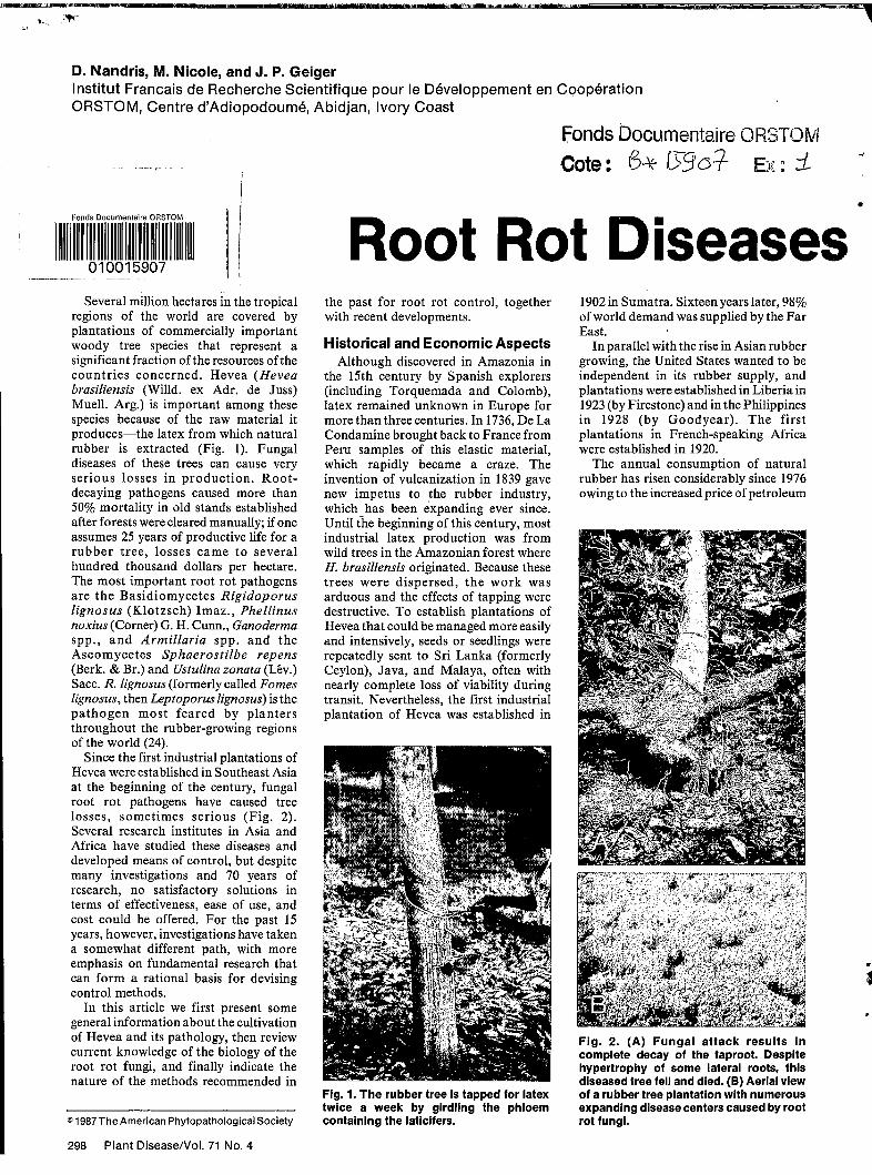
Fig. 2. (A) Fungal attack results in complete decay of the taproot. Despite hypertrophy of some lateral roots, this diseased tree fell and died. (B) Aerial view of a rubber tree plantation with numerous expanding disease centers caused by root rot fungi.
. 8
?
.
c
+-of Rubber
1
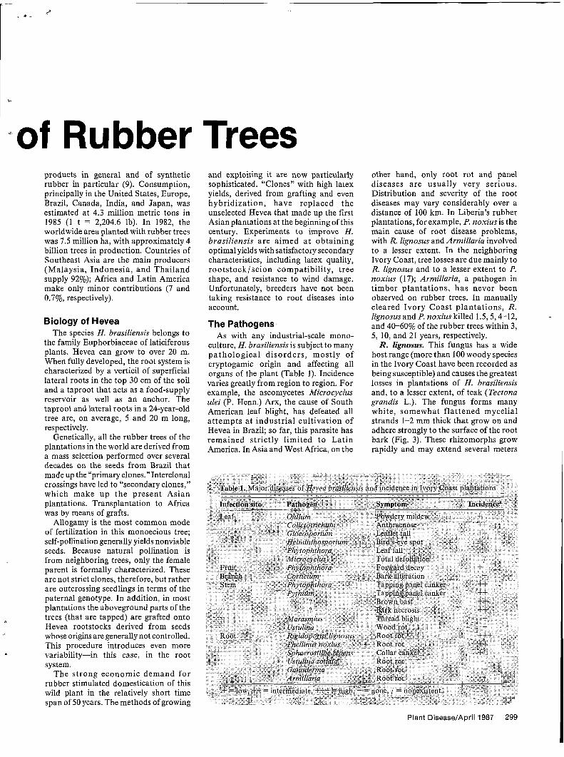
products in general and of synthetic rubber in particular (9). Consumption, principally in the United States, Europe, Brazil, Canada, India, and Japan, was estimated at 4.3 million metric tons in 1985 (1 t = 2,204.6 lb). In 1982, the worldwide area planted with rubber trees was 7.5 million ha, with approximately 4 billion trees in production. Countries of Southeast Asia are the main producers (Malaysia, Indonesia, and Thailand supply 92%); Africa and Latin America make only minor contributions (7 and 0.7%, respectively).
Biology of Hevea The species H. brasiliensis belongs to
the family Euphorbiaceae of laticiferous plants. Hevea can grow to over 20 m. When fully developed, the root system is characterized by a verticil of superficial lateral roots in the top 30 cm of the soil and a taproot that acts as a food-supply reservoir as well as an anchor. The taproot and lateral roots in a 24-year-old tree are, on average, 5 and 20 m long, respectively.
Genetically, all the rubber trees of the plantations in the world are derived from a mass selection performed over several decades on the seeds from Brazil that made up the “primary clones.”Interclonal crossings have led to “secondary clones,” which make up the present Asian plantations. Transplantation to Africa was by means of grafts.
Allogamy is the most common mode of fertilization in this monoecious tree; self-pollination generally yields nonviable seeds. Because natural pollination is from neighboring trees, only the female parent is formally characterized. These are not strict clones, therefore, but rather are outcrossing seedlings in terms of the paternal genotype. In addition, in most plantations the aboveground parts of the trees (that are tapped) are grafted onto Hevea rootstocks derived from seeds whose origins are generally not controlled. This procedure introduces even more variability-in this case, in the root system.
The strong economic demand for rubber stimulated domestication of this wild plant in the relatively short time span of 50 years. The methods of growing
and exploiting it are now particularly sophisticated. “Clones” with high latex yields, derived from grafting and even hybridization, have replaced the unselected Hevea that made up the first Asian plantations at the beginning of this century. Experiments to improve H. brasiliensis are aimed a t obtaining optimal yields with satisfactory secondary characteristics, including latex quality, rootstock/scion compatibility, tree shape, and resistance to wind damage. Unfortunately, breeders have not been taking resistance to root diseases into account.
The Pathogens As with any industrial-scale mono-
culture, H. brasiliensis is subject to many pathological disorders , mostly of cryptogamic origin and affecting all organs of the plant (Table 1). Incidence varies greatly from region to region. For example, the ascomycetes Microcyclus ulei (P. Henn.) Arx, the cause of South American leaf blight, has defeated all at tempts a t industrial cultivation of Hevea in Brazil; so far, this parasite has remained strictly limited to Latin America. In Asia and West Africa, on the
other hand, only root rot and panel diseases are usually very serious. Distribution and severity of the root diseases may vary considerably over a distance of 100 km. In Liberia’s rubber plantations, for example, P. noxius is the main cause of root disease problems, with R. lignosus and Armillaria involved to a lesser extent. In the neighboring Ivory Coast, tree losses are due mainly to R. lignosus and to a lesser extent to P. noxius (17); Armillaria, a pathogen in timber plantations, has never been observed on rubber trees. In manually cleared Ivory Coast plantations, R. lignosus and P. noxius killed 1.5,5,4-12, and 4 6 6 0 % of the rubber trees within 3, 5, 10, and 21 years, respectively.
R. lignosus. This fungus has a wide host range (more than 100 woody species in the Ivory Coast have been recorded as being susceptible) and causes the greatest losses in plantations of H. brasiliensis and, to a lesser extent, of teak (Tectona grandis L.). The fungus forms many white, somewhat flattened mycelial strands 1-2 mm thick that grow on and adhere strongly to the surface of the root bark (Fig. 3). These rhizomorphs grow rapidly and may extend several meters
Plant Disease/April 1987 299
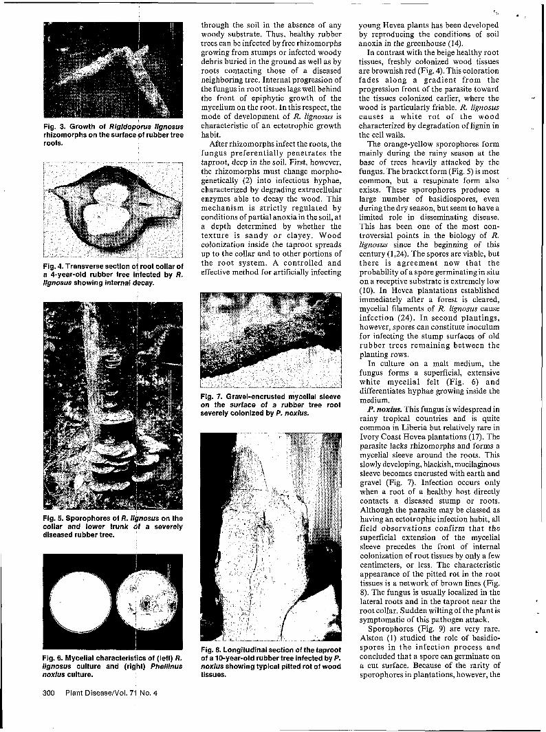
Fig. 3. Growth of Rlgldoporus llgnosus rhizomorphs on the surface of rubber tree roots.
Fig. 4. Transverse section of root collar of a 4-year-old rubber tree infected by R. Iignosus showing internal decay.
Fig. 5. Sporophores of R. llgnosus on the collar and lower trunk of a severely diseased rubber tree.
Fig. 6. Mycelial characteristics of (left) R. llgnosus culture and (right) Phellinus noxlus culture.
through the soil in the absence of any woody substrate. Thus, healthy rubber trees can be infected by free rhizomorphs growing from stumps or infected woody debris buried in the ground as well as by roots contacting those of a diseased neighboring tree. Internal progression of the fungus in root tissues lags well behind the front of epiphytic growth of the mycelium on the root. In this respect, the mode of development of R. Zignosus is characteristic of an ectotrophic growth habit.
After rhizomorphs infect the roots, the fungus preferentially penetrates the taproot, deep in the soil. First, however, the rhizomorphs must change morpho- genetically (2) into infectious hyphae, characterized by degrading extracellular enzymes able to decay the wood. This mechanism is strictly regulated by conditions of partial anoxiain the soil, at a depth determined by whether the texture is sandy o r clayey. Wood colonization inside the taproot spreads up to the collar and to other portions of the root system. A controlled and effective method for artificially infecting
Fig. 7. Gravel-encrusted mycelial sleeve on the surface of a rubber tree root severely colonized by P. noxius.
Fig. 8. Longitudinal section of the taproot of a 10-year-old rubber tree infected by P. noxlus showing typical pitted rot of wood tissues.
~
- 2 r-,
young Hevea plants has been developed by reproducing the conditions of soil anoxia in the greenhouse (14).
In contrast with the beige healthy root tissues, freshly colonized wood tissues are brownish red (Fig. 4). This coloration fades along a gradient from the progression front of the parasite toward the tissues colonized earlier, where the wood is particularly friable. R. Zignosus causes a white ro t of the wood characterized by degradation of lignin in the cell walls.
The orange-yellow sporophores form mainly during the rainy season at the base of trees heavily attacked by the fungus. The bracket form (Fig. 5) is most common, but a resupinate form also exists. These sporophores produce a large number of basidiospores, even during the dry season, but seem to have a limited role in disseminating disease. This has been one of the most con- troversial points in the biology of R. Zignosus since the beginning of this century (1,24). The spores are viable, but there is agreement now tha t the probability of a spore germinating in situ on a receptive substrate is extremely low (10). In Hevea plantations established immediately after a forest is cleared, mycelial filaments of R. Zignosus cause infection (24). I n second plantings, however, spores can constitute inoculum for infecting the stump surfaces of old rubber trees remaining between the planting rows.
In culture on a malt medium, the fungus forms a superficial, extensive white mycelial felt (Fig. 6) and differentiates hyphae growing inside the medium. P. noxius. This fungus is widespread in
rainy tropical countries and is quite common in Liberia but relatively rare in Ivory Coast Hevea plantations (17). The parasite lacks rhizomolrphs and forms a mycelial sleeve around the roots. This slowly developing, blackish, mucilaginous sleeve becomes encrusted with earth and gravel (Fig. 7). Infection occurs only when a root of a healthy host directly contacts a diseased stump or roots. Although the parasite may be classed as having an ectotrophic infection habit, all field observations confirm that the
4
superficial extension of the mycelial sleeve precedes the front of internal colonization of root tissues by only a few centimeters, or less. The characteristic appearance of the pitted rot in the root tissues is a network of brown lines (Fig. 8). The fungus is usually localized in the lateral roots and in the taproot near the root collar. Sudden wiltingof the plant is symptomatic of this pathogen attack.
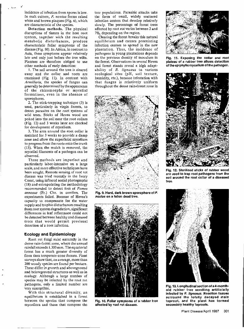
Sporophores (Fig. 9) are very rare. Alston (1) studied the role of basidio- spores in the infection process and concluded that a spore can germinate on a cut surface. Because of the rarity of sporophores in plantations, however, the
300 Plant DiseaseNol. 71 No. 4
a I '-
incidence of infection from spores is low. In malt culture, P. noxius forms raised white and brown plaques (Fig. 6), which are characteristic of the species.
Detection methods. The physical disruptions of tissues in the host root system, together with the resulting metabol ic d i s turbances , produce
disease (Fig. 10). In Africa, in contrast to Asia, these symptoms appear relatively
Planters are therefore obliged to use other methods of early detection: I. The soil around the tree is cleared
away and the collar and roots are examined (Fig. 11). In contrast with Amiillaria, the species of fungus can generally be determined by the appearance of the rhizomorphs o r mycelial formations, even in the absence of sporophores.
2. The stick-trapping technique (3) is used, particularly in virgin forests, to detect parasites on the root systems of wild trees. Sticks of Hevea wood are poked into the soil near the root collars (Fig. 12) and 3 weeks later are checked for development of mycelium. 3. The area around the root collar is
mulched for 3 weeks to provide a damp zone and allow the superficial mycelium to progress from the roots onto the trunk (13). When the mulch is removed, the mycelial filaments of a pathogen can be observed.
These methods a re imperfect and particularly labor-intensive on a large scale, and more effective techniques have been sought. Remote sensing of root rot disease was tried recently in the Ivory Coast, using infrared aerial photographs (18) and extrapolating the methodology recommended to detect foci of Fomes annosus (Fr.) Cke. in conifers. The experiments failed. Because of Hevea's capacity to compensate for the water supply and trophic disturbances resulting from root system degradation, significant differences in leaf reflectance could not be detected between healthy and diseased trees tha t would permit previsual detection of a root infection.
.. characteristic foliar symptoms of the
late and only just before the tree wilts. c
Ecology and Epidemiology Root rot fungi exist naturally in the
dense rain-forest zone, where the annual rainfall exceeds 1,300 mm. The equatorial forest has a much greater diversity of flora than temperate-zone forests. Plant surveys show that, on average, more than 200 woody species are found per hectare. These differ in growth and aboveground and belowground structures as well as in ecology. Although a large number of species may be infected by the root rot pathogens, only a limited number are very susceptible.
With this structural diversity, a n equilibrium is established in a forest between the species that compose the mycoflora and those that compose the
tree populations. Parasitic attacks take the form of small, widely scattered infection centers that develop relatively slowly. The percentage of forest trees affected by root rot varies between 2 and 7%, depending on the region.
Clearing the forest breaks this natural equilibrium and causes preexisting infection centers to spread in the new plantation. Thus, t he incidence of pathogens in young plantations depends on the previous density of inoculum in the forest. Observations in several Hevea and forest stands reveal a high adapt- ability of R. lignosus t o various ecological sites (pH, soil texture, humidity, etc.), because infestation with tha t fungus is relatively uniform throughout the dense rain-forest zone in
Fig. 9. Hard, dark brown sporophore of P. noxius on a fallen dead tree.
Fig. 10. Foliar symptoms of a rubber tree affected by root rot disease.
Fig. 11. Exposing the collar and root plateau of a rubber tree allows detection of the epiphytic mycelium of the pathogen.
Fig. 12. Sterilized sticks of rubber wood are used to trap root pathogens from the soil around the root collar of a diseased tree.
Fig. 13. Longitudinal section of a4-month- old rubber tree seedling artificially infected by R. lignosus. Reaction tissues surround the totally decayed main taproot, and the plant has formed secondary healthy taproots.
Plant Disease/Apr¡l 1987 301
the Ivory Coast. Therefore, types of soil that are more conducive than others to the development of roo t rtot a re impossible to define, as has been done for some other soil pathogens in temperate regions. This suggests that the methods of forest clearing and ground preparation before planting, alone, can quantitatively modulate the future incidence of root pathogens in plantations established in forest zones.
Two phases characterize the spatial spread of root rot disease in plantations (15). The first is infection and colonization of the root system of young rubber trees by mycelial filaments growing from stumps of infected forest trees (primary inoculum) remaining om or under the ground despite clearing operations. The second is propagation along roots of the pathogen from infected rubber trees (secondary inoculum) toward neighboring healthy rubber trees. Mycelial growth rates on the superficial roots of Hcevea are estimated to be 2.5 and 0.7 m per year for R. lignosus and P. noxius, respectively. Because trees are planted every 2 m in rows 8 m apart, the pathogen spreads within rather than between rows. In a
I
row segment with diseased trees, only the two infected trees on either end of the segment are considered infectious, each to a single healthy neighboring tree. The diseased trees between the ends of the segment play no part in further spread of the epidemic because the healthy trees in the opposite rows are seldom infected across the 8 m. Consequently, t he circular openings observed in plantations result from simultaneous development of disease centers in neighboring rows.
Disease development and mortality are most rapid during the first few years after planting (15), when areas of the initial disease centers increase and new centers appear. A clear drop in disease development is observed from the fifth or sixth year of culture, then the epidemic stagnates after the 10th year. A similar pattern was observed in a 6-month greenhouse experiment with artificially infected young rubber plants (21).
Two a t tendant phenomena may explain stagnation of the epidemic:
1. The capacity of Hevea to react to root decay increases considerably with age and may contain the parasite within the taproots being colonized. Secondary
302 Plant DiseaseNol. 71 No. 4
I*
taproots and cicatricial swellings form (Fig. 13), and lateral roots hypertrophy. These reactions compensate trophically for the loss of the decayed taproot for some time. The nature of a tree’s defenses to fungal attack on its root system has also been characterized at cellular and molecular levels (see box, page 305).
2. With time, the pathogen loses aggressiveness and ability to spread toward a healthy tree. As the disease center expands, the progression front where the pathogen is fully active in taproots of young trees gets farther and farther away from the primary inoculum (i.e., colonized dead stumps) at the center of the opening. These residual woody materials disintegrate under the influence of either the root rot fungus or the saprophytic microorganisms of the soil, causing the trophic connections between the primary inoculum and the fungus within the infected taproots to rupture. The pathogen gradually passes from a state of saprophytism in the dead wood to a state of near-absolute parasitism in the root systems of trees tha t a re becoming less and less susceptible with age. In “weak” parasites such as R.
D. Nandris
Dr. Nandris, a plant lpathologist with ORSTOlM, has been working in the Ivory Coast since 1977, mainly investigiating the physiology, biology, and epidemiology of root rot fungi in rubber tree plantations and also studying bark necrosis of rubbertrees. He received his Ph.D. degree in plant pathology from P. et M. Curie University of Paris in 1985.
M. Nicole
Dr. Nicole, a researcher in the Plant Pathology Department of ORSTOM, received his Ph.D. degree in plant physiology from the University of Toulouse in France. Since 1977 h e has been concerned with the biology of parasitism and the cellular aspects of pathogen aggression and host defense, mainly in root rot disease and phloem necrosis of rubber trees.
J. P. Geiger
IDr. Geiger, head ofthe Plant Pathology Department of ORSTOM, obtained his Ph.D. degree from the University of Paris and his D.Sc. from the University of Strasbourg. His research activities concern sclerotial morphogenesis of Sclerotium rolfsii, extracellular enzyme production by S. rolfsiiand Rigidoporus lignosus, popullation analysis of Colletotrichum species in the Ivory Coast, and biochemical aspects of host-pathogen interactions.
lignosus and P. noxius, this phenomenon may induce an important decrease in pathogenicity (4). This decrease does not mean degeneration or senescence of the fungus, however; stick traps around the
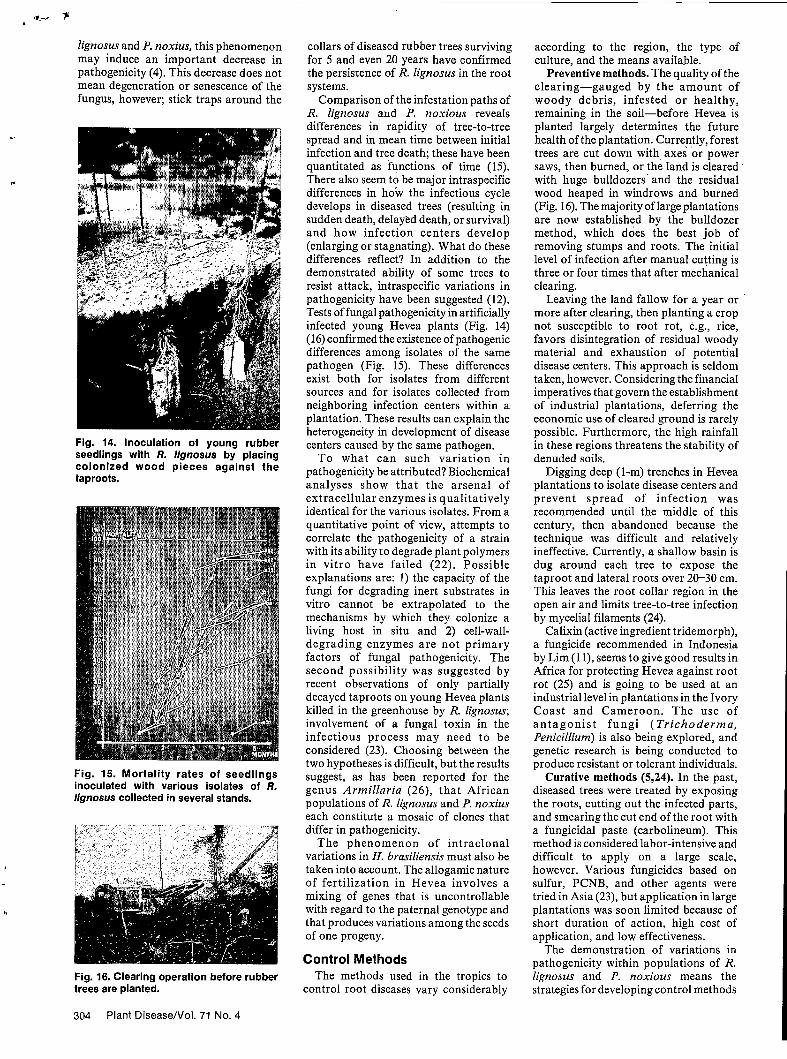
Fig. 14. Inoculation of young rubber seedlings with R. Mgnosus by placing colonized wood pieces against the taproots.
Fig. 15. Mortality rates of seedlings inoculated with various isolates of R. lignosus collected in several stands.
Fig. 16. Clearing operation before rubber trees are planted.
collars of diseased rubber trees surviving for 5 and even 20 years have confirmed the persistence of R. lignosus in the root systems.
Comparison of the infestation paths of R. Iignosus and P. noxious reveals differences in rapidity of tree-to-tree spread and in mean time between initial infection and tree death; these have been quantitated as functions of time (15). There also seem to be major intraspecific differences in how the infectious cycle develops in diseased trees (resulting in sudden death, delayed death, or survival) and how infection centers develop (enlarging or stagnating). What do these differences reflect? In addition to the demonstrated ability of some trees to resist attack, intraspecific variations in pathogenicity have been suggested (12). Tests of fungal pathogenicity in artificially infected young Hevea plants (Fig. 14) (16) confirmed the existence of pathogenic differences among isolates of the same pathogen (Fig. 15). These differences exist both for isolates from different sources and for isolates collected from neighboring infection centers within a plantation. These results can explain the heterogeneity in development of disease centers caused by the same pathogen.
To what can such variation in pathogenicity be attributed? Biochemical analyses show that the arsenal of extracellular enzymes is qualitatively identical for the various isolates. From a quantitative point of view, attempts to correlate the pathogenicity of a strain with its ability to degrade plant polymers in vitro have failed (22). Possible explanations are: 1) the capacity of the fungi for degrading inert substrates in vitro cannot be extrapolated to the mechanisms by which they colonize a living host in situ and 2) cell-wall- degrading enzymes are not primary factors of fungal pathogenicity. The second possibility was suggested by recent observations of only partially decayed taproots on young Hevea plants killed in the greenhouse by R. lignosus; involvement of a fungal toxin in the infectious process may need to be considered (23). Choosing between the two hypotheses is difficult, but the results suggest, as has been reported for the genus Armillaria (26), that African populations of R. Iignosus and P. noxius each constitute a mosaic of clones that differ in pathogenicity.
The phenomenon of intraclonal variations in H. brasiliensis must also be taken into account. The allogamic nature of fertilization in Hevea involves a mixing of genes that is uncontrollable with regard to the paternal genotype and that produces variations among the seeds of one progeny.
Control Methods The methods used in the tropics to
control root diseases vary considerably
according to the region, the type of culture, and the means available.
Preventive methods. The quality of the clearing-gauged by the amount of woody debris, infested or healthy, remaining in the soil-before Hevea is planted largely determines the future health of the plantation. Currently, forest trees are cut down with axes or power saws, then burned, or the land is cleared ' with huge bulldozers' and the residual wood heaped in windrows and burned (Fig. 16). The majority of large plantations are now established by the bulldozer method, which does the best job of removing stumps and roots. The initial level of infection after manual cutting is three or four times that after mechanical c 1 e ar in g.
Leaving the land fallow for a year or more after clearing, then planting a crop not susceptible to root rot, e.g., rice, favors disintegration of residual woody material and exhaustion of potential disease centers. This approach is seldom taken, however. Considering the financial imperatives that govern the establishment of industrial plantations, deferring the economic use of cleared ground is rarely possible. Furthermore, the high rainfall in these regions threatens the stability of denuded soils.
Digging deep (I-m) trenches in Hevea plantations to isolate disease centers and prevent spread of infection was recommended until the middle of this century, then abandoned because the technique was difficult and relatively ineffective. Currently, a shallow basin is dug around each tree to expose the taproot and lateral roots over 20-30 cm. This leaves the root collar region in the open air and limits tree-to-tree infection by mycelial filaments (24).
Calixin (active ingredient tridemorph), a fungicide recommended in Indonesia by Lim (1 I), seems to give good results in Africa for protecting Hevea against root rot (25) and is going to be used at an industrial level in plantations in the Ivory Coast and Cameroon. The use of a n t a g o n i s t fungi (Trichoderma, Penicillium) is also being explored, and genetic research is being conducted to produce resistant or tolerant individuals.
Curative methods (5,24). In the past, diseased trees were treated by exposing the roots, cutting out the infected parts, and smearing the cut end of the root with a fungicidal paste (Carbolineum). This method is considered labor-intensive and difficult to apply on a large scale, however. Various fungicides based on sulfur, PCNB, and other agents were tried in Asia (23), but application in large plantations was soon limited because of short duration of action, high cost of application, and low effectiveness.
The demonstration of variations in pathogenicity within populations of R. Iignosus and P. noxious means the strategies for developing control methods
304 Plant Disease/Vol. 71 No. 4
t :.
?
Interactions Between the Pathogens and the Host
Aggression by the root rot fungi Rigidoporus lignosus and Phellinus noxius and the reactions of the host Hevea brasiliensis to the pathogenic stress have been investigated.
Aggression by the pathogens. Infection takes place in three stages: penetration of hyphae into the root system, colonization of the tissues, and degradation of the host’s cell structures. The mycelium penetrates the roots either actively, by enzymatic digestion of the tissues (Fig. 17A) with differentiation of specialized structures (20), or mechanically, through natural openings or wounds. Tissues are colonized either by perforation and digestion of cell walls (Fig. 17B) or by penetration through pores and pits of the cells (phloem and xylem). Fungal hyphae have been observed within and between cells and within cell walls.
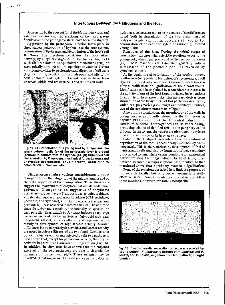
Fig. 17. (A) Penetration of a young root by R. Ilgnosus; the space between cells (c) of the peridermic layer is eroded (arrows) in contact with the hyphae (h). (B) Suberized cell wall alteration by R. Ilgnosus; mechanical forces (arrows) and enzymatic degradation (double arrows) contribute to colonization of phellem cells.
Ultrastructural observations unambiguously show disorganization, then digestion of the middle lamella and of the walls, regardless of their composition. These alterations suggest the involvement of enzymes that can degrade plant polymers. Disorganization suggestive of enzymatic activities-glycosidases (P-glucosidase, a-galactosidase, and &galactosidase), polysaccharidases (CM-cellulase, pectinase, and xylanase), and phenol oxidases (laccase and peroxidase)-was observed in infected tissues. The nature of these disturbances, especially the intensity, is specific for each parasite. Thus, attack by P. noxius induces a very large increase in hydrolytic activities (glycosidases and polysaccharidases), whereas attack by R. lignosus results mainly in development of high laccase activity. Similar differences between hydrolytic activities and laccase activity are noted in culture filtrates of the two fungi. Comparisons of healthy tissues with tissues infected by the two pathogens have shown that, except for peroxidase activity, the enzyme activities in parasitized tissues are of fungal origin (Fig. 18). In addition, in vitro tests have shown that the enzymes excreted by the two pathogens are able to degrade the polymers of the cell wall (6,7). These enzymes may be involved in pathogenesis. The differences in the ratios of
hydrolases to laccase seem to be the source of the differences noted both in degradation of the two main types of polysaccharide and lignin polymers (8) and in the colonization of phloem ahd xylem of artificially infected young plants.
Reactions of the host. During the initial stages of penetration, the most characteristic reactions occur in the young cork, where hyperplasia and cell hypertrophy are seen (19). These reactions are associated generally with a st imulation of the phenolic pool, as revealed by cytochemical tests.
At the beginning of colonization of the cortical tissues, phellogen activity leads to formation of supernumerary cell layers at the points of penetration. Certain cell walls thicken after suberification or lignification of their constituents. Lignification can be explained by a considerable increase in the activity of one of the host isoperoxidases. Investigations of adult trees have shown that this increase derives from stimulation of the biosynthesis of this particular isoenzyme, which can polymerize p-coumaryl and coniferyl alcohols, two of the constituent monomers of lignin.
Also during colonization, the morphology of the walls in young cork is profoundly altered by the formation of papillae (wall appositions). In the central cylinder, the cambium becomes heterogeneous in its functioning, producing islands of lignified cells in the periphery of the phloem. In the xylem, the vessels are obstructed by tyloses formation, and some walls have an extra layer.
Later in the host-pathogen interaction the anatomical organization of the root is occasionally disturbed by tissue neogenesis. This is characterized by development of foci of meristematic cells and also by formation of new secondary phloem and xylem. These tissues constitute an anatomical barrier resisting the fungal attack. In adult trees, these tissues also contain a major isoperoxidase, identical to that mentioned above, that is probably involved in lignification.
Some of the reactions described here limit the progress of the parasite locally, but only tissue neogenesis is really effective, since it compartmentalizes infected tissues. All of these reactions, however, are totally nonspecific.
Fig. 18. Electrophoretic separation of laccases secreted by (top to bottom) R. llgnosus, a mixture of R. llgnosus and P. noxius, and P. noxius; migration from left (cathode) to right (anode).
Plant Disease/April 1987 305
must be modified. If results are to be extrapolated to all the strains making up
j the parasite population of a region, breeders can no longer use only a single fungal isolate f o r testing antifungal compounds or for screening resistant individual plants. Either the tests will have to be repeated with different strains or a highly pathogenic strain previously characterized under controlled conditions will have to be used.
Because of the biology of the root rot fungi and the complexity of the medium in which they develop, less is known about these soil-inhabiting parasites than abou t many leaf parasites. I n our laboratory and in other research institutes, studies are in progress on interisolate sexual compatibility, bio- degradation of lignin, elicitors of defense reactions in Hevea, mathematical modeling of epidemics, characterization of competing and antagonistic fungi, and testing of new antifungal compounds. In parallel with these investigations, prospecting expeditions have been mounted in recent years in the Amazonian basin, with the goal of collecting wild Hevea as sources of new genetic variability. This improvement of the genotype of cultivated Hevea should lead to the production of clones that are better defined genetically and that perform better. Testing of these new gene sources for resistance to root diseases would be desirable.
Literature Cited 1. Alston, R. A. 1953. Annual reports,
1949-1951, Pathological Division, Rubber Research Institute of Malaya.
2. Boisson, C. 1972. Etude de la formation du rhizomorphe du Leptoporus lignosus (Kl.) Heim: Le déterminisme de l’agrégation des filaments en palmettes. C. R. Acad. Sci. Paris Ser. D 274:2481-2484.
3. Declert, C. 1986. Une technique nouvelle de détection des agents de pourridié: La bûchette-piège. Son application à l’étude
du Leptoporus lignosus. Rev. Mycol.
4. Fassi, B. 1964. Eyolution du pourridié blanc, dû au Fomes lignosus (KI.) Bres. dans une plantation de Hevea brasiliensis aménagée immédiatement aprks l’abattage de la forêt. Publ. Inst. Nat. Etude Agron. Congo INEAC 105.54 pp.
5. Fox, R. A. 1977. The impact of ecological, cultural and biological factors on the strategy and costs of controlling root diseases in tropical plantation crops as exemplified by Hevea brasiliensis. J. Rubber Res. Inst. Sri Lanka 54329-362.
6. Geiger, J. P., Huguenin, B., Nandris, D., and Nicole, M. 1986. The laccases of Rigidoporus lignosus and Phellinus noxius. II. Effect. of R. lignosus laccase LI on thioglycolic lignin of Hevea. Appl. Biochem. Biotechnol. 13:97-111.
7. Geiger, J. P., Nicole, M., Nandris, D., and Rio, B. 1986. Root rot diseases of Hevea brasiliensis. I. Physiological and biochemical aspects of root aggression. Eur. J. For. Pathol. 16:22-37.
8. Geiger, J. P., Rio, B., Nicole, M., and Nandris, D. 1986. Biodegradation of Hevea brasiliensis wood by Rigidoporus lignosus and Phellinus noxius. Eur. J. For. Pathol. 16:147-155.
9. International Rubber Study Group. 1985. Rubber Stat. Bull. Vol. 40, No. 1. 46 pp.
10. John, K. P. 1965. Some observations on spore infection of Hevea stumps by Fomes lignosus (Kl.) Bres. J. Rubber Res. Inst. Malaya 19:17-21.
11. Lim, T. M. 1972. Southeast Asia Regional Symposium on Crop Protection, Jogjakarta, Indonesia.
12. Liyanage, G. W., Liyanage, A, de S., Peries, O. S., and Halangoda, L. 1977. Studies of the variability and pathogenicity of Rigidoporus lignosus. J. Rubber Res. Inst. Sri Lanka 54362-372.
13. Martin, R. 1964. Sur la propension de Fomes lignosus, agent de la pourriture des racines blanches d’Hévéa, à la fonction rhizomorphique hors du milieu souterrain. Etdde d’une méthode de détection. Rev. Gen. Caoutch. 41:279-284.
14. Nandris, D., Nicole, M., and Geiger, J. P. 1983. Inoculations of young plants of Hevea brasiliensis by Rigidoporus
26:119-127. lignosus and Phellinus noxius. Eur. J. For. Pathol. 13:65-76.
15. Narudris, D., Nicole, M., and Geiger, J. P. 1985. Epidemiology of rubber root rot in Ivory Coast. Pages 21-30 in: Proc. Colloq. Soc. Fr. Phytopathol. 28th.
16. Nandris, D., Nicole, M., and Geiger, J. P. 1987. Variations and virulence among Rigidoporus lignosus and Phellinus noxius isolates from West Africa. Eur. J. For. Pathol. In press.
17. Nandris, D., Nicole, M., Geiger, J. P., and Mallet, B. 1983. Root rot diseases in Ivory Coast forest and plantations. Pages 286- 295 in: Proc. Colloq. IUFRO 6th.
18. Nandris, D., Tran Van Cahn, Geiger, J. P., Omont, H., and Nicole, M. 1985. Remote sensing in plant diseases using infrared color aerial photography: Application trials in Ivory Coast to root diseases of Hevea brasiliensis. Eur. J. For. Pathol. 15:ll-21.
19. Nicole, M., Geiger, J. P., and Nandris, D. 1986. Root rot diseases of Hevea brasiliensis. II. Some host reactions. Eur. J. For. Pathol. 16:37-55.
20. Nicole, M., Geiger, J. P., and Nandris, D. 1986. Penetration and degradation of suberized cells of Hevea brasiliensis infected with root rot fungi. Physiol. Mol. Plant Pathol. 28:181-185.
21. Nicole, M., Nandris, D., and Geiger, J. P. 1983. Cinétique de l’infection de plants d’Hevea brasiliensis par Rigidoporus lignosus. Can. J. For. Res. 13:359-364.
22. Nicole, M., Nandris, D., Geiger, J. P., and Rio, B. 1985. Variability among African populations of Rigidoporus lignosus and Phellinus noxius. Eur. J. For. Pathol.
23. Peries, O. S. 1965. Recent developments in the control of the diseases of the Hevea rubber tree. Rubber Res. Inst. Ceylon
24. Pichel, R. J. 1956. Les pourridiés de l’Hévéa dans le cuvette congolaise. INEAC Ser. Tech. 49.480 pp.
25. Tran Van Cahn. 1982. Control of Fomes: New test method. Rev. Gen. Caoutch. Plast. No. 617/618.
26. Wargo, P. M., and Shaw, C. G., III. 1985. Armillaria root rot: The puzzle is being solved. Plant Dis. 69:826-832.
15 :293-300.
41:33-46.
I.
i
c
306 Plant Disease/Vol. 71 No. 4
i