Embed Size (px)
DESCRIPTION
Liver sonography, ultrasound
Citation preview

Ultrasound Of The Ultrasound Of The LiverLiver
Steve Geiersbach MS, RT(R), RDMS
Size And LocationSize And Location
Largest abdominal Largest abdominal organorgan
Intraperitoneal Intraperitoneal except for the except for the “bare area”“bare area”
Location: RUQLocation: RUQ Weighs from 1400-Weighs from 1400-
1600 grams 15-17 1600 grams 15-17 cms in lengthcms in length

Lobar AnatomyLobar Anatomy RIGHT LOBE RIGHT LOBE
Largest lobe Largest lobe makes up 80% of makes up 80% of the liverthe liver
LEFT LOBE LEFT LOBE Comprises 20% of Comprises 20% of the liverthe liver
CAUDATE LOBE CAUDATE LOBE Located posterior Located posterior to the porta to the porta hepatishepatis

BARE A
REA
Right Lobe
Left Lobe

R. Kidney
Liver

Anatomical BordersAnatomical Borders
POSTERIORPOSTERIOR: Right : Right kidney and IVCkidney and IVC
ANTERIORANTERIOR: Free : Free margin & GB fossamargin & GB fossa
POSTIOR/INFERIORPOSTIOR/INFERIOR: : DiaphragmDiaphragm
SUPERIORSUPERIOR: : Diaphragm outlines Diaphragm outlines this surfacethis surface

Cell TypesCell Types
BILIARY EPITHELIALBILIARY EPITHELIAL: : Line vascular sinusoidsLine vascular sinusoids
KUPFERKUPFER: Portion of the : Portion of the reticuloendothelial reticuloendothelial system; phagocytizes system; phagocytizes bacteria and foreign bacteria and foreign materialsmaterials
HEPATOCYTESHEPATOCYTES: : Synthesize, metabolize Synthesize, metabolize and excrete a variety of and excrete a variety of compoundscompounds

Double Vascular SupplyDouble Vascular Supply
PORTAL VEINPORTAL VEIN: : 80%80% of the blood of the blood supply is unfiltered coming from supply is unfiltered coming from the stomach, intestines, and the stomach, intestines, and pancreaspancreas
COMMON HEPATIC ARTERYCOMMON HEPATIC ARTERY: : 20%20% is clean oxygenated blood is clean oxygenated blood originates at the celiac axis of the originates at the celiac axis of the abdominal aorta runs adjacent to abdominal aorta runs adjacent to the PV and CBD at the porta the PV and CBD at the porta hepatishepatis

Double Vascular SupplyDouble Vascular Supply
AcinusAcinus: Terminal : Terminal branchesbranches of the portal of the portal veins & accompanying hepatic arterioles veins & accompanying hepatic arterioles and bile ducts.and bile ducts.

Branches Of CHABranches Of CHA
Proper Hepatic Artery (PHA) Proper Hepatic Artery (PHA) Continuation of the CHA at the Continuation of the CHA at the junction of the gastroduodenal junction of the gastroduodenal artery artery
Gastroduodenal Artery (GDA) Gastroduodenal Artery (GDA) Curves caudally and runs along the Curves caudally and runs along the RT anterolateral surface of the RT anterolateral surface of the pancreatic headpancreatic head

Branches of CHABranches of CHA
Right hepatic artery courses in Right hepatic artery courses in the rt. Intersegmental fissure.the rt. Intersegmental fissure.
Left hepatic artery courses Left hepatic artery courses anterior to the lt. Hepatic duct anterior to the lt. Hepatic duct
Middle hepatic artery courses Middle hepatic artery courses in the mid intersegmental in the mid intersegmental fissure fissure

Venous Return ViaVenous Return ViaHepatic VeinsHepatic Veins
Valveless venous outflowValveless venous outflow Left hepatic veinLeft hepatic vein Middle hepatic veinMiddle hepatic vein Right hepatic veinRight hepatic vein

Ligaments Ligaments Ligament Of TeresLigament Of Teres: Originates as the : Originates as the
umbilical vein in embryology;umbilical vein in embryology; FalciformFalciform LigamentLigament Becomes the Becomes the
ligament of teres at termination point; ligament of teres at termination point; separates medial and lateral segments separates medial and lateral segments of the left lobeof the left lobe
Ligament Of Venosum Ligament Of Venosum Separates the Separates the left lobe from the caudate lobe left lobe from the caudate lobe originates from ductus venosum in originates from ductus venosum in embryologyembryology

Hepatic Veins (HV) - Anechoic structuresThin walls
Portal Vein (PV) -Echogenic walls from periportalfat

hv
IVC
PVHA
CHD

Hepatic FissuresHepatic Fissures Right Intersegmental Right Intersegmental
FissureFissure Left Intersegmental Left Intersegmental
FissureFissure Main Lobar FissureMain Lobar Fissure: :
Courses between the Courses between the right portal vein and right portal vein and the gallbladder / also the gallbladder / also referred to as the major referred to as the major hepatic fissure; useful hepatic fissure; useful to locate the to locate the gallbladder fossagallbladder fossa

Collagen fat greater in echogenicity Collagen fat greater in echogenicity than pancreas, liver, and kidneythan pancreas, liver, and kidney
PV walls very echogenic due to PV walls very echogenic due to collagen contentcollagen content
HV less echogenic HV less echogenic PV, HA, and CD = portal triad / PV, HA, and CD = portal triad /
portal hepatisportal hepatis Liver is Homogeneous ehotexture Liver is Homogeneous ehotexture
interspersed with tubular fluid filled interspersed with tubular fluid filled structuresstructures
Sonographic AppearancesSonographic Appearances

AnteriorRPV
Main PVPosteriorRPV

Sonographic AppearancesSonographic Appearances
Glisson’s CapsuleGlisson’s Capsule: : The capsule of The capsule of the liver. A layer of connective tissue the liver. A layer of connective tissue surrounding the liver and ensheathing the surrounding the liver and ensheathing the hepatic artery, portal vein, and bile ducts hepatic artery, portal vein, and bile ducts within the liver. Named for the British within the liver. Named for the British physician, anatomist, physiologist, and physician, anatomist, physiologist, and pathologist Francis Glisson (1597-1677). pathologist Francis Glisson (1597-1677).

Distinguishing PVs & HVsDistinguishing PVs & HVs
Portal VeinsPortal Veins– Very echogenic wallsVery echogenic walls– High collagen contentHigh collagen content– Course transverselyCourse transversely

Hepatic VeinsHepatic Veins– Less echogenic wallsLess echogenic walls– Low collagen contentLow collagen content– Course longitudinallyCourse longitudinally– Get larger as they course towards Get larger as they course towards
the IVCthe IVC
Distinguishing HVs & PVsDistinguishing HVs & PVs

Physiology Of The LiverPhysiology Of The Liver Metabolizes bile pigmentsMetabolizes bile pigments Synthesizes proteins as Synthesizes proteins as
albumin and fibrinogenalbumin and fibrinogen Bilirubin becomes water Bilirubin becomes water
solublesoluble Forms urea from ammoniaForms urea from ammonia Metabolizes fats and Metabolizes fats and
carbohydratescarbohydrates Phagocytizes bacteria Phagocytizes bacteria

Laboratory Function TestsLaboratory Function Tests
SGOT/ASTSGOT/AST SGPT/ALTSGPT/ALT Alkaline phosphataseAlkaline phosphatase Serum bilirubinSerum bilirubin Serum albuminSerum albumin Prothrombin timeProthrombin time
– Each LFT may rise or fall depending Each LFT may rise or fall depending on pathologic condition.on pathologic condition.

Scan PreparationScan Preparation
NPO After midnight or for at NPO After midnight or for at least six hours. This allows the least six hours. This allows the biliary system to distend and biliary system to distend and fill the gallbladder with bile. It fill the gallbladder with bile. It also reduces bowel gas. Bowel also reduces bowel gas. Bowel gas limits the exam.gas limits the exam.

Indications For ExamIndications For Exam
RUQ PainRUQ Pain NauseaNausea VomitingVomiting Palpable massPalpable mass JaundiceJaundice Abnormal LFTsAbnormal LFTs

SCAN PLANESSCAN PLANESPATIENT POSITIONSPATIENT POSITIONS
SCAN PLANES:SCAN PLANES: SAGITTALSAGITTAL� TRANSVERSETRANSVERSE
PATIENT POSITIONSžSUPINEžLPOžLATERALžUPRIGHT

LiverLiver
RIGHT LOBE
RT. KIDNEY
Longitudinal

LiverLiver
RIGHT LOBEOF LIVER
Longitudinal

RIGHT LOBE OF LIVER
PVs
HEPATICVEINS
DIAPHRAGM
Longitudinal

Left LobeLeft Lobe
Longitudinal

Caudate LobeCaudate Lobe
LIGAMENT OFVENOSUM
CAUDATELOBE
LEFT LOBE
Longitudinal

Ultrasound Of The Ultrasound Of The AbdomenAbdomen
SPLEENSPLEEN

Size And LocationSize And Location Intraperitoneal located in the Lt. Intraperitoneal located in the Lt.
hypochondrium between fundus of hypochondrium between fundus of stomach and diaphragmstomach and diaphragm– Bare area anterior to Lt. kidneyBare area anterior to Lt. kidney
Ovid in shapeOvid in shape 12 cm in length;12 cm in length; 7 cm in breadth7 cm in breadth 4 cm in thickness4 cm in thickness Weighs 150 gm Weighs 150 gm
– Varies 80-300 gmsVaries 80-300 gms

StructureStructure
Red Pulp - Venous sinuses Red Pulp - Venous sinuses serving as blood reservoir, serving as blood reservoir, resembles a lymph glandresembles a lymph gland
White Pulp - Malpighian White Pulp - Malpighian corpuscles / lymph tissuecorpuscles / lymph tissue

Blood ExchangeBlood Exchange Splenic Artery - Originates at Splenic Artery - Originates at
the celiac trunkthe celiac trunk Splenic Vein - Tributaries to Splenic Vein - Tributaries to
the SV connect with the sinus the SV connect with the sinus pulppulp– Joins the superior mesenteric Joins the superior mesenteric
vein to form the portal vein and vein to form the portal vein and is considered a sonographic is considered a sonographic landmark (portal venous landmark (portal venous confluence)confluence)

PhysiologyPhysiology
HemopoiesisHemopoiesis ProtectionProtection Blood Blood ReservoirReservoir Culling FunctionCulling Function Pitting FunctionPitting Function

Indications For StudyIndications For Study
Palpable splenomegalyPalpable splenomegaly Lt. Upper quadrant traumaLt. Upper quadrant trauma Focal and congenital Focal and congenital
abnormalitiesabnormalities Solid massesSolid masses HIV / AIDSHIV / AIDS

TerminologyTerminology
Leukopenia - Decrease in Leukopenia - Decrease in WBCsWBCs
LeukocystosisLeukocystosis - Increase in - Increase in WBCs
Leukemia - Abnormal numbers and Leukemia - Abnormal numbers and forms of WBCsforms of WBCs

Sonographic Sonographic TechniquesTechniques
Patient PositionsPatient Positions– SUPINESUPINE– RPORPO– LATERALLATERAL– UPRIGHTUPRIGHT

Sonographic Sonographic TechniquesTechniques
Scan PlanesScan Planes– SAGITTALSAGITTAL– TRANSVERSETRANSVERSE– CORONALCORONAL

SpleenSpleen
Longitudinal
Lt Kidney
Adrenal
Spleen
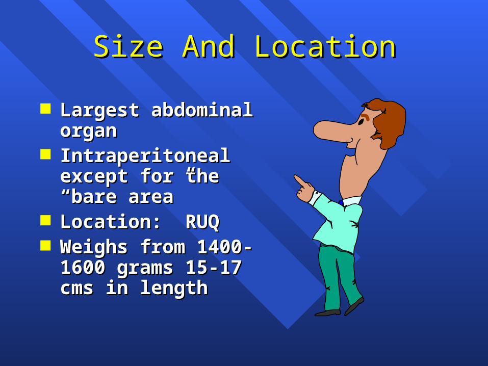
Splenic HiliumSplenic Hilium
Longitudinal
Splenic hilum

Spleen
Longitudinal
Diaphragm


![Ultrasound versus liver function tests for diagnosis of ... · [Diagnostic Test Accuracy Review] Ultrasound versus liver function tests for diagnosis of common bile duct stones Kurinchi](https://img.pdfslide.us/doc/110x75/601bcce3144189465e124f14/ultrasound-versus-liver-function-tests-for-diagnosis-of-diagnostic-test-accuracy.jpg)

![Endoscopic ultrasound-guided biopsy in chronic liver ...scopic ultrasound-guided liver biopsy (EUS-LB) is another method of acquiring liver tissue [8,9]. The feasibility of EUS-LB](https://img.pdfslide.us/doc/110x75/600c40491939a52c585d9ae9/endoscopic-ultrasound-guided-biopsy-in-chronic-liver-scopic-ultrasound-guided.jpg)