Embed Size (px)
Citation preview

1SCIENTIfIC RePoRts | (2018) 8:13562 | DOI:10.1038/s41598-018-31971-9
www.nature.com/scientificreports
Ultra-diluted Toxicodendron pubescens attenuates pro-inflammatory cytokines and ROS- mediated neuropathic pain in ratsShital Magar1, Deepika Nayak2, Umesh B. Mahajan1, Kalpesh R. Patil1, Sachin D. Shinde1, Sameer N. Goyal3, Shivang Swaminarayan4, Chandragouda R. Patil1, Shreesh Ojha5 & Chanakya Nath Kundu2
Despite the availability of multiple therapeutic agents, the search for novel pain management of neuropathic pain is still a challenge. Oxidative stress and inflammatory signaling are prominently involved in clinical manifestation of neuropathic pain. Toxicodendron pubescens, popularly known as Rhus Tox (RT) is recommended in alternative medicines as an anti-inflammatory and analgesic remedy. Earlier, we reported anti-inflammatory, anti-arthritic and immunomodulatory activities of Rhus Tox. In continuation, we evaluated antinociceptive efficacy of Rhus Tox in the neuropathic pain and delineated its underlying mechanism. Initially, in-vitro assay using LPS-mediated ROS-induced U-87 glioblastoma cells was performed to study the effect of Rhus Tox on reactive oxygen species (ROS), anti-oxidant status and cytokine profile. Rhus Tox decreased oxidative stress and cytokine release with restoration of anti-oxidant systems. Chronic treatment with Rhus Tox ultra dilutions for 14 days ameliorated neuropathic pain revealed as inhibition of cold, warm and mechanical allodynia along with improved motor nerve conduction velocity (MNCV) in constricted nerve. Rhus Tox decreased the oxidative and nitrosative stress by reducing malondialdehyde (MDA) and nitric oxide (NO) content, respectively along with up regulated glutathione (GSH), superoxide dismutase (SOD) and catalase activity in sciatic nerve of rats. Notably, Rhus Tox treatment caused significant reductions in the levels of tumor necrosis factor (TNF-α), interleukin-6 (IL-6) and interleukin-1β (IL-1β) as compared with CCI-control group. Protective effect of Rhus Tox against CCI-induced sciatic nerve injury in histopathology study was exhibited through maintenance of normal nerve architecture and inhibition of inflammatory changes. Overall, neuroprotective effect of Rhus Tox in CCI-induced neuropathic pain suggests the involvement of anti-oxidative and anti-inflammatory mechanisms.
Neuropathic pain represents an important clinical condition and there is a continuous search for novel treat-ments for treating neuropathy1. It is manifested as sensory abnormalities like dysesthesia (unpleasant sensation), hyperalgesia (increased sensitivity to noxious stimuli), allodynia (increased sensitivity to non-noxious stimuli) and spontaneous development of pain2,3. Neuropathic pain has a complex pathology with distinct mechanisms. Global occurrences of neuropathic pain are escalating due to an increase in the ageing population, enhanced survival from cancer chemotherapy and progressively increasing incidence of diabetes mellitus1. Despite the emergence of novel drug discovery technologies and advancements in the field of neuroscience, the issue of neu-ropathic pain management with safe and effective remedies is still unresolved4,5. Many randomized controlled trials have presented the clinical efficacy of opioids, gabapentinoids, tricyclic antidepressants, serotonin reuptake
1Department of Pharmacology, R. C. Patel Institute of Pharmaceutical Education and Research, Shirpur- 425405, Dist. Dhule, Maharashtra, India. 2School of Biotechnology, Kalinga Institute of Industrial technology (a deemed to be University), Campus-11, Patia, Bhubaneswar, Odisha, Pin-751024, India. 3SVKM’s Institute of Pharmacy, Dhule-424001, Dist-Dhule, Maharashtra, India. 4Janmangal Homeopathy and Wellness Centre, Bopal, Ahmedabad, Gujarat, 380058, India. 5Department of Pharmacology & Therapeutics, College of Medicine & Health Sciences, UAE University, Al Ain, UAE. Correspondence and requests for materials should be addressed to C.R.P. (email: [email protected]) or S.O. (email: [email protected]) or C.N.K. (email: [email protected])
Received: 14 July 2018
Accepted: 28 August 2018
Published: xx xx xxxx
OPEN
www.nature.com/scientificreports/
2SCIENTIfIC RePoRts | (2018) 8:13562 | DOI:10.1038/s41598-018-31971-9
inhibitors and calcium channel ligands in the treatment of neurogenic pain. However, these agents do not suffice the targets of managing of neuropathic pain. The efficacy of available anti-neuropathic drugs is limited due to occurrence of several side effects and inadequate or delayed pain relief4,6. The need to discover novel treatment modalities for neuropathic conditions still prevails.
Various factors contribute to the peripheral sensitization and initiation of neuropathic pain. Raised oxida-tive stress, increased vascular permeability and release of different inflammatory mediators including substance P and calcitonin gene related peptide produced by nociceptive terminals, formation and/or release of brady-kinin, prostaglandins, growth factors and cytokines leads to the occurrence of neuropathic pain7. Recently, the anti-inflammatory and antioxidant agents like N-acetyl carnitine and alpha lipoic acid are being investigated as add-on medications for the management of neuropathic pain8.
Natural products or complementary and alternative medicines are frequently used to manage the chronic neu-rological disorders like neuropathic pain4. Few preclinical studies on homeopathic remedies have also demon-strated the efficacy of homeopathic remedies in the management of neuropathic pain9,10. Evidence based surveys have revealed that patients with chronic pain prefer to use herbal treatments for these painful conditions11,12. Several pre-clinical and clinical studies have reported the beneficial effect of medicinal plants in the therapy of painful neuropathy4,8,13–17. Toxicodendron pubescens P. Mill belonging to family anacardiaceae is known as Rhus toxicodendron or Rhus tox (RT) in alternative medicines18,19. RT is commonly used homeopathic remedy for the management of inflammatory conditions, rheumatic pain, typhoid type fever and mucous membrane affections20. Experimental studies have shown that RT possesses immunomodulatory21, anti-inflammatory19,22, anti-arthritic23,24 and anti-melanoma activity25. Recently, a prospective observational study in breast cancer patients has shown that RT may decrease joint pain and stiffness in women with early breast cancer26.
Surgical lesion of peripheral nerves in experimental animals are usually performed to produce the animal models resembling human neuropathic pain27. In this regard, chronic constriction injury (CCI) model appears to be well established and frequently used animal model for the study of neuropathic pain27,28. In this model, unilat-eral loose ligation of sciatic nerve mimics the pathological neuropathic pain state in humans29. In this report, we demonstrated antinociceptive effects of RT in neuropathic pain using in-vitro and in-vivo assays. Initially, in-vitro study using U-87 primary glioblastoma cell line was executed to delineate the effect of RT on LPS-induced ROS production, anti-oxidant status and cytokine levels. Furthermore, in-vivo efficacy of RT was assessed in neu-ropathic pain using well characterized animal model of sciatic nerve constriction injury-induced neuropathic pain in rats. Various behavioral testing paradigms (cold, warm and mechanical allodynia), physiological param-eter (MNCV), biochemical estimations (MDA, NO, GSH, SOD, catalase, TNF-α, IL-1β, IL-6) and histopathol-ogy was executed to explore the underlying analgesic mechanisms of RT in the experimental neuropathic pain. Gabapentin served as a positive control in this study.
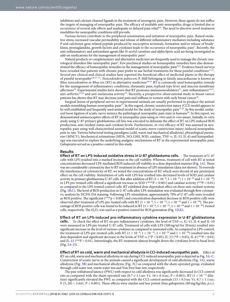
ResultsEffect of RT on LPS-induced oxidative stress in U-87 glioblastoma cells. The treatment of U-87 cells with LPS resulted into a marked increase in the cell viability. Whereas, treatment of cell with RT at tested concentrations decreased LPS-mediated ROS induced cell viability in a dose dependent manner (Fig. 1A). There was no considerable cytotoxicity due to RT treatment in absence of LPS stimulation (data not shown). To overrule the interference of cytotoxicity of RT, we tested the concentrations of RT which were devoid of any prominent effect on the cell viability. Stimulation of cells with LPS has resulted into decreased levels of SOD and catalase activity in primary glioblastoma U-87 cells. But after addition of RT (1 × 10⁻⁸; 1 × 10⁻¹²; 1 × 10⁻²⁴ and 1 × 10⁻³⁰) in LPS pre-treated cells offered a significant increase in SOD (***P < 0.001) and catalase activity (**P < 0.01) as compared to the LPS-treated control cells. RT exhibited dose dependent effect on these anti-oxidant systems (Fig. 1B,C). The level of ROS production in U-87 cells after LPS-stimulation was evaluated through flow cytomet-ric analysis by DCFH-DA staining. Following LPS-stimulation, approximately 70% of U-87 cells were revealed as ROS positive. The significant (***p < 0.001) and concentration dependent decrease in ROS positive cells were observed after treatment of LPS-pre-treated cells with RT (1 × 10⁻⁸; 1 × 10⁻¹²; 1 × 10⁻²⁴ and 1 × 10⁻³⁰). The per-centage of ROS positive cells was found to be reduced in RT (1 × 10⁻⁸; 1 × 10⁻¹²; 1 × 10⁻²⁴ and 1 × 10⁻³⁰) treated cells, respectively. The H2O2 was used as a positive control for ROS generation (Fig. 1D,K).
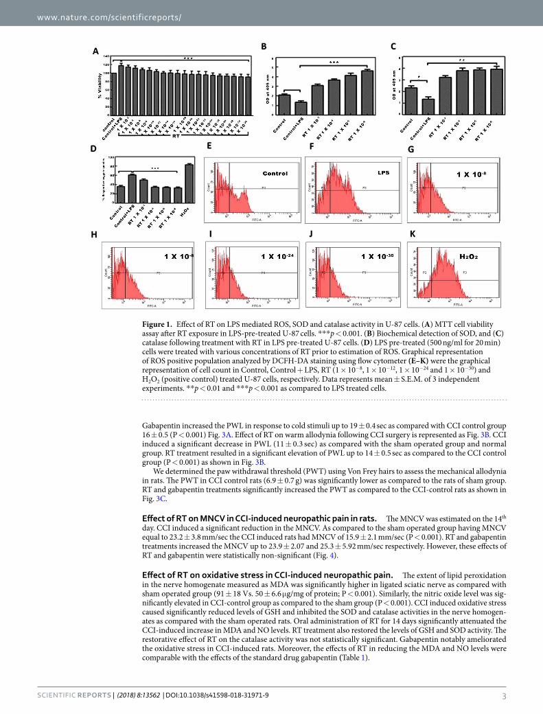
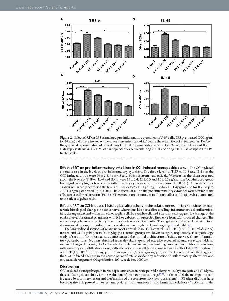
Effect of RT on LPS-induced pro-inflammatory cytokine expression in U-87 glioblastoma cells. To check the effect of RT on pro-inflammatory cytokines, the level of TNF-α, IL-1β, IL-6 and IL-10 were measured in LPS pre-treated U-87 cells. Treatment of cells with LPS (500 ng/ml for 20 min) resulted into significant increase in the level of various cytokines as compared to untreated cells. As compared to LPS-control, the treatment of LPS-pre-treated cells with RT (1 × 10⁻⁸; 1 × 10⁻¹²; 1 × 10⁻²⁴ and 1 × 10⁻³⁰) resulted into the dose dependent and significant decrease in the levels of TNF-α (*P < 0.05), IL-1β (*P < 0.05), IL-6 (**P < 0.01) and IL-1β (**P < 0.01). Interestingly, the RT treatment almost brought down the cytokines level to basal level (Fig. 2A–D).
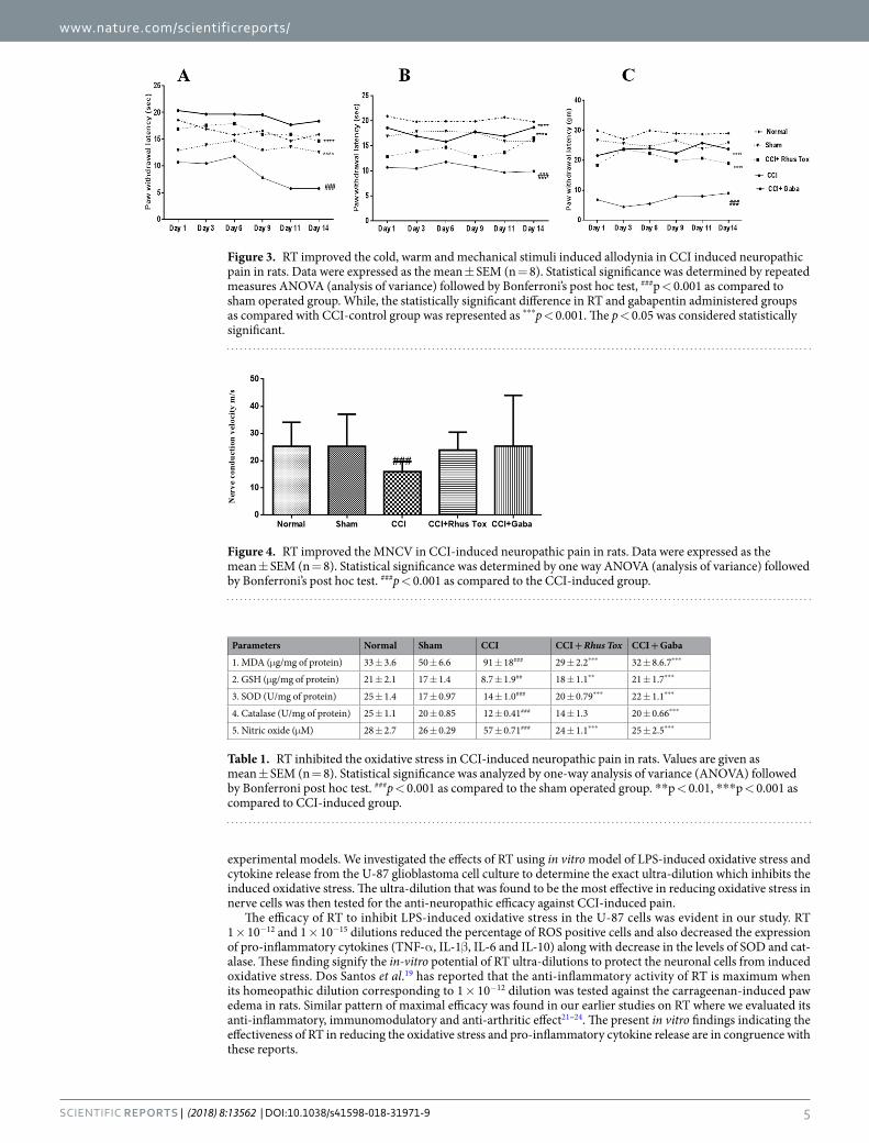
Effect of RT on cold, warm and mechanical allodynia in CCI-induced neuropathic pain. Effect of RT on cold, warm and mechanical allodynia in rats during CCI-induced neuropathic pain is depicted as Fig. 3A–C. Constriction of sciatic nerve in the animals caused a significant development of cold allodynia (Fig. 3A), warm allodynia (Fig. 3B) and mechanical allodynia (Fig. 3C) as compared with the sham operated group, evaluated through cold water test, warm water test and Von Frey hair test, respectively.
The paw withdrawal latency (PWL) with respect to cold allodynia was significantly decreased in CCI-control rats as compared with the sham operated rats (8.7 ± 1.1 sec Vs. 16 ± 0.4 sec, P < 0.001). RT (1 × 10⁻¹² dilu-tion) significantly elevated the PWL as compared with the CCI-control animals (13 ± 0.3 sec Vs. 8.7 ± 1.1 sec; F (5, 20) = 5.641, P < 0.001). These effects were similar and less potent than gabapentin (60 mg/kg/day, p.o.).
www.nature.com/scientificreports/
3SCIENTIfIC RePoRts | (2018) 8:13562 | DOI:10.1038/s41598-018-31971-9
Gabapentin increased the PWL in response to cold stimuli up to 19 ± 0.4 sec as compared with CCI control group 16 ± 0.5 (P < 0.001) Fig. 3A. Effect of RT on warm allodynia following CCI surgery is represented as Fig. 3B. CCI induced a significant decrease in PWL (11 ± 0.3 sec) as compared with the sham operated group and normal group. RT treatment resulted in a significant elevation of PWL up to 14 ± 0.5 sec as compared to the CCI control group (P < 0.001) as shown in Fig. 3B.
We determined the paw withdrawal threshold (PWT) using Von Frey hairs to assess the mechanical allodynia in rats. The PWT in CCI control rats (6.9 ± 0.7 g) was significantly lower as compared to the rats of sham group. RT and gabapentin treatments significantly increased the PWT as compared to the CCI-control rats as shown in Fig. 3C.
Effect of RT on MNCV in CCI-induced neuropathic pain in rats. The MNCV was estimated on the 14th day. CCI induced a significant reduction in the MNCV. As compared to the sham operated group having MNCV equal to 23.2 ± 3.8 mm/sec the CCI induced rats had MNCV of 15.9 ± 2.1 mm/sec (P < 0.001). RT and gabapentin treatments increased the MNCV up to 23.9 ± 2.07 and 25.3 ± 5.92 mm/sec respectively. However, these effects of RT and gabapentin were statistically non-significant (Fig. 4).
Effect of RT on oxidative stress in CCI-induced neuropathic pain. The extent of lipid peroxidation in the nerve homogenate measured as MDA was significantly higher in ligated sciatic nerve as compared with sham operated group (91 ± 18 Vs. 50 ± 6.6 μg/mg of protein; P < 0.001). Similarly, the nitric oxide level was sig-nificantly elevated in CCI-control group as compared to the sham group (P < 0.001). CCI induced oxidative stress caused significantly reduced levels of GSH and inhibited the SOD and catalase activities in the nerve homogen-ates as compared with the sham operated rats. Oral administration of RT for 14 days significantly attenuated the CCI-induced increase in MDA and NO levels. RT treatment also restored the levels of GSH and SOD activity. The restorative effect of RT on the catalase activity was not statistically significant. Gabapentin notably ameliorated the oxidative stress in CCI-induced rats. Moreover, the effects of RT in reducing the MDA and NO levels were comparable with the effects of the standard drug gabapentin (Table 1).
Figure 1. Effect of RT on LPS mediated ROS, SOD and catalase activity in U-87 cells. (A) MTT cell viability assay after RT exposure in LPS-pre-treated U-87 cells. ***p < 0.001. (B) Biochemical detection of SOD, and (C) catalase following treatment with RT in LPS pre-treated U-87 cells. (D) LPS pre-treated (500 ng/ml for 20 min) cells were treated with various concentrations of RT prior to estimation of ROS. Graphical representation of ROS positive population analyzed by DCFH-DA staining using flow cytometer (E–K) were the graphical representation of cell count in Control, Control + LPS, RT (1 × 10⁻⁸, 1 × 10⁻¹², 1 × 10⁻²⁴ and 1 × 10⁻³⁰) and H2O2 (positive control) treated U-87 cells, respectively. Data represents mean ± S.E.M. of 3 independent experiments. **p < 0.01 and ***p < 0.001 as compared to LPS treated cells.
www.nature.com/scientificreports/
4SCIENTIfIC RePoRts | (2018) 8:13562 | DOI:10.1038/s41598-018-31971-9
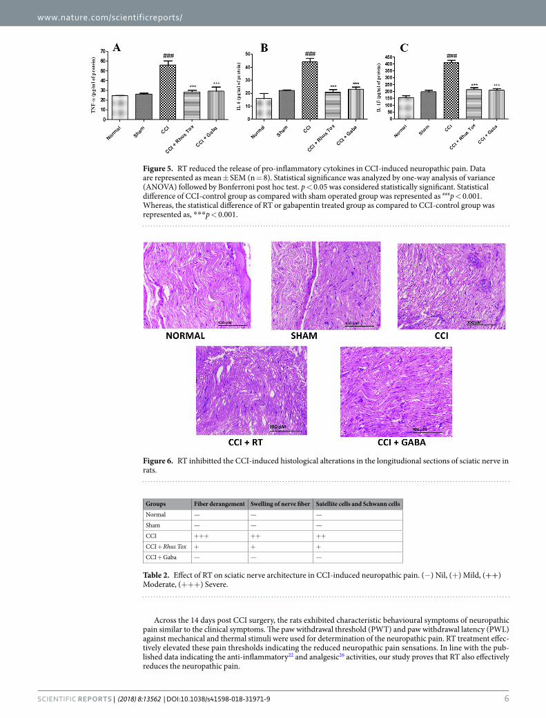
Effect of RT on pro-inflammatory cytokines in CCI-induced neuropathic pain. The CCI induced a notable rise in the levels of pro-inflammatory cytokines. The tissue levels of TNF-α, IL-6 and IL-1β in the CCI-induced group were 56 ± 2.6, 44 ± 4.8 and 64 ± 6.8 pg/mg respectively. Whereas, in the sham operated group the levels of TNF-α, IL-6 and IL-1β were 26 ± 0.4, 22 ± 0.3 and 22 ± 0.3 pg/mg. The CCI-induced group had significantly higher levels of proinflammatory cytokines in the nerve tissue (P < 0.001). RT treatment for 14 days remarkably decreased the levels of TNF-α to 25 ± 1.1 pg/mg, IL-6 to 20 ± 1.4 pg/mg and for IL-1β up to 20 ± 1.4 pg/mg of protein (p < 0.001). These effects of RT on the pro-inflammatory cytokines were similar to the effects exerted by gabapentin (Fig. 5). RT exerted more prominent inhibitory effect on IL-1β levels as compared to the effect of gabapentin.
Effect of RT on CCI-induced histological alterations in the sciatic nerve. The CCI induced charac-teristic histological changes in sciatic nerve. Alterations like nerve fibre swelling, inflammatory cell infilteration, fibre derangement and activation of neuroglial cell like satellite cells and Schwann cells suggest the damage of the sciatic nerve. Treatment of animals with RT or gabapentin protected the nerve from CCI-induced changes. The nerve samples from rats receiving these treatments revealed that both RT and gabapentin had reduced structural derangements, along with inhibition nerve fiber and neuroglial cell swelling (Fig. 6 and Table 2).
The longitudional sections of sciatic nerve of normal, sham, CCI-control, CCI + RT (1 × 10¹²; 0.1 ml/day, p.o.) treated and CCI + gabapentin (60 mg/kg, p.o) treated groups are shown as Fig. 6, respectively. Histopathology study of sections from normal rats demonstrated the normal architecture of sciatic nerve with no inflamma-tory perturbations. Sections obtained from the sham operated rats also revealed normal structure with no marked changes. However, the CCI-control rats showed nerve fibre swelling, derangement of fibre architecture, inflammatory cell infiltration along with alterations in satellite cells and schwann cells (Table 2). Treatment with RT (1 × 10⁻¹²; 0.1 ml/day, p.o.) or gabapentin (60 mg/kg/day, p.o.) exibited ameliorative effect against the CCI-induced changes in the sciatic nerve of rats as evident by reduction in inflammatory alterations and structural derangement (Magnification 100×, scale bar, 1000 µm).
DiscussionCCI-induced neuropathic pain in rats represents characteristic painful behaviors like hyperalgesia and allodynia, thus validating its suitability for the evaluation of anti-neuropathic drugs29–31. In this model, the neuropathic pain is caused by a primary lesion and dysfunction of the somatosensory nervous system3,14. RT ultra-dilutions have been consistently proved to possess analgesic, anti-inflammatory22 and immunomodulatory21 activities in the
Figure 2. Effect of RT on LPS stimulated pro-inflammatory cytokines in U-87 cells. LPS pre-treated (500 ng/ml for 20 min) cells were treated with various concentrations of RT before the estimation of cytokines. (A–D) Are the graphical representation of optical density of cell supernatants at 405 nm for TNF-α, IL-1β, IL-6 and IL-10. Data represents mean ± S.E.M. of 3 independent experiments. **p < 0.01 and ***p < 0.001 as compared to LPS treated cells.
www.nature.com/scientificreports/
5SCIENTIfIC RePoRts | (2018) 8:13562 | DOI:10.1038/s41598-018-31971-9
experimental models. We investigated the effects of RT using in vitro model of LPS-induced oxidative stress and cytokine release from the U-87 glioblastoma cell culture to determine the exact ultra-dilution which inhibits the induced oxidative stress. The ultra-dilution that was found to be the most effective in reducing oxidative stress in nerve cells was then tested for the anti-neuropathic efficacy against CCI-induced pain.
The efficacy of RT to inhibit LPS-induced oxidative stress in the U-87 cells was evident in our study. RT 1 × 10⁻¹² and 1 × 10⁻¹⁵ dilutions reduced the percentage of ROS positive cells and also decreased the expression of pro-inflammatory cytokines (TNF-α, IL-1β, IL-6 and IL-10) along with decrease in the levels of SOD and cat-alase. These finding signify the in-vitro potential of RT ultra-dilutions to protect the neuronal cells from induced oxidative stress. Dos Santos et al.19 has reported that the anti-inflammatory activity of RT is maximum when its homeopathic dilution corresponding to 1 × 10⁻¹² dilution was tested against the carrageenan-induced paw edema in rats. Similar pattern of maximal efficacy was found in our earlier studies on RT where we evaluated its anti-inflammatory, immunomodulatory and anti-arthritic effect21–24. The present in vitro findings indicating the effectiveness of RT in reducing the oxidative stress and pro-inflammatory cytokine release are in congruence with these reports.
Figure 3. RT improved the cold, warm and mechanical stimuli induced allodynia in CCI induced neuropathic pain in rats. Data were expressed as the mean ± SEM (n = 8). Statistical significance was determined by repeated measures ANOVA (analysis of variance) followed by Bonferroni’s post hoc test, ###p < 0.001 as compared to sham operated group. While, the statistically significant difference in RT and gabapentin administered groups as compared with CCI-control group was represented as ***p < 0.001. The p < 0.05 was considered statistically significant.
Figure 4. RT improved the MNCV in CCI-induced neuropathic pain in rats. Data were expressed as the mean ± SEM (n = 8). Statistical significance was determined by one way ANOVA (analysis of variance) followed by Bonferroni’s post hoc test. ###p < 0.001 as compared to the CCI-induced group.
Parameters Normal Sham CCI CCI + Rhus Tox CCI + Gaba
1. MDA (μg/mg of protein) 33 ± 3.6 50 ± 6.6 91 ± 18### 29 ± 2.2*** 32 ± 8.6.7***
2. GSH (μg/mg of protein) 21 ± 2.1 17 ± 1.4 8.7 ± 1.9## 18 ± 1.1** 21 ± 1.7***
3. SOD (U/mg of protein) 25 ± 1.4 17 ± 0.97 14 ± 1.0### 20 ± 0.79*** 22 ± 1.1***
4. Catalase (U/mg of protein) 25 ± 1.1 20 ± 0.85 12 ± 0.41### 14 ± 1.3 20 ± 0.66***
5. Nitric oxide (μM) 28 ± 2.7 26 ± 0.29 57 ± 0.71### 24 ± 1.1*** 25 ± 2.5***
Table 1. RT inhibited the oxidative stress in CCI-induced neuropathic pain in rats. Values are given as mean ± SEM (n = 8). Statistical significance was analyzed by one-way analysis of variance (ANOVA) followed by Bonferroni post hoc test. ###p < 0.001 as compared to the sham operated group. **p < 0.01, ***p < 0.001 as compared to CCI-induced group.
www.nature.com/scientificreports/
6SCIENTIfIC RePoRts | (2018) 8:13562 | DOI:10.1038/s41598-018-31971-9
Across the 14 days post CCI surgery, the rats exhibited characteristic behavioural symptoms of neuropathic pain similar to the clinical symptoms. The paw withdrawal threshold (PWT) and paw withdrawal latency (PWL) against mechanical and thermal stimuli were used for determination of the neuropathic pain. RT treatment effec-tively elevated these pain thresholds indicating the reduced neuropathic pain sensations. In line with the pub-lished data indicating the anti-inflammatory22 and analgesic26 activities, our study proves that RT also effectively reduces the neuropathic pain.
Figure 5. RT reduced the release of pro-inflammatory cytokines in CCI-induced neuropathic pain. Data are represented as mean ± SEM (n = 8). Statistical significance was analyzed by one-way analysis of variance (ANOVA) followed by Bonferroni post hoc test. p < 0.05 was considered statistically significant. Statistical difference of CCI-control group as compared with sham operated group was represented as ###p < 0.001. Whereas, the statistical difference of RT or gabapentin treated group as compared to CCI-control group was represented as, ***p < 0.001.
Figure 6. RT inhibitted the CCI-induced histological alterations in the longitudional sections of sciatic nerve in rats.
Groups Fiber derangement Swelling of nerve fiber Satellite cells and Schwann cells
Normal — — —
Sham — — —
CCI +++ ++ ++
CCI + Rhus Tox + + +
CCI + Gaba — — —
Table 2. Effect of RT on sciatic nerve architecture in CCI-induced neuropathic pain. (−) Nil, (+) Mild, (++) Moderate, (+++) Severe.
www.nature.com/scientificreports/
7SCIENTIfIC RePoRts | (2018) 8:13562 | DOI:10.1038/s41598-018-31971-9
The CCI induces ischemic hypoxic imbalance in the secondary metabolites in affected nerve fibres and pro-duces oxidative stress32,33. Degeneration of nerve fibre and decreased nerve energy is reported to reduce the MNCV. RT ameliorated the nerve damage mediated reduction in the MNCV following CCI in sciatic nerve indicating its neuroprotective effect. Oxidative stress and inflammation are interconnected events leading to nerve injury and persistent pain34,35. Various plant extracts, their isolated fractions or phytoconstituents have been reported to exert their anti-neuropathic activity through the anti-inflammatory and anti-oxidant mecha-nisms35–40. The concentrations of MDA and NO are commonly measured to study the extent of reactive oxygen species34. ROS like superoxide, NO and peroxynitrite have an important role in the development of neuroinflam-matory responses41. Increase in lipid peroxidation following CCI of sciatic nerve has also been reported41. Efficacy of free radical scavengers towards the reduction of lipid peroxidation suggests the involvement of ROS in the nerve sensitization and production of allodynia42.
NO is the vital signaling molecule which has significant role in the peripheral and central pain43. Involvement of endogenous nitric oxide in the development of CCI-induced neuropathic pain is well documented in earlier studies44. Significant rise in the levels of NO in sciatic nerve suggested the up regulation of NO and its role in the production and maintenance of neuropathic pain44,45. In the present study significant increase in the levels of MDA and NO were observed in the chronically constricted nerve. It denotes elevated damage to the macro-molecules such as proteins and lipids which may result into the neuropathic pain. Chronic administration of RT resulted in the decreased levels of MDA and NO indicating the anti-lipid peroxidation activity along with inhibi-tion of nitrosative tissue damage and successive neuropathic pain.
Our findings are in line with previous studies reporting the decline in GSH content of sciatic nerve following CCI in rats46. Catalase and SOD are main anti-oxidant enzymes that help to scavenge the free radicals and offer protection against oxidative stress46,47. In our study, decrease in the level of catalase and SOD after CCI in the rats are indicative of increased oxidative stress or declined anti-oxidant defense mechanisms. These results are in tune with earlier reports and reinforce the contribution of oxidative stress in the development of neuropathic pain47–49. Oxidative stress mediated decline in the levels of GSH, SOD and catalase were restored in the CCI rats by chronic treatment with RT ultra-dilution (1 × 10⁻¹²). Thus, RT mediated decrease in the CCI-induced oxidative stress could be one of the mechanisms involved its antinociceptive activity in neuropathic pain.
Different cytokines including TNF-α IL-1β, and IL-6 are released following the nerve injury and contribute to the development of neuropathic pain34. Peripheral nerve damage induces release of TNF-α rapidly from mac-rophages, Schwann cells and mast cells26,50. TNF-α is associated with decline in pain threshold and anti-TNF-α treatments are reported to alleviate the CCI-induced pain in rats51. In this study, we observed a significant decrease in the TNF-α level in sciatic nerve in the group treated with RT as compared with CCI-control group. This finding revealed that RT modulated the neuro-inflammation in peripheral nerve through its anti-TNF-α action and it could be one of its anti-nociceptive mechanisms. IL-1β is known to be expressed in nociceptive neurons and it has important role in several pain models52,53. The IL-1β increases the neuropathic pain sensitiza-tion through its action on the adjacent neurons34. We observed a significant increase in the production of IL-1β in sciatic nerve after CCI in rats. Treatment of animals with RT inhibited IL-1β production which may account for its antinociceptive effect. IL-6 is a pro-inflammatory cytokine having important role in the development of inflammatory and neuropathic pain following peripheral nerve injury. Results of present study revealed that RT has ability to decrease the IL-6 level which signifies the anti-IL-6 effect of RT in CCI-induced neuropathic pain model. Overall findings through biochemical estimation denote that RT favorably altered the cytokine pro-file in the experimental model of neuropathic pain.
In summary, this study substantiated the antinociceptive potential of RT ultra-dilutions in the validated model of CCI-induced peripheral neuropathic pain. Interestingly, RT was found to be efficacious in reducing not only the thermal nociception but also the mechanical allodynia in rats. RT demonstrated notable anti-oxidant and anti-inflammatory effects in the sciatic nerve and exhibited potential free radical scavenging activity that would reduce its anti-neuropathic activity. Attenuated oxidative stress and inflammatory pathways may contribute to the therapeutic potential of RT in the treatment of neuropathic pain. Although, the results of present study sug-gested the anti-neuropathic effect of RT, further pre-clinical and clinical studies are warranted to confirm these effects. Several other biochemical mechanisms may be involved in RT mediated anti-neuropathic effect. Results of present study are suggestive of the anti-nociceptive effect of RT against neuropathic pain and deserve further validation of its effectiveness in various painful conditions.
MethodsChemicals. The authenticated dried powder of leaves of Rhus Tox (Family: Anacardiaceae) was obtained from Homeopathic Pharmacopoeia laboratory, Ghaziabad, Uttar Pradesh, India. Gabapentin was gifted by Mylan lab-oratories, India. Cytokine ELISA Ready SET-Go kits for TNF-α (Cat: 837324-22: Batch No. E09479-1645), IL-1β (Cat: 887013-22; Batch No. E09323-1645) and IL-6 (Cat: 837064-22: Batch No. E09358-1645) were purchased from e-Biosciences Incorporation, USA. Lipopolysaccharide (LPS) from Escherichia coli O55:B5 (Cat: L2880; Lot No. 025M4040V) was purchased from Sigma-Aldrich, USA.
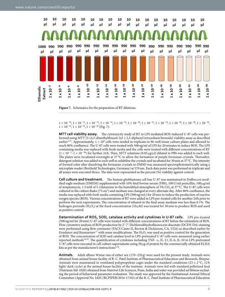
Preparation of RT ethanolic extract and dilutions. The procedure prescribed in the monograph of Indian Homeopathic pharmacopoeia was followed for the preparation of RT extract and its ultra-dilutions except the characteristic successions used in preparation of homeopathic dilutions. Dried and coarse powder of RT leaves was pulverized. Exactly weighed (10 gm) powder was mixed with 100 mL of ethanol (70%) and kept in the glass jar for cold maceration up to 7 days with occasional shaking during each day36,54. On 8th day, mixture was filtered through Whatman filter paper and the filtrate was used as an alcoholic extract of RT. Various dilutions of extract in ethanol were prepared to obtain the final RT concentrations of 1 × 10⁻², 1 × 10⁻⁴, 1 × 10⁻⁶, 1 × 10⁻⁸,
www.nature.com/scientificreports/
8SCIENTIfIC RePoRts | (2018) 8:13562 | DOI:10.1038/s41598-018-31971-9
1 × 10⁻¹⁰, 1 × 10⁻¹², 1 × 10⁻¹⁴, 1 × 10⁻¹⁶, 1 × 10⁻¹⁸, 1 × 10⁻²⁰, 1 × 10⁻²², 1 × 10⁻²⁴, 1 × 10⁻²⁶, 1 × 10⁻²⁸, 1 × 10⁻³⁰, 1 × 10⁻³², 1 × 10⁻³⁴, 1 × 10−36 (Fig. 7).
MTT cell viability assay. The cytotoxicity study of RT in LPS mediated ROS-induced U-87 cells was per-formed using MTT [3-(4,5-dimethylthiazol-2yl-)-2,5-diphenyl tetrazolium bromide] viability assay as described earlier5,35. Approximately, 1 × 10⁴ cells were seeded in triplicate in 96-well tissue culture plates and allowed to reach 80% confluence. The U-87 cells were treated with 500 ng/ml of LPS for 20 minutes to induce ROS. The LPS containing media was replaced with fresh media and the cells were treated with different concentrations of RT (1 × 10⁻²–1 × 10−36) for further 24 h. Then, MTT solutions (0.05 μg/μl) diluted in PBS was added to each well. The plates were incubated overnight at 37 °C to allow the formation of purple formazan crystals. Thereafter, detergent solution was added to each well to solubilise the crystals and incubated for 30 min at 37 °C. The intensity of formed color after dissolving the formazan crystals in DMSO was measured spectrophotometrically using a microplate reader (Berthold Technologies, Germany) at 570 nm. Each data point was performed in triplicate and all assays were executed thrice. The data were represented as the percent (%) viability against control.
Cell culture and treatment. The human glioblastoma cell line U-87 was maintained in Dulbecco’s mod-ified eagle medium (DMEM) supplemented with 10% fetal bovine serum (FBS), 100 U/ml penicillin, 100 μg/ml of streptomycin, 1.5 mM of L-Glutamine in the humidified atmosphere of 5% CO2 at 37 °C. The U-87 cells were cultured in the culture flasks (75 cm2) and medium was changed at every alternate day. After 80% confluence, the media was replaced with fresh media containing LPS (500 ng/mL) for 20 min to induce the production of reactive oxygen species (ROS). Various concentrations of RT were added in LPS pre-treated cells for another 24 h prior to perform the next experiments. The concentration of ethanol in the final assay medium was less than 0.1%. The hydrogen peroxide (H2O2) at the fixed concentration (10 μM) was treated for 30 min to produce ROS and used as positive control.
Determination of ROS, SOD, catalase activity and cytokines in U-87 cells. LPS pre-treated (500 ng/ml for 20 min) U-87 cells were treated with different concentrations of RT before the estimation of ROS. Flow cytometry analysis of ROS production by 2′-7′-Dichlorodihydrofluorescein diacetate (DCFH-DA) staining were performed using flow cytometer (FACS Canto II, Becton & Dickinson, CA, USA) as described earlier by Eruslanov and Kusmartsev37 with some modifications. The H2O2 was used as positive control for the generation of ROS. The concentration of SOD and catalase level in LPS-pretreated U-87 cells were measured using earlier reported methods38,39. The quantification of cytokines including TNF- α, IL-1β, IL-6, IL-10 in LPS-pretreated U-87 cells were executed in cell culture supernatants using 50 μg of protein by the commercially obtained ELISA kits as per the manufacturer’s instructions5,38.
Animals. Adult albino Wistar rats of either sex (170–220 g) were used for the present study. Animals were obtained from animal house facility of R. C. Patel Institute of Pharmaceutical Education and Research, Shirpur. Animals were maintained in ventilated polypropylene cages under the standard conditions (25 ± 2 °C, 12 h light/ dark cycle) at the animal house facility of the institute. Animals were fed with standard pelletized feed (Nutrimix Std-1020) obtained from Nutrivet Life Sciences, Pune, India and water was provided ad libitum exclud-ing the period of behavioral parameter evaluation. The study was approved by the Institutional Animal Ethical Committee (Approval No. IAEC/RCPIPER/2016-17/02) of the R. C. Patel Institute of Pharmaceutical Education
Figure 7. Schematics for the preparation of RT dilutions.
www.nature.com/scientificreports/
9SCIENTIfIC RePoRts | (2018) 8:13562 | DOI:10.1038/s41598-018-31971-9
and Research, Shirpur, India (Reg. No. 651/PO/ReBi/S/02/CPCSEA). All the experimental procedures involving the use of animals were carried out in accordance with the regulations laid down by Committee for the Purpose of Control and Supervision of Experimentation on animals (CPCSEA) constituted under the Prevention of Cruelty to Animals Act, 1960, Ministry of Environment and Forests, Government of India.
Induction of chronic constriction injury (CCI) in rats. The surgery was performed to induce the CCI as described earlier by Chanchal et al.5. Briefly, the animals were anaesthetized with intraperitoneal administration of pentobarbital sodium (60 mg/kg). A blunt dissection through biceps femoris was executed to expose the com-mon sciatic nerve of right hind limb at the middle of the thigh. Approximately, 5–7 mm of the nerve was freed off the adhering tissue proximal to the trifurcation of sciatic nerve and four ligatures (6.0 silk) were loosely tied around it about 1 mm apart. Following nerve ligation, the muscular and skin layers were instantly sutured and povidone-iodine solution was applied externally. The rats were kept in individual cages and allowed to recover29. The respective drug treatments were started on the next day after the surgery.
Drug treatment and groups. Animals were randomly divided into five groups, each consisting of 8 rats (n = 8). Group I: Normal control group of rats were orally administered once daily with 1 ml saline for 14 days. Group II: Sham operated group of rats were treated with 1 ml saline once daily for 14 days. Group III: CCI-induced neuropathy control group of rats orally received 1 ml saline once daily for 14 days. Group IV: CCI-induced neuropathy + RT treated group of rats orally received 0.1 ml of RT (1 × 10⁻¹² dilution) with 1 ml of distilled water once daily for 14 days. Group V: CCI-induced neuropathy + gabapentin treated group of rats orally received Gabapentin (60 mg/kg/day, p.o.) suspended in 0.5% carboxymethyl cellulose (CMC) once daily for 14 days.
Experimental design. Subsequent to the induction of CCI, rats were habituated for 3 days. RT treatment was started on the next day after the CCI surgery. The thermal and mechanical allodynia were measured on Day-3, Day-7, Day-11 and Day-14 following the surgery by earlier reported methods5,40. Paw withdrawal latency (PWL) was recorded with the maximum cut off time of 20 sec. Right hind paw of each rat up to the ankle joint was immersed in warm water (40 ± 1 °C) and cold water (12 ± 1 °C) for the determination of thermal (warm and cold) allodynia. Mechanical allodynia was noted using electronic Von-Frey apparatus comprising of super-tip probes (2390 series, IITC Life Sciences Incorporation). Paw withdrawal threshold (PWT) was recorded with cut-off pres-sure of 30 gm. Rats were kept in polypropylene cages with metal mesh floor and acclimatized for approximately 10 min before the measurements. Mid-plantar surface of operated hind paw were probed with Von Frey filaments through the mesh floor, when the paw was in contact with floor. Each filament was applied to the planter surface until it just bent and kept in position for about 6–8 sec. Probes were applied in ascending order and the smallest filament which provoked paw withdrawal response was measured as threshold stimulus55.
On the 14th day after surgery, the animals were anesthetized with intraperitoneal injection of pentobarbital sodium (60 mg/kg). The body temperature of animal was maintained at 37 °C. Sciatic-tibial motor nerve con-duction velocity (MNCV) was measured by the stimulation of sciatic and tibial nerves at the sciatic notch and tibial notch through the bipolar needle electrodes (Power Lab/ML856; AD Instruments, Australia) at the 0.10 Hz frequency, 0.1 ms duration and 1.5 V amplitude. After single stimulus the compound muscle action potential was measured from the first interosseous muscle of the hind-paw by unipolar pin electrodes. The recording was typical biphasic response with an initial M-wave which is a direct motor response owing to stimulation of motor fibers. The MNCV was calculated as the ratio of the distance (mm) between both sites of stimulation divided by the difference between proximal and distal latencies recorded in ms32,56.
Following the recording of MNCV, the rats were sacrificed using overdose of pentobarbital sodium. The injured sciatic nerve was isolated along with 1 cm segments on the both sides of CCI injury. The central 5 mm portion of the isolated nerve segment was processed for histological examination. The sections of 4 μm thickness were stained with haematoxylin and eosin. The stained sections were examined under the light microscope for structural alterations including fiber derangement, swelling of nerve fiber and presence of activated satellite cells and Schwann cells. Paraformaldehyde-fixed nerve tissues were dehydrated in ascending graded series of alcohol and embedded in paraffin. The specimens were cut into the sections of 4 μm thickness using microtome and stained with hematoxylin and eosin according to routine staining protocols. The stained sections were examined under the light microscope (Leica D1000, LED) for structural alterations like nerve fiber swelling, fiber derange-ment and presence of activated satellite cells and Schwann cells13,57.
Segments of sciatic nerve from the rats were process in ice chilled phosphate buffer (pH 7.4) to obtain the 10% homogenate. Homogenate was centrifuged at 2000 g for 20 min at 4 °C and aliquots were used for the determina-tion of malondialdehyde (MDA)58, reduced glutathione (GSH)59, superoxide dismutase (SOD)38 and catalase39,60.
Nitric oxide (NO) was estimated using earlier reported method by Kumar et al.41 with some modifications. Briefly, 50 μl of tissue supernatant was mixed with 500 μl of Griess reagent and the absorbance was determined spectrophotometrically at 540 nm using Powerwave XS microplate spectrophotometer (Biotek, USA). Calibration curve was obtained by using Sodium nitrite as a standard. The concentration of NO was expressed in μM of NO per mg of protein.
The quantification of cytokines like TNF- α, IL-1β and IL-6 was determined in homogenate and using 50 μg of protein by the commercially obtained ELISA kits as per the manufacturer’s instructions5,38.
References 1. Colloca, L. et al. Neuropathic pain. Nature Reviews Disease Primers 3, 17002 (2017). 2. Liu, N. et al. Antinociceptive effects of gentiopicroside on neuropathic pain induced by chronic constriction injury in mice: a
behavioral and electrophysiological study. Canadian journal of physiology and pharmacology 94, 769–778 (2016).
www.nature.com/scientificreports/
1 0SCIENTIfIC RePoRts | (2018) 8:13562 | DOI:10.1038/s41598-018-31971-9
3. Kim, H. J. Berberine ameliorates allodynia induced by chronic constriction injury of the sciatic nerve in rats. Journal of medicinal food 18, 909–915 (2015).
4. Shahid, M., Subhan, F., Ahmad, N. & Ullah, I. A bacosides containing Bacopa monnieri extract alleviates allodynia and hyperalgesia in the chronic constriction injury model of neuropathic pain in rats. BMC complementary and alternative medicine 17, 293 (2017).
5. Chanchal, S. K. et al. In vivo and in vitro protective effects of omeprazole against neuropathic pain. Scientific reports 6, 30007 (2016). 6. Attal, N. Pharmacologic treatment of neuropathic pain. Acta Neurologica Belgica 101, 53–64 (2001). 7. Cohen, S. P. & Mao, J. Neuropathic pain: mechanisms and their clinical implications. BMJ: British Medical Journal (Online) 348
(2014). 8. Jia, S., Zhang, Y. & Yu, J. Antinociceptive effects of isosakuranetin in a rat model of peripheral neuropathy. Pharmacology 100,
201–207 (2017). 9. Kishore, L. & Singh, R. Effects of different homeopathic potencies of Cephalendra indica in treatment of neuropathic pain in
streptozotocin induced diabetes. Bulletin of Faculty of Pharmacy, Cairo University 55, 273–280 (2017). 10. Mohammadi, R., Amini, K. & Charehsaz, S. Homeopathic treatment for peripheral nerve regeneration: an experimental study in a
rat sciatic nerve transection model. Homeopathy 101, 141–146 (2012). 11. Boyd, A. et al. Herbal medicinal products or preparations for neuropathic pain. The Cochrane Library (2017). 12. Boyd, A. et al. Herbal medicinal products or preparations for neuropathic pain and fibromyalgia. Cochrane Database of Systematic
Reviews 5 (2013). 13. Muthuraman, A. & Singh, N. Attenuating effect of Acorus calamus extract in chronic constriction injury induced neuropathic pain
in rats: an evidence of anti-oxidative, anti-inflammatory, neuroprotective and calcium inhibitory effects. BMC complementary and alternative medicine 11, 24 (2011).
14. Kaur, G., Bali, A., Singh, N. & Jaggi, A. S. Ameliorative potential of Ocimum sanctum in chronic constriction injury-induced neuropathic pain in rats. Anais da Academia Brasileira de Ciências 87, 417–429 (2015).
15. Zhou, J. et al. Paeoniflorin and albiflorin attenuate neuropathic pain via mapk pathway in chronic constriction injury rats. Evidence-based Complementary and Alternative Medicine 2016 (2016).
16. Nurmikko, T. J. et al. Sativex successfully treats neuropathic pain characterised by allodynia: a randomised, double-blind, placebo-controlled clinical trial. Pain® 133, 210–220 (2007).
17. Babbar, S. et al. Pharmacokinetic analysis of capsaicin after topical administration of a high-concentration capsaicin patch to patients with peripheral neuropathic pain. Therapeutic drug monitoring 31, 502–510 (2009).
18. Fisher, P. & Scott, D. A randomized controlled trial of homeopathy in rheumatoid arthritis. Rheumatology 40, 1052–1055 (2001). 19. Dos Santos, A., Perazzo, F., Cardoso, L. & Carvalho, J. In vivo study of the anti-inflammatory effect of Rhus toxicodendron.
Homeopathy 96, 95–101 (2007). 20. Verma, P. & Vaid, I. (Jain Publishers, New Delhi, India, 2002). 21. Patil, C. et al. Immunomodulatory activity of Toxicodendron pubescens in experimental models. Homeopathy 98, 154–159 (2009). 22. Patil, C. R. et al. Dual effect of Toxicodendron pubescens on Carrageenan induced paw edema in rats. Homeopathy 98, 88–91 (2009). 23. Patel, D. R. et al. Toxicodendron pubescens retains its anti-arthritic efficacy at 1M, 10M and CM homeopathic dilutions. Homeopathy
101, 165–170 (2012). 24. Patil, C. R. et al. Modulation of arthritis in rats by Toxicodendron pubescens and its homeopathic dilutions. Homeopathy 100,
131–137 (2011). 25. Guimarães, F. S. et al. Stimulation of lymphocyte anti-melanoma activity by co-cultured macrophages activated by complex
homeopathic medication. BMC cancer 9, 293 (2009). 26. Karp, J.-C. et al. Treatment with Ruta graveolens 5CH and Rhus toxicodendron 9CH may reduce joint pain and stiffness linked to
aromatase inhibitors in women with early breast cancer: results of a pilot observational study. Homeopathy 105, 299–308 (2016). 27. Chen, L., Chen, W., Qian, X., Fang, Y. & Zhu, N. Liquiritigenin alleviates mechanical and cold hyperalgesia in a rat neuropathic pain
model. Scientific reports 4, 5676 (2014). 28. De Vry, J., Kuhl, E., Franken-Kunkel, P. & Eckel, G. Pharmacological characterization of the chronic constriction injury model of
neuropathic pain. European journal of pharmacology 491, 137–148 (2004). 29. Bennett, G. J. & Xie, Y.-K. A peripheral mononeuropathy in rat that produces disorders of pain sensation like those seen in man.
Pain 33, 87–107 (1988). 30. Espinosa‐Juárez, J. V., Jaramillo‐Morales, O. A. & López‐Muñoz, F. J. Haloperidol Decreases Hyperalgesia and Allodynia Induced
by Chronic Constriction Injury. Basic & clinical pharmacology & toxicology (2017). 31. Dowdall, T., Robinson, I. & Meert, T. F. Comparison of five different rat models of peripheral nerve injury. Pharmacology
Biochemistry and Behavior 80, 93–108 (2005). 32. Morani, A. S. & Bodhankar, S. L. Neuroprotective effect of vitamin E acetate in models of mononeuropathy in rats. Neuroanatomy
7, 33–37 (2008). 33. Asiedu, M., Ossipov, M. H., Kaila, K. & Price, T. J. Acetazolamide and midazolam act synergistically to inhibit neuropathic pain. Pain
148, 302–308 (2010). 34. Bhat, R. A. et al. Effect of ursolic acid in attenuating chronic constriction injury‐induced neuropathic pain in rats. Fundamental &
clinical pharmacology 30, 517–528 (2016). 35. Patil, K. R. et al. Pentacyclic triterpenoids inhibit IKKβ mediated activation of NF-κB pathway: in silico and in vitro evidences. PloS
one 10, e0125709 (2015). 36. Shanbhag, D. & Khandagale, A. Screening and standardization of Terminalia arjuna used as medicine in homeopathy using HPTLC
method. Int J Anal Bioanal Chem 1, 57–60 (2011). 37. Eruslanov, E. & Kusmartsev, S. In Advanced protocols in oxidative stress II 57–72 (Springer, 2010). 38. Marklund, S. & Marklund, G. Involvement of the superoxide anion radical in the autoxidation of pyrogallol and a convenient assay
for superoxide dismutase. The FEBS Journal 47, 469–474 (1974). 39. Aebi, H. In Methods in enzymology Vol. 105 121–126 (Elsevier, 1984). 40. Goecks, C. S. et al. Assessment of oxidative parameters in rat spinal cord after chronic constriction of the sciatic nerve. Neurochemical
research 37, 1952–1958 (2012). 41. Kumar, A., Meena, S. & Pottabathini, R. Effect of Ashwagandha (Withania somnifera) against chronic constriction injury induced
behavioral and biochemical alterations: possible involvement of nitric oxide mechanism. International Journal of Nutrition, Pharmacology, Neurological Diseases 4, 131 (2014).
42. Kim, H. K. et al. Reactive oxygen species (ROS) play an important role in a rat model of neuropathic pain. Pain 111, 116–124 (2004). 43. Freire, M. A. M., Guimarães, J. S., Gomes-Leal, W. & Pereira, A. Pain modulation by nitric oxide in the spinal cord. Frontiers in
neuroscience 3, 24 (2009). 44. Naik, A. K., Tandan, S. K., Kumar, D. & Dudhgaonkar, S. P. Nitric oxide and its modulators in chronic constriction injury-induced
neuropathic pain in rats. European journal of pharmacology 530, 59–69 (2006). 45. Kumar, A., Meena, S., Kalonia, H., Gupta, A. & Kumar, P. Effect of nitric oxide in protective effect of melatonin against chronic
constriction sciatic nerve injury induced neuropathic pain in rats. (2011). 46. Naik, A. K. et al. Role of oxidative stress in pathophysiology of peripheral neuropathy and modulation by N‐acetyl‐l‐cysteine in rats.
European Journal of Pain 10, 573–573 (2006).
www.nature.com/scientificreports/
1 1SCIENTIfIC RePoRts | (2018) 8:13562 | DOI:10.1038/s41598-018-31971-9
47. Bansode, V. J. et al. Ameliorative effect of ethyl pyruvate in neuropathic pain induced by chronic constriction injury of sciatic nerve. Indian Journal of Pain 28, 82 (2014).
48. Khalil, Z. & Khodr, B. A role for free radicals and nitric oxide in delayed recovery in aged rats with chronic constriction nerve injury. Free Radical Biology and Medicine 31, 430–439 (2001).
49. Nazıroğlu, M., Dikici, D. M. & Dursun, Ş. Role of oxidative stress and Ca2+ signaling on molecular pathways of neuropathic pain in diabetes: focus on TRP channels. Neurochemical research 37, 2065–2075 (2012).
50. Shubayev, V. I. & Myers, R. R. Upregulation and interaction of TNFα and gelatinases A and B in painful peripheral nerve injury. Brain research 855, 83–89 (2000).
51. Andrade, P. et al. Tumor necrosis factor‐α inhibitors alleviation of experimentally induced neuropathic pain is associated with modulation of TNF receptor expression. Journal of neuroscience research 92, 1490–1498 (2014).
52. Singh, A. K., Kumar, S. & Vinayak, M. Recent development in antihyperalgesic effect of phytochemicals: anti-inflammatory and neuro-modulatory actions. Inflammation Research, 1–22 (2018).
53. Piccinelli, A. C. et al. Limonene reduces hyperalgesia induced by gp120 and cytokines by modulation of IL-1β and protein expression in spinal cord of mice. Life sciences 174, 28–34 (2017).
54. Jadhav, H. P. et al. Standardization of homeopathic mother tincture of Toxicodendron pubescens and correlation of its flavonoid markers with the biological activity. Homeopathy 105, 48–54 (2016).
55. Tsuda, M. et al. Mechanical allodynia caused by intraplantar injection of P2X receptor agonist in rats: involvement of heteromeric P2X2/3 receptor signaling in capsaicin-insensitive primary afferent neurons. Journal of Neuroscience 20, RC90–RC90 (2000).
56. Kandhare, A. D., Raygude, K. S., Ghosh, P., Ghule, A. E. & Bodhankar, S. L. Neuroprotective effect of naringin by modulation of endogenous biomarkers in streptozotocin induced painful diabetic neuropathy. Fitoterapia 83, 650–659 (2012).
57. Sudoh, Y. et al. Neurologic and histopathologic evaluation after high-volume intrathecal amitriptyline. Regional anesthesia and pain medicine 29, 434–440 (2004).
58. Ohkawa, H., Ohishi, N. & Yagi, K. Assay for lipid peroxides in animal tissues by thiobarbituric acid reaction. Analytical biochemistry 95, 351–358 (1979).
59. Moron, M. S., Depierre, J. W. & Mannervik, B. Levels of glutathione, glutathione reductase and glutathione S-transferase activities in rat lung and liver. Biochimica et Biophysica Acta (BBA)-General Subjects 582, 67–78 (1979).
60. Muthuraman, A. & Singh, N. Neuroprotective effect of saponin rich extract of Acorus calamus L. in rat model of chronic constriction injury (CCI) of sciatic nerve-induced neuropathic pain. Journal of ethnopharmacology 142, 723–731 (2012).
AcknowledgementsThe authors acknowledge the Homeopathic pharmacopoeia laboratory, Ghaziabad, India for the generous gift sample of authenticated sample Toxicodendron pubescens. The authors also acknowledge the financial support to the research works in the laboratory of Dr. Shreesh Ojha from United Arab Emirates University, United Arab Emirates.
Author ContributionsConceived and designed the experiments: C.R.P. and C.N.K. Performed the experiments: S.M., D.N., U.B.M. and S.D.S. Analyzed the data: S.M., D.N., S.N.G., C.R.P., U.B.M., C.N.K. and S.O. Contributed reagents, materials and analysis tools: C.N.K., C.R.P., S.O. and S.N.G. Wrote the paper: C.N.K., C.R.P., S.M., K.R.P., S.S. and U.B.M. Wrote the first draft: S.M., K.R.P., D.N., C.N.K. and U.B.M. Analyzed the data, collected references and drafted the manuscript: C.R.P., S.O., C.N.K., S.N.G., K.R.P., S.S. and U.B.M.
Additional InformationCompeting Interests: The authors declare no competing interests.Publisher's note: Springer Nature remains neutral with regard to jurisdictional claims in published maps and institutional affiliations.
Open Access This article is licensed under a Creative Commons Attribution 4.0 International License, which permits use, sharing, adaptation, distribution and reproduction in any medium or
format, as long as you give appropriate credit to the original author(s) and the source, provide a link to the Cre-ative Commons license, and indicate if changes were made. The images or other third party material in this article are included in the article’s Creative Commons license, unless indicated otherwise in a credit line to the material. If material is not included in the article’s Creative Commons license and your intended use is not per-mitted by statutory regulation or exceeds the permitted use, you will need to obtain permission directly from the copyright holder. To view a copy of this license, visit http://creativecommons.org/licenses/by/4.0/. © The Author(s) 2018