Embed Size (px)
Citation preview
Research ArticleGlycomacropeptide Attenuates Inflammation, Pruritus, andTh2 Response Associated with Atopic Dermatitis Induced by2,4-Dinitrochlorobenzene in Rat
Fabiola Carolina Muñoz,1 Maritza Montserrat Cervantes,1 Daniel Cervantes-García,1
Mariela Jiménez,1 Javier Ventura-Juárez,2 and Eva Salinas1
1Department of Microbiology, Basic Science Center, Autonomous University of Aguascalientes,Av. Universidad No. 940, 20131 Aguascalientes, AGS, Mexico2Department of Morphology, Basic Science Center, Autonomous University of Aguascalientes,Av. Universidad No. 940, 20131 Aguascalientes, AGS, Mexico
Correspondence should be addressed to Eva Salinas; [email protected]
Received 6 December 2016; Accepted 12 January 2017; Published 7 February 2017
Academic Editor: Daniel Ortuno-Sahagun
Copyright © 2017 Fabiola Carolina Munoz et al.This is an open access article distributed under the Creative Commons AttributionLicense, which permits unrestricted use, distribution, and reproduction in anymedium, provided the originalwork is properly cited.
Atopic dermatitis (AD) is one of the most common skin diseases, whose incidence is increasing in industrialized countries.The epicutaneous application of a hapten, such as 2,4-dinitrochlorobenzene (DNCB), evokes an experimental murine AD-likereaction. Glycomacropeptide (GMP) is a dairy bioactive peptide derived from hydrolysis of 𝜅-casein by chymosin action. Ithas anti-inflammatory, prebiotic, and immunomodulatory effects. The present study was aimed to investigate the effect of GMPadministration on DNCB-induced AD in rats. The severity of inflammatory process, pruritus, production of cytokines, and totalimmunoglobulin E (IgE) content were measured, and the histopathological features were analyzed. GMP reduced the intensityof inflammatory process and edema of DNCB-induced dermatitis, with a significant decrease in eosinophils recruitment andmast cells hyperplasia. In addition GMP suppressed the serum levels of total IgE and IL-4, IL-5, and IL-13 expression in AD-lesions. Besides, the levels of IL-10 were significantly increased. Remarkably, GMP administration before AD-induction abolishedpruritus in dermatitis-like reactions in the rats. Taken together, these results indicate that GMP has an inhibitory effect on AD bydownregulating Th2 dominant immune response, suggesting GMP as a potential effective alternative therapy for the preventionand management of AD.
1. Introduction
Atopic dermatitis (AD) is a chronic and relapsing skin diseasethat is characterized by skin inflammation and pruritus. Itis one of the most common skin diseases, affecting about15–30% of children and 2–10% of adults worldwide, withan increasing prevalence rate in industrialized countries[1]. Although it is not a life-threatening disease, AD has asignificant impact on patients’ quality of life and on economyof health services. Besides, AD is often the first manifestationof allergic disease, as most patients with AD will furtherdevelop another atopic disorder, such as allergic rhinitis orasthma [2].
The precise etiology of AD is not yet determined, but onepossibility is a deregulation of adaptive and innate immuneresponse raised by environmental and genetic factors [3].In AD patients, genetic conditions, external stimuli, orscratching episodes disrupt barrier skin that facilitates aller-gen penetration and activation of keratinocytes to producethymic stromal lymphopoietin that triggers dendritic cellsto induce a Th2-cell mediated response [4]. In the acutephase of disease, infiltrated CD4+ T cells in skin lesionspredominantly secrete IL-4, IL-5, and IL-13. These Th2cytokines orchestrate a skin inflammation characterized byeosinophil recruitment and mast cells hyperplasia. Besides,IL-4 induces immunoglobulin (Ig)E isotype switching in B
HindawiJournal of Immunology ResearchVolume 2017, Article ID 6935402, 11 pageshttps://doi.org/10.1155/2017/6935402

2 Journal of Immunology Research
cells, increasing serum IgE levels which is associated withthe pathogenesis of the disease [5]. In the chronic phase ofthe AD,Th1 cells appear and secrete interferon-gamma (IFN-𝛾) that is mainly associated with epidermal hyperplasia [1].Therefore, the imbalance in the rate of Th1 and Th2 cells, orin Treg cells that maintain immune homeostasis locally, hasspecial consideration in AD [6].
Animal models for human diseases are very importantto analyze the mechanisms involved in the onset and devel-opment of pathologies and to establish treatment strategiesfor the disease [7]. Mice model has been widely used forthe detailed study of AD and for the development of rapidtrials of possible therapies for the disease [8]. Dermatitismodel induced by skin repeated application of haptens causeshistopathological, immunological, and clinical features sim-ilar to human AD [7, 9]. Although most of AD-models byhapten repeated application are developed in mice, thicknessof the cornea layer and chemical permeability of skin inmouse are greater than rat and human, so rat skin suffersAD-like injuries less severe than mouse and more similar tohuman [10–12].
Many kinds of bioactive peptides that might preventlifestyle-related diseases are released from food proteins afterenzymatic digestion. Glycomacropeptide (GMP) is an activebiopeptide derived from milk 𝜅-casein that is released tothe whey during cheese-making process by the action ofchymosin [13]. It is composed of 64 amino acids extensivelyglycosylated with units of N-acetylneuraminic (sialic) acidthat confers several nutraceutical and biological properties[14]. GMP has an excellent safe record and is not immuno-genic [15]. As component of the whey, it is included in infantfood formulas as a source of amino acids; besides, it is addedto nutritional formulas for phenylketonuria patients due tothe lack of phenylalanine [16]. Recently, GMP has deservedmuch interest for its proposed prebiotic, anti-inflammatory,and immunoregulatory properties. It has anti-inflammatoryactivity in rat models of colitis and ileitis induced bytrinitrobenzene-sulphonic acid [17–19] and prevents exten-sive damage in colon in amodel of colonic damage induced bydimethyl hydrazine [20]. Both GMP effects are mediated bythe regulation of lymphocytes differentiation. Recent studiescarried out in our laboratory show the prophylactic effect oforally administered GMP on the development of immuneresponse associated with allergic sensitization, protectinganimals from the severity of urticarial reaction and systemicanaphylaxis induced by allergens. This effect is related tochanges in gutmicrobiota composition, upregulation of TGF-𝛽 and downregulation of IL-13 production by splenocytes,reduction in allergen-specific IgE production, and mast cellsinhibition [21, 22]. GMP has also immunoregulatory activityin allergic asthma models, as it effectively suppresses bloodand lung eosinophilia, goblet cell hyperplasia, and collagendeposition in airways. Beneficial effect of GMP in asthmais associated with downregulation of IL-5 and IL-13 andupregulation of IL-10 expression in asthmatic lung tissue [23].
The aim of this study was to evaluate whether oralGMP administration, previously or once pathology wasestablished, can influence the development of AD. Firstly,we characterized a rat model of dermatitis by systemic
sensitization followed by hapten repeated application. Wefurther examined the effect of GMP in skin inflammation,pruritus, as well as Th2-immune response associated withAD to determine its potential prophylactic and therapeuticactivity.
2. Material and Methods
2.1. Animals. Male Wistar rats (150–180 g) obtained from theLaboratory Animal Service of the Autonomous University ofAguascalientes were used throughout the study. Rats werehoused under controlled conditions of temperature (22–24∘C) and illumination (12 h light cycle) and fed with RodentLaboratoryChow 5001 (Purina,MexicoCity,Mexico) and tapwater ad libitum. All experiments were carried out with strictadherence to ethical guidelines approved by the InstitutionalNormative Welfare Standards.
2.2. Protocol for Induction of Experimental Atopic Dermatitis.Ear cutaneous reaction was induced by repeated applicationsof 2,4-dinitrochlorobenzene (DNCB; Sigma, St. Louis, MO,USA) after systemic sensitization, as previously described[24]. Briefly, animals were sensitized at day 0, with an intra-muscular injection of 1mg of dinitrophenyl-bovine serumalbumin (DNP-BSA) precipitated in 7.8mg of aluminumhydroxide gel (Al(OH)
3; Thermo Scientific, Waltham, MA,
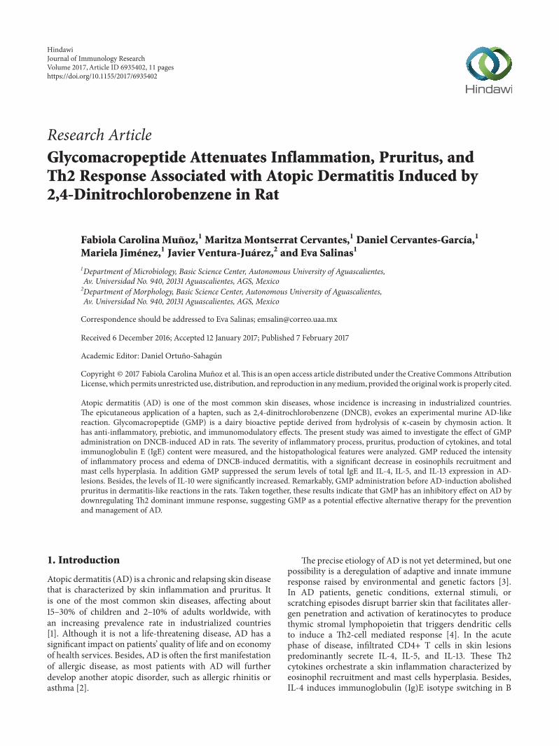
USA) in 1mL of saline solution. Simultaneously, and as anadjuvant, 0.5mL of Bordetella pertussis vaccine (Zuvirac,Mexico City, Mexico) containing 10–15 × 109 heat-killedbacilli/mLwas injected subcutaneously. On days 14, 16, 18, 20,22, and 36, animals were resensitized with a topical applica-tion of 60𝜇L of 1.5%w/v DNCB prepared in acetone-olive oil(A-OO) solution (4 : 1) to both sides of the right ear lobe ofthe rats. Control group was only injected with adjuvants andtopically applied with A-OO solution (Figure 1).
2.3. Experimental Design. For characterization of dermatitismodel, rats were randomly assigned to two different groups(5 rats per group): control and DNCB sensitized. For analysisof GMP effect, rats were randomly assigned to five differentgroups (8 rats per group): control, not sensitized and wateradministered before AD-induction; DNCB-P, DNCB sensi-tized and water administered before AD-induction; GMP-P, DNCB sensitized and GMP administered before AD-induction; DNCB-T, DNCB sensitized and water adminis-tered after AD-induction; and GMP-T, DNCB sensitized andGMP administered after AD-induction. GMP (Lacprodan�cGMP-10; a gift from Arla Foods Amba, Viby, Denmark) wasorally administered to animals at 500mg/kg/day dissolved intap water. Oral intake of GMP was started from 3 days beforesensitization to day 36 as prophylaxis (GMP-P) and fromday 23 to day 36 when employed in a therapeutic manner,that is, once AD was established (GMP-T). Control, DNCB-P, and DNCB-T groups were administered orally with tapwater during corresponding times (Figure 1). An esophagealcatheter was used to deliver GMP solution or water. Allanimals were sacrificed with an overdose of ether at day 37,and blood and ear samples were obtained.
Journal of Immunology Research 3
Sensitization: Resensitization:
14 16 18 20 22 23 36 37
Oral administration of GMP or water before AD-induction(Prophylactic treatment)
Oral administration of GMP or water after AD-induction(Therapeutic treatment)
0
i i Sacrifice1.5% DNCB/A-OO (4 : 1)
−3
DNP-BSA + Al(OH)3 + B. Pertussis
Figure 1: Schematic diagram of experimental dermatitis induction protocol and GMP administration. Rats were sensitized on day 0with injection of DNP-BSA mixed with Al(OH)
3gel and simultaneously with B. pertussis vaccine. Animals were resensitized with topical
application of DNCB in A-OO on days 14, 16, 18, 20, 22, and 36. Control group was injected with the adjuvants but without DNP-BSA andapplied topically with A-OO mixture. GMP or water was administered, daily and orally, from 3 days before AD-induction or from day 23after AD-induction, and until day 36 to analyze the prophylactic or therapeutic effect, respectively. Animals were sacrificed at day 37.
2.4. Evaluation of Ear Cutaneous Inflammatory Reaction andEdema. Cutaneous reaction was evaluated by ear swellinginduced by the challenge with DNCB. Ear thickness wasmeasured using a dial thickness gauge (Milomex, Ltd., Bed-fordshire, UK) at 0, 1, 6, and 24 h after DNCB application onday 36. Ear swellingwas calculated based in the increase of earthickness as RT-LT, where RT and LT represent the thicknessof the right and left ear, respectively, at the correspondingtime point. At day 37 animals were sacrificed, the earswere excised from the base, and identical portions of themiddle of the ears were removed using a metallic punch. Thetissue samples were individually weighted on an analyticalbalance (Precisa XT220A,Dietikon, Switzerland). Edemawascalculated based on the increase of ear weight as RW-LW,where RW and LW represent the weight of the fragment ofthe right and left ear, respectively.
2.5. Evaluation of Scratching Behavior. The total number ofscratching events was counted during 10 minutes immedi-ately after the application of DNCB on days 16, 22, and 36.For that purpose, rats were placed into an acrylic cage dividedinto eight compartments. Their behavior was recorded usinga digital video camera (Samsung HMX-W350, New Jersey,USA). Videoswerewatched by two observers and the numberof scratching events was counted. One scratching event orepisode was defined as a series of one or more scratchingmovements by the hind paw directed toward the applicationsite and ended when the rat either licked its hind paw orplaced it back on the floor [25].
2.6. Histological Analysis. Upper portions of the right ears ofeach rat were fixed in 10% neutral formalin, embedded inparaffin, and sectioned into 5 𝜇m slices. Slices were stainedwith hematoxylin and eosin for evaluation of eosinophilsinfiltration and with toluidine blue for evaluation of mastcells number. After microscopic fields were photographed,the numbers of stained eosinophils and mast cells werecounted in random areas (40,000 𝜇m2) with an AxioPlanCarl Zeiss microscope (Oberkochen, Germany) at 400xmagnification. Three slides were stained per rat and three
fields were examined per slide. Morphometric assessmentwas performed using AxioVision Rel 4.8 software by twoobservers whowere not aware of the group of rats fromwhichthe samples originated.
2.7. Determination of Total IgE. Serum samples preparedfrom blood obtained on day 37 were stored at −70∘C untilused to IgE determination. Total IgE level in serum wasquantified using a rat IgE ELISAkit (Abcam,Cambridge,UK)according to the manufacturer’s instructions.
2.8. RNA Purification and Semiquantitative or Real-TimeQuantitative PCR (qRT-PCR). Total RNA was isolated fromthe lower ear tissue using the SV Total RNA Isolation System(Promega, Madison,WI, USA). Purified RNAwas quantifiedwith a NanoDrop 2000 Spectrophotometer (Thermo Scien-tific) with the A260/280 ratio. Only samples with ratio >1.8were employed for cDNA synthesis. Reverse transcriptions of2 𝜇g of RNAwere performedwith the RETROscript�ReverseTranscription kit (Thermo Scientific). Semiquantitative PCRwas performed with 1𝜇L of 1 : 10 diluted cDNA product,5 𝜇L of PCR Master Mix 2x (Thermo Scientific), and 1 𝜇L offorward and reverse primers at 5 𝜇M each (listed on Table 1);all reactions were completed with nuclease-free water to10 𝜇L. PCR conditions were as follows: initial denaturing at95∘C for 3min, with 25, 30, or 35 cycles of 95∘C for 30 sec,60∘C for 30 sec, and 72∘C for 10 sec, and later for all reactionsa final extension of 72∘C for 3min was included. Ampliconswere separated in 2% agarose gels containing GelRed�Nucleic Acid Gel Stain (Biotium, Hayward, CA, USA) asrecommended by the manufacturer, in TBE 1x (89mM Tris,89mM boric acid, 2mM EDTA, pH 8). Gels were visualizedunder UV light in a MiniBis Pro documentation system(DNR Bio-Imaging Systems, Jerusalem, ISR). For RT-PCR,2 𝜇L of diluted cDNA reaction was used as template forthe detection of IL-4, IL-5, IL-13, IL-10, and 𝛽-actin withthe GoTaq� qPCR Master Mix (Promega) in an Eco Real-Time PCR System (Illumina, San Diego, CA, USA). Relativequantification was determined with ΔΔCt method using 𝛽-actin as housekeeping gene for normalization.
4 Journal of Immunology Research
Table 1: Oligonucleotides for gene expression quantification.
Gene Oligonucleotides Accession number
IL-4 Fw: CACCTTGCTGTCACCCTGTT NM 201270.1Rv: ACATCTCGGTGCATGGAGTC
IL-5 Fw: CAGTGGTGAAAGAGACCTTG NM 021834.1Rv: GTATGTCTAGCCCCTGAAAG
IL-13 Fw: ATCGAGGAGCTGAGCAACAT NM 053828.1Rv: ATCCGAGGCCTTTTGGTTAC
IFN-𝛾 Fw: GCCTAGAAAGTCTGAAGAAC NM 138880.2Rv: GAGATAATCTGGCTCTCAAG
IL-10 Fw: CACCTTGCTGTCACCCTGTT NM 012854.2Rv: ACATCTCGGTGCATGGAGTC
𝛽-Actin Fw: GTCGTACCACTGGCATTGTG NM 031144.3Rv: GCTGTGGTGGTGAAGCTGTA
2.9. Data Analysis. Data were expressed as mean ± standarderror of the mean (SEM). Statistical analysis was performedby Student’s 𝑡-test. Ear thickness data were analyzed bymulticomparative Bonferroni test. Significance was set at 𝑝 <0.05.
3. Results
3.1. Characterization of Dermatitis Evoked by Repeated Chal-lenges withDNCBafter Systemic Sensitization. First, rats weresystemically sensitized with DNP-BSA and later challenged 6times by painting the right ear with DNCB/A-OO solution.As shown in Figures 2(a)-2(b), repeated impregnation withDNCB solution caused potent inflammatory changes inthe ear skin, such as the thickening of both dermis andepidermis, edema, and the accumulation of eosinophils andmast cells. The number of eosinophils and mast cells indermis of rats from DNCB group increased by 12.6- and2.3-fold (Figure 2(c)). The ear thickness, measured as anindicator of skin inflammation [11], increased after eachapplication of DNCB. On day 36, the ear thickness picket at1 h after DNCB painting and maintained significantly greaterthan control rats at 6 and 24 h (Figure 2(d)). On day 37,edema in DNCB group was 98-fold higher than that incontrol rats (Figure 2(e)). Scratching toward the ear receivingDNCB application was observed from day 16. Scratchingoccurred immediately after the application of DNCB, with itsfrequency decreasing as time passed, and no scratching wasobserved at 1 h and thereafter. The scratching events countedfor the first 10min, as shown in Figure 2(f), significantlyincreased at day 16 and were almost equal at day 22, witha slight decrease at day 36. Total RNA was extracted fromthe skin lesions excised 24 h after the sixth DNCB challengeand the expression of inflammatory cytokines was examined.As shown in Figure 2(g), the IFN-𝛾, IL-5, and IL-13 mRNAexpression in skin of control rats was very weak, but itwas potentiated in DNCB group. Furthermore, although theexpression of IL-4mRNAs in skinwas undetectable in controlrats, DNCB-treatment induced their expression in dermatitislesion.
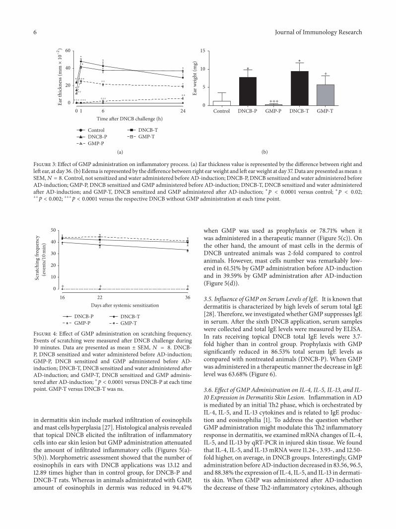
3.2. Oral GMP Administration Diminishes Inflammatory Pro-cess in Dermatitis. First we investigated whether oral intakeof GMP might modify the development of the inflammatoryresponse associated with dermatitis. So, ear thickness wasmeasured after DNCB-repeated applications. On day 36,before the sixth DNCB application (0 h), DNCB-P andDNCB-T animals reported an increase of 0.15 and 0.23mmover control animals. But animals administered with GMPreduced in 95.6 and 54.55% the thickness induced by theprevious five DNCB applications when used in a prophylacticor therapeutic manner, respectively. One hour after the lastDNCB application, ear thickness presented a peak of 0.41and 0.48mm in the ears of DNCB-P and DNCB-T animals,which was sustained at 6 h and presented a slight decrease at24 h. However, when animals were GMP administered beforeAD-induction the inflammatory process was reduced in 99.4,93.98, and 85.89% at 1, 6, and 24 h after challenge, and if theyreceived GMP after AD-induction the ear inflammation wasdiminished in 47.16, 49.41, and 34.06% (Figure 3(a)).
Another way to assess changes in the inflammatoryprocess is to evaluate the ear edema as the increment inear weight. As shown in Figure 3(b), when animals wererepeatedly challenged with DNCB the ear edema was 6.64(DNCB-P) and 8.05 (DNCB-T) higher than in control ani-mals.However, when animalswereGMPadministered beforeAD-induction there was a decrease of 97.03% on ear DNCB-induced edema. Besides, animals that received GMP oncedermatitis was established showed a decrease of 39.87% onedema when compared to untreated group (DNCB-T).
3.3. Scratching Behavior Is Inhibited by GMP-Prophylaxis.Pruritus is one of the major symptoms of AD and impactsquality of life of patients in a significantmanner [26]. Controlanimals did not show any scratching event in the rightear during 10min immediately after the application of A-OO mixture (data not shown). The chronological profile ofscratching behavior in DNCB challenged rats, treated or notwith GMP, is shown in Figure 4. In DNCB-P and DNCB-T rats the number of scratching events remained almostconstant during 10min afterDNCB topical application at days

Journal of Immunology Research 5
Control DNCB
(a)
Control DNCB
(b)
ControlDNCB
Eosinophils Mast cells
∗
∗
Cel
l num
ber/40
,000
m2
0
10
20
30
40
(c)
ControlDNCB
0 1 6 24Time after DNCB challenge (h)
∗
∗∗
∗
0
10
20
30
40
50
Ear t
hick
ness
(mG×10
−2)
(d)
DNCBControl
∗
0
2
4
6
8
10
Ear w
eigh
t (m
g)
(e)
ControlDNCB
Days after systemic sensitization16 22 36
Scra
tchi
ng fr
eque
ncy ∗
∗
∗
0
20
40
60
80
100
(eve
nts/10
min
)
(f)
IL-5
IL-4
IL-13
Control DNCB
IFN-
-Actin
(g)
Figure 2: Characteristics of dermatitis-like reaction in rats challenged with DNCB after systemic sensitization. Histopathological featuresof the ears of control and DNCB challenged rats, 24 h after the sixth challenge, stained with (a) hematoxylin and eosin and (b) toluidineblue. Arrows indicated (a) eosinophils and (b) mast cells. (c) Eosinophils and mast cells were counted in dermis with a microscope at amagnification of 400x. (d) Ear thickness was measured at 0, 1, 6, and 24 h after last DNCB challenge. (e) To measure ear edema, equal areasfrom ears were punched and weighed 24 h after last challenge. (f) Scratching frequency was measured during the first 10 min after DNCBapplication and reported at days 16, 22, and 36. (g) Inflammatory cytokine mRNA expression in the skin lesion 24 h after the last DNCBchallenge. Values represent mean ± SEM;𝑁 = 5 rats. ∗𝑝 < 0.001 versus control at each time point.
16, 22, and 36, with an average of 36.87 and 41.71 scratch-ing events. Oral GMP administration before AD-inductionresulted in a significant and dramatic inhibition of morethan 99% in the number of scratching episodes of DNCB-applied animals during the same days, pruritus being almostcompletely abolished. In contrast, there were no differences
in scratching behavior betweenGMP-T andDNCB-T groups,indicating that GMP has no effect on pruritus when it wasadministered once dermatitis was established.
3.4. GMPAdministration Reduces the Infiltration of Inflamma-tory Cells into DNCB-Induced Skin Lesions. Cellular changes
6 Journal of Immunology Research
ControlDNCB-PGMP-P
DNCB-TGMP-T
+++
+++ +++ +++
+++++
++
0
20
40
60
Ear t
hick
ness
(mG×10
−2)
1 6 240Time after DNCB challenge (h)
∗
∗
∗
∗
∗
∗
∗
∗
+
(a)
Control DNCB-P GMP-P DNCB-T GMP-T
+++0
5
10
15
Ear w
eigh
t (m
g) ∗
∗+
(b)
Figure 3: Effect of GMP administration on inflammatory process. (a) Ear thickness value is represented by the difference between right andleft ear, at day 36. (b) Edema is represented by the difference between right ear weight and left earweight at day 37. Data are presented asmean±SEM,𝑁 = 8. Control, not sensitized andwater administered before AD-induction; DNCB-P, DNCB sensitized andwater administered beforeAD-induction; GMP-P, DNCB sensitized and GMP administered before AD-induction; DNCB-T, DNCB sensitized and water administeredafter AD-induction; and GMP-T, DNCB sensitized and GMP administered after AD-induction; ∗𝑝 < 0.0001 versus control; +𝑝 < 0.02;++𝑝 < 0.002; +++𝑝 < 0.0001 versus the respective DNCB without GMP administration at each time point.
Scra
tchi
ng fr
eque
ncy
22 3616Days after systemic sensitization
(eve
nts/10
min
)
0
10
20
30
40
50
DNCB-PGMP-P
DNCB-TGMP-T
+ + +
Figure 4: Effect of GMP administration on scratching frequency.Events of scratching were measured after DNCB challenge during10 minutes. Data are presented as mean ± SEM, 𝑁 = 8. DNCB-P, DNCB sensitized and water administered before AD-induction;GMP-P, DNCB sensitized and GMP administered before AD-induction; DNCB-T, DNCB sensitized and water administered afterAD-induction; and GMP-T, DNCB sensitized and GMP adminis-tered after AD-induction; +𝑝 < 0.0001 versus DNCB-P at each timepoint. GMP-T versus DNCB-T was ns.
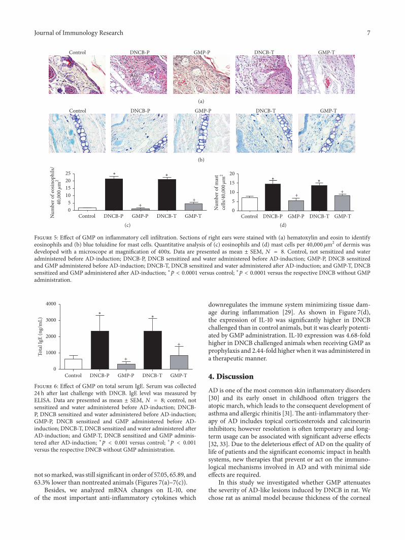
in dermatitis skin include marked infiltration of eosinophilsandmast cells hyperplasia [27]. Histological analysis revealedthat topical DNCB elicited the infiltration of inflammatorycells into ear skin lesion but GMP administration attenuatedthe amount of infiltrated inflammatory cells (Figures 5(a)-5(b)). Morphometric assessment showed that the number ofeosinophils in ears with DNCB applications was 13.12 and12.89 times higher than in control group, for DNCB-P andDNCB-T rats. Whereas in animals administrated with GMP,amount of eosinophils in dermis was reduced in 94.47%
when GMP was used as prophylaxis or 78.71% when itwas administered in a therapeutic manner (Figure 5(c)). Onthe other hand, the amount of mast cells in the dermis ofDNCB untreated animals was 2-fold compared to controlanimals. However, mast cells number was remarkably low-ered in 61.51% by GMP administration before AD-inductionand in 39.59% by GMP administration after AD-induction(Figure 5(d)).
3.5. Influence of GMP on Serum Levels of IgE. It is known thatdermatitis is characterized by high levels of serum total IgE[28].Therefore, we investigated whether GMP suppresses IgEin serum. After the sixth DNCB application, serum sampleswere collected and total IgE levels were measured by ELISA.In rats receiving topical DNCB total IgE levels were 3.7-fold higher than in control group. Prophylaxis with GMPsignificantly reduced in 86.53% total serum IgE levels ascompared with nontreated animals (DNCB-P). When GMPwas administered in a therapeuticmanner the decrease in IgElevel was 63.68% (Figure 6).
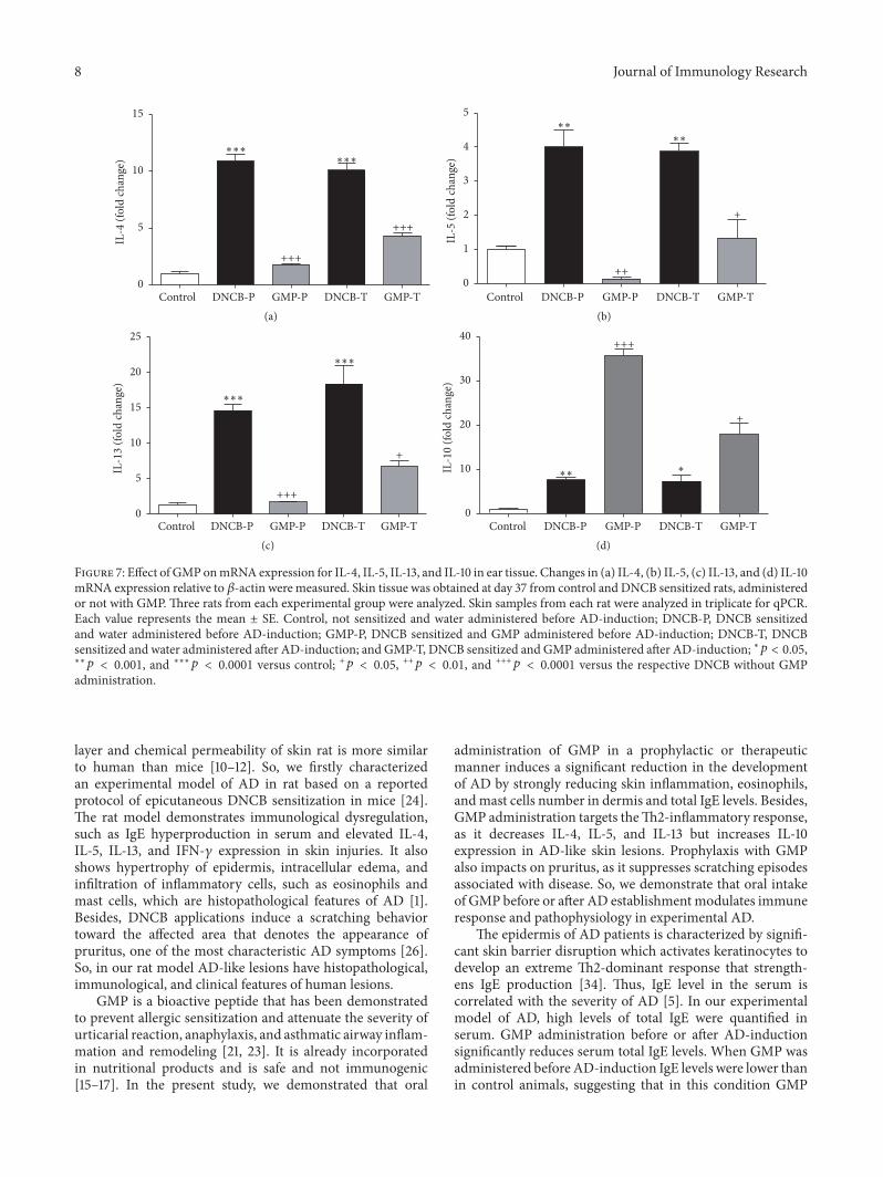
3.6. Effect of GMP Administration on IL-4, IL-5, IL-13, and IL-10 Expression in Dermatitis Skin Lesion. Inflammation in ADis mediated by an initial Th2 phase, which is orchestrated byIL-4, IL-5, and IL-13 cytokines and is related to IgE produc-tion and eosinophilia [1]. To address the question whetherGMP administration might modulate this Th2 inflammatoryresponse in dermatitis, we examined mRNA changes of IL-4,IL-5, and IL-13 by qRT-PCR in injured skin tissue. We foundthat IL-4, IL-5, and IL-13 mRNAwere 11.24-, 3.93-, and 12.50-fold higher, on average, in DNCB groups. Interestingly, GMPadministration beforeAD-induction decreased in 83.56, 96.5,and 88.38% the expression of IL-4, IL-5, and IL-13 in dermati-tis skin. When GMP was administered after AD-inductionthe decrease of these Th2-inflammatory cytokines, although
Journal of Immunology Research 7
Control DNCB-P DNCB-TGMP-P GMP-T
(a)
Control DNCB-P DNCB-TGMP-P GMP-T
(b)
Control DNCB-P GMP-P DNCB-T GMP-TNum
ber o
f eos
inop
hils/
∗ ∗
++
05
10152025
40
,000G2
(c)N
umbe
r of m
ast
Control DNCB-P GMP-P DNCB-T GMP-T
∗ ∗
++
0
5
10
15
20
cells
/40
,000
m2
(d)
Figure 5: Effect of GMP on inflammatory cell infiltration. Sections of right ears were stained with (a) hematoxylin and eosin to identifyeosinophils and (b) blue toluidine for mast cells. Quantitative analysis of (c) eosinophils and (d) mast cells per 40,000 𝜇m2 of dermis wasdeveloped with a microscope at magnification of 400x. Data are presented as mean ± SEM, 𝑁 = 8. Control, not sensitized and wateradministered before AD-induction; DNCB-P, DNCB sensitized and water administered before AD-induction; GMP-P, DNCB sensitizedand GMP administered before AD-induction; DNCB-T, DNCB sensitized and water administered after AD-induction; and GMP-T, DNCBsensitized and GMP administered after AD-induction; ∗𝑝 < 0.0001 versus control; +𝑝 < 0.0001 versus the respective DNCB without GMPadministration.
Control DNCB-P GMP-P DNCB-T GMP-T0
1000
2000
3000
4000
Tota
l IgE
(ng/
mL)
∗∗
+
+
Figure 6: Effect of GMP on total serum IgE. Serum was collected24 h after last challenge with DNCB. IgE level was measured byELISA. Data are presented as mean ± SEM, 𝑁 = 8; control, notsensitized and water administered before AD-induction; DNCB-P, DNCB sensitized and water administered before AD-induction;GMP-P, DNCB sensitized and GMP administered before AD-induction; DNCB-T, DNCB sensitized and water administered afterAD-induction; and GMP-T, DNCB sensitized and GMP adminis-tered after AD-induction; ∗𝑝 < 0.001 versus control; +𝑝 < 0.001versus the respective DNCB without GMP administration.
not somarked,was still significant in order of 57.05, 65.89, and63.3% lower than nontreated animals (Figures 7(a)–7(c)).
Besides, we analyzed mRNA changes on IL-10, oneof the most important anti-inflammatory cytokines which
downregulates the immune system minimizing tissue dam-age during inflammation [29]. As shown in Figure 7(d),the expression of IL-10 was significantly higher in DNCBchallenged than in control animals, but it was clearly potenti-ated by GMP administration. IL-10 expression was 4.68-foldhigher in DNCB challenged animals when receiving GMP asprophylaxis and 2.44-fold higher when it was administered ina therapeutic manner.
4. Discussion
AD is one of the most common skin inflammatory disorders[30] and its early onset in childhood often triggers theatopic march, which leads to the consequent development ofasthma and allergic rhinitis [31].The anti-inflammatory ther-apy of AD includes topical corticosteroids and calcineurininhibitors; however resolution is often temporary and long-term usage can be associated with significant adverse effects[32, 33]. Due to the deleterious effect of AD on the quality oflife of patients and the significant economic impact in healthsystems, new therapies that prevent or act on the immuno-logical mechanisms involved in AD and with minimal sideeffects are required.
In this study we investigated whether GMP attenuatesthe severity of AD-like lesions induced by DNCB in rat. Wechose rat as animal model because thickness of the corneal
8 Journal of Immunology Research
Control DNCB-P GMP-P DNCB-T GMP-T0
5
10
15IL
-4 (f
old
chan
ge)
+++
+++
∗∗∗∗∗∗
(a)Control DNCB-P GMP-P DNCB-T GMP-T
+
++
∗∗∗∗
0
1
2
3
4
5
IL-5
(fol
d ch
ange
)
(b)
Control DNCB-P GMP-P DNCB-T GMP-T
+
+++
∗∗∗
∗∗∗
0
5
10
15
20
25
IL-1
3 (fo
ld ch
ange
)
(c)Control DNCB-P GMP-P DNCB-T GMP-T
+
+++
∗∗∗
0
10
20
30
40
IL-1
0 (fo
ld ch
ange
)
(d)
Figure 7: Effect of GMPonmRNA expression for IL-4, IL-5, IL-13, and IL-10 in ear tissue. Changes in (a) IL-4, (b) IL-5, (c) IL-13, and (d) IL-10mRNA expression relative to 𝛽-actin were measured. Skin tissue was obtained at day 37 from control and DNCB sensitized rats, administeredor not with GMP. Three rats from each experimental group were analyzed. Skin samples from each rat were analyzed in triplicate for qPCR.Each value represents the mean ± SE. Control, not sensitized and water administered before AD-induction; DNCB-P, DNCB sensitizedand water administered before AD-induction; GMP-P, DNCB sensitized and GMP administered before AD-induction; DNCB-T, DNCBsensitized and water administered after AD-induction; and GMP-T, DNCB sensitized and GMP administered after AD-induction; ∗𝑝 < 0.05,∗∗𝑝 < 0.001, and ∗∗∗𝑝 < 0.0001 versus control; +𝑝 < 0.05, ++𝑝 < 0.01, and +++𝑝 < 0.0001 versus the respective DNCB without GMPadministration.
layer and chemical permeability of skin rat is more similarto human than mice [10–12]. So, we firstly characterizedan experimental model of AD in rat based on a reportedprotocol of epicutaneous DNCB sensitization in mice [24].The rat model demonstrates immunological dysregulation,such as IgE hyperproduction in serum and elevated IL-4,IL-5, IL-13, and IFN-𝛾 expression in skin injuries. It alsoshows hypertrophy of epidermis, intracellular edema, andinfiltration of inflammatory cells, such as eosinophils andmast cells, which are histopathological features of AD [1].Besides, DNCB applications induce a scratching behaviortoward the affected area that denotes the appearance ofpruritus, one of the most characteristic AD symptoms [26].So, in our rat model AD-like lesions have histopathological,immunological, and clinical features of human lesions.
GMP is a bioactive peptide that has been demonstratedto prevent allergic sensitization and attenuate the severity ofurticarial reaction, anaphylaxis, and asthmatic airway inflam-mation and remodeling [21, 23]. It is already incorporatedin nutritional products and is safe and not immunogenic[15–17]. In the present study, we demonstrated that oral
administration of GMP in a prophylactic or therapeuticmanner induces a significant reduction in the developmentof AD by strongly reducing skin inflammation, eosinophils,andmast cells number in dermis and total IgE levels. Besides,GMP administration targets theTh2-inflammatory response,as it decreases IL-4, IL-5, and IL-13 but increases IL-10expression in AD-like skin lesions. Prophylaxis with GMPalso impacts on pruritus, as it suppresses scratching episodesassociated with disease. So, we demonstrate that oral intakeof GMP before or after AD establishmentmodulates immuneresponse and pathophysiology in experimental AD.
The epidermis of AD patients is characterized by signifi-cant skin barrier disruption which activates keratinocytes todevelop an extreme Th2-dominant response that strength-ens IgE production [34]. Thus, IgE level in the serum iscorrelated with the severity of AD [5]. In our experimentalmodel of AD, high levels of total IgE were quantified inserum. GMP administration before or after AD-inductionsignificantly reduces serum total IgE levels. When GMP wasadministered before AD-induction IgE levels were lower thanin control animals, suggesting that in this condition GMP

Journal of Immunology Research 9
administration can suppress serum total IgE. It is knownthat IgE released from B cells binds to mast cells. Allergensinduce mast cells degranulation through IgE-Fc𝜀RI complexand the release of several biological mediators involved inskin inflammation [35]. So, a lessened level of IgE is inline with the reduction of edema and skin inflammation ofAD-lesions observed in animals with GMP administration.Previously, it has been demonstrated that GMP inhibits mastcells activation by allergens [22] and we observed a reducednumber of mast cells in dermis of GMP-treated animals,so the reduction in edema and skin inflammation as aconsequence of GMP administration might also be mediatedby alterations in mast cells number and function.
One of the central causes of the AD is the dysregu-lated Th1 and Th2 response that induces the characteristicTh2-dominant skin allergic inflammation [36]. In this Th2response, the involvement of IL-4, IL-5, and IL-13 is crucialin humans [37]. In transgenic mice that overproduce IL-4,IL-5, and IL-13, investigators have demonstrated a positivecorrelation between the onset and progression of AD-likedisease and the expression of these Th2 cytokines [38]. Inour experimental model of AD the expression of IL-4, IL-5, and IL-13 was increased in skin lesions. It is known thatIL-5 plays an important role in eosinophil differentiation,activation, proliferation, and chemotaxis [39, 40]. The num-ber of eosinophils and levels of IL-5 have previously beenshown to be elevated in injured skin of patients with AD[5, 41].We show thatGMP administration before or afterAD-induction induces a significant reduction in IL-5 expressionin AD-lesions, which is correlated with the decrease in thenumber of eosinophils infiltrated in dermis. On the otherhand, transgenic mice overexpressing epidermal IL-4 or IL-13 spontaneously developed signs and symptoms associatedwith AD, including elevated IgE levels [42, 43]. So, reducedlevels of IL-4 and IL-13 in skin of animals treated with GMPin a prophylactic or therapeutic manner are in concordancewith the decrease in total IgE.The downregulation of theTh2-dominant skin inflammation by GMP administrationmay beassociated with the increased expression of IL-10, a knownregulatory cytokine. It has been reported that IL-10 inhibitsboth the proliferation and the cytokine synthesis of CD4+Th2 cells [44]. Recently, the role of IL-10 in the control of ADdevelopment and maintenance has been highlighted by thefact that polymorphisms in the IL-10 gene could represent agenetic marker for AD in childhood [45]. As Th2 cytokinesdestabilize cutaneous barrier function [46, 47] and IFN-𝛾is crucial in dermal thickening and in the progression tochronic AD skin lesions [1], the study of the effect of GMPadministration on the recovery of skin barrier integrity andon levels of IFN-𝛾 expression is the aim of our currentresearch.
Pruritus is a clinical manifestation of AD [26] that causesa great deterioration in patient’s quality of life [48]. Besides,scratching worsens the dermatitis, increasing lesions in skinand thereby aggravating pruritus [49]. Thus, proper treat-ment of pruritus is the critical part of therapeutic approachto AD. Our rats with dermatitis showed an intense pruritusafter DNCB application, but prophylaxis with GMP totallyabolished the scratching episodes of the rats. A wide range
of itch-inducing stimuli generated within the skin are able totrigger pruritus. Among them, histamine is recently consid-ered relevant, as combined H1R/H4R antagonists therapy issuccessfully addressing pruritus in AD [50]. The decrease inIgE levels andmast cells number observed in animals admin-istered with GMP before AD-induction, together with thereported inhibitory action of GMP on mast cells activationby allergen [22], might cause a decrease in histamine levelsin skin, impacting on itching. Besides, it has been reportedthat transgenicmice expressing IL-13 in skin develops intensepruritus [43]. Dupilumab, a monoclonal antibody that bindsto IL-4R𝛼 and blocks both IL-4 and IL-13 signaling pathways,induces a reduction in the pruritus score of patients withmoderate to severe AD [51, 52]. These data indicate a role ofIL-4 and IL-13 in triggering pruritus.Thus, antipruritic actionof GMP-prophylaxis might be alsomediated by the reductionof IL-4 and IL-13 expression in skin. However, due to the widerange of stimuli able to trigger pruritus in AD we cannotexclude a possible effect of GMP on other itching-inducingelement.
GMP exerted a clearly superior therapeutic effect when itwas given beforeAD-induction thanwhen administered onceAD-lesions were established. This is a common observationwith GMP, because when it is used as anti-inflammatorytherapy in experimental colitis its effect is greater whenused as prophylaxis [17]. We recently demonstrated thatGMP administration before allergen sensitization inducesa significant increase in the amount of Lactobacillus, Bifi-dobacterium, and Bacteroides in the gut of sensitized animals[22]. In this regard, data about the effect of probiotics in theprevention and treatment of AD remain elusive, with negativeand positive results, but evidencing that their positive effectsdepend on factors such as the type of probiotic strain,methodof administration, onset time, duration of exposure, anddosage [53]. Particularly Lactobacillus and Bifidobacteriumas therapy in AD show a promissory effect on preventionof pediatric AD, while there is less convincing informationabout their effects when used in a therapeutic manner [54],which is in concordance with our results. It is important tohighlight that even after AD-induction most of the beneficialeffects of GMP were retained, with exception of antipruriticeffect. This may be due to the lesser reduction in IgElevels, mast cells number, and IL-4 and IL-13 expression inanimals administered with GMP once AD was established.The remaining levels of these immune elements might besufficient to maintain pruritus in the animals. However, wemust consider that patients with AD may benefit from anti-inflammatory and Th2-downregulation properties of GMPused in a therapeutic manner.
In conclusion, the present study shows that GMP pos-sesses prophylactic and therapeutic effects in the develop-ment of AD. GMP effectively suppresses skin inflammation,eosinophils recruitment, and mast cells hyperplasia in der-mis, as well as total IgE in serum. Beneficial effect of GMPis associated with downregulation of IL-4, IL-5, and IL-13expression together with upregulation of IL-10. Prophylacticadministration of GMP also abolished pruritus. This studyprovides the first experimental basis for the potential use ofGMP in the prevention and therapy of AD.
10 Journal of Immunology Research
Disclosure
Cervantes-Garcıa is a CONACYT research fellow in Auton-omous University of Aguascalientes.
Competing Interests
The authors confirm that there are no competing interests.
Authors’ Contributions
All authors read the paper and participated in revising itfor intellectual content and style. The authors participatedas follows: for experimental design: Eva Salinas and DanielCervantes-Garcıa; for acquisition of data: Fabiola CarolinaMunoz, Maritza Montserrat Cervantes, Daniel Cervantes-Garcıa, and Mariela Jimenez; for analysis and interpreta-tion of data: Eva Salinas, Daniel Cervantes-Garcıa, FabiolaCarolina Munoz, and Javier Ventura-Juarez; for drafting ofthe paper: Eva Salinas, Daniel Cervantes-Garcıa, FabiolaCarolina Munoz, and Mariela Jimenez.
Acknowledgments
This work was supported by the Autonomous University ofAguascalientes, Grant PIBB 15-9N, and CONACYT, Grant240921. The authors wish to thank Claudia Berenice Barronfor excellent technical assistance and MVZ Jose Luis Ponceand MVZ Jesus Humberto Estrada Calderon for providingthe animals for the study.
References
[1] T. Bieber, “Atopic dermatitis,” Annals of Dermatology, vol. 22,no. 2, pp. 125–137, 2010.
[2] L. Schneider, S. Tilles, P. Lio et al., “Atopic dermatitis: apractice parameter update 2012,” Journal of Allergy and ClinicalImmunology, vol. 131, no. 2, pp. 295–299.e27, 2013.
[3] T. Bieber, “Atopic dermatitis,”New England Journal of Medicine,vol. 358, no. 14, pp. 1430–1494, 2008.
[4] K. Kabashima, “New concept of the pathogenesis of atopicdermatitis: interplay among the barrier, allergy, and pruritus asa trinity,” Journal of Dermatological Science, vol. 70, no. 1, pp.3–11, 2013.
[5] F.-T. Liu, H. Goodarzi, and H.-Y. Chen, “IgE, mast cells, andeosinophils in atopic dermatitis,”Clinical Reviews in Allergy andImmunology, vol. 41, no. 3, pp. 298–310, 2011.
[6] M. Auriemma, G. Vianale, P. Amerio, andM. Reale, “Cytokinesand T cells in atopic dermatitis,” European Cytokine Network,vol. 24, no. 1, pp. 37–44, 2013.
[7] N. Inagaki and H. Nagai, “Analysis of the mechanism for thedevelopment of allergic skin inflammation and the applicationfor its treatment: mouse models for the development of reme-dies for human allergic dermatitis,” Journal of PharmacologicalSciences, vol. 110, no. 3, pp. 251–259, 2009.
[8] T. Shiohara, J. Hayakawa, and Y.Mizukawa, “Animal models foratopic dermatitis: are they relevant to human disease?” Journalof Dermatological Science, vol. 36, no. 1, pp. 1–9, 2004.
[9] H. Kitagaki, S. Fujisawa, K. Watanabe, K. Hayakawa, andT. Shiohara, “Immediate-type hypersensitivity response fol-lowed by a late reaction is induced by repeated epicutaneous
application of contact sensitizing agents in mice,” Journal ofInvestigative Dermatology, vol. 105, no. 6, pp. 749–755, 1995.
[10] D. P. Arfsten, C. M. Garrett, W. W. Jederberg, E. R. Wilfong,and J. N. McDougal, “Characterization of the skin penetrationof a hydrocarbon-based weapons maintenance oil,” Journal ofOccupational and Environmental Hygiene, vol. 3, no. 9, pp. 457–464, 2006.
[11] Y. Fujii, H. Takeuchi, S. Sakuma, T. Sengoku, and S. Takakura,“Characterization of a 2,4-dinitrochlorobenzene-inducedchronic dermatitis model in rats,” Skin Pharmacology andPhysiology, vol. 22, no. 5, pp. 240–247, 2009.
[12] K. Sato, K. Sugibayashi, and Y. Morimoto, “Species differencesin percutaneous absorption of nicorandil,” Journal of Pharma-ceutical Sciences, vol. 80, no. 2, pp. 104–107, 1991.
[13] E. P. Brody, “Biological activities of bovine glycomacropeptide,”British Journal of Nutrition, vol. 84, no. 1, pp. S39–S46, 2000.
[14] Neelima, R. Sharma, Y. S. Rajput, and B. Mann, “Chemical andfunctional properties of glycomacropeptide (GMP) and its rolein the detection of cheese whey adulteration in milk: a review,”Dairy Science and Technology, vol. 93, no. 1, pp. 21–43, 2013.
[15] T. L. Mikkelsen, E. Rasmussen, A. Olsen, V. Barkholt, and H.Frøkiær, “Immunogenicity of 𝜅-casein and glycomacropeptide,”Journal of Dairy Science, vol. 89, no. 3, pp. 824–830, 2006.
[16] M. H. Abd El-Salam, “Separation of casein glycomacropeptidefrom whey: methods of potential industrial application,” Inter-national Journal of Dairy Science, vol. 1, no. 1, pp. 93–99, 2006.
[17] A. Daddaoua, V. Puerta, A. Zarzuelo, M. D. Suarez, F.Sanchez DeMedina, and O.Martınez-Augustin, “Bovine glyco-macropeptide is anti-inflammatory in rats with hapten-inducedcolitis,” Journal of Nutrition, vol. 135, no. 5, pp. 1164–1170, 2005.
[18] P. Requena, A. Daddaoua, E. Martınez-Plata et al., “Bovine gly-comacropeptide ameliorates experimental rat ileitis by mech-anisms involving downregulation of interleukin 17,” BritishJournal of Pharmacology, vol. 154, no. 4, pp. 825–832, 2008.
[19] P. Requena, R. Gonzalez, R. Lopez-Posadas et al., “The intestinalantiinflammatory agent glycomacropeptide has immunomod-ulatory actions on rat splenocytes,” Biochemical Pharmacology,vol. 79, no. 12, pp. 1797–1804, 2010.
[20] Q. S. Chen, J. F. Wang, and Y. L. Yan, “Effects of caseinGlycomacropeptide on dimethyl hydrazine-induced alterationof cytokine network in rats,” Journal of Food Science, vol. 35, pp.192–198, 2014.
[21] M. Jimenez, N. A. Chavez, and E. Salinas, “Pretreatmentwith glycomacropeptide reduces allergen sensitization, allevi-ates immediate cutaneous hypersensitivity and protects fromanaphylaxis,” Clinical and Experimental Immunology, vol. 170,no. 1, pp. 18–27, 2012.
[22] M. Jimenez, D. Cervantes-Garcıa, Y. H. Munoz, A. Garcıa, L.M. Jr. Haro, and E. Salinas, “Novel mechanisms underlying thetherapeutic effect of glycomacropeptide on allergy: change ingut microbiota, upregulation of TGF-𝛽, and inhibition of mastcells,” International Archives of Allergy and Immunology, vol. 171,no. 3-4, pp. 217–226, 2016.
[23] N. R. Roldan,M. Jimenez,D.Cervantes-Garcıa, E.Marın, andE.Salinas, “Glycomacropeptide administration attenuates airwayinflammation and remodeling associated to allergic asthma inrat,” Inflammation Research, vol. 65, no. 4, pp. 273–283, 2016.
[24] H. Yamashita, T. Ito, H. Kato et al., “Comparison of theefficacy of tacrolimus and cyclosporine A in a murine modelof dinitrofluorobenzene-induced atopic dermatitis,” EuropeanJournal of Pharmacology, vol. 645, no. 1–3, pp. 171–176, 2010.
Journal of Immunology Research 11
[25] H. Nojima and E. Carstens, “5-hydroxytryptamine (5-HT)2receptor involvement in acute 5-HT-evoked scratching but notin allergic pruritus induced by dinitrofluorobenzene in rats,”Journal of Pharmacology and Experimental Therapeutics, vol.306, no. 1, pp. 245–252, 2003.
[26] K. Kim, “Neuroimmunological mechanism of pruritus in atopicdermatitis focused on the role of serotonin,” Biomolecules andTherapeutics, vol. 20, no. 6, pp. 506–512, 2012.
[27] N. Inagaki, N. Shiraishi, K. Igeta et al., “Inhibition of scratch-ing behavior associated with allergic dermatitis in mice bytacrolimus, but not by dexamethasone,” European Journal ofPharmacology, vol. 546, no. 1–3, pp. 189–196, 2006.
[28] A. S.Moreno, R.McPhee, L. K. Arruda, andM.D. Howell, “Tar-geting the T helper 2 inflammatory axis in atopic dermatitis,”International Archives of Allergy and Immunology, vol. 171, no.2, pp. 71–80, 2016.
[29] R. Sabat, G.Grutz, K.Warszawska et al., “Biology of interleukin-10,” Cytokine and Growth Factor Reviews, vol. 21, no. 5, pp. 331–344, 2010.
[30] R. J.Hay,N. E. Johns,H.C.Williams et al., “The global burden ofskin disease in 2010: an analysis of the prevalence and impact ofskin conditions,” Journal of Investigative Dermatology, vol. 134,no. 6, pp. 1527–1534, 2014.
[31] S. C. Dharmage, A. J. Lowe, M. C. Matheson, J. A. Burgess, K.J. Allen, and M. J. Abramson, “Atopic dermatitis and the atopicmarch revisited,” Allergy, vol. 69, no. 1, pp. 17–27, 2014.
[32] J. D. Ference and A. R. Last, “Choosing topical corticosteroids,”American Family Physician, vol. 79, no. 2, pp. 135–140, 2009.
[33] M. M. Tollefson, A. L. Bruckner, B. A. Cohen et al., “Atopicdermatitis: skin-directed management,” Pediatrics, vol. 134, no.6, pp. e1735–e1744, 2014.
[34] M. Boguniewicz and D. Y. M. Leung, “Atopic dermatitis: adisease of altered skin barrier and immune dysregulation,”Immunological Reviews, vol. 242, no. 1, pp. 233–246, 2011.
[35] K. Amin, “The role of mast cells in allergic inflammation,”Respiratory Medicine, vol. 106, no. 1, pp. 9–14, 2012.
[36] M. Grewe, C. A. F. M. Bruijnzeel-Koomen, E. Schopf et al., “Arole for Th1 andTh2 cells in the immunopathogenesis of atopicdermatitis,” Immunology Today, vol. 19, no. 8, pp. 359–361, 1998.
[37] E. B. Brandt and U. Sivaprasad, “Th2 cytokines and atopic der-matitis,” Journal of Clinical & Cellular Immunology, vol. 2, no. 3,2011.
[38] G. R. Lee and R. A. Flavell, “Transgenic mice which overpro-duce Th2 cytokines develop spontaneous atopic dermatitis andasthma,” International Immunology, vol. 16, no. 8, pp. 1155–1160,2004.
[39] D. Simon, L. R. Braathen, and H.-U. Simon, “Eosinophils andatopic dermatitis,” Allergy, vol. 59, no. 6, pp. 561–570, 2004.
[40] N. A. Molfino, D. Gossage, R. Kolbeck, J. M. Parker, and G. P.Geba, “Molecular and clinical rationale for therapeutic target-ing of interleukin-5 and its receptor,” Clinical and ExperimentalAllergy, vol. 42, no. 5, pp. 712–737, 2012.
[41] C.-W. Jeong, K.-S. Ahn, N.-K. Rho et al., “Differential in vivocytokine mRNA expression in lesional skin of intrinsic vs.extrinsic atopic dermatitis patients using semiquantitative RT-PCR,”Clinical and Experimental Allergy, vol. 33, no. 12, pp. 1717–1724, 2003.
[42] L. S. Chan, N. Robinson, and L. Xu, “Expression of interleukin-4 in the epidermis of transgenic mice results in a pruriticinflammatory skin disease: an experimental animal model tostudy atopic dermatitis,” Journal of Investigative Dermatology,vol. 117, no. 4, pp. 977–983, 2001.
[43] T. Zheng, M. H. Oh, S. Y. Oh, J. T. Schroeder, A. B. Glick, andZ. Zhu, “Transgenic expression of interleukin-13 in the skininduces a pruritic dermatitis and skin remodeling,” Journal ofInvestigative Dermatology, vol. 129, no. 3, pp. 742–751, 2009.
[44] G. Del Prete, M. De Carli, F. Almerigogna, M. G. Giudizi, R.Biagiotti, and S. Romagnani, “Human IL-10 is produced byboth type 1 helper (Th1) and type 2 helper (Th2) T cell clonesand inhibits their antigen-specific proliferation and cytokineproduction,” Journal of Immunology, vol. 150, no. 2, pp. 353–360,1993.
[45] M. H. Sohn, J. S. Song, K.-W. Kim, E.-S. Kim, K.-E. Kim,and J. M. Lee, “Association of interleukin-10 gene promoterpolymorphism in children with atopic dermatitis,” The Journalof Pediatrics, vol. 150, no. 1, pp. 106–108, 2007.
[46] P. Y. Ong, T. Ohtake, C. Brandt et al., “Endogenous antimi-crobial peptides and skin infections in atopic dermatitis,” NewEngland Journal of Medicine, vol. 347, no. 15, pp. 1151–1160, 2002.
[47] M. Suarez-Farinas, S. J. Tintle, A. Shemer et al., “Nonlesionalatopic dermatitis skin is characterized by broad terminal differ-entiation defects and variable immune abnormalities,” Journalof Allergy and Clinical Immunology, vol. 127, no. 4, pp. 954–964,2011.
[48] A. Buske-Kirschbaum, A. Geiben, and D. Hellhammer, “Psy-chobiological aspects of atopic dermatitis: an overview,” Psy-chotherapy and Psychosomatics, vol. 70, no. 1, pp. 6–16, 2001.
[49] C.-F. Wahlgren, “Itch and atopic dermatitis: an overview,”Journal of Dermatology, vol. 26, no. 11, pp. 770–779, 1999.
[50] M. Albrecht and A. M. Dittrich, “Expression and function ofhistamine and its receptors in atopic dermatitis,”Molecular andCellular Pediatrics, vol. 2, article no. 16, 2015.
[51] L. A. Beck,D.Thaci, J. D.Hamilton et al., “Dupilumab treatmentin adults with moderate-to-severe atopic dermatitis,” NewEngland Journal of Medicine, vol. 371, no. 2, pp. 130–139, 2014.
[52] D.Thaci, E. L. Simpson, L. A. Beck et al., “Efficacy and safety ofdupilumab in adults with moderate-to-severe atopic dermatitisinadequately controlled by topical treatments: a randomised,placebo-controlled, dose-ranging phase 2b trial,” The Lancet,vol. 387, no. 10013, pp. 40–52, 2016.
[53] I. A. Rather, V. K. Bajpai, S. Kumar, J. Lim,W. K. Paek, and Y.-H.Park, “Probiotics and atopic dermatitis: an overview,” Frontiersin Microbiology, vol. 7, article 507, 2016.
[54] K. L. B. Nole, E. Yim, and J. E. Keri, “Probiotics and prebiotics indermatology,” Journal of the AmericanAcademy of Dermatology,vol. 71, no. 4, pp. 814–821, 2014.

Submit your manuscripts athttps://www.hindawi.com
Stem CellsInternational
Hindawi Publishing Corporationhttp://www.hindawi.com Volume 2014
Hindawi Publishing Corporationhttp://www.hindawi.com Volume 2014
MEDIATORSINFLAMMATION
of
Hindawi Publishing Corporationhttp://www.hindawi.com Volume 2014
Behavioural Neurology
EndocrinologyInternational Journal of
Hindawi Publishing Corporationhttp://www.hindawi.com Volume 2014
Hindawi Publishing Corporationhttp://www.hindawi.com Volume 2014
Disease Markers
Hindawi Publishing Corporationhttp://www.hindawi.com Volume 2014
BioMed Research International
OncologyJournal of
Hindawi Publishing Corporationhttp://www.hindawi.com Volume 2014
Hindawi Publishing Corporationhttp://www.hindawi.com Volume 2014
Oxidative Medicine and Cellular Longevity
Hindawi Publishing Corporationhttp://www.hindawi.com Volume 2014
PPAR Research
The Scientific World JournalHindawi Publishing Corporation http://www.hindawi.com Volume 2014
Immunology ResearchHindawi Publishing Corporationhttp://www.hindawi.com Volume 2014
Journal of
ObesityJournal of
Hindawi Publishing Corporationhttp://www.hindawi.com Volume 2014
Hindawi Publishing Corporationhttp://www.hindawi.com Volume 2014
Computational and Mathematical Methods in Medicine
OphthalmologyJournal of
Hindawi Publishing Corporationhttp://www.hindawi.com Volume 2014
Diabetes ResearchJournal of
Hindawi Publishing Corporationhttp://www.hindawi.com Volume 2014
Hindawi Publishing Corporationhttp://www.hindawi.com Volume 2014
Research and TreatmentAIDS
Hindawi Publishing Corporationhttp://www.hindawi.com Volume 2014
Gastroenterology Research and Practice
Hindawi Publishing Corporationhttp://www.hindawi.com Volume 2014
Parkinson’s Disease
Evidence-Based Complementary and Alternative Medicine
Volume 2014Hindawi Publishing Corporationhttp://www.hindawi.com


![Research Paper Deguelin Attenuates Allergic Airway ... · Research Paper Deguelin Attenuates Allergic Airway Inflammation via ... pathophysiology of asthma [4]. ... schematic diagram](https://img.pdfslide.us/doc/110x75/5b396a827f8b9ab9068e82d4/research-paper-deguelin-attenuates-allergic-airway-research-paper-deguelin.jpg)