Embed Size (px)
Citation preview


Our program
Breast cancer treatment
Breast cancer basics
Follow-up care
What to expect when
diagnosed with breast cancer
Managing symptoms and
side effects
Understanding
your diagnosis
Breast cancer dictionaryUCLA Breast Center
S A N T A M O N I C A


UCLA Breast Center
Dear Patient,
We have designed this binder to help organize personalized materials about your treatment,
appointments, medications and other vital information. You can use this binder to write notes,
questions or concerns that you may have between your appointments. Bring this with you to
your appointments so you can track your health and organize your information.
As partners in your care, we want you to have the most up-to-date information so you can
make the most informed decisions. Sometimes the information may seem confusing. Feel free to
ask us questions or seek clarification. In addition to your physicians, we have a patient navigator
to help guide you through the process. This can be a difficult time for you and your family.
Our team is here to make your experience as comfortable as possible.
Sincerely,
UCLA Breast Center
1223 16th Street, Suite 1100 Santa Monica, CA 90404
breastcenter.ucla.edu

UCLA Breast Center
1
“ The experience could not have been more perfect. After
the shock of the diagnosis, there are many options, tests and clinicians
to see. It was a godsend to have all the expertise in one place and to
have a coordinator who oversees the many facets of my care.”


UCLA Breast Center
Our program The UCLA Breast Center in Santa Monica is designed to personalize breast cancer care. Our mission is to deliver state-of-the-art, personalized, multidisciplinary breast care in a compassionate and supportive environment.
At your first appointment, you will meet with our team of board-certified specialists in breast surgery, reconstructive plastic surgery, medical oncology and radiation oncology. Our physicians will attend to your individual needs and develop a comprehensive, personalized plan of care — all in a single day to eliminate delays between diagnosis and the start of treatment.
Our team reviews all biopsies and imaging studies in collaboration with our pathology, breast radiology and genetics experts.
In addition, a specially trained psychologist or clinical social worker from the Simms/Mann — UCLA Center for Integrative Oncology will meet with you to optimize your physical and emotional well-being and identify other resources that may be helpful to you through this process.
Our nurse navigator is a nurse practitioner who specializes in breast cancer care and will act as your guide, assisting you with care coordination, planning, education and follow up.
UCLA Breast Center
Our team
Breast Surgery
Amy Kusske, MD
Maggie DiNome, MD
Phone: 424-259-8791
Breast Medical Oncology
Sara Hurvitz, MD
Parvin Peddi, MD
Rena Desai Callahan, MD
Saeed Sadeghi, MD
Aashini Master, DO
Kelly McCann, MD
Phone: 310-829-5471
Radiology
Anne Hoyt, MD
Laura Doepke, MD
Melissa Joines, MD
Jane Dascalos, MD
Irene Tsai, MD
Nanette DeBruhl, MD
Stephanie Lee-Felker, MD
Cheryce Fischer, MD
Phone: 310-393-5153
Plastic Surgery
Charles Tseng, MD
Christopher Crisera, MD
Jason Roostaeian, MD
Jaco Festekjian, MD
Andrew Da Lio, MD
Phone: 310-825-5510
Pathology
Nicole Dawson, MD
Steve Hart, MD
David Y. Lu, MD
Tobi Quinto, MD
Thomas Lawton, MD
Phone: 424-259-8111
Breast Radiation Oncology
Susan McCloskey, MD
Phone: 424-259-8777
Genetic Counselor
Erica Silver, MS, LCGC
Phone: 310-998-4747
Nurse Navigator
Amy Jacobson, RN, NP-BC
Phone: 424-259-8791
Simms/Mann UCLA Center for Integrative Oncology
Phone: 310-794-6644

UCLA Breast Center
2
“ I am very grateful for discovering this center. I was simply
lost in shock and confusion over my diagnosis. I had no idea how to feel
about all the experts I was told I would need. This center is so comprehensive
in its approach; I only wish everyone, no matter their health issue, would
be as lucky as I have been.”


UCLA Breast Center
Breast cancer — it’s the diagnosis virtually all women fear, and understandably so. Breast cancer strikes one out of every eight women in this country, and almost everyone knows someone who has been diagnosed with this disease.
However, with early diagnosis and advances in treatment, breast cancer can be treated successfully and, in many cases, cured.
At first, the treatment options can seem scary and confusing. Your surgeon or oncologist should be able to explain individual treatment recommendations in detail, but here is a brief outline of how breast cancer is generally treated.
Surgery
Surgery is usually the first line of treatment for breast cancer unless the tumor is very large, is of a certain subtype or has spread to other parts of the body, in which case chemotherapy may be needed first. The surgical options for breast cancer include breast-conserving surgery (also called lumpectomy or partial mastectomy) and mastectomy. Lumpectomy removes the tumor and some normal surrounding breast tissue, whereas mastectomy removes the entire breast. Which option is best depends on tumor size, breast size and personal preference, as well as other aspects of the medical history. If mastectomy is performed, breast reconstruction can be done during the same surgery or at a later time. During the lumpectomy or mastectomy, some of the lymph nodes in the armpit will likely be removed to determine if the cancer has spread (lymph nodes in the underarm are the first place breast cancer spreads). For early breast cancer, a sentinel lymph node biopsy is performed to sample and examine the first lymph node draining the cancer from the breast. If cancer is not found in the sentinel nodes, no additional lymph nodes need to be removed. Advantages of this technique include faster recovery time and a lower risk of lymphedema (swelling of the arm).
Radiation treatment
Radiation treatment is almost always used after lumpectomy, and may be used after mastectomy, to help decrease the risk of recurrence. High-energy X-rays are used to treat the breast and sometimes the surrounding lymph nodes. Radiation treatment is usually well tolerated and has few side effects.
What to expect when diagnosed with breast cancer

UCLA Breast Center
Systemic therapies
Chemotherapy
Chemotherapy, often given intravenously after surgery, allows anti-cancer drugs to enter the bloodstream and target cancer cells wherever they are in the body. Unfortunately, many of these drugs do not only affect cancer cells. When they act on other cells, such as those lining the intestine or hair follicles, side effects like nausea and hair loss can result. The good news is that most of these side effects go away when chemotherapy is completed. Major strides have been made in developing drugs that treat the cancer more effectively and have fewer side effects.
Anti-hormonal therapies
Sometimes anti-hormonal therapy will be recommended as an additional treatment. These drugs block the estrogen hormone’s potential to stimulate breast-cancer cell growth. While side effects from these drugs are usually minimal, drug suitability may vary depending on the person.
Targeted therapies
HER2
A newer form of therapy called HER2-targeted therapy may also be recommended if your tumor tests positive for the HER2 protein. This therapy is generally very well tolerated and works well with chemotherapy to prevent cancer recurrence.
Specific treatment recommendations may vary depending on each patient’s circumstances. When patients have questions about their treatment plans, they should ask their surgeon, radiation oncologist or medical oncologist to explain their treatment more thoroughly. The American Cancer Society is also an excellent resource for additional information.

UCLA Breast Center
3
“ We love that the whole process is under one team and that
all the different aspects — chemo, surgery, radiation, etc. — are so
well coordinated that nothing is lost switching between departments.
We are also very impressed by how committed and passionate
everyone is to their respective areas of expertise.”


UCLA Breast Center
The breast is mostly made up of fatty tissue. Within this tissue is a network of lobes, which are made up of small, tube-like structures called lobules that contain milk glands. Tiny ducts connect the glands, lobules and lobes, carrying the milk from the lobes to the nipple, located in the middle of the areola (darker area that surrounds the nipple). Blood and lymph vessels also run throughout the breast. Blood nourishes the cells while the lymphatic system drains bodily waste products. The lymph vessels connect to lymph nodes, which are tiny, bean-shaped organs that help fight infection.
Breast cancer basics
Clavicle
2nd ribAxillary lymph nodes
Pectoralis major muscle
Mammary gland
Lactiferous sinus
Nipple
Breast
Areola
Lactiferous ducts
Gland lobules
Fat

UCLA Breast Center
Breast cancer development In the United States, breast cancer is the most common cancer diagnosed in women (excluding skin cancer). Men may also develop breast cancer, but less than 1 percent of all people with breast cancer are men. Breast cancer begins when healthy cells in the breast change and grow uncontrollably, forming a mass called a tumor. A tumor can be benign (non-cancerous) or malignant (cancerous). A benign tumor is rarely life threatening and does not spread to other parts of the body. A malignant tumor can spread beyond where it began to other parts of the body. Almost 75 percent of all breast cancers begin in the cells lining the milk ducts; they are called ductal carcinomas. Cancer that begins in the lobules is called lobular carcinoma. The difference between ductal and lobular cancer is determined by a pathologist (a doctor who specializes in interpreting laboratory tests and evaluating cells, tissues and organs to diagnose disease) after examining a sample of the tumor removed during a biopsy. If the disease has spread outside the duct or lobule into the surrounding tissue, it is called invasive or infiltrating ductal or lobular carcinoma. Cancer that is only located in the duct or lobule is called in situ, meaning “in place.” Most in situ breast cancers are classified as ductal carcinoma in situ (DCIS). Currently, surgeons recommend surgery to remove DCIS to help prevent the cancer from becoming an invasive breast cancer and spreading to other parts of the breast or body. Radiation therapy and hormonal therapy may also be recommended for DCIS. Lobular carcinoma in situ (LCIS) is not considered cancer and is usually monitored by your doctor. LCIS in one breast is a risk factor for developing invasive breast cancer in both breasts.
Other less common types of breast cancer include medullary, mucinous, tubular and papillary breast cancer, as well as other rarer types. Inflammatory breast cancer is a faster-growing type of cancer that accounts for about 1 to 5 percent of all breast cancers. It may be misdiagnosed as a breast infection because there is often breast swelling and redness that starts suddenly. Paget’s disease is a type of cancer that begins in the ducts of the nipple. The skin often appears scaly and may be itchy. Although it is usually in situ, it can be an invasive cancer. These rarer types of breast cancer are not covered in this guide, but information about them can be found at cancer.net/cancer-types.

UCLA Breast Center
Breast cancer markers/receptors
The breast tissue that was removed during your biopsy and/or surgery will be
analyzed to determine:
• Hormone receptor status: Some breast cancers need hormones to grow. These cancers have hormone receptors for estrogen and/or progesterone hormones. If the hormone receptor tests show that the breast cancer has these receptors, then anti-hormone therapy is usually recommended as part of the treatment plan.
• HER2 status: Some breast cancers have large amounts of a protein called HER2, which helps them to grow. The HER2 test shows whether a woman’s breast cancer has a large amount of HER2. If so, then targeted therapy against HER2 will usually be recommended.
About 15 in every 100 American women with breast cancer have triple-negative
breast cancer. These women have breast cancer cells that:
• Do not have estrogen receptors (estrogen receptor negative)• Do not have progesterone receptors (progesterone receptor negative)• Do not have a large amount of HER2 (HER2 normal/negative)
How breast cancer can spread As a cancerous breast tumor grows, cancer cells may break away and spread to other parts of the body through the bloodstream or lymphatic system. During this process, called metastasis, the cancer cells grow and develop into new tumors. One of the first places breast cancer usually spreads to is the regional lymph nodes.
Breast cancer can also spread farther away from the breast to other parts of the body, such as the bones, lungs and liver. Less commonly, breast cancer may spread to the brain. However, even if the cancer has spread, it is still named after the area of the body where it first began to grow. For example, if breast cancer spreads to the lungs, it is called metastatic breast cancer, not lung cancer. No matter the size, location or metastasis, breast cancer can be treated and/or managed.

UCLA Breast Center
Breast cancer geneticsThe UCLA Cancer Genetics Program provides clinical services for patients who may be at risk for hereditary cancer syndromes. In the rapidly growing area of genetic testing and technology, genetic counselors provide expertise to analyze, interpret and communicate complex genetic information to patients and their families. To determine their risk of having hereditary cancer in their family, individuals can take advantage of our cancer genetic counseling services.
Cancer genetic counseling includes:
Personal cancer history
• Diagnosed with breast cancer before age 50• Diagnosed with breast cancer and a family history of one or more female relatives
with breast cancer• Diagnosed with triple-negative breast cancer before the age of 60• Diagnosed with breast cancer at any age in an Ashkenazi Jewish individual
Family history of cancer
• Any individual with a first-degree or second-degree relative meeting the above criteria• Family history of related cancers in two or more first/second/third-degree relatives on
the same side of the family
During your genetic counseling appointment, we will:• Review medical history, family history and other cancer risk factors• Provide an estimate of your risks for developing specific cancers• Estimate the likelihood that the cancers in a family are hereditary• Provide cancer screening and risk-reduction recommendations tailored to
your level of risk• Discuss the risks, benefits and limitations of genetic testing• Provide genetic testing for appropriate persons and follow-up counseling to discuss
the results within the context of your personal and family history of cancer• Discuss insurance coverage and potential discrimination issues

UCLA Breast Center
4“ I came to UCLA for a second opinion and was so impressed
during my visit when I met with the entire team in one day — I mean
anyone that may have anything to do with my care and treatment! It was
a no-brainer that UCLA was the place to be. So far, care and follow-up
have been great. Can’t say enough!”

ALAMY_RF (B2H4DP) EFFECTIVE RES: 418 PPI

UCLA Breast Center
The stage of breast cancer depends on the size of the breast tumor and whether it has spread to lymph nodes or other parts of the body. Doctors describe the stages of breast cancer using the Roman numerals 0, I, II, III and IV and the letters A, B and C. A cancer that is Stage I is early-stage breast cancer, and a cancer that is Stage IV is advanced cancer that has spread to other parts of the body, such as the liver. The stage is often unknown until after surgery is performed to remove the breast tumor and one or more underarm lymph nodes.
Understanding your diagnosis
Stage 0
Stage 0 is carcinoma in situ. In ductal carcinoma in situ (DCIS), abnormal cells are in the lining of a breast duct, but the abnormal cells have not invaded nearby breast tissue or spread outside the duct.
Stage IA
The breast tumor is no more than 2 centimeters (3/4 of an inch) across. Cancer has not spread to the lymph nodes.
Stage IB
The tumor is no more than 2 centimeters across. Cancer cells are found in lymph nodes.
Stage IIA
The tumor is no more than 2 centimeters across, and the cancer has spread to underarm lymph nodes. Or, the tumor is between 2 and 5 centimeters (between 3/4 of an inch and 2 inches) across, but the cancer has not spread to underarm lymph nodes.
Stage IIB
The tumor is between 2 and 5 centimeters across, and the cancer has spread to underarm lymph nodes. Or, the tumor is larger than 5 centimeters across, but the cancer has not spread to underarm lymph nodes.
5 cm
2 cm
4 cm1 cm
0 1 2
Centimeters (cm)
3 4 5
Pea Peanut Walnut Lime
UCLA Breast Center
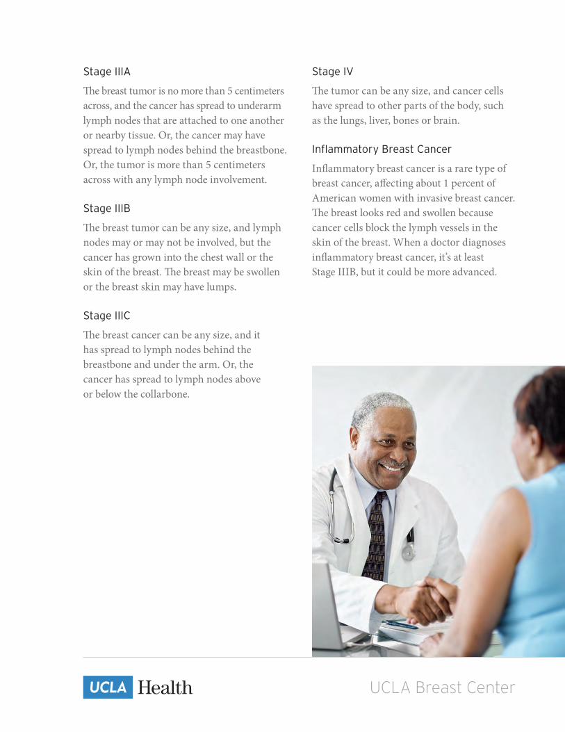
Stage IIIA
The breast tumor is no more than 5 centimeters across, and the cancer has spread to underarm lymph nodes that are attached to one another or nearby tissue. Or, the cancer may have spread to lymph nodes behind the breastbone. Or, the tumor is more than 5 centimeters across with any lymph node involvement.
Stage IIIB
The breast tumor can be any size, and lymph nodes may or may not be involved, but the cancer has grown into the chest wall or the skin of the breast. The breast may be swollen or the breast skin may have lumps.
Stage IIIC
The breast cancer can be any size, and it has spread to lymph nodes behind the breastbone and under the arm. Or, the cancer has spread to lymph nodes above or below the collarbone.
Stage IV
The tumor can be any size, and cancer cells have spread to other parts of the body, such as the lungs, liver, bones or brain.
Inflammatory Breast Cancer
Inflammatory breast cancer is a rare type of breast cancer, affecting about 1 percent of American women with invasive breast cancer. The breast looks red and swollen because cancer cells block the lymph vessels in the skin of the breast. When a doctor diagnoses inflammatory breast cancer, it’s at least Stage IIIB, but it could be more advanced.

UCLA Breast Center
Lymph node biopsy
Surgeons use a method called sentinel lymph node biopsy to remove the lymph node most likely to have breast cancer cells. If cancer cells are not found in the sentinel node, the woman may be able to avoid further lymph node removal. Removing additional lymph nodes to check for cancer cells is called axillary dissection.
CT scan
An X-ray machine connected to a computer takes a series of detailed pictures of your chest, abdomen and/or pelvis. You may receive contrast material (used to highlight abnormal areas of the body) orally or intravenously. The pictures from a CT scan can show cancer that has spread to the lungs or liver.
MRI
A strong magnet linked to a computer is used to create detailed pictures of your breasts. Sometimes, contrast material is used to make abnormal areas show up more clearly on the picture.
Bone scan
The doctor injects a small amount of a radioactive substance called tracers into a blood vessel. It travels through the bloodstream and collects in the bones. Higher amounts of the tracers collect in areas where there is cancer. A scanner is used to detect and measure radioactivity in the tracers. The scanner then creates images of the bones to determine if the cancer has spread there.
PET-CT scan
You will receive a small injection of radioactive sugar that will circulate throughout your body and accumulate in cancerous areas that consume more sugar than normal cells. The radioactive sugar emits signals that are detected by the PET scanner and converted into images of your body. These pictures are then analyzed to determine if cancer is present in other parts of the body.
Additional tests
Additional testing may be necessary to determine if cancer cells have spread to other parts of the body. Not all tests are indicated for every woman with breast cancer. Your doctors will inform you if any of the following tests are necessary. After learning that you have breast cancer, you may need other tests to help choose the best treatment for you.

UCLA Breast Center
5
“ The physicians are so informed and create a very
real connection with you over the details of your care. It seems as if
everyone is striving toward the same goal: to take the best care of you,
with attention to all the details.”


UCLA Breast Center
In cancer care, different specialty doctors often work to create an overall treatment plan that combines different types of treatments. At the UCLA Breast Center in Santa Monica, our team of board-certified specialists in breast surgery, reconstructive plastic surgery, medical oncology and radiation oncology will meet with you at your first appointment.
Our specialty team works in collaboration with our pathology and diagnostic radiology experts who have specialty training in women’s imaging to develop a treatment plan that is designed specifically for you.
The treatment options and recommendations the team gives you will depend on several factors, including the stage and grade of your tumor, whether the cancer has spread, your menopausal state, your age and overall health, the tumors’ hormone receptor (ER, PR) and HER2 status, and the presence of known mutations in the inherited breast cancer genes (BRCA1 or BRCA2).
When making treatment decisions, you are also encouraged to consider participating in a clinical trial. A clinical trial is a research study that tests whether a new treatment is safe, effective and better than the standard treatment.
Before treatment begins, it is important to discuss the goals and possible side effects of treatment with your team, including the likelihood that the treatment will be successful and its potential effects on your quality of life.
Breast cancer treatment options
A few questions to ask our specialists
• What are my treatment options?• Will I need more than one type of treatment?• What treatment plan do you recommend for me? Why?• What is the goal of the treatment(s) you are recommending? Is it to eliminate the cancer?
To relieve my symptoms? Or both?• What is the expected timeline for my treatment plan?• When do I need to make a decision about starting treatment?

UCLA Breast Center
Surgery Surgery to remove the tumor from the breast and/or evaluate the lymph nodes for cancer is often one of the first treatments for someone diagnosed with breast cancer. Our breast surgeon performs this procedure. The goal of breast cancer surgery is to remove the tumor along with a small area of healthy tissue around it, known as a margin. Generally, the smaller the tumor, the more surgical options a person has. These options include:
Lumpectomy: This surgery removes the tumor as well as a small, clear (cancer-free) margin of healthy tissue around it. Most of the breast remains. For both DCIS and invasive cancer, follow-up radiation therapy to the remaining breast tissue is typically recommended. A lumpectomy may also be called breast-conserving surgery, a partial mastectomy or segmental mastectomy.
Mastectomy: This surgery removes the entire breast and may or may not be combined with reconstructive (plastic) surgery.• In some cases, a skin-sparing or skin- and nipple-sparing mastectomy may be an option.
For this approach, the surgeon removes as little skin as possible and may preserve the nipple.• In total (simple) mastectomy, the surgeon removes the whole breast but not the underarm
lymph nodes.• In modified radical mastectomy, the surgeon removes the whole breast and most or all of
the lymph nodes under the arm. Often, the lining over the chest muscles is removed. A small chest muscle may also be taken out to make it easier to remove the lymph nodes.
The choice between breast-conserving surgery and mastectomy depends on
many factors:
• The size, location and stage of the tumor• The size of your breast• How you feel about how surgery will change your breast• How you feel about radiation therapy• Your ability to travel to a radiation treatment center for daily treatment sessions

UCLA Breast Center
Surgery is also used to evaluate nearby lymph nodes for cancer cells. This helps the doctor figure out the most appropriate treatment. Lymph nodes serve as collecting stations for lymph, a clear fluid that flows throughout the body. As lymph drains out of the breast and into nearby lymph nodes, it can transport cancer cells that may have detached from the original tumor.
The type of procedure you have will depend on a variety of factors, including the type of breast cancer and whether there is evidence of cancer in the lymph nodes before surgery. The options are sentinel lymph node biopsy and axillary lymph node dissection.
Sentinel lymph node biopsy
During a sentinel lymph node biopsy, the surgeon finds and removes the sentinel lymph nodes, the first group of nodes that receive drainage from the breast. The pathologist then examines them for cancer cells. To find the sentinel lymph nodes, the surgeon injects a dye and/or a radioactive tracer into the area of the cancer and/or around the nipple. The dye travels to the lymph nodes, arriving at the sentinel nodes first. The surgeon locates these nodes once they change color. If the sentinel lymph nodes are cancer-free, research has shown that there is a good possibility that the remaining lymph nodes will also be free of cancer and no further surgery will be needed. If the sentinel lymph nodes show evidence of cancer, then the surgeon may perform an axillary lymph node dissection to remove more lymph nodes and look for additional cancer cells.
Axillary lymph node dissection
The surgeon’s decision to perform an axillary lymph node dissection depends on the type of breast surgery planned (lumpectomy versus mastectomy), the stage of the cancer and the number of cancer cells found in the sentinel lymph nodes. During an axillary lymph node dissection, the surgeon removes lymph nodes from under the arm, which are then examined by a pathologist for cancer cells.
After surgery (lumpectomy or mastectomy) to treat breast cancer, the breast may be scarred and may be a different shape or size than before surgery. The area around the surgical site may also be harder. If any lymph nodes were removed as part of the surgery, or were affected during treatment, lymphedema (swelling of the hand and/or arm) may occur; this is a lifelong risk.
Lymphedema develops when a blockage in the lymphatic system causes fluid to build up in the arm. Lymphedema can be acute (lasting no more than six months) or chronic (lasting years). Talk with your team about ways to reduce your risk of lymphedema, symptoms you may experience and ways to manage this condition.

UCLA Breast Center
Questions you may want to ask your doctor about surgery
• Am I a candidate for a lumpectomy? Why or why not?• If I have a lumpectomy, will my treated breast differ in size and shape from my
other breast?• If I have a lumpectomy, will I be able to breastfeed if I have a child in the future?• Do I need to have a mastectomy?• Do the lymph nodes under my arm need to be removed? Do you recommend a sentinel
lymph node biopsy? Do you recommend an axillary node biopsy?• Will I need to be admitted to the hospital for the operation? If so, how long will I need
to stay in the hospital?• Am I at risk for developing lymphedema? If so, how can we reduce this risk?• Where will the scar be and what will it look like?• Will my arm be affected by surgery? If so, for how long? Will I need physical therapy?

UCLA Breast Center
Breast reconstruction Breast reconstruction is surgery to recreate the shape of a breast after mastectomy. It is an option for most women who have had their breasts removed because of cancer. Breast reconstruction may help a woman feel better about her appearance. However, it is important to note that the reconstructed breast will look and feel somewhat different than a natural breast.
Immediate and delayed reconstruction
Breast reconstruction may be done at the same time as mastectomy (called immediate reconstruction) or weeks to years later (delayed reconstruction). In either case, it is important to discuss options with your breast surgeon early on, before your mastectomy. The choices you make may influence where incisions are made and how much skin is saved.
Types of breast reconstruction
There are two basic types of breast reconstruction, implant reconstruction and tissue reconstruction. Our plastic surgeon will explain which option is best for your age, overall health, body type, lifestyle, treatment history and personal goals.
Reconstruction with implants
With implant reconstruction, a two-step procedure is usually required to stretch the breast skin and chest muscle. The first step involves placing a temporary tissue expander under the chest muscle. Over the next few weeks to months, the tissue expander is gradually filled with a saline (salt-water) solution until the tissue has been adequately stretched. Next, the expander is replaced by an implant (saline or silicone gel). If desired, nipple and areola reconstruction requires additional procedures.
Possible problems
In addition to risks associated with all surgery, the most common long-term problems with implant reconstruction are rupture (breakage of the implant cover) and capsular contracture (scar tissue forming around the implant). It should also be noted that breast implants do not last forever. One or more replacement surgeries may be needed.

UCLA Breast Center
Reconstruction with your own tissue
Tissue reconstruction uses a woman’s own tissue to rebuild the shape of a breast. The tissue may be taken from the back, abdomen, buttocks or more rarely, the thighs. With tissue reconstruction, a flap of skin, fat and muscle is moved to the chest where it is formed into the shape of a breast. The two most common methods are called TRAM flap (transverse rectus abdominis flap) and LAT flap (latissimus dorsi flap). The TRAM flap uses tissue from the lower abdomen; the LAT flap uses tissue from the upper back. Some situations may also require the use of an implant. Tissue reconstruction is not usually recommended for women who smoke or have diabetes, vascular or connective tissue diseases.
Possible problems
Tissue reconstruction is a major operation. Large surgical wounds, considerable discomfort, swelling and bruising after surgery are common. Decreased strength in the area of the body from which the tissue was taken is also common. Complications such as excessive bleeding, excessive scar tissue, fluid collection and problems with healing, including flap failure, are not typical but are possible. The chance that the cosmetic result will not be as pleasing as expected is a possible problem with any breast reconstruction.
Nipple and areola reconstruction
Reconstruction of the nipple and areola (the small, darker area around the nipple) is an option with either implant or tissue reconstruction. It is usually performed on an outpatient basis, under local anesthesia and after the reconstructed breast has had time to heal (about two to four months). A variety of techniques may be used to create the new nipple and areola. Tattooing is often used for matching the areola to a woman’s natural color.

UCLA Breast Center
Questions you may want to ask your doctor about plastic surgery
• What type of reconstruction surgery do you recommend? Why?• Will this surgery interfere with chemotherapy or radiation therapy?• Will the type of surgery impact what sort of breast imaging I can have in the future?• What results can I expect?• Do you have photographs of reconstructed breasts I can see?• How will my reconstructed breast feel? Will it match my other breast in size and shape?• What type of sensation (feeling) will the reconstructed breast have?• What if I become pregnant in the future?• Is a prosthesis a better option for me?• How can I get fitted for a breast prosthesis?• Will my insurance cover this?• Where will the scar be and what will it look like?• Will my arm be affected by surgery? If so, for how long? Will I need physical therapy?

UCLA Breast Center
Radiation therapy Radiation therapy uses high-energy X-rays or other particles to destroy cancer cells. It only affects cells in the part of the body that is treated. The most common type of radiation treatment is called external-beam radiation therapy, which uses machines called linear accelerators to generate beams of radiation outside the body and deliver them to cancerous areas of the body. You will not see or feel the radiation as it is being delivered. A doctor who specializes in giving radiation therapy to treat cancer is called a radiation oncologist.
Radiation therapy is most often administered after a lumpectomy to help lower the risk of recurrence in the breast. By combining modern surgery and radiation therapy, recurrence rates in the breast may now be less than 5 percent in the 10 years following treatment, and survival is often the same as a mastectomy.
The radiation oncologist will discuss personalized treatment options in detail. Radiation therapy for breast cancer can be delivered to the tumor cavity (area where the breast tumor was removed), to the whole breast following breast conservation surgery or to the chest wall/reconstructed breast following mastectomy. Depending on the extent of your particular cancer, you may also receive radiation therapy to the surrounding lymph node regions.
The radiation is delivered on a daily basis, Monday through Friday. The entire radiation course is typically six to seven weeks long, but it may be as short as two to three weeks depending on your particular breast cancer. The typical daily treatment sessions last approximately 10 – 15 minutes.
Side effects are typically limited to gradual changes in skin coloration much like a sunburn reaction, and possibly fatigue. There may be some long-term cosmetic effects of radiation; however, risks to normal organs such as the heart and lung are very low with modern radiation treatment planning.

UCLA Breast Center
Questions you may want to ask your doctor about radiation therapy• Which type of radiation therapy can I consider? Are there options for me? • When will treatment start? When will it end? How often will I have treatment?• How will I feel during treatment? • Will I need to stay in the hospital? • Will I be able to drive myself to and from the appointments? • What can I do to take care of myself before, during and after treatment?• How will we know the treatment is working? • Will radiation therapy harm my skin?• How will my chest look afterward?• Are there any lasting effects?• What is the chance that the cancer will come back in my breast?

UCLA Breast Center
Chemotherapy
Adjuvant and neoadjuvant therapy
Depending on how it is used, your doctor may refer to chemotherapy as either adjuvant or neoadjuvant therapy.
Adjuvant therapy
Adjuvant therapy describes chemotherapy that is used after surgery to lower the risk of breast cancer recurrence (in the breast and throughout the body). Even when all of the cancer appears to be gone, doctors will sometimes recommend chemotherapy as an added measure of safety in case some cancer cells have escaped into the bloodstream. Over time, these cells could spread cancer to other places in the body. Chemotherapy helps to lower this risk.
Neoadjuvant therapy
Neoadjuvant therapy is used before surgery to shrink cancer. Shrinking the size of a cancer gives some women with larger cancers the option of choosing breast-conserving surgery over mastectomy. Using chemotherapy before surgery also gives doctors and patients a chance to see how well a certain drug or combination of drugs is going to work in an individual woman’s case. In some instances, chemotherapy may be the main treatment (instead of surgery) for women diagnosed with advanced breast cancer.
Possible problems
Prior to initiating chemotherapy, patients will spend time with their oncologist (or oncology nurse) reviewing the potential risks, side effects and benefits of therapy. The side effects of chemotherapy vary depending on the drugs used, the dosages, the overall length of treatment and the individual woman. The most common side effects are weakness and fatigue, nausea and vomiting, loss of appetite, weight changes, nail changes and hair loss. (The hair usually grows back after treatment. The use of a scalp-cooling devise during chemotherapy infusion may limit hair loss and can be discussed with your oncologist.) Mouth sores, diarrhea or constipation are also fairly common. Ask your doctor about medicines and methods that can be used for managing these and other possible side effects. Infections are more likely during treatment so patients should take special care to avoid situations that increase this risk. Short- or longer-term changes in thinking and memory are also possible. Certain chemotherapy drugs can cause lasting damage to the heart, lungs, liver and kidneys. In younger women, chemotherapy can cause infertility or premature menopause. Women planning to become pregnant (or able to become pregnant but not taking birth control) should talk with their doctors before starting treatment.

UCLA Breast Center
Anti-hormonal therapy
Anti-hormonal therapy is another form of systemic therapy. It combats breast cancer by blocking the action or lowering the amount of specific hormones in the body. It is used for women whose breast cancers rely on hormones to grow (these are called hormone receptor-positive breast cancers). A hormone-receptor test will tell you and your doctor if your breast cancer is hormone receptor-positive. About two-thirds of breast cancers are hormone receptor-positive.
Types of anti-hormonal therapy
There are different types of anti-hormonal therapy. Most are pill-based and are given for five to 10 years after surgery. Some block the effect of hormones while others lower the amount of hormones in the body. The most effective treatment may involve using more than one type over the course of several years. Your doctor will work with you to determine the most appropriate plan for you.
Tamoxifen, for example, works to stop or slow cancer by blocking the effect of estrogen hormones on cancer cells. It has been the standard drug for treating women with hormone receptor-positive breast cancer for more than 30 years. Taken after surgery for invasive cancers, tamoxifen lowers the risk of cancer recurrence by about half and improves the chances for long-term survival.
Tamoxifen also helps women whose cancer has spread and women who have no personal history of breast cancer but whose risk for developing breast cancer is higher than average.
Aromatase inhibitors
Aromatase inhibitors are newer drugs that work to lower the amount of hormones in the body. Examples are anastrozole (Arimidex), letrozole (Femara) and exemestane (Aromasin). Used either alone or after a course of tamoxifen, these drugs have been found to work as well or better than tamoxifen alone for reducing the risk of cancer recurrence.
Unlike tamoxifen, which can be used for women who are either premenopausal (still having menstrual periods) or postmenopausal (no longer having menstrual periods), aromatase inhibitors can only be used for women without ovarian function (e.g., women who have gone into menopause or have had their ovarian function suppressed by drugs such as leuprolide or goserelin).
Possible problems
Anti-hormonal therapy can cause similar side effects to those of menopause (hot flashes, weight gain, vaginal dryness, headaches, mood swings, hair thinning, etc.). Rare but serious side effects of tamoxifen include increased risk for cancers of the uterus, blood clots, stroke, vision problems such as cataracts, liver toxicities and fertility issues. Aromatase inhibitors have less serious side effects than tamoxifen. Possible problems include upset stomach, an increase in cholesterol, joint stiffness or pain, and potential loss of bone strength. Aromatase inhibitors do not increase risk for uterine cancers and very rarely cause blood clots.

UCLA Breast Center
Targeted therapy
Targeted therapy is a newer systemic therapy option. Women with breast cancers that contain too much of the HER2 protein (called HER2-positive breast cancer) are often helped by a drug called trastuzumab (more commonly known as Herceptin) and, in some cases, pertuzumab (also known as Perjeta). About one in five women with breast cancer have HER2-positive cancer. Used with chemotherapy, trastuzumab (alone or with pertuzumab) can lower the risk of cancer recurrence after surgery. It can also shrink or slow the growth of HER2-positive breast cancer that has spread. Lapatinib, another targeted therapy drug, is used for treating HER2-positive breast cancer in women with metastatic cancer that no longer responds to trastuzumab. Other targeted drugs are being studied in clinical trials.
Possible problems
Flu-like symptoms, such as fever, chills and nausea, are common with trastuzumab, especially after the first dose. Less commonly, it can cause mild to severe heart damage. In combination with chemotherapy, trastuzumab may increase risk for other side effects as well, such as anemia and/or infection. Rarely, it can cause severe or life-threatening breathing problems and/or allergic reactions.
Questions you may want to ask your doctor about chemotherapy• Which type of chemotherapy do you recommend? Why?• When do you recommend I have chemotherapy — before surgery or after?• How long will I need to receive chemotherapy?• How will the treatment be given? Do I need a port?• How will we know if the treatment is working?• How will chemotherapy affect my daily life? Will I be able to work, exercise
and perform my usual activities?• Can I stay alone after my treatments or do I need someone to stay with me?• What are the potential short- and long-term side effects of each medication?
Will I lose my hair?• Where can I get more information about the medication(s) I will be taking?• If I am worried about the cost of treatment, who can help me with this concern?

UCLA Breast Center
Clinical trialsDoctors are always looking for better ways to treat people with breast cancer. To make scientific advances, doctors conduct clinical trials, or research studies, involving volunteers.
Many clinical trials are focused on evaluating whether a new treatment is safe, effective and better than the current (standard) treatment. These types of studies evaluate new drugs, different combinations of existing treatments, new approaches to radiation therapy or surgery, and new methods of treatment.
Those who participate in clinical trials are often among the first to receive new treatments, before they are widely available. However, there is no guarantee that a new treatment will be safe, effective or better than the standard treatment.
People decide to participate in clinical trials for many reasons. For some people with breast cancer, a clinical trial is the best treatment option available. Because standard treatments may not be optimal for some patients, they are often willing to face the added uncertainty of a clinical trial in the hope of a better result. Other people volunteer for clinical trials because they know these studies are the only way to make progress in treating breast cancer. Even if they will not benefit directly from the clinical trial, their participation may help other people with breast cancer in the future.
If you decide to participate in a clinical trial, you will participate in a process called informed consent. During informed consent, the doctor should list all of your options and help you understand how the new treatment is different from the standard treatment. The doctor must also list all of the risks of the new treatment, which may or may not be different from the risks of the standard treatment. Finally, the doctor must explain what will be required of each patient in order to participate in the clinical trial, including the number of doctor visits, tests, the treatment schedule, and the associated costs.
Keep in mind, even if you decide to participate in a clinical trial, you may stop participating at any time for any personal or medical reason.

UCLA Breast Center
6
“ You have the best service so I really can’t think
of anything else you could do better. Thanks to the whole staff for
making me feel very comfortable throughout my ordeal.”


UCLA Breast Center
Managing symptoms and side effectsFearing the side effects of breast cancer treatment is normal, but it may help to know that preventing and controlling side effects is a major focus of your healthcare team. Before starting treatment, talk with your team about which side effects are most likely to happen. Then, once treatment begins, let your healthcare team know what side effects you are experiencing so they can help manage them.
Everyone’s experience with breast cancer treatment is difficult. The specific side effects that may occur during and after treatment depend on a number of factors, including the cancer’s location, your treatment plan and your overall health. However, some of the potential physical, emotional and social effects experienced by people receiving treatment for breast cancer are described in this section.
Physical effects
Fatigue
Cancer and its treatment often cause a persistent sense of tiredness or exhaustion. Most people receiving cancer treatment experience some type of fatigue, which can make even a small effort, such as walking across a room, seem overwhelming. Fatigue often seriously affects people’s daily activities, including the ability to be involved with their family or to socialize. It is important to tell your doctor if you are experiencing fatigue because there are medical and lifestyle interventions your healthcare team can recommend to help.
Pain
Pain can be caused by the tumor, the cancer treatment or from causes not related to the cancer. Untreated pain can make other aspects of cancer seem worse, such as fatigue, weakness, nausea, constipation, sleep disturbance, depression, anxiety and mental confusion. Your doctor can help you find an effective pain-relief strategy.
Lymphedema
Lymphedema is the abnormal buildup of fluid in the arm or leg caused by a blockage in the lymphatic system. It can happen immediately after surgery or radiation therapy, or months or years after cancer treatment has ended. In some cases, the swelling goes away on its own as the body heals and normal lymph fluid flow resumes. However, lymphedema may become chronic when the lymphatic system changes and can no longer meet the body’s demand for fluid drainage. There is no cure for chronic lymphedema; however, there are ways to treat it.

UCLA Breast Center
Infertility
Some breast cancer treatments may cause temporary or permanent infertility (inability to become pregnant or have children). If this is a concern for you, please talk with your healthcare team about any fertility-related side effects of your treatment plan and the options for preserving your fertility before treatment begins.
Psychosocial effects
We feel it is important to address the needs of the whole person, not just his or her medical needs. Providing a clinical social worker or psychologist who specializes in breast cancer is an important component of the multidisciplinary care our team provides to women and families dealing with the emotional roller coaster of a cancer diagnosis. Having an expert validate and normalize patients’ concerns and provide guidance on coping with cancer is tremendously helpful. We want to make sure our patients have all the support and resources they need to move forward with their treatment. The clinicians from Simms/Mann — UCLA Center for Integrative Oncology also provide information about support services available at the Center, such as nutritional consultations, support groups and individual counseling.

UCLA Breast Center
7
“ I have received the very best care and courtesy —
absolutely outstanding care! Everyone has treated me so well. I’m thankful
to the entire staff; my experience with them couldn’t have been better!”


UCLA Breast Center
Follow-up careFollow-up care will be determined by the type of treatment you received/will be receiving and your personal preferences. Follow-up care may be provided by your breast surgeon, radiation oncologist, medical oncologist, plastic surgeon and/or your primary-care doctor. It is important to keep a regular check-up schedule and share details about your status with your treating doctors, even years after treatment is completed. The recommended follow-up schedule for patients with breast cancer is the following:
If you have had breast-conserving surgery, you should have mammograms once a year, with the first mammogram typically scheduled one year from the most recent mammogram that preceded your diagnosis. Regardless of the type of surgery, you should have a clinical breast exam at least every six months for five years, then yearly. Any additional imaging recommendations, including a breast MRI and/or breast ultrasound, will be discussed with you if necessary.
The purpose of follow-up care is to monitor and manage any late, long-term effects of treatment and to check for any signs that the cancer may have returned.
During these visits, your healthcare provider should conduct a comprehensive clinical breast exam to inspect your lymph nodes and look for any changes in normal breast appearance.
Your doctor will also ask about any symptoms you may be experiencing. Imaging tests may be ordered. Women taking tamoxifen should have yearly pelvic exams. Patients being treated with an aromatase inhibitor should have a bone density test before, during and after treatment, as recommended by their doctors.

UCLA Breast Center
Monthly breast self-examination
You may also choose to conduct a monthly breast self-examination. Your primary-care physician can show you the proper method to check for breast changes. The goal is to immediately report any of the following so that possible problems can be diagnosed and treated as soon as possible:
• A new lump in the breast or chest area• A new lump in the armpit or neck• A change in the shape of the breast• A skin rash, swelling or change in the color of the skin over the breast or chest
Women recovering from breast cancer
Women recovering from breast cancer are also encouraged to follow established guidelines for good health, such as maintaining a healthy weight, not smoking, minimizing alcohol intake, eating a balanced diet and receiving recommended cancer screening tests. Talk with your team to develop a plan that is best for your needs. Moderate physical activity can help rebuild your strength and energy level and may lower the risk of cancer recurrence. Your team can help you create a safe exercise plan based upon your needs, physical abilities and fitness level.
Questions to ask your team about follow-up care• How often should I see a doctor for follow-up care?• Whom will I see for my follow-up visits?• What follow-up tests will I need and how often will I need them?• What is the chance that the cancer will return?• Is there anything I can do to reduce the risk of recurrence?• What are the most common long-term and late effects associated with
the treatment I received?

UCLA Breast Center 8
“ At a time when even the most basic or simple decisions
can seem monumental, the staff at the UCLA Breast Center Santa Monica provides
unparalleled courtesy, professionalism and care. They help navigate the new
territory and lift some of the burden. They are awesome!”


UCLA Breast Center
Breast cancer dictionaryAromatase inhibitor (uh-ROH-muh-tayz in-HIH-bih-ter)A drug that prevents the formation of estradiol, a female hormone, by interfering with an aromatase enzyme. Aromatase inhibitors are used as a type of hormone therapy for postmenopausal women who have a hormone-dependent breast cancer.
Axilla (ak-SIL-a) The underarm or armpit.
Axillary dissection (AK-sih-LAYR-ee dy-SEK-shun) Surgery to remove lymph nodes found in the armpit region. Also called axillary lymph node dissection.
Axillary lymph node (AK-sih-LAYR-ee limf)A lymph node in the armpit region that drains lymph from the breast and nearby areas.
Benign (beh-NINE)Not cancer. Benign tumors may grow larger but do not spread to other parts of the body.
Biopsy (BY-op-see) The removal of cells or tissues for examination by a pathologist. The pathologist may study the tissue under a microscope or perform other tests on the cells or tissue.
Blood vesselA tube through which the blood circulates in the body. Blood vessels include a network of arteries, arterioles, capillaries, venules and veins.
Breast-conserving surgery An operation to remove the breast cancer but not the breast itself. Types of breast-sparing surgery include lumpectomy (removal of the lump), quadrantectomy (removal of one quarter, or quadrant, of the breast) and segmental mastectomy (removal of the cancer as well as some of the breast tissue around the tumor and the lining over the chest muscles below the tumor).
Cancer (KAN-ser)A term for diseases in which abnormal cells divide uncontrollably and can invade nearby tissues. Cancer cells can also spread to other parts of the body through the blood and lymphatic systems.
Carcinoma in situ (KAR-sih-NOH-muh in SY-too)A group of abnormal cells that remain where they first formed. They have not spread. These abnormal cells may become cancer and spread into nearby normal tissue. Also called stage 0 disease.
CellThe individual unit that makes up the tissues of the body. All living things are made up of one or more cells.
Chemotherapy (KEE-moh-THAYR-uh-pee)Treatment with drugs that kill cancer cells.
Clinical trialA type of research study that tests the efficacy of new medical approaches. These studies test new methods of screening, prevention, diagnosis or treatment of a disease.
CT scanA series of detailed pictures of areas inside the body taken from different angles. The pictures are created by a computer linked to an X-ray machine. Also called a CAT scan, computed tomography scan, computerized axial tomography scan and computerized tomography.
Duct (dukt)In medicine, a tube or vessel of the body through which fluids pass.
Ductal carcinoma (DUK-tul KAR-sih-NOH-muh)The most common type of breast cancer. It begins in the cells that line the breast’s milk ducts.

UCLA Breast Center
Ductal carcinoma in situ (DCIS)(DUK-tal KAR-sih-NOH-muh in SYE-too) A noninvasive condition in which abnormal cells are found in the lining of a breast duct. The abnormal cells have not spread outside the duct to other tissues in the breast. In some cases, ductal carcinoma in situ may become invasive cancer and spread to other tissues, although it is not known at this time how to predict which lesions will become invasive. Also called intraductal carcinoma.
Early-stage breast cancerBreast cancer that has not spread beyond the breast or the axillary lymph nodes. This includes ductal carcinoma in situ and stage I, stage IIA, stage IIB and stage IIIA breast cancers.
Estrogen (ES-truh-jin) A type of hormone produced by the body that helps develop and maintain female sex characteristics and the growth of long bones. Estrogen can also be made in the laboratory. It may be used as a type of birth control and as a treatment for the symptoms of menopause, menstrual disorders, osteoporosis and other conditions.
External radiation therapy (RAY-dee-AY-shun THAYR-uh-pee) A type of radiation therapy that uses a machine to aim high-energy rays at the cancer from outside of the body. Also called external-beam radiation therapy.
Fibrous Containing or resembling fibers.
Gland An organ that makes one or more substances, such as hormones, digestive juices, sweat, tears, saliva or milk.
HER2 A protein involved in normal cell growth. It is found on some types of cancer cells, including breast and ovarian. Cancer cells removed from the body may be tested for the presence of HER2/neu to help decide the best type of treatment. Also called c-erbB-2, human EGF receptor 2 and human epidermal growth factor receptor 2.
Hormone receptor (HOR-mone reh-SEP-ter)A cell protein that binds a specific hormone. The hormone receptor may be on the surface of the cell or inside the cell. Many changes take place in a cell after a hormone binds to its receptor.
Hormone therapy (HOR-mone THAYR-uh-pee) Treatment that adds, blocks or removes hormones. For certain conditions (such as diabetes or menopause), hormones are given to adjust low hormone levels. To slow or stop the growth of certain cancers (such as prostate and breast cancer), synthetic hormones or other drugs may be given to block the body’s natural hormones. Sometimes surgery is needed to remove the gland that produces a certain hormone. Also called endocrine therapy, hormonal therapy and hormone treatment.
Inflammatory breast cancer (in-FLA-muh-TOR-ee) A type of breast cancer in which the breast looks red and swollen and feels warm. The skin of the breast may also show the pitted appearance called peau d’orange (like the skin of an orange). The redness and warmth occur because the cancer cells block the lymph vessels in the skin.
Intravenous (IN-truh-VEE-nus) Into or within a vein. Intravenous usually refers to a way of giving a drug (or other substance) through a needle or tube inserted into a vein. Abbreviated as IV.
Lobe A portion of an organ, such as the liver, lung, breast, thyroid or brain.
Lobular carcinoma (LAH-byuh-ler KAR-sih-NOH-muh) Cancer that begins in the lobules (the glands that make milk) of the breast. Lobular carcinoma in situ (LCIS) is a condition in which abnormal cells are only found in the lobules. When cancer has spread from the lobules to surrounding tissues, it is invasive lobular carcinoma. LCIS does not become invasive lobular carcinoma very often, but having LCIS in one breast increases the risk of developing invasive cancer in either breast.

UCLA Breast Center
Lobule (LOB-yule) A small lobe or a subdivision of a lobe.
Lumpectomy (lum-PEK-toh-mee) Surgery to remove abnormal tissue or cancer from the breast and a small amount of normal tissue around it. It is a type of breast-conserving surgery.
Lymph node (limf)A rounded mass of lymphatic tissue that is surrounded by a capsule of connective tissue. Lymph nodes filter lymph (lymphatic fluid) and store lymphocytes (white blood cells). They are located along lymphatic vessels. Also called a lymph gland.
Lymph vessel (limf)A thin tube that carries lymph (lymphatic fluid) and white blood cells through the lymphatic system. Also called a lymphatic vessel.
Lymphedema (LIM-fuh-DEE-muh) A condition in which excess fluid collects in surrounding tissues and causes swelling. It may occur in the arm or leg after lymph vessels or lymph nodes in the underarm or groin are removed or treated with radiation.
Magnetic resonance imaging (MRI)A procedure in which radio waves and a powerful magnet linked to a computer are used to create detailed pictures of areas inside the body. These pictures can show the difference between normal and diseased tissue. MRI makes better images of organs and soft tissue than other scanning techniques, such as computed tomography (CT) or X-ray. MRI is especially useful for imaging the brain, the spine, the soft tissue of joints and the inside of bones.
Malignant (muh-LIG-nunt) Cancerous. Malignant tumors can invade and destroy nearby tissue and spread to other parts of the body.
Mammogram (MAM-o-gram) An X-ray of the breast.
Mastectomy (ma-STEK-toh-mee) Surgery to remove the breast (or as much of the breast tissue as possible).
Medical oncologist (MEH-dih-kul on-KAH-loh-jist) A doctor who specializes in diagnosing and treating cancer using chemotherapy, targeted therapy, hormonal therapy and biological therapy. A medical oncologist often is the main healthcare provider for someone who has cancer. A medical oncologist also gives supportive care and may coordinate treatment given by other specialists.
Menopause (MEH-nuh-PAWZ) The time of life when a woman’s ovaries stop producing estrogen and progesterone, and menstrual periods end. Natural menopause usually occurs around age 50. A woman is said to be in menopause when she has not had a period for 12 consecutive months. Symptoms of menopause include hot flashes, mood swings, night sweats, vaginal dryness, trouble concentrating, and infertility.
Menstrual period (MEN-stroo-al) The periodic discharge of blood and tissue from the uterus. From puberty until menopause, menstruation occurs about every 28 days, but does not occur during pregnancy.
Metastatic (meh-tuh-STA-tik)Having to do with metastasis, which is the spread of cancer from one part of the body to another.
Modified radical mastectomy (RA-dih-kul ma-STEK-toh-mee) Surgery for breast cancer in which the breast, most or all of the lymph nodes under the arm and the lining over the chest muscles are removed. Sometimes the surgeon also removes part of the chest wall muscles.
Oncology nurse (on-KAH-loh-jee)A nurse who specializes in treating and caring for people who have cancer.
OrganA part of the body that performs a specific vital function. For example, the heart is an organ.

UCLA Breast Center
Ovary (OH-vuh-ree)One of a pair of female reproductive glands in which the ova, or eggs, are formed. The ovaries are located in the pelvis, one on each side of the uterus.
Partial mastectomy (ma-STEK-toh-mee) The removal of cancer as well as some of the breast tissue around the tumor and the lining over the chest muscles below the tumor. Usually some of the lymph nodes under the arm are also taken out. Also called segmental mastectomy.
Physical therapistA health professional who teaches physical activities that help condition muscles and restore strength and movement.
Plastic surgery (SER-juh-ree)An operation that restores or improves the appearance of body structures.
Positron emission tomography scan (PET)A procedure in which a small amount of radioactive glucose (sugar) is injected into a vein, and a scanner is used to make detailed, computerized pictures of areas inside the body where the glucose is used. Because cancer cells often use more glucose than normal cells, the pictures can be used to find cancer cells in the body.
Progesterone (proh-JES-tuh-RONE)A type of hormone produced by the body that plays a role in the menstrual cycle and pregnancy. Progesterone can also be made in the laboratory. It may be used as a type of birth control and to treat menstrual disorders, infertility, symptoms of menopause and other conditions.
Radiation (RAY-dee-AY-shun)Energy released in the form of particle or electromagnetic waves. Common sources of radiation include radon gas, cosmic rays from outer space, medical X-rays and energy emitted by a radioisotope (unstable form of a chemical element that releases radiation as it breaks down and becomes more stable).
Radiation oncologist (RAY-dee-AY-shun on-KAH-loh-jist) A doctor who specializes in using radiation to treat cancer.
Radiation therapy (RAY-dee-AY-shun THAYR-uh-pee)The use of high-energy radiation from X-rays, gamma rays, neutrons, protons and other sources to kill cancer cells and shrink tumors. Radiation may come from a machine outside the body (external-beam radiation therapy) or it may come from radioactive material placed in the body near cancer cells (internal- radiation therapy). Systemic radiation therapy uses a radioactive substance, such as a radiolabeled monoclonal antibody, that travels in the blood to tissues throughout the body. Also called irradiation and radiotherapy.
Radioactive (RAY-dee-oh-AK-tiv)Emitting radiation.
Segmental mastectomy (seg-MEN-tul ma-STEK-toh-mee)The removal of cancer as well as some of the breast tissue around the tumor and the lining over the chest muscles below the tumor. Usually some of the lymph nodes under the arm are also taken out. Also called partial mastectomy.
Sentinel lymph node biopsyRemoval and examination of the sentinel node(s) (the first lymph node(s) to which cancer cells are likely to spread from a primary tumor). To identify the sentinel lymph node(s), the surgeon injects a radioactive substance and/or blue dye near the tumor. The surgeon then uses a scanner to find the sentinel lymph node(s) containing the radioactive substance, or looks for the lymph node(s) stained with dye. The surgeon then removes the sentinel node(s) to check for the presence of cancer cells.
Side effectA problem that occurs when treatment affects healthy tissues or organs. Some common side effects of cancer treatment are fatigue, pain, nausea, vomiting, decreased blood cell counts, hair loss and mouth sores.
Social workerA professional trained to talk with people and their families about emotional or physical needs. Also helps them find support services.

UCLA Breast Center
Surgery (SER-juh-ree)A procedure to remove or repair a part of the body, or to determine whether disease is present. An operation.
Tamoxifen (tuh-MOK-sih-FEN)A drug used to treat certain types of breast cancer in women and men. It is also used to prevent breast cancer in women who have had ductal carcinoma in situ (abnormal cells in the ducts of the breast) and in women who are at a high risk of developing breast cancer. It blocks the effects of the estrogen hormone in the breast.
Targeted therapy (TAR-geh-ted THAYR-uh-pee)A type of treatment that uses drugs or other substances, such as monoclonal antibodies, to identify and attack specific cancer cells. Targeted therapy may have fewer side effects than other types of cancer treatments.
Tissue (TISH-oo)A group or layer of cells that work together to perform a specific function.
Total mastectomy (ma-STEK-toh-mee)Removal of the breast. Also called simple mastectomy.
Tumor (TOO-mer)An abnormal mass of tissue that results when cells divide more than they should or do not die when they should. Tumors may be benign (not cancer) or malignant (cancer). Also called neoplasm.
X-rayA type of high-energy radiation. In low doses, X-rays are used to diagnose diseases by creating images of the inside of the body. In high doses, X-rays are used to treat cancer.