Embed Size (px)
Citation preview
UPPER GI CASESurgery 2
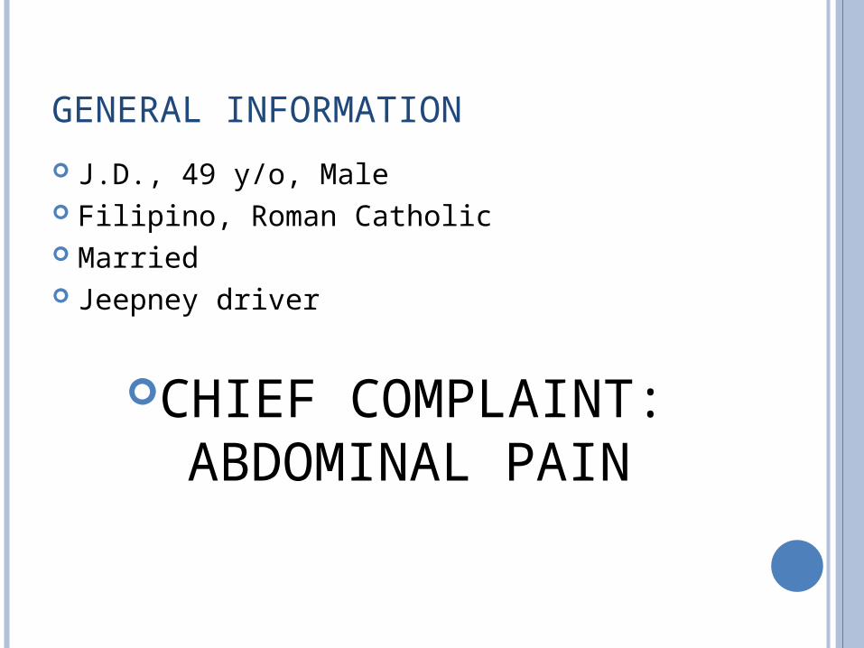
GENERAL INFORMATION
J.D., 49 y/o, Male Filipino, Roman Catholic Married Jeepney driver
CHIEF COMPLAINT: ABDOMINAL PAIN
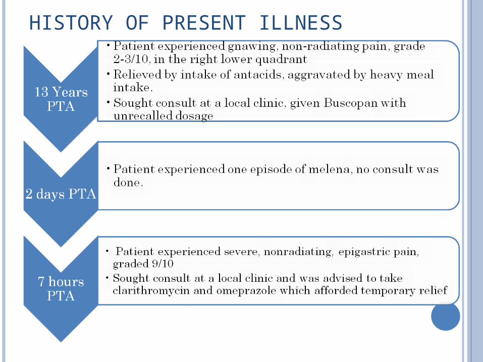
HISTORY OF PRESENT ILLNESS
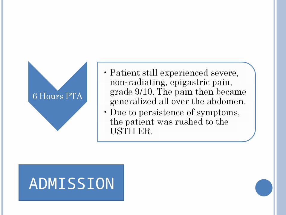
ADMISSION
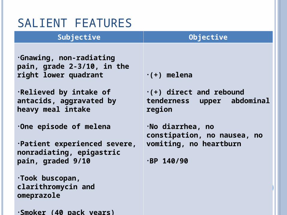
SALIENT FEATURESSubjective Objective
•Gnawing, non‐radiating pain, grade 2‐3/10, in the right lower quadrant
•Relieved by intake of antacids, aggravated by heavy meal intake
•One episode of melena
•Patient experienced severe, nonradiating, epigastric pain, graded 9/10
•Took buscopan, clarithromycin and omeprazole
•Smoker (40 pack years)
•(+) melena
•(+) direct and reboundtenderness upper abdominal region
•No diarrhea, no constipation, no nausea, no vomiting, no heartburn
•BP 140/90
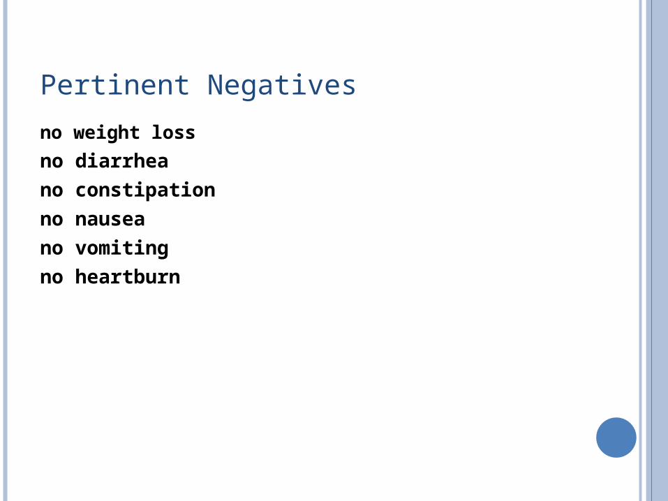
Pertinent Negatives
no weight loss
no diarrhea
no constipation
no nausea
no vomiting
no heartburn
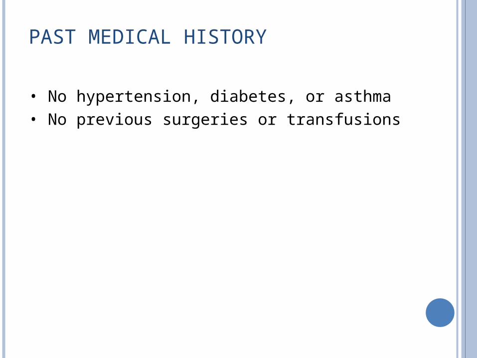
PAST MEDICAL HISTORY
• No hypertension, diabetes, or asthma• No previous surgeries or transfusions
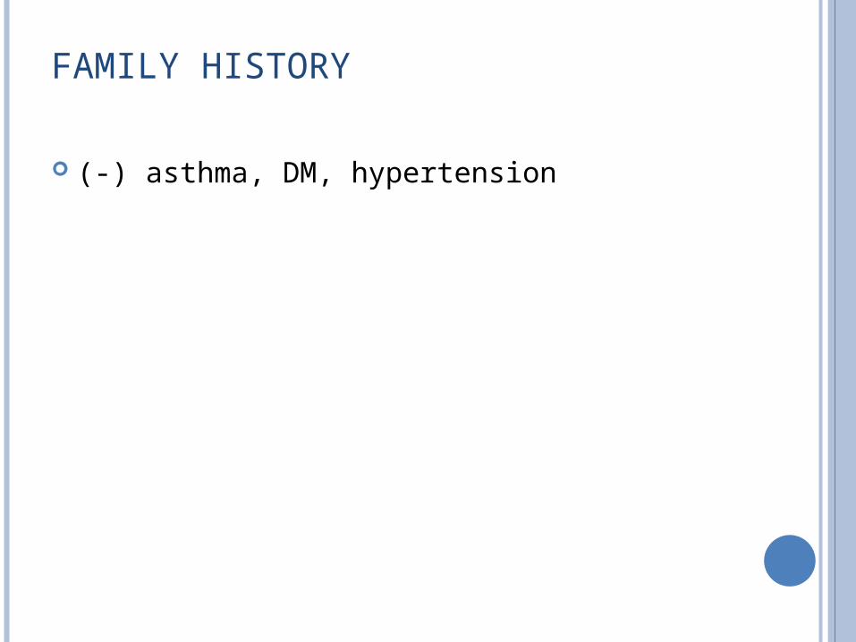
FAMILY HISTORY
(‐) asthma, DM, hypertension
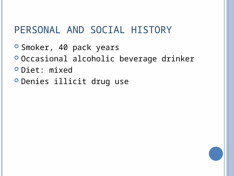
PERSONAL AND SOCIAL HISTORY
Smoker, 40 pack years Occasional alcoholic beverage drinker Diet: mixed Denies illicit drug use
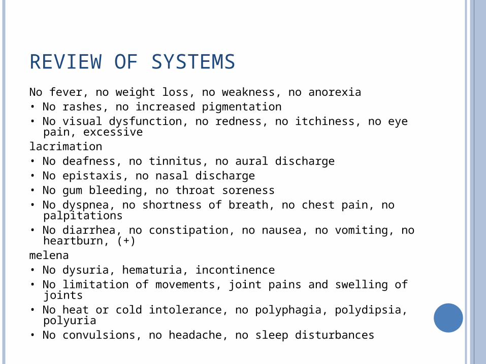
REVIEW OF SYSTEMSNo fever, no weight loss, no weakness, no anorexia• No rashes, no increased pigmentation• No visual dysfunction, no redness, no itchiness, no eye pain,
excessivelacrimation• No deafness, no tinnitus, no aural discharge• No epistaxis, no nasal discharge• No gum bleeding, no throat soreness• No dyspnea, no shortness of breath, no chest pain, no
palpitations• No diarrhea, no constipation, no nausea, no vomiting, no
heartburn, (+)melena• No dysuria, hematuria, incontinence• No limitation of movements, joint pains and swelling of joints• No heat or cold intolerance, no polyphagia, polydipsia, polyuria• No convulsions, no headache, no sleep disturbances
PHYSICAL EXAM
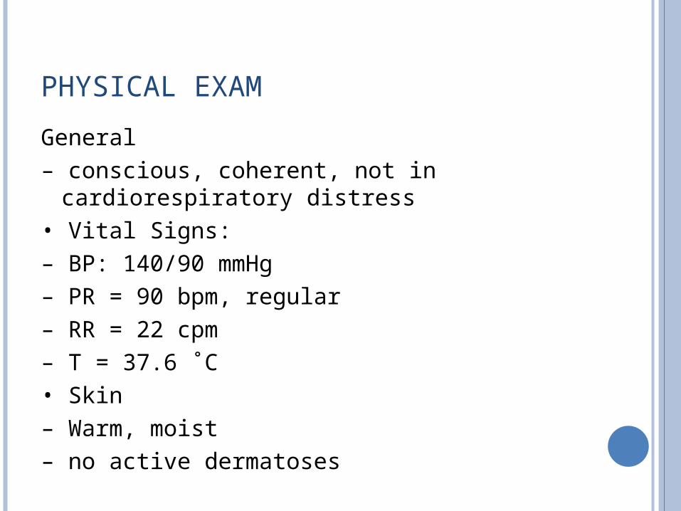
General– conscious, coherent, not in cardiorespiratory
distress• Vital Signs:– BP: 140/90 mmHg– PR = 90 bpm, regular– RR = 22 cpm– T = 37.6 ˚C• Skin– Warm, moist– no active dermatoses
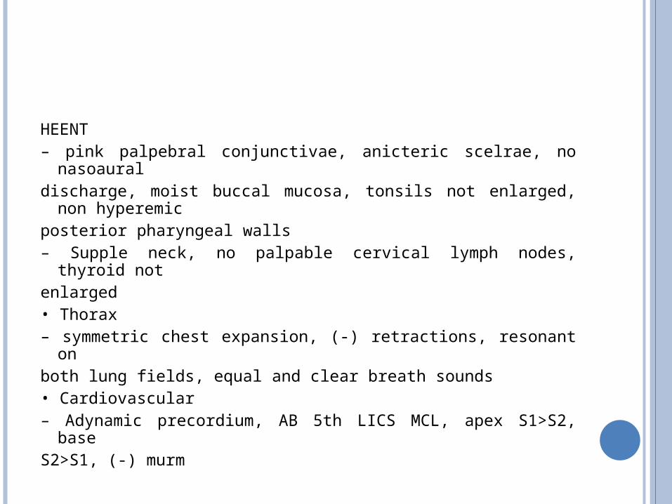
HEENT– pink palpebral conjunctivae, anicteric scelrae, no
nasoauraldischarge, moist buccal mucosa, tonsils not enlarged, non
hyperemicposterior pharyngeal walls– Supple neck, no palpable cervical lymph nodes, thyroid
notenlarged• Thorax– symmetric chest expansion, (‐) retractions, resonant onboth lung fields, equal and clear breath sounds• Cardiovascular– Adynamic precordium, AB 5th LICS MCL, apex S1>S2,
baseS2>S1, (‐) murm
Abdomen– Flat, no scars or striae, NABS, tympanitic upon
percussion, Traube’s space not obliterated, (+) direct and rebound tenderness upper abdominal region with guarding (‐) Rovsing’s sign, (‐) psoas sign
• DRE:– no skin tags seen, tight sphincteric tone, smooth
rectalmucosa, (‐) palpated masses, (‐) pararectal
tenderness,brown stool on tactating finger
Extremities– Pulses were full and equal, no cyanosis, no edema, nolimitation of movement in all extremities were noted.• Neurological Examination– Conscious, coherent, oriented to 3 spheres– Cranial nerves: pupils 2‐3 mm ERTL, EOMs full and
equal,V1V2V3 intact, can clench teeth, can raise eyebrows,
canclose eyes tightly, can smile, can frown, can puff cheeks,
nofacial asymmetry, no hearing loss, can turn head from
sideto side with resistance, can shrug shoulders, tonguemidline on protrusion
Neurologic Exam – Motor: MMT of 5/5 on all extremi4es – Cerebellar: can do FTNT & APST – DTR’s: ++ on all extremi4es – No sensory deficit – (‐) Babinski – (‐) nuchal rigidity
Assessment:
Acute Abdomen secondary to
perforated viscus secondary to PUD.
DIFFERENTIAL DIAGNOSES
The list of gastrointestinal and non-gastrointestinal disorders that can mimic ulceration of the stomach or duodenum is quite extensive.
Harrisons Principle of internal medicine, 17th ed.
Several additional disease processes that may present with “ulcerlike” symptoms include proximal gastrointestinal tumors, gastroesophagealreflux disease (GERD), vascular disease, pancreaticobiliary disease (biliary colic,chronic pancreatitis), and gastroduodenal Crohn’s disease.
Harrisons Principle of interal medicine, 17th ed.
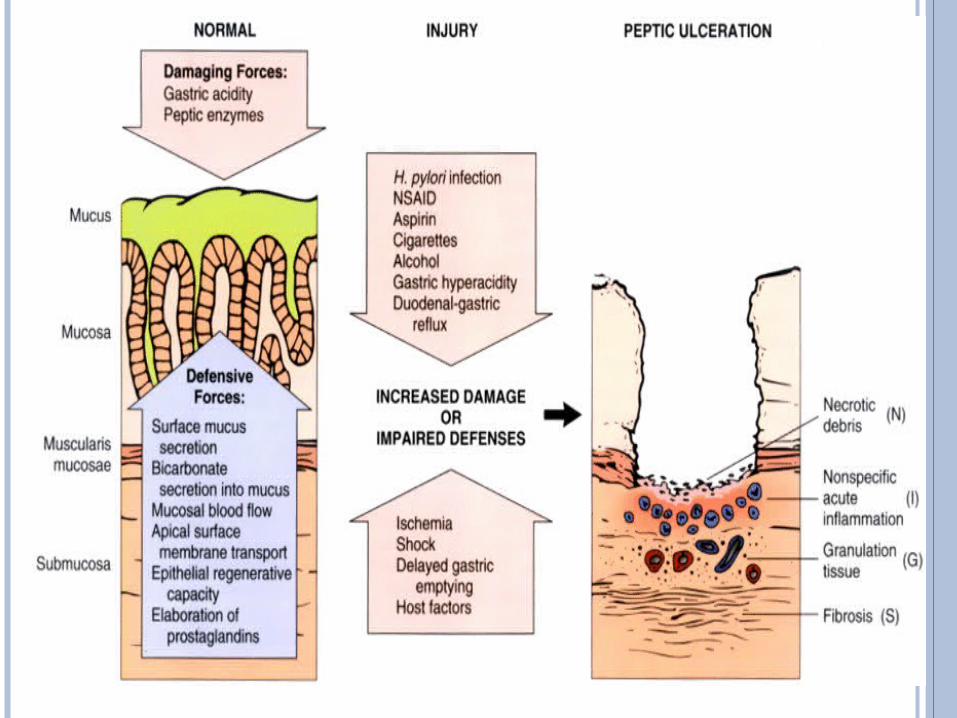
PATHOPHYSIOLOGIC BASIS OF PEPTIC ULCER DISEASE
PUD encompasses both gastric and duodenal ulcers. Ulcers are defined as a break in the mucosal surface 5 mm in size, with depth to the submucosa.
Harrisons Principle of interal medicine, 17th ed.
A major causative factor (60% of gastric and up to 90% of duodenal ulcers) is chronic inflammation due to Helicobacter pylori that colonizes (i.e. settles there after entering the body) the antral mucosa. The immune system is unable to clear the infection, despite the appearance of antibodies. Thus, the bacterium can cause a chronic active gastritis, resulting in a defect in the regulation of gastrin production by that part of the stomach, and gastrin secretion is increased
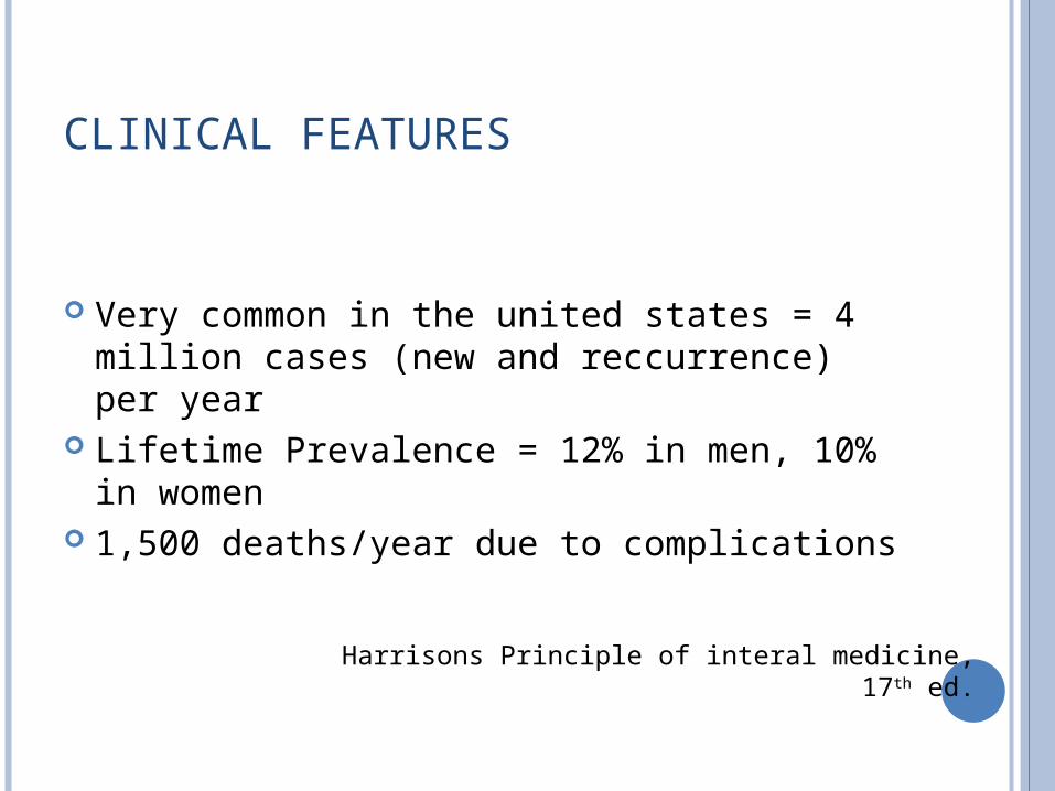
CLINICAL FEATURES
Very common in the united states = 4 million cases (new and reccurrence) per year
Lifetime Prevalence = 12% in men, 10% in women
1,500 deaths/year due to complications
Harrisons Principle of interal medicine, 17th ed.
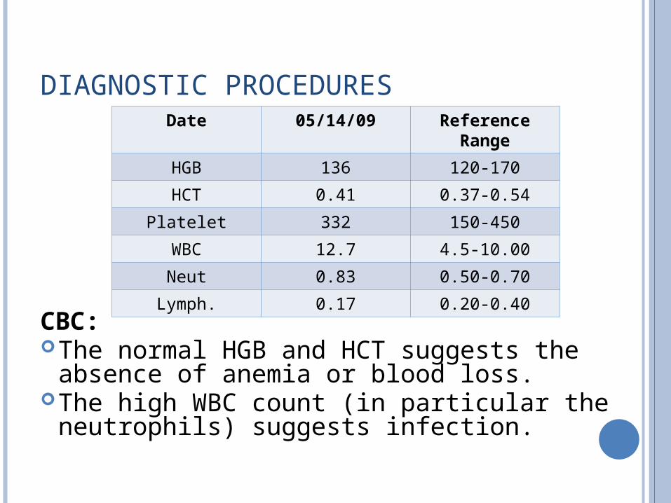
DIAGNOSTIC PROCEDURES
CBC:The normal HGB and HCT suggests the
absence of anemia or blood loss.The high WBC count (in particular the
neutrophils) suggests infection.
Date 05/14/09 Reference Range
HGB 136 120-170
HCT 0.41 0.37-0.54
Platelet 332 150-450
WBC 12.7 4.5-10.00
Neut 0.83 0.50-0.70
Lymph. 0.17 0.20-0.40
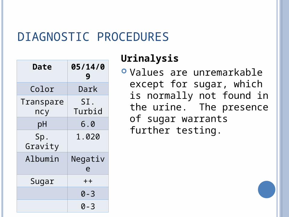
DIAGNOSTIC PROCEDURES
Urinalysis Values are unremarkable
except for sugar, which is normally not found in the urine. The presence of sugar warrants further testing.
Date 05/14/09
Color Dark
Transparency SI. Turbid
pH 6.0
Sp. Gravity 1.020
Albumin Negative
Sugar ++
0-3
0-3
DIAGNOSTIC PROCEDURES
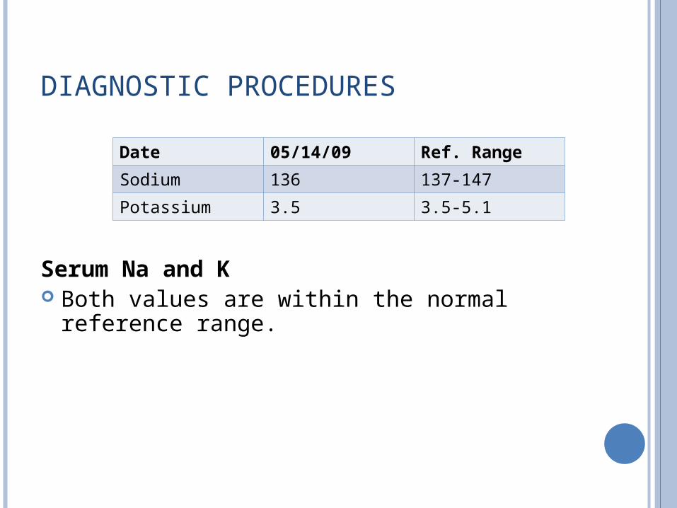
Serum Na and K Both values are within the normal reference
range.
Date 05/14/09 Ref. Range
Sodium 136 137-147
Potassium 3.5 3.5-5.1
DIAGNOSTIC PROCEDURES
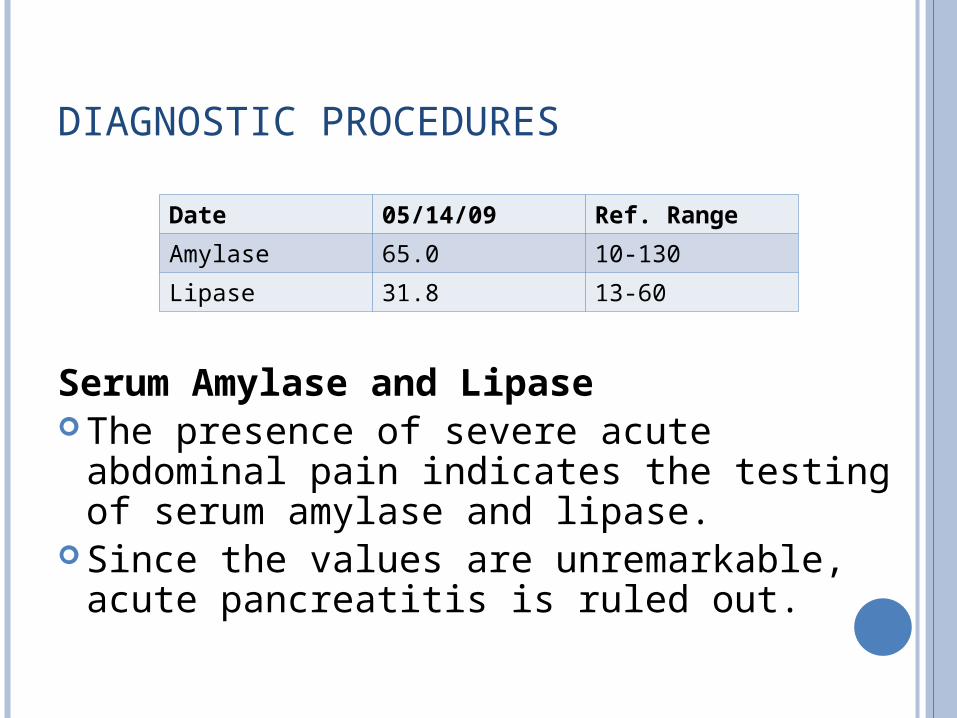
Serum Amylase and Lipase The presence of severe acute abdominal
pain indicates the testing of serum amylase and lipase.
Since the values are unremarkable, acute pancreatitis is ruled out.
Date 05/14/09 Ref. Range
Amylase 65.0 10-130
Lipase 31.8 13-60
DIAGNOSTIC PROCEDURES The 12-L ECG taken at 05/14/09 presents
with normal findings. The ECG records the electrical activity of the
heart over time via skin electrodes. The normal levels of serum sodium and
potassium is also consistent with the normal ECG.
This rules out the presence of cardiovascular involvement in the patient.
DIAGNOSTIC PROCEDURES
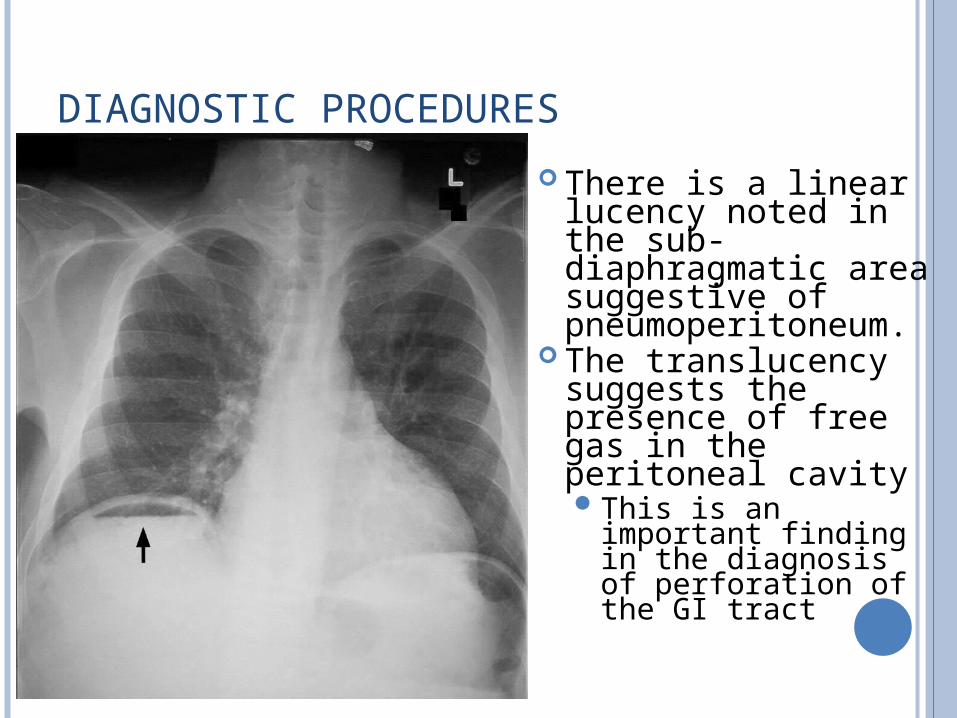
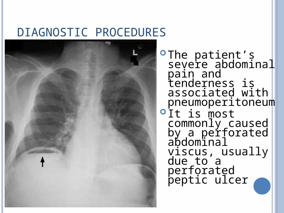
There is a linear lucency noted in the sub‐diaphragmatic area suggestive of pneumoperitoneum.
The translucency suggests the presence of free gas in the peritoneal cavityThis is an important
finding in the diagnosis of perforation of the GI tract
DIAGNOSTIC PROCEDURES
The patient’s severe abdominal pain and tenderness is associated with pneumoperitoneum
It is most commonly caused by a perforated abdominal viscus, usually due to a perforated peptic ulcer
DIAGNOSTIC PROCEDURES
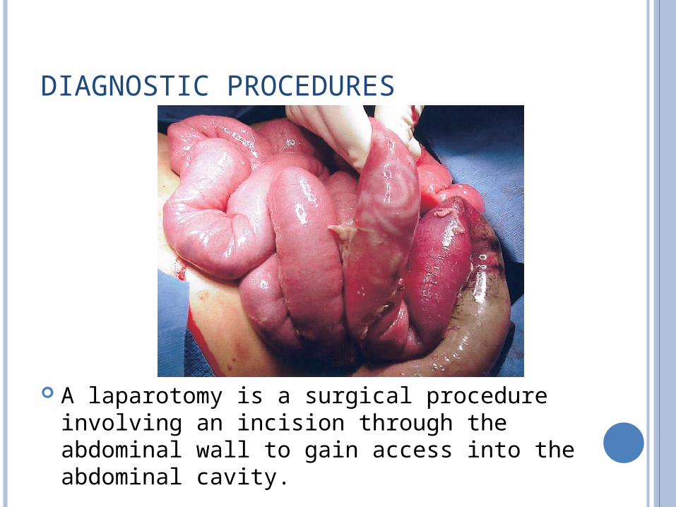
A laparotomy is a surgical procedure involving an incision through the abdominal wall to gain access into the abdominal cavity.
DIAGNOSTIC PROCEDURES
In diagnostic laparotomy (also known as exploratory laparotomy), the disease nature is unknown, and laparotomy is deemed the best way to identify the cause.
Surgical Intervention
The omental buttress is done in order to close the perforated peptic ulcer (‘omental patch repair’).
Pre and Post Op Care
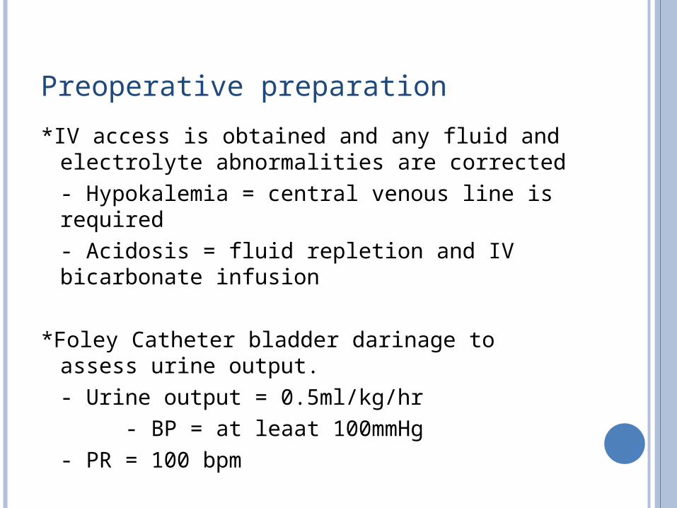
Preoperative preparation
*IV access is obtained and any fluid and electrolyte abnormalities are corrected- Hypokalemia = central venous line is required- Acidosis = fluid repletion and IV bicarbonate infusion
*Foley Catheter bladder darinage to assess urine output.- Urine output = 0.5ml/kg/hr
- BP = at leaat 100mmHg- PR = 100 bpm
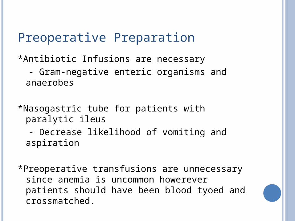
Preoperative Preparation
*Antibiotic Infusions are necessary - Gram-negative enteric organisms and
anaerobes
*Nasogastric tube for patients with paralytic ileus - Decrease likelihood of vomiting and aspiration
*Preoperative transfusions are unnecessary since anemia is uncommon howerever patients should have been blood tyoed and crossmatched.
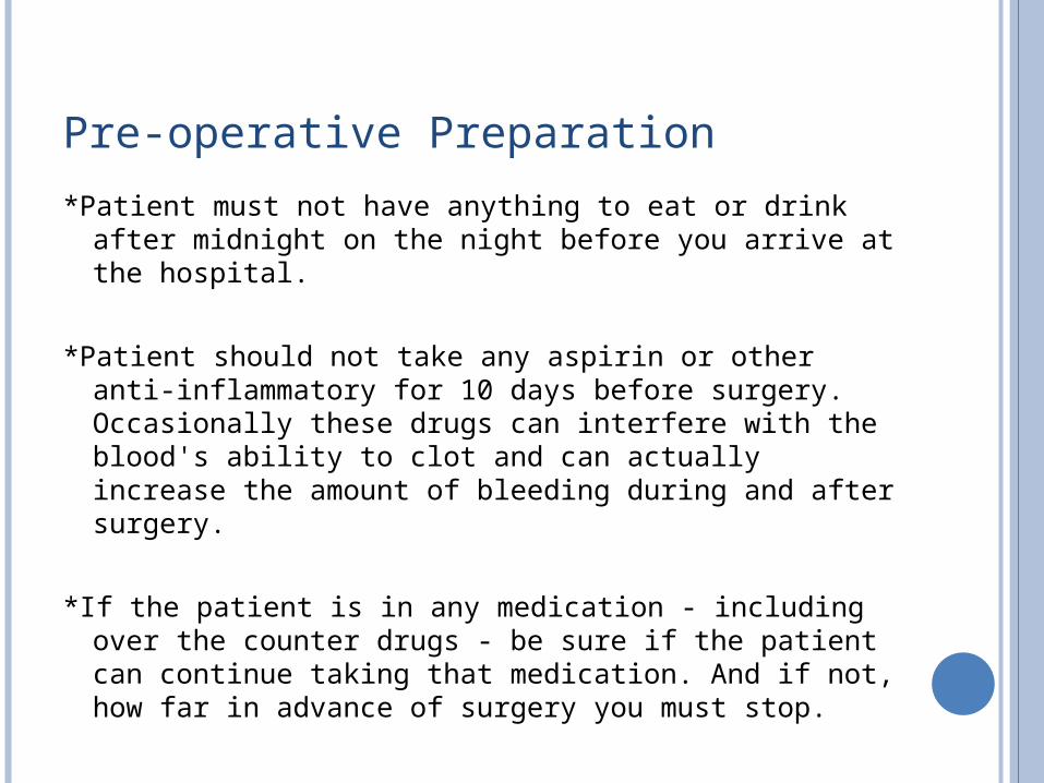
Pre-operative Preparation
*Patient must not have anything to eat or drink after midnight on the night before you arrive at the hospital.
*Patient should not take any aspirin or other anti-inflammatory for 10 days before surgery. Occasionally these drugs can interfere with the blood's ability to clot and can actually increase the amount of bleeding during and after surgery.
*If the patient is in any medication - including over the counter drugs - be sure if the patient can continue taking that medication. And if not, how far in advance of surgery you must stop.
Post-op Care
*Most patients experience at least some pain following surgery, but if properly handled, it shouldn't present any serious problems.
*check on the patient – monitor the patients progress following surgery note any inflammations or infections on the site of surgery, complications may arise such as vomiting and diarrhea
*Note if there is bleeding on the site of incision or any leaks in that matter
*Look for any signs of infection near the incision - increased swelling, redness, bleeding or other discharge
![Solar Jeepney-prs.ppt - Star 8 · Title: Microsoft PowerPoint - Solar Jeepney-prs.ppt [Compatibility Mode] Author: Ian Lancaster Created Date: 10/3/2016 9:27:53 AM](https://img.pdfslide.us/doc/110x75/6045df5154e5401c2e09c70f/solar-jeepney-prsppt-star-8-title-microsoft-powerpoint-solar-jeepney-prsppt.jpg)